Embed Size (px)
Citation preview

41
3 Functional anatomy and physiology 42
Genetic disease and inheritance 50Meiosis 50Patterns of disease inheritance 51Classes of genetic variant 53Consequences of genetic variation 57Constitutional genetic disease 58Somatic genetic disease 59
Investigation of genetic disease 60General principles of diagnosis 60Genetic testing in pregnancy and
pre-implantation genetic testing 62Genetic testing in children 63Identifying a disease gene in families 63Genetic investigation in populations 64Predictive genetic testing 64
Molecular and genetic factors in disease
Research frontiers in molecular medicine 69Gene therapy 69Induced pluripotent stem cells and
regenerative medicine 69Pathway medicine 70
D.R. FitzPatrickJ.R. Seckl
Presenting problems in genetic disease 64
Major categories of genetic disease 64Inborn errors of metabolism 64Neurological disorders 65Connective tissue disorders 65Learning disability, dysmorphism and
malformations 66Familial cancer syndromes 66
Genetic counselling 67
Genetics of common diseases 68Measuring the genetic contribution to complex
disease 68Genetic testing in complex disease 69Pharmacogenomics 69

Molecular and genetic factors in disease
3
42
Almost all diseases have a genetic component. In chil-dren and young adults in particular, many of the disor-ders causing long-term morbidity and mortality are genetically determined. The molecular basis of most Mendelian (or ‘single-gene’) diseases has now been determined, and our understanding of the abnormalities in cell function responsible for the clinical presentation is improving. It has also become clear that variants in many genes contribute to the pathogenesis of several common diseases such as asthma, rheumatoid arthritis and osteoporosis. In this chapter, we review key princi-ples of cell biology, cellular signalling and molecular genetics, with emphasis on the diagnosis and assess-ment of patients with genetic diseases.
FUNCTIONAL ANATOMY AND PHYSIOLOGY
Cell and molecular biologyAll human cell types are derived from a single totipotent stem cell, the zygote (the fertilised ovum). During devel-opment, organs and tissues are formed by the integra-tion of four closely regulated cellular processes: cell division, migration, differentiation and programmed cell death. In many adult tissues such as skin, liver and the intestine, these processes continue throughout life, mediated by populations of stem cells that are responsi-ble for tissue maintenance and repair. Cell biology is the study of these processes and of intracellular compart-ments, called organelles, which maintain cellular homeo-stasis. Dysfunction of any of these processes may lead to disease.
DNA, chromosomes and chromatinThe nucleus is a membrane-bound compartment found in all cells except erythrocytes and platelets. The human nucleus contains 46 chromosomes, each a single linear molecule of deoxyribonucleic acid (DNA) complexed with proteins to form chromatin. The basic protein unit of chromatin is the nucleosome, comprising 147 base pairs (bp) of DNA wound round a core of four different histone proteins. The vast majority of chromosomal DNA is double-stranded, with the exception of the ends of chromosomes, where ‘knotted’ domains of single-stranded DNA, called telomeres, are found. Telomeres prevent degradation and accidental fusion of chromo-somal DNA.
The genome comprises approximately 3.1 billion bp of DNA. Humans are diploid organisms, meaning that each nucleus contains two copies of the genome, visible microscopically as 22 identical chromosomal pairs – the autosomes – named 1 to 22 in descending size order (see Fig. 3.11, p. 57), and two sex chromosomes (XX in females and XY in males). Each DNA strand consists of a linear sequence of four bases – guanine (G), cytosine (C), adenine (A) and thymine (T) – covalently linked by phosphate bonds. The sequence of one strand of double-stranded DNA determines the sequence of the opposite strand because the helix is held together by hydrogen bonds between adenine and thymine or guanine and cytosine nucleotides.
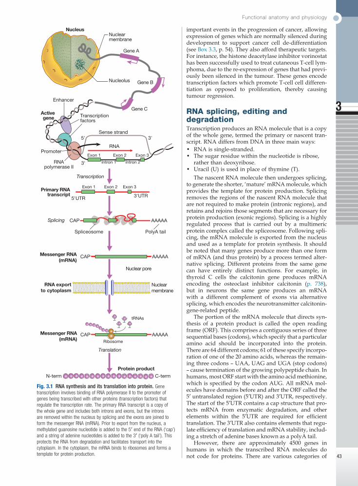
Genes and transcriptionGenes are functional elements on the chromosome that are capable of transmitting information from the DNA template via the production of messenger ribonucleic acid (mRNA) to the production of proteins. The human genome contains an estimated 21 500 genes, although many of these are inactive or silenced in different cell types. For example, although the gene for parathyroid hormone (PTH) is present in every cell, activation of gene expression and production of PTH mRNA is virtu-ally restricted to the parathyroid glands. Genes that are active in different cells undergo transcription, which requires binding of an enzyme called RNA polymerase II to a segment of DNA at the start of the gene termed the promoter. Once bound, RNA polymerase II moves along one strand of DNA, producing an RNA molecule that is complementary to the DNA template. A DNA sequence close to the end of the gene, called the poly-adenylation signal, acts as a signal for termination of the RNA transcript (Fig. 3.1). The activity of RNA polymerase II is regulated by transcription factors. These proteins bind to specific DNA sequences at the promoter, or to enhancer elements that may be many thousands of base pairs away from the promoter. A loop in the chromosomal DNA brings the enhancer close to the promoter, enabling the bound proteins to interact.
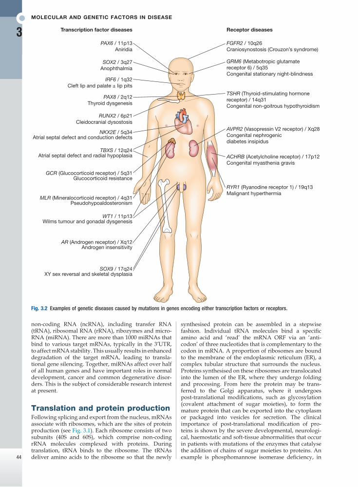
The human genome encodes approximately 1200 dif-ferent transcription factors, and mutations in many of these can cause genetic diseases (Fig. 3.2). Mutation of the transcription factor binding sites within promoters or enhancers also causes genetic disease. For example, the blood disorder alpha-thalassaemia can result from loss of an enhancer located more than 100 000 bp from the alpha-globin gene promoter, leading to greatly reduced transcription. Similarly, variation in the pro-moter of the gene encoding intestinal lactase determines whether or not this is ‘shut off’ in adulthood, producing lactose intolerance.
The accessibility of promoters to RNA polymerase II depends on the structural configuration of chromatin. Transcriptionally active regions have decondensed (or ‘open’) chromatin (euchromatin). Conversely, tran-scriptionally silent regions are associated with densely packed chromatin called heterochromatin. Chemical modification of both the DNA and core histone proteins allows heterochromatic regions to be distinguished from open chromatin. DNA can be modified by addition of a methyl group to cytosine molecules (methylation). In promoter regions, this silences transcription, since methyl cytosines are usually not available for tran-scription factor binding or RNA transcription. The core histones can also be modified via methylation, phosphorylation, acetylation or sumoylation at specific amino acid residues in a pattern that reflects the func-tional state of the chromatin; this is called the histone code – reflecting an emerging understanding of the ‘rules’ by which specific modifications mark transcrip-tionally activating (trimethylation of lysine 4 on histone H3; acetylation of many histone residues) or silencing (methylation of lysine 9 on histone H4; deacetylation of many histone residues) effects. Such DNA and protein modifications are termed epigenetic, as they do not alter the primary sequence of the DNA code but have biological significance in chromosomal function. Abnor-mal epigenetic changes are increasingly recognised as
Functional anatomy and physiology
3
43
Fig. 3.1 RNA synthesis and its translation into protein. Gene transcription involves binding of RNA polymerase II to the promoter of genes being transcribed with other proteins (transcription factors) that regulate the transcription rate. The primary RNA transcript is a copy of the whole gene and includes both introns and exons, but the introns are removed within the nucleus by splicing and the exons are joined to form the messenger RNA (mRNA). Prior to export from the nucleus, a methylated guanosine nucleotide is added to the 5′ end of the RNA (‘cap’) and a string of adenine nucleotides is added to the 3′ (‘poly A tail’). This protects the RNA from degradation and facilitates transport into the cytoplasm. In the cytoplasm, the mRNA binds to ribosomes and forms a template for protein production.
Protein product
N-term C-term
CAPMessenger RNA(mRNA)
Messenger RNA(mRNA)
AAAAA
tRNAs
Ribosome
Nuclearmembrane
Primary RNAtranscript
Spliceosome
RNA exportto cytoplasm
CAP
CAP
AAAAA
PolyA tail
3'UTR5'UTR
Nuclear pore
AAAAA
Splicing
Transcription
3'5'
3'
Sense strand
Enhancer
Transcriptionfactors
RNApolymerase II
Exon 1
Intron 2Intron 1
Exon 2 Exon 3
Exon 1 Exon 2 Exon 3
Promoter
Nucleolus
Nuclearmembrane
Gene A
Gene B
Gene CActive gene
RNA
Nucleus
Translation
important events in the progression of cancer, allowing expression of genes which are normally silenced during development to support cancer cell de-differentiation (see Box 3.3, p. 54). They also afford therapeutic targets. For instance, the histone deacetylase inhibitor vorinostat has been successfully used to treat cutaneous T-cell lym-phoma, due to the re-expression of genes that had previ-ously been silenced in the tumour. These genes encode transcription factors which promote T-cell cell differen-tiation as opposed to proliferation, thereby causing tumour regression.
RNA splicing, editing and degradationTranscription produces an RNA molecule that is a copy of the whole gene, termed the primary or nascent tran-script. RNA differs from DNA in three main ways:• RNA is single-stranded.• The sugar residue within the nucleotide is ribose,
rather than deoxyribose.• Uracil (U) is used in place of thymine (T).
The nascent RNA molecule then undergoes splicing, to generate the shorter, ‘mature’ mRNA molecule, which provides the template for protein production. Splicing removes the regions of the nascent RNA molecule that are not required to make protein (intronic regions), and retains and rejoins those segments that are necessary for protein production (exonic regions). Splicing is a highly regulated process that is carried out by a multimeric protein complex called the spliceosome. Following spli-cing, the mRNA molecule is exported from the nucleus and used as a template for protein synthesis. It should be noted that many genes produce more than one form of mRNA (and thus protein) by a process termed alter-native splicing. Different proteins from the same gene can have entirely distinct functions. For example, in thyroid C cells the calcitonin gene produces mRNA encoding the osteoclast inhibitor calcitonin (p. 738), but in neurons the same gene produces an mRNA with a different complement of exons via alternative splicing, which encodes the neurotransmitter calcitonin-gene-related peptide.
The portion of the mRNA molecule that directs syn-thesis of a protein product is called the open reading frame (ORF). This comprises a contiguous series of three sequential bases (codons), which specify that a particular amino acid should be incorporated into the protein. There are 64 different codons; 61 of these specify incorpo-ration of one of the 20 amino acids, whereas the remain-ing three codons – UAA, UAG and UGA (stop codons) – cause termination of the growing polypeptide chain. In humans, most ORF start with the amino acid methionine, which is specified by the codon AUG. All mRNA mol-ecules have domains before and after the ORF called the 5′ untranslated region (5′UTR) and 3′UTR, respectively. The start of the 5′UTR contains a cap structure that pro-tects mRNA from enzymatic degradation, and other elements within the 5′UTR are required for efficient translation. The 3′UTR also contains elements that regu-late efficiency of translation and mRNA stability, includ-ing a stretch of adenine bases known as a polyA tail.
However, there are approximately 4500 genes in humans in which the transcribed RNA molecules do not code for proteins. There are various categories of
Molecular and genetic factors in disease
3
44
synthesised protein can be assembled in a stepwise fashion. Individual tRNA molecules bind a specific amino acid and ‘read’ the mRNA ORF via an ‘anti-codon’ of three nucleotides that is complementary to the codon in mRNA. A proportion of ribosomes are bound to the membrane of the endoplasmic reticulum (ER), a complex tubular structure that surrounds the nucleus. Proteins synthesised on these ribosomes are translocated into the lumen of the ER, where they undergo folding and processing. From here the protein may be trans-ferred to the Golgi apparatus, where it undergoes post-translational modifications, such as glycosylation (covalent attachment of sugar moieties), to form the mature protein that can be exported into the cytoplasm or packaged into vesicles for secretion. The clinical importance of post-translational modification of pro-teins is shown by the severe developmental, neurologi-cal, haemostatic and soft-tissue abnormalities that occur in patients with mutations of the enzymes that catalyse the addition of chains of sugar moieties to proteins. An example is phosphomannose isomerase deficiency, in
non-coding RNA (ncRNA), including transfer RNA (tRNA), ribosomal RNA (rRNA), ribozymes and micro-RNA (miRNA). There are more than 1000 miRNAs that bind to various target mRNAs, typically in the 3′UTR, to affect mRNA stability. This usually results in enhanced degradation of the target mRNA, leading to transla-tional gene silencing. Together, miRNAs affect over half of all human genes and have important roles in normal development, cancer and common degenerative disor-ders. This is the subject of considerable research interest at present.
Translation and protein productionFollowing splicing and export from the nucleus, mRNAs associate with ribosomes, which are the sites of protein production (see Fig. 3.1). Each ribosome consists of two subunits (40S and 60S), which comprise non-coding rRNA molecules complexed with proteins. During translation, tRNA binds to the ribosome. The tRNAs deliver amino acids to the ribosome so that the newly
Fig. 3.2 Examples of genetic diseases caused by mutations in genes encoding either transcription factors or receptors.
Transcription factor diseases
SOX2 / 3q27Anophthalmia
IRF6 / 1q32Cleft lip and palate ± lip pits
PAX8 / 2q12Thyroid dysgenesis
RUNX2 / 6p21Cleidocranial dysostosis
NKX2E / 5q34Atrial septal defect and conduction defects
TBXS / 12q24Atrial septal defect and radial hypoplasia
GCR (Glucocorticoid receptor) / 5q31 Glucocorticoid resistance
MLR (Mineralocorticoid receptor) / 4q31 Pseudohypoaldosteronism
WT1 / 11p13Wilms tumour and gonadal dysgenesis
AR (Androgen receptor) / Xq12 Androgen insensitivity
SOX9 / 17q24XY sex reversal and skeletal dysplasia
PAX6 / 11p13Aniridia
FGFR2 / 10q26Craniosynostosis (Crouzon’s syndrome)
TSHR (Thyroid-stimulating hormonereceptor) / 14q31Congenital non-goitrous hypothyroidism
AVPR2 (Vasopressin V2 receptor) / Xq28Congenital nephrogenicdiabetes insipidus
ACHRB (Acetylcholine receptor) / 17p12Congenital myasthenia gravis
RYR1 (Ryanodine receptor 1) / 19q13Malignant hyperthermia
GRM6 (Metabotropic glutamatereceptor 6) / 5q35Congenital stationary night-blindness
Receptor diseases

Functional anatomy and physiology
3
45
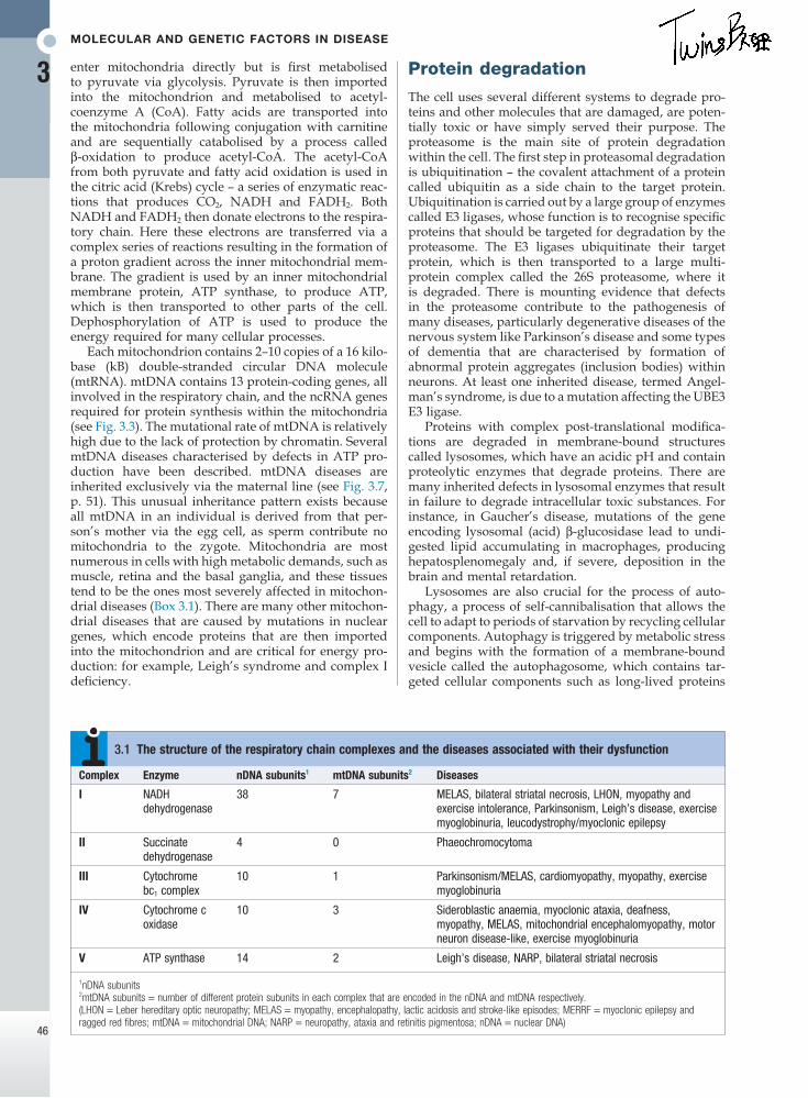
Mitochondria and energy productionThe mitochondrion is the main site of energy pro-duction within the cell. Mitochondria arose during evolution via the symbiotic association with an intra-cellular bacterium. They have a distinctive structure with functionally distinct inner and outer membranes. Mitochondria produce energy in the form of adenosine triphosphate (ATP). ATP is mostly derived from the metabolism of glucose and fat (Fig. 3.3). Glucose cannot
which there is a defect in the conversion of fructose-6-phosphate to mannose-6-phosphate. This results in a defect in supply of D-mannose derivatives for glycosyla-tion of a variety of proteins, resulting in a multi-system disorder characterised by protein-losing enteropathy, hepatic fibrosis, coagulopathy and hypoglycaemia. Post-translational modifications can also be disrupted by the synthesis of proteins with abnormal amino acid sequences. For example, the most common mutation in cystic fibrosis (ΔF508) results in an abnormal protein that cannot be exported from the ER and Golgi.
Fig. 3.3 Mitochondria. A Mitochondrial structure. There is a smooth outer membrane surrounding a convoluted inner membrane, which has inward projections called cristae. The membranes create two compartments: the inter-membrane compartment, which plays a crucial role in the electron transport chain, and the inner compartment (or matrix), which contains mitochondrial DNA and the enzymes responsible for the citric acid (Krebs) cycle and the fatty acid β-oxidation cycle. B Mitochondrial DNA. The mitochondrion contains several copies of a circular double-stranded DNA molecule, which has a non-coding region, and a coding region which encodes the genes responsible for energy production, mitochondrial tRNA molecules and mitochondrial rRNA molecules. ATP = adenosine triphosphate; NADH = nicotinamide adenine dinucleotide. C Mitochondrial energy production. Fatty acids enter the mitochondrion conjugated to carnitine by carnitine-palmityl transferase type 1 (CPT I) and, once inside the matrix, are unconjugated by CPT II to release free fatty acids (FFA). These are broken down by the β-oxidation cycle to produce acetyl-CoA. Pyruvate can enter the mitochondrion directly and is metabolised by pyruvate dehydrogenase (PDH) to produce acetyl-CoA. The acetyl-CoA enters the Krebs cycle, leading to the production of NADH and flavine adenine dinucleotide (reduced form) (FADH2), which are used by proteins in the electron transport chain to generate a hydrogen ion gradient across the inter-membrane compartment. Reduction of NADH and FADH2 by proteins I and II respectively releases electrons (e), and the energy released is used to pump protons into the inter-membrane compartment. As these electrons are exchanged between proteins in the chain, more protons are pumped across the membrane, until the electrons reach complex IV (cytochrome oxidase), which uses the energy to reduce oxygen to water. The hydrogen ion gradient is used to produce ATP by the enzyme ATP synthase, which consists of a proton channel and catalytic sites for the synthesis of ATP from ADP. When the channel opens, hydrogen ions enter the matrix down the concentration gradient, and energy is released that is used to make ATP.
L strand
H strand
Outermembrane
Innermembrane
NADH
NAD
III
IIIQCytC
IV
NADH
FADH2
Fatty acidβ-oxidation
cycle
Citric acid(Krebs)cycle
H+
e 2e
FADH2
FADH2
LactatePyruvate
PDH
Acetyl-CoA
Glucose
22 tRNAs
NADH dehydrogenase 7subunits
Cytochrome B/C oxidase 4subunits
2 ribosomal RNA subunits
2 ATP synthase subunits
Intragenic DNA
Innermembrane
Cristae
Matrix
Outermembrane
FFA
CPT I
CPT II
Carnitine
Carnitine-FA ester
C
A B
FFAFAD
2H+
H2O
O2
ATP
ADP+ Pi
H+ H+
ATPsynthase
Carnitine
ee e
Molecular and genetic factors in disease
3
46
Protein degradation
The cell uses several different systems to degrade pro-teins and other molecules that are damaged, are poten-tially toxic or have simply served their purpose. The proteasome is the main site of protein degrada tion within the cell. The first step in proteasomal degradation is ubiquitination – the covalent attachment of a protein called ubiquitin as a side chain to the target protein. Ubiquitination is carried out by a large group of enzymes called E3 ligases, whose function is to recognise specific proteins that should be targeted for degradation by the proteasome. The E3 ligases ubiquitinate their target protein, which is then transported to a large multi-protein complex called the 26S proteasome, where it is degraded. There is mounting evidence that defects in the proteasome contribute to the pathogenesis of many diseases, particularly degenerative diseases of the nervous system like Parkinson’s disease and some types of dementia that are characterised by formation of abnormal protein aggregates (inclusion bodies) within neurons. At least one inherited disease, termed Angel-man’s syndrome, is due to a mutation affecting the UBE3 E3 ligase.
Proteins with complex post-translational modifica-tions are degraded in membrane-bound structures called lysosomes, which have an acidic pH and contain proteolytic enzymes that degrade proteins. There are many inherited defects in lysosomal enzymes that result in failure to degrade intracellular toxic substances. For instance, in Gaucher’s disease, mutations of the gene encoding lysosomal (acid) β-glucosidase lead to undi-gested lipid accumulating in macrophages, producing hepatosplenomegaly and, if severe, deposition in the brain and mental retardation.
Lysosomes are also crucial for the process of auto-phagy, a process of self-cannibalisation that allows the cell to adapt to periods of starvation by recycling cellular components. Autophagy is triggered by metabolic stress and begins with the formation of a membrane-bound vesicle called the autophagosome, which contains tar-geted cellular components such as long-lived proteins
enter mitochondria directly but is first metabolised to pyruvate via glycolysis. Pyruvate is then imported into the mitochondrion and metabolised to acetyl-coenzyme A (CoA). Fatty acids are transported into the mitochondria following conjugation with carnitine and are sequentially catabolised by a process called β-oxidation to produce acetyl-CoA. The acetyl-CoA from both pyruvate and fatty acid oxidation is used in the citric acid (Krebs) cycle – a series of enzymatic reac-tions that produces CO2, NADH and FADH2. Both NADH and FADH2 then donate electrons to the respira-tory chain. Here these electrons are transferred via a complex series of reactions resulting in the formation of a proton gradient across the inner mitochondrial mem-brane. The gradient is used by an inner mitochondrial membrane protein, ATP synthase, to produce ATP, which is then transported to other parts of the cell. Dephosphorylation of ATP is used to produce the energy required for many cellular processes.
Each mitochondrion contains 2–10 copies of a 16 kilo-base (kB) double-stranded circular DNA molecule (mtRNA). mtDNA contains 13 protein-coding genes, all involved in the respiratory chain, and the ncRNA genes required for protein synthesis within the mitochondria (see Fig. 3.3). The mutational rate of mtDNA is relatively high due to the lack of protection by chromatin. Several mtDNA diseases characterised by defects in ATP pro-duction have been described. mtDNA diseases are inherited exclusively via the maternal line (see Fig. 3.7, p. 51). This unusual inheritance pattern exists because all mtDNA in an individual is derived from that per-son’s mother via the egg cell, as sperm contribute no mitochondria to the zygote. Mitochondria are most numerous in cells with high metabolic demands, such as muscle, retina and the basal ganglia, and these tissues tend to be the ones most severely affected in mitochon-drial diseases (Box 3.1). There are many other mitochon-drial diseases that are caused by mutations in nuclear genes, which encode proteins that are then imported into the mitochondrion and are critical for energy pro-duction: for example, Leigh’s syndrome and complex I deficiency.
Complex Enzyme nDNA subunits1 mtDNA subunits2 Diseases
I NADH dehydrogenase
38 7 MELAS, bilateral striatal necrosis, LHON, myopathy and exercise intolerance, Parkinsonism, Leigh’s disease, exercise myoglobinuria, leucodystrophy/myoclonic epilepsy
II Succinate dehydrogenase
4 0 Phaeochromocytoma
III Cytochrome bc1 complex
10 1 Parkinsonism/MELAS, cardiomyopathy, myopathy, exercise myoglobinuria
IV Cytochrome c oxidase
10 3 Sideroblastic anaemia, myoclonic ataxia, deafness, myopathy, MELAS, mitochondrial encephalomyopathy, motor neuron disease-like, exercise myoglobinuria
V ATP synthase 14 2 Leigh’s disease, NARP, bilateral striatal necrosis
1nDNA subunits2mtDNA subunits = number of different protein subunits in each complex that are encoded in the nDNA and mtDNA respectively.(LHON = Leber hereditary optic neuropathy; MELAS = myopathy, encephalopathy, lactic acidosis and stroke-like episodes; MERRF = myoclonic epilepsy and ragged red fibres; mtDNA = mitochondrial DNA; NARP = neuropathy, ataxia and retinitis pigmentosa; nDNA = nuclear DNA)
3.1 The structure of the respiratory chain complexes and the diseases associated with their dysfunction
Functional anatomy and physiology
3
47
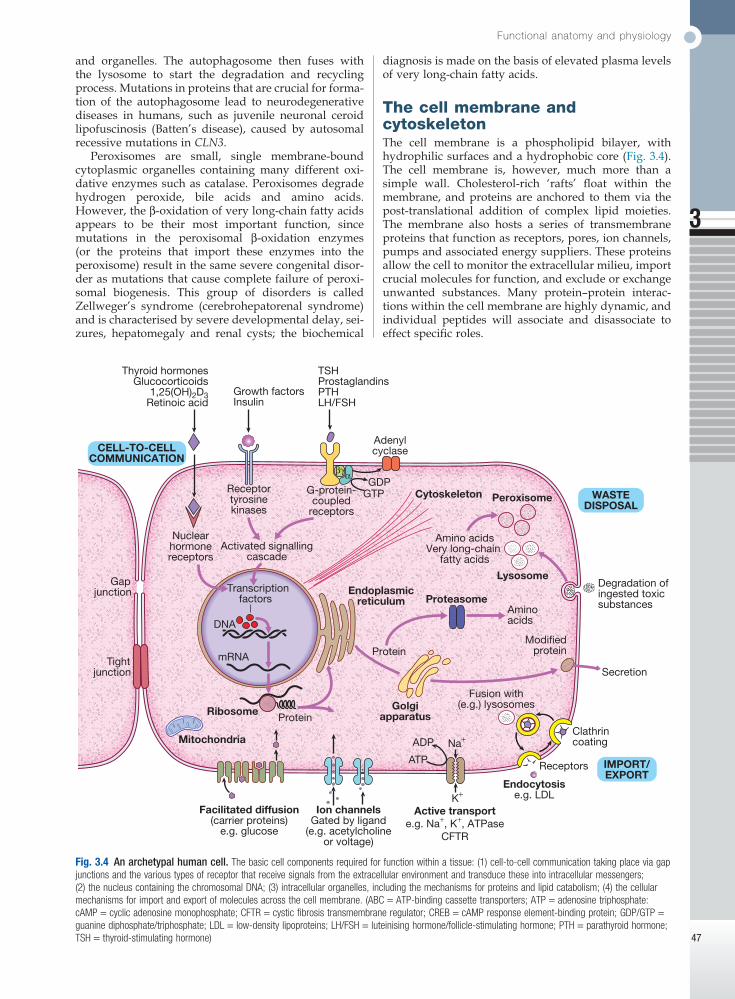
Fig. 3.4 An archetypal human cell. The basic cell components required for function within a tissue: (1) cell-to-cell communication taking place via gap junctions and the various types of receptor that receive signals from the extracellular environment and transduce these into intracellular messengers; (2) the nucleus containing the chromosomal DNA; (3) intracellular organelles, including the mechanisms for proteins and lipid catabolism; (4) the cellular mechanisms for import and export of molecules across the cell membrane. (ABC = ATP-binding cassette transporters; ATP = adenosine triphosphate: cAMP = cyclic adenosine monophosphate; CFTR = cystic fibrosis transmembrane regulator; CREB = cAMP response element-binding protein; GDP/GTP = guanine diphosphate/triphosphate; LDL = low-density lipoproteins; LH/FSH = luteinising hormone/follicle-stimulating hormone; PTH = parathyroid hormone; TSH = thyroid-stimulating hormone)
Active transporte.g. Na+, K+, ATPase
CFTR
Ribosome
Mitochondria
Golgiapparatus
Secretion
Degradation ofingested toxicsubstances
Protein
Modifiedprotein
K+
Na+
Ion channelsGated by ligand
(e.g. acetylcholineor voltage)
Facilitated diffusion(carrier proteins)
e.g. glucose
Endocytosise.g. LDL
Clathrincoating
Receptors
Fusion with(e.g.) lysosomes
IMPORT/EXPORT
ATP
ADP
Tightjunction
Gapjunction
PeroxisomeCytoskeleton
Thyroid hormonesGlucocorticoids
1,25(OH)2D3Retinoic acid
Growth factorsInsulin
TSHProstaglandinsPTHLH/FSH
AdenylcyclaseCELL-TO-CELL
COMMUNICATION
WASTEDISPOSAL
Endoplasmicreticulum
Lysosome
Receptortyrosinekinases
Activated signallingcascade
G-protein-coupled
receptors
Nuclearhormonereceptors
ProteasomeTranscription
factors
mRNA
DNA
Protein
Aminoacids
Amino acidsVery long-chain
fatty acids
βγ
GTP
αGDP
and organelles. The autophagosome then fuses with the lysosome to start the degradation and recycling process. Mutations in proteins that are crucial for forma-tion of the autophagosome lead to neurodegenerative diseases in humans, such as juvenile neuronal ceroid lipofuscinosis (Batten’s disease), caused by autosomal recessive mutations in CLN3.
Peroxisomes are small, single membrane-bound cytoplasmic organelles containing many different oxi-dative enzymes such as catalase. Peroxisomes degrade hydrogen peroxide, bile acids and amino acids. However, the β-oxidation of very long-chain fatty acids appears to be their most important function, since mutations in the peroxisomal β-oxidation enzymes (or the proteins that import these enzymes into the peroxisome) result in the same severe congenital disor-der as mutations that cause complete failure of peroxi-somal biogenesis. This group of disorders is called Zellweger’s syndrome (cerebrohepatorenal syndrome) and is characterised by severe developmental delay, sei-zures, hepatomegaly and renal cysts; the biochemical
diagnosis is made on the basis of elevated plasma levels of very long-chain fatty acids.
The cell membrane and cytoskeletonThe cell membrane is a phospholipid bilayer, with hydrophilic surfaces and a hydrophobic core (Fig. 3.4). The cell membrane is, however, much more than a simple wall. Cholesterol-rich ‘rafts’ float within the membrane, and proteins are anchored to them via the post-translational addition of complex lipid moieties. The membrane also hosts a series of transmembrane proteins that function as receptors, pores, ion channels, pumps and associated energy suppliers. These proteins allow the cell to monitor the extracellular milieu, import crucial molecules for function, and exclude or exchange unwanted substances. Many protein–protein interac-tions within the cell membrane are highly dynamic, and individual peptides will associate and disassociate to effect specific roles.
Molecular and genetic factors in disease
3
48
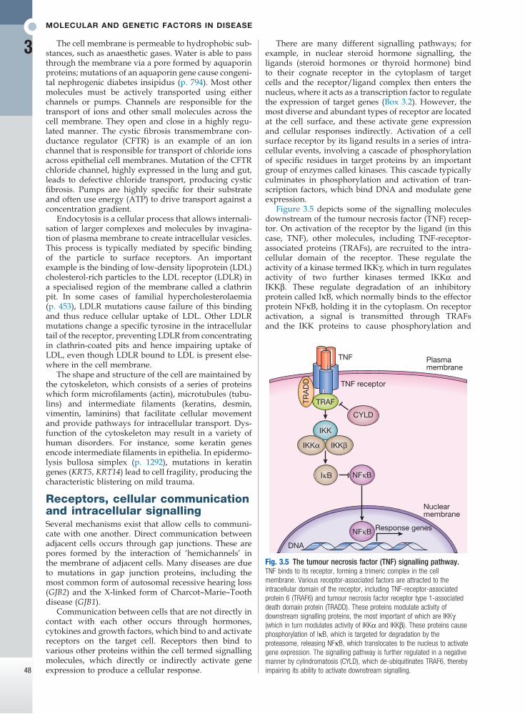
Fig. 3.5 The tumour necrosis factor (TNF) signalling pathway. TNF binds to its receptor, forming a trimeric complex in the cell membrane. Various receptor-associated factors are attracted to the intracellular domain of the receptor, including TNF-receptor-associated protein 6 (TRAF6) and tumour necrosis factor receptor type 1-associated death domain protein (TRADD). These proteins modulate activity of downstream signalling proteins, the most important of which are IKKγ (which in turn modulates activity of IKKα and IKKβ). These proteins cause phosphorylation of IκB, which is targeted for degradation by the proteasome, releasing NFκB, which translocates to the nucleus to activate gene expression. The signalling pathway is further regulated in a negative manner by cylindromatosis (CYLD), which de-ubiquitinates TRAF6, thereby impairing its ability to activate downstream signalling.
TNF
TNF receptor
Plasmamembrane
TRAFTRA
DD
CYLD
IKKα
IκB NFκB
NFκB
IKKβ
IKK
Nuclearmembrane
DNA
Response genes
There are many different signalling pathways; for example, in nuclear steroid hormone signalling, the ligands (steroid hormones or thyroid hormone) bind to their cognate receptor in the cytoplasm of target cells and the receptor/ligand complex then enters the nucleus, where it acts as a transcription factor to regulate the expression of target genes (Box 3.2). However, the most diverse and abundant types of receptor are located at the cell surface, and these activate gene expression and cellular responses indirectly. Activation of a cell surface receptor by its ligand results in a series of intra-cellular events, involving a cascade of phosphorylation of specific residues in target proteins by an important group of enzymes called kinases. This cascade typically culminates in phosphorylation and activation of tran-scription factors, which bind DNA and modulate gene expression.
Figure 3.5 depicts some of the signalling molecules downstream of the tumour necrosis factor (TNF) recep-tor. On activation of the receptor by the ligand (in this case, TNF), other molecules, including TNF-receptor-associated proteins (TRAFs), are recruited to the intra-cellular domain of the receptor. These regulate the activity of a kinase termed IKKγ, which in turn regulates activity of two further kinases termed IKKα and IKKβ. These regulate degradation of an inhibitory protein called IκB, which normally binds to the effector protein NFκB, holding it in the cytoplasm. On receptor activation, a signal is transmitted through TRAFs and the IKK proteins to cause phosphorylation and
The cell membrane is permeable to hydrophobic sub-stances, such as anaesthetic gases. Water is able to pass through the membrane via a pore formed by aquaporin proteins; mutations of an aquaporin gene cause congeni-tal nephrogenic diabetes insipidus (p. 794). Most other molecules must be actively transported using either channels or pumps. Channels are responsible for the transport of ions and other small molecules across the cell membrane. They open and close in a highly regu-lated manner. The cystic fibrosis transmembrane con-ductance regulator (CFTR) is an example of an ion channel that is responsible for transport of chloride ions across epithelial cell membranes. Mutation of the CFTR chloride channel, highly expressed in the lung and gut, leads to defective chloride transport, producing cystic fibrosis. Pumps are highly specific for their substrate and often use energy (ATP) to drive transport against a concentration gradient.
Endocytosis is a cellular process that allows internali-sation of larger complexes and molecules by invagina-tion of plasma membrane to create intracellular vesicles. This process is typically mediated by specific binding of the particle to surface receptors. An important example is the binding of low-density lipoprotein (LDL) cholesterol-rich particles to the LDL receptor (LDLR) in a specialised region of the membrane called a clathrin pit. In some cases of familial hypercholesterolaemia (p. 453), LDLR mutations cause failure of this binding and thus reduce cellular uptake of LDL. Other LDLR mutations change a specific tyrosine in the intracellular tail of the receptor, preventing LDLR from concentrating in clathrin-coated pits and hence impairing uptake of LDL, even though LDLR bound to LDL is present else-where in the cell membrane.
The shape and structure of the cell are maintained by the cytoskeleton, which consists of a series of proteins which form microfilaments (actin), microtubules (tubu-lins) and intermediate filaments (keratins, desmin, vimentin, laminins) that facilitate cellular movement and provide pathways for intracellular transport. Dys-function of the cytoskeleton may result in a variety of human disorders. For instance, some keratin genes encode intermediate filaments in epithelia. In epidermo-lysis bullosa simplex (p. 1292), mutations in keratin genes (KRT5, KRT14) lead to cell fragility, producing the characteristic blistering on mild trauma.
Receptors, cellular communication and intracellular signallingSeveral mechanisms exist that allow cells to communi-cate with one another. Direct communication between adjacent cells occurs through gap junctions. These are pores formed by the interaction of ‘hemichannels’ in the membrane of adjacent cells. Many diseases are due to mutations in gap junction proteins, including the most common form of autosomal recessive hearing loss (GJB2) and the X-linked form of Charcot–Marie–Tooth disease (GJB1).
Communication between cells that are not directly in contact with each other occurs through hormones, cytokines and growth factors, which bind to and activate receptors on the target cell. Receptors then bind to various other proteins within the cell termed signalling molecules, which directly or indirectly activate gene expression to produce a cellular response.
Functional anatomy and physiology
3
49
situs inversus (left–right laterality reversal) as a result of failure of a specific signalling process in very early embryogenesis. Mutations in proteins that are essential for non-motile cilia formation or function are responsi-ble for a large number of autosomal recessive disorders known collectively as ciliopathies, which are commonly associated with intellectual disability, renal cystic dys-plasia and retinal degeneration. For example, in the Bardet–Biedl syndrome, mutations in a series of genes encoding ciliary structure cause polydactyly, obesity, hypogonadism, retinitis pigmentosa and renal failure.
Cell division, differentiation and migrationIn normal tissues, molecules such as hormones, growth factors and cytokines provide the signal to activate the cell cycle, a controlled programme of biochemical events that culminates in cell division. During the first phase, G1, synthesis of the cellular components necessary to complete cell division occurs. In S phase, the cell pro-duces an identical copy of each chromosome – which carries the cell’s genetic information – via a process called DNA replication. The cell then enters G2, when any errors in the replicated DNA are repaired before proceeding to mitosis, in which identical copies of all chromosomes are segregated to the daughter cells. The progression from one phase to the next is tightly con-trolled by cell cycle checkpoints. For example, the check-point between G2 and mitosis ensures that all damaged DNA is repaired prior to segregation of the chromo-somes. Failure of these control processes is a crucial driver in the pathogenesis of cancer, as discussed in Chapter 11 (p. 262).
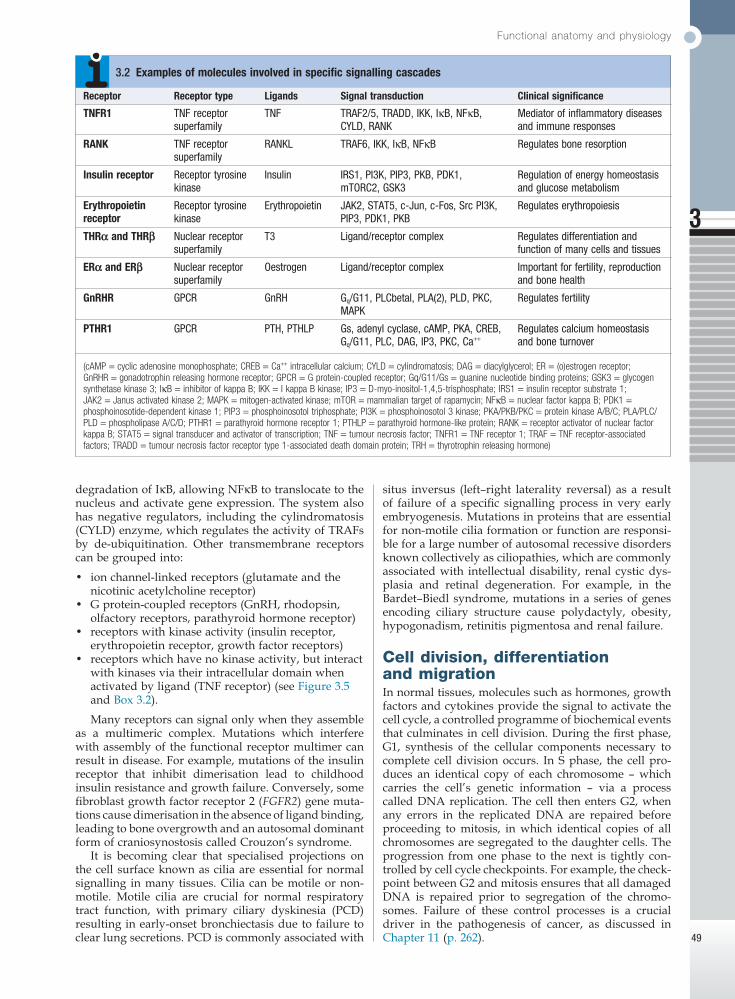
Receptor Receptor type Ligands Signal transduction Clinical significance
TNFR1 TNF receptor superfamily
TNF TRAF2/5, TRADD, IKK, IκB, NFκB, CYLD, RANK
Mediator of inflammatory diseases and immune responses
RANK TNF receptor superfamily
RANKL TRAF6, IKK, IκB, NFκB Regulates bone resorption
Insulin receptor Receptor tyrosine kinase
Insulin IRS1, PI3K, PIP3, PKB, PDK1, mTORC2, GSK3
Regulation of energy homeostasis and glucose metabolism
Erythropoietin receptor
Receptor tyrosine kinase
Erythropoietin JAK2, STAT5, c-Jun, c-Fos, Src PI3K, PIP3, PDK1, PKB
Regulates erythropoiesis
THRα and THRβ Nuclear receptor superfamily
T3 Ligand/receptor complex Regulates differentiation and function of many cells and tissues
ERα and ERβ Nuclear receptor superfamily
Oestrogen Ligand/receptor complex Important for fertility, reproduction and bone health
GnRHR GPCR GnRH Gq/G11, PLCbetal, PLA(2), PLD, PKC, MAPK
Regulates fertility
PTHR1 GPCR PTH, PTHLP Gs, adenyl cyclase, cAMP, PKA, CREB, Gq/G11, PLC, DAG, IP3, PKC, Ca++
Regulates calcium homeostasis and bone turnover
(cAMP = cyclic adenosine monophosphate; CREB = Ca++ intracellular calcium; CYLD = cylindromatosis; DAG = diacylglycerol; ER = (o)estrogen receptor; GnRHR = gonadotrophin releasing hormone receptor; GPCR = G protein-coupled receptor; Gq/G11/Gs = guanine nucleotide binding proteins; GSK3 = glycogen synthetase kinase 3; IκB = inhibitor of kappa B; IKK = I kappa B kinase; IP3 = D-myo-inositol-1,4,5-trisphosphate; IRS1 = insulin receptor substrate 1; JAK2 = Janus activated kinase 2; MAPK = mitogen-activated kinase; mTOR = mammalian target of rapamycin; NFκB = nuclear factor kappa B; PDK1 = phosphoinosotide-dependent kinase 1; PIP3 = phosphoinosotol triphosphate; PI3K = phosphoinosotol 3 kinase; PKA/PKB/PKC = protein kinase A/B/C; PLA/PLC/PLD = phospholipase A/C/D; PTHR1 = parathyroid hormone receptor 1; PTHLP = parathyroid hormone-like protein; RANK = receptor activator of nuclear factor kappa B; STAT5 = signal transducer and activator of transcription; TNF = tumour necrosis factor; TNFR1 = TNF receptor 1; TRAF = TNF receptor-associated factors; TRADD = tumour necrosis factor receptor type 1-associated death domain protein; TRH = thyrotrophin releasing hormone)
3.2 Examples of molecules involved in specific signalling cascades
degradation of IκB, allowing NFκB to translocate to the nucleus and activate gene expression. The system also has negative regulators, including the cylindromatosis (CYLD) enzyme, which regulates the activity of TRAFs by de-ubiquitination. Other transmembrane receptors can be grouped into:
• ion channel-linked receptors (glutamate and the nicotinic acetylcholine receptor)
• G protein-coupled receptors (GnRH, rhodopsin, olfactory receptors, parathyroid hormone receptor)
• receptors with kinase activity (insulin receptor, erythropoietin receptor, growth factor receptors)
• receptors which have no kinase activity, but interact with kinases via their intracellular domain when activated by ligand (TNF receptor) (see Figure 3.5 and Box 3.2).
Many receptors can signal only when they assemble as a multimeric complex. Mutations which interfere with assembly of the functional receptor multimer can result in disease. For example, mutations of the insulin receptor that inhibit dimerisation lead to childhood insulin resistance and growth failure. Conversely, some fibroblast growth factor receptor 2 (FGFR2) gene muta-tions cause dimerisation in the absence of ligand binding, leading to bone overgrowth and an autosomal dominant form of craniosynostosis called Crouzon’s syndrome.
It is becoming clear that specialised projections on the cell surface known as cilia are essential for normal signalling in many tissues. Cilia can be motile or non-motile. Motile cilia are crucial for normal respiratory tract function, with primary ciliary dyskinesia (PCD) resulting in early-onset bronchiectasis due to failure to clear lung secretions. PCD is commonly associated with
Molecular and genetic factors in disease
3
50
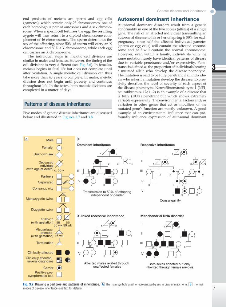
Fig. 3.6 Meiosis and gametogenesis. The main chromosomal stages of meiosis in both males and females. A single homologous pair of chromosomes is represented in different colours. The final step is the production of haploid germ cells. Each round of meiosis in the male results in four sperm cells; in the female, however, only one egg cell is produced, as the other divisions are sequestered on the periphery of the mature egg as peripheral polar bodies.
Egg
Sperm
Father Mother
Meiotic cell divisions
1stpolar bodies
2ndpolar body
DNA replication
Sister chromatids
Homologous pairing
Swapping ofgenetic material
between homologues:Recombination
Individual chromosomepair (homologues)
Non-disjunction ofchromosomes is a common
error in human meiosis,resulting in trisomy of
individual chromosomesor uniparental disomy
(both chromosomes fromsingle parent)
During development, cells must become progres-sively less like a stem cell and acquire the morphological and biochemical configuration of the tissue to which they will contribute. This process is called differentiation and it is achieved by activation of tissue-specific genes and inactivation or silencing of genes that maintain the cell in a progenitor state. This epigenetic process enables cells containing the same genetic material to have very different structures and functions. The programme of differentiation is often deranged or partially reversed in cancer cells. A similar mechanism allows adult stem cells to maintain and repair tissues. Cell migration is a process that is also necessary for development and wound healing. Migration also requires the activation of a specific set of genes, such as the transcription factor TWIST, that give the cell polarity and enable the leading edge of the cell to interact with the extracellular environ-ment to control the speed and direction of travel. Again, this process can be reactivated in cancer cells and is thought to facilitate tumour metastasis.
Cell death, apoptosis and senescenceWith the exception of stem cells, human cells have only a limited capacity for cell division. The Hayflick limit is the number of divisions a cell population can go through in culture before division stops and the cell enters a state known as senescence. This ‘biological clock’ is of great interest in the study of the normal ageing process. Rare human diseases associated with premature ageing, called progeric syndromes, have been very helpful in identifying the importance of DNA repair mechanisms in senescence (p. 168). For example, in Werner syndrome, a DNA helicase (an enzyme that separates the two DNA strands) is mutated, leading to failure of DNA repair and premature ageing. A distinct mechanism of cell death is seen in apoptosis, or pro-grammed cell death.
Apoptosis is an active process that occurs in normal tissues and plays an important role in development, tissue remodelling and the immune response. The signal that triggers apoptosis is specific to each tissue or cell type. This signal activates enzymes, called caspases, which actively destroy cellular components, including chromosomal DNA. This degradation results in cell death, but the cellular corpse contains characteristic vesicles called apoptotic bodies. The corpse is then rec-ognised and removed by phagocytic cells of the immune system, such as macrophages, in a manner that does not provoke an inflammatory response.
A third mechanism of cell death is necrosis. This is a pathological process in which the cellular environment loses one or more of the components necessary for cell viability. Hypoxia is probably the most common cause of necrosis.
GENETIC DISEASE AND INHERITANCE
Meiosis
Meiosis is a special form of cell division that only occurs in the post-pubertal testis and the fetal and adult ovary (Fig. 3.6). Meiosis differs from mitosis in two main ways; there are two separate cell divisions and before the first
of these there is extensive swapping of genetic material between homologous chromosomes, a process known as recombination. The result of recombination is that each chromosome that a parent passes to his or her off-spring is a mix of the chromosomes that the parent inherited from his or her own mother and father. The
Genetic disease and inheritance
3
51
end products of meiosis are sperm and egg cells (gametes), which contain only 23 chromosomes: one of each homologous pair of autosomes and a sex chromo-some. When a sperm cell fertilises the egg, the resulting zygote will thus return to a diploid chromosome com-plement of 46 chromosomes. The sperm determines the sex of the offspring, since 50% of sperm will carry an X chromosome and 50% a Y chromosome, while each egg cell carries an X chromosome.
The individual steps in meiotic cell division are similar in males and females. However, the timing of the cell divisions is very different (see Fig. 3.6). In females, meiosis begins in fetal life but does not complete until after ovulation. A single meiotic cell division can thus take more than 40 years to complete. In males, meiotic division does not begin until puberty and continues throughout life. In the testes, both meiotic divisions are completed in a matter of days.
Patterns of disease inheritance
Five modes of genetic disease inheritance are discussed below and illustrated in Figures 3.7 and 3.8.
Autosomal dominant inheritanceAutosomal dominant disorders result from a genetic abnormality in one of the two copies (alleles) of a single gene. The risk of an affected individual transmitting an autosomal disease to his or her offspring is 50% for each pregnancy, since half the affected individual gametes (sperm or egg cells) will contain the affected chromo-some and half will contain the normal chromosome. However, even within a family, individuals with the same mutation rarely have identical patterns of disease due to variable penetrance and/or expressivity. Pene-trance is defined as the proportion of individuals bearing a mutated allele who develop the disease phenotype. The mutation is said to be fully penetrant if all individu-als who inherit a mutation develop the disease. Expres-sivity describes the level of severity of each aspect of the disease phenotype. Neurofibromatosis type 1 (NF1, neurofibromin, 17q11.2) is an example of a disease that is fully (100%) penetrant but which shows extremely variable expressivity. The environmental factors and/or variation in other genes that act as modifiers of the mutated gene’s function are mostly unknown. A good example of an environmental influence that can pro-foundly influence expression of autosomal dominant
Fig. 3.7 Drawing a pedigree and patterns of inheritance. A The main symbols used to represent pedigrees in diagrammatic form. B The main modes of disease inheritance (see text for details).
SB
Male
Clinically affected
Deceasedindividual
(with age at death)
Separated
Consanguinity
Clinically affected, several diagnoses
CarrierPositive pre-
symptomatic test
Monozygotic twins
Dizygotic twins
Stillbirth(with gestation)
Termination
Miscarriage,affected
(with gestation)
Unknown sex
Female
Partners
Recessive inheritanceDominant inheritance
Mitochondrial DNA disorderX-linked recessive inheritance
Transmission to 50% of offspringindependent of gender
Consanguinity
Affected males related through unaffected females
Both sexes affected but onlyinherited through female meiosis
I
II
III
IV
I
II
III
IV
I
II
III
IV
V
I
II
III
IV
1
1
1
2
2 3 4
2 3 4 5 6
1 2 3 4
1
1
1
2
2 3 4
2 3 4 5 6
3 6 7
5 6 7 8
1
1
1
2
2 3 4
2 3 4 5 6
1 2 3 4
1
1
1
2
2 3 4
2 3 4 5 6
1 2 3 4
d. 50 y
30 wkSB
39 wk
16 wk
A B
Molecular and genetic factors in disease
3
52
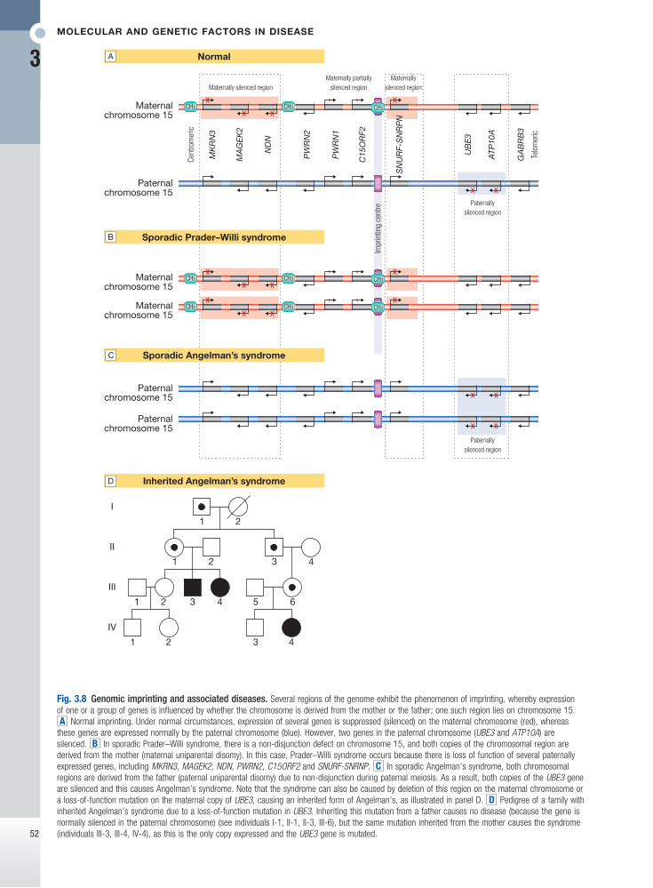
Fig. 3.8 Genomic imprinting and associated diseases. Several regions of the genome exhibit the phenomenon of imprinting, whereby expression of one or a group of genes is influenced by whether the chromosome is derived from the mother or the father; one such region lies on chromosome 15. A Normal imprinting. Under normal circumstances, expression of several genes is suppressed (silenced) on the maternal chromosome (red), whereas these genes are expressed normally by the paternal chromosome (blue). However, two genes in the paternal chromosome (UBE3 and ATP10A) are silenced. B In sporadic Prader–Willi syndrome, there is a non-disjunction defect on chromosome 15, and both copies of the chromosomal region are derived from the mother (maternal uniparental disomy). In this case, Prader–Willi syndrome occurs because there is loss of function of several paternally expressed genes, including MKRN3, MAGEK2, NDN, PWRN2, C15ORF2 and SNURF-SNRNP. C In sporadic Angelman’s syndrome, both chromosomal regions are derived from the father (paternal uniparental disomy) due to non-disjunction during paternal meiosis. As a result, both copies of the UBE3 gene are silenced and this causes Angelman’s syndrome. Note that the syndrome can also be caused by deletion of this region on the maternal chromosome or a loss-of-function mutation on the maternal copy of UBE3, causing an inherited form of Angelman’s, as illustrated in panel D. D Pedigree of a family with inherited Angelman’s syndrome due to a loss-of-function mutation in UBE3. Inheriting this mutation from a father causes no disease (because the gene is normally silenced in the paternal chromosome) (see individuals I-1, II-1, II-3, III-6), but the same mutation inherited from the mother causes the syndrome (individuals III-3, III-4, IV-4), as this is the only copy expressed and the UBE3 gene is mutated.
1
I
II
III
IV
2
1 2 3 4
21 43 5 6
3 41 2
Maternalchromosome 15
Paternalchromosome 15
Paternalchromosome 15
MK
RN
3
Cent
rom
eric
Impr
intin
g ce
ntre
Telo
mer
ic
MA
GE
K2
ND
N
PW
RN
2
PW
RN
1
C15
OR
F2
SN
UR
F-S
NR
PN
UB
E3
ATP
10A
GA
BR
B3
Maternally silenced regionMaternally
silenced region
Paternallysilenced region
Paternallysilenced region
Maternally partiallysilenced region
CH3 CH3 CH3
Maternalchromosome 15
CH3 CH3 CH3
Maternalchromosome 15
CH3 CH3 CH3
Paternalchromosome 15
Normal
Sporadic Angelman’s syndrome
Sporadic Prader–Willi syndrome
A
C
Inherited Angelman’s syndromeD
B
Genetic disease and inheritance
3
53
disease is seen in the triggering of malignant hyper-pyrexia by anaesthetic agents in the presence of RYR1 mutations. Autosomal dominant disorders may be the result of either loss or gain of function of the affected gene. For example, adult polycystic kidney disease type 1 is caused by loss-of-function mutations in PKD1, which encodes polycystin I on 16p13.1. Hereditary motor and sensory neuropathy type 1 is caused by increased number of copies (resulting in increased gene dosage) of PMP22, encoding peripheral myelin protein 22 on 17p11.2.
Autosomal recessive inheritanceIn autosomal recessive disorders, both alleles of a gene must be mutated before the disease is manifest in an individual, and an affected individual must inherit one mutant allele from each parent. What distinguishes autosomal dominant and recessive diseases is that carrying one mutant allele does not produce a disease phenotype. Autosomal recessive disorders are rare in most populations. For example, the most common serious autosomal recessive disorder in the UK is cystic fibrosis, which has a birth incidence of 1 : 2000. The fre-quency of autosomal recessive disorders increases with the degree of inbreeding of a population because the risk of inheriting the same mutant allele from both parents (homozygosity) is increased. Genetic risk calculation for a fully penetrant autosomal recessive disorder is straightforward. Each subsequent pregnancy of a couple who have had a previous child affected by an autosomal recessive disorder will have a 25% (1 : 4) risk of being affected; a healthy individual who has a sibling with an autosomal recessive disorder will have 2/3 chance of being a carrier. The risk of an affected individual having children with the same condition is usually low but is dependent on the carrier rate of the mutant allele in the population.
X-linked inheritanceGenetic diseases caused by mutations on the X chro-mosome have specific characteristics. X-linked diseases are mostly recessive and restricted to males who carry the mutant allele. This is because males have only one X chromosome, whereas females have two. Thus females who carry a single mutant allele are generally unaffected. Occasionally, female carriers may exhibit signs of an X-linked disease due to a phenomenon called skewed X-inactivation. All female embryos, at about 100 cells in size, stably inactivate one of their two X chromosomes in each cell. This process is random in each cell but if, by chance, there is a dis-proportionate inactivation of normal X chromosomes carrying the normal allele, then an affected female carrier will be more likely, an extreme example being the rare cases of carrier females affected with Duch-enne muscular dystrophy. X-linked recessive disorders have a recognisable pattern of inheritance, with trans-mission of the disease from carrier females to affected males and absence of father-to-son transmission. The risk of a female carrier having an affected child is 25% (1 : 4; half of her male offspring). If the carrier status of a woman is unclear, then the risk may be altered by conditional information, as discussed in the autosomal dominant disease section above. Bayes’ theorem is
commonly used to calculate such modified risks and this is discussed in more detail later in this chapter (p. 68).
Mitochondrial inheritanceThe inheritance of mtDNA disorders is characterised by transmission from females, but males and females are generally affected equally. Unlike the other inheritance patterns mentioned above, mitochondrial inheritance has nothing to do with meiosis but reflects the fact that mitochondrial DNA is transmitted by oöcytes. Mito-chondrial disorders tend to be very variable in pene-trance and expressivity within families, and this is mostly accounted for by the fact that only a proportion of multiple mtDNA molecules within mitochondria contain the causal mutation (the degree of mtDNA heteroplasmy).
Epigenetic inheritance and imprintingSeveral chromosomal regions (loci) have been identified where gene repression is inherited in a parent-of-origin-specific manner; these are called imprinted loci. Within these loci the paternal alleles of a gene may be active while the maternal one may be silenced, or vice versa (see Fig. 3.8). Mutations within imprinted loci lead to a very unusual pattern of inheritance in which the pheno-type is only manifest if inherited from the parent who contributes the transcriptionally active allele (see Fig. 3.8). Examples of these disorders are given in Box 3.3.
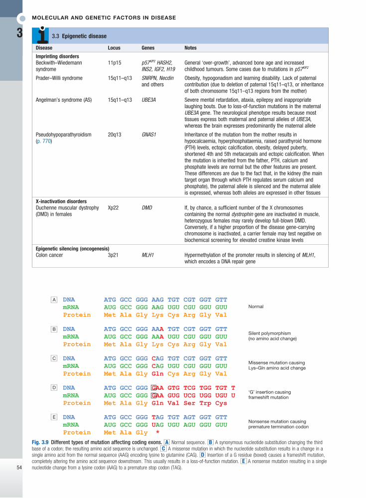
Classes of genetic variant
There are many different classes of variation in the human genome (Figs 3.9 and 3.10). Rare genetic varia-tions that result in a disease are generally referred to as mutations, whereas common variations and those that do not cause disease are referred to as polymorphisms. These different types of variation are further categorised by the size of the DNA segment involved and/or by the mechanism giving rise to the variation.
Nucleotide substitutionsThe substitution of one nucleotide for another is the most common type of variation in the human genome. Depending on their frequency and functional conse-quences, these changes are known as a point mutation or a single nucleotide polymorphism (SNP). They occur by misincorporation of a nucleotide during DNA syn-thesis or by the action of a chemical mutagen. When these substitutions occur within ORFs of a protein-coding gene, they are further classified into:• synonymous – resulting in a change in the codon
but no change in the amino acid and thus no phenotype
• missense – altering a codon, resulting in an amino acid change in the protein
• nonsense – introducing a premature stop codon, resulting in truncation of the protein
• splicing – occurring at the junction of an intron and an exon, thereby adversely affecting splicing.
Examples of these types of variation are shown in Figures 3.9 and 3.10.
Molecular and genetic factors in disease
3
54
Normal
Silent polymorphism(no amino acid change)
Missense mutation causingLys–Gln amino acid change
‘G’ insertion causingframeshift mutation
Nonsense mutation causingpremature termination codon
A
B
C
D
E
Fig. 3.9 Different types of mutation affecting coding exons. A Normal sequence. B A synonymous nucleotide substitution changing the third base of a codon; the resulting amino acid sequence is unchanged. C A missense mutation in which the nucleotide substitution results in a change in a single amino acid from the normal sequence (AAG) encoding lysine to glutamine (CAG). D Insertion of a G residue (boxed) causes a frameshift mutation, completely altering the amino acid sequence downstream. This usually results in a loss-of-function mutation. E A nonsense mutation resulting in a single nucleotide change from a lysine codon (AAG) to a premature stop codon (TAG).
Disease Locus Genes Notes
Imprinting disordersBeckwith–Wiedemann syndrome
11p15 p57KIP2 HASH2, INS2, IGF2, H19
General ‘over-growth’, advanced bone age and increased childhood tumours. Some cases due to mutations in p57KIP2
Prader–Willi syndrome 15q11–q13 SNRPN, Necdin and others
Obesity, hypogonadism and learning disability. Lack of paternal contribution (due to deletion of paternal 15q11–q13, or inheritance of both chromosome 15q11–q13 regions from the mother)
Angelman’s syndrome (AS) 15q11–q13 UBE3A Severe mental retardation, ataxia, epilepsy and inappropriate laughing bouts. Due to loss-of-function mutations in the maternal UBE3A gene. The neurological phenotype results because most tissues express both maternal and paternal alleles of UBE3A, whereas the brain expresses predominantly the maternal allele
Pseudohypoparathyroidism (p. 770)
20q13 GNAS1 Inheritance of the mutation from the mother results in hypocalcaemia, hyperphosphataemia, raised parathyroid hormone (PTH) levels, ectopic calcification, obesity, delayed puberty, shortened 4th and 5th metacarpals and ectopic calcification. When the mutation is inherited from the father, PTH, calcium and phosphate levels are normal but the other features are present. These differences are due to the fact that, in the kidney (the main target organ through which PTH regulates serum calcium and phosphate), the paternal allele is silenced and the maternal allele is expressed, whereas both alleles are expressed in other tissues
X-inactivation disordersDuchenne muscular dystrophy (DMD) in females
Xp22 DMD If, by chance, a sufficient number of the X chromosomes containing the normal dystrophin gene are inactivated in muscle, heterozygous females may rarely develop full-blown DMD. Conversely, if a higher proportion of the disease gene-carrying chromosome is inactivated, a carrier female may test negative on biochemical screening for elevated creatine kinase levels
Epigenetic silencing (oncogenesis)Colon cancer 3p21 MLH1 Hypermethylation of the promoter results in silencing of MLH1,
which encodes a DNA repair gene
3.3 Epigenetic disease
Genetic disease and inheritance
3
55
Insertions and deletionsOne or more nucleotides may be inserted or lost in a DNA sequence, resulting in an insertion/deletion (indel) polymorphism or mutation (see Fig. 3.9). If an indel change affects one or two nucleotides within the ORF of a protein-coding gene, this can have serious con-sequences because the triple nucleotide sequence of the codons is disrupted, resulting in a frameshift mutation. The effect upon the gene is typically severe because the amino acid sequence is totally disrupted.
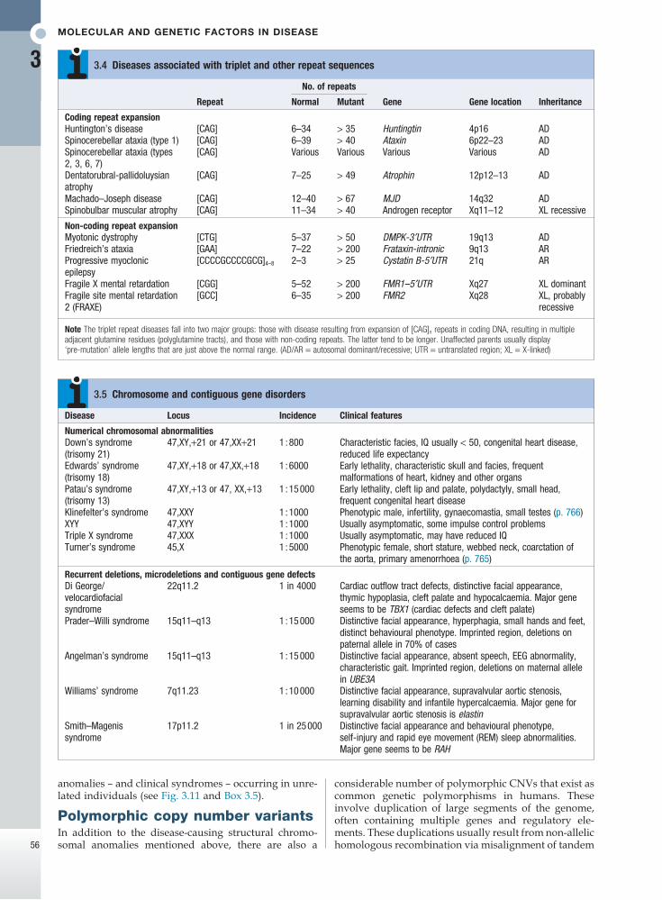
Simple tandem repeat mutationVariations in the length of simple tandem repeats of DNA are thought to arise as the result of slippage of DNA during meiosis and are termed microsatellite (small) or minisatellite (larger) repeats. These repeats are unstable and can expand or contract in different generations. This instability is proportional to the size of the original repeat, in that longer repeats tend to be more unstable. Many microsatellites and minisatellites occur in introns or in chromosomal regions between genes and have no obvious adverse effects. However, some genetic diseases, including Huntington’s disease and myotonic dystrophy, are caused by microsatellite repeats, which result in duplication of amino acids within the affected gene product or affect gene expres-sion (Box 3.4).
Copy number variationsVariation in the number of copies of an individual segment of the genome from the usual diploid (two copies) content can be categorised by the size of the
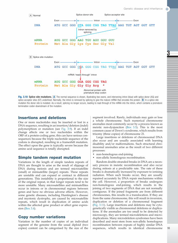
Fig. 3.10 Splice site mutations. A The normal sequence is shown, illustrating two exons, and intervening intron (blue) with splice donor (AG) and splice acceptor sites (GT) underlined. Normally, the intron is removed by splicing to give the mature mRNA that encodes the protein. B In a splice site mutation the donor site is mutated. As a result, splicing no longer occurs, leading to read-through of the mRNA into the intron, which contains a premature termination codon downstream of the mutation.
Normal
Splice site mutation
Splice donor site
Exon ExonIntron
Exon ExonIntron
Intron removed bysplicing
Splice acceptor site
mRNA ‘reads through’ intron
Abnormal protein withpremature stop codon
A
B
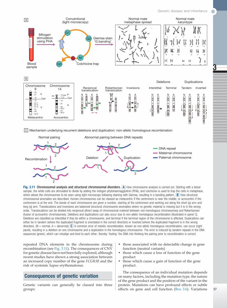
segment involved. Rarely, individuals may gain or lose a whole chromosome. Such numerical chromosome anomalies most commonly occur by a process known as meiotic non-dysjunction (Box 3.5). This is the most common cause of Down’s syndrome, which results from trisomy (three copies) of chromosome 21.
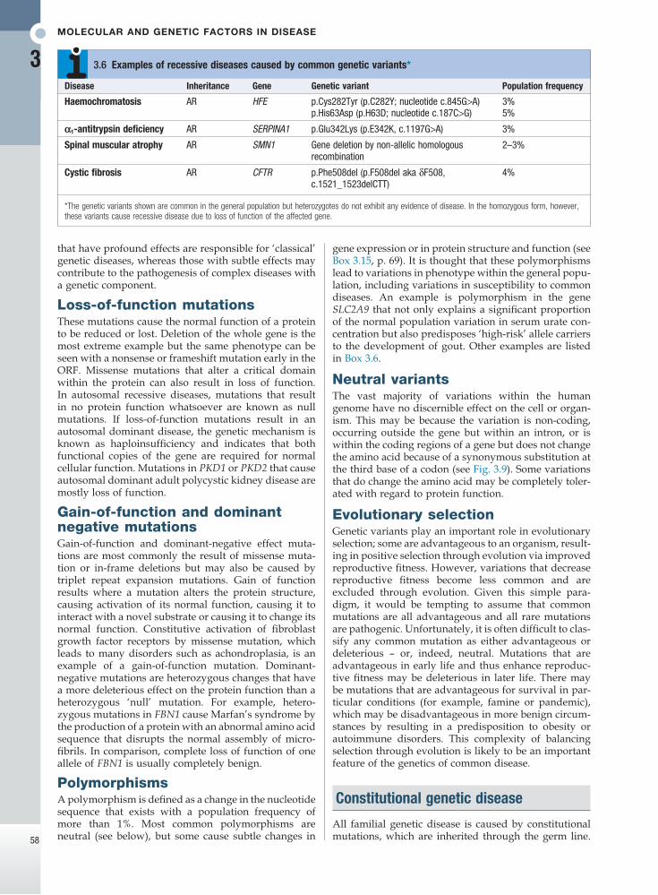
Large insertions or deletions of chromosomal DNA also occur and are usually associated with learning disability and/or malformations. Such structural chro-mosomal anomalies arise as the result of two different processes:• non-homologous end-joining• non-allelic homologous recombination.
Random double-stranded breaks in DNA are a neces-sary process in meiotic recombination and also occur during mitosis at a predictable rate. The rate of these breaks is dramatically increased by exposure to ionising radiation. When such breaks occur, they are usually repaired accurately by DNA repair mechanisms within the cell. However, a proportion of breaks undergoes non-homologous end-joining, which results in the joining of two segments of DNA that are not normally contiguous. If the joined fragments are from different chromosomes, this results in a translocation. If they are from the same chromosome, this will result in inversion, duplication or deletion of a chromosomal fragment (Fig. 3.11). Large insertions and deletions may be cyto-genetically visible as chromosomal deletions or duplica-tions. If the anomalies are too small to be detected by microscopy, they are termed microdeletions and micro-duplications. Many microdeletion syndromes have been described and most stem from non-allelic homologous recombination between repeats of highly similar DNA sequences, which results in identical chromosome
Molecular and genetic factors in disease
3
56
No. of repeats
Repeat Normal Mutant Gene Gene location Inheritance
Coding repeat expansionHuntington’s disease [CAG] 6–34 > 35 Huntingtin 4p16 ADSpinocerebellar ataxia (type 1) [CAG] 6–39 > 40 Ataxin 6p22–23 ADSpinocerebellar ataxia (types 2, 3, 6, 7)
[CAG] Various Various Various Various AD
Dentatorubral-pallidoluysian atrophy
[CAG] 7–25 > 49 Atrophin 12p12–13 AD
Machado–Joseph disease [CAG] 12–40 > 67 MJD 14q32 ADSpinobulbar muscular atrophy [CAG] 11–34 > 40 Androgen receptor Xq11–12 XL recessive
Non-coding repeat expansionMyotonic dystrophy [CTG] 5–37 > 50 DMPK-3′UTR 19q13 ADFriedreich’s ataxia [GAA] 7–22 > 200 Frataxin-intronic 9q13 ARProgressive myoclonic epilepsy
[CCCCGCCCCGCG]4–8 2–3 > 25 Cystatin B-5′UTR 21q AR
Fragile X mental retardation [CGG] 5–52 > 200 FMR1–5′UTR Xq27 XL dominantFragile site mental retardation 2 (FRAXE)
[GCC] 6–35 > 200 FMR2 Xq28 XL, probably recessive
Note The triplet repeat diseases fall into two major groups: those with disease resulting from expansion of [CAG]n repeats in coding DNA, resulting in multiple adjacent glutamine residues (polyglutamine tracts), and those with non-coding repeats. The latter tend to be longer. Unaffected parents usually display ‘pre-mutation’ allele lengths that are just above the normal range. (AD/AR = autosomal dominant/recessive; UTR = untranslated region; XL = X-linked)
3.4 Diseases associated with triplet and other repeat sequences
Disease Locus Incidence Clinical features
Numerical chromosomal abnormalitiesDown’s syndrome (trisomy 21)
47,XY,+21 or 47,XX+21 1 : 800 Characteristic facies, IQ usually < 50, congenital heart disease, reduced life expectancy
Edwards’ syndrome (trisomy 18)
47,XY,+18 or 47,XX,+18 1 : 6000 Early lethality, characteristic skull and facies, frequent malformations of heart, kidney and other organs
Patau’s syndrome (trisomy 13)
47,XY,+13 or 47, XX,+13 1 : 15 000 Early lethality, cleft lip and palate, polydactyly, small head, frequent congenital heart disease
Klinefelter’s syndrome 47,XXY 1 : 1000 Phenotypic male, infertility, gynaecomastia, small testes (p. 766)XYY 47,XYY 1 : 1000 Usually asymptomatic, some impulse control problemsTriple X syndrome 47,XXX 1 : 1000 Usually asymptomatic, may have reduced IQTurner’s syndrome 45,X 1 : 5000 Phenotypic female, short stature, webbed neck, coarctation of
the aorta, primary amenorrhoea (p. 765)
Recurrent deletions, microdeletions and contiguous gene defectsDi George/velocardiofacial syndrome
22q11.2 1 in 4000 Cardiac outflow tract defects, distinctive facial appearance, thymic hypoplasia, cleft palate and hypocalcaemia. Major gene seems to be TBX1 (cardiac defects and cleft palate)
Prader–Willi syndrome 15q11–q13 1 : 15 000 Distinctive facial appearance, hyperphagia, small hands and feet, distinct behavioural phenotype. Imprinted region, deletions on paternal allele in 70% of cases
Angelman’s syndrome 15q11–q13 1 : 15 000 Distinctive facial appearance, absent speech, EEG abnormality, characteristic gait. Imprinted region, deletions on maternal allele in UBE3A
Williams’ syndrome 7q11.23 1 : 10 000 Distinctive facial appearance, supravalvular aortic stenosis, learning disability and infantile hypercalcaemia. Major gene for supravalvular aortic stenosis is elastin
Smith–Magenis syndrome
17p11.2 1 in 25 000 Distinctive facial appearance and behavioural phenotype, self-injury and rapid eye movement (REM) sleep abnormalities. Major gene seems to be RAH
3.5 Chromosome and contiguous gene disorders
anomalies – and clinical syndromes – occurring in unre-lated individuals (see Fig. 3.11 and Box 3.5).
Polymorphic copy number variantsIn addition to the disease-causing structural chromo-somal anomalies mentioned above, there are also a
considerable number of polymorphic CNVs that exist as common genetic polymorphisms in humans. These involve duplication of large segments of the genome, often containing multiple genes and regulatory ele-ments. These duplications usually result from non-allelic homologous recombination via misalignment of tandem
Genetic disease and inheritance
3
57
Fig. 3.11 Chromosomal analysis and structural chromosomal disorders. A How chromosome analysis is carried out. Starting with a blood sample, the white cells are stimulated to divide by adding the mitogen phytohaemagglutinin (PHA), and colchicine is used to trap the cells in metaphase, which allows the chromosomes to be seen using light microscopy following staining with Giemsa, resulting in a banding pattern. B How structural chromosomal anomalies are described. Human chromosomes can be classed as metacentric if the centromere is near the middle, or acrocentric if the centromere is at the end. The bands of each chromosome are given a number, starting at the centromere and working out along the short (p) arm and long (q) arm. Translocations and inversions are balanced structural chromosome anomalies where no genetic material is missing but it is in the wrong order. Translocations can be divided into reciprocal (direct swap of chromosomal material between non-homologous chromosomes) and Robertsonian (fusion of acrocentric chromosomes). Deletions and duplications can also occur due to non-allelic homologous recombination (illustrated in panel C). Deletions are classified as interstitial if they lie within a chromosome, and terminal if the terminal region of the chromosome is affected. Duplications can either be in tandem (where the duplicated fragment is orientated in the correct direction) or inverted (where the duplicated fragment is in the wrong direction). (N = normal; A = abnormal) C A common error of meiotic recombination, known as non-allelic homologous recombination, can occur (right panel), resulting in a deletion on one chromosome and a duplication in the homologous chromosome. The error is induced by tandem repeats in the DNA sequences (green), which can misalign and bind to each other, thereby ‘fooling’ the DNA into thinking the pairing prior to recombination is correct.
A
Bloodsample
Chromosome9
Cen
p2321
12
1221.121.321.2313334.2
q
Cen
p N N A N A N A N AN AN A A13 (sa)(st)11.2
12
21
2324.23132.2
q
Metacentric Acrocentric
Chromosome14 Reciprocal
translocationRobertsoniantranslocation
Inversions Interstitial Terminal Tandem Inverted
Mechanism underlying recurrent deletions and duplication: non-allelic homologous recombination
Deletions Duplications
Mitogenstimulationusing PHA
Giemsa stain‘G banding’
Conventional(light microscopy)
Normal malemetaphase spread
Normal malekaryotype
Colchicine trapG0
M
G2
G1
C
B
Recombination
Normal pairing Abnormal pairing between DNA repeats
Deletion Duplication
1 1
1
2
1 2
1
2
2
2
2 DNA repeatMaternal chromosomePaternal chromosome
1 1
21
2
repeated DNA elements in the chromosome during recombination (see Fig. 3.11). The consequences of CNV for genetic disease have not been fully explored, although recent studies have shown a strong association between an increased copy number of the gene FCGR3B and the risk of systemic lupus erythematosus.
Consequences of genetic variation
Genetic variants can generally be classed into three groups:
• those associated with no detectable change in gene function (neutral variants)
• those which cause a loss of function of the gene product
• those which cause a gain of function of the gene product.
The consequence of an individual mutation depends on many factors, including the mutation type, the nature of the gene product and the position of the variant in the protein. Mutations can have profound effects or subtle effects on gene and cell function (Box 3.6). Variations
Molecular and genetic factors in disease
3
58
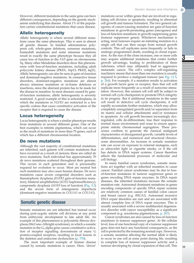
gene expression or in protein structure and function (see Box 3.15, p. 69). It is thought that these polymorphisms lead to variations in phenotype within the general popu-lation, including variations in susceptibility to common diseases. An example is polymorphism in the gene SLC2A9 that not only explains a significant proportion of the normal population variation in serum urate con-centration but also predisposes ‘high-risk’ allele carriers to the development of gout. Other examples are listed in Box 3.6.
Neutral variantsThe vast majority of variations within the human genome have no discernible effect on the cell or organ-ism. This may be because the variation is non-coding, occurring outside the gene but within an intron, or is within the coding regions of a gene but does not change the amino acid because of a synonymous substitution at the third base of a codon (see Fig. 3.9). Some variations that do change the amino acid may be completely toler-ated with regard to protein function.
Evolutionary selectionGenetic variants play an important role in evolutionary selection; some are advantageous to an organism, result-ing in positive selection through evolution via improved reproductive fitness. However, variations that decrease reproductive fitness become less common and are excluded through evolution. Given this simple para-digm, it would be tempting to assume that common mutations are all advantageous and all rare mutations are pathogenic. Unfortunately, it is often difficult to clas-sify any common mutation as either advantageous or deleterious – or, indeed, neutral. Mutations that are advantageous in early life and thus enhance reproduc-tive fitness may be deleterious in later life. There may be mutations that are advantageous for survival in par-ticular conditions (for example, famine or pandemic), which may be disadvantageous in more benign circum-stances by resulting in a predisposition to obesity or autoimmune disorders. This complexity of balancing selection through evolution is likely to be an important feature of the genetics of common disease.
Constitutional genetic disease
All familial genetic disease is caused by constitutional mutations, which are inherited through the germ line.
that have profound effects are responsible for ‘classical’ genetic diseases, whereas those with subtle effects may contribute to the pathogenesis of complex diseases with a genetic component.
Loss-of-function mutationsThese mutations cause the normal function of a protein to be reduced or lost. Deletion of the whole gene is the most extreme example but the same phenotype can be seen with a nonsense or frameshift mutation early in the ORF. Missense mutations that alter a critical domain within the protein can also result in loss of function. In autosomal recessive diseases, mutations that result in no protein function whatsoever are known as null mutations. If loss-of-function mutations result in an autosomal dominant disease, the genetic mechanism is known as haploinsufficiency and indicates that both functional copies of the gene are required for normal cellular function. Mutations in PKD1 or PKD2 that cause autosomal dominant adult polycystic kidney disease are mostly loss of function.
Gain-of-function and dominant negative mutationsGain-of-function and dominant-negative effect muta-tions are most commonly the result of missense muta-tion or in-frame deletions but may also be caused by triplet repeat expansion mutations. Gain of function results where a mutation alters the protein structure, causing activation of its normal function, causing it to interact with a novel substrate or causing it to change its normal function. Constitutive activation of fibroblast growth factor receptors by missense mutation, which leads to many disorders such as achondroplasia, is an example of a gain-of-function mutation. Dominant-negative mutations are heterozygous changes that have a more deleterious effect on the protein function than a heterozygous ‘null’ mutation. For example, hetero-zygous mutations in FBN1 cause Marfan’s syndrome by the production of a protein with an abnormal amino acid sequence that disrupts the normal assembly of micro-fibrils. In comparison, complete loss of function of one allele of FBN1 is usually completely benign.
PolymorphismsA polymorphism is defined as a change in the nucleotide sequence that exists with a population frequency of more than 1%. Most common polymorphisms are neutral (see below), but some cause subtle changes in
*The genetic variants shown are common in the general population but heterozygotes do not exhibit any evidence of disease. In the homozygous form, however, these variants cause recessive disease due to loss of function of the affected gene.
Disease Inheritance Gene Genetic variant Population frequency
Haemochromatosis AR HFE p.Cys282Tyr (p.C282Y; nucleotide c.845G>A) 3%p.His63Asp (p.H63D; nucleotide c.187C>G) 5%
α1-antitrypsin deficiency AR SERPINA1 p.Glu342Lys (p.E342K, c.1197G>A) 3%
Spinal muscular atrophy AR SMN1 Gene deletion by non-allelic homologous recombination
2–3%
Cystic fibrosis AR CFTR p.Phe508del (p.F508del aka δF508, c.1521_1523delCTT)
4%
3.6 Examples of recessive diseases caused by common genetic variants*
Genetic disease and inheritance
3
59
However, different mutations in the same gene can have different consequences, depending on the genetic mech-anism underlying that disease. About 1% of the popula-tion carries constitutional mutations that cause disease.
Allelic heterogeneityAllelic heterogeneity is where several different muta-tions cause the same phenotype. This is seen in almost all genetic disease. In familial adenomatous poly-posis coli, whole-gene deletions, nonsense mutations, frameshift mutations and some missense mutations result in exactly the same phenotype because they all cause loss of function in the FAP gene on chromosome 5q. Many other Mendelian disorders show this phenom-enon with loss-of-function mutations, including adult polycystic kidney disease (PKD1, 16p13; PKD2, 4q21). Allelic heterogeneity can also be seen in gain-of-function and dominant-negative mutations. In connective tissue disorders, dominant-negative mutations are almost always missense mutations or in-frame deletions or insertions, since the aberrant protein has to be made for the disease to manifest. In most diseases caused by gain-of-function mutations, allelic heterogeneity is severely restricted. A good example of this is achondroplasia, in which the mutations in FGFR3 are restricted to a few specific codons that cause constitutive activation of the receptor that is required to cause the disease.
Locus heterogeneityLocus heterogeneity is where a similar phenotype results from mutations in several different genes. One of the best examples is retinitis pigmentosa, which can occur as the result of mutations in more than 75 genes, each of which has a different chromosomal location.
De novo mutationsAlthough the vast majority of constitutional mutations are inherited, each gamete will contain mutations that have occurred as a result of meiosis; these are called de novo mutations. Each individual has approximately 70 de novo mutations scattered throughout their genome. This occurs in each generation and is presumably required for evolution to occur. Most are neutral but such mutations may also cause human disease. De novo mutations cause severe congenital disorders such as thanatophoric dysplasia (FGFR3 gain-of-function muta-tion), bilateral anophthalmia (SOX2 haploinsufficiency), campomelic dysplasia (SOX9 loss of function) (Fig. 3.2) and the severe form of osteogenesis imperfecta (dominant-negative mutations in COL1A1 or COL1A2).
Somatic genetic disease
Somatic mutations are not inherited but instead occur during post-zygotic mitotic cell divisions at any point from embryonic development to late adult life. An example of this phenomenon is polyostotic fibrous dys-plasia (McCune–Albright syndrome), in which a somatic mutation in the GS alpha gene causes constitutive activa-tion of receptor signalling downstream of many G protein-coupled receptors, resulting in focal lesions in the skeleton and endocrine dysfunction (p. 770).
The most important example of human disease caused by somatic mutations is cancer. Here, ‘driver’
mutations occur within genes that are involved in regu-lating cell division or apoptosis, resulting in abnormal cell growth and tumour formation. The two general cat-egories of cancer-causing mutation are gain-of-function mutations in growth-promoting genes (oncogenes) and loss-of-function mutations in growth-suppressing genes (tumour suppressor genes). Whichever mechanism is acting, most tumours require an initiating mutation in a single cell that can then escape from normal growth controls. This cell replicates more frequently or fails to undergo programmed death, resulting in clonal expan-sion. As the size of the clone increases, one or more cells may acquire additional mutations that confer further growth advantage, leading to proliferation of these subclones, which may ultimately lead to aggressive metastatic cancer. The cell’s complex self-regulating machinery means that more than one mutation is usually required to produce a malignant tumour (see Fig. 11.3, p. 264). For example, if a mutation results in activation of a growth factor gene or receptor, then that cell will replicate more frequently as a result of autocrine stimu-lation. However, this mutant cell will still be subject to normal cell cycle checkpoints to promote DNA integrity in its progeny. But if additional mutations in the same cell result in defective cell cycle checkpoints, it will rapidly accumulate further mutations, which may allow completely unregulated growth and/or separation from its matrix and cellular attachments and/or resistance to apoptosis. As cell growth becomes increasingly dys-regulated, cells de-differentiate, lose their response to normal tissue environment and cease to ensure appro-priate mitotic chromosomal segregation. These pro-cesses combine to generate the classical malignant characteristics of disorganised growth, variable levels of differentiation, and numerical and structural chromo-some abnormalities. An increase in somatic mutation rate can occur on exposure to external mutagens, such as ultraviolet light or cigarette smoke, or if the cell has defects in DNA repair systems. Cancer therefore affects the fundamental processes of molecular and cell biology.
In many familial cancer syndromes, somatic muta-tions act together with an inherited mutation to cause cancer. Familial cancer syndromes may be due to loss-of-function mutations in tumour suppressor genes or genes encoding DNA repair enzymes. In DNA repair diseases, the inherited mutations increase the somatic mutation rate. Autosomal dominant mutations in genes encoding components of specific DNA repair systems are relatively common causes of familial colon cancer and breast cancer (e.g. BRCA1). Autosomal recessive DNA repair disorders are rare and are associated with almost complete loss of DNA repair enzymes. This is usually associated with a severe multifaceted degenera-tive disorder with cancer susceptibility as a significant component (e.g. xeroderma pigmentosum, p. 267).
Cancer syndromes are also caused by loss-of-function mutations in tumour suppressor genes. At the cellular level, loss of one functional copy of a tumour suppressor gene does not have any functional consequences, as the cell is protected by the remaining normal copy. However, a somatic mutation affecting the normal allele is likely to occur in one cell at some point during life, resulting in complete loss of tumour suppressor activity and a tumour developing by clonal expansion of that cell. This
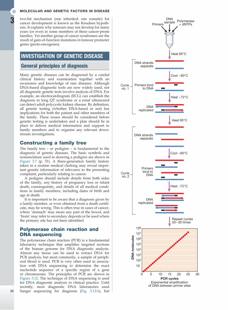
Molecular and genetic factors in disease
3
60
Cool ~60°C
DNAsample
DNA strandsseparate
Primersbind to
DNA
DNAreplicated
Heat 95°C
Heat 95°C
Cool ~60°C
Repeat cycles20–30 times
108
107
106
105
104
103
102
10
0 5 10 15PCR cycles
Exponential amplificationof DNA between primer sites
DN
A m
ole
cule
s
20 25 30
DNA strandsseparate
Primers bindto DNA
Cycleno. 1
DNAreplicated
Heat ~72°C
Heat ~72°C
Polymerase+ dNTPsPrimers
Cycleno. 2
two-hit mechanism (one inherited, one somatic) for cancer development is known as the Knudsen hypoth-esis. It explains why tumours may not develop for many years (or ever) in some members of these cancer-prone families. Yet another group of cancer syndromes are the result of gain-of-function mutations in tumour promoter genes (proto-oncogenes).
INVESTIGATION OF GENETIC DISEASE
General principles of diagnosis
Many genetic diseases can be diagnosed by a careful clinical history and examination together with an awareness and knowledge of rare diseases. Although DNA-based diagnostic tools are now widely used, not all diagnostic genetic tests involve analysis of DNA. For example, an electrocardiogram (ECG) can establish the diagnosis in long QT syndrome or a renal ultrasound can detect adult polycystic kidney disease. By definition, all genetic testing (whether DNA-based or not) has implications for both the patient and other members of the family. These issues should be considered before genetic testing is undertaken and a plan should be in place to deliver medical information and support to family members and to organise any relevant down-stream investigations.
Constructing a family treeThe family tree – or pedigree – is fundamental to the diagnosis of genetic diseases. The basic symbols and nomenclature used in drawing a pedigree are shown in Figure 3.7 (p. 51). A three-generation family history taken in a routine medical clerking may reveal impor-tant genetic information of relevance to the presenting complaint, particularly relating to cancer.
A pedigree should include details from both sides of the family, any history of pregnancy loss or infant death, consanguinity, and details of all medical condi-tions in family members, including dates of birth and age at death.
It is important to be aware that a diagnosis given by a family member, or even obtained from a death certifi-cate, may be wrong. This is often true in cases of cancer, where ‘stomach’ may mean any part of the bowel, and ‘brain’ may refer to secondary deposits or be used where the primary site has not been identified.
Polymerase chain reaction and DNA sequencingThe polymerase chain reaction (PCR) is a fundamental laboratory technique that amplifies targeted sections of the human genome for DNA diagnostic analysis. Almost any tissue can be used to extract DNA for PCR analysis, but most commonly, a sample of periph-eral blood is used. PCR is very often used in associa-tion with DNA sequencing to determine the exact nucleotide sequence of a specific region of a gene or chromosome. The principles of PCR are shown in Figure 3.12. The technique of DNA sequencing is used for DNA diagnostic analysis in clinical practice. Until recently, most diagnostic DNA laboratories used Sanger sequencing for diagnosis (Fig. 3.13A), but
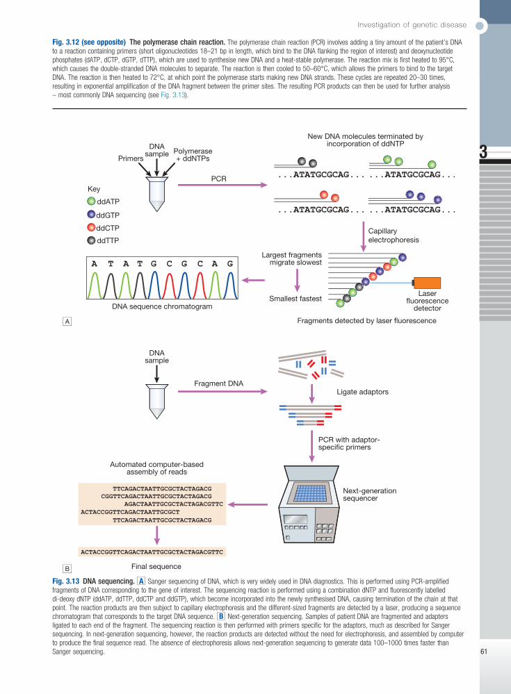
Investigation of genetic disease
3
61
Fig. 3.12 (see opposite) The polymerase chain reaction. The polymerase chain reaction (PCR) involves adding a tiny amount of the patient’s DNA to a reaction containing primers (short oligonucleotides 18–21 bp in length, which bind to the DNA flanking the region of interest) and deoxynucleotide phosphates (dATP, dCTP, dGTP, dTTP), which are used to synthesise new DNA and a heat-stable polymerase. The reaction mix is first heated to 95°C, which causes the double-stranded DNA molecules to separate. The reaction is then cooled to 50–60°C, which allows the primers to bind to the target DNA. The reaction is then heated to 72°C, at which point the polymerase starts making new DNA strands. These cycles are repeated 20–30 times, resulting in exponential amplification of the DNA fragment between the primer sites. The resulting PCR products can then be used for further analysis – most commonly DNA sequencing (see Fig. 3.13).
Fig. 3.13 DNA sequencing. A Sanger sequencing of DNA, which is very widely used in DNA diagnostics. This is performed using PCR-amplified fragments of DNA corresponding to the gene of interest. The sequencing reaction is performed using a combination dNTP and fluorescently labelled di-deoxy dNTP (ddATP, ddTTP, ddCTP and ddGTP), which become incorporated into the newly synthesised DNA, causing termination of the chain at that point. The reaction products are then subject to capillary electrophoresis and the different-sized fragments are detected by a laser, producing a sequence chromatogram that corresponds to the target DNA sequence. B Next-generation sequencing. Samples of patient DNA are fragmented and adapters ligated to each end of the fragment. The sequencing reaction is then performed with primers specific for the adaptors, much as described for Sanger sequencing. In next-generation sequencing, however, the reaction products are detected without the need for electrophoresis, and assembled by computer to produce the final sequence read. The absence of electrophoresis allows next-generation sequencing to generate data 100–1000 times faster than Sanger sequencing.
A
B
Capillaryelectrophoresis
Largest fragmentsmigrate slowest
Smallest fastestLaser
fluorescencedetector
ddTTP
ddCTP
ddATP
ddGTP
DNAsample
PCR
DNA sequence chromatogram
Key
Fragments detected by laser fluorescence
New DNA molecules terminated byincorporation of ddNTP
Polymerase+ ddNTPsPrimers
DNAsample
Automated computer-basedassembly of reads
Final sequence
Fragment DNALigate adaptors
PCR with adaptor-specific primers
Next-generationsequencer
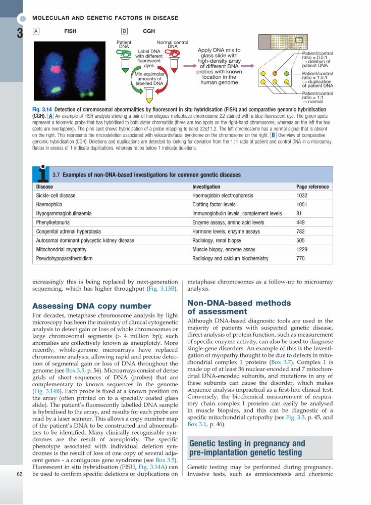
Molecular and genetic factors in disease
3
62
Fig. 3.14 Detection of chromosomal abnormalities by fluorescent in situ hybridisation (FISH) and comparative genomic hybridisation (CGH). A An example of FISH analysis showing a pair of homologous metaphase chromosome 22 stained with a blue fluorescent dye. The green spots represent a telomeric probe that has hybridised to both sister chromatids (there are two spots on the right-hand chromosome, whereas on the left the two spots are overlapping). The pink spot shows hybridisation of a probe mapping to band 22q11.2. The left chromosome has a normal signal that is absent on the right. This represents the microdeletion associated with velocardiofacial syndrome on the chromosome on the right. B Overview of comparative genomic hybridisation (CGH). Deletions and duplications are detected by looking for deviation from the 1 : 1 ratio of patient and control DNA in a microarray. Ratios in excess of 1 indicate duplications, whereas ratios below 1 indicate deletions.
FISH CGH
PatientDNA
Label DNAwith differentfluorescent
dyes
Mix equimolaramounts of
labelled DNA
Apply DNA mix toglass slide with
high-density arrayof different DNA
probes with knownlocation in the
human genome
Patient/controlratio = 0.5:1→ deletion ofpatient DNA
Patient/controlratio = 1.5:1→ duplicationof patient DNA
Patient/controlratio = 1:1→ normal
Normal controlDNA
A B
metaphase chromosomes as a follow-up to microarray analysis.
Non-DNA-based methods of assessmentAlthough DNA-based diagnostic tools are used in the majority of patients with suspected genetic disease, direct analysis of protein function, such as measurement of specific enzyme activity, can also be used to diagnose single-gene disorders. An example of this is the investi-gation of myopathy thought to be due to defects in mito-chondrial complex 1 proteins (Box 3.7). Complex 1 is made up of at least 36 nuclear-encoded and 7 mitochon-drial DNA-encoded subunits, and mutations in any of these subunits can cause the disorder, which makes sequence analysis impractical as a first-line clinical test. Conversely, the biochemical measurement of respira-tory chain complex I proteins can easily be analysed in muscle biopsies, and this can be diagnostic of a specific mitochondrial cytopathy (see Fig. 3.3, p. 45, and Box 3.1, p. 46).
Genetic testing in pregnancy and pre-implantation genetic testing
Genetic testing may be performed during pregnancy. Invasive tests, such as amniocentesis and chorionic
increasingly this is being replaced by next-generation sequencing, which has higher throughput (Fig. 3.13B).
Assessing DNA copy numberFor decades, metaphase chromosome analysis by light microscopy has been the mainstay of clinical cytogenetic analysis to detect gain or loss of whole chromosomes or large chromosomal segments (> 4 million bp); such anomalies are collectively known as aneuploidy. More recently, whole-genome microarrays have replaced chromosome analysis, allowing rapid and precise detec-tion of segmental gain or loss of DNA throughout the genome (see Box 3.5, p. 56). Microarrays consist of dense grids of short sequences of DNA (probes) that are complementary to known sequences in the genome (Fig. 3.14B). Each probe is fixed at a known position on the array (often printed on to a specially coated glass slide). The patient’s fluorescently labelled DNA sample is hybridised to the array, and results for each probe are read by a laser scanner. This allows a copy number map of the patient’s DNA to be constructed and abnormali-ties to be identified. Many clinically recognisable syn-dromes are the result of aneuploidy. The specific phenotype associated with individual deletion syn-dromes is the result of loss of one copy of several adja-cent genes – a contiguous gene syndrome (see Box 3.5). Fluorescent in situ hybridisation (FISH, Fig. 3.14A) can be used to confirm specific deletions or duplications on
Disease Investigation Page reference
Sickle-cell disease Haemoglobin electrophoresis 1032
Haemophilia Clotting factor levels 1051
Hypogammaglobulinaemia Immunoglobulin levels, complement levels 81
Phenylketonuria Enzyme assays, amino acid levels 449
Congenital adrenal hyperplasia Hormone levels, enzyme assays 782
Autosomal dominant polycystic kidney disease Radiology, renal biopsy 505
Mitochondrial myopathy Muscle biopsy, enzyme assay 1229
Pseudohypoparathyroidism Radiology and calcium biochemistry 770
3.7 Examples of non-DNA-based investigations for common genetic diseases
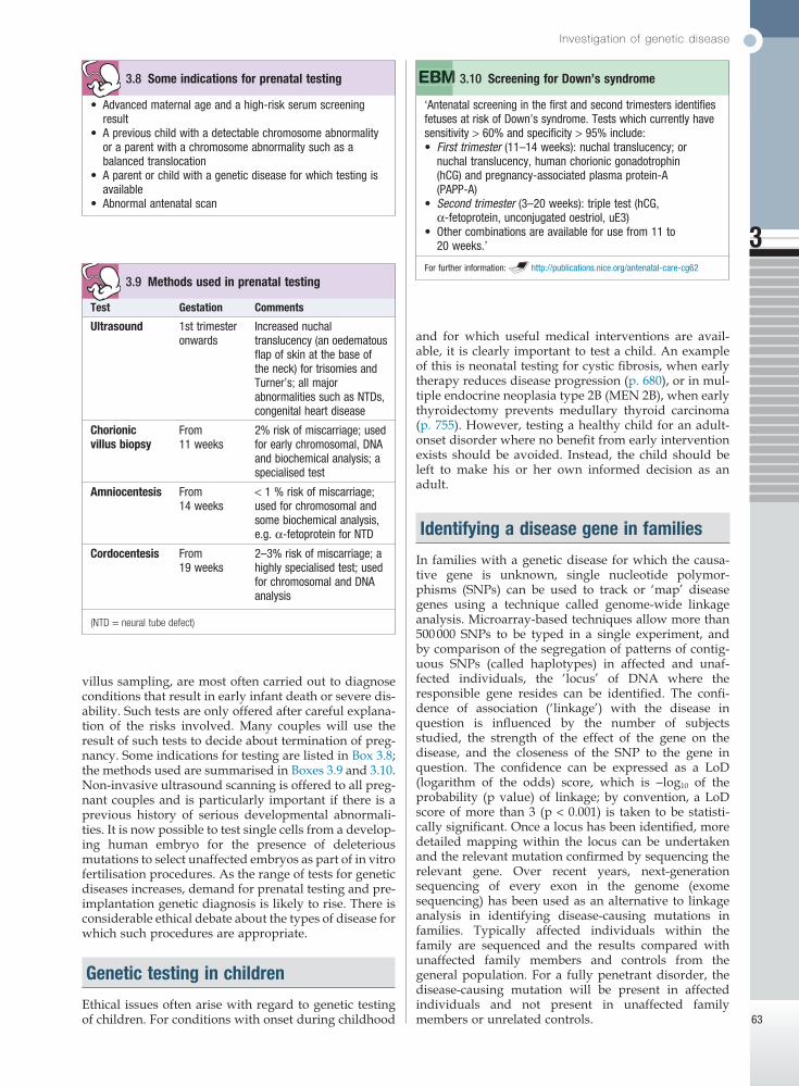
Investigation of genetic disease
3
63
and for which useful medical interventions are avail-able, it is clearly important to test a child. An example of this is neonatal testing for cystic fibrosis, when early therapy reduces disease progression (p. 680), or in mul-tiple endocrine neoplasia type 2B (MEN 2B), when early thyroidectomy prevents medullary thyroid carcinoma (p. 755). However, testing a healthy child for an adult-onset disorder where no benefit from early intervention exists should be avoided. Instead, the child should be left to make his or her own informed decision as an adult.
Identifying a disease gene in families
In families with a genetic disease for which the causa-tive gene is unknown, single nucleotide polymor-phisms (SNPs) can be used to track or ‘map’ disease genes using a technique called genome-wide linkage analysis. Microarray-based techniques allow more than 500 000 SNPs to be typed in a single experiment, and by comparison of the segregation of patterns of contig-uous SNPs (called haplotypes) in affected and unaf-fected individuals, the ‘locus’ of DNA where the responsible gene resides can be identified. The confi-dence of association (‘linkage’) with the disease in question is influenced by the number of subjects studied, the strength of the effect of the gene on the disease, and the closeness of the SNP to the gene in question. The confidence can be expressed as a LoD (logarithm of the odds) score, which is −log10 of the probability (p value) of linkage; by convention, a LoD score of more than 3 (p < 0.001) is taken to be statisti-cally significant. Once a locus has been identified, more detailed mapping within the locus can be undertaken and the relevant mutation confirmed by sequencing the relevant gene. Over recent years, next-generation sequencing of every exon in the genome (exome sequencing) has been used as an alternative to linkage analysis in identifying disease-causing mutations in families. Typically affected individuals within the family are sequenced and the results compared with unaffected family members and controls from the general population. For a fully penetrant disorder, the disease-causing mutation will be present in affected individuals and not present in unaffected family members or unrelated controls.
• Advanced maternal age and a high-risk serum screening result
• A previous child with a detectable chromosome abnormality or a parent with a chromosome abnormality such as a balanced translocation
• A parent or child with a genetic disease for which testing is available
• Abnormal antenatal scan
3.8 Some indications for prenatal testing
‘Antenatal screening in the first and second trimesters identifies fetuses at risk of Down’s syndrome. Tests which currently have sensitivity > 60% and specificity > 95% include:• First trimester (11–14 weeks): nuchal translucency; or
nuchal translucency, human chorionic gonadotrophin (hCG) and pregnancy-associated plasma protein-A (PAPP-A)
• Second trimester (3–20 weeks): triple test (hCG, α-fetoprotein, unconjugated oestriol, uE3)
• Other combinations are available for use from 11 to 20 weeks.’
For further information: http://publications.nice.org/antenatal-care-cg62
3.10 Screening for Down’s syndrome
Test Gestation Comments
Ultrasound 1st trimester onwards
Increased nuchal translucency (an oedematous flap of skin at the base of the neck) for trisomies and Turner’s; all major abnormalities such as NTDs, congenital heart disease
Chorionic villus biopsy
From 11 weeks
2% risk of miscarriage; used for early chromosomal, DNA and biochemical analysis; a specialised test
Amniocentesis From 14 weeks
< 1 % risk of miscarriage; used for chromosomal and some biochemical analysis, e.g. α-fetoprotein for NTD
Cordocentesis From 19 weeks
2–3% risk of miscarriage; a highly specialised test; used for chromosomal and DNA analysis
(NTD = neural tube defect)
3.9 Methods used in prenatal testing
villus sampling, are most often carried out to diagnose conditions that result in early infant death or severe dis-ability. Such tests are only offered after careful explana-tion of the risks involved. Many couples will use the result of such tests to decide about termination of preg-nancy. Some indications for testing are listed in Box 3.8; the methods used are summarised in Boxes 3.9 and 3.10. Non-invasive ultrasound scanning is offered to all preg-nant couples and is particularly important if there is a previous history of serious develop mental abnormali-ties. It is now possible to test single cells from a develop-ing human embryo for the presence of deleterious mutations to select unaffected embryos as part of in vitro fertilisation procedures. As the range of tests for genetic diseases increases, demand for prenatal testing and pre-implantation genetic diagnosis is likely to rise. There is considerable ethical debate about the types of disease for which such procedures are appropriate.
Genetic testing in children
Ethical issues often arise with regard to genetic testing of children. For conditions with onset during childhood
Molecular and genetic factors in disease
3
64
Genetic investigation in populations
Genetic screening may be applied to whole populations. The criteria for the use of population screening are well established; they depend on the incidence of specific conditions in individual populations and on whether an intervention is available to ameliorate the effects of the disease. In the UK, examples include screening for phenylketonuria and cystic fibrosis in the newborn, and prenatal screening for neural tube defects and Down’s syndrome in pregnant women (see Box 3.10). Screening for carriers of haemoglobinopathies and Tay–Sachs disease is also carried out in some countries where the incidence of these conditions may be high enough to merit screening the entire population (p. 1031).
Predictive genetic testing
In the absence of symptoms or signs of disease in an individual at risk, a genetic test can be used to determine whether that individual carries the disease-causing mutation. This is known as pre-symptomatic or predic-tive genetic testing. Predictive tests are usually carried out for adult-onset disorders such as familial cancer syn-dromes and neurodegenerative disorders such as Hunt-ington’s disease (Box 3.11), or when a positive result in children will affect screening and management, such as in familial polyposis coli (p. 911). However, many com-plicated ethical issues arise with testing of children and such tests should only be carried out by clinicians expe-rienced in their use.
Whilst a negative predictive test is clearly a favour-able outcome for the individual concerned, a positive test may have significant negative consequences. These should have been explained fully in the counselling process (see below), and include employment discrimi-nation and psychological effects. Providing this is done, current evidence suggests that serious psychological sequelae are uncommon.
genetics a fascinating clinical specialty but it does mean that it is difficult, if not impossible, for any individual clinician to memorise the features associated with all these disorders. It is therefore important to have an awareness of the existence of genetic diseases and some general rules or ‘triggers’ in mind. Although single-gene disorders can present at any age (Box 3.12) and affect any tissue or organ system, they share some general characteristics:• positive family history• early age of onset• multisystem involvement• no obvious non-genetic explanation.
It is important to recognise any unusual clinical pres-entation and to consider genetic disease in the context of the clinical findings and the family history. Publicly accessible online catalogues of Mendelian diseases can be useful sources of potential diagnoses.
MAJOR CATEGORIES OF GENETIC DISEASE
It is clearly impossible to discuss all Mendelian disease in this chapter, as there are many thousands of single-gene disorders. However, the major categories of genetic disease that are commonly encountered by clinical geneticists in adult practice are discussed below.
Inborn errors of metabolism
Inborn errors of metabolism (IEM) are caused by muta-tions that disrupt the normal function of a biochemical pathway. A good example is the glycogen storage diseases (see Box 16.23, p. 450), which are caused by mutations in various genes involved in regulating glucose metabolism. Most IEM are due to autosomal or X-linked recessive loss-of-function mutations in genes encoding specific enzymes or enzymatic co-factors. Knowledge of the biochemical pathway involved means that specific blocks have predictable consequences, including deficiency of the end product and build-up of intermediary compounds. Many hundreds of different IEM have been identified and these disorders have con-tributed a great deal to our understanding of human biochemistry. Most IEM are very rare and some are restricted to paediatric practice; however, a growing number may now present during adult life and some of these are discussed below.
Intoxicating IEMA subgroup of IEM, termed ‘intoxicating IEM’, can present as a sudden deterioration in a previously well individual. Such deteriorations are usually precipitated
• Currently, no medical benefit can be derived from knowing pre-symptomatic genetic status. In this situation, predictive testing is not offered to children but is available to capable adults
• Individuals have several reasonably spaced appointments with a genetic counsellor (or medical geneticist or specialist psychiatrist) prior to testing to ensure that the implications of testing are fully understood
• Fully informed patient consent is required prior to testing and individuals must be free to withdraw from testing at any time
• Follow-up support must be available and the result must not be disclosed to any other person without written consent of the tested individual
3.11 Predictive testing for Huntington’s disease
• Genetic disease: may present for the first time in elderly patients, e.g. Huntington’s disease.
• Family investigation: remains essential in the management of genetic disease presenting in old age and referral to clinical genetics services should be considered.
3.12 Genetic disease and counselling in old age
PRESENTING PROBLEMS IN GENETIC DISEASE
There are many thousands of known single-gene dis-eases. Individually these are rare, but collectively they are relatively common. This diversity makes clinical

Major categories of genetic disease
3
65
disorders cause an interesting group of syndromes and have specific features that are dealt with below.
Huntington’s diseaseHuntington’s disease (HD) is the paradigm of triplet repeat disorders. This condition can present with a movement disorder, weight loss or psychiatric symp-toms (depression, addiction, psychosis, dementia), or with a combination of all three. The disease is the result of a [CAG]n triplet repeat expansion mutation in the HD gene on chromosome 4. Since CAG is a codon for glutamine and this mutation is positioned in the ORF, this results in an expansion of a polyglutamine tract in the protein. The mutation probably leads to gain of func-tion, as deletions of the gene do not cause HD. The function of the protein encoded by the HD gene is not fully understood, but expansion of the repeat to above the normal range of 3–35 results in neurological disease. In general, the severity of disease and age at onset are related to the repeat length. In HD, atrophy of the caudate nuclei and the putamen is obvious on magnetic resonance imaging (MRI) of the brain, and in later stages cerebral atrophy is also apparent. There is currently no therapy that will alter the progression of the disease, which will often be the cause of the patient’s death. Within families there is a tendency for disease severity to increase and age at onset to fall due to further expan-sion of the repeat, a phenomenon known as anticipation. The mutation is more likely to expand through the male germ line than through female meiosis.
Other triplet repeat disordersOther progressive neurological disorders caused by triplet repeat expansion mutations in different genes include several forms of autosomal dominant spino-cerebellar ataxias, dentatorubral-pallidoluysian atro-phy (DRPLA), Machado–Joseph disease and Kennedy disease. These polyglutamine disorders all show intra-cellular inclusions in affected cells. It is thought that this accumulation may, in itself, be deleterious and is the result of defective protein degradation. Myotonic dys-trophy and Friedreich’s ataxia are also triplet repeat expansion disorders but the pathogenetic mechanism associated with these diseases is different, as these repeats do not lie within the coding regions of the affected genes.
Connective tissue disorders
Mutations in different types of collagen, fibrillin and elastin make up the majority of connective tissue disor-ders. The clinical features of these disorders vary, depending on the structural function and tissue distri-bution of the protein that is mutated. For example, auto-somal dominant loss-of-function mutations in the gene encoding elastin cause either supravalvular aortic steno-sis, cutis laxa or a combination of both conditions. The most commonly involved systems are:• skin (increased or decreased elasticity, poor wound
healing)• eyes (myopia, lens dislocation)• blood vessels (vascular fragility)• bones (osteoporosis, skeletal dysplasia)• joints (hypermobility, dislocation, arthropathy).
by physiological stress, such as infection, pregnancy, exercise or changes in diet. The intoxication is due to the build-up of intermediary, water-soluble compounds, which will vary according to the pathway involved. For example, in urea cycle disorders ammonia is the toxic substance, whereas in maple syrup urine disease it is branched-chain amino acids. The intoxication is often associated with derangement of the acid–base balance and, if not recognised and treated, will often proceed to multi-organ failure, coma and death. In the porphyrias (Box 16.32, p. 460), the intoxication is caused by a build-up in the metabolites involved in haem synthesis. The diagnosis of these disorders requires specialist bio-chemical analysis of blood and/or urine. In some disor-ders, treatment relies on removal of the toxic substance using haemodialysis or chemical conjugation, or preven-tion of further accumulation by restricting intake of the precursors, such as total protein restriction in urea cycle disorders and avoidance of branched-chain amino-acid intake in maple syrup urine disease. In other disorders, such as the porphyrias, treatment is based on avoiding precipitating factors and supportive care (p. 460).
Mitochondrial disordersDisorders of energy production are the most common type of IEM presenting in adult life, and some of these disorders have been mentioned in the section on mito-chondrial function (see Fig. 3.3, p. 45, and Box 3.1, p. 46). The tissues that are most commonly affected in this group of disorders are those with the highest meta-bolic energy requirements, such as muscle, heart, retina and brain. Therapy in this group of disorders is based on giving antioxidants and co-factors, such as vitamin C and ubiquinone, that can improve the function of the respiratory chain.
Storage disordersStorage disorders involve enzyme deficiency in lyso-somal degradation pathways. The clinical consequences depend on the specific enzyme involved. For example, Fabry disease, an X-linked recessive deficiency of α-galactosidase A, results in abdominal pain, episodic diarrhoea, renal failure and angiokeratoma. Niemann–Pick disease type C is caused by autosomal recessive loss-of-function mutations in either the NPC1 or NPC2 gene. These result in lysosomal cholesterol accumula-tion, causing hepatosplenomegaly, dysphagia, loss of speech, very early dementia, spasticity and dystonia. An increasing number of storage disorders are treatable with enzyme replacement or substrate depletion thera-pies, making awareness and diagnosis more important. More details of specific disorders are provided in Chapter 16 (Box 16.24, p. 451).
Neurological disorders
Progressive neurological deterioration is one of the most common presentations of adult genetic disease. These diseases are mostly autosomal dominant and can be grouped into specific neurological syndromes and early-onset forms of well-known, non-Mendelian clinical entities. In the latter group, the best examples would be early-onset familial forms of dementia, Parkinson’s disease and motor neuron disease. The triplet repeat

Molecular and genetic factors in disease
3
66
Learning disability, dysmorphism and malformations
Congenital global cognitive impairment (also called mental handicap or learning disability) affects about 3% of the population. It is commonly divided into broad categories of mild to moderate (IQ 50–70), moderate to severe (IQ 20–50), and severe to profound (IQ < 20). There are important ‘environmental’ causes of global cognitive impairment, including:• teratogen exposure during pregnancy (alcohol,
anticonvulsants)• congenital infections (cytomegalovirus, rubella,
toxoplasmosis, syphilis)• the sequelae of prematurity (intraventricular
haemorrhage)• birth injury (hypoxic ischaemic encephalopathy).
Genetic disorders contribute very significantly to the aetiology of global cognitive impairment. Given the complexity of brain development, it is not surprising that global cognitive impairment shows extreme locus heterogeneity. The three most important groups of dis-order are reviewed below.
Chromosome disordersAny significant gain or loss of autosomal chromosomal material (known as aneuploidy) usually results in learn-ing disability and other phenotypic abnormalities (see Fig. 3.11, p. 57). Down’s syndrome is the most frequently found and best known of these disorders, and is caused by an increased dosage of genes on chromosome 21. Most cases of Down’s syndrome are due to a numerical chromosome abnormality with trisomy of chromosome 21, e.g. 47,XX,+21 or 47,XY,+21. The clinical features are:• globally delayed development• characteristic facial appearance• a significant risk of specific malformations
(atrioventricular septal defect, duodenal atresia)• a predisposition to several late-onset disorders,
including hypothyroidism, acute leukaemias and Alzheimer’s disease.Recent surveys have shown that DNA microarray
analysis can identify causative structural chromosome abnormalities in 10–25% of cases of significant learning disability. These deletions and duplications are mostly de novo and unique. An interest group of recurrent dele-tions and duplication caused by non-allelic homologous recombination events has been mentioned above. These result in specific microdeletion or microduplication syn-dromes, such as:• velocardiofacial syndrome due to deletion of 22q11.2
(learning disability, malformations of the cardiac outflow tract, cleft palate, distinctive facial appearance and immune disorders)
• Williams’ syndrome due to deletion of 7q11.23 (learning disability, supravalvular aortic stenosis and mild cutis laxa as a result of deletion of the elastin gene, distinctive facial appearance and over-friendly, chatty personality).
Dysmorphic syndromesThere are several thousand different dysmorphic syn-dromes; all are rare but they are characterised by the
occurrence of cognitive impairment, malformations and a distinctive facial appearance – or ‘gestalt’ – associated with various other clinical features. Making the correct diagnosis is important, as it has profound implications on immediate patient management, detection of future complications and assessment of recurrence risks in the family. Clinical examination remains the mainstay of diagnosis and the patient often needs to be evaluated by a clinician who specialises in the diagnosis of these syn-dromes. The differential diagnosis in dysmorphic syn-dromes is often very wide and this has resulted in computer-aided diagnosis becoming an established clin-ical tool. Dysmorphology databases such as POSSUM and LMD have been established that are curated cata-logues of the many thousands of known syndrome enti-ties; they can be searched to identify possible explanations of unusual combinations of clinical features. The clinical diagnosis may then be confirmed by specific genetic investigations, as the genetic basis of a wide range of dysmorphic syndromes has been identified.
X-linked mental handicapX-linked mental handicap (XLMH) accounts for approx-imately 10% of cases of moderate to severe learning dis-ability. There are over 100 genes on the X chromosome that can cause learning disability but the most common disorder is fragile X syndrome, characterised by a dis-tinctive facial appearance, attention deficit, joint hyper-mobility, macro-orchism (increased testicular size) and a non-staining gap on the X chromosome on chromo-some analysis. Fragile X is caused by a triplet repeat expansion mutation but of a different type from the polyglutamine repeat disorders mentioned above. The repeat in fragile X syndrome is not in the coding region and is a [CGG]n expansion (see Box 3.4, p. 56). Methyla-tion of the expanded repeat results in silencing of a specific gene called FMR1, which encodes an RNA-binding protein.
De novo mutationsNext-generation sequencing technology has made pos-sible trio-based, whole-exome sequencing, in which the affected individual and both of their parents are ana-lysed. It has recently become clear that de novo muta-tions in the coding regions of one of the many genes that are involved in normal brain development are collec-tively responsible for severe intellectual disability in at least 25% of affected patients. Trio-exome sequencing is thus likely to become a first-line diagnostic test for such cases in the near future.
Familial cancer syndromes
Most cancers are not inherited but occur as the result of an accumulation of somatic mutations, as discussed pre-viously in this chapter. However, it has been recognised for many decades that some families are prone to one or more specific types of cancer. Affected individuals tend to present with tumours at an early age and are more likely to have multiple primary foci of carcinogenesis.
RetinoblastomaPatients with autosomal dominant familial retinoblast-oma have an inherited mutation in one copy of the RB

Genetic counselling
3
67
primarily with the effects of non-ionising radiation. The skin is particularly involved, and affected patients develop skin cancers with increased frequency.
GENETIC COUNSELLING
Genetic counselling provides information about the medical and family implications of a specific disease in a clear and non-directive manner. Such counselling aims to help individuals make informed decisions about plan-ning a family, taking part in screening programmes and accepting prophylactic therapies. Genetic counselling may be provided by a medical geneticist, a specialist nurse, or a clinician with particular skills in this area, such as an obstetrician or paediatrician (Box 3.13). Per-ception of genetic risks clearly depends on perceived hazard. For example, a 5% (or 1 : 20) risk of genetic disease may be perceived as low if the disease is treat-able, but unacceptably high if not.
Specific problems encountered in genetic counselling include:• accurate assessment of genetic risk• identification of children at risk of genetic disorders• the increase in genetic risks associated with
consanguinity• non-paternity as an incidental finding in DNA.
gene, which is a tumour suppressor. This strongly pre-disposes individuals to the formation of retinoblastoma in one or both eyes. It is possible for more than one primary tumour to form in the same eye and for retino-blastoma to occur in the pineal gland. From a clinical perspective, it is important to screen the eyes and pineal gland of such individuals regularly so that tumours can be treated early and sight preserved. This gene is widely expressed and it is not clear why the retina is the main site of oncogenesis in this syndrome. An increased inci-dence of osteogenic sarcoma is also seen in affected individuals.
Familial adenomatous polyposis coliFamilial adenomatous polyposis coli (FAP) is an auto-somal dominant condition due to inactivation mutations in the FAP tumour suppressor gene on 5q. The gene product is thought to modulate a specific signalling cascade (Wnt signalling) that regulates cell proliferation. Mutation carriers usually develop many thousands of intestinal polyps in their second and third decades and have a very high risk of malignant change in the colon. Prophylactic colectomy in the third decade is necessary in most cases. Regular screening for polyps in the upper gastrointestinal tract is also recommended.
Li–Fraumeni syndromeHeterozygous loss-of-function mutations in the gene encoding p53 cause Li–Fraumeni syndrome. Families with this condition have a very significant increased predisposition to early-onset leukaemias, sarcomas, and breast and brain malignancies. Screening for pre-symptomatic tumours in this condition is very difficult and of unproven benefit, as almost any tissue can be affected.
Hereditary non-polyposis colorectal cancerHereditary non-polyposis colorectal cancer (HNPCC) is an autosomal dominant disorder that presents with early-onset familial colon cancer, particularly affecting the proximal colon. Other cancers, such as endometrial cancer, are often observed in affected families. This dis-order shows marked locus heterogeneity, as mutations can occur in several different genes encoding proteins involved in DNA mismatch repair.
Familial breast cancerFamilial breast cancer is an autosomal dominant disor-der that is most often due to mutations in genes encod-ing either BRCA1 or BRCA2. Both of these proteins are involved in DNA repair. Individuals who carry a BRCA1 or BRCA2 mutation are at high risk of early-onset breast and ovarian tumours, and require regular screening for both of these conditions. Because of the very high risk of cancer, many women who carry these mutations elect to have prophylactic bilateral mastectomy and oophorec-tomy in the absence of a detectable tumour.
Xeroderma pigmentosumXeroderma pigmentosum (XP) is the name given to a group of rare disorders in which there are autosomal recessive defects in DNA repair genes that deal
Component Role
Medical geneticist Diagnosis and management of genetic disease, assessment of genetic risk, managing screening programmes, interpretation of genetic test results. Subspecialties include prenatal genetics, dysmorphology (syndrome identification), cancer genetics
Genetic counsellor Assessing genetic risk, provision of genetic counselling (providing accurate risk information in a comprehensible format), predictive testing for genetic disease and provision of information and support
DNA diagnostic laboratory
Identifying and reporting disease-causing mutations in validated disease genes. Some laboratories also provide linkage analysis to track diseases in families. Laboratories often work in a consortium, as so many different disease genes have now been identified
Cytogenetics laboratory
Identifying pathogenic numerical and structural chromosome anomalies in prenatal, postnatal and oncology samples
Biochemical genetics laboratory
Metabolite and enzymatic-based diagnosis of IEM. Metabolite-based monitoring of treatment of IEM
Newborn screening laboratory
Provision of population-based newborn screening, e.g. PKU, cystic fibrosis, etc.
(IEM = inborn errors of metabolism; PKU = phenylketonuria)
3.13 Clinical genetics services
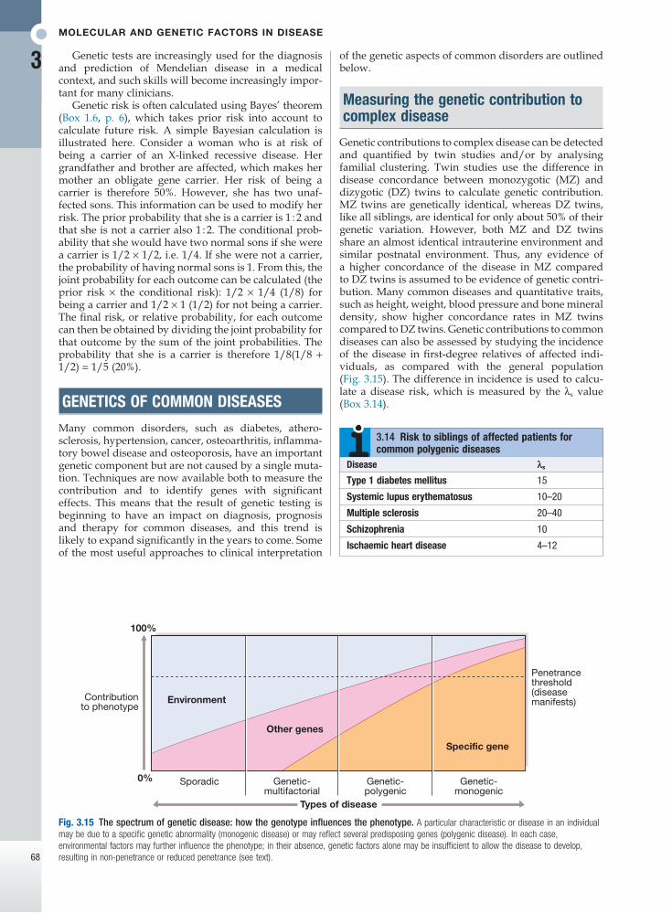
Molecular and genetic factors in disease
3
68
of the genetic aspects of common disorders are outlined below.
Measuring the genetic contribution to complex disease
Genetic contributions to complex disease can be detected and quantified by twin studies and/or by analysing familial clustering. Twin studies use the difference in disease concordance between monozygotic (MZ) and dizygotic (DZ) twins to calculate genetic contribution. MZ twins are genetically identical, whereas DZ twins, like all siblings, are identical for only about 50% of their genetic variation. However, both MZ and DZ twins share an almost identical intrauterine environment and similar postnatal environment. Thus, any evidence of a higher concordance of the disease in MZ compared to DZ twins is assumed to be evidence of genetic contri-bution. Many common diseases and quantitative traits, such as height, weight, blood pressure and bone mineral density, show higher concordance rates in MZ twins compared to DZ twins. Genetic contributions to common diseases can also be assessed by studying the incidence of the disease in first-degree relatives of affected indi-viduals, as compared with the general population (Fig. 3.15). The difference in incidence is used to calcu-late a disease risk, which is measured by the λs value (Box 3.14).
Genetic tests are increasingly used for the diagnosis and prediction of Mendelian disease in a medical context, and such skills will become increasingly impor-tant for many clinicians.
Genetic risk is often calculated using Bayes’ theorem (Box 1.6, p. 6), which takes prior risk into account to calculate future risk. A simple Bayesian calculation is illustrated here. Consider a woman who is at risk of being a carrier of an X-linked recessive disease. Her grandfather and brother are affected, which makes her mother an obligate gene carrier. Her risk of being a carrier is therefore 50%. However, she has two unaf-fected sons. This information can be used to modify her risk. The prior probability that she is a carrier is 1 : 2 and that she is not a carrier also 1 : 2. The conditional prob-ability that she would have two normal sons if she were a carrier is 1/2 × 1/2, i.e. 1/4. If she were not a carrier, the probability of having normal sons is 1. From this, the joint probability for each outcome can be calculated (the prior risk × the conditional risk): 1/2 × 1/4 (1/8) for being a carrier and 1/2 × 1 (1/2) for not being a carrier. The final risk, or relative probability, for each outcome can then be obtained by dividing the joint probability for that outcome by the sum of the joint probabilities. The probability that she is a carrier is therefore 1/8(1/8 + 1/2) = 1/5 (20%).
GENETICS OF COMMON DISEASES
Many common disorders, such as diabetes, athero-sclerosis, hypertension, cancer, osteoarthritis, inflamma-tory bowel disease and osteoporosis, have an important genetic component but are not caused by a single muta-tion. Techniques are now available both to measure the contribution and to identify genes with significant effects. This means that the result of genetic testing is beginning to have an impact on diagnosis, prognosis and therapy for common diseases, and this trend is likely to expand significantly in the years to come. Some of the most useful approaches to clinical interpretation
Fig. 3.15 The spectrum of genetic disease: how the genotype influences the phenotype. A particular characteristic or disease in an individual may be due to a specific genetic abnormality (monogenic disease) or may reflect several predisposing genes (polygenic disease). In each case, environmental factors may further influence the phenotype; in their absence, genetic factors alone may be insufficient to allow the disease to develop, resulting in non-penetrance or reduced penetrance (see text).
100%
Contributionto phenotype
Environment
Other genes
Sporadic0% Genetic-multifactorial
Specific gene
Genetic-polygenic
Genetic-monogenic
Types of disease
Penetrancethreshold(diseasemanifests)
Disease λs
Type 1 diabetes mellitus 15
Systemic lupus erythematosus 10–20
Multiple sclerosis 20–40
Schizophrenia 10
Ischaemic heart disease 4–12
3.14 Risk to siblings of affected patients for common polygenic diseases
Research frontiers in molecular medicine
3
69
patients who have increased sensitivity to the effects of azathioprine and who can be treated with lower doses than normal.
RESEARCH FRONTIERS IN MOLECULAR MEDICINE
Gene therapy
Replacing or repairing mutated genes (gene therapy) is very difficult in humans. Retroviral-mediated ex vivo replacement of the defective gene in bone marrow cells for the treatment of severe combined immune deficiency syndrome (p. 80) has been partially successful. There have been two major problems with the clinical trials of virally delivered gene therapy conducted to date:• The random integration of the retroviral DNA
(which contains the replacement gene) into the genome has caused leukaemia in some treated children via activation of proto-oncogenes.
• A severe immune response to the viral vector may be induced. It has not yet been possible to use non-viral means to introduce sufficient numbers of copies of replacement genes to produce significant biological effects.Other therapies for genetic disease include PTC124,
a compound that can ‘force’ cells to read through a mutation that results in a premature termination codon in an ORF with the aim of producing a near-normal protein product. This therapeutic approach could be applied to any genetic disease caused by nonsense mutations.
Induced pluripotent stem cells and regenerative medicine
Adult stem cell therapy has been in wide use for decades in the form of haematopoietic stem cell transplantation. The identification of adult stem cells for other tissues, coupled with the ability to purify and maintain such cells in vitro, now offers exciting therapeutic potential for other diseases. Many different adult cell types can be transdifferentiated to form cells termed induced pluri-potent stem cells (iPS cells) with almost all the charac-teristics of embryonic stem cells. In mammals, iPS cells can be used to regenerate various tissues such as the
Genetic testing in complex disease
Most common diseases are determined by interactions between a number of genes and the environment. In this situation, the genetic contribution to disease is termed polygenic. Until recently, very little progress had been made in identifying the genetic variants that predispose to common diseases, but this has been changed by the advent of genome-wide association studies (GWAS). A GWAS typically involves genotyping many (> 500 000) genetic markers spread across the genome in a large group of individuals with the disease and controls. By comparing the genotypes in cases and controls, it is pos-sible to identify regions of the genome and candidate genes that contribute to the disease under study. Some of the candidate genes for common diseases identified by this approach are listed in Box 3.15.
Pharmacogenomics
Pharmacogenomics is the science of dissecting the genetic determinants of drug kinetics and effects using information from the human genome. For more than 50 years, it has been appreciated that polymorphic mutations within genes can affect individual responses to some drugs, such as loss-of-function mutations in CYP2D6 causing hypersensitivity to debrisoquine, an adrenergic-blocking medication formerly used for the treatment of hypertension, in 3% of the population. This gene is part of a large family of highly polymorphic genes encoding cytochrome P450 proteins, mostly expressed in the liver, which determine the metabolism of a host of specific drugs. Polymorphisms in the CYP2D6 gene also determine codeine activation, while those in the CYP2C9 gene affect warfarin inactivation. Polymorphisms in these and other drug metabolic genes determine the persistence of drugs and, therefore, should provide information about dosages and toxicity. At the present time, genetic testing for assessment of drug response is seldom used routinely, but in the future it may be possible to predict the best specific drugs and dosages for individual patients based on genetic profiling: so-called ‘personalised medicine’. An example is the enzyme thiopurine methyltransferase (TPMT), which catabolises azathioprine, a drug that is used in the treatment of autoimmune diseases and in cancer chemotherapy. Genetic screening for polymor-phic variants of TPMT can be useful in identifying
Disease Inheritance No. of loci Pathways
Colorectal cancer Polygenic 20 DNA repair enzymes, growth factor signalling pathways, Wnt signalling
Osteoporosis Polygenic 56 Cytokines, receptors and intracellular signalling molecules in RANK and Wnt pathways. Regulators of stem cell differentiation and bone mineralisation
Systemic lupus erythematosus
Polygenic 60 Cytokines, receptors and intracellular signalling pathways that regulate immune function; complement, intracellular nucleases
Gout Polygenic 9 Renal solute transporters; regulators of glucose and insulin metabolism
(RANK = receptor activator of nuclear factor κ B)
3.15 Polymorphisms that predispose to common diseases

Molecular and genetic factors in disease
3
70
model of Marfan’s syndrome, showing promising effects in early human clinical trials.
Further information
Books and journal articlesAlberts B, Bray D, Hopkin K, et al. Essential cell biology.
New York: Garland Science. 3rd edn; 2009.Read A, Donnai D. New clinical genetics. 2nd edn. Banbury:
Scion; 2010.Strachan T, Read A. Human molecular genetics. 3rd edn.
Wilmington: Wiley–Liss; 2003.
Websiteswww.bshg.org.uk British Society for Human Genetics; has
report on genetic testing of children.www.ensembl.org Annotated genome databases from multiple
organisms.www.genome.ucsc.edu Excellent source of genomic information.www.ncbi.nlm.nih.gov Online Mendelian Inheritance in Man
(OMIM).
heart and brain. They have great potential both to develop tissue models of human disease and for regen-erative medicine. In mammalian model species, such cells can be taken and used to regenerate differentiated tissue cells, such as in heart and brain.
Pathway medicine
The ability to manipulate pathways that have been altered in genetic disease has tremendous therapeutic potential for Mendelian disease, but a firm understand-ing of both disease pathogenesis and drug action at a biochemical level is required. An exciting example of this has been the discovery that the vascular pathology associated with Marfan’s syndrome is due to the defec-tive fibrillin molecules causing up-regulation of trans-forming growth factor (TGF)-β signalling in the vessel wall. Losartan is an antihypertensive drug that is mar-keted as an angiotensin II receptor antagonist. However, it also acts as a partial antagonist of TGF-β signalling and is effective in preventing aortic dilatation in a mouse