Embed Size (px)
Citation preview
Presented by:
Dr. Md. Zareer Tafadar
Post Graduate Student
Deptt. Of Anaesthesiology & Critical Care
Silchar Medical College & Hospital.
Anaesthetic Management Of A Patient With Ischaemic Heart Disease Undergoing Non
Cardiac Surgery
Ischaemic heart disease (IHD) also known as coronary artery
disease (CAD) is the generic designation for a group of closely related syndromes resulting from myocardial ischemia—an imbalance between the supply (perfusion) and demand of the heart for oxygenated blood. Ischemia comprises not only insufficiency of oxygen, but also reduced availability of nutrient substrates and inadequate removal of metabolites
Coronary heart disease (CHD) is the most common form of heart disease.
5% of patients over 35 years of age have asymptomatic ischaemic heart disease.
May be present in up to 30% of older pts undergoing surgery
Cardiac dysrhythmias[VF] are a major cause of sudden death.
Introduction
Male gender
Increasing age
Hypercholesterolemia
Hypertension
Cigarette smoking
Diabetes mellitus
Obesity
Sedentary lifestyle
Genetic factors
Family history of premature ischemic heart disease (male <55 yrs of age, female <65 yrs)
Risk Factors for Development of Ischemic Heart Disease

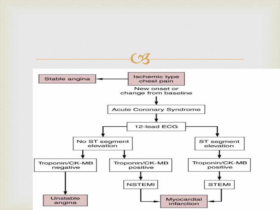
IHD
Angina Pectoris
Stable angina,
Prinzmetal angina,
Unstable angina—
Acute MI
STEMI
NSTEMI
Chronic IHD with Heart
Failure
Sudden Cardiac Death
Spectrum Of IHD
Angina Pectoris: symptom complex of IHD characterized by paroxysmal and usually
recurrent attacks of substernal or precordial chest discomfort (variously described as constricting, squeezing,choking, or knifelike) caused by transient (15 seconds to 15 minutes) myocardial ischemia
Stable Angina Chronic pattern of transient angina pectoris precipitated by
physical activity or emotional upset, relieved by rest with in few minutes.
caused by the reduction of coronary perfusion to a critical level by chronic stenosing coronary atherosclerosis; this renders the heart vulnerable to further ischemia whenever there is increased cardiac workload.
Typical angina pectoris is usually relieved by rest (thereby decreasingdemand) or nitroglycerin, a strong vasodilator
Temporary depression of ST segment with no permanent myocardial damage.
Angina Pectoris
Variant Angina/Prinzmetal Angina
Typical anginal discomfort usually at rest.
Develops due to coronary artery spasm rather than increase myocardial oxygen demand.
The anginal attacks are unrelated to physical activity,heartrate, or blood pressure. Generally responds promptly to vasodilators.
Transient shifts of ST segment – ST elevation.
Unstable Angina
Refers to a pattern of pain that occurs with progressively increasing frequency, is precipitated with progressively less effort, often occurs at rest, and tends to be of more prolonged duration.
In most patients, unstable angina is induced by disruption of an atherosclerotic plaque with superimposed partial (mural) thrombosis and possibly embolization or vasospasm (or both). of subsequent acute MI.
Thus this syndrome is sometimes referred to as preinfarction angina, and in the spectrum of IHD, unstable angina lies between stable angina on the one hand and MI on the other.
MI, also known as "heart attack," is the death of cardiac muscle
resulting from ischemia.
Region of myocardial necrosis due to prolonged cessation of blood supply
Results from acute thrombus at side of coronary atherosclerotic stenosis
Typical symptoms of myocardial infarction include
• Sudden Chest Pain,
• Shortness Of Breath,
• Nausea, Vomiting,
• Palpitations, Sweating
• Weakness, Light-headedness
• Collapse/syncope
Myocardial Infarction (MI)
Diagnosis of Acute MI:
At least 2 of the following
• Ischaemic symptoms
• Diagnostic ECG changes
• Serum cardiac marker elevations
Screening & Evaluation
Goals
• Identify the risk for heart disease based on risk factors.
• Identify the presence and severity of heart disease from symptoms, physical findings, or diagnostic tests .
• Determine the need for preoperative interventions.
• Modify the risk for perioperative adverse events.
History:
Aim: To elicit severity progression and functional limitations imposed by IHD
Chest discomfort (pain, pressure, tightness), the duration of the discomfort, precipitating factors, associated symptoms, and methods of relief.
Exercise tolerance: In the absence of significant lung disease limitation indicates decreased cardiac reserve.
If a patient can climb two to three flights of stairs without symptoms, it is likely that cardiac reserve is adequate.
Risk factors : Smoking, hypertension, age, male
gender, hypercholesterolemia, and family history.
H/O MI
Acute MI: 1-7 days
Recent MI: 8- 30 days
H/O cardiac revascularisation- PCI/CABG
Co-Existing Noncardiac Diseases: CVA, COPD, DM, Renal insufficiency
Current Medications: Anti-hypertensives, Anti platelet agents, Anticoagulants, Others.
Physical Examination
• Abnormal physical findings are often absent and non-specific.
• Quick assessment of patients vital signs
• Signs of right and left ventricular dysfunction must be sought.
• Jugular venous distention and peripheral edema are signs of right ventricular failure.
• Auscultation of the chest may reveal evidence of left ventricular dysfunction such as an S3 gallop or rales.
• A carotid bruit may indicate cerebrovascular disease.
• Orthostatic hypotension may reflect attenuated autonomic nervous system activity due to treatment with antihypertensive drugs.
12-lead ECG: Preoperative resting 12-lead ECG is reasonable for patients
with known coronary heart disease or other significant structural heart disease, except for low-risk surgery (IIa)
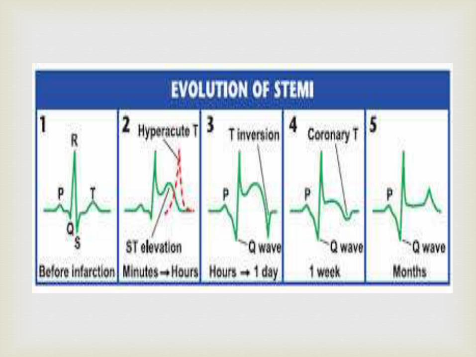
May not show any abnormalities at rest or with no symptoms, or may show evidence of old MI (Q waves) in 2 or more leads & >1/3 of the QRS complex length)
• May reveal ST segment depression >1 mm from baseline in case of angina pectoris or ST-segment elevation in association with AMI or variant angina
• Other changes with symptoms of angina pectoris: reversible T-wave inversion
• Other findings with AMI: increased T-wave amplitude, followed by ST elevation, followed by Q-wave development & resolution of ST elevation
Specialised Preoperative Testing
• Exercise ECG is useful for detecting signs of myocardial ischaemia as well as for evaluation of exercise capacity
Contraindications:• Severe AS
• Severe hypotension
• Acute myocarditis
• Uncontrolled HF
• Infective Endocarditis
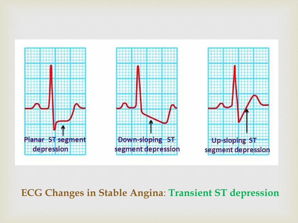
ECG Changes in Stable Angina: Transient ST depression
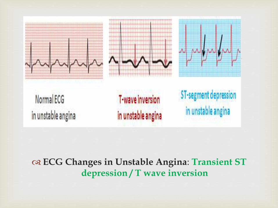
ECG Changes in Unstable Angina: Transient ST depression / T wave inversion
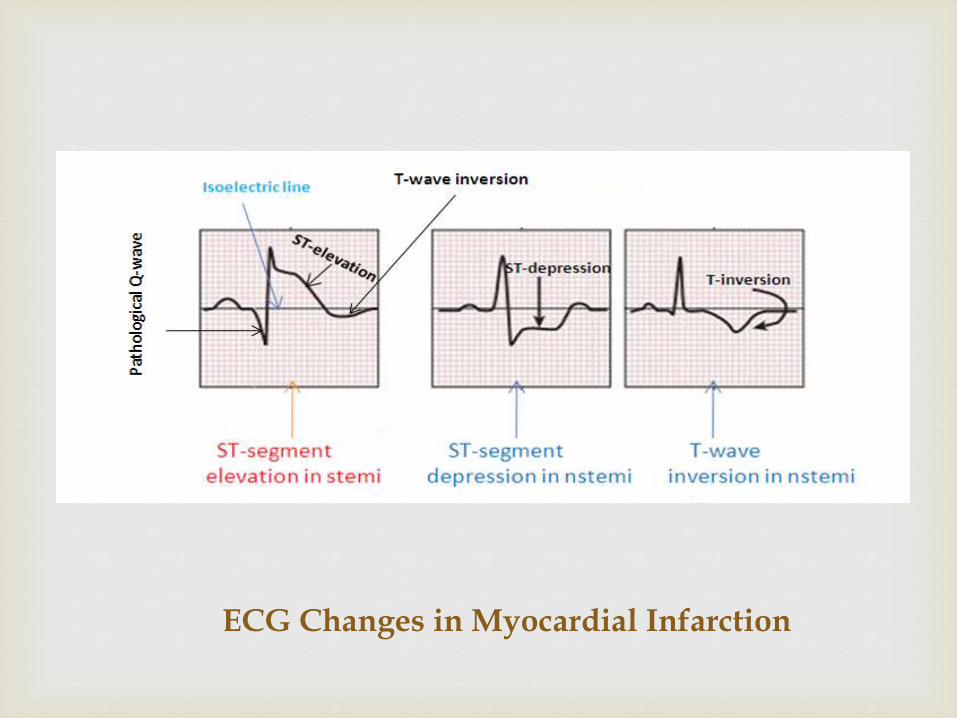
ECG Changes in Myocardial Infarction
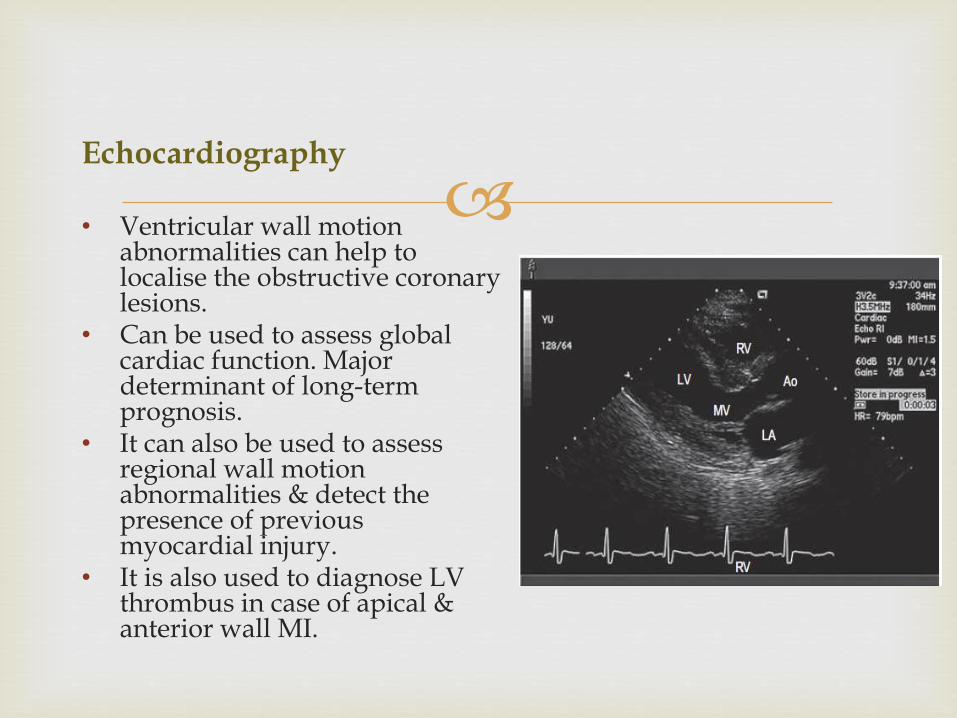
Echocardiography
• Ventricular wall motion abnormalities can help to localise the obstructive coronary lesions.
• Can be used to assess global cardiac function. Major determinant of long-term prognosis.
• It can also be used to assess regional wall motion abnormalities & detect the presence of previous myocardial injury.
• It is also used to diagnose LV thrombus in case of apical & anterior wall MI.
Stress echocardiography: This is used with
pharmacologic induction of cardiac stress (dobutamine) or exercise to look at LV segmental wall function at rest & with stress. This can be also used to differentiate between viable (hibernating, stunned) & nonviable (infarcted) myocardial segments
Nuclear Stress Imaging
• Thallium scan and technetium scan shows areas of reduced uptake of radioactive isotope by the myocardium. Useful for assessing coronary perfusion• A perfusion defect present during stress but not all rest indicates reversible myocardial ischemia, whereas a persistent perfusion defect on scan during both phases (rest and stress) usually indicates previous myocardial infarction.• Thallium scanning is positive in 75-90% of patients with significant coronary disease. False positive test may occur in women due to breast tissue.
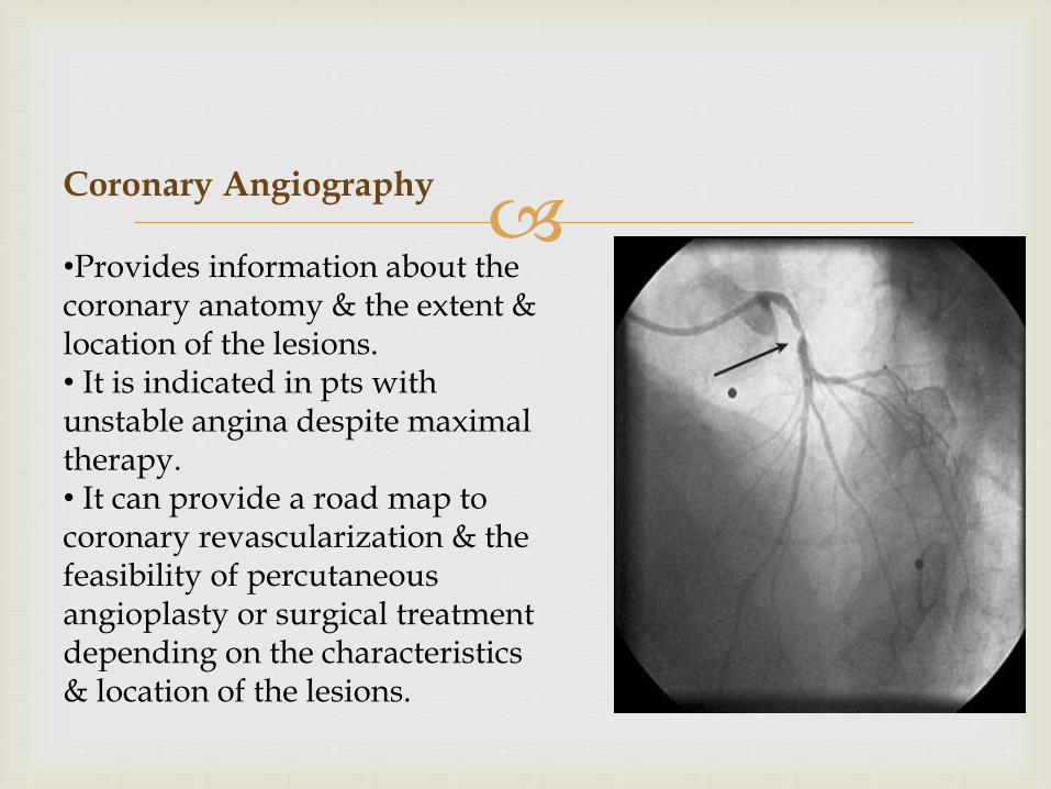
Coronary Angiography
•Provides information about the coronary anatomy & the extent & location of the lesions.• It is indicated in pts with unstable angina despite maximal therapy.• It can provide a road map to coronary revascularization & the feasibility of percutaneousangioplasty or surgical treatment depending on the characteristics & location of the lesions.
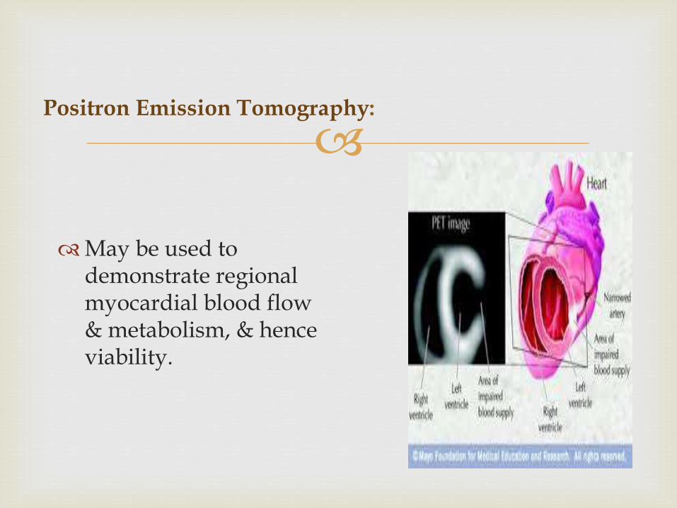
Positron Emission Tomography:
May be used to demonstrate regional myocardial blood flow & metabolism, & hence viability.
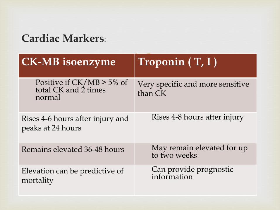
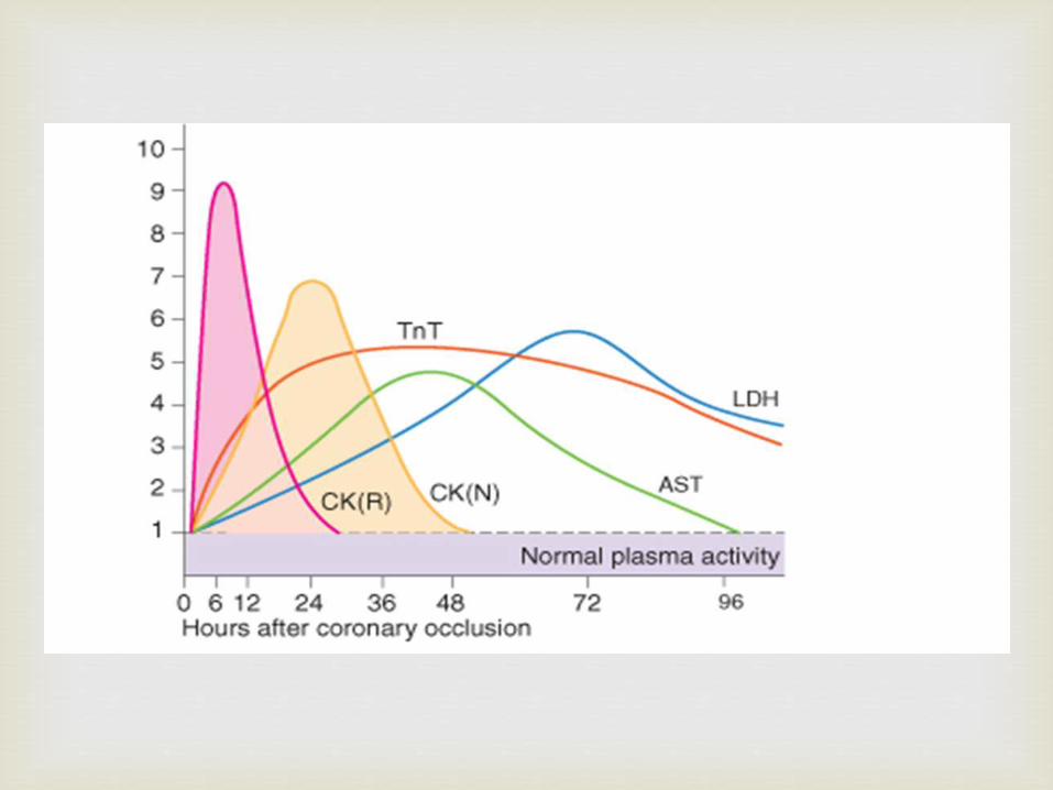
Cardiac Markers:
CK-MB isoenzyme Troponin ( T, I )
Positive if CK/MB > 5% of total CK and 2 times normal
Very specific and more sensitive than CK
Rises 4-6 hours after injury and peaks at 24 hours
Rises 4-8 hours after injury
Remains elevated 36-48 hours May remain elevated for up to two weeks
Elevation can be predictive of mortality
Can provide prognostic information
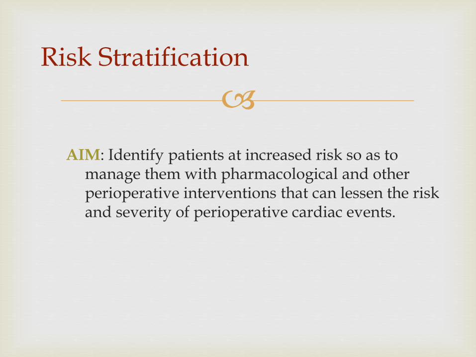
AIM: Identify patients at increased risk so as to manage them with pharmacological and other perioperative interventions that can lessen the risk and severity of perioperative cardiac events.
Risk Stratification
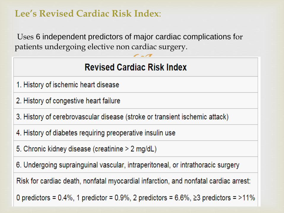
Lee’s Revised Cardiac Risk Index:
Uses 6 independent predictors of major cardiac complications for patients undergoing elective non cardiac surgery.
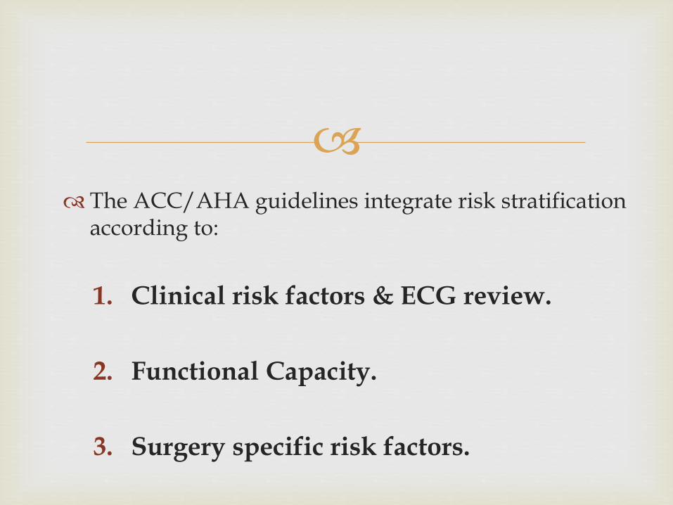
The ACC/AHA guidelines integrate risk stratification according to:
1. Clinical risk factors & ECG review.
2. Functional Capacity.
3. Surgery specific risk factors.
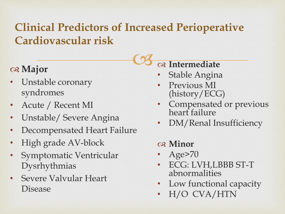
Clinical Predictors of Increased PerioperativeCardiovascular risk
Major
• Unstable coronary syndromes
• Acute / Recent MI
• Unstable/ Severe Angina
• Decompensated Heart Failure
• High grade AV-block
• Symptomatic Ventricular Dysrhythmias
• Severe Valvular Heart Disease
Intermediate• Stable Angina• Previous MI
(history/ECG)• Compensated or previous
heart failure• DM/Renal Insufficiency
Minor• Age>70• ECG: LVH,LBBB ST-T
abnormalities• Low functional capacity• H/O CVA/HTN
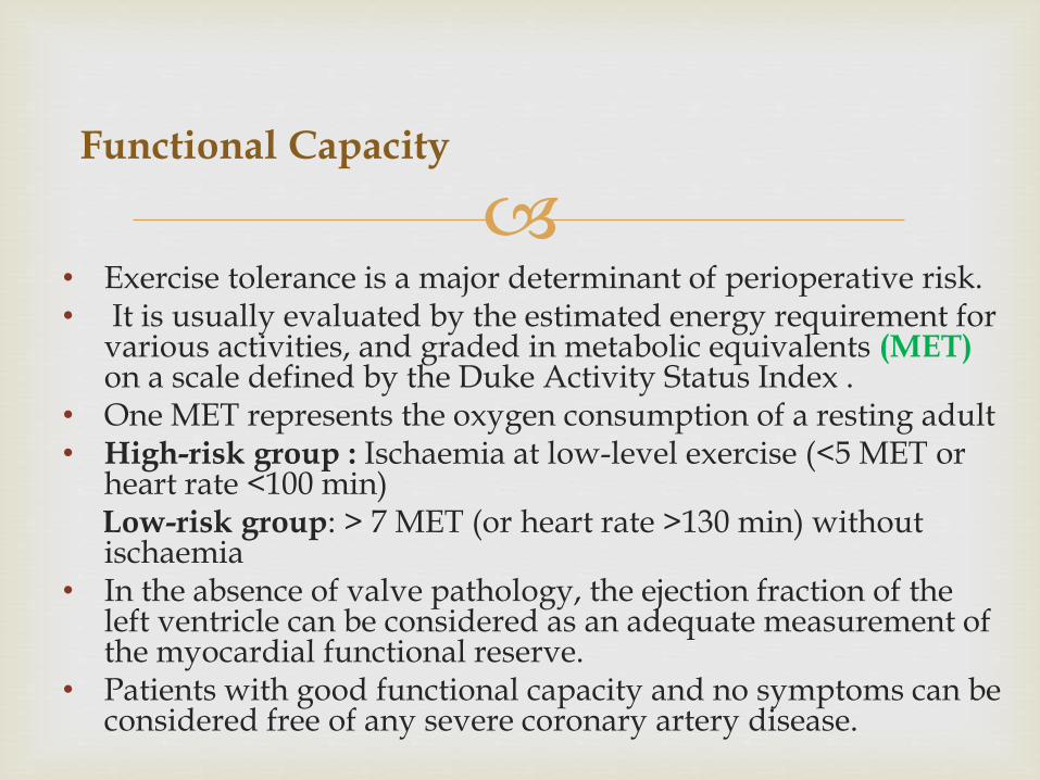
Functional Capacity
• Exercise tolerance is a major determinant of perioperative risk.• It is usually evaluated by the estimated energy requirement for
various activities, and graded in metabolic equivalents (MET) on a scale defined by the Duke Activity Status Index .
• One MET represents the oxygen consumption of a resting adult • High-risk group : Ischaemia at low-level exercise (<5 MET or
heart rate <100 min)Low-risk group: > 7 MET (or heart rate >130 min) without ischaemia
• In the absence of valve pathology, the ejection fraction of the left ventricle can be considered as an adequate measurement of the myocardial functional reserve.
• Patients with good functional capacity and no symptoms can be considered free of any severe coronary artery disease.
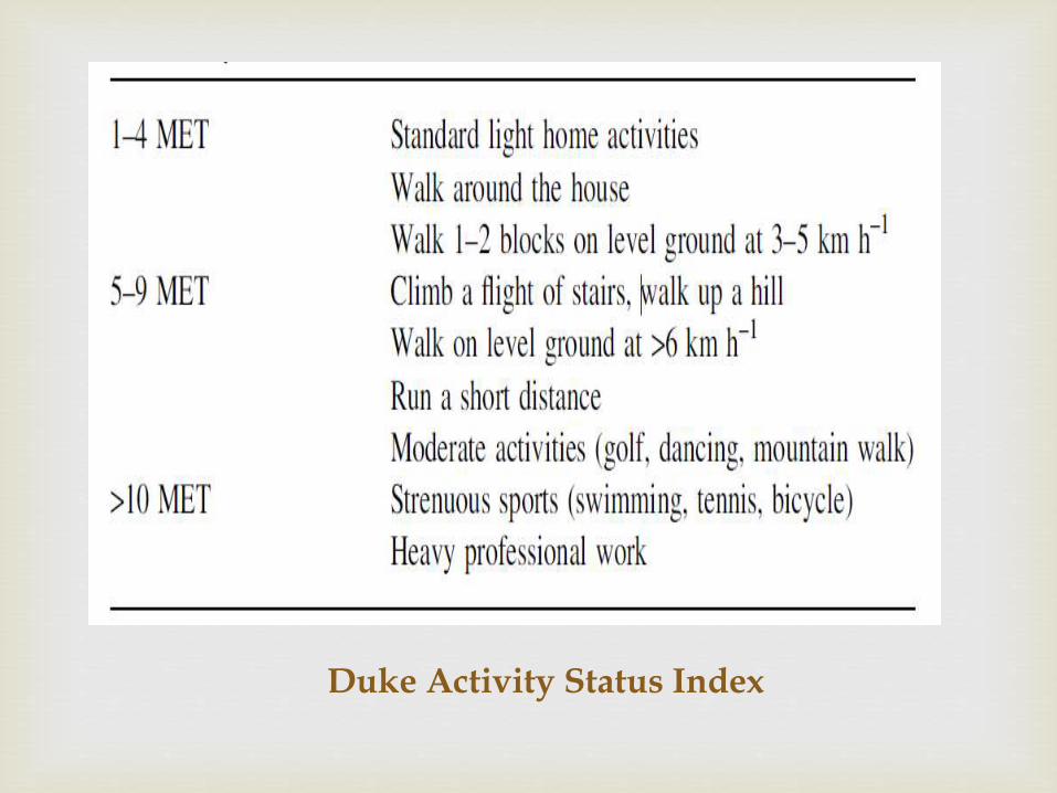
Duke Activity Status Index
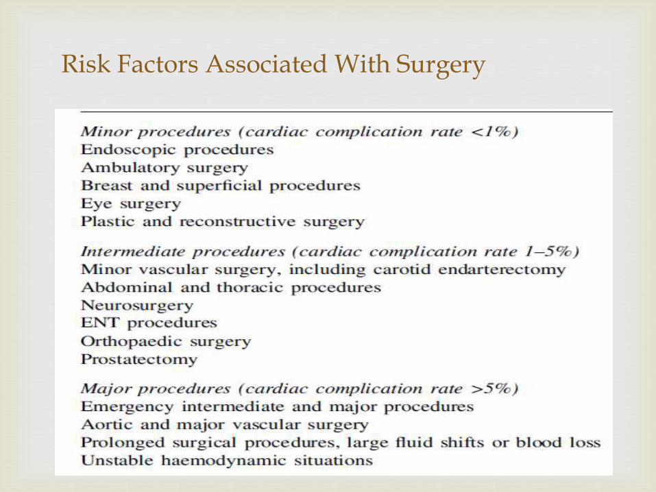
Risk Factors Associated With Surgery
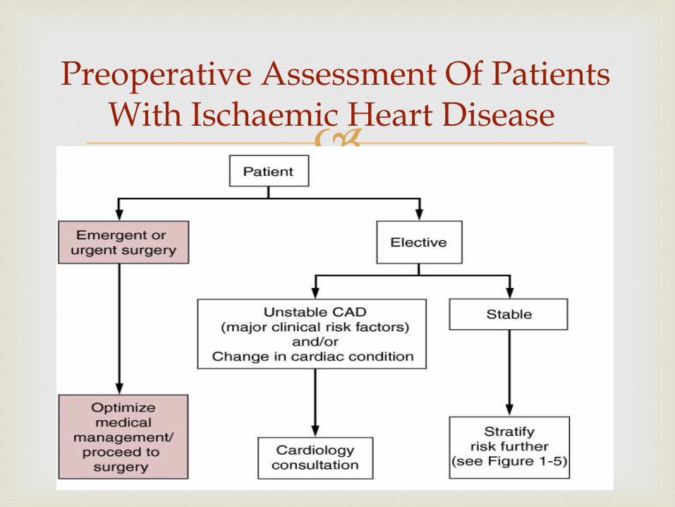
Preoperative Assessment Of Patients With Ischaemic Heart Disease
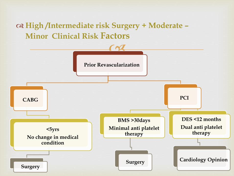
High /Intermediate risk Surgery + Moderate –
Minor Clinical Risk Factors
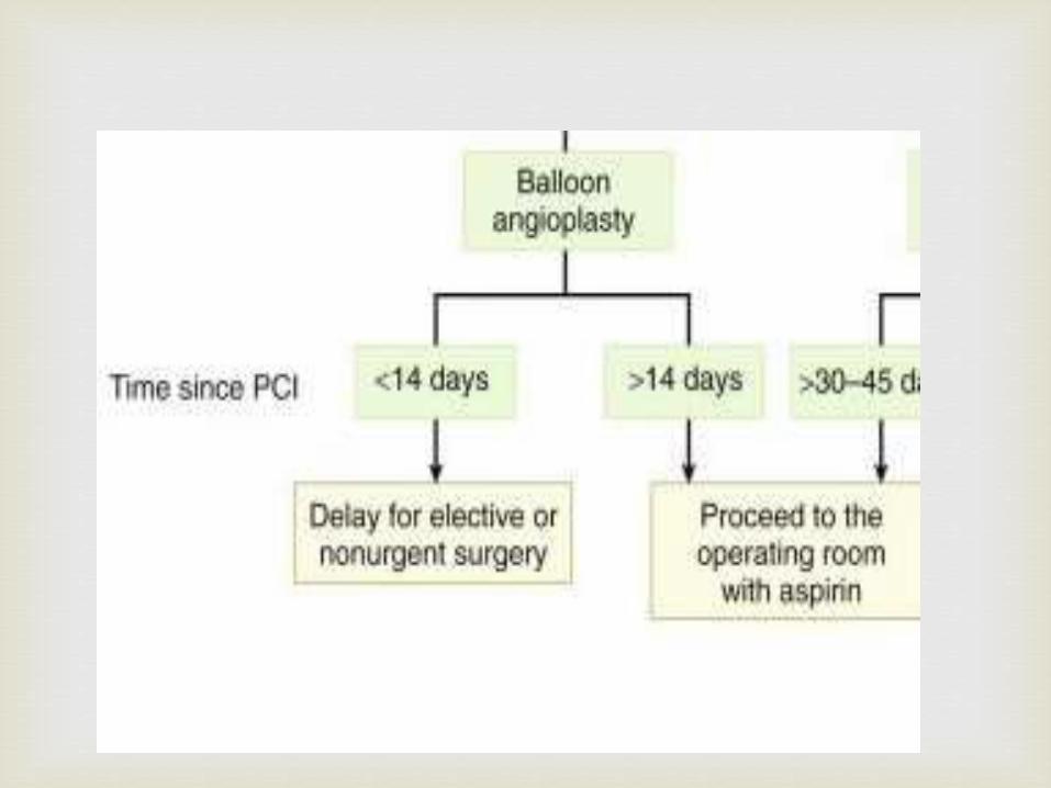
Prior Revascularization
CABG
<5yrs
No change in medical condition
Surgery
PCI
BMS >30days
Minimal anti platelet therapy
Surgery
DES <12 months
Dual anti platelet therapy
Cardiology Opinion
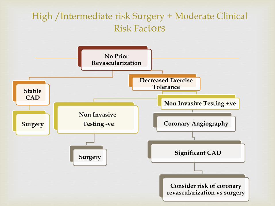
High /Intermediate risk Surgery + Moderate Clinical
Risk Factors
No Prior Revascularization
Stable CAD
Surgery
Decreased Exercise Tolerance
Non Invasive
Testing -ve
Surgery
Non Invasive Testing +ve
Coronary Angiography
Significant CAD
Consider risk of coronary revascularization vs surgery
Three options
Revascularization by surgery (CABG)
Revascularization byPCI
Optimal Medical Management
Management Prior to Surgery
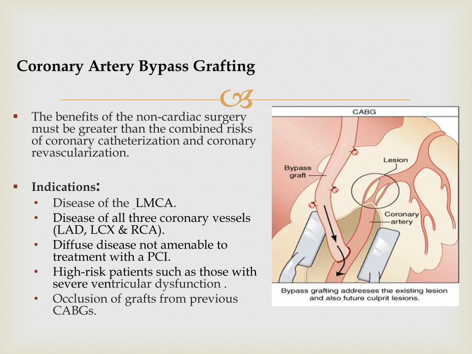
Coronary Artery Bypass Grafting
The benefits of the non-cardiac surgery must be greater than the combined risks of coronary catheterization and coronary revascularization.
Indications:• Disease of the LMCA.• Disease of all three coronary vessels
(LAD, LCX & RCA).• Diffuse disease not amenable to
treatment with a PCI.• High-risk patients such as those with
severe ventricular dysfunction .• Occlusion of grafts from previous
CABGs.
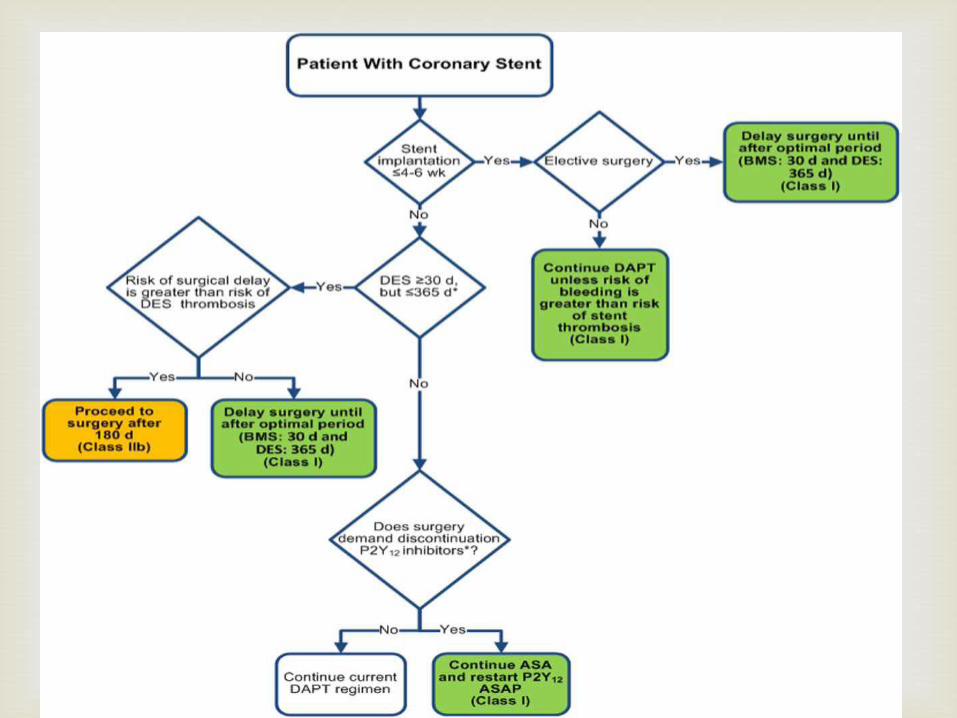
Traditional bare-metal stent (BMS) coronary stents provide a
mechanical framework that holds the artery wall open, preventing stenosis of coronary arteries.
Newer drug-eluting stents (DES) are traditional stents coated with drugs, which, when placed in the artery, release certain drugs over time. These types of stents help revent restenosis of the artery by suppressing tissue growth at the stent site and local modulation of the body's inflammatory and immune responses. They may be susceptible to an event known as "late stent thrombosis“.
DAPT (DAPT; aspirin plus platelet P2Y12 receptor blocker) significantly lowers the risk of stent thrombosis.
Percutaneous Coronary Intervention
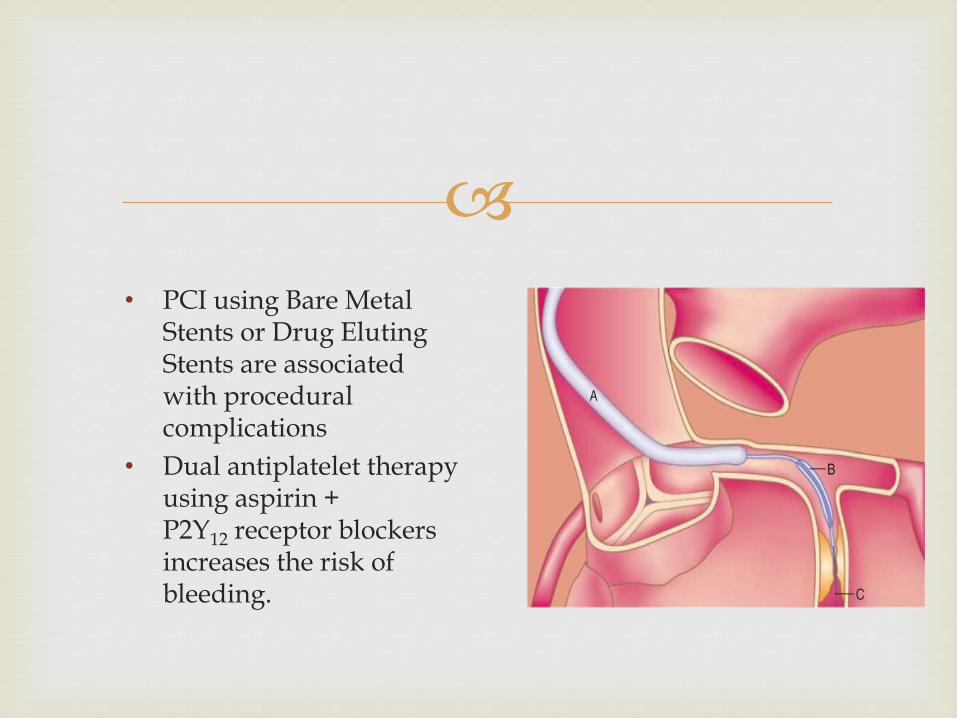
• PCI using Bare Metal Stents or Drug Eluting Stents are associated with procedural complications
• Dual antiplatelet therapy using aspirin + P2Y12 receptor blockers increases the risk of bleeding.
Pharmacological Management (ACC/AHA + European
Guidelines)
Perioperative beta-blocker therapy
• Patients already on beta blockers chronically (I)
• In patients with intermediate- or high-risk preoperative tests (IIb)
• In patients with ≥3 RCRI factors (IIb)
• If used for prophylaxis should be initiated atleast 1 week prior to elective surgery.
Nitrates: Prophylactic use of niroglycerin has not been shown to reduce perioperative mortality or morbidity.(III)
Perioperative statin therapy
• To be continued in patients already taking statins (I)
• Patients with vascular disease should receive statins regardless of the need of surgery (IIa)
• Perioperative initiation of statins may be considered in patients with a clinical risk factor who areundergoing elevated-risk procedures (IIb)
• Should be started 1-4 weeks prior to surgery and continued perioperatively.
Alpha-2 agonists
• Alpha-2 agonists are not recommended for prevention of cardiac events (III)
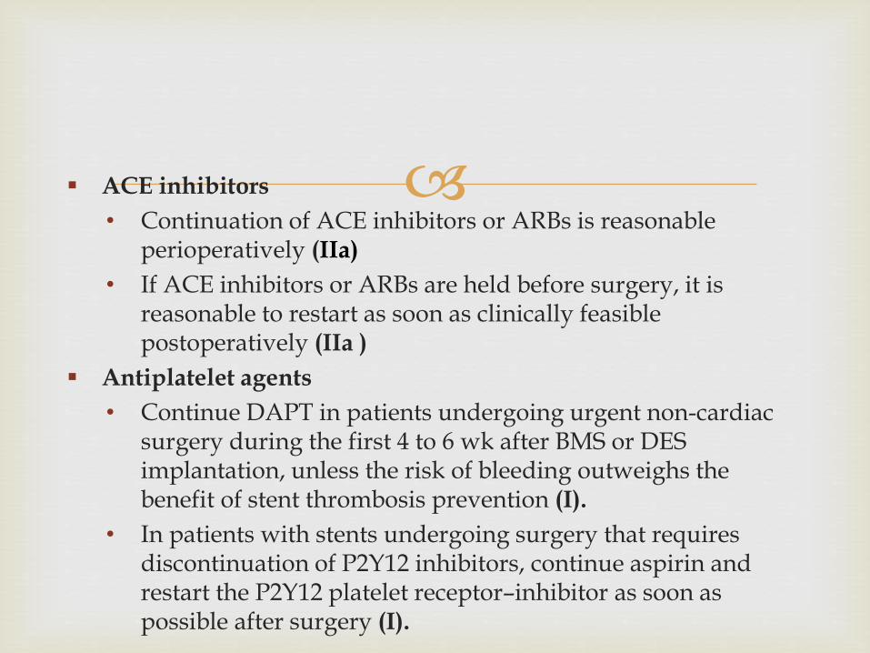
ACE inhibitors
• Continuation of ACE inhibitors or ARBs is reasonable perioperatively (IIa)
• If ACE inhibitors or ARBs are held before surgery, it is reasonable to restart as soon as clinically feasible postoperatively (IIa )
Antiplatelet agents
• Continue DAPT in patients undergoing urgent non-cardiac surgery during the first 4 to 6 wk after BMS or DES implantation, unless the risk of bleeding outweighs the benefit of stent thrombosis prevention (I).
• In patients with stents undergoing surgery that requires discontinuation of P2Y12 inhibitors, continue aspirin and restart the P2Y12 platelet receptor–inhibitor as soon as possible after surgery (I).
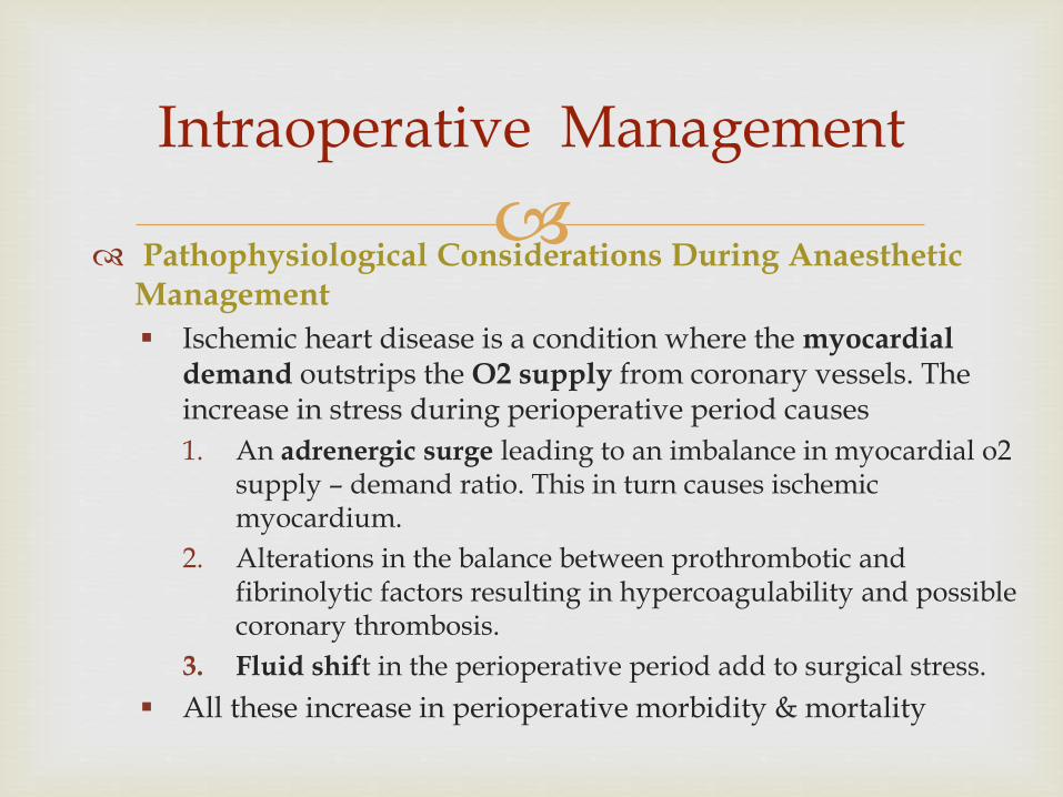
Pathophysiological Considerations During Anaesthetic
Management
Ischemic heart disease is a condition where the myocardial demand outstrips the O2 supply from coronary vessels. The increase in stress during perioperative period causes
1. An adrenergic surge leading to an imbalance in myocardial o2 supply – demand ratio. This in turn causes ischemic myocardium.
2. Alterations in the balance between prothrombotic and fibrinolytic factors resulting in hypercoagulability and possible coronary thrombosis.
3. Fluid shift in the perioperative period add to surgical stress.
All these increase in perioperative morbidity & mortality
Intraoperative Management
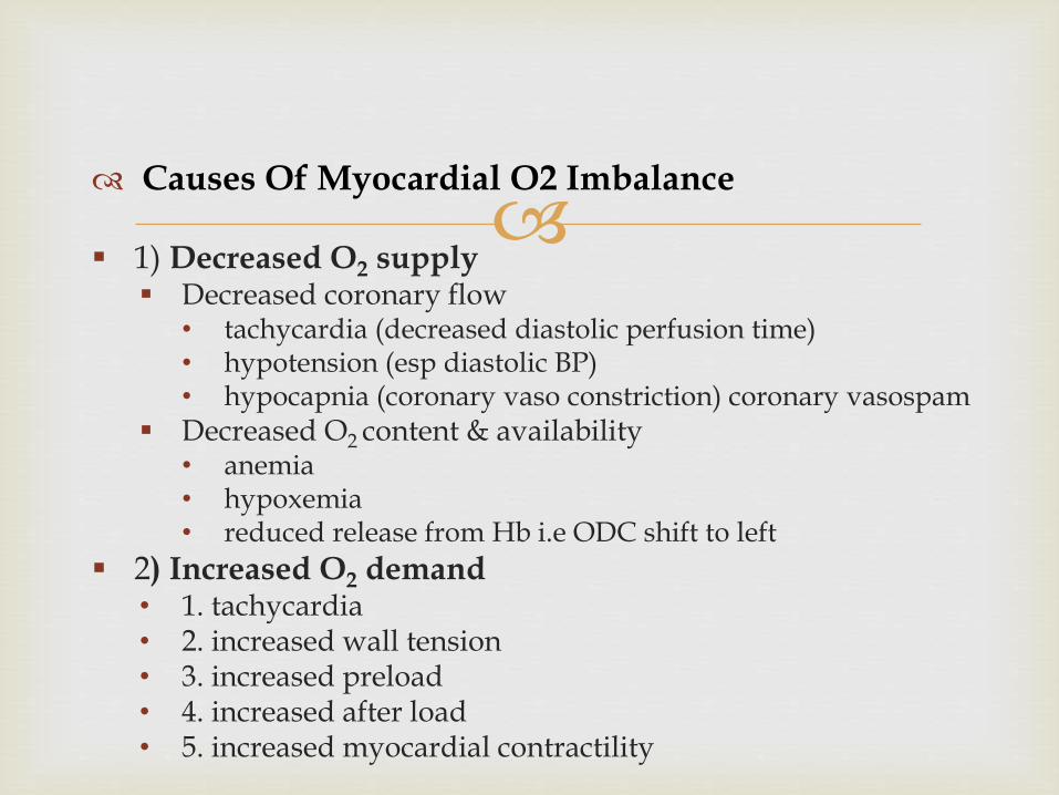
Causes Of Myocardial O2 Imbalance
1) Decreased O2 supply Decreased coronary flow
• tachycardia (decreased diastolic perfusion time) • hypotension (esp diastolic BP) • hypocapnia (coronary vaso constriction) coronary vasospam
Decreased O2 content & availability • anemia • hypoxemia • reduced release from Hb i.e ODC shift to left
2) Increased O2 demand • 1. tachycardia • 2. increased wall tension • 3. increased preload • 4. increased after load • 5. increased myocardial contractility
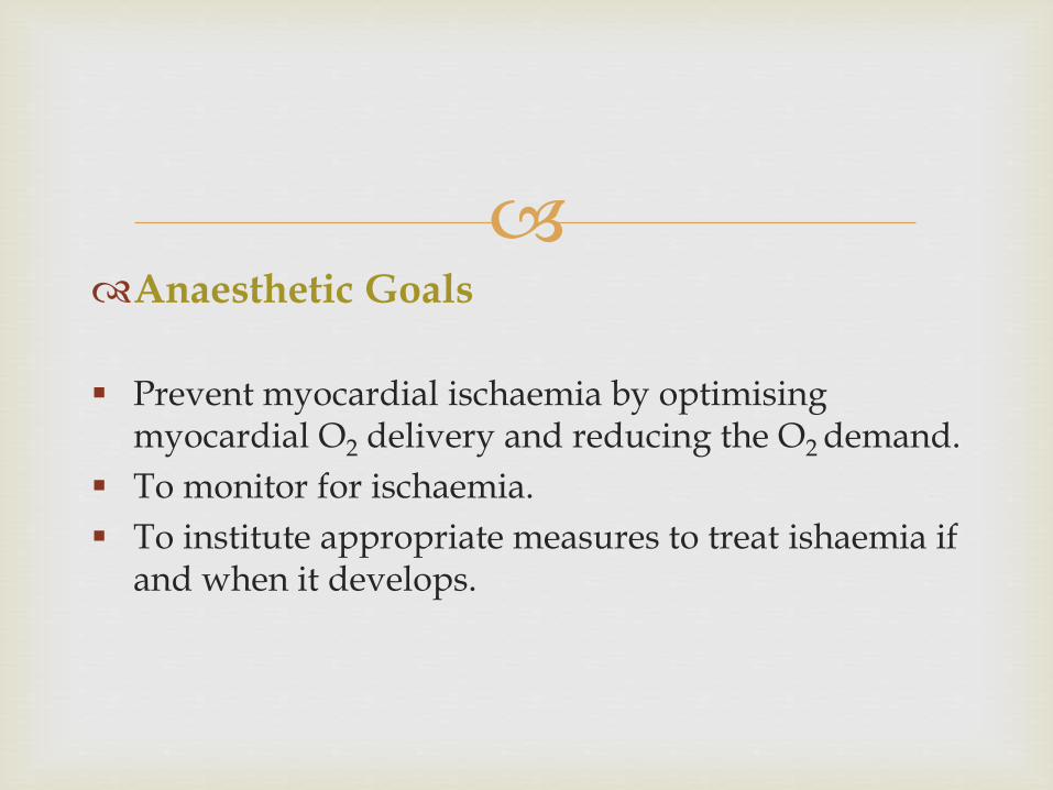
Anaesthetic Goals
Prevent myocardial ischaemia by optimisingmyocardial O2 delivery and reducing the O2 demand.
To monitor for ischaemia.
To institute appropriate measures to treat ishaemia if and when it develops.
Tachycardia
• Increased O2 demand through increased myocardial work
• Also shortens diastolic filling time thereby reducing time for optimal coronary perfusion.
Diastolic BP • In the absence of left ventricle volume overload , a
diastolic arterial pressure of 60 mmHg should be sufficient to maintain coronary perfusion in most patients with CAD .
• Above 90 mmHg is counterproductive as this level invariably requires a higher left ventricular wall tension, thereby increasing O2 demand as well.
• The aim should be to avoid persistent changes in BP.
Anaesthetic Considerations During The Intraoperative Period
Hyperventilation:
• Must be avoided as hypocapnia can cause coronary artery vasoconstriction
Maintain normothermia: • Minimizing body heat loss is vital to avoid postop shivering &
precipitation of ischemic myocardial events. This can be achieved with warm IV fluids, warm operating room atmosphere, forced warm air covers & irrigation of the surgical site with warm fluids.
Correct Anaemia
Blood loss to be taken care of – anaemia can cause critical reduction in myocardial oxygen supply in IHD pt.
Pre Medication
Benzodiazepines –• Quells anxiety
• Hemodynamic stability
• Extended duration of action
• Potential for hypoxia
Intravenous narcotics (e.g. Fentanyl)• Effective control of catecholamine surge
• Respiratory depression
• Prolonged ventilation
General Anaesthesia
Induction
Induction agent:
• Thiopentone: Decreases myocardial contractility, pre-load & BP and causes slight increase of the heart rate.
• Propofol: Causes dose dependent decrease in myocardial contractility and produces significant decrease of BP and HR. Not suitable for patients in whom LV function is compromised.
• Etomidate: Causes minimal haemodynamic changes and is a good choice for patients with poor cardiac reserve.
• Midazolam: It produces decrease in the MAP and increase in the HR.
• Ketamine is to be avoided as it increases myocardial oxygen demand by increasing HR and BP.
Intubation: To be facilitated by the use of succinyl choline or a
NDMR.
Blunting of haemodynamic response to tracheal intubation by
• Keeping the duration short (<15s)
• Use of laryngotracheal Lidocaine, intravenous lidocaine, esmolol, fentanyl, dexmedetomidine.
Maintenance
Volatile anesthetics
• May be used to achieve controlled myocardial depression to minimise the intense SNS activity associated laryngoscopy and surgical stimulation
• Decrease myocardial O2 requirements and pre-condition the myocardium to tolerate ischaemic events.
• AHA guidelines recommend volatile anaesthetic agents for maintenance of anaesthesia in patients with IHD in a haemodynamically stable patient with no evidence of CHF. (IIa)
• Halothane has the disadvantage of myocardial depression and potential of dysrhythmias.
• Although Isoflurane reduces coronary vascular resistance there is no evidence o suggest that it increases the incidence of intra-op myocardial ischaemia.
Nitrous Oxide
• The use of N2O in patients with IHD is questionable since it increases PVR, and predisposes to diastolic dysfunction and subsequent myocardial ischaemia.
Muscle Relaxants • Vecuronium, rocuronium, cisatracurium are attractive choices for
patients with ischemic heart disease
• Atracurium causes histamine release and subsequently fall in blood pressure which makes it a less desirable agent
• Pancuronium should be avoided to due to its sympathomimeticactivity.
• Increased sensitivity to muscle relaxants may be seen in pts on CCBs.
Opioids
• Opioids have an important role in supplementing anaesthesia. They have the Advantage of providing stable haemodynamics due to─ Lack of myocardial depressant effect.
─ Absence of histamine release (fentanyl congeners).
─ Supression of stress response to surgery.
• Opioids may be selected as the primary anaestheticin patients with compromised LV function.
Regional anesthesia may be preferred to GA if possible,
as it tends to better block the stress response to surgery.
Associated hypotension should be corrected by fluids & sympathomimetic agents
Benefits • excellent pain control.
• decreased incidence of deep vein thrombosis.
• postoperative analgesia.
However, the incidence of postoperative cardiac morbidity and mortality does not appear to be significantly different between general and regional anesthesia.
Regional Anaesthesia
The combination of thoracic epidural and GA can be used
for upper abdominal, thoracic and major vascular surgery.
Preoperative epidural analgesia may be considered to decrease the incidence of preoperative adverse cardiac events (IIb).
The main advantages of epidural blockade are superior postoperative analgesia and less diminution of vital capacity.
Epidural analgesia by suppressing pain improves transmural distribution of regional myocardial blood flow and thus minimises myocardial ischaemia.
Combined Regional-General Anaesthesia
• Early extubation is possible and desirable in many patients as long as they fulfill the criteria for extubation.
• However, patients with IHD can become ischemic during emergence from anesthesia and/or weaning with an increased heart rate and blood pressure.
• These hemodynamic alterations must be managed diligently. Pharmacologic therapy with a β-blocker or combined α- and β-blockers such as labetalol can be very helpful.
• Proper pain control.• Continuous ECG monitoring with ST-segment analysis is
important to detect any myocardial ischemic events.• Supplemental O2 to maintain adequate oxygen saturation is
important.• Avoid & treat shivering.• Prevent hypoxemia, hypercarbia hypovolaemia, hypotension.
Post Operative And Long-term Management
• An important goal when selecting monitors for
patients with IHD is to select those that allow early detection of myocardial ischemia
• Most myocardial ischemia occurs in the absence of hemodynamic alterations
• So one should be cautious when endorsing routine use of expensive or complex monitors to detect myocardial ischemia
Perioperative Monitoring
ECG
Simplest and most cost-effective method for detecting perioperative myocardial ischaemia.
3 lead monitoring II, V4 & V5 or V3 V4&V5 is recommended
• Lead II, III, aVF- RCA
• Lead I, aVL- CxCA
• Lead V3-V5- LCA
Pulmonary Artery Catheter
Intra operative myocardial ischaemia can manifest as an acute increase in pulmonary artery occlusion pressure.
Can be used to guide fluid replacement, and to evaluate the efficacy of vasopressor , vasodilator and inotropic therapy.
3 variables are particularly important in assessing benefit versus risk of pulmonary artery catheter use:─ disease severity, ─ magnitude of anticipated surgery, ─ and practice setting .
Patients most likely to benefit from perioperative use of a pulmonary artery catheter • recent MI complicated by HF, • significant CAD undergoing procedures associated with significant
hemodynamic stress, systolic or diastolic LV dysfunction, cardiomyopathy, and/or valvular disease who are undergoing high-risk operations
Use of pulmonary artery catheterization may be considered when underlying medical conditions that significantly affect hemodynamics cannot be corrected before surgery (IIa)
Central Venous Pressure Monitoring: CVP may correlate with
PCWP if EF = 0.5 & there is no evidence of LV dysfunction
Transesophageal echocardiography: • Most sensitive to detect intraoperative myocardial ischemia by
detecting new onset of regional wall motion abnormality
• Emergency use of perioperative TEE in patients with hemodynamic instability is reasonable in patients undergoing noncardiac surgery if expertise is readily available (IIa)
Perioperative myocardial ischaemia and infarction
(PMI) is a major cause of short and long term morbidity and mortalityin the surgical population.
The incidence of perioperative MI in patients who undergo elective high-risk vascular surgery is between 5% and 15%.
The risk is even higher for emergency surgery.
The incidence of perioperative cardiac injury is a cumulative result of preoperative medical condition, the specific surgical procedure, expertise of the surgeon, the diagnostic criteria used to define MI, and the overall medical care at a particular institution.
Perioperative Myocardial Ischaemia
Pathophysiology
Two distinct mechanisms may lead to PMI:
1.Acute coronary syndrome.
• Occurs when an unstable or vulnerable plaque undergoes spontaneous rupture, fissuring, or erosion, leading to acute coronary thrombosis, ischemia, and infarction.
• The sympathetic overactivity induced Tachycardia and Hypertension, common in the perioperative period, may exert shear stress, leading to rupture of plaques.
• Increased postoperative procoagulants (fibrinogen, factor VIII coagulant, von Willebrand factor, α1-antitrypsin), increased platelet reactivity, decreased endogenous anticoagulants (protein C, antithrombin III), and decreased fibrinolysis have been reported.
2. Myocardial O2 Supply – Demand Imbalance
• Tachycardia is the most common cause of postoperative oxygen supply-demand imbalance. Heart rates >80 bpm in patients with significant CAD can lead to prolonged ischemia and PMI.
• Postoperative hypotension (hypovolemia, bleeding, or systemic vasodilatation), hypertension (elevated stress hormones, vasoconstriction), anemia, hypoxemia, and hypercarbiaaggravate ischemia.
• Stress-induced and ischemia-induced coronary vasoconstriction impairs coronary perfusion.
Intraoperative Diagnosis
Complicated by lack of symptomatic presentation in about half of patients with perioperative MI. 75% of Intraop MIs are Silent, present atypically, without ST changes, Q waves or chest pain.
Signs:• instability of HR/BP, desaturation, shock refractory to
vasopressors, poor perfusion, new murmur, skin discolored, pulmonary edema
ECG:• LBBB, arrhythmias, ST changes, T wave inversion, QRS & T
wave axis deviations, and R/U wave changes• ST depression will be 1mm or less in anaesthetized patients.
Biochemistry:• Elevated lactate, CK & Troponin levels.
Prevention
Avoid tachycardia and hypertension.• Maintenance of adequate depth of anaesthesia
• Control pain & anxiety
• Measures to attenuate pressor responses to laryngoscopy and
endotracheal intubation.
Ensure adequate ventilation.
Adequate pain management.
Maintain normothermia.
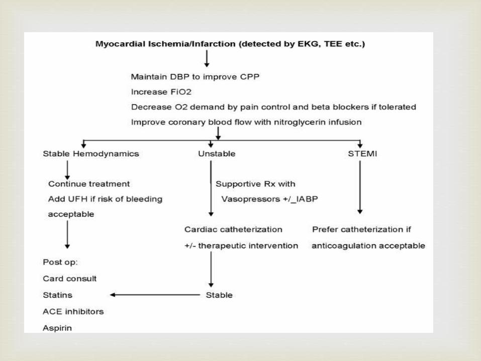
Management
If ischemia is detected early in the intraoperative period before the surgical incision is made, the procedure should be delayed and patient stabilized by improving the oxygen supply and demand ratio.
On the other hand, if surgery cannot be stopped, it should be expedited while the patient is stabilized by improving coronary blood flow and oxygen delivery and reducing oxygen demand.
Discontinue volatile anaesthetic agents & start 100% O2
Administer 325mg Aspirin via ryles’s tube
Tachycardia : Treated with beta blockers.
Nitroglycerine is the drug of choice for accompanying hypertension.
Morphine is a venodilator that reduces ventricular preload and oxygen requirements and it is also effective in patients with pulmonary vascular congestion.
Treatment of cardiac dysrrhythmias
Hypotension
Moderate hypotension responds to volume expansion with 300-500ml of crystalloid
Severe hypotension: Vasopressors Refractory hypotension
• PCWP <12mm Hg – Continue volume expansion> 12 mm Hg - inotropic support with dopamine 3-5/
dobutamine is considered.• If hypotension persists, considerepinephrine or milrinone.
In some patients who don't respond, use of percutaneous IABP is life saving.
Thrombolytic/ reperfusion therapy
• Thromboplastin activator (t-PA) or streptokinase is recommended to minimize the damage caused by intraoperative infarct.
• Should be given within 4 hrs (maximum up to 12 hrs).
• Contraindicated in patients with fresh surgical wounds.
• Antithrombotics and antiplatelet drugs can be started to ensure maximum myocardial salvage.
• Heparin should be started in patients in whom thrombolytic is not given. Heparin has been shown to reduce morbidity andmortality from thromboembolism.
Anaesthetists must be able to:
Identify patients with pre-existing IHD or those at risk for perioperative myocardial ischaemia.
Apply diagnostic measures and guide medical management of patients with IHD .
Optimise patients with pre-existing IHD for surgery.
Minimize physiologic alterations and stress during surgery and extend the care upto the post –opeartiveperiod.
To Sum Up