Embed Size (px)
Citation preview

By-Dr.Anupam

:
1. Overview of hemostasis
2. Clinical approach in making a diagnosis
3. LAB diagnosis
4. Review the most common bleeding conditions

Active process that clots blood in areas of blood vessel injury yet simultaneously limits the clot size only to the areas of injury.
HEMOSTASIS
Components:1. Vessel wall2. Platelets3. Coagulation proteins4. Anticoagulant proteins5. Fibrinolytics

Overview of Haemostasis
INJURY
Collagen Exposure
Platelet Adhesion and release
reaction
Platelet aggregation
VASOCONSTRICTION
Serotonin Platelet Phospolipid
Thromboxane A2
ADP
Primary haemostatic plug
Stable haemostatic plug
Tissue Factor
Coagulation
Thrombin
Fibrin
Fibrinolysis

CLOTTING FACTORS

Protein C/SFactor 8 & 5
Antithrombin3

PRACTICAL APPROACH TO A CHILD WITH BLEEDING HISTORY
HISTORYA.onset of symptomsB. Sites of bleedingC.Perinatal historyD.Gynecologic bleedingE. MedicationF. Diet
Family history

HISTORY
A)Onset-
• age
• acute vs chronic
• Timing of bleeding-immediate vs delayed
1. Mucocutaneous
a. Epistaxis
• Duration, frequency,
• Associated trauma (nose picking, allergy,infection)
b. Oral (bleeding after tooth brushing, after dental
extractions requiring sutures/packing)
c. Bruising (number, sites, size, raised [other than
extremities],
d. Gastrointestinal bleeding
SITES OF BLEEDING

2. Deepa. Musculoskeletal• Hemarthroses, unexplained arthropathy• Intramuscular hematomasb. Central nervous system hemorrhagec. Genitourinary tract3. Surgicala. Minor (sutures, lacerations, poor or delayed woundhealing)b. Major• Tonsillectomy and adenoidectomy• Abdominal surgery
Contd……

C. Perinatal historya. Superficial (bruising, petechiae)b. Deep• Circumcision• Central nervous system bleeding• Gastrointestinal bleeding• Cephalohematoma• Unexplained anemia or hyperbilirubinemia• Delayed cord separation, bleeding after cordseparationc. Vitamin K administrationd. Maternal drugs

D.MEDICATIONSa. Aspirin and NSAIDSb. Anticoagulantsc. Antibioticsd. AnticonvulsantsE. Dieta. Vitamin Kb. Vitamin C
II. Family History.
PHYSICAL EXAMINATION-
• Healthy and sick looking• Vitals signs and growth parameters• Joint examination for :
A)chronic arthropathyB)joint laxity
• Extremities examination fot thumb or radial anomalies(TAR)
• Lymphadenopathy/hepatosplenomegaly
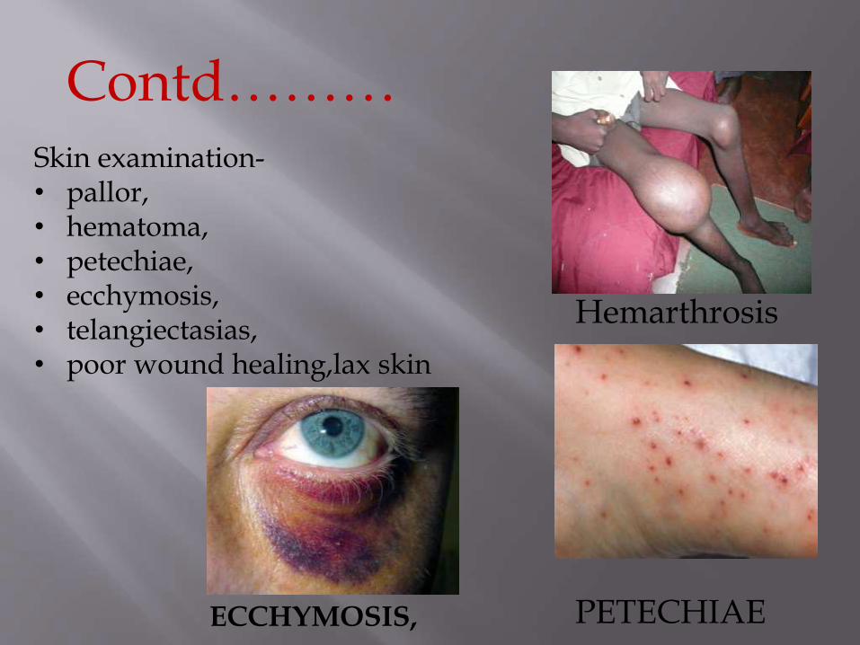
Contd………Skin examination-• pallor,• hematoma,• petechiae,• ecchymosis,• telangiectasias,• poor wound healing,lax skin
Hemarthrosis
PETECHIAEECCHYMOSIS,

Approach to a bleeding patient
LABORATORY WORK UP-
1) C.B.C./PLATELET COUNT2) PERIPHERAL SMEAR- MORPHOLOGY3) P.T. [Prothrombin time]4) a.P.T.T. [ Activated partial thromboplastin
time]

Control bleeding; give fluids arrange blood;order
CBC,PT,aPTT stat
Normal PT,aPTT and decreased platelet
Normal morphology and size of platelets
Perform bone marrow examination
Normal megakaryocytes Decreased megakaryocytes
Peripheral platelet destruction
1. ITP
2. Alloimmune thrombocytopenia
3. Drug induced throbocytopenia
4. TTP
5. HELLP
6. Splenic sequestration
7. Others(sepsis,malignancy)
Decreased production
1. Heriditary thrombocytopenia
2. Fanconi anemia
3. Myelopthisic disorders
4. Drug induced aplasia
5. Viral aplasia
6. Acquired megakaryocytic purpura

Control bleeding; give fluids arrange blood;order
CBC,PT,aPTT stat
Prolonged PT,aPTT and decreased
platelet
Prolonged PT and /or aPTT
Normal platelets
Sick child Healthy child
Investigate for liver disease
1. Acute viral hepatitis
2. Drug induced hepatitis
3. Autoimmune hepatitis
4. Hepatotoxic agent
ingestion
Both PT and
aPTT prolonged
PT normal and
PTT prolonged
VIT K def
• Give vit k
• probiotics
Clotting factor
def
Hemophilia
A
B
C

Control bleeding; give fluids arrange blood;order
CBC,PT,aPTT stat
Healthy child Sick child
Bleeding due to
local factors like
trauma,
anatomic
abnormalities
Qualitative platelet defects
1. Order platelet
aggregation studies
2. Order platelet surface
GP analysis
Compromised vascular
integrity
1. Sepsis
2. Hypoxia
3. Acidosis
4. prematurity
Normal PT and APTT and normal platelet counts

PT PTT FIBRINO-GEN
PLATELETS FDPs Clinical keys
DIC ↑ ↑ ↓ ↓ ↑ Shock
Liver failure↑ ↑ ↓ N/↓ N Jaundice
Vitamin K def ↑ ↑ N N N Malabsorpt
ion
Sepsis without shock
↑ ↑ N N ↑/N Fever
DD’s for DIC

Some common bleeding conditions

Causesa) Infectious-CMV,HCV,HIV
b) Vaccination
c) SLE
d) APLA synd
e) Drugs-quinidine,sulfonamide,heparin
f) CVID
g) Lymphoproliferative disorder

Newly diagnosed ITP-diagnosis to 3months
Persistent ITP-3to 12months of diagnosis
Chronic ITP->12months
Corticosteroid dependence ITP-need for ongoing or repeated administration of steroid to maintain platelet count in excess of 30x109
or avoid bleeding.
Severe ITP-bleeding at presentation of sufficient magnitude requiring treatment or occurrence of new bleeding symptoms requiring intervention
RefractoryITP-presence of severe ITP occuringafter splenectomy

MANAGEMENT
GRADE 1 MINOR BLEEDINGFEW PETECHIAE(<100)<5 SMALL MUCOSAL BLEED
Observation
GRADE 2 MILD BLEEDING>100 PETECHIAE>5LARGE BRUISE(>3cm)NO MUCOSAL BLEEDS
ObservationTreatment in selected childred
GRADE 3 MODERATE BLEEDINGOVERT MUCOSAL BLEEDMENORRHAGIA
Intervention to reach grade1 or gr2 in selected children
GRADE 4 MUCOSAL BLEEDS OR SUSPECTED INTERNAL BLEEDS
INTERVENTION

DRUGS APPROX RESPONSE
RESPONSETIME
TOXICITY
Iv anti D 50-75mcg/kg
50-77% Within24hrs
Headache,fever
IVIG0.8-1g/kg SD
>80% 1-2days Fever
Prednisolone1-2mg/kg-14d or4mg/kg 4days
¾ patient responddepending on dose
2-7days Gastritis
Wait & watch
2/3 ptimprove spontaneouslywithin 6months
Days to month
-

a)Children and adolscents
PLTs <50,000/dl and bleeding
PLTs<50,000/dl and invasive procedure
PLTs <20,000/dl and marrow failure and hemorragic factors
PLTs any counts,but with PLTs dysfunction plus bleeding or on an invasive procedure

Infants within the first 4 months of life:
PLTs <1lakh/dl and bleeding
PLTs<50,000/and an invasive procedure
PLTs<1lakh/dl and clinically unstable
PLTs any counts,but with PLTs dysfunction plus bleeding or on an invasive procedure

Caused by an absence or decreased amount of a procoagulant –
1. VIII -Hemophilia A affects ~ 1:5000 males2. IX -Hemophilia B affects ~ 1:30000 males3. XI –Hemophilia C – Rare /Ethnicity

• Hallmark –HEMARTHROSIS• Easy bruising ,intramuscular
hematomas,heamarthrosis• Life threatening condition like bleed into
iliopsoas muscle,GI bleed

Prolonged PTT(reduced level of 8 & 9)
Other screening test of hemostatic mechanism(platelet,bleeding time,PT)are normal.
Specific assay for factors VIII & IX confirms the diagnosis

Dose calculation of r factor 8 and factor 9
Dose of factor 8
=%desired(rise in f8)xbody weight(kg)x0.5
Dose of factor 9
=%desired(rise in f9)xbody weight(kg)x1.4

Factor 8 concentrates-
A)Plasma -derived factor VIII as well as factor VIII/von Willebrand factor (vWf) concentrates are prepared from cyroprecipitates and contain vWf and moderately enriched factorVIII.
B) Recombinant factor -concentrates are prepared in animal cell cultures using biotechnological procedures
Package Sizes-250/500/1000/1,500/2,000 U/package.
storage temperature for concentrates is 2–8 °C

Diluent-sterile waterLyophilised powderRoute-IV
baxject

-DDAVP’s

VWF tethers platelet to injured endothelium
VWF serves as carrier protein for factor 8
Stored in WEIBEL PALADE BODIES in endothelial cells
Facilitates its ability to bind to platelets by GP1b receptor.

Easy bruisability
Epistaxis or gingival bleeding
Menorrhagia
Post-surgical bleeding
Bleeding post-dental extraction

Sub types of VW
Type 1 Partial quantitative deficiency of vWF
Type 2 Qualitative variants of vWF
A - Absence of HMW vWF multimersB - Same as 2A and increased affinity for platelet gp IbM - Abnormal function not caused by absence of HMW N - Decreased affinity for factor VIII
Type 3 Complete deficiencey of vWF & Behave as Severe Hemophilia A

VWf concentrates-
Desmopressin sprays
antifibrinolytics

VWD treatment

factor XIII is responsible for the crosslinking of fibrin to stabilize the fibrin clot,
symptoms -delayed hemorrhage
Typically, patients have trauma 1 day and then have a bruise or hematoma the next day.
C/f
mild bruising, delayed separation of the umbilical stump beyond 4 wk in neonates,
poor wound healing,

screening tests –for hemostasis are normal
The normal clot remains insoluble in the presence of 5M urea, whereas in a patient with XIII deficiency, the clot dissolves.
The half-life of factor XIII is 5-7 days
There is a heat-treated, lyophilized concentrate of coagulation factor XIII available to treat bleeding episodes or for prophylaxis.


Blood products


• Indication1)to increase oxygen delivery in an anemic patient2)acute blood loss greater than 25-40% in blood volume.
Dose-10ml/kg of RBC will increase Hb by 2.5-3g/dl
Recommneded temp-33-35 C

Can be single or pooled whole blood
1U/10 kg=increase upto 50,000
Stored in mechanical agitator at 22 C upto5days


Freezing plasma at -18 C within 8hrs of separation from whole blood
Contains all coagulation factors
Stored at -18C for 1yr
Prior to administration—thawed at 36-38C
10-15ml/kg over 30-120mins


Contains fibrinogen(150mg/bag),
factor 8(80u/bag),VWf,factor 13
Highly concentrated form of plasma protein that settle down at bottom as ppte when frozen plasma is slowly thawed at 1-6 C
Dose-10-20 ml/kg

Thank you