Embed Size (px)
Citation preview
OUTLINE
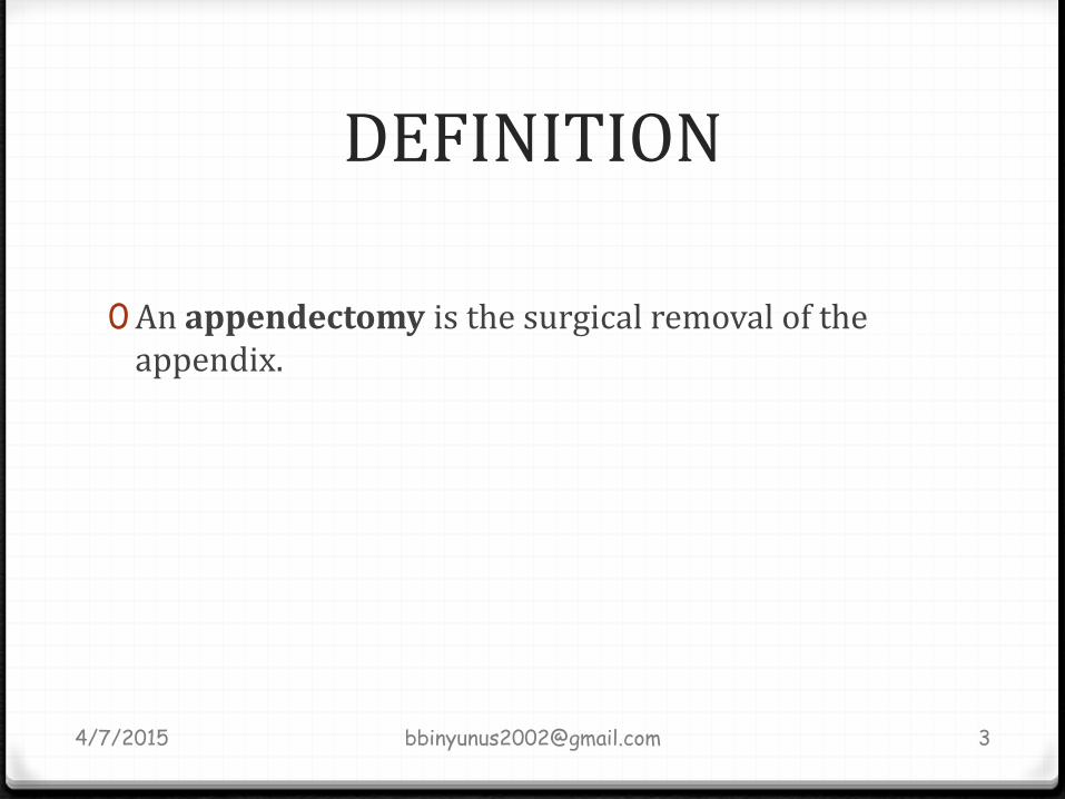
0 DEFINATION
0 INDICATIONS
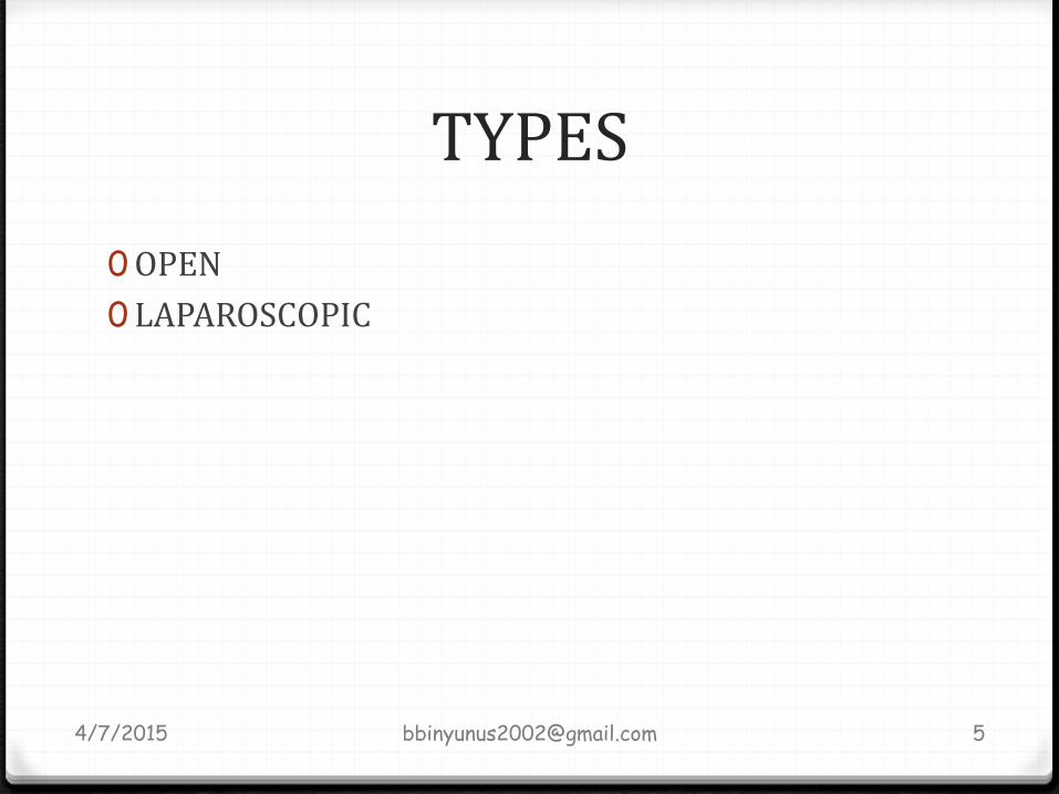
0 TYPES
0 PRE-OPERATIVE PREPARATION
0 ANAESTHESIA
0 POSITION
0 EXPOSURE AND PROCEDURE
0 POST-OP MANAGEMENT
0 COMPLICATIONS
0 REFERENCES
4/7/2015 [email protected] 2
INDICATIONS
0Acute appendicitis
0Recurrent appendicitis
0As Interval appendectomy after drainage of abcess or in appendicial mass
0 Carcinoid tumour : at the tip. <2cm
0Mucocele of the appendix
0Appendicular graft; ileal conduit
0On table colonic lavage
4/7/2015 [email protected] 4
PRE-OP PREPARATION
0 INVESTIGATION
0 Urinalysis- exclude infection
0 Full blood count- leucocytosis
0 Ultrasound scan – noncompressible diameter of > 6mm
0Rehydrate patient with IV fluids; N/S
0 Pass urethral catheter
0N-G tube
0 IV antibiotics prophylaxis- broad spectrum
4/7/2015 [email protected] 6

ANAESTHESIA
0General anesthesia with endotracheal intubation and muscle relaxation
0 Local anesthesia may be indicated in the very ill patient.
4/7/2015 [email protected] 7
POSITION
0 Patient is placed in supine position.
0Routine scrubbing and gowning
0The skin is cleaned from the nipple line to the mid-thigh and draped exposing the operation field.
4/7/2015 [email protected] 8

PROCEDURE
0The surgeon stand on the right side of the patient, the assistant on the left and the nurse on the left side of the assistant
4/7/2015 [email protected] 9
INCISION0 The incision is placed at the point of maximum tenderness.
0 APPROACHES;
0 Mc Burney’s/Grid iron ; an incision placed perpendicular to the McBurney’s point i.e an lateral 1/3 and medial 2/3 of an imaginary line joining the ASIS and the umbilicus.
0 Lanz; skin crease incision. Cosmetically better. approximately 2 cm below the umbilicus centred on the mid-clavicular–midinguinal line.
0 Rutherford Morison’s ; muscle cutting. The muscles are cut upwards and laterally- cutting the internal oblique and trasversus abdominis- extension of Mc Burney
0 Lower mid-line; when in doubt of peritonitis, pelvic appendix,
0 Right Paramedian;
0 Fowler- weir incision; by cutting the muscle medially over the rectus
0 Others; para rectal, kochers, Battle, Rocky Davis
4/7/2015 [email protected] 10
EXPOSURE AND PROCEDURE
0 Skin incision is deepened through the subcutenous tissue to expose the external oblique aponeurosis, hemostasis is secured.
0 Edges are retracted
0 A small incision is made on the external oblique aponeurosis along the line of it fibers
0 The superior and the inferior edges are grasp and the incision are extended with a Mc Indo or Metzenbaum to expose the internal oblique muscle.
0 The fibers are splited along the fibers with curved artery forceps and retracted with langenberg
0 This exposes the transversus abdominis muscles which is also splited and retractor adjusted, the peritoeum is exposed
4/7/2015 [email protected] 11
0 The surgeon grasps the peritoneum with an artery forceps, carefully verifying that intra-abdominal viscera is not inadvertently grasped.
0 A small incision is made on the peritoneum with a size 15 blade.
0 Aspirate taken for mcs and the secretions suctioned
0 Edges of the the peritoneum graspeed with artery forceps and extended
0 The langenbarg retractor is placed within the peritoneal cavity to elevate the anterior abdominal wall
4/7/2015 [email protected] 12
0The caecum is delivered into the wound and the taenia coli is followed to identify the appendix
0Before the appendix is delivered, the wound edges are protected with moist laparotomy pads.
0 If difficulty is encountered in delivering the cecum, the peritoneal lining along the lateral paracolic area may need to be divided to mobilize the cecum
4/7/2015 [email protected] 13
0Once the appendix is delivered, it is held in a Babcock's forceps, while the mesentry is viewed against light to identify the anatomy of the appendicular vessels.
0A small window in the mesoappendix near the base is created this allow application of artery forceps the clamped and ligated with 2-0 suture and divided
0However, it is advisable to divide the mesentry in separate bites if; the artery has divided early into individual branches, fat-laden, inflammed, oedematous
0While addressing the mesoappendix, it is advisable to wrap the inflamed appendix in a gauze sponge to avoid direct contact with the wound margins and thus prevent wound infections.
4/7/2015 [email protected] 14
0The base of the appendix is then gently crushed with a straight artery forceps.(this is to reduce swelling of the tissue to be ligated and reduce likelihood of suture cutting through the edematous tissue, however if the base of the appendix is inflamed, it should not be crushed but ligated just tight enough to occlude the lumen)
0The base is then doubly ligated with 2-0 absorbable sutures.
0A straight hemostat is placed on the appendix approximately 1.5 cm distal to the ligature, and the appendix is transected with a scalpel (between the suture and the forceps).
0The specimen and the contaminated instruments are removed from the operative field.
4/7/2015 [email protected] 15
0The stump;
0 One way of managing the stump is to cleanse it with Betadine or spirit and then electrocoagulate its mucosa.
0 Alternatively, some surgeons prefer placing a purse-string suture (sero-muscular) on the caecum 1.25cm from the base of the appendix using 3-0 absorbable sutures and then inverting the appendiceal stump. This is contra-indicated if the caecum is inflamed
4/7/2015 [email protected] 16
0 If an acutely inflamed appendix had been found and removed, the rest of the abdomen does not need to be explored.
0 Local toileting- lavage
0However, if the appendix is not inflamed, the surgeon needs to exclude other pathologic processes;0 Terminal ileitis
0 Meckel’s diverticulum
0 Tubal or ovarian cause in female
0 Crohn’s disease
4/7/2015 [email protected] 17
CLOSURE 0 The peritoneum is grasped with curved Kelly clamps and
approximated with 3-0 continuous absorbable sutures.0 The transversus and internal oblique muscle layers are
irrigated and loosely approximated with 2-0 absorbable sutures
0 The external oblique fascia is repaired with continuous 0-0 absorbable sutures
0 The subcutaneous tissue is irrigated, and the skin is approximated with staples.
0 If there had been excessive contamination of the wound, it should be left open and the subcutaneous tissue packed with saline-soaked gauze. A delayed primary closure can be performed by day 3 to 4.
4/7/2015 [email protected] 18
POST-OP MANAGEMENT
0 In uncomplicated case, oral fliud are started 12hrs after recovery followed by light diet 24hrs later.
0 In complicated, iv fluids, iv antibiotics and NPO with NG tube drainage until bowel activity recommence and temperature subsides
0 Stiches removed in 7-10days
4/7/2015 [email protected] 19
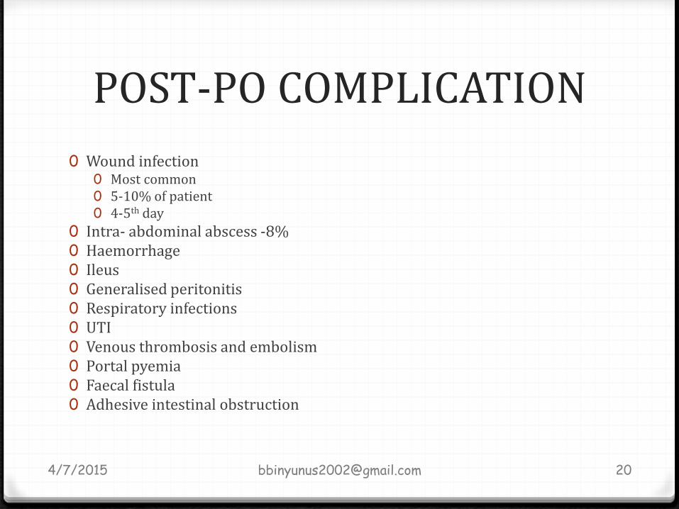
POST-PO COMPLICATION
0 Wound infection0 Most common0 5-10% of patient 0 4-5th day
0 Intra- abdominal abscess -8%0 Haemorrhage 0 Ileus0 Generalised peritonitis 0 Respiratory infections 0 UTI0 Venous thrombosis and embolism0 Portal pyemia0 Faecal fistula0 Adhesive intestinal obstruction
4/7/2015 [email protected] 20

REFERENCES
0VIJAY P. KHATIR, JUAN A ASENSEO. OPERATIVE SURGERY MANUAL 1ST EDITION.
0 FARQUHARSON’S TEXTBOOK OF OPERATIVE GENERAL SURGERY 9TH EDITION
0BALEY AND LOVE’S SHORT PRACTICE OF SURGERY 26TH EDITION
0E.A BADOE ET AL. PRINCIPLES AND SURGERY INCLUDING PATHOLOGY IN THE TROPICS 4TH
EDITION
4/7/2015 [email protected] 21


![Appendectomy Case Report[1]](https://img.pdfslide.net/doc/110x75/546ff242b4af9fc2738b45a1/appendectomy-case-report1.jpg)