Embed Size (px)
Citation preview


APPLIED KINESIOLOGY

APPLIED KINESIOLOGY
A Training Manual and Reference Book
of Basic Principles and Practices
*
Robert Frost
� NORTH ATLANTIC BOOKS
� BERKELEY, CALIFORNIA

Copyright © 2002 by Robert Frost. All rights reserved. No portion of this book, except for brief
review, may be reproduced, stored in a retrieval system, or transmitted in any form or by any
means-electronic, mechanical, photocopying, recording or otherwise-without the written per
mission of the publisher. For information contact North Atlantic Books.
Published by North Atlantic Books
p.o. Box 12327
Berkeley, California 94712
Printed in the United States of America
Cover design © Ayelet Maida, NM Studios
Book design by Jan Camp
Photography by Andreas Werda, Lukas von Saint-George
Illustrations by Tatjana Schuba
Applied Kinesiology is sponsored by the Society for the Study of Native Arts and Sciences, a non
profit educational corporation whose goals are to develop an educational and crosscultural
perspective linking various scientific, social, and artistic fields; to nurture a holistic view of arts,
sciences, humanities, and healing; and to publish and distribute literature on the relationship of
mind, body, and nature.
North Atlantic Books' publications are available through most bookstores. For further
information, call 800-733-3000 or visit our website at www.northatlanticbooks.com.
ISBN-13: 978-1-55643-374-0
Library of Congress Cataloging-in-Publication Data
Frost, Robert 1950-
Applied Kinesiology: A Training Manual and Reference Book of Basic Principles and
Practice / Robert Frost.
p. cm
ISBN 1-55643-374-3 (alk. paper) 1. Kinesiology-Handbooks, manuals, etc. 2. Human mechanics-Handbooks, manuals,etc.
3. Musculoskeletal system-Diseases-Patients-Rehabilitation-Handbooks, manuals, etc.
I. Title.
RZ999 .F76 2002
612.7'6-dc21 2002044729
CIP
5 6 7 8 9 10 11 12 United 14 13 12 11 10 09 08
TABLE OF CONTENTS
Foreword by George J. Goodheart x
Acknowledgements xi
I ntrod uction xii
Chapter 1 FROM BIOMECHANICS TO APPLIED KINESIOLOGY
How Muscles Are Tested in Applied Kinesiology 1
The Development of Traditional Kinesiology or Biomechanics 3
A Short History of Applied Kinesiology 3
A Short Discussion of the Anatomy and Physiology of Muscles 6
Medical Definitions 8
AK Definitions 8
History of Applied Kinesiology (continued) 8
Applied Kinesiology Today 12
Chapter 2
SCIENTIFIC PRINCIPLES OF APPLIED KINESIOLOGY 15
Anatomy and Physiology of the Muscles and Related Structures 15
Neurophysiology: The Nervous System 17
The Nerve Receptors 21
Good Posture and the Central Nervous System 32
Stress Research and Applied Kinesiology 35
A Change in Worldview: From Newtonian Physics
to Quantum Mechanics and Cbaos Theory 39
Traditional Worldviews 40
Quantum and Chaos Theories 43
A Comparison Between Traditional and Modern Models of Reality 44
Fractal Geometry 47
Holograms 49
Biological Medicine and the Systems of Regulation 51

CONTENTS
Chapter 3
THE MUSCLE TEST 63
Theory, Procedure, and Interpretation of Muscle Testing 63
Examiner Prejudice or Impartiality 65
Applications of Muscle Testing 66
Challenge 67
1. Structural or Mechanical Challenge 68
2. Emotional Challenge 71
3. Functional-Neurological Challenge 72
4. Chemical-PhysicallEnergetic-Electromagnetic Challenge 72
Therapy Localization 73
Surrogate Testing 76
Technique for Surrogate Testing 77
Chapter 4 PRETESTS 79
How to Prepare an Indicator Muscle for Accurate Testing 79
A. Is Dehydration Present? 79
B. Does the Muscle Test Strong in the Clear? 79
C. Can the Muscle Be Weakened? 82
D. Is the Individual Muscle in a Hypertonic State? 83
Is Neurological Disorganization (Switching) Present? 84
E. Ocular Lock 85
F. Kidney 27 and Ocular Lock Correction 86
G. Auxiliary K 27 89
H. Central (Conception) Vessel and Governing Vessel 89
Muscle Testing Pretests - A Summarized Overview 90
General Hypertonicity 93
How to Detect Hypertonic Muscles 96
How to Correct Hypertonicity in Individual Muscles 96
How to Correct Hypertonicity in Bilateral Muscle Pairs 97
How to Correct General Hypertonicity 97
Chapter 5
DIAGNOSIS AND CORRECTION TECHNIQUES 99
The Origin-Insertion Technique 99
How to Perform the Origin-Insertion Technique 100

Neurolymphatic Reflexes 100
Neurolymphatic Reflex Point Technique 106
Neurovascular Reflexes 107
Vascular Circulation from the Arteries to the Veins 108
Neurovascular Reflex Point Technique 109
The Meridian System 111
The Meridians 115
Diagnosis of the Reaction to Substances and Other Stimuli 119
Testing for Possible Positive Effects of Stimuli 121
CONTENTS
Testing for Possible Negative Effects of Nutrition or Other Non-Toxic Substances 121
Hidden Problems 121
Finding Hidden Problems 121
Activation of the Right and Left Halves of the Brain 122
Detecting Hidden Problems Through Activating the Halves of the Brain 123
Repeated Muscle Testing 123
Repeated Muscle Testing Technique 125
Fascial Release or Chill and Stretch Techniques for Muscle Stretch Reaction 126
Testing for Muscle Stretch Reaction 130
Draining Excess Fluids from a Muscle 130
Performing the Fascial Release Technique 130
Performing the Chill and Stretch Technique 131
Reactive Muscles 131
The Reactive Muscle Technique 135
Exercise 139
Basic Formula for an Exercise Routine 139
Basic Formula for Muscle Injuries: "RICE" 140
Tho 1)'pes of Exercise: Aerobic and Anaerobic 140
Improving Posture 141
Typical Postural Difficulties 141
Specific Exercises for the Most Commonly Neglected Muscles 142
Chapter 6
MUSCLE TESTS 1 53
Adductors 154
Deltoids: Anterior, Middle, and Posterior 156
Gluteus Maximus 160

CONTENTS
Gluteus Medius 162
Hamstrings 164
Iliopsoas 166
Infraspinatus 170
Latissimus Dorsi 172
Pectoralis Major Clavicular 174
Pectoralis Major Sternal 176
Pectoralis Minor 178
Peroneus Longus and Brevis 182
Peroneus Tertius 184
Piriformis 186
Popliteus 190
Rectus Abdominis Group 192
Rectus Femoris 196
Rhomboid Major and Minor 198
Sacrospinalis Group 202
Sartorius 204
Serratus Anticus 206
Sternocleidomastoideus: Neck Flexors 208
Subclavius 212
Subscapularis 214
Supraspinatus 216
Tensor Fascia Lata 218
Teres Major 222
Teres Minor 224
Trapezius, Lower 226
Trapezius, Middle 228
Trapezius, Upper 230
Appendices 233
1. Glossary of Anatomical Terminology 233
Vocabulary 236
II. Correspondences of Meridians, Muscles, and Organs/Glands 244
III. Sedation Points 245
IV. Step-By-Step Plan for Conducting a Session with AK Techniques 246
V. Applied Kinesiology Techniques of Examination and Diagnosis 249

VI. Additional Tips for Correcting Weak-Testing Muscles 251
Locating Active Reflex Points 251
Challenge All Corrections 251
"Unsolvable" Problems 252
VII. How to Improve and Maintain Optimal Health 253
General Health Tips for the Therapist to Tell to His Patients 253
VIII. Case Histories 257
IX. Bibliography 261
X. Contact Addresses and Sources 264
Orthomolecular (Nutritional) Products 264
Tables and Tools 266
Diagnostic Labs 266
Web Sites 266
ICAK Chapter Contacts 267
Fractals 278
Index 269
·Index of the Main Muscles Discussed in this Text 273
CONTENTS

FOREWORD
Applied Kinesiology had a simple beginning in 1964,
based on the concept that muscle weakness is involved
in most muscle spasms and, indeed, is primary.
Applied Kinesiology is based on the fact that body
language never lies. The opportunity of understand
ing body language is enhanced by the ability to use
muscles as indicators for body language. The original
method of testing muscles and determining their func
tion, first brought to my attention by Kendall, Kendall,
and Wadsworth, remains the prime diagnostic device.
Once muscle weakness has been ascertained, a
variety of therapeutic options is available, too numer
ous to enumerate here. The opportunity to use the
body as an instrument of laboratory analysis is unpar
alleled in modem therapeutics because the response
of the body is unerring; if one approaches the problem
correctly, making the proper and adequate diagnosis
and treatment, the response is adequate and satis
factory both to the doctor and to the patient.
The name of the game, to quote a phrase, is to get
people better. The body heals itself in a sure, sensi
ble, practical, reasonable, and observable manner.
"The healer within" can be approached from with
out. Man possesses a potential for recovery through
the innate intelligence or the physiological home
ostasis of the human structure. The recovery poten
tial with which he is endowed merely waits for the
hand and the heart and the mind of a trained indi
vidual to bring it into manifestation, allowing health
to come forth; this is man's natural heritage.
x
DR. GEORGEJ. GOODHEART, JR.
This benefits mankind individually and collectively.
It benefits the doctor who has rendered the service,
and it allows the force which created the structure to
operate unimpeded. This benefit can be performed
with knowledge, with physiological facts, with pre
dictable certainty. It should be done, it can be done,
and this book offers a means and a measure of how it
can be done. My appreciation to the author and his
staff for the excellent job he has performed in advanc
ing these principles, and my best wishes are extended
to all who read this manual.
-George 1. Goodheart, Jr., D.c., Flee
Diplomate, leAK

ACKNOWLEDGEMENTS
First of all, I would like to give a heartfelt thanks to
the founder of Applied Kinesiology, George Good
heart, D.C His insights and research are the reason
this field exists at all.
Next, I am indebted to the excellent texts of David
Walther, D.C, David Leaf, D.C, and Wolfgang Gerz,
M.D. These were my most-used references for the
writing of this book. Dr. Gerz was also kind enough to
read the text, answer questions by phone and fax, pro
vide various diagrams, and to help with specific ques
tions including the correct translations of Applied
Kinesiology terminology (Fachbegriffe) for the Ger
man edition. His critical reading of this text, correc
tions and suggestions greatly assisted its accuracy and
completeness.
My deepest thanks go out also to my personal
teachers of kinesiology: John Grahme, Andres
Bernard, Richard Harnack, J immy Scott, Gordon
Stokes, Daniel Whiteside, John Thie, Frank Mahoney,
Dominique Monette, Richard Utt, Sheldon Deal, Joan
and Bruce Dewe, John Varun Maguire, Hap and Eliz
abeth Barhydt, Irene Yaychuk Arabei, and Andrew
Verity. Their dedication and personal love of kinesi
ology constitute an ongoing inspiration. A special
thanks to Irene Yaychuk Arabei and Andrew Verity
for the personal balancing sessions that helped me
rid myself of various health and personal problems,
making the writing of this book and the achievement
of other life goals possible.
Parts of this text were derived from my doctoral
thesis. While I was writing that thesis, my father played
the role of the interested but uninformed student of
kinesiology. Through his continual questioning, I
rewrote and rewrote until a beginner could under
stand what I meant. Through the magic of electronic
mail (between California and Switzerland/Germany),
he assisted me in clarifying this text as well. He taught
me to seek unity, coherence, and emphasis in my writ
ing. I hear his guiding words whenever I write. Thanks
to you, Joe Frost.
A special thanks to Tatjana Schuba (Hei/praktik
erin, acupuncturist, fitness trainer, designer). Her
design and precision craftsmanship produced the var
ious anatomical and other graphic drawings. During
the initial writing of this book, Tatjana sat next to me
and translated the text into German. Through her
extensive knowledge of anatomy and physiology, the
text achieved scientific accuracy. In particular the parts
about the nervous system, neurophysiology, hormones
and the meridian system have, through her research
and reworking of my text, achieved greater precision.
W riting together made the work fun and stimulated
us both to keep at it for long hours. Through her ques
tioning of exactly what I meant to say, many unclear
sections of the text were rewritten and greatly
improved.
xi

INTRODUCTION
This book is for those who want a detailed introduc
tion to Applied Kinesiology (AK) as it is performed
by qualified chiropractors, medical doctors and health
professionals. The goal of this book is to present the
principles and basic practices of AK in their original
form as developed by George Goodheart, but in a
manner and a format which may be understood even
by the reader with no prior medical training. Stan
dard medical terminology as used in AK is adhered
to in this text. However, since most every specific term
or concept is defined and logically presented, even
the complete beginner should be able to follow and
understand the ideas. Since I especially wish to pres
ent these concepts using the vocabulary common to
occupational groups with medical background, I uti
lize the following terminology which is also typical in
AK literature: T he "examiner" tests the "patient,"
"diagnoses" and provides corrective "treatments."
At the beginning of the first chapter, I present short
definitions of traditional kinesiology (biomechanics),
Applied Kinesiology and muscle testing so that the
reader may more easily understand these topics. Then
a short history of Applied Kinesiology, its methods
and techniques is provided. In order to describe how
living beings move (the original meaning of kinesi
ology or biomechanics), I describe the anatomy and
physiology of muscles and related structures. Since
muscles are driven by nerves, sections on neuro
physiology and nerve receptors are included. T he
stress concept of Hans Selye and how this relates to
muscular dysfunction follows. Since many of the phe
nomena of Applied Kinesiology cannot be adequately
described within the limitations of the old Newton
ian cause-and-effect scientific model, this is contrasted
with the new worldviews provided by quantum and
xii
chaos theories. Biological medicine, which uses quan
tum and chaos theories to provide a basis for a holis
tic model of healing, and which often uses Applied
Kinesiology for diagnostic purposes, is then described
at length. There follows a section on how to use the
concepts of biological medicine to improve and main
tain optimal health.
For those with some experience in muscle testing,
the main portion of this book will provide the theo
retical background necessary to deeply understand
and to explain to others how muscle testing is per
formed and how muscle strengthening techniques
function. The testing and strengthening of thirty-three
muscles are illustrated and carefully described. The
muscle strengthening techniques discussed ill this text
include Goodheart's original origin-insertion tech
nique, neurolymphatic reflex point massage, neu
rovascular reflex point holding, appropriate nutrition,
and manipulation of the neuromuscular spindle cells
and Golgi tendon organs. The detailed explanations of
how these techniques are performed in AK will
enable the "apprentice" muscle-tester to use muscle
testing and strengthening techniques with improved
precision and effectiveness. The advanced AK diag
nostic and treatment techniques explained in this
book include therapy localization, challenge, nutri
tional and other substance testing, individual activa
tion of the right and left halves of the brain, repeated
muscle testing, muscle stretch response, and reactive
muscles. Use of these techniques will produce much
greater ability to locate and correct the energy imbal
ances that affect health and optimal functioning. These
basic and advanced AK techniques are described in a
step-by-step format I designed for easy application
in a therapeutic session. A selection of case histories

INTRODUCTION
using this format is presented to help the reader
bridge the gap from theory to practice. Most anatom
ical and other specific terms used in this text are
defined in the glossary.
The AK techniques in this book should give the
student a thorough theoretical grounding in muscle
testing and its application. However, nothing can
replace "hands-on" experience. It is highly advisable
to seek training with a health professional experi
enced with AK techniques before attempting to per
form them. Readers who already have experience in
muscle testing will find the techniques that are new
for them described in enough detail here that they
will be able to put them directly into use. It is hoped
that this text will also whet their appetite for more.
For all those who have the required prior training in
a health profession, it is recommended that they
acquire training under the guidance of a qualified
teacher of Applied Kinesiology.
Sports trainers and physical therapists of all sorts
will learn ·useful techniques from this book and
thereby be better able to help their clients. Mastery
of the practical techniques in this text should give any
health professional who practices them the ability to
help patients dispel health problems, improve pos
ture and coordination, increase endurance, eliminate
pains, increase the recuperative powers and many
other salutary effects.
Applied Kinesiology was created in the 1960s by
the American chiropractor, Dr. George Goodheart.
It has been further developed by other chiropractors
and by medical doctors. The requirements for the
highest accreditation, the "diplomate" of Goodheart's
International College of Applied Kinesiology (ICAK),
are high indeed. To join the organization, or take train
ing courses, you must already be a chiropractor, med
ical doctor, or other health professional with a four
year medical training and the legal right to diagnose.
Then you must have at least 300 hours of accredited
instruction in AK, publish two AK research papers
and practice AK for two years. Finally, you must pass
intensive written, oral and practical examinations. The
ICAK diplomates have tremendous training, knowl-
xiii
edge, and experience behind them. But due to the
stringent and extensive requirements for accredita
tion, there are not many of them, and the successful
work they do is not yet very widely known.
In the German branch (ICAK-D), membership
and specially designed AK training programs are
available for accredited practitioners of all state-rec
ognized health professions including Heilpraktiker,
Krankengymnasten, Physiotherapeuten and Psy
chologen. A special branch of ICAK-D, the Interna
tional Medical Society for Applied Kinesiology
(IMAK), exists to serve the interests of medical doc
tors and dentists, offering an exclusive AK training
program for them. Germany, Austria and Switzerland
are the first countries where the medical community
is beginning to take serious interest in AK. In fact,
there are more medical doctors who use AK tech
niques in the German- speaking countries than in the
rest of the world combined.
In an of itself, AK is not a profession. Therefore,
in the world of AK, there are no "applied kinesiol
ogists." As mentioned, to study AK one must already
be a chiropractor, medical doctor, or at least a state
approved therapist. For simplicity in this book, qual
ified therapists who use AK will be referred to as
"examiners" or "therapists who use AK."
John Thie (chiropractor and first president of
Goodheart's International College of Applied Kine
siology) gave some of his patients AK techniques for
self-application as "homework." He saw that the
patients who did this homework had better and
swifter results than those who didn't. Excited by these
practical results, he then urged Dr. Goodheart to write
a popular book about his discoveries in AK. Dr.
Goodheart gave the job back to Dr. Thie. First with
the help of Mary Marks, and then with both research
and writing assistance from Richard Duree and Gor
don Stokes, Dr. Thie wrote the now famous Touch for
Health book, first published in 1973. This was designed
for use by lay persons. The only requirements were
that the chosen techniques be easy to learn, would
(even in simplified form) be able to do a lot of good
and, even if done incorrectly, would cause no harm.

xiv
It is an excellent system for mothers to help improve
the health and performance of their children. As far
as it goes, the system works very well. In fact, it works
so well, that many people use it professionally as a
therapy system. This was a great surprise to its
founders. No one ever intended that Touch for Health
become a professional system of healing. Through its
widespread popularity, Touch for Health has greatly
increased the awareness of Applied Kinesiology. More
than two million people world-wide have been intro
duced to kinesiology muscle testing techniques
through Touch for Health. The many "kinesiologies"
that have been developed from the root of Touch for
Health are today referred to as belonging to the field
of "Specialized Kinesiology."
In many countries such as Germany, Touch for
Health was being taught long before Goodheart's
Applied Kinesiology became known at large. And
many of the practitioners of Touch for Health and
related kinesiologies called their work "Angewandte
Kinesiologie," the German translation of Applied
Kinesiology. At that time there were few therapists
using Applied Kinesiology and there seemed to be
no reason not to translate from English and use the
term themselves. This can be compared with Califor
nia calling its sparkling wines "champagne." The
French complained bitterly but to no avail. Although
no one denies that Champagne is a province in
France, the French had not internationally patented
the word "champagne." Similarly, Goodheart never
patented the term "Applied Kinesiology." One unfor
tunate consequence is that many therapists believe
that Touch for Health and Applied Kinesiology are
identical. And seeing that Touch for Health is for lay
persons, they do not pursue studies in Applied Kine
siology. In order to avoid further confusion, Good
heart's original work, even in foreign language texts,
is now called "Applied Kinesiology" with no transla
tion of the term.
xiv
APPLIED KINESIOLOGY
The simplified techniques of Touch for Health do
not go as far or do as much good as can be achieved
by the original and more complicated techniques of
AK. For example, Touch for Health advises, as a mus
cle strengthening technique, that the neurovascular
points be gently held. AK teaches that neurovascu
lar points be not only held, but gently tugged in var
ious directions, until the direction that produces
maximum pulsation is detected. Then the points are
held in this exact direction for 20 seconds longer. Just
holding the points will often strengthen the muscle
test. Careful experimentation has revealed that the
best effects upon the associated organ and bodily
areas are only achieved with the precise application
taught in AK (and explained in this book) .
In most systems of Specialized Kinesiology, there
is a conspicuous absence of descriptive detail of the
anatomy and physiology involved. And an explana
tion of how the techniques function is also lacking.
This is to be expected, because Touch for Health was
designed for lay persons. For those who began with
Touch for Health and/or other branches of Special
ized Kinesiology and are now ready for more detailed
knowledge and precision, this book will provide a
bridge toward the deeper understandings and appli
cations of the original techniques of Applied Kinesi
ology. It is hoped that this book will demonstrate the
professional level of knowledge, the wide range of
application and the practical usefulness of the tech
niques of AK and thereby attract more health pro
fessionals to study AK.
Since I lived in Switzerland and Germany for
twenty years, this book first appeared in the German
language in 1999 with the title Grundlagen der
Applied Kinesiology. I am living in California again
and I'm pleased to now present the English version.
-Robert Frost, Ph.D.
Carlsbad, California, September 2001

* CHAPTER 1
From Biomechanics
To Applied Kinesiology
KINESIOLOGY (from the Greek kinesis, movement) began in antiquity as the study of human and animal movement. Over the course of many centuries, this original, traditional form of kinesiology (biomechan
ics) has produced a broad body of knowledge of how nerves stimu).ate muscles to act upon bones in order to produce posture and movement. Like physiotherapy, kinesiology is a therapeutic profession with a long history. Medical muscle testing existed in biomechanics long before the emergence of Applied Kinesiology.
The biomechanic principles of kinesiology (such as the application of the minimal force necessary to produce maximal result) have been successfully applied to a wide variety of ergonomic problems of industry, sports, and medicine. The application of biomechanics in industry has resulted in the design of tools, chairs, work stations, etc., that are "user friendly." It has stimulated the development of ergonomic work techniques (e.g. how to lift heavy objects without endangering the body) that result in fewer injuries and yield greater productivity. Athletes work with kinesiologists to learn how to more efficiently and successfully perform the movements required by their sports. And biomechanic principles have many applications in the various fields of medicine including the designing of artificial joints and the development of more effective rehabilitation methods.
The research and developments of biomechanics or "traditional" kinesiology can be traced back over
thousands of years and continue into the present. By contrast,Applied Kinesiology (shortened in this book to AK) began in 1964 with the research of the American chiropractor, George J. Goodheart, Jr., ne. His extraordinary powers of observation, his curiosity, his drive to research the causes of what he observed and the resulting discoveries have been the source of most of the diagnostic techniques used today in this relatively new discipline.
Various kinds of health professionals schooled in AK use standard medical muscle tests of biomechanics to directly assess the functional integrity of the nervous system and the muscles. Muscle testing is described at length later in this book. As a preliminary introduction, a brief description of muscle testing as performed in AK is given below:
How Muscles Are Tested in Applied Kinesiology
1. Most muscles are attached through their tendons at both ends to bones that meet in a moveable joint. When muscles contract, they shorten. This shortening pulls one of the attached bones toward the other.
2. To prepare for the muscle test, one bends the joint over which the muscle is attached. This shortens the muscle, bringing it into a position of contraction. The examiner places his hand in a position to resist the further contraction of the muscle.
1

2
3. The patient initiates the test by steadily contracting the muscle from zero force up to the maximum force of contraction against the examiner's unmoving hand. During this short period, the examiner provides an equal and opposite, steadily increasing resistance to maintain the starting position of the muscle test. When the patient has willfully contracted his or her muscle as much as possible, the examiner applies a bit more force. The whole test procedure should not last longer than 2-3 seconds. If the patient can maintain the original test position against this small extra force without movement, the muscle "tested strong." If not, it "tested weak."
4. In the first part of the muscle test, one is testing the determination and ability of the patient to strongly contract the muscle. In the second part of the muscle test, one is also testing the ability of the patient's nervous system, "on its own," to provide a little more contraction than the patient can willfully provide. By this technique, one is actually assessing the functional integrity of the complete circuit of the muscle and the portion of the nervous system involved with that muscle. This initial muscle test is performed "in the clear," i.e., with no extra stimulus of any kind. The muscle is contracted as strongly as the patient is consciously able. In the second part of the muscle test, the question asked is: After the patient has completely contracted the muscle, and the examiner then applies additional pressure, can her nervous system finely coordinate the muscle to contract just a bit more than he or she is consciously able to do?
5. AK uses not only muscle testing "in the clear" as described above, but also "indicator" muscle testing. In this type of muscle testing, a muscle that previously tested strong in the clear is used as an indicator for testing some other stimulus. The extra stimulus can be provided by touching an area of the patient's body that is "disturbed" or dysfunctional because of injury, infection, etc. If this is done while repeating the test of a previously
APPLIED KINESIOLOGY
strong-testing indicator muscle, such stimulus may cause that muscle to test weak. The stimulus provided by the patient touching himself or herself is referred to as "therapy localization."
In practice, many examiners touch the patient to therapy-localize, which is often easier, faster, and usually produces the same results. However, on occasion, when the examiner touches the patient, the results of therapy localization are different than when the patient touches the same area of the body. Therefore it is recommended that the patient touch herself for therapy localization. When the patient is presented with some other kind of stimulus besides touch, or performs some kind of activity and the effect is then measured with muscle testing, this is called "challenge."
Much of the fascination of Applied Kinesiology lies in the fact that most factors influencing health may be tested using an indicator muscle and therapy localization or challenge. As will be described later, health professionals familiar with AK techniques use standard muscle testing "in the clear" and indicator muscle testing of various stimuli to evaluate the structural, mental/emotional, and biochem'ical functions of the human organism.
Applied Kinesiology is primarily a diagnostic technique. Although extensive methods for the evaluation (diagnosis) of dysfunction were developed early within the field of AK, most of the treatments used in AK have been gathered from other (sometimes quite foreign) areas of healing. Besides its well-developed diagnostic techniques, the practical advantage of AK is that one can determine which of many possible therapy methods will be the most effective for the individual problems of specific patients. In this way, before applying any therapeutic technique, the examiner can determine the relative effectiveness and thus choose accurately from a wide variety of treatments.
The diagnostic techniques of AK allow one to determine which body system is disturbed and which treatment modalities are best suited to the correction

FROM BIOMECHANICS TO APPLIED KINESIOLOGY
of the disturbance. Interventions of all sorts (structural, chemical, nutritional, mental, electromagnetic, etc.) may be individually tested in advance to assess their worth in treating a specific problem. After treatment, the same techniques can be applied to determine whether the treatment was appropriate, correctly applied, and effective.
The Development of Traditional Kinesiology or Biomechanics Beginning with the ideas of Aristotle (384-322 B.C.), who is often called the "father of biology," the study of movement (the original kinesiology) has for centuries centered upon anatomy and mechanics. Leonardo da Vinci (1452-1519) is especially well known for studies of human structure and function. These make him one of the best known pioneers of the study of movement, or kinesiology.
Mechanics is the branch of physical science that deals with energy and forces and their effect upon bodies. The central interest of the early kinesiologists was the mechanical consideration of how muscles act upon bones and joints to produce posture and movement. Eventually, in the modern age, representing the bones as levers, the joints as fulcrums, and the muscles as springs provided a simple model of body mechanics for mathematical calculations. Although idealized, such models do provide useful insights into human and other animal movement. Kinesiology was originally defined as the study of the structures and their functions that produce animal and human movement. Today this study is called biomechanics, and it is sometimes referred to as "traditional" kinesiology.
After the spectacular contributions of Leonardo da Vinci in the fifteenth century, traditional kinesiology made little progress for more than two hundred years. When Luigi Galvani made his discovery (1780) that muscular contraction is produced by electrical impulses, the development of kinesiology began again. Galvani applied a small electrical voltage to a frog's leg, which produced a twitch contraction in the mus-
3
cles of the leg. From this he correctly concluded that muscular contraction is initiated by electrical impulses. Until his time, a muscle was considered to have a will of its own. This thinking is still to be observed in certain phrases such as "the biceps act to bring the wrist toward the shoulder". Galvani's experiment demonstrated that muscular contraction, and thus movement of the body, is the result of electrical stimulation of the muscles. With the further discovery that electrical impulses were, in the living animal, provided by nerves under central nervous system (brain and spinal cord) control, the study of the function of nerves and the central nervous system (neurophysiology) automatically came to be included in the study of movement (kinesiology). Neurophysiology will be discussed at greater length in the following chapter. First we will continue with the historical development of kinesiology.
A Short History of Applied Kinesiology Applied Kinesiology grew out of Dr. George J. Goodheart's analysis of his day-to-day chiropractic practice. Accepted chiropractic procedures served him well in his practice, most of the time. However, he was keenly disturbed by his lack of techniques to adequately diagnose the occasional set of paradoxical (or just plain puzzling) symptoms. And without adequate diagnosis, he was at a loss to devise effective treatments. When stumped by a patient's unusual symptoms and diagnostic results, Goodheart continually asked himself the age-old query of the scientistresearcher: "Why is this?"
In his search for explanations that might lead him to effective corrective procedures, Goodheart carefully considered the anatomy and physiology involved
in his patients' problems. This knowledge often led him directly to possible interventions. Examples of new methods he deduced include his theory and treatment of reactive muscles and his effective treatment for sustained muscle use. (Both of these methods are described in detail later in this text.)

4
Goodheart explored beyond the boundaries of his formal chiropractic training to consider the concepts of other innovative healers and scientists. He studied the traditional knowledge and research findings of many other healing systems (Chinese acupuncture, lymphatic drainage, nutrition, neurology, etc.) and then found ways to incorporate them into Applied Kinesiology. He experimented with many alternative treatment modes such as Chapman's reflexes, Bennett's reflexes, synchronization of pulses in reflex points, etc. When a puzzling diagnostic situation could not be solved with the techniques he already knew, he experimented, even with highly unusual measures. Out of his uniquely open-minded search for procedures came most of the techniques used in Applied Kinesiology today.
In 1964, Goodheart made the discovery that marks the birth of Applied Kinesiology. As a chiropractor, he assumed that correcting structural imbalances in the body (postural problems, false alignment of bones, etc.) will reduce or eliminate most health problems. Structural balance, optimal postural alignment of the parts of the body, is the goal of chiropractic. But structural balance cannot be obtained when muscles are overly tense or too limp.
For several months, Goodheart unsuccessfully treated a patient whose presenting symptom was one shoulder blade that stuck out away from his back. He remembered reading in Kendall and Kendall's classic, Muscles: Testing and Function, about a muscle that pulled the shoulder blade down upon the back. He used the muscle testing technique described by Kendall and Kendall to test this muscle, the serratus anticus (often called the anterior serratus). It tested weak only on the side where the shoulder blade protruded. The serratus muscle is called "serrated" like a saw because of its "toothed" shape (see illustration on page 207). It connects the upper eight or nine ribs to the inner vertebral side of the scapula, the shoulder blade. The muscle was not less developed on the weak-testing side and Goodheart found no other reason for the one-sided test weakness. Exploring the weak-testing muscle with his fingers revealed painful
APPLIED KINESIOLOGY
little lumps (nodules) where the tendons of serratus anticus attach to the ribs. When he firmly rubbed one of these nodules, it disappeared. As an experiment, he firmly massaged all of these nodules and upon retesting found an immediate increase in the "test strength" of the muscle. Encouraged by this discovery, Goodheart used Kendall and Kendall's book to teach himself how to muscle-test many other muscles as well. This was the first discovery of Applied Kinesiology and the beginning point of ongoing and very fruitful research.
This surprising discovery that a weak-testing muscle may be made to test strong through the massage of its extreme ends where its tendons attach to bone
is referred to in Applied Kinesiology today as the "origin-insertion technique." This technique worked often enough in establishing muscular balance (and thus structural balance also) that many chiropractors began to use manual muscle testing to assess structural balance, the goal of chiropractic. When the origin-insertion massage strengthened weak-testing muscles, many other health problems often disappeared without further treatment. This provided more confirmation of the basic chiropractic premise that structural balance affects all aspects of health.
However, the origin-insertion technique often failed to strengthen weak-testing muscles and reestablish muscular balance. Muscle-building exercises didn't help either. Such exercises, specifically designed to strengthen the weak-testing muscle, often did increase the mass of the muscle and its weight-bearing strength, but it still "muscle-tested" weak. Factors other than pure physical strength were at work that needed to be unearthed. Goodheart's further research revealed that muscular imbalances may be the result of problems not just in the origin-insertion area of the muscle itself, but also in any of the areas represented by the three sides of the chiropractic "triad of health"-that is, dysfunction could be the result of structural, chemical, and/or mental problems.
The interaction of the three sides of the triad of health is an important and very useful principle in Applied Kinesiology evaluation. Some examples of
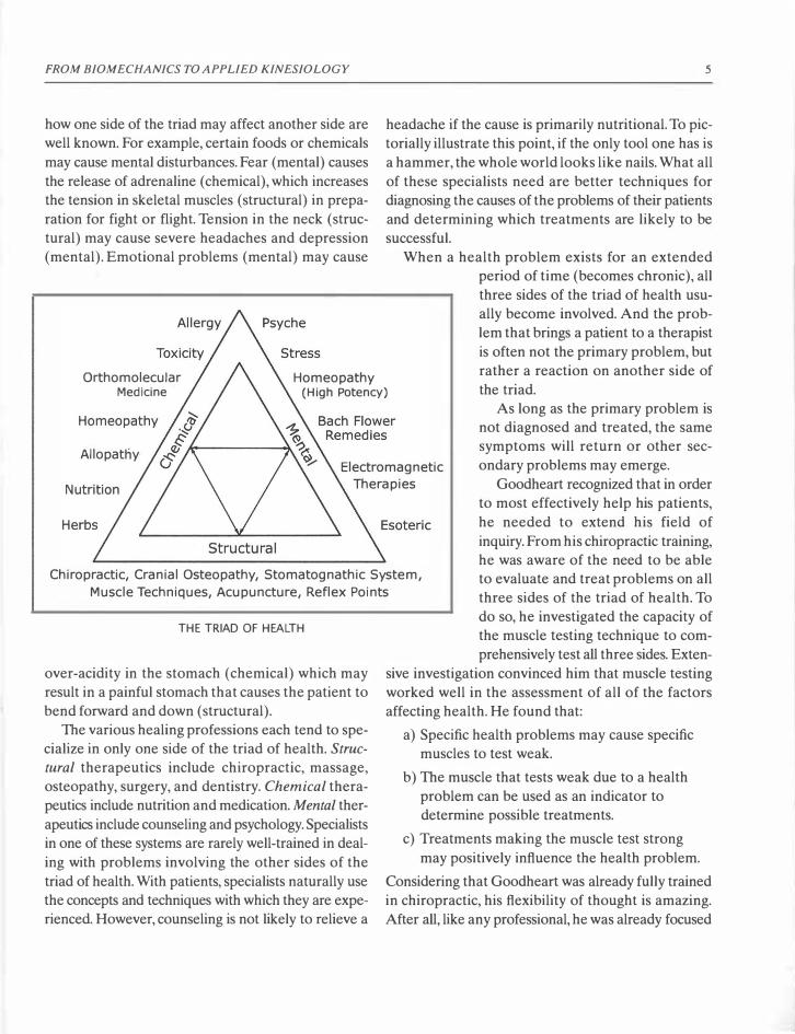
FROM BIOMECHANICS TO APPLIED KINESIOLOGY
how one side of the triad may affect another side are well known. For example, certain foods or chemicals may cause mental disturbances. Fear (mental) causes the release of adrenaline (chemical), which increases the tension in skeletal muscles (structural) in preparation for fight or flight. Tension in the neck (structural) may cause severe headaches and depression (mental). Emotional problems (mental) may cause
5
headache if the cause is primarily nutritional. To pictorially illustrate this point, if the only tool one has is a hammer, the whole world looks like nails. What all of these specialists need are better techniques for diagnosing the causes of the problems of their patients and determining which treatments are likely to be successful.
Orthomolecular Medicine
Homeopathy (High Potency)
When a health problem exists for an extended period of time (becomes chronic), all three sides of the triad of health usually become involved. And the problem that brings a patient to a therapist is often not the primary problem, but rather a reaction on another side of the triad.
Allopatny Electromagnetic
Therapies
As long as the primary problem is not diagnosed and treated, the same symptoms will return or other secondary problems may emerge.
Goodheart recognized that in order to most effectively help his patients, he needed to extend his field of inquiry. From his chiropractic training, he was aware of the need to be able to evaluate and treat problems on all three sides of the triad of health. To
Chiropractic, Cranial Osteopathy, Stomatognathic System,
Muscle Techniques, Acupuncture, Reflex Points
THE TRIAD OF HEALTH
over-acidity in the stomach (chemical) which may result in a painful stomach that causes the patient to bend forward and down (structural).
The various healing professions each tend to specialize in only one side of the triad of health. Struc
turaL therapeutics include chiropractic, massage, osteopathy, surgery, and dentistry. ChemicaL therapeutics include nutrition and medication. MentaL therapeutics include counseling and psychology. Specialists in one of these systems are rarely well-trained in dealing with problems involving the other sides of the triad of health. With patients, specialists naturally use the concepts and techniques with which they are experienced. However, counseling is not likely to relieve a
do so, he investigated the capacity of the muscle testing technique to comprehensively test all three sides. Exten-
sive investigation convinced him that muscle testing worked well in the assessment of all of the factors affecting health. He found that:
a) Specific health problems may cause specific muscles to test weak.
b) The muscle that tests weak due to a health problem can be used as an indicator to determine possible treatments.
c) Treatments making the muscle test strong may positively influence the health problem.
Considering that Goodheart was already fully trained in chiropractic, his flexibility of thought is amazing. After all, like any professional, he was already focused
6
upon the limiting concepts of his specialized field of knowledge. With uninhibited enthusiasm, Goodheart evaluated a wide variety of therapeutic approaches in his attempts to achieve the chiropractic goal of structural balance. He thoroughly researched any procedure that resulted in the strengthening of a weaktesting muscle.
Sometimes, patients with the same symptoms require very different therapies. Many of the most successful interventions that Goodheart studied had been previously developed but were seldom used due to a lack of diagnostic techniques that could identify when a specific intervention would be helpful. The use of muscle testing provided him with the needed diagnostic tool to choose among the many possible interventions for each disturbance. Since muscle testing uses the patient's body itself as the instrument for performing diagnostics, it provides a direct method for studying the effects upon the body of just about any kind of healing modality. Goodheart found muscle testing to be the most direct method to locate the treatment best suited to the needs of each particular patient.
For use in his own practice and for the benefit of other practitioners, Goodheart gathered, adapted, developed, and codified many techniques useful in the strengthening of weak-testing muscles. The greater portion of the techniques known and used in AK today stem from his research.
Goodheart's research is remarkable for its intuitive conceptual leaps. For example, he first determined that a correct treatment measure almost always swiftly returns a weak-testing muscle to testing strong. He then intuitively jumped to the proposition that muscle testing conversely might be used to test for the effectiveness of any treatment after it has been applied. Further careful research proved the inspiration to be true. He established the rule that by using muscle testing after the treatment, one can determine whether the applied treatment has been effective. Health professionals using AK today stand on the shoulders of the giants like Goodheart that have gone before. From this perspective, we can mistakenly feel that such intuitive jumps are actually obvious. The challenge is to be such a giant and to discover some of
APPLIED KINESIOLOGY
the similar "jumps" of realization still waiting to be made in this young field of research.
A Short Discussion of the Anatomy
and Physiology of Muscles In order to understand the depth of Goodheart's work, a very short discussion of the anatomy and physiology of muscles is included here. Definitions of the words used will also be discussed briefly. These topics will be discussed at greater length in later sections of the book.
Many muscles work in functional pairs (agonistantagonist), with one contracting to open a joint (moving the attached bones apart) and the other contracting to close the same joint (moving the attached bones together). The biceps and triceps form a clear example of two such opposing muscles. Contraction of the biceps brings the wrist toward the shoulder, closing the elbow joint. Contraction of the triceps brings the wrist away from the shoulder, straightening the arm and opening the elbow joint. A more complex example is provided by the upper trapezius muscles, one function of which is to elevate the shoulders, and the latissimus dorsi muscles which, among other functions, pull the should�rs down.
Muscle tone is defined as the level of continual contraction while the muscle is at rest, meaning not actively contracting. In medical terminology, when a muscle has too much tone, and feels hard by palpation (examination through touch), it is said to be hypertonic. When a muscle has optimal tone, it is said to be normotonic . When a muscle has too little tone and feels somewhat limp, it is hypotonic. When a muscle has no tone, it is atonic (flaccid or limp). In AK, these same medical terms have slightly different meanings: A normotonic muscle tests strong and can be weakened by specific methods. A hypertonic muscle tests strong but cannot be weakened. The term hypotonic is found in the literature and refers to a weak-testing muscle.
A hypertonic muscle, by palpation, feels hard and usually muscle-tests very strong. Sheldon Deal (through the work of his patient, Richard Utt) was

FROM BIOMECHANICS TO APPLIED KINESIOLOGY 7
AGONIST-ANTAGONIST MUSCLE PAIRS:
BICEPS-TRICEPS, LATISSIMUS DORSI-UPPER TRAPEZIUS
the first in AK to present a term to define a muscle that tests strong but cannot, by usual means, be made to test weak. He called the state of such a muscle "frozen." Others describe this state as "hypertonic"
or "over-facilitated." Referring to the results of muscle testing, Goodheart stated that the muscle is "weak" or "strong." He also mentioned the existence of hypertonic muscles once by stating that "strong"testing muscles can test "too strong." Unfortunately,
Weak-Testing Normotonic
Schwach Normoton
Weak Strong
Under-Facilitated Normal-Facilitated
Hyporeaktive Normoreaktive
Unlocked Locked
Flaccid Homeostasis
Goodheart never deeply explored the state of muscles that test "too strong," nor were his terms "weak" and "strong" really accurate descriptions. And he never "laid down the law" by choosing the precise nomenclature to be used by all in AK. Therefore various AK authorities use different vocabulary for the same items, which can lead to confusion for those studying the AK literature.
For simplicity and clarity in this book, the terms
Hypertonic Author
Hyperton (Wolfgang Gerz)
Too Strong (George Goodheart)
Over-Facilitated Uoe Schaffer)
Hyperreaktive (Hans Garten)
Blocked (Richard Utt)
Frozen (Sheldon Deal)

8
used for the response of a muscle in muscle testing will be "weak-testing," "normotonic," and "hypertonic." The medical term hypertonic will be referred to in this text as "palpatory hypertonic," an accurate description of a medically hypertonic muscle. Goodheart's term "weak" will be rendered here as "weaktesting." Goodheart's terminology of a muscle testing "strong" as used so far in this text will be now further differentiated into "normotonic" and "hypertonic." The table shows the various terms used by various authorities for these three basic possible reactions of a muscle when tested.
It is interesting to note the similarity of the terminology that the medical doctor, F. X. Mayr (Austria), used in the 1920s in the diagnosis of the various states of the intestinal tract in problems of digestion. He used the terms Hypoton, Normoton and Hyperton to describe the muscle tone of the intestines.
MEDICAL DEFINITIONS
Muscle tone: the continual tension in a muscle that occurs without any conscious effort.
Hypotonic: the medical term for a muscle that has too little tone and is soft to the touch.
Palpatory Normotonic: the state in which a muscle has a normal amount of tone and feels firm but not hard when touched.
Palpatory Hypertonic: the state in which a muscle has too much tone, feels hard and often is painful when touched.
AK DEFINITIONS
The three responses of a muscle when tested:
1. Weak-testing: The muscle cannot contract sufficiently to prevent the bones to which it is attached from moving during the muscle test.
2. Normotonic:The muscle can contract sufficiently to prevent the bones to which it is attached from moving during the muscle test. And the muscle can be weakened by standard methods such as TL to its sedation point.
APPLIED KINESIOLOGY
3. Hypertonic: The muscle tests strong but cannot be weakened, e.g. by touching the appropriate sedation point.
Challenge: applying some stimulus and measuring the effect it has upon the results of muscle testing.
Therapy Localization or TL: This is a special form of challenge in which the patient touches himself or herself upon an area where some problem is suspected. The effect is assessed with muscle testing.
Active: A point upon the body is active when touching it (TL) changes the results of a muscle test.
Sedation Point: Precise locations on the body are used in acupuncture to drain energy away from a particular meridian. In AK, therapy localization of the sedation point should cause the muscle(s) related to this meridian to test weak.
History of Applied Kinesiology (continued)
Early in his research, Goodheart noticed that the opponent (antagonist) to a weak-testing muscle was often painfully over-contracted (palpatory hypertonic). Strengthening the weak-testing muscle alone often caused the overly tight antagonist muscle to relax and become less painful. For example, it is common for the upper trapezius muscle, which lifts the shoulder toward the head, to be overly tight and painful. In such cases, its agonist partner (the latissimus dorsi, which pulls the shoulder down) usually tests weak. Strengthening the latissimus dorsi through the techniques used in Applied Kinesiology often causes the trapezius to relax, allowing the shoulder to drop and thus relieving pain. Pressing on the trapezius before and after the strengthening of the latissimus dorsi will reveal that softening of the muscle tissue of the upper trapezius has occurred as well. The patient will also typically report that after such treatment, digital (finger) pressure on the trapezius causes less pain.
An unopposed muscle is known to contract and

FROM BIOMECHANICS TO APPLIED KINESIOLOGY
shorten. For example, a muscle that is torn away from one of its attachments no longer has opposition and, as a result, bunches up in a cramp. Knowing this mechanism, Goodheart deduced that lack of adequate muscle tone in one muscle (as revealed by muscle testing) is often the cause of overly tense (hypertonic) opposing (antagonist) muscles. Direct treatment to relax an overly tense antagonist muscle through massage, heat, or-other means does not affect the weak-testing muscle, which soon causes its treated antagonist to tense up again. For this reason, the results of massage or other treatment directed solely toward an overly tight muscle are often temporary. It is interesting to note that when such treatment is successful, it is so by causing the overly tight antagonist to also test weak like its agonist partner. Thus such treatment results in two pathologic muscles instead of just one. This theme is discussed in greater detail in the section on nerve receptors (page 21).
While gathering data from his daily work with patients, Goodheart observed identical organ or gland problems in various patients who had the same muscle weakness. He then began to make a list associating specific muscles with specific organs and glands. The first correspondence he discovered was that patients with stomach problems often had a weaktesting pectoralis major clavicular muscle (see illustration on page 175), the upper part of the breast muscle that brings the arm up and in (cranial and medial). Furthermore, he found that treatment which made a weak-testing muscle test strong usually improved the health condition of the gland and organ associated with that muscle. These correspondences form an important portion of the diagnostic techniques of Applied Kinesiology. When a particular muscle tests weak, the examiner knows to check the corresponding gland and organ. When a gland or organ is malfunctioning, the examiner may use any existing weakness in the corresponding muscles to identify the causes and to indicate a proper treatment, i.e., a treatment that strengthens the muscle (makes it normotonic). More than one type of treatment may need to be determined and applied before the vari-
9
ous causes of the presenting problem are corrected. In the 1930s Frank Chapman, an osteopath, devel
oped a system of reflex point massage to increase the lymphatic drainage of particular organs and glands and to positively affect specific health problems. In Chapman's system, these points were diagnosed by palpation. Chapman believed that swelling and tenderness in these points indicated the need for massage to these points to increase the lymphatic drainage in the corresponding bodily areas, organs, and glands. Goodheart experimented with the Chapman reflexes and found that many of them were capable of strengthening weak-testing muscles. The reflex point that Goodheart found to strengthen a weak-testing pectoralis major clavicular muscle is Chapman's "emotional reflex." Goodheart, through his own research with muscle testing, had already observed a correlation between weakness in the pectoralis major clavicular and stomach problems. And it is well known that emotional problems often adversely affect the stomach. Inspired by this similar research finding, he began in 1965 to correlate the other Chapman reflexes with weaknesses in specific muscles. As Chapman's reflexes are all associated with organs and glands, these correlations helped Goodheart complete his growing list of the correspondences of specific muscles with specific organs and glands. In Applied Kinesiology, the Chapman reflexes are called the neurolym
phatic reflexes (page tOO). Also in the 1930s, the chiropractor Terrence Ben
nett found that when he touched certain points upon the skin, the flow of blood to specific organs increased. This increased flow favorably affected one or more bodily functions. Excited by the possibilities of his discovery, Dr. Bennett spent hundreds of hours touching various points on patients' skin and scalp, while observing the reactions of their organs under an xray fluoroscope. Tragically, this is reported to have led to his death by radiation poisoning. Through experimentation, Goodheart found out which of Bennett's reflex points can strengthen weak-testing muscles. Bennett's reflex points and the organ associations that Goodheart confirmed and tabulated are known

10
in Applied Kinesiology as the neurovascular reflexes
(pages 106). The points lie mostly in the skin of the face and scalp.
In 1969, Goodheart explored the mechanics of cranial motion (movement of the bones of the skull) and experimented with methods to affect it. From his studies and experiments, he defined the Applied Kinesiology concepts of cranial motion. From this he developed effective techniques for the diagnosis and correction of misalignments of the cranial bones (cranial faults).
He found that correcting cranial faults did make some weak-testing muscles test strong. And he found
a few cranial faults that almost always caused a specific muscle to test weak (for example, weak-testing abdominals with a sagittal suture fault-the only cranial fault correction technique included in the Touch for Health system). In these cases, correcting the cranial fault strengthened the associated muscle. But such specific correlations were the exception. Most individual cranial faults produced different weak-testing muscles in different patients. His cranial studies, though important therapeutically, did not add a new dimension to his growing set of muscle-organ/gland correspondences.
In 1970, Goodheart researched and detected a direct correspondence between the muscle-organ /gland associations from his own research and the meridian-organ/gland associations found in acupuncture. In the ancient Chinese system of acupuncture,
a known correspondence exists between the meridians (upon which the acupuncture points lie) and the organs and glands. Meridians are believed in Chinese medicine to be channels for Chi energy, or life force. When the energy in a meridian is deficient, the corresponding organs and glands are weakened and may become diseased or otherwise dysfunctional. Goodheart found that techniques to correct specific meridian imbalances positively affected the organs and glands associated with the meridian in the acupuncture system. More of interest, balancing the energy of a meridian strengthened the various weak-testing muscles associated with the same glands and organs
APPLIED KINESIOLOGY
(in his research) as the meridian is associated with in the acupuncture system. He also discovered that through strengthening weak-testing muscles, he could affect the meridians. Now he had a system of muscleorgan/gland-meridian relationships. This provided both a confirmation and a great extension of his own research results. In fact, in AK today, the fourteen
main meridians of Chinese medicine provide the basis for defining the systems of regulation of all the structures and functions of the human body. (Acupuncture, meridians, acupuncture points and Chi are defined and further discussed on pages 111).
In 1971, Goodheart presented his discovery of the correlation between muscles, organs, and glands with the meridian system of Eastern medicine. He determined that lack of sufficient energy in a meridian can be associated with both muscle weaknesses and disturbances in the functions of specific organs and glands.
However, an excess of energy in a meridian can also disturb the function of its corresponding organ and gland without causing the associated muscle to weaken. Indeed, in such a case, the muscle may test hypertonic. The correlation of meridians with muscles, organs, and glands led to more complete diag
nostic measures in Applied Kinesiology, including methods that test the existence of excess energy in a meridian and its associated muscle.
Chemicals, including nutritional compounds, medicines, and pollutants, have also been correlated with the muscle, organ, gland, and meridian groupings. The compounds known to affect a specific organ generally affect the associated muscle as well. The nutrition that affects specific muscles is included in the section on muscle tests (pages 153-231).
The correlations of muscles with organs/glands, and meridians provide the therapist who uses AK a framework within which to systematically research the causes of any health problem. However, at times an organ will be improperly functioning, but the associated muscle will test strong. In the beginning of Applied Kinesiology, such cases threw doubt upon the correlation of muscles with organs and glands.

FROM 810M ECHANICS TO APPLIED KINESIOLOGY
One cause of such incongruent test results is the existence of hypertonic muscles. In other cases, the body can compensate for a problem, strengthening weaktesting muscles and thus hiding these correlations. This compensation may make the muscle test normotonic, but the underlying problem remains. Applied Kinesiology techniques, such as therapy localization of the various reflex points can reveal hidden problems (see page 121). The correlations of muscles with organs/glands, and meridians have been confirmed in thousands of t�sts. Seeming exceptions, when studied with techniques for detecting hidden problems, have led to a broader understanding of how the body compensates for imbalances. Research with such hidden problems has turned seeming exceptions into confirmations of Goodheart's correspondences between muscles, organs/glands and meridians.
Through careful research, Goodheart demonstrated that the relative test strength of a muscle may reflect the influence or the condition of one or more of five main systems in the body. These are the nerv
ous, lymphatic, vascular, cerebrospinal fluid, and merid
ian systems. All of these systems are represented in the spaces between each vertebrae on both sides of the spinal column (the intervertebral foramina). Passing through each "foramen" are nerves, lymph vessels, veins and arteries. In an experiment, radioactive tracing chemicals injected into the cerebrospinal fluid were later detected in the spinal nerves that pass through the foramina. This demonstrated that cerebrospinal fluid is also present in the foramina.
That the acupuncture "associated points" are located along each side of the vertebral column near the foramina is not coincidental and is for the therapist quite useful. These paired points have a connection with each of the meridians. When the energy in a meridian is out of balance, its associated point may become tender and swollen, and the muscles in the area may tighten inappropriately. This swelling and muscular tension may cause the neighboring vertebra to move out of its natural position (to subluxate). When a vertebra is out of position, the nerves that enter and exit above and below this vertebra may
11
become mechanically irritated, causing extensive disturbance to the function of the organs innervated by these irritated nerves. The concept that active meridian associated points may cause vertebral lesions provides a model for understanding the interrelatedness of chiropractic and meridian theories. Stimulating the associated point will increase the function of the meridian-related organ. The associated points appear to be closely related in function to the neurolymphatic points that are located in the same area.
Since all five main systems of the body are present in the area of the foramina, they are referred to as the five factors of the intervertebral foramen or IVF. An understanding of these five factors is very important to the examiner using AK because most of the treatments used in Applied Kinesiology can be identified as belonging to one of the above five systems.
In AK, imbalances due to disturbances on any side of the triad of health (structural, chemical, or mental) are approached through the five systems. Thus it was a logical choice that the symbol of a man standing in a triangle (the triad of health) with five points balanced around him in a circle (the five factors of the IVF) has become the logo of Applied Kinesiology.
THE LOGO OF APPLIED KINESIOLOGY

12
Applied Kinesiology Today
In Applied Kinesiology, as in any science, phenomena are encountered for which no current explanation exists. But an attempt is always made to generate a hypothesis-a theory that if proved true would explain why a particular technique works. Such theories are very useful in that they generate new ideas and suggest new procedures to test. As knowledge grows, some of these theories must inevitably prove inaccurate and be discarded. Although disappointing to the originator, such discarding of a theory is no disgrace. Applied Kinesiology is a very young science. Theories underlying and attempting to explain its effectiveness will surely continue to evolve. To the extent that its procedures are successful in both diagnosing the underlying causes and locating effective treatments for many human difficulties, the techniques of AK will continue to grow in popularity and use, not only by chiropractors and medical doctors but also by many others involved in healing professions.
George Goodheart is to be honored for his outstandingly incisive powers of observation and deduction, and for his openness to the consideration of systems of healing that had not yet received official recognition. His pioneering contributions are an inspiration to those who continue research into this fascinating new field of Applied Kinesiology. Many follow in his footsteps, using Applied Kinesiology muscle testing to avidly experiment with both new methods and with procedures from a wide variety of the fields of healing.
In the late 1960s, Goodheart's first working group of twelve associates came to be known as the "dirty dozen." In 1974, the much-expanded group of mostly chiropractors founded the International College of Applied Kinesiology ( ICAK). As various members suggested new techniques, criteria were developed to determine which techniques would be officially accepted into the ICAK system of Applied Kinesiology. In their deliberations, the group recognized that sometimes the practitioner of AK is dealing with very subtle energies; and that some practitioners can pro-
APPLIED KINESIOLOGY
duce excellent therapeutic results with techniques that use these subtle energies and others cannot. It was decided that the only techniques to be officially accepted as ICAK techniques would be those that anyone with proper training could readily reproduce.
The ICAK meets twice each year. Diplomate members (health professionals who have taken the required training and passed all the ICAK exams) are expected to present new research. Their research reports are printed into large journals after each meeting (The Collected Papers of the Members of the Inter
national College of Applied Kinesiology). These journals (and Dr. Goodheart's prolific personal research writings) are only available to members. Furthermore, only chiropractors, psychologists and medical doctors may join the ICAK in America. It appears that the ICAK doesn't want to make its techniques available to anyone except members of ICAK. Most other professional organizations in the world have their research journals at every major library, but not the ICAK. Unfortunately, this reluctance to make their research journals generally available has resulted in many professionals concluding that AK is unscientific and not worthy of further consideration.
Although the journals are not generally available, two of the founding members have written monumental works, compiling the knowledge and accepted techniques of Applied Kinesiology. David Walther has performed a superhuman task of gathering most of the techniques of Applied Kinesiology into his series of books entitled Applied Kinesiology. Beginning with the basic concepts and describing in detail how to perform hundreds of the techni
.ques known
to Applied Kinesiology, his texts are likely the best source books for professionals who study and use AK, and they are generally available. However, some techniques are not described in enough detail to enable the student examiner to put them directly into practice. And a rather high level of prior knowledge of anatomy and physiology plus medical vocabulary is assumed. The most complete single book on Applied Kinesiology is Walther's Applied Kinesiology
Synopsis.

FROM BIOMECHANICS TO APPLIED KINESIOLOGY
David Leaf has produced an excellent workbook for the professional entitled Applied Kinesiology
Flowchart Manual . However, this book assumes a more thorough understanding of the field than Walther's and is thus suited only for the therapist with extensive experience and understanding of AK.
The most complete work upon Applied Kinesiology currently available in the German language was written by Wolfgang Gerz, a German medical doctor who became so fascinated with Applied Kinesiology that he studied to became a diplomate of the International College of Applied Kinesiology. His own personal additions to the field plus his gatherings from the works of David Leaf and David Walther are to be found in his Lehrbuch der Applied Kinesiology in
der naturheilkundlichen Praxis.
The book you are now reading was originally published in German and is often used for teaching the fundamentals of AK in German-speaking countries.
13
For detailed information about all references, see the bibliography.
AK in German-speaking countries is structured somewhat differently than in the U.S. Most state-recognized health professionals may join the ICAK-D, the German branch of the ICAK. In German-speaking countries, more medical doctors use AK than in all other countries combined. The ICAK-D and the IMAK ( International Medical Society for AK), the ICAK group for medical doctors, publishes the Med
ical Journal of Applied Kinesiology which is generally available. Due to the efforts of the members of IMAK, Vienna ( Z ahnarztlicher Interressenverband bsterreich-Z IV) has the first medical school program to formally include studies in Applied Kinesiology. Due to the efforts of ICAK-D and the lMAK,AK in German-speaking countries is being taught to and used by a great variety of health professionals.

THIS PAGE INTENTIONALLY LEFT BLANK

* CHAPTER 2
Scientific Principles
of Applied Kinesiology
Anatomy and Physiology of the Muscles and Related Structures
Biology is the scientific study of life and living mat
ter, including all of its forms and processes. Anatomy
is a branch of biology that deals with the structure of
organisms. Physiology is the branch of biology that
deals with the functions and activities of living organ
isms and their parts such as organs, tissues, and cells.
We will now consider in more detail the anatomy (the
structures) and the physiology (the functions of these
structures) involved in human movement.
Within each muscle is a huge number of tiny con
tractile fibers that contain filaments of the chemicals,
actin and myosin. When the brain sends electrical sig
nals through the motor (movement) nerves to the
muscles, actin and myosin filaments slide together,
shortening the muscle. This is the mechanism of mus
cular contraction. Muscles merge into strong con
nective-tissue tendons before attaching to bones.
Tendon-like tissues that connect bones directly to
bones are called ligaments. Ligaments are relatively
unstretchable and thereby provide structural stabil
ity to joints. The shortening (contraction) of a mus
cle pulls upon the bones to which the muscle is
attached at each end. In most cases, there is a joint
(articulation) between the two bones to which the
two ends of the muscle attach. Stabilization of the
bones by muscular tension creates posture, the ability
to hold a body position. Muscular contraction moves
a bone with respect to the adjoining bone around the
joint connecting them. This is the basic mechanism of
human movement. Representing the muscles as
springs, the bones as levers and the joints as fulcrums
gives us an approximate model of the action of
muscles.
There are two types of muscle fibers, the slow con
tracting and the fast contracting. These are also known
as "slow twitch" and "fast twitch" fibers. Both types
of fibers utilize a chemical substance, ATP (adeno
sine triphosphate), as their direct energy source. The
splitting of ATP releases the energy used in muscu
lar contraction. There is only a limited amount of ATP
within muscles. When it is split, it must be quickly
resynthesized to support further contraction of the
muscle. The energy for this synthesis is won for the
slow fibers from the oxidation of sugar and fat, and
for the fast fibers by the splitting of sugar in the
absence of oxygen.
The slow fibers are capable of long periods of con
traction and thus provide endurance. They are found
in the highest ratio in "tonic" or postural muscles,
which must work for long periods of time without
pause. They have a high concentration of myoglobin
(muscle hemoglobin), which supplies them with the
oxygen needed (aerobic) for their long-continuing
contractions. The red-colored myoglobin has a six
times greater affinity for oxygen than the hemoglo
bin (which carries the oxygen in the blood). So it is
easy for the myoglobin to take the oxygen from the
15

16
hemoglobin. It is the presence of oxygen-bearing
myoglobin which gives the slow fibers their red color.
The slow fibers utilize sugar (glucose) for fuel and
can completely oxidize it to carbon dioxide and water.
Twenty times more ATP is produced by the oxida
tion of glucose in the slow fibers than by the splitting
of glucose in the fast fibers. The slow fibers can also
use fatty acids for their fuel.
Fast fibers contain little or no myoglobin and are
therefore white in color. They are thicker than the
red slow fibers. They don't require oxygen (anaero
bic). The end product of the anaerobic splitting of
glucose is lactic acid. When muscles contract strongly,
lactic acid builds up in the muscle, and the muscle
becomes more acidic, it hurts, and it fatigues (gradu
ally fails to further contract). In order to dilute the
lactic acid, extra water is retained in the muscle, which
produces the swollen, "full" feeling of muscles after
strong exercise.
To "refresh" the tired muscle and prepare it for
further contraction, the lactic acid needs to be
absorbed into the capillaries and carried out of the
muscle through the veins. All activities that increase
the circulation, including massage, hot baths, and gen
tle exercise of the same muscles the next day, can
speed this process of elimination of lactic acid from
fatigued muscles. Ice is often applied for a maximum
of fifteen minutes to reduce the pain of sore muscles.
Ice, while applied, reduces the circulation in a mus
cle. However, after the ice is removed, the circulation
increases strongly and remains so for an extended
period of time. For this reason, the application of ice
is an excellent way to increase the circulation in
selected areas of the body.
Since fast fibers use glucose much less efficiently
than slow fibers, fast fibers run out of fuel and fatigue
more quickly. However, the rate of contraction of fast
fibers may be ten times faster than that of slow fibers.
"Phasic" muscles which are required to contract
quickly and precisely, such as the muscles that move
the eyes (extraocular muscles), have a high ratio of
fast fibers.
Every muscle has both fast and slow fibers. As
would be expected, those having a higher ratio of slow
APPLIED KINESIOLOGY
fibers are more red in color. As an example, the
lifestyle of the chicken requires it to make only occa
sional bursts of flight. Thus, its pectoralis muscles (the
breast muscles used in flying) are white. The same
muscles are dark red in a duck, which is typically "on
the wing" for longer periods of time. Postural mus
cles (those that must work without pause) contain a
higher percentage of slow fibers, giving them their
ability to sustain continued contraction for long peri
ods of time without tiring.
At birth, the rate of contraction of every muscle
fiber is identical. In response to the type of nerve sig
nal a muscle receives, it develops a greater ratio of
either fast or slow fibers. This development consists
of both the transformation of the muscle fibers pres
ent at birth and the creation of new muscle fibers. The
basic pattern of nerve signaling to a muscle, and thus
the resultant ratio of fibers developed, appears to be
determined genetically. For decades, the Russians and
Scandinavians have analyzed the ratio of red and
white fibers in muscle samples from young potential
athletes to determine the types of sports in which they
are genetically predisposed to succeed. If there are
proportionally more white fast fibers, they are guided
to train for short "burst of energy" events like sprint
ing. If they have more slow red fibers, they train for
endurance activities like the 5,OOO-meter dash. As a
result of these techniques, many youths have been
guided into sports in which they indeed become world
class athletes.
Although the proportion of muscle fibers is par
tially determined by genetic predisposition, the ratio
of fast and slow fibers is also adjusted by a change of
use. Even in the adult, new requirements upon the
muscle may change the proportion of fast and slow
fibers. If incorrect posture places constant strain upon
a muscle that is rich in fast fibers, some of the fast
fibers will be transformed into slow fibers. Walking
with a cast upon one leg, or limping to avoid pain, will
place greater than normal stresses upon the muscles
used. As this imbalanced use continues, the concen
tration of fast and slow fibers in the over-stressed
muscles may be altered. This change of muscle fiber
concentration may result in continuing patterns of

SCIENTIFIC PRINCIPLES OF APPLIED KINESIOLOGY
misuse, even after the injury is healed.
If through athletic training, a muscle is called upon
to make short bursts of intense activity, many slow
fibers will be transformed into fast fibers. For exam
ple, the calf muscles of a sprinter develop a greater
ratio of fast fibers. '1hls principle of changing the ratio
of muscle fibers is important in considering the effects
(upon the muscles themselves) of faulty posture and
use, or of specific activities required in work or sports.
The names of the two ends of a muscle (the origin
and the insertion) are functionally defined. The ori
gin of a muscle is attached (through its tendon) to the
bone that does not move when the muscle contracts.
The insertion of a muscle is attached to the bone that
is moved toward the origin. So when a muscle con
tracts, its insertion moves toward its origin.
In some cases, either end of the muscle may be cor
rectly called the origin, depending upon the activity
of the muscle. For example, when the rectus femoris
(one of the quadriceps, the four-headed muscle on
the front of the thigh) lifts the leg (flexes the thigh on
the hip), its origin is on the hip, which is stable in this
motion. When the thigh is stabilized and the rectus
femoris causes the body to bend forward (flexing the
hip on the thigh), the hip attachment is its insertion,
and its attachment to the knee becomes the origin.
At any one time, a muscle may have one of four
functionally defined roles. It may be the main mus
cle involved in a movement (prime mover or agonist).
It may work with another muscle (synergist), against
a muscle (antagonist), or hold bones in position to
make other movements possible (stabilizer).
Muscles contract in response to electrical signals
sent through nerves from the central nervous sys
tem. Some active muscle contractions (such as the
muscles used in walking and talking) are initiated by
conscious decision and will. Other active muscle con
tractions, such as those of the stomach and intestines
in the process of digestion, are effected by the cen
tral nervous system without any conscious involve
ment. Muscles also have a passive level of continuous
contraction called muscle tone. As already mentioned,
a hypertonic muscle has too much tone. A hypotonic
muscle has too little tone. A flaccid muscle has very
17
little or no tone at all. It is the balance of tone among
groups of muscles pulling upon their bones through
out the body which creates posture and structural bal
ance. When the level of tone in some muscles is too
high (hypertonic) or too low (hypotonic), the struc
ture of the body will become imbalanced, creating
poor posture, malfunction of organs, and even mental
problems. The level of tone in a muscle, its synergists,
stabilizers, and antagonists is controlled, adjusted, and
fine-tuned mainly by the activity of the nerve recep
tors within the muscle.
NEUROPHYSIOLOGY:
THE NERVOUS SYSTEM
Of necessity, the following description of the brain
and nervous system will be superficial and simplistic.
There is not enough space in this book to do more
than name the basic structures and define some of
their functions. Only the aspects that concern the mus
cles will be considered in any depth.
The main organ of the nervous system is the brain.
More complex than the most advanced computers in
existence, the brain is beyond our ability to under
stand it. 1hls is logical. After all, if the brain were sim
ple enough for us to understand, we (who use the
brain to think with) would not be intelligent enough
to understand it. The brain's three distinct divisions
have a structural relationship to the story of the evo
lution of all vertebrates (animals with a spinal col
umn). Starting at the base of the skull where it meets
the vertebral column and moving forward, we have
the hindbrain (rhombencephalon), midbrain (mes
encephalon) and forebrain (prosencephalon).
The most ancient evolutionary structure of the
brain is the hindbrain which consists of the medulla
oblongata, the pons and the cerebellum. These three
together are often referred to collectively as the
"brainstem." The medulla oblongata connects the rest
of the brain with the spinal cord through a hole at the
base of the skull. The medulla controls the rate of
breathing, the rapidity of the heartbeat, and the blood
pressure. Immediately above the medulla, the brain
stem enlarges to form the pons and then continues
onward as the midbrain. The pons is the signal bridge

18
between the medulla and the middle brain. The neu
rotransmitter dopamine is produced in the middle
brain. Dopamine has a direct effect upon muscle tone.
In the absence of sufficient dopamine, various symp
toms and syndromes may emerge including muscle
spasticity, tremors and even psychic disturbances such
as found in Parkinson's disease.
Wrapped around the brainstem and filling the back
bottom of the cranium is the cerebellum. The cere
bellum is largest part of the hindbrain and the
second-largest part of the brain. It controls the coor
dination of body movements and muscle tone. The
input to the cerebellum comes mainly from the mus
cle, tendon and joint receptors, the eyes, skin and
labyrinthine of the inner ear. All this information
about the position of the limbs and posture of the
body is integrated within the cerebellum. Messages
from the cerebral cortex can override and change the
action of the cerebellum. Impulses, influenced by the
cerebral cortex, are generated in the cerebellum and
from there travel via the spinal cord to the muscles
to maintain or change the positioning and posture of
the body.
The forebrain is the most massive and recently
evolved portion of the brain. It consists of two parts,
the interbrain (diencephalon) and the cerebrum
(telencephalon). The interbrain (often called the mid
brain) includes the thalamus, hypothalamus and pineal
gland plus the nerves connecting these to the cere
brum. The cerebrum consists of the two massive cere
bral hemispheres, which are connected by a massive
nerve bundle called the corpus callosum. A cross-sec
tion of the cerebrum reveals the thick grey cerebral
cortex of nerve cells surrounding a white core of nerve
fibers. All mental pictures and the patterns of bodily
movement are stored within the cerebrum. The cere
brum gathers and processes the reports of sensory
impressions (sight, sound, touch, etc.) and is the cen
ter of memory. Conscious thought and awareness
appear to occur here. Today, the differing concepts of
the functions of these "two halves of the brain" are
the basis for much research and discussion in various
medical and non-medical disciplines, including
psychology.
APPLIED KINESIOLOGY
Located in the interbrain (diencephalon) are the
most important control centers for unconscious body
function, the thalamus and the hypothalamus. The thal
amus receives and transmits incoming nerve signals
from the body and between most parts of the brain.
Information from the cerebellum is processed in the
thalamus, where it is prepared for becoming conscious.
For this reason, the thalamus is called the "door of
consciousness." It integrates the functions of the brain
and is involved with drive, emotion, personality, and
with the perception and interpretation of pain.
The hypothalamus is the main director of the auto
nomic nervous system which directs all the functions
like digestion, secretion, metabolism, etc., that are not
normally under the control of the will. The hypo
thalamus also controls instinctive responses like
hunger, thirst, sex and self-preservation. The hypo
thalamus is located above and controls the functions
of the pituitary gland, the "master gland" of the body.
Indeed, directly or indirectly through controlling the
pituitary, the hypothalamus controls all of the
endocrine glands.
The brain and the spinal cord together form the
central nervous system of the body. Attached to the
base of the cranium is the spine or backbone, the ver
tebral column, consisting of a group of twenty-four
separate bones (vertebrae) stacked vertically, one on
top of another. Under these twenty-four are the
sacrum and the coccyx (the tailbone), which form the
lower end of the vertebral column. The vertebral col
umn has a vertical hole passing through each verte
bra, combining to form a tube. Passing from the
hindbrain through and protected by this long tube is
the spinal cord, the largest bundle of nerves in the
body with the exception of the brain itself ..
The central nervous system has two main jobs.
The first one is to maintain structural integrity by
keeping the internal environment of the body at a
homeostasis, that is, at a constant ievel of tempera
ture, pressure, balance of chemistry, etc. Its second
job is to direct the body to respond (adapt) to the
changing external environment.
Most of the physiological processes of the body
are under the control of the central nervous system.

SCIENTIFIC PRINCIPLES OF APPLIED KINESIOLOGY
Many occur without any conscious awareness. The
central nervous system has two main routes of com
munication with and control over the rest of the body:
chemical and electrical.
GLands are organs that produce, store and release
chemicals that produce an effect upon the physiol
ogy of the body. Exocrine gLands deliver their pro
duced chemicals (sweat, lubricating oils, odor
producing chemicals, etc.) onto the inner or outer skin
of the body (mucous membranes, intestinal lining,
outer skin). Endocrine gLands produce chemicals
called hormones, released directly into the blood
stream. Chemical communication is effected by the
central nervous system through the control of the
release of those hormones into the bloodstream.
The hypothalamus produces two hormones, oxy
tocin and vasopressin (the anti-diuretic hormone,
ADH). These are excreted from the posterior por
tion of the hypothalamus into the bloodstream. Oxy
tocin causes contraction of the musculature of the
uterus in the birth process. When the uterus is pro
ducing adequate contractions, a nerve signal goes back
to the hypothalamus, causing the release of oxytocin
to cease. The suckling of the baby on the breast sends
a nerve signal to the hypothalamus to produce more
oxytocin, which also causes the contraction of the
milk-producing glands and consequent excretion of
milk from the breast.
Vasopressin, or ADH, has an effect upon the kid
neys, which filter wastes out of the blood and deliver
this "urine" to the bladder. Vasopressin causes the
kidneys to reabsorb water, making urine more con
centrated. This process conserves bodily water when
water intake is low and/or loss through perspiration
is high. In higher concentrations than required to
affect the kidneys, vasopressin stimulates the con
traction of the musculature around arteries and
veins, which raises the blood pressure.
The pituitary gland is located behind the nose in a
depression of the sphenoid bone above the roof of
the mouth. This depression is referred to as the "Turk
ish saddle" because of its shape. In the Eastern mys
tical traditions, the pituitary is associated in location
and function with the "third eye." It is the "opening
19
of the third eye" which is supposed to proffer the
supersensory ability to see, hear, feel and otherwise
sense at a distance. These functions are called clair
voyance, clairaudience and clairsentience. In many
countries, police use such "seers" to locate missing
persons. A scientific explanation for these abilities
does not yet exist.
Notwithstanding the metaphysical phenomena
described above, the pituitary is considered to be the
most important gland in the body and is often referred
to as the "master gland." The posterior part of the
pituitary consists mainly of nerve tissues. The ante
rior part consists of endocrine tissue that secretes at
least ten different hormones into the bloodstream. It
is the excretion of these pituitary hormones into the
bloodstream which chemically signals the other glands
to act. This is the most important function of the pitu
itary known to science today. It is likely that the pitu
itary gland and the hypothalamus which controls it
have still more differentiated functions not yet elu
cidated by science.
Through the hypothalamus, the central nervous
system controls the pituitary which, in turn, controls
all other endocrine glands of the body. As the inte
grator of the functions of the endocrine and nervous
systems, the hypothalamus has a central role in the
control of the body's physiological functions.
Endocrine glands exist in the head, the neck and
the trunk of the body. They include the thyroid,
parathyroid, thymus, pancreas, ovaries, testes and
adrenals. As noted above, release of their various hor
mones is controlled primarily by the prior release of
hormones from the pituitary gland. Other organs such
as the liver, lungs, kidneys and heart also produce hor
mones, though only in very small quantities.
Although some glands are controlled by nerves,
the level of secretion of the various endocrine glands
in general is "feedback controLLed." For example, when
the hypothalamus detects that the amount of estro
gen in a woman's blood is not sufficient, it sends its
estrogen "releasing hormone" (RH) directly to the
pituitary gland. This transport occurs along the nerves
that connect the hypothalamus and the pituitary. As
a result, the pituitary releases its follicle stimulating

20
hormone (FSH) into the bloodstream. When the fol
licle stimulating hormone reaches the ovaries, it stim
ulates them to produce and release estrogen into the
blood. When the hypothalamus detects that the estro
gen circulating in the blood has reached the desired
level, it stops sending its releasing hormone. As a
result, the pituitary stops producing FSH and the
ovaries stop producing and releasing estrogen.
Hormone control is slow and is thus suited for
body functions that occur slowly such as the men
strual cycle. The other main type of communication
within the central nervous system (eNS) and between
the eNS and the rest of the body is electrical. Elec
trical impulses travel along nerves from the muscles,
skin, inner organs and glands to the eNS. The eNS
processes these signals and generates new signals that
return to the organs. In the next example, the eNS
sends nerve signals to the adrenal glands much more
rapidly than hormones could arrive there. In com
parison with communication via hormones, "neural
signaling" is much faster and more specific.
Situations such as the "fight or flight" reaction
require an immediate release of hormones. More so
in the distant past than now, an immediate reaction
could mean the difference between life and death. In
such situations, survival may depend upon the speed
with which the body can prepare to "fight for its life"
or "flee to save itself" The hormone adrenaline from
the adrenal glands is needed for the body to swiftly
prepare for such action. However, in a situation of
danger, a chemical signal via the bloodstream to the
adrenal glands to start producing and releasing adren
aline would be too slow. The adrenal glands can be
activated by chemicals in the blood, including hor
mones. But they are more swiftly stimulated to release
their hormones (particularly adrenaline) by electri
cal stimulation via the nerves.
Adrenaline in the blood causes many generalized
effects throughout the body. Organs required for the
fight or flight reaction experience increased blood cir
culation and increased activity. Organs not required
receive less blood and slow their rate of activity. Thus
adrenaline causes constriction of superficial capillar
ies, causing the skin to pale. Pupil dilation is increased
APPLIED KINESIOLOGY
to let in more light. The heartbeat and respiration are
increased. A generalized increase of the circulation
and tension of all the voluntary muscles prepares the
body for dynamic muscular activity. The activity of
non-voluntary muscles such as those that move food
through the intestines is reduced. Although some hor
mones have a specific effect upon only one particu
lar gland, other hormones like adrenaline have a very
general and widespread effect.
Nerve cells (neurons) consist of a cell body con
taining the nucleus and various long, thin extensions.
Many short extending fibers called dendrites trans
mit impulses into the nerve cell body. One long
extending fiber called the axon or nerve fiber trans
mits impulses out of the nerve cell body. The axon of
one nerve meets and stimulates dendrites of other
nerves or acts directly upon other kinds of cells.
Nerves are not directly connected with one
another. Where an axon meets dendrites is a fluid
filled cavity called a synapse. When the electrical
impulse from an axon meets the synapse, it causes a
chemical reaction in the synapse. If the incoming
impulse is strong enough, the chemical reaction in the
synapse usually causes the next nerve to "fire," car
rying the new electrical signal onward. However, when
the same nerve circuit fires through the same synapse
many times, the fluids in the synapse become
exhausted. When that happens, the incoming nerve
signal can no longer stimulate the following nerve to
fire. The result is that the signal dies at the exhausted
synapse.
We have all experienced mental exhaustion when
we "just can't get our thoughts together." This state
ment is quite literally true on a neural level. We start
a thought, but since the synaptic fluid is exhausted,
the signal dissipates. The result in this case is that we
forget what we were thinking. The underlying cause of
this "forgetting" due to exhausted synapses is often
a deficit of B vitamins, which are the main "precur
sors" of the synaptic fluids. A precursor is a chemical
the body uses to make some other chemical that it
needs. The body can store great amounts of BI2 but
can only partially store the other B vitamins, uses
them up quickly under any kind of stress, and requires

SCIENTIFIC PRINCIPLES OF APPLIED KINESIOLOGY
a new supply daily. When we take a good supply of
B vitamins (which should be taken together with vita
min C), the synaptic fluids are renewed and mental
exhaustion quickly disappears. As long as one has no
sensitivity to yeast, a mixture of orange juice, some
vitamin C powder, plus a few spoonfuls of brewer's
yeast will supply the C and B vitamins needed. About
twenty to thirty minutes after drinking this brew, the
synaptic· fluids will be renewed, mental exhaustion
will disappear, and one will be mentally refreshed.
As mentioned, the largest nerve bundle in the body
outside the brain is the spinal cord. When the spinal
cord is transected (horizontally severed) and the cut
surface observed, a white substance with the shape
of a butterfly is seen embedded in a grey substance.
The two "butterfly wings" that extend anteriorly are
called the ventral horns. The two that extend poste
riorly are called the dorsal horns . All of the outgoing
motor or efferent nerves emerge from the ventral
horns of the spinal cord. All of the incoming sensory
or afferent nerves go into the dorsal horns. "Sensory"
as used here does not refer to conscious awareness,
only to the fact that the nervous system receives a
signal. The white substance consists of the nerves that
carry signals vertically through the spinal cord. These
nerves connect various segments of the spinal cord
and carry signals to and from the brain.
On both the right and the left side of two adjacent
vertebrae in one "segment" of the spinal cord, an
incoming (afferent) nerve brings sensory information
from specific muscles and other organs to the central
nervous system. From the same segment emerges an
outgoing (efferent) nerve that directs the function of
the same muscles and organs. The related bodily
region is also referred to as a segment. The name of
the segment, for example Cl, in determined accord
ing to the location of the nerves entering into and
emerging from the vertebral column. In our exam
ple, the nerves emerge and enter between the sev
enth cervical (neck) vertebra (C7) and the first
thoracic (trunk) vertebra (T1).
The ventral side of the vertebral column is less
exposed and more protected from injury than the dor
sal side. Since the motor nerves emerge from the ven-
21
tral side of the spinal cord, it would appear that evo
lution has given the motor nerves a higher priority
for protection than the sensory nerves. Perhaps sur
vival is better served by giving a higher priority to the
brain and spinal cord being able to send the correct
nerve signals out to the body than the ability to sense
the incoming signals. Before the efferent and affer
ent nerves emerge from the "intervertebral" spaces
and connect with the body, they join together into
one paired nerve.
The speed of signals through nerves is very rapid.
However, every synapse through which the signal
must pass slows the overall speed significantly. Thus
the overall speed of a nervous signal is determined
by how far the signal must travel through the nerve(s)
and the number of synapses through which it must
pass. Some reactions must occur so fast that a path
way via the brain would be too slow. These kinds of
signals, which take place between the body and the
spinal cord and within the spinal cord itself with no
involvement of the brain whatsoever, and the mus
cular reactions they produce are called reflexes. In
simple reflexes such as the knee-jerk, the afferent
(sensory) nerve from the knee tendon meets and stim
ulates, in one single synapse in the spinal cord, the
efferent (motor) nerve that signals the rectus femoris
muscle (page 196) to contract, causing the leg to
swiftly extend. For a more complete description of
the knee reflex, see page 24. Because the total dis
tance traversed by the efferent and afferent nerves
is short and because there is only one "interneuronal"
synapse, such reflex reactions can occur very swiftly,
possibly saving us from (further) injury.
THE NERVE RECEPfORS
As described in the last chapter, efferent nerves orig
inate in the central nervous system and transmit sig
nals to other parts of the central nervous system
(CNS) and the body. The afferent nerves are stimu
lated by various types of nerve receptors to send
signals into the CNS.
Mechanical receptors (mechanoreceptors) gather
information about outside mechanical forces acting
upon the body (sound, pressure, touch, movement,

22
and gravity) and transmit this information to the cen
tral nervous system. The cochlea in the ear contains
mechanoreceptors that transform external sound
vibrations striking the eardrum into auditory nerve
signals for the function of hearing. The brain uses
input from receptors within the eye to orient the head
upright (visual righting reflexes). The cochlea and
visual receptors both respond to stimuli (light and
sound) produced outside the organism.
Proprioceptors are a large group of nerve recep
tors that respond to stimuli produced within the
organism. The central nervous system (mainly the
cerebellum) compares signals from all types of pro
prioceptors to assess and regulate body integrity,
movement, and positioning. There are three main
groups of proprioceptors: ligament, joint and skin pro
prioceptors; the neck and labyrinthine propriocep
tors; and muscle proprioceptors. All measure the
tensions acting upon body parts (through posture,
motion and acceleration) and produce correcting
effects upon the function of the muscles.
The three main groups of proprioceptors are:
1. The proprioceptors in the ligaments, joints and
skin, (Ruffini 's end organs, Pacinian corpuscles
and free nerve endings)
These will not be discussed in this text.
2. The proprioceptors in the neck and the labyrinthine
The neck (especially the small muscles of the neck)
contains an extremely high concentration of propri
oceptors that are responsible for righting the head
upon the neck and the neck upon the body. The
labyrinthine is an organ within the inner ear that is
responsible for the sense of equilibrium. It consists
of a very compact hollow bone (the petrous bone)
within which are the sensory organs of hearing and
equilibrium. The labyrinthine is filled with a special
lymph fluid. In its center is the vestibule, which con
tains the foramen ovale, a "window" in the hollow
bone of the labyrinthine. The mechanical impulses
that sound produces upon the eardrum are transmit
ted to the labyrinthine fluid through this window. As
APPLIED KINESIOLOGY
a result, the fluid moves through the vestibule into
one end of the labyrinthine called the cochlea, which
is shaped like a snail shell. In the cochlea, the mov
ing fluid compresses sensitive cells that generate nerve
signals. These nerve signals are transmitted to the
brain. The processing of these signals in the brain
results in the subjective awareness of sound.
At the other end of the labyrinthine are three semi
circular canals, one oriented in each of the three
dimensions. Like the whole of the labyrinthine, the
semicircular canals are filled with lymph fluid. At the
end of each one is an ampulla, a spherical cavity con
taining a fluid-filled sack lined with specialized hair
cells. Nerves attached to these hairs measure the
movement of the lymph fluid in the canals and
thereby signal the nervous system as to the position
and motion of the head in all three dimensions.
3. Muscle proprioceptors
Muscles perform two kinds of work: The active work
of muscles is called contraction, which produces move
ment of the bodily parts. The passive work of mus
cles is called muscle tone, which is the state of tension
in a muscle when it is at rest. Muscle tone is produced
by the gamma motor neurons described in detail
below.
The proprioceptors most involved with the work
of muscles are the neuromuscular spindle cell recep
tors and the Golgi tendon organs. These muscle pro
prioceptors act directly upon human posture and
movement. Neuromuscular spindle cell receptors
monitor the length of a muscle. Golgi tendon organs
monitor the tension in a muscle.
Neuromuscular Spindle Cells
Throughout the muscle, but concentrated in its
"belly," are neuromuscular spindle cells. A neuro
muscular spindle is 2-20 rom in length and is enclosed
in a fluid-filled sheath of connective tissue. Within
are 3-10 thin intrafusal muscle fibers. These lie par
allel to the much larger extrafusal muscle fibers
located outside the sheath. The extrafusal fibers make
up the bulk of a muscle and are responsible for the

SCIENTIFIC PRINCIPLES OF APPLIED KINESIOLOGY
THE NEUROMUSCULAR SPINDLE CELL
strength of contraction. Spiraling around the central
portion of the intrafusal fibers are the neuromuscu
lar spindle cell receptors. These stretch-sensing nerve
receptors send their messages along the "primary"
type fa afferent nerves to the central nervous system.
The efferent nerves that send the response of the eNS
back to the muscle are the alpha motor neurons and
gamma motor neurons.
As stated above, a neuron is one complete nerve
cell. It consists of:
1 . the cell body with its nucleus;
2. a long extension called the axon or nerve fiber;
3. many short branching extensions (dendrites) that
connect the nerve via synapses with nerve recep
tors or other nerves, and
4. the synapse.
23
The signal that a nerve conducts cannot be stronger or
weaker. The nerve impulse always has the same
strength. The intensity of a nerve signal is determined
by how often the nerve "fires" per second.
Alpha motor neurons have their origin in the ven
tral horn of the gray matter of the spinal cord. When
a nerve signal from type Ia afferent nerves reaches
the spinal cord, the response returns to the muscle
along the alpha motor neurons. There are two types of
alpha motor neurons. "Tonic" alpha motor neurons
innervate postural muscles that are active for
extended periods of time. "Phasic" alpha motor
neurons innervate phasic muscles that only contract
for short periods of time. About 70% of the motor
neurons going to the muscles are alpha motor neu
rons. These innervate the thick extrafusal muscle fibers
that are responsible for the force of muscular con
traction. The remaining 30% are the gamma motor
neurons, which innervate the intrafusal muscle fibers.
The intrafusal muscle fibers do not contribute to the
strength of muscular contraction. Instead, gamma
stimulation causes contraction of the intrafusal mus
cle fibers, which stretch the neuromuscular spindle.
This process produces fine motor control, not raw
force. The nerve impulses traveling through gamma
motor neurons to the intrafusal muscle fibers origi
nate in the cerebellum.
At each end of the neuromuscular spindle, the
intrafusal muscle fibers attach to the muscle sheath
and are thereby automatically lengthened or short
ened with the rest of the muscle. In the middle of each
intrafusal fiber is an area with no actin and myosin,
which therefore does not contract. So when the
gamma motor neurons stimulate the intrafusal mus
cle fibers to contract, the intrafusal muscle fibers pull
toward each end and thus lengthen the central por
tion upon which the Ia nerve receptor fibers are
wound.
Ia nerves are afferent nerves. They and their nerve
receptors are called "primary" to distinguish them
from the finer type II nerves. The primary Ia receptors
of neuromuscular spindle cells are always sending sig
nals into the spinal cord. When the central portion is

24
stretched, the output from the "primary" receptors is
increased.Jype Ia nerves are thick and the nerve sig
nals travel through them swiftly. When the receptor
area between the contractile portions of the intra
fusal fibers is suddenly lengthened, it sends an espe
cially intense signal (high number of nerve firings per
second) through its type Ia nerves to the dorsal hom
of the spinal cord. Only one synapse in the spinal cord
needs to be stimulated before the speedy "monosy
naptic" response is sent back to the same muscle to
contract. This is the basis of reflex reactions such as
the knee-jerk reflex. This type of response is called
"monophasic" because it occurs once and immedi
ately stops.
"Secondary" type II afferent nerves connect directly
into the contractile portions of the intrafusal muscle
fibers. Type II nerves are slender and slower than type
Ia nerves. They are responsible for the second type
of neuromuscular spindle response which, when stim
ulated, causes the muscle to raise its tension slowly
and remain in a state of elevated tonus.
During the muscle test or any weight-bearing
activity, there is both alpha and gamma efferent stim
ulation to the muscle. Alpha stimulation of the extra
fusal fibers produces the force of contraction. Gamma
stimulation of the intrafusal fibers stretches the cen
tral receptor area of the neuromuscular spindle cell.
The receptor, which is always sending impulses, then
sends a greater than normal impulse into the spinal
cord and cerebellum. This is, in effect, an order for
greater contraction in the muscle to meet the cur
rent demand. This results in a greater nerve signal
being sent back through the alpha motor nerves to
the extrafusal fibers of the muscle, increasing the
force of contraction.
The neuromuscular spindle cells are responsible
for signaling the nervous system to increase the ten
sion in a muscle that is lacking adequate tone. As the
examiner applies more force in the muscle test, the
patient's neuromuscular spindle cells monitor the
amount of force applied and signal the nervous system
to produce the appropriate intensity of alpha nerve
signal to contract the muscle enough to hold the limb
APPLIED KINESIOLOGY
or other body part in position. Thus if the neuro
muscular spindle cells in a muscle are not sending an
adequate signal, the muscle will test weak.
The activity of the neuromuscular spindle cells
excites its own muscle to contract. It further facili
tates contraction of the muscle's synergists and its
stabilizers. At the same time, the antagonists to the
muscle are inhibited. As mentioned, the reflex exci
tation of the muscle itself occurs swiftly because only
one single synapse in the spinal cord must be tra
versed between the neuromuscular spindle cell and
the extrafusal muscles that provide the force of con
traction. This allows the body to protect itself by very
quickly jerking away from potentially damaging stim
uli. The neural circuits that facilitate the synergists
and stabilizers, and those that inhibit the antagonists,
each have two synapses in the spinal cord to traverse
and thus are somewhat slower.
It is the coupled activity of the alpha and gamma
nerves in the neuromuscular spindle cells that makes
muscular contraction smooth and coordinated. Pos
tural or "tonic" muscles typically hold relatively high
levels of tone for long periods of time. Such muscles
have a high proportion of slow tonic fibers. The activ
ity of such muscles does not require fine coordina
tion. Therefore, tonic muscles have few neuromuscular
spindle cells. "Phasic" muscles have a higher propor
tion of fast "phasic" fibers and far more neuromus
cular spindle cells to provide for their faster, more
intricate, finely coordinated movements.
Normally, the neuromuscular spindle cells contin
uously send signals to the nervous system concern
ing the length of the muscle. The length of a muscle
may be manually adjusted by manipulation of the
neuromuscular spindle cells. Pushing or pinching the
neuromuscular spindle cells together (parallel to the
length of the muscle) reduces the tension on the intra
fusal muscle fibers. They then send signals of less
intensity than normal through their Ia afferent nerves
to the spinal cord. This temporarily reduces the level
of alpha efferent nerve signaling, which results in less
tension in the extrafusal fibers responsible for muscle
strength. Thus, pinching the spindle cells together will

SCIENTIFIC PRINCIPLES OF A PPLIED KINESIOLOGY 25
INHIBITION AND FACILITATION OF MUSCULAR CONTRACTION
THROUGH THE ACTIVITY OF THE NEUROMUSCULAR SPINDLE CELLS
cause a muscle to temporarily test weak. This may be
used to determine whether a muscle is responding
correctly (tests normotonic, see page 82).
Stretching and activating the neuromuscular spin
dle cells by pushing the two hands or fingers into the
belly of a muscle and pulling apart stretches the intra
fusal fibers, increasing their output to the spinal cord.
As a result, more nerve impulses per second are sent
through the alpha efferent nerves to the extrafusal
fibers of the muscles, which then contract more
strongly. Thus, pulling the neuromuscular cells apart
increases the tension in a muscle.
It is believed that trauma, extreme contraction or
stretching, or stretching performed too rapidly may
cause the neuromuscular spindle cell to continuously
send an impulse of inappropriate strength into the
spinal cord. As a result, the muscle, its synergists,
stabilizers and antagonists will all have inappropri-
ate levels of tension. This may disturb the posture,
causing other muscles to contract in excess of their
normal levels in order to support the incorrect pos
ture. Their excess contraction will improperly inhibit
their antagonists, etc. This chain of adaptations to
an incorrectly functioning neuromuscular spindle
cell is a common cause of neurological disorganiza
tion (page 84).
When a muscle (the agonist) acts, the action of its
neuromuscular spindle cells causes its antagonist to
be inhibited. When the antagonist to a muscle acts,
the agonist is inhibited. This principle is call "recip
rocal inhibition." Were both the agonist and antago
nist to act simultaneously, they would be fighting
against each other, which would be a waste of energy.
Also, the two bones in the joint over which the two
muscles act would be jammed together. Reciprocal
inhibition occurs automatically at the level of the

26
spinal cord where the synapses meet between the
afferent nerve from the neuromuscular spindle cell
and the efferent nerve that returns to the antagonist.
However, higher brain centers can override this effect.
A conscious decision can be made in the cerebral cor
tex to flex whole groups of agonists and their antag
onists simultaneously. This is conveyed to the
cerebellum and from there to the appropriate spinal
segments and then on to the muscles. This is what
occurs when a bodybuilder flexes most of his or her
muscles at once to make an impressive pose.
It is normal that while a muscle is active, its antag
onists are inhibited. An incorrectly functioning neu
romuscular spindle cell may send impulses so overly
strong that any activity of the muscle causes its antag
onist(s) to subsequently test weak, even after the ago
nist has relaxed. This condition is called "reactive
muscles." For a full discussion of this condition and
its treatment, see page 131.
A dysfunctioning neuromuscular spindle cell will
usually therapy-localize (TL). That is, if the muscle
APPLIED KINESIOLOGY
tests weak because of dysfunction of the neuromus
cular spindle cell, touching the neuromuscular spindle
cell will cause the muscle to test strong. Conversely,
just about any normotonic indicator muscle will
weaken when a dysfunctioning neuromuscular spin
dle cell is therapy-localized.
A dysfunctioning neuromuscular spindle cell is
usually palpable as a hard lump. Locating it through
therapy localization and palpation makes treatment
more direct and easy to perform.
To strengthen a muscle that tests weak because of
a dysfunctioning neuromuscular spindle cell , the
examiner presses his fingers rather deeply into the
muscle on each side of the neuromuscular spindle cell
and then pulls his fingers apart from each other along
the direction of the fibers of the muscle. This also can
be used to "wake up" normally functioning muscles
and thereby prepare them for strong contraction
before or even during athletic competition.
To weaken a muscle that tests hypertonic (one that
tests strong but cannot be weakened by usual means)
PRESSING THE NEUROMUSCULAR SPINDLE CELL TOGETHER
TO DECREASE TENSION IN A MUSCLE

SCIENTIFIC PRINCIPLES OF APPLIED KINESIOLOGY
because of a dysfunctioning neuromuscular spindle
cell, the examiner presses his fingers into the muscle
on each side of the problematic neuromuscular spin
dle cell and then repeatedly presses his fingers
together along the line of the direction of muscular
contraction (the line of the muscle fibers themselves).
This effectively "pinches" the neuromuscular spindle
cell together. This type of manipulation of the neu
romuscular spindle cells can swiftly return full exten
sion to shortened, tight muscles. It is not the most
pleasant type of massage, but it is one of the most
effective in promoting the relaxation of overly con
tracted muscles. For example, the application of this
technique to a tight upper trapezius muscle between
the neck and shoulder can swiftly reduce tension and
pain in the muscle. This is assuming that the tension in
the upper trapezius is primary and not caused only
by its antagonist, the latissimus dorsi, having inade
quate tone. Such treatment to the upper trapezius
muscle may lengthen it so much that the shoulder
sinks a few centimeters.
27
Treatment of neuromuscular spindle cells requires
1-7 kilograms of pressure. Sometimes even more pres
sure is required for the desired effect. Patients who
have l ittle tone in their muscles should be treated
with less pressure. The pressure should be applied to
a particular neuromuscular spindle cell several times.
If the muscle is very wide and several neuromuscu
lar spindle cells are involved, the treatment is repeated
upon each active spindle cell, or simply across the
whole width of the muscle. After adequate treatment,
the neuromuscular spindle cells should no longer ther
apy-localize and the muscle should have the proper
level of tension as measured by muscle testing.
If the same dysfunction of the neuromuscular spin
dle cell returns, or if there are neuromuscular spin
dle cell problems in many separate muscles, a
nutritional correction is indicated. Goodheart rec
ommends that the patient chew raw bone concentrate
or raw bone nucleoprotein extract. Standard Process
(see "Contact Addresses and Sources") supplies these
nutrients. He suspects that the helpful factor in the
PULLING THE NEUROMUSCULAR SPINDLE CELL APART
TO INCREASE TENSION IN A MUSCLE

28
bone concentrate is phosphatase. As long as one has
no allergy to the nightshade family of plants (pota
toes, tomatoes, eggplant and peppers), the phos
phatase in raw potato also seems to work well for this
purpose.
Correction of a Muscle that Tests Weak Due
to Dysfunctioning Neuromuscular Spindle Cells
Indications of possible dysfunction of the neuromus
cular spindle cells:
a) The muscle has too little tone (is hypotonic) .
b) The muscle is palpatory hypertonic but tests
weak.
c) There are firm lumps in the belly of a muscle
that hurt when pressed upon.
d) The muscle contains neuromuscular spindle
cells that therapy-localize.
Test:
1. Palpate the muscle for firm lumps. They will usu
all y be in the bel ly of the muscle but may be
located anywhere in the contractile fibers.
2. Touch (therapy-localize) the suspect area (on the
lumps if any are palpated).
3. Test the muscle again while the suspect area is
therapy-localized.
4. If the previously weak-testing muscle now tests
strong, there are dysfunctioning neuromuscular
spindle cells in the area therapy-localized.
Correction:
1 . To raise the tone of a muscle that tests weak
because it contains a dysfunctioning neuromus
cular spindle cel l , push the fingers (or sides of the
hands) into the muscle on each side of it and pull
the tissues apart along the direction of the mus
cle fibers. Do this several times with 1-7 kilos of
pressure.
2. Retest the previously weak-testing muscle. It
should now test strong. If not, return to test step 1,
check for and correct other areas of dysfunction.
APPLIED KINESIOLOGY
3. Therapy-localize the previously dysfunctioning
neuromuscular spindle cell and retest the muscle.
The muscle should again test strong. If not, repeat
correction step 1.
4. Have the patient chew raw potato or raw bone
concentrate at this time to lock in the correction.
Correction of a Muscle that is Hypertonic Due
to a Dysfunctioning Neuromuscular Spindle Cell
Indications of possible dysfunction of the neuromus
cular spindle cells:
a) The muscle has too much tone (palpatory
hypertonic) and is hard and painful .
b) There are firm lumps in the belly of a muscle
that hurt when pressed upon.
c) Neuromuscular spindle cells therapy-localize.
d) The existence of "primary muscles" (see
"Reactive Muscles" page 131).
Test:
1. Palpate the muscle for firm lumps.
2. Therapy-localize the suspect area (on the lumps
if any are palpated).
3. Test another normotonic indicator muscle while
therapy localizing the suspect area.
4. If the previously normotonic indicator muscle now
tests weak, there are dysfunctioning neuromus
cular spindle cells in the area therapy-localized.
Correction:
1. To correct a muscle that is too tight because of
dysfunctioning neuromuscular spindle cells, push
the fingers (or sides of the hands) into the mus
cle on each side of the area that therapy-localized
and push the tissues together (pinch the neuro
muscular spindle cell together) along the direc
tion of the muscle fibers. Do this several times
with 1-7 kilos of pressure.
2. Therapy-localize the previously dysfunctioning
neuromuscular spindle cell area and retest the

SCIENTIFIC PRINCIPLES OF APPLIED KINESIOLOGY
indicator muscle. The indicator muscle should now
remain strong. If not, repeat correction step l .
3. Test if the muscle that could not be weakened pre
viously (hypertonic), now can be weakened (is
normotonic).
4. Have the patient chew raw potato or raw bone
concentrate to lock in the correction.
Golgi Tendon Organs
The treatment of the Golgi tendon organs can also
be used to adjust the level of tension in a muscle. The
Golgi tendon organs are proprioceptors located in
the junction where muscles blend into tendons before
attaching to bones. An average of 10-15 muscle fibers
attach to each Golgi tendon organ. Golgi tendon
organs have a function that is the opposite from that
of the neuromuscular spindle cells: Neuromuscular
spindle cells monitor the length of a muscle. Golgi
tendon organs monitor the tension in a muscle.
When a muscle strongly contracts and thus quickly
shortens, the neuromuscular spindle cells are short
ened, but the Golgi tendon organs are stretched and
activated. Neuromuscular spindle cells are always
active and increase their output when a muscle is
lengthened. Golgi tendon organs are passive until the
muscle is quickly or extremely shortened (contracted).
The activation of neuromuscular spindle cells
increases the tension of (facilitates) the muscles in
which they are found. This also facilitates the syner
gists and stabilizers while inhibiting the antagonists.
The activation of Golgi tendon organs produces a
decrease in the tension of (inhibits) the muscles in
which they are found. Muscles that work together
with the muscle containing the Golgi tendon organ
(synergists and stabilizers) are also inhibited. The
antagonists to the muscle are stimulated to increase in
tone (facilitated).
The tendon receptors of the Golgi tendon organs
are connected to a large type I afferent nerve, just
like the neuromuscular spindle cells. To prevent con
fusion, the tendon receptor nerves are called Ib affer
ent nerves (to differentiate them from the Ia nerves
29
attached to the neuromuscular spindle cells). Ib nerves
go to the spinal cord where they meet and have an
effect upon the alpha motor nerves that return to the
muscle. Ib nerves also pass their signal through the
spinal cord on to the cerebellum, which is highly
involved with posture, balance and the integration of
the movements of all parts of the body.
Many muscles are functionally strong enough to
overstretch and thereby strain their own fibers or even
pull their tendon away from its attachment to bone.
Rapid or extreme stretching of the Golgi tendon
organ results in a lowering of muscular tension,
lengthening the muscle containing the Golgi tendon
organ. Golgi tendon organs produce a greater output
signal, and thus a greater inhibiting effect, when the
muscle containing them is strongly contracted. This
inhibiting effect of the Golgi tendon organ serves to
prevent damage to the muscle fibers, the tendons, the
attachment of the tendons to the bones, and to the
bones themselves that could be caused by extreme
tension in the muscles.
When the Golgi tendon organ signaling is strong
enough, it turns the alpha motor neurons off. Then
little or no signal to contract passes through the alpha
motor nerves back to the muscle, which abruptly
relaxes. This phenomenon can be seen in arm
wrestling where the loser's arm typically gives out
and goes down very suddenly. The protective mech
anism of the Golgi tendon organs can be overridden
by conscious decision and practice. Thus weight-lifters
and arm wrestlers seeking to achieve maximum con
traction may consciously prevent the Golgi tendon
organs from inhibiting the muscle's contraction, even
to the point of damaging the muscles, tendons or
bones. It is postulated that in such cases, higher brain
centers in the cerebrum send messages to the cere
bellum and from the cerebellum on through the
spinal cord to the segment level where the Ib affer
ent nerves form a junction with and affect the alpha
efferent nerves. The end result is that the inhibiting
effect of the Golgi tendon organs is itself inhibited
and prevented from affecting the alpha motor neuron
signaling.

30
The level of tension in a muscle (muscle tone) and
its response to muscle testing can be directly affected
by manipulation of the Golgi tendon organs. This can
be demonstrated in a normally functioning muscle,
as long as the location of the Golgi tendon organs can
be correctly determined. In a normally functioning
muscle, the Golgi tendon organs will not therapy
localize. This means that therapy localization cannot
be used to determine their location. However, there
is an easy way to locate the Golgi tendon organs, espe
cially in a muscle with a narrow tendon. Activating
the Golgi tendon organ by pulling either or both of
the tendinous ends of a muscle toward the bone
where the tendon attaches will cause a normotonic
muscle containing this Golgi tendon organ to subse
quently test weak. The effect will last from twenty
seconds to several minutes. It may take a few tries to
APPLIED KINESIOLOGY
locate the exact position of the Golgi tendon organ.
In a muscle that tests weak due to a dysfunction
of the Golgi tendon organ, the affected Golgi tendon
organ will usually therapy-localize. Touching it will
strengthen the weak-testing muscle itself and will also
weaken most normotonic indicator muscles. Usually,
there will be a lump in the junction between the ten
don and the muscle that can be easily felt. Pulling this
lump parallel to the fibers of the muscle toward the
belly of the muscle several times should cause the
muscle to return to testing normotonic.
Less often, a muscle may be hypertonic due to an
overactive Golgi tendon organ. This can cause the
muscle to become a primary muscle and cause other
muscles to be reactive to it (see "Reactive Muscles",
page 131). In this case, the Golgi tendon organ needs
to be pulled away from the belly of the muscle.
Golgi Tendon Organ
INHIBITIO N AND FACI LITATIO N OF MUSCULAR CO NTRACTIO N
THRO UGH THE ACTIV ITY OF THE GOl GI TENDO N O RGAN

SCIENTIFIC PRINCIPLES OF APPLIED KINESIOLOGY
The Golgi tendon organs at both ends of the mus
cle should be evaluated. Either or both may be
involved in a dysfunction. As with neuromuscular
spindle cells, adjusting the signaling levels of Golgi
tendon organs requires a pressure of about one to
seven kilograms. The pushing or pulling motions
should be repeated several times. Manual muscle test
ing before and after the treatment will reveal if the
manipulation has been effective.
As with neuromuscular spindle cells, if the same
dysfunction of the Golgi tendon organ returns, or if
PUSHING THE GOlGI TENDON ORGANS TOGETHER TO
INC REASE THE TENSION IN A MUSC LE
31
there are Golgi tendon organ problems in many sep
arate muscles, a nutritional correction may be needed.
Raw bone concentrate or raw bone nucleoprotein
extract contain phosphatase, which Goodheart
believes to be useful for problems with propriocep
tors. For those who don't want to chew raw bone con
centrate, as long as they have no adverse reaction to
the nightshade family of plants, phosphatase can be
obtained from raw potato. These same recommen
dations apply for the treatment of neuromuscular
spindle cells.
PULLING THE GOlGI TENDON ORGANS APART
TO DEC REASE THE TENSION IN A MUSC LE

32
Correction of a Muscle that Tests Weak
Due to Dysfunctioning Golgi Tendon Organs
Suspect dysfunctioning Golgi tendon organs when:
a) The muscle tests weak and cannot be
strengthened with normal methods.
b) The muscle starts out testing strong and then,
during strong contraction, suddenly becomes
completely weak.
c) A lump which is tender to the touch is present
at the junction of the muscle and its tendon.
Test:
1. Therapy-localize the junction of the muscle and
its tendon on one end and retest the muscle.
2. Therapy-localize the junction of the muscle and
its tendon on the other end of the muscle and
retest the muscle.
3. If the muscle tested strong with therapy localiza
tion on one or both of its ends, it has a dysfunc
tioning Golgi tendon organ at that one end or
both ends.
Correction:
1. Press the fingers into the muscle on the tendon
side of the dysfunctioning Golgi tendon organ and
pull it toward the belly of the muscle along the
direction of the fibers of the muscle. Do this sev
eral times with 1-7 kilos of pressure.
Confirm the correction:
2. Retest the muscle. If the treatment was success
ful, it should now test strong and any previously
tender lump in the Golgi tendon organ should
be less tender.
3. Have the patient chew some raw potato or raw
bone concentrate to lock in the correction.
Correction of a Muscle that Tests Hypertonic Due to
Dysfunctioning Golgi Tendon Organs (rare)
Suspect dysfunctioning Golgi tendon organs when:
APPLIED KINESIOLOGY
a) The muscle tests hypertonic, and/or is hard
and painful (palpatory hypertonic) but the
neuromuscular spindle cells are not found to
be responsible for the hypertonic state.
b) The muscle is a primary muscle but the
neuromuscular spindle cells are not found to
be responsible for the reactive condition.
Test:
1. Therapy-localize the junction of the muscle and
its tendon on one end and test a previously nor
motonic indicator muscle (not the muscle itself).
2. Therapy-localize on the junction of the muscle
and its tendon at the other end of the muscle and
retest a previously normotonic indicator muscle.
3. If the indicator muscle tested weak with therapy
localization on one or both of the ends of the sus
pect muscle, it has a dysfunctioning Golgi tendon
organ at that one end or both ends.
Correction:
1.Press the fingers into the muscle on the muscle belly
side of the dysfunctioning Golgi tendon organ and
pull it toward the bone, away from the belly of the
muscle, along the direction of the fibers of the
muscle. Do this several times with 1-7 kilos of
pressure.
Confirm the correction:
2. Retest the muscle. If the treatment was success
ful, the muscle should now test normotonic, be
less hard, less painful and/or cause no further
weakening of previously reactive muscles.
3. Have the patient chew some raw potato or raw
bone concentrate to lock in the correction.
Good Posture and the Central Nervous System If the muscle proprioceptors are out of adjustment
and improperly signaling, they send false messages
through the afferent sensory nerves into the central

SCIENTIFIC PRINCIPLES OF APPLIED KINESIOLOGY
nervous system (spinal cord and cerebellum). When
the CNS responds correctly to incorrect incoming sig
nals, incorrect outgoing signals will be sent to the mus
cles through the efferent motor nerves. As a result,
some muscles will be palpatory hypertonic (too tense)
while others will be hypotonic (too relaxed). This
results in poor structural alignment of the body (incor
rect posture) and uncoordinated movements.
The central nervous system has been compared to
a computer. If the input signals are incorrect, the out
put will also be incorrect. In the computer world, this
principle is stated, "Garbage in, garbage out."
Although this formula fits the central nervous system
well, all in all the CNS cannot be accurately compared
to a computer but rather to an "action-reaction pat
tern." This is in harmony with modern communica
tion theories, especially that of Huberto Maturana
and Franzisco Varela as presented in their book, Der
Baum der Erkenntnis (The Tree of Knowing). This
book treats the principles of systems theory as they
are currently presented in the areas of sociology, psy
chotherapy and medicine. Concerning the compari
son of the central nervous system to a computer,
Maturana and Varela write:
"It would be a mistake to look upon the nervous
system as a simple input-output device. That would
mean by definition that in the organization of the sys
tem there must be inputs and outputs like there is in
a computer or a machine. These definitions would be
fitting in the case of a man-made machine whose most
important characteristic is our interaction with it.
However, the nervous system (or the organism) is
not created by anyone. The nervous system is the
result of a phylogenetic drift of biological entities cen
tered on their own dynamic conditions. It is therefore
fitting to look upon the nervous system as an entity
that is defined by its internal relations, within which
the interactions work only as modulations of its struc
tural dynamic; as a unity that is operationally closed.
In other words: The nervous system receives no
information-like one often says. More correctly, it
brings forth a world within which it determines the
representation of the perturbations of the environ
ment and which alterations these release within the
33
organism. The popular metaphor of the brain as a
computer is not only misleading, it rather is simply
false." (MaturanaNarela, 1987, p. 185, translation by
the author).
This quote sounds complicated. Summarized and
simplified, it means that the central nervous system
does not only react to incoming information, it defines
how this information is represented to itself. This, in
turn, leads to internal processing of the information
and consequent signaling to and control of other parts
of the body. You might compare this to the differing
reactions of various people to the same stimulus. Each
person's representation (interpretation) of the stim
ulus is determined individually. Therefore each per
son's reaction is also individual.
Signaling levels from Golgi tendon organs and
from neuromuscular spindle cells are responsible for
the quality of our posture and patterns of motion, and
for most of the subjective sense of posture and move
ment. Poor posture may cause some discomfort, but
unless it becomes extreme, we simply ignore it. After
all, our poor posture is a habit of long standing. And
habits, even bad ones, come to feel correct with
enough repetition.
When someone with poor posture is ordered to sit
or stand straight, he does so by tightening the antag
onists to his already overly tense and shortened mus
cles. The posture thus produced looks straighter but is
twice as tight! The shortened, tense agonists are still
tight. Their antagonists are now also tight, fighting
against the tight agonists. The result is a jamming
together of the joints, which often results in more dis
comfort than experienced before the attempt.
Being told not to slouch and to stand straight does
not create lasting postural change because proprio
ceptor signaling has not been changed. The result of
the attempt to attain better posture is that the old
tensions are at least partially retained. They are not
released but only counteracted with additional new
tensions. Furthermore, the new posture feels wrong
and the old poor posture still feels more comfortable.
In such a case, one soon returns to the old posture
(often with a sense of relief) as soon as one tires of
the attempt . . . or as soon as the person who ordered,

34
"Stand straight!" is no longer looking.
Typical patterns of poor posture (poor structural
alignment) include collapsing the front of the torso
downward, lifting the shoulders, putting the head for
ward of the line of gravity, and pulling the head back
upon the neck. Clearly, when the alignment of the
body moves away from the line of gravity, muscles
must contract to support it or the body will fall over.
Continually holding such posture requires certain
muscles to be excessively contracted. This inhibits
their antagonists. Other muscles less designed for the
task must tighten excessively to take over the job of
the inhibited antagonists. This increasingly compli
cated chain of inappropriate tensions can produce
discomfort, neurological disorganization and even
tually organic illnesses in the organs associated with
the involved muscles.
AK muscle correction techniques have the goal of
breaking the chain of compensations described above
through precisely adjusting the signaling of the neu
romuscular spindle cells and the Golgi tendon organs.
The central nervous system receives this more accu
rate input and responds with more accurate output,
which is sent back to the muscles. When each muscle
then has balanced, optimal tone, the result is better
structural alignment. The balanced muscular tone in
the agonists and antagonists brings the vertebral col
umn more into alignment with the line of gravity. Then
the weight of the body passes through the bones of
the vertebral column, pelvis and legs with less effort
in the muscles. This results in better posture, better
coordination and smoother, more graceful movement.
This explains the observed improvement of posture
and body use after an Applied Kinesiology muscle
testing and correction session.
The Golgi tendon organ and neuromuscular spin
dle cell manipulation techniques described above are
used to balance muscular tone by adjusting the sig
nal levels produced by these proprioceptors. Through
such treatment, the body's sense of its own balance
and orientation changes, making the new postural
tension levels in the muscles feel correct. Pain is less
ened and the patient has better posture and use with
no conscious effort at all.
APPLIED KINESIOLOGY
As described above, the neuromuscular spindle
cells and the Golgi tendon organs have functionally
opposite, yet often simultaneous activities. Their recip
rocal effect upon muscular tone makes human move
ment coordinated and graceful. For example, when
one flexes the arm, as the biceps nears complete con
traction, the shortening of its length deactivates its
neuromuscular spindle cells and the increased ten
sion activates its Golgi tendon organs.
At the same time, the triceps is extended, which
lengthens its neuromuscular spindle cells. The result
is an inhibition of the biceps and a facilitation of its
antagonist, the triceps (reciprocal inhibition). Thus
the speed of contraction automatically slows as the
joint nears complete flexion. Without this reciprocal
mechanism, our movements would be jerky and pro
duce excess strain upon the tissues. A similar effect
is observed when braking in a car. Were the driver to
press firmly and continuously upon the brake until
the car completely stopped, all in the car would be
thrown sharply forward during the last few seconds
of stopping. Instead, as the car nears a complete halt,
a good driver slowly lifts his foot from the brake to
slow down the process of deceleration. The effect of
the muscle proprioceptors is similar to slowing down
movements as a joint approaches complete flexion or
complete extension.
As mentioned, muscles, even when inactive, have a
continuous level of tension called muscle tone. When
a muscle has too little tone, its antagonists react by
tightening and shortening. This tightening of the
antagonist may cause a further reduction of tension in
the agonist. This result then causes the antagonist to
tighten even more. In such cases, this reciprocal inhi
bition and facilitation (produced by the effects of the
neuromuscular spindle cells and Golgi tendon organs)
can result in a self-perpetuating problem. When a
patient visits a massage therapist with such a prob
lem, the therapist typically massages the overly-tight
muscle. This sounds logical. After all, it is the tight
muscle that hurts. But since the cause of the original
problem (too little tone in the agonist) was not
addressed, such corrections are short lived. The excess
tension in the antagonist soon returns.
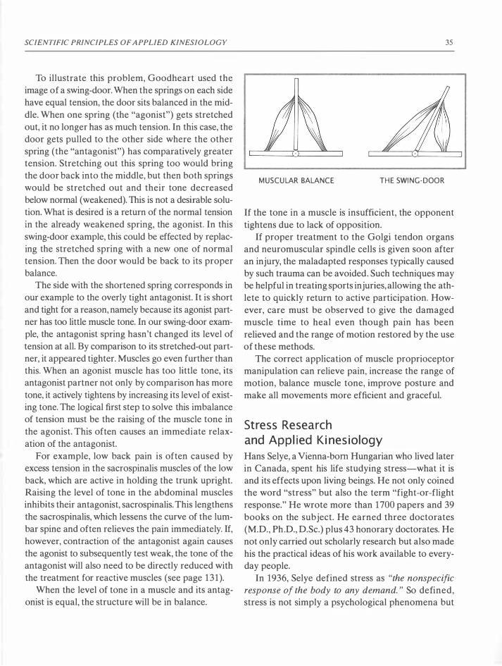
SCIENTIFIC PRINCIPLES OF APPLIED KINESIOLOGY
To illustrate this problem, Goodheart used the
image of a swing-door. When the springs on each side
have equal tension, the door sits balanced in the mid
dle. When one spring (the "agonist") gets stretched
out, it no longer has as much tension. In this case, the
door gets pulled to the other side where the other
spring (the "antagonist") has comparatively greater
tension. Stretching out this spring too would bring
the door back into the middle, but then both springs
would be stretched out and their tone decreased
below normal (weakened). This is not a desirable solu
tion. What is desired is a return of the normal tension
in the already weakened spring, the agonist. In this
swing-door example, this could be effected by replac
ing the stretched spring with a new one of normal
tension. Then the door would be back to its proper
balance.
The side with the shortened spring corresponds in
our example to the overly tight antagonist. It is short
and tight for a reason, namely because its agonist part
ner has too little muscle tone. In our swing-door exam
ple, the antagonist spring hasn't changed its level of
tension at all. By comparison to its stretched-out part
ner, it appeared tighter. Muscles go even further than
this. When an agonist muscle has too little tone, its
antagonist partner not only by comparison has more
tone, it actively tightens by increasing its level of exist
ing tone. The logical first step to solve this imbalance
of tension must be the raising of the muscle tone in
the agonist. This often causes an immediate relax
ation of the antagonist.
For example, low back pain is often caused by
excess tension in the sacrospinalis muscles of the low
back, which are active in holding the trunk upright.
Raising the level of tone in the abdominal muscles
inhibits their antagonist, sacrospinalis. This lengthens
the sacrospinalis, which lessens the curve of the lum
bar spine and often relieves the pain immediately. If,
however, contraction of the antagonist again causes
the agonist to subsequently test weak, the tone of the
antagonist will also need to be directly reduced with
the treatment for reactive muscles (see page 131).
When the level of tone in a muscle and its antag
onist is equal, the structure will be in balance.
35
MUSCULAR BALANCE TH E SWI NG- DOOR
If the tone in a muscle is insufficient, the opponent
tightens due to lack of opposition.
If proper treatment to the Golgi tendon organs
and neuromuscular spindle cells is given soon after
an injury, the maladapted responses typically caused
by such trauma can be avoided. Such techniques may
be helpful in treating sports injuries, allowing the ath
lete to quickly return to active participation. How
ever, care must be observed to give the damaged
muscle time to heal even though pain has been
relieved and the range of motion restored by the use
of these methods.
The correct application of muscle proprioceptor
manipulation can relieve pain, increase the range of
motion, balance muscle tone, improve posture and
make all movements more efficient and graceful.
Stress Research and Applied Kinesiology Hans Selye, a Vienna-born Hungarian who lived later
in Canada, spent his life studying stress-what it is
and its effects upon living beings. He not only coined
the word "stress" but also the term "fight-or-flight
response." He wrote more than 1700 papers and 39
books on the subject. He earned three doctorates
(M.D., Ph.D., D.Sc.) plus 43 honorary doctorates. He
not only carried out scholarly research but also made
his the practical ideas of his work available to every
day people.
In 1936, Selye defined stress as "the nonspecific
response of the body to any demand." So defined,
stress is not simply a psychological phenomena but

36
also includes all processes of response or adaptation.
It encompasses all physical and psychological reac
tions to the demands placed upon the individual by
both his internal and his external environments. With
out stress, life is not possible. Stress is normal and can
not be avoided. Selye indicated this by further dividing
stress into eustress and distress. Eustress is produced
by doing what we like to do and what is good for us.
Distress is elicited by those activities that we do not
like to do but must (Gerz, 1996, pp. 30-33; Heine 1997,
pp. 201-209; Selye, 1952).
The Three-Phase Reaction to Stress
Selye found out that every living being has a three
phase reaction to stress, which he called the general
ized adaptation syndrome (GAS) as follows:
2
THREE· PHASE STRESS REACTIO N
ACCORDING TO SELY E
1 . Alarm reaction:
In this first phase, the reaction of the body to stress
is an "alarm reaction" in which the ability to respond
temporarily decreases. This is a kind of shock reac
tion that is usually of short duration.
2. State of resistance:
If stress continues, extra cortisone and adrenaline are
produced and released increasing the body's ability
to adapt to the stress and improve performance of
the activities required by the stressor. This adapta
tion to the stressor consists, for example, in a sharp
ening of the senses, an increase in the precision of
APPLIED KINESIOLOGY
motor control of the body, increased wakefulness
and alertness, and a high metabolic rate. This results
in increased performance of activities carried out at
this time.
If the stress still does not stop, the quantity of these
chemicals in the bloodstream increases until the body
reaches its peak of ability to react to the stress. Selye
calls this maximal adaptation the "state of resistance."
1bis keeps the organism able to react swiftly and pow
erfully. This is a wise reaction of the body that pro
motes survival in conditions of extended stress. How
long the body can support this process is individual.
3. State of exhaustion:
When this state of resistance continues too long, there
comes what Selye calls the "state of exhaustion."
When more or less completely exhausted, the body
cannot produce the extra chemicals and energy for
further resistance. The reserves are used up. In com
mon parlance, this is referred to as "bum out." In this
state, rest, therapy, good nutrition and a change of
lifestyle are desperately needed for the body (and
mind) to be relieved from stress and to recover from
this exhausted state.
Selye found the following symptoms to be typically
associated with stress:
- recurring infections
- allergies and hay fever
- stomach and other digestive symptoms
- insomnia, irritability, lack of energy, unusual
mental symptoms
- lack of concentration, confusion of thought
- chronic fatigue, depressive tendencies
- trembling, nervous tics, stuttering
- teeth grinding at night, leading to a greater sensi-
tivity to sweet/sour, hot/cold, perhaps problems
with the temporomandibular joint (TMJ)
- diarrhea
- migraine, premenstrual syndrome
- neck and back pain
- too great or too small an appetite
- a strong or even addictive desire for nicotine,
alcohol, coffee or other stimulants/drugs

SCIENTIFIC PRINCIPLES OF APPLIED KINESIOLOGY
- recurring minor injuries or chronic aches and pains
which, in spite of good treatment, refuse to go away
Note that this list includes the most frequent com
plaints of most patients everywhere today. This indi
cates how widespread the problem of stress is.
According to Selye, any animal responds to pro
longed stress with hypertonicity of the skeletal mus
cles. This casts light upon the problem of hypertonic
muscles, which is receiving increased attention in AK
today. In fact, it is fascinating that Selye's concept of
the three stages of stress has such a direct relation to
all three states of reaction of a muscle in muscle test
ing: weak-testing, normotonic, and hypertonic.
Normotonic
A normotonic muscle is the ideal state in which both
negative and positive stimuli are recognized and
reacted to in a natural manner.
Weak-testing as a response to a stimulus ->
Alarm reaction
A normotonic indicator muscle may test weak in
response to TL to a scar or challenge by the oral pres
entation of an allergen. This can be compared to
Selye's alarm reaction of the body in response to a
new stress.
Hypertonic -> State of resistance
A single hypertonic muscle usually indicates a mal
function of a proprioceptor that can usually be cor
rected by proprioceptor manipulation.
A bilateral hypertonic muscle indicates that the
corresponding muscle-organ-meridian circuit is
stressed and is in a stress-adaptation phase.
General hypertonicity of muscles indicates that
the whole system is in a massive stress-adaptation
phase and working "overtime" to deal with the stress.
In this phase, Selye says that rest and recuperation
are the most needed treatment factors.
It is important that the patient correct the prob
lem at this stage before a general state of exhaustion
sets in. Natural healing techniques needed at this stage
include clearing the body of toxins and focal infec-
37
tions, and obtaining exercise, fresh air, water, needed
nutrients, and a change of lifestyle that includes less
emotional stress.
Weak-testing muscles (without TL or challenge) -> State of exhaustion
An individual weak-testing muscle indicates that the
body's ability to adapt to a stress has been exhausted,
but only in the one specific regulating circuit related
to the muscle tested.
If the general hypertonicity condition continues
too long, the result will be a general weakness of most
or all muscles. This corresponds to Selye's state of
exhaustion. In this stage, the body can no longer adapt
to the stress and is "giving up the fight." Typically with
patients in this stage, it is difficult or impossible to
find a single muscle that can be strengthened and used
as an indicator muscle. The same steps listed for gen
eral hypertonicity are required at this stage, but they
need to be applied more carefully and regularly. Most
of all, such patients need rest, quiet, water, warmth,
positive energy and a supporting environment of car
ing family or friends. This problem and its treatment
are considered in detail in the section "General
Hypertonicity," page 93).
Gerz compares the response of muscles to Selye's
three states of stress as follows:
1. A previously strong-testing muscle tests weak =
The stimulus that made the muscle test weak is
greater than the ability of the body to adapt to it.
2. The normotonic muscle stays normotonic = The
adaptation mechanism of the body, with respect
to the stimulus, is adequate.
3. The normotonic muscle becomes hypertonic =
The stimulus creates an alann reaction in the body,
which is expressed as a pathological increase of
muscular tension. This can only take place at the
cost of a "pre-damage"-the memory of a previ
ous stress adaptation. In principle, this is what
occurs in the reaction of the immune system to an
allergen. For the body to have an allergic reaction,
38
it must have been previously exposed to the sub
stance. It "learns" to react in an allergic manner.
Hypertonic reaction is also a "learned response."
In reaction to various kinds and amounts of stress,
the body learns to tighten muscles.
4. A weak-testing muscle becomes normotonic =
The stimulus (challenge) has a positive effect that
enables the body able to improve its neuromus
cular reaction.
5. A weak-testing muscle remains weak-testing =
The challenge was not recognized by the body as
significantly positive.
6. A weak-testing muscle becomes hypertonic = The
body reacts to the challenge with an extreme
alarm reaction. This is also a learned response to
stress as in #3 above. (Gerz, 1996, p. 33).
In experiments with animals, Selye found that enough
stress of any kind led to death. Three particular
organs, the stomach, the thymus, and the adrenals,
were always affected by extreme stress and usually
in the same way each time.
1. The stomach became inflamed and developed
ulcers.
2. The thymus shrunk in size and function.
3. The adrenal glands were filled with fat and their
function reduced.
4. Other organs were affected by specific stressors,
but these three were always affected by stress of
any kind.
Although stress appears to produce similar symptoms
in humans, there is still some controversy as to what
extent these animal findings parallel the human reac
tion to stress. Human medicine widely recognizes the
connection between stress and the stomach. And the
thymus is well known as the main organ of the
immune system (whose response to stress is common
knowledge). Less attention is given to the extremely
common problem of functional weakness of the adre
nal glands. For that reason, this problem will be con
sidered more completely here.
APPLIED KINESIOLOGY
Typical problems associated with adrenal weak
ness include allergies, states of fear, arthritis, chronic
tiredness, colitis, exhaustion, hormonal dysfunction,
hypoglycemia (low blood sugar), dysfunction of the
immune system, learning difficulties, excess weight,
and ulcers of the duodenum. Besides laboratory tests
for the detection of these conditions, there are also
five simple tests for adrenal weakness:
1. The examiner compares blood pressure readings,
first while the patient is lying, then while he is
standing. When standing, the adrenal glands
should release extra noradrenalin to elevate the
blood pressure, ideally about 8 mm Hg (mercury).
This keeps the blood from pooling in the abdomen
and legs. If the lying blood pressure remains
unchanged when the patient stands, or especially
if it sinks, very likely the adrenal glands are weak
ened and are not producing and releasing enough
noradrenaline. In medicine, this reaction is called
the "Ragland sign."
2. A further test for adrenal weakness involves deter
mining the pupil reaction to light. Light in the eyes
should cause the pupils to contract. When the
adrenal glands are weakened, the body loses
sodium and the intercellular potassium increases.
This extra potassium interferes with the function
of the pupil sphincter muscles, which then are
unable to adequately contract. If the pupils widen
immediately when a light is shined upon the eyes
or fail to contract even after about thirty seconds,
an adrenal weakness is indicated.
3. The region where the last rib meets the
sacrospinalis muscle is an adrenal reflex area. Pain
in this area indicates possible problems with the
adrenal glands. This response is known as
"Rogoff's sign."
4. In AK, the sartorius muscle is associated with the
adrenal glands. When the sartorius muscle tests
weak, which it often does when the adrenal glands
are not functioning properly, the ilium subluxates
posteriorly. This "posterior ilium" is a classic sign
of adrenal weakness.
SCIENTIFIC PRINCIPLES OF APPLIED KINESIOLOGY
5. The anterior neurolymphatic reflex points for sar
torius (5 cm superior and 2.5 cm lateral to the
navel) are approximately over the adrenal glands
and are used in AK to therapy-localize the adre
nal glands. A weak sartorius muscle or an active
TL to these points provides an ideal starting point
to determine measures that might help strengthen
the weakened adrenal glands. For a complete dis
cussion of this issue, the reader is directed to the
relevant literature, especially Gerz, Applied Kine
siology; Schmitt, Common Glandular Dysfunc
tions in the General Practice, and Goodheart,
You 'll Be Better.
The stress-related degeneration of these three organs,
the stomach, thymus and adrenal glands, occurs in the
second and third stage of the "Generalized Adapta
tion Syndrome" (GAS). The important point here is
that stress of any type, if intense and constant enough,
will lead to damage and degeneration of these three
organs and eventually to death.
Selye's research led him to the conclusion that to
promote health, the best attitude and lifestyle for all
living beings is "altruistic egotism" (Selye, 1974). He
coined this phrase as an alternative to the nearly
impossible recommendation of Jesus to " love thy
neighbor as thyself." For Selye, altruistic egotism
implies that being benevolent to others promotes self
preservation. This means that the best way to be
assured that others will help you when you need them
is to help them when they need you.
Simplified and summarized, Selye believed that
one should arrange one's life to have as much eustress
and as little distress as possible. His further recom
mendations include:
• Find your own natural stress level.
• Learn the difference between eustress and distress.
• Precisely analyze your problems.
• Don't force and overexert yourself. Rather learn
to know and follow your own need for rest and
relaxation.
• Avoid table salt. It makes the negative effects of
stress worse. Instead use potassium chloride (KCI).
• Clear out all hidden infections such as in teeth,
39
impacted colon, sinuses. Selye noted that these are
a cause of continual stress for the body, even when
no noticeable symptoms exist.
• Avoid one-sided stresses. The body wants to bal
ance one kind of stress like hard physical work
with another kind of stress like reading or making
music.
• Observe your stress quotient with relation to your
specific stress and general stress. When there is too
much specific stress, one needs to get involved in
other kinds of activities. When there is too much
general stress, one needs rest and relaxation in a
quiet non-stressing environment.
Selye's research indicates that stress weakens the
immune system whose job it is to recognize and fight
against undesirable substances in the body. Since
immune system weakness is the root of many of
today's most prevalent and difficult medical prob
lems, a deeper consideration of Selye's concepts by
medical doctors and other health professionals is
indicated.
Applied Kinesiology is an excellent instrument for
diagnosis in the field of immunology. With the tech
niques of AK, the examiner can assess the state of
the immune system and the reactions of the immune
system to various kinds of stimuli. Muscle testing the
body of the patient with the challenge techniques pro
vides the perfect tool for revealing both the individ
ually problematic stress factors and the specific
treatments best suited to the patient. Seen in this light,
Applied Kinesiology is a kind of "applied immunol
ogy" or "applied stressology."
A Change i n Worldview: From Newton ian Physics to Quantum Mechan ics and Chaos Theory
Modern medicine is in a time of great upheaval. The
paradigms that were useful in solving the problem
of infectious diseases in the past do not provide
adequate solutions for the many chronic and degen
erative diseases prevalent today. It appears that a new
way of thinking, a new way of looking at health and

40
disease is required. The modem scientific concepts of
quantum mechanics and chaos theory provide the
needed new viewpoint. Although these new theories
have been demonstrated to more accurately repre
sent reality than the worldviews of the past, in the
field of medicine as in other areas of society, there is
resistance to accepting and applying new ideas.
However, a diverse group of medical doctors,
researchers and biologists has embraced these new
concepts and is using them to develop a new medical
viewpoint. With this new medical "Weltanschauung,"
they have already had ground-breaking success in
providing new understandings and solutions to many
of the medical problems of today. "Biological medi
cine" is the generic term used to describe this new
conceptual field. Unlike classical medicine, biologi
cal medicine no longer follows the Newtonian "cause
and effect" way of thinking. Classical medicine seeks
single causes of illness and fights them. By contrast,
the doctor using biological medicine draws together
and analyzes all the possible factors that could be
causing the health problem and attempts to positively
influence them. Alternative medicine can thus be seen
as an aspect of biological medicine. The therapy meth
ods of biological medicine can be classified as follows
(after Heine, 1997) .
Phytotherapy (therapy using plant products):
1. Homeopathy
2. Traditional Chinese medicine
3. Physiotherapy
Therapy with the goal of specifically affecting the
system of ground regulation (Pischinger):
1. Acupuncture
2. Neural therapy
3. Bioresonance
Therapy to stimulate the system of ground regulation
in a general way:
1. Nutrition
2. Kneipp techniques (external application
of water)
3. Anthroposophic medicine
4. Alternating activity and rest with the goal
of strengthening the powers of recuperation
APPLIED KINESIOLOGY
5. Music and art therapy
6. Psychotherapy; therapy that uses talking as
the medium in general
Detoxification and elimination techniques
1 . Purging and vomiting
2. Bloodletting
3. Sweating methods (exercise, sauna, etc.)
4. Stimulation/irritation of the skin
5. Diuretic (water-removing) interventions
The techniques of classical medicine (surgery,
chemotherapy, pharmacology, the use of artificial
organs, hormone and other biochemical substitution)
seek to fight the apparent, superficial cause of illness.
With such methods, the body has a more or less pas
sive role. When treatment is effective, the body is said
to have been "freed" from the illness. All these meth
ods are oriented toward illness.
By contrast, the therapy procedures of biological
medicine give the body an active role-for example,
in eliminating toxins. They help support and stimu
late the self-healing processes. Special attention is
given to the re-establishment of the natural biological
processes (sleep and waking rhythm, body tempera
ture, digestion, etc.). These methods strengthen the
powers of recuperation and the resistance to illness.
Biological medicine is oriented toward promoting
and maintaining health.
In order to understand this new medical ideology,
the old "classical" worldviews prevalent in medicine
will be reviewed here and contrasted with the new
concepts of quantum and chaos theories (reviewed
in detail later in this chapter, page 43).
ThAomoNAL WORLDVIEWS
It appears that man's concept of reality, his concept
of himself and his immediate environment, is inex
tricably tied to the currently accepted concepts of the
nature of the cosmos, in which he exists. Everything he
thinks occurs within the framework of the currently
believed facts of nature as defined by the popular
thinkers and scientists of his time. In modem times,
the physicists are most responsible for our definitions
of reality. In earlier times, reality was defined by the
metaphysicians and the religious scholars.

SCIENTIFIC PRINCIPLES OF APPLIED KINESIOLOGY
Until the time of the Renaissance, when scientific
inquiry and the growth of secular values blossomed,
man existed within an earth-centered, three-level,
natural universe. Generally, his concept involved a
heaven above the earth, man on the flat surface, and
hell below. Believing this concept of reality, sea explor
ers had a real fear of sailing off the edge of the earth,
which would not only mean their certain death but
also their consequent eternal damnation.
Prior to the Renaissance, scientific thinking was
not based upon the concept of cause and effect as we
understand it today but rather upon Aristotelian logic.
Aristotle (384-322 B.C.) defined four kinds of causes.
If a chair is created, its builder is the efficient cause,
the structure that makes it a chair is its formal cause,
the matter that has been made into the chair is its
material cause and the need it was made to fulfill
(namely, for sitting upon) is its purposeful cause. Thus,
in Aristotle's natural philosophy, all things were con
sidered to have a kind of inherent purpose. According
to this line of reasoning, the cause of rain falling was
believed to be the plants and animals below that
require it to live and grow. The purpose is the cause.
The church chose to make Aristotle's ideas accepted
dogma. This came about in part because his idea of
purposeful cause was easily adapted to justify church
dogma. Thus God created the world so that animals
and humans could live here. And woman was created
to serve man. The church claimed the ultimate author
ity to declare the purposeful causes of all things.
Aristotle believed that when one is familiar with
a subject, its governing principles become evident to
the faculty of reason. For example, Aristotle stated
that a body with twice the weight, when lifted and
released in the air, would fall twice as fast. This was
logical and therefore considered true. In Aristotle's
time, it was not yet common to test the veracity of a
logically deduced idea. Familiarity with the topic plus
reason were considered adequate tools for the deter
mination of truth.
With the spread of Christianity, the (Catholic)
church attained increasing influence upon scientific
thinking and research. The church adopted Aristo
tle's concepts, such as those about weight and grav-
41
ity and declared them to be correct. The church was
placed in a position of great consternation and embar
rassment when Galileo Galilei (1564-1642) made his
famous experiment (-1604) of dropping iron balls of
various weights from the tower of Pisa. All the balls
fell at the same rate! The mainly Catholic witnesses
were put into a true moral conflict. They could not
believe their eyes or they would become heretics, and
likely subjected to torture by the Inquisition. Even
worse, they would most certainly be excluded from
paradise and suffer eternal damnation. So, they chose
not to believe their eyes, and to consider that what
they saw was unreal.
In his De revolutionibus orbium caelestium libri VI
(Six books about the revolutions of the heavenly
spheres, 1543) Nicolaus Copernicus was the first to
publicly question the idea of the geocentric cosmology
accepted since the time of Aristotle. The Danish
astronomer, IYcho Brahe (1546-1601 ), produced the
most accurate measurements of the planets ever
obtained without a telescope. In 1572 he observed a
new star in the constellation of Cassiopeia. In 1573,
he published his first work, proving that the observed
object was a star beyond the moon's orbit. This
revealed a flaw in the Aristotelian world view of the
immutability of the heavens. Although Brahe, unlike
his colleague Johannes Kepler (1571-1630), did not
believe in the heliocentric theories of Copernicus, he
invited Kepler in 1600 to stay with him in Prague
where Brahe was the astronomer and mathematician
at the court of Emperor Rudolph II . Brahe was
impressed with Kepler's work. One year later, Brahe
died and Kepler inherited his scientific legacy, includ
ing Brahe's planetary observations. Kepler studied
these and proved that the orbit of Mars is an ellipse
with the sun as one of its foci. This became Kepler's
first law of planetary motion. His second law con
cerned planetary velocity, and his third law defined a
relationship between the orbital periods and the dis
tances of the planets from the sun. His work clearly
demonstrated that the planets orbit around the sun.
His belief that the sun governs planetary velocity
formed the foundation for Isaac Newton's theory of
universal gravitation.

42
In 1609, Galileo learned of the newly invented
Dutch telescope and in the same year constructed
one with 20-power magnification and turned it upon
the heavens. There he saw mountains upon the moon,
the starry nature of the Milky Way, and some kind of
"planets" orbiting Jupiter. His discoveries again ran
against accepted church dogma. He argued for free
dom of inquiry, stating that sensory evidence and
mathematical proofs should not be subject to inter
pretations of scripture. The church had another idea
in these matters and declared him a heretic, impris
oned him (soon commuted to house arrest) and
banned all of his books in Italy. However, outside
Italy, his ideas were pursued eagerly.
Isaac Newton (1643-1727) is often revered as the
greatest scientific genius of all. During his first two
years at Trinity College at Cambridge (1661-1662),
students were only allowed to learn the philosophy
of Aristotle. However, during his third year, some
freedom of choice was instituted and Newton stud
ied mechanics, algebra, analytical geometry and
Copernican astronomy. When the plague closed the
University in 1665, he returned home to Lin
colnshire, England, where he swiftly made revolu
tionary advances in physics, optics, mathematics and
astronomy.
Newton reasoned that planetary orbits are the
result of two forces: a centrifugal force which drives
the body away from the center, and a centripetal force
which draws the body toward the center. He imag
ined that the force of the earth's gravity extended to
the moon, providing the centripetal force. Using
Kepler's third law of planetary motion, he deduced
that the force of gravity between any two bodies must
decrease with the inverse square of the distance. Thus
if the distance between the bodies is doubled, the
force of gravitation is one-fourth as much. If the dis
tance is tripled, the force is one-ninth as much. New
ton was the first to mathematically prove that if a
body obeys Kepler's second law, which states that a
line connecting a planet to the sun sweeps over equal
areas in equal times, then the force of gravity must
obey the inverse square law. Newton published his
APPLIED KINESIOLOGY
Philosophiae naturalis principia mathematica (The
Mathematical Principles of Natural Philosophy or
"Principia") in 1687. The Principia is universally rec
ognized as the greatest scientific book ever written.
Using his law of gravitation and the laws of motion,
Newton was able to explain a wide variety of phe
nomena including the eccentric orbits of comets, the
tides, the effect of the sun's gravitation in producing
variations in the orbit of the moon, the motion of pro
jectiles, falling bodies and pendula. Thus the New
tonian law of gravitation and system of mechanics
was able to explain, within the accuracy of existing
measurements, most of the observed phenomena of
nature. The divergent works of Copernicus, Galileo
and Kepler were united into one coherent whole. The
Copernican world view of heliocentricity at last had
a firm physical and mathematical basis. Thus Newton
completed the scientific revolution of his times and
formed the content of modern science. By looking
out into and analyzing the motions of the planets of
our solar system and starry universe beyond, a new
worldview was created which is still influential today.
As the concepts of Newtonian mechanics pre
vailed and became the accepted scientific belief, the
model of the three-level universe was no longer con
vincing and lost its validity. Gradually the general
populace came to accept the principles of Newton
ian physics, which established that rather than being
the center of the universe, the earth is but a mere
speck in the total order of things. In this new world
view, the earth is just one planet rotating around our
sun. And our sun is only one of a seemingly endless
number of other stars in our universe. The whole
worldview of reality changed. And with it, the power
of the church over the minds of humanity decreased
greatly. Freedom of thought and scientific inquiry,
requested by Galileo but denied him, became the
ideal of the new age.
Perhaps most importantly, Newton experimen
tally demonstrated the relationship between cause
and effect and postulated this concept to be the basic
defining principle of the universe. Today this princi
ple is seldom questioned. It is, for us, a priori-

SCIENTIFIC PRINCIPLES OFAPPLIED KINESIOLOGY
unquestionably true. In fact, it is quite difficult for
us to conceive of any other way of thinking. In the
still-prevalent Newtonian thinking of our times, the
universe is considered by many to be absolutely
ordered and all phenomena defined by clear, logi
cal, mathematical laws. Anything that does not
appear to follow these laws is considered to be illu
sion and unreality. Chaos and uncertainty were not
only ruled out, as long as Newtonian thinking ruled,
they were not even conceived of as possible. As a
new worldview becomes popular, even the unedu
cated often wonder how prior generations could have
been so narrow-minded as to accept the old concept
of reality.
QUANTUM AND CHAOS ThEORIES
In very recent times, physicists looking within the
atom found both a firm natural order and a totally
unanticipated chaos and uncertainty. In order to
express and work with these new ideas mathemati
cally, scientists developed the uncertainty principle
coupled with the science of quantum mechanics and
chaos theory. Curiously, as the new conception of the
nature of the universe began to emerge, reality again
was defined a new type of three-level universe. The
first layer consists of those aspects of our everyday
world that may be accurately described by Newtonian
physics. The third level consists of all objects and phe
nomena that are best described by chaos theory.
Located between these two levels is the area of rel
ative uncertainty (determined chaos) within which
all the phenomena of life exist. Within this new and
still emerging worldview, it is believed that life can
not exist either in rigid form or in chaos. Life can only
exist within defined limits, between strict rules of struc
ture and utter chaos.
With this new definition of reality, our worldview
has once again drastically changed. In this new light,
people are beginning to review their assumptions,
concepts, and even their prejudices. Many find it hard
to give up the comfortable certainty of Newtonian
mechanics, but forward thinkers everywhere are
adopting and adapting to the new concepts. Expres-
43
sion of the new worldview may be seen in various sci
ences, art and music. Chaos theory has even been suc
cessfully applied in predicting trends in the fields of
economics and business. With the framework of these
new concepts, repeatable but previously unexplain
able phenomena in the field of alternative medicine
are receiving a strong theoretical basis. It took sev
eral centuries after the Renaissance before the estab
lished dogmas gave way and the new concepts of
Newtonian physics were generally accepted. Today,
it may take decades before the general consensus of
the established medical profession, which is still
greatly dominated by the Newtonian concepts of
cause and effect, accepts those facets of alternative
medicine that are now being explained and proved
in the light of quantum and chaos theories.
In the AK literature, one can see that explanations
of muscle testing have been limited to the most widely
accepted medical concepts of physiology and neuro
physiology. However, within the generally accepted
medical model, many of the observed phenomena of
AK simply cannot be explained. Bioresonance and
multi-resonance therapy systems such as electro
acupuncture (Voll) are used by some in AK. Exam
iners using AK often test subtle energy substances
such as homeopathic remedies, Bach Flower Reme
dies and gemstones held in the hand. An explanation
of how the above methods could function does not
exist within the models of classical medicine. Expla
nations and verifications, however, are now being
developed in terms of quantum mechanics and chaos
theory.
For both diagnosis and treatment, examiners using
AK often utilize systems of reflex that have no, or
only tenuous, basis in conventional anatomy. Exam
ples of such reflex systems include neurolymphatic
reflexes, neurovascular reflexes, hand and foot reflexes
(reflexology), cranial stress receptors, the meridian
system, and ear acupuncture. 10chen Gleditsch in his
book, Mundakupunktur, has demonstrated that such
reflex areas exist all over the body. According to his
research, one can affect any chosen part of the body
by stimulating reflex points located more or less upon

44
every other part of the body. In all of these systems,
remote areas are stimulated to produce an effect in
a target organ or group of organs. Although attempts
and some progress have been made, all efforts to
explain these phenomena using the models of classi
cal medicine have had limited success.
In this difficult situation, and as outlined above, a
review of the evolution in mathematics and physics
over the past century may provide a path-finder for
understanding the new thinking in medicine. During
that time, most of the hallowed Newtonian concepts
gave way to Einsteinian theories, and particularly to
quantum mechanics and chaos theory. In order to
provide an explanation of the peculiar, very useful,
and hitherto unexplainable AK phenomena, classic
scientific and especially medical thinking will now
be contrasted with quantum mechanics and chaos
theory.
A COMPARISON BETWEEN ThADmONAL
AND MODERN MODELS OF REALITY
Classical linear cause-and-effect thinking developed
during the Renaissance and ascended to its peak in
Newtonian mechanics. The "one disease-one cure"
concept still dominant in medicine today is a reflection
of this thinking. In the classical scientific model (and
also in classical medicine), a separation between the
observer and the observed phenomena is assumed.
In this model, it is believed that separate objects act
one upon one another without any influence being
caused by the act of observation. The basic premise
of this viewpoint is that one object can cause an effect
upon another object, fully independent of the
observer.
In modem quantum theory, it has been determined
that independent observers (those who have no effect
upon that which is observed) do not exist. That which
is sought, and the act of seeking itself, have an influ
ence upon what is found. In physics, the validity of
this principle was clearly demonstrated by the wave
particle duality of electrons. It was not known if elec
trons were waves or particles. In an experiment to
detect waves, electrons behaved like waves. In another
APPLIED KINESIOLOGY
experiment to detect particles, electrons behaved like
particles. So which are they? These experiments
demonstrated that electrons appear to be what the
experimenter is looking for and measuring! The act
of observation, more than any inherent reality, deter
mines how they appear. A philosophical expression
of this idea is reflected in the popular saying, "If you
are looking for trouble, you will probably find it."
In another experiment, researchers were presented
with two groups of rats. They were told that one group
was bred for increased intelligence and that the other
group was rather stupid. The researchers were asked
to test each group of rats to see how much faster the
intelligent rats learned to run through a labyrinth. As
expected, the intelligent rats did indeed learn signif
icantly faster to run through the maze. Then came the
surprise: All the rats from both groups were geneti
cally from the same stock. The only difference was
the expectations of the researchers. In a similar exper
iment, school teachers were told that certain students
were especially intelligent. And although the students
were of only average intelligence, they did excel in
their courses when their teachers expected them to
excel. These surprising results, which are in perfect
agreement with quantum theory, cast doubt upon the
accuracy of much of the scientific research performed
throughout history. These examples imply that in this
universe of ours, there is a tendency to find what you
are looking for!
Another basic premise of quantum theory is the
"superposition principle," which states that every
thing is related to and connected with everything
else. In a universe such as this implies, it is impossible
to make any definite statement about cause and
effect. In order to make some sense of observed phe
nomena, it is necessary to isolate and abstract them
from the all-encompassing quantum world into the
special, classical world of time and space. Then the
system researched has individuality, measurements
can be taken, and statements about its nature can be
made. But any such measurements and statements
about the "reality" of the system researched are
dependent upon the point of reference, the context of

SCIENTIFIC PRINCIPLES OF APPLIED KINESIOLOGY
the investigations and the expectations of the
researchers themselves.
Although quantum theory has been accepted by
most branches of science, many in the world of med
icine cling to the cause-effect mentality defined by
Newtonian mechanics. Classical medicine still bases its
diagnostic and treatment techniques upon the con
cept of the lock and key, in which the lock is a disease
and the key is a medicine or medical technique (chem
ical medicines, surgery, etc.) . This linear thinking
model implies that each part of the body and indeed
each cell is an independent entity that may be treated
for its independent disturbances. However, in a multi
cellular being such as the human body, cells do not
exist individually. Historically, the abstract concept
of the separate cell and its pathologies (Virchow, 1858)
led to the linear idea of "for one disease, one specific
cure." Within this thinking, the individual phenom
ena one experiences when ill are ignored in favor of
defined models of illness. Symptoms are quantified
and used as indicators of specific syndromes. If the
values of a certain test are above a defined level, you
have the syndrome. After effective treatment, the val
ues must again fall below the defined level. An effec
tive treatment within this perspective means
"normalizing" the values-bringing them back into
the accepted range of tolerance. At its worst, this sci
entific dogma leads to treating the model of the dis
ease instead of the patient.
For example, a patient had breast cancer that had
spread to many other areas of her body. In May, after
surgery to the breast and chemotherapy, she was told
to prepare for death before September. Her T-lym
phocyte cell count was 1 8/�1. The normal level is
defined as being between 1000 and 3500/�1. She
enjoyed running and continued to run 17 kilometers
each day. In February, she was still alive and her T
cell count had increased to 76/�1. The doctors said
that this increase was too small to have any signifi
cance whatsoever. The idea that her regular exercise
might be keeping her alive and improving her health
was not even considered. She was told that the
increase in the number ofT cells had no meaning and
45
not to allow it to produce unrealistic hopes, because
she would soon be dead.
This type of medical viewpoint automatically places
limits upon which possible therapeutic efforts may
even be considered. By only considering lab test val
ues, doctors may diagnose and prescribe swiftly and
thereby see more patients per day. The problem is
that in this simplistic model, there is no place for indi
vidual variation. There is no room in this thinking for
the fact that the same symptoms may be produced
by different causes. And there is no place for the fact
that the same medicine may produce different effects
in two individual patients, even though they have the
same presenting symptoms. More attention is given
to the numerical results of medical tests than to how
the patient is feeling or the causes of her symptoms
viewed within her personal biology/constitution, or
considered with reference to the quality of her
lifestyle.
In acute illnesses where physical damage to tissues
or infection by a micro-organism is the main cause,
traditional medical diagnosis and treatment are
extremely effective. In most acute illnesses, one spe
cific cause outweighs all the others. In such cases, the
"one disease-one treatment" mentality is usually suc
cessful. Doctors using this method have developed
effective treatments for most acute medical problems.
Because of these great successes, the world is largely
free from many of the complaints that were often a
cause of death in the past. Today the situation has
changed. The most common medical complaints are
no longer acute "diseases," but are instead tumors
and other chronic degenerative illnesses. And tradi
tional medical thinking is at a loss in dealing with
these problems.
Because people today seldom die young from
injuries, infections and infectious diseases, the aver
age age of death has risen significantly. Bodily func
tions and the ability to recover and regenerate
decrease with increasing years. Old people typically
die from degenerative diseases. That there are more
older people is one reason degenerative diseases are
more prevalent today. But the fact that many more

46
young people suffer from degenerative diseases today
than in years past indicates that there are other causes
besides increasing age.
Thinking along the lines of the lock-and-key model,
the doctor prescribes a chemical medicine to kill bac
teria, or in the case of a viral infection, a chemical that
inhibits viral reproduction. But medical research has
revealed that many of the disease-producing bacte
ria and viruses live upon and within our bodies con
stantly. Often, the question is not asked as to why the
body was susceptible at this particular time to infec
tion. To even be aware that such a question exists, the
doctor needs to expand his or her thinking from the
classical Newtonian mechanics style of thinking that
typically dominates the field of medicine to the more
expansive, multi-dimensional concepts of modern
quantum theory and chaos theory.
Anyone who has had a garden has observed that
insects prefer to infest plants that are already weak.
Similarly, anyone who has observed their own chang
ing state of health knows that they are more likely to
succumb to the ever-present cold viruses when they
are exhausted, chilled, hungry, emotionally upset or
otherwise excessively stressed. When the body is
under stress, the bacteria that normally live on the
skin can more easily infect a cut.
Seen in this light, it seems likely that taking med
icines to combat an infecting agent, although tem
porarily successful against the acute disease, may be
ignoring the multiple other causes of why the person
got ill in the first place. And evidence indicates that,
if not corrected, these multiple stresses may cause a
repeat attack or eventually even result in a degener
ative disease.
Furthermore, it is well known that those who are
prone to infections and get sick a few times each year
have less probability of developing our most preva
lent disease, cancer. However, this benefit is lost if
they take medicines that prevent their immune sys
tem from building strength by performing its natural
fight against the infecting agents. Research strongly
indicates that the immune system needs to battle dis
eases from time to time to keep in shape. When infec-
APPLIED KINESIOLOGY
tious diseases are severe, medicines may save lives.
But their use for every little infectious disease that
occurs may actually be one of the causes of severe
diseases such as cancer!
Newtonian mechanics defines physical systems as
functioning in a linear manner. An example of a lin
ear system is a single pendulum. Its oscillating behav
ior is independent of the influence of any separate
objects in its environment and is relatively inde
pendent of the effect of the observer. This is similar
to the medical model of the single, isolated cell dis
cussed above.
However, the concepts of cybernetics (Wiener,
1963), chaos theory and thermodynamic open sys
tems of energy (Prigogine, 1979) have demonstrated
that biological systems are non-linear. A simple exam
ple of a non-linear system is many different pendula
linked together with springs. When one is moved, the
energy dissipates first into the nearby and then into
the more distant pendula and then back again until
all are moving together in harmonic resonance. Like
these coupled pendula, most structures within non
linear systems are self-repetitive. The same patterns of
structure and motion are seen on many levels. This
principle is observed throughout nature.
In the eighteenth century, Ernst Chaldni placed
sand upon a thin plate of glass and made the plate
resonate by stroking it with the bow of a violin. The
sand vibrated into beautiful repetitive geometric fig
ures across the surface (Chaldnian figures) . Chaldni
publicized his discoveries in his book, Entdeckungen
iiber die Theorie des Klanges, (Leipzig, 1787). The
Swiss scientist, Hans Jenny, spent ten years investi
gating the power of sound to form geometric patterns
in various inorganic substances. The patterns pro
duced by sound vibration in his experiments look like
starfish, bacteria, organs and other patterns seen in
forms of life. In Cymatics (1974), he concluded that
where organization is concerned, the harmonic fig
ures of physics are essentially similar to the harmonic
patterns of organic nature.
These patterns of physics are seen in all living sys
tems. Their principal characteristic is redundance, i.e.,

SCIENTIFIC PRINCIPLES OF APPLIED KINESIOLOGY
the same patterns repeat again and again on many
levels until the available space is filled. For example,
in the circulatory system each artery branches again
and again into smaller and smaller, but in form essen
tially identical, arterioles and capillaries. The lung pro
vides a similar example. The lung is basically a bag of
air that is subdivided into smaller and smaller bags
of air. This redundancy principle may be seen also in
the form of most organs, which are segmented with
each segment also segmented and so on until each
sub-segment is completely filled with nearly identi
cal cells.
FRACTAL GEOMETRY
Mathematics recognizes such redundant systems of
organization in which the same patterns appear
repeatedly on various levels of complexity until the
available space is filled. In mathematics, these geo
metric structures are called "broken dimensions" or
"fractals." Fractal geometry was developed by the
Polish-born French mathematician Benoit Mandel
brot. He coined the word "fractal" from the Latin
verb frangere, "to break," and the related adjective
DIAGRAM OF A BASIC MANDELBROT SET
47
fractus, "irregular and fragmented." Fractal geome
try deals with structures that repeat on finer and finer
scales (Mandelbrot, 1991). Fractal geometric figures
are self-similar. This means that when enlarged, their
parts are identical with the whole. Fractals are not
smooth figures like the curves and circles that exist
in Euclidean geometry. They instead have a step-wise,
jagged quality.
Mathematical fractals are perfectly symmetrical
and remain geometrically identical on any level of
magnification. They exist between strict order and
chaos in the realm of "determined chaos." If the def
inition of fractals is expanded a bit to include some
non-linear qualities (some deviation from mathe
matical perfection), such geometric forms can be
found in nearly every natural phenomenon. Outside
certain defining limits, forms may be very rough (but
not self-similar) or very smooth-thus defining them
as not fractal but rather Euclidean. As already
described, life can only exist in an area between chaos
and structure. Too rough is too chaotic. Too smooth
is too structured. Within these limits, virtually every
thing is fractal. For example, almost any-sized piece
MAGNIFIED TOP SECTION OF THE FIRST DIAGRAM

48
DIAGRAM OF A J ULIA SET
of cauliflower looks much like a whole head of cau
liflower. The pattern "cauliflower" is clearly present
on all these levels of observation. Thus the design of
cauliflower is fractal. Fractals are an essential part of
the mathematics of all natural phenomena, including
life.
Fractal geometry has been used to understand dis
order (chaos) in natural systems. In 1961, Mandelbrot
applied his fractal theories successfully to turbulence
in moving fluids, the distribution of galaxies, and even
to predicting fluctuations of the stock market. In 1967,
he showed that the irregular shorelines of the Eng
lish coast are fractal.
Tho American mathematicians, John Hubbard and
Adrien Douady, developed the most known and use
ful set of non-linear fractals and named it after Man
delbrot. The more the bud-like geometrical figures
derived from this set are magnified, the more the
unpredictability increases. The Mandelbrot set is cen
tral to the science of dynamic systems.
Note that whereas the first figure of a Mandelbrot
Set has one single line extending from it, in the mag-
APPLIED KINESIOLOGY
MAGNIFICATION OF THE TOP PORTION
nified figure the line divides into two lines of unequal
length. In further magnifications, the amount of such
unpredictable changes continues to increase.
The French mathematician, Gaston Julia, used the
Mandelbrot set to develop a mathematics of non-lin
ear transformations in a complex plane. The "Julia
sets" he devised are used to produce computer
graphic images that often strikingly resemble natu
ral forms.
Note that when the top portion of the first figure
is magnified, nearly identical geometrical figures are
revealed. This is the nature of fractals, within which
the same figures can be found on many levels of com
plexity.
The chaos theory of mathematical physics uses
fractals to describe the routes a dynamic system takes
from order to chaos. A chaotic system is sensitive to
initial conditions. A slight uncertainty in the begin
ning develops into greater and greater unpredictability
(chaos) over time. In all chaotic systems, a tiny change
in the beginning produces great effects later. Chaotic
systems include all living forms, ecosystems, social

SCIENTIFIC PRINCIPLES OF APPLIED KINESIOLOGY
systems and the universe as a whole including the
atomic, geological and astronomic levels.
On the atomic level, fractals and chaos theory have
contributed to the understanding of chemical reac
tions, wave motion and electrical currents in semi
conductors. Geological fractals exist both in the
external structures of the earth and also in internal
structures such as faults. Earthquakes occur in a frac
tal pattern across the surface of the earth. Earth
quakes under a magnitude of 6 occur in self-similar
(fractal) clusters over time. Rain falls in a fractal pat
tern over time. In biology, the temperature-related
shape of proteins is fractal. In astrophysics, fractal
patterns have been observed in the pulsations of vari
able stars (stars whose observed intensity of light vary
over time). The pattern of atoms on the surface of a
protein molecule is fractally distributed. And since
proteins are the basic chemical building blocks of all
biological systems, the patterns of living structures
are fractal.
Biological systems are many-sided, complex and
highly interconnected. They can take in energy (food,
light) and use it to more highly organize themselves.
They are interconnected in a complex manner and
have feedback systems that allow them flexibility to
react to the situation of the moment in spontaneous
ways. They can optimize their behavior. Biological
systems, like all non-linear systems, exist within nat
ural laws but tend toward a chaotic state ("deter
mined chaos") . They exist in a dynamic balance
between utter chaos and rigid order in both struc
ture and function. Too much order (as in a crystal)
or too much chaos means death, i .e . , destruction
of the system. In the determined chaos of biological
systems, small changes in the current state of the sys
tem can produce large future changes. This fact pro
vides a possible explanation for the effectiveness of
such healing treatments as acupuncture or homeop
athy in which tiny stimuli may produce system-wide
changes that cannot be explained within traditional
medical models.
In a complex interconnected system with great
redundance such as within a specific organ with mil-
49
lions of essentially identical cells, many parts can be
destroyed without reducing the quality of the func
tion of the organ. The quantity of the chemicals pro
duced or the other functions of the organ will be
reduced but it will essentially function as before. This
redundant quality of non-linear systems is an impor
tant factor for survival.
HOLOGRAMS
It is interesting to note that a hologram demonstrates
characteristics similar to those of life. In a hologram
as in a living being, "the whole information is pres
ent and repeated in all parts" and "portions of parts
may be removed without disturbing the function qual
itatively." These attributes of a hologram are in har
mony with the superposition principle that states that
everything exists in relation to and has an effect upon
everything else.
A visual hologram is produced on photographic
film by utilizing one laser beam of light divided into
two. One of these beams goes directly to the photo
film. The other one bounces off the object to be pho
tographed and then onto the film. The two light beams
interact and form interference patterns over the sur
face of the film. When that film image is viewed by
ordinary light, there is no recognizable image. But
when a laser beam is directed toward the film, a three
dimensional image of the item photographed appears
in the air above the film.
In such a visual hologram, a complete three-dimen
sional picture is present in all parts of the film. Cut a
small corner of the film and the whole picture is still
present, though in reduced clarity. This may be com
pared to an organ in which many cells may be
destroyed but the organ still performs the same activ
ity with no change in quality, only a reduction in the
quantity of activity. A hologram is clearly a quantum
mechanical phenomenon in which all parts are inter
connected and the whole is present in the parts.
There is much evidence to suggest that the brain
stores information holographically, that is, that each
specific bit of information is replicated over wide areas
of the brain. Rats that have learned to run a maze still

50
remember how to do so when up to 50% of their brain
cortex has been removed. And it doesn't matter which
50% ! This implies that the learning is stored all over
the brain like visual information in a hologram.
Holograms can be produced with almost any
source of energy, not only light. In producing a holo
graph, there are always two reference sources of
energy. The bilateral structure of the human body
may provide similar energy pairs for the formation
of holographs within the nervous system. Evidence
indicates that the nervous system functions holo
graphically with respect to sensory and motor sys
tems. Perhaps this is why there are two hemispheres
of the brain that each receive signals from two eyes
and two ears. These separate pairs of images likely
are the two references necessary to form holographic
sensory images in the brain.
If the nervous and organ systems function holo
graphically, then it is logical that one may act upon
one part and produce effects on any other part. This
is the concept underlying the many systems of reflex
therapy.
When two similar but not identical holograms are
placed one upon the other in laser light, interference
patterns appear. These are called "Newton rings."
Goodheart believes that something similar occurs in
the human body. Goodheart proposed that the brain
has a perfect holographic image of all the parts of the
body. When the local hologram in one part of the
body (for example, an injured knee) does not corre
spond accurately with the perfect hologram of the
knee in the brain, the brain is alerted that something
is wrong. Symptoms then occur, and the healing
process is initiated (Goodheart, 1986; Walther 1988,
p. 26).
The many similarities between human functioning
and the properties of the hologram suggest that many
of the structures and functions of the human body
have holographic qualities and may be accurately
described and analyzed within the models of quan
tum theory.
One advantage of a non-linear system is that when
it is disturbed and moved out of its normal position
APPLIED KINESIOLOGY
or activity, it quickly returns to its basic position or
rhythm. In chaos theory, this quality of a non-linear
system is called an attractor. Biological systems have
attractor activities that repeat on a constant time cycle.
Under normal conditions, they can react and adapt
to small disturbances and swiftly return to their basic
rhythm. Examples of such rhythms are heartbeat, res
piration, temperature, permeability and metabolic
rate. In chronic illnesses and especially in tumor tis
sues, such activities no longer occur rhythmically. If
these rhythms cannot be reestablished, the progno
sis for cure is poor. For example, the temperature of
the surface of the skin changes regularly with a
24-hour cycle. However, changes in the temperature
of the skin over a tumor have no regularity. Thus
chronic illnesses are marked by a loss of natural rhyth
mic periodicity in the affected tissues. Therapeutic
efforts that return normal periodicity to diseased tis
sues are especially important and effective in restor
ing health.
Under normal physiological circumstances, the
homeostasis of an organism is extremely stable. Outer
and inner stressful influences are quickly adapted to
and the system returns to balance. For example, if
adequate food is not available, the system will adapt
to the situation in a reversible way. Minerals may be
removed from the bones to replace those not cur
rently in the diet. If this process goes far enough, there
will be noticeable symptoms. Later, when the miss
ing food elements are provided, the system can return
to its prior state of balance. This exemplifies the inter
connectedness and redundance of non-linear systems.
However, when a biological system is sufficiently
stressed, it may not spontaneously return to balance.
Once such an imbalance becomes established, small
further disturbances of various types are not auto
matically corrected but rather may resonate with and
thereby add their weight to the existing imbalances.
When the combination of imbalances stresses the sys
tem enough, illness is the result. Seen in this light, ill
nesses are recognized to be system-wide disturbances
that are often the result of many different causes.
The inferences of quantum mechanics concerning

SCIENTIFIC PRINCIPLES OF APPLIED KINESIOLOGY
the state of determined chaos of living organisms
allow us to understand nature more deeply and accu
rately than the classical models of cause and effect.
The possibly multiple causes of a patient's disease
may each be considered within their own individual
contexts. Internal and external factors (bio-chemical
individuality, diet, posture, exercise, weather, social
situation, the effect of prior experiences, expectation,
occupational stress, etc.) may be analyzed for their
contributing part in health and disease. The attention
of the patient is focused upon his personal responsi
bility in promoting or combating disease. The patient
is helped to help himself. Both the patient and his
physician are operating from a far more complicated
multi-dimensional picture of the processes of health
and disease. Because this picture more accurately rep
resents reality, it provides a better basis for both an
evaluation of the totality of the health situation and
for the application of the wider spectrum of thera
peutic efforts needed to solve health problems, par
ticularly those of chronic and degenerative diseases.
These "new worldview" ideas are being pursued today
by medical doctors and other scientists in the emerg
ing field of "biological medicine."
Biological Med icine and the Systems of Regulation This section is for the advanced student who desires
to better understand the biochemical and electrical
aspects of health and disease. Although an attempt
has been made throughout this book to define every
new term used, in this chapter I won't even try. The
complete definitions would more than double its size.
Obviously only a most superficial view is possible
given the small amount of space that can be devoted
here to this huge field. By giving an overview of this
fascinating world of knowledge I hope to inspire my
readers to inquire further on their own. Because so
much of the new knowledge has been recently gath
ered, I urge you to read only the newest standard text
books of anatomy, physiology, neurology and
functional histology. For the advanced student with
51
a good understanding of the German language, I can
highly recommend the best single textbook I have
found on this topic: Hartmut Heine'S, Lehrbuch der
biologischen Medizin.
In the new medical discipline of "biological med
icine," attention is given in both diagnosis and treat
ment to almost every factor that may make an
individual ill.
Modern medicine conceives the gene to be one of
the controllers, or the direct controller of biological
functioning. The contrary view of biological medicine
is that:
1. the gene is the repository of blueprints of instruc
tions on how cells and higher systems should oper
ate, not a direct activator, and
2. the actual regulation of biological activities in cells
and all higher levels of organization lies in a so
called "system of ground regulation."
The tissues of higher life forms are built of three basic
structures; the capillary, the ground substance or
matrix, and the cell. The basic structure of the system
of ground regulation is the ground substance. The
ground substance is a complex connective tissue that
lies between all the cells of the body. It consists of two
groups of components, the amorphous ground sub
stance and the structural ground substance. The amor
phous ground substance is a transparent, half-fluid
gel produced and sustained by the fibroblast cells of
the connective tissues. It consists of highly polymer
ized sugar-protein complexes. One of its main fami
lies of components is the glycosaminoglycanes.
Glycosaminoglycanes are bound to proteins in the
tissues, forming huge chains (polymers) with a molec
ular weight of several million. They do not bend
around into a circle like most other proteins but rather
lie flat and take up lots more room than other mole
cules of the same molecular weight. On their flat sides
they bind large amounts of water and positively
charged ions (especially sodium), which assists the
amorphous ground substance to become viscous and
gives the structural ground substance turgor (fluid
pressure) or even hardness. They twist together and

52
partially form the permeability barrier around cells.
The structural strength of the ground substance is
also provided by various kinds of fibers. The struc
tural ground substance consists mainly of bendable
reticulin fibers that can mature into inelastic colla
gen, stretchable elastin fibers, and the recently dis
covered structural glycoproteins fibronectin and
laminin that are a part of cell surface membranes. The
components of the structural ground substance form
other types of connective tissues such as those of fas
cia, tendons, ligaments and cartilage as well as fat tis
sues, veins, arteries, lymphatic vessels, bones and teeth.
These other types of connective tissues are durable,
strong and long-lasting. They represent a kind of
"long-term memory" of the ground substance.
The smallest structural element of the ground sub
stance is the matrisome, which has a polygon network
structure formed of various glycoproteins and tran
sitorily bound proteins. The matrisome structure is
arranged in many self-repetitive layers. Each layer is
slightly rotated the same amount with respect to the
next layer. We will return to the matrisome again later
in this section.
The ground substance surrounds and interconnects
the cells. It acts as a molecular sieve, determining what
chemicals enter and exit the cells. The maintenance
of homeostasis in the cells requires that the ground
substance reacts swiftly and precisely to complex
changes. This is made possible by the diversity of
molecular structures of the sugar polymers of the
ground substance, the ability to swiftly generate new
such substances, and their high interconnectedness.
This creates a redundance that makes possible the
controlled oscillation of values above and below the
dynamic homeostasis present in all living creatures.
This is a kind of fast-responding, "short-term mem
ory" of the ground substance. Without this capacity,
the system would quickly move to an energetic equi
librium, which would bring inactivity and death.
The ground substance contains a network of
fibrous and non-fibrous connective tissue paths
through which interactive information can flow. Such
information flows into the ground substance as chem-
APPLIED KINESIOLOGY
ical, electrical and electromagnetic signals from blood,
lymph, nerves, glands, etc., plus formatted informa
tion on proper "how to's" from the genes into the
ground substance; then from the ground substance to
the cells defining "what" to do. This interchange of
information over the interactive network of the
ground substance determines the reactions to internal
and environmental influences. These stereotyped
responses form the ever-repeating and yet ever-indi
vidual patterns of metabolism, development, growth,
repair and behavior, etc.
Only about 2 % of all illnesses are produced by the
functioning or malfunctioning of a single genetic ele
ment. Biological medicine contends that most of the
other diseases are produced by a combination of fac
tors that interfere with the processes of the system of
ground regulation. The system of ground regulation
controls the relationship between cells and their envi
ronment-how energy and matter are exchanged in
the thermodynamics of open energy systems of the
human body. A knowledge of this system allows doc
tors to recognize malfunctions in tissues early so that
corrective measures may be taken before disease
develops. Thus the biological medicine approach pro
vides for true preventative health care.
The function and composition of the ground sub
stance can change swiftly under the various influences
of the nervous and immune systems plus a wide vari
ety of chemical influences including hormones, neu
ropeptides, enzymes, growth factors and cytokines.
The end of the motor nerves of the vegetative nerv
ous system project out from their insulating myelin
sheaths into the ground substance. They do not form
synapses or enter the cells that they influence but
rather end naked in the ground substance. Their sig
nals are carried up to 2000 nm through the ground
substance along fine collagen fibers to the basal mem
brane of the connective tissue or organ cells. Thus the
nervous control of cells does not occur through
synapses but through the chemical medium of the
ground substance. Not only electric signals emerge
from the end of nerves. Chemicals (neurotransmit
ters, neuropeptides and cytokines) are also released.

SCIENTIFIC PRINCIPLES OF APPLIED KINESIOLOGY
These bind to receptors on the connective tissue and
organ cells and cause the cells to release short-lived
chemicals (cytokines) that have a reciprocal influence
upon the cells themselves, upon nearby cells, and upon
distant cells via the circulatory system. Fine hollow
tubes within the cells carry the chemical signals into
and out of the cells, providing for fast communica
tion and reciprocal reaction. The chemicals that are
released by nerve endings are also involved in the
process of inflammation.
Inflammation is a basic reaction of the tissues of
the body against various damaging stimuli. These may
be mechanical (friction, pressure, foreign bodies),
chemical (acids, bases, toxins), physical (temperature,
radiation), damaging internal processes (uremia, tis
sues destroyed by tumors, or microorganisms (bac
teria, virus, yeast, parasites). Thus inflammation is the
basic reaction of the body in all diseases. Inflamma
tion of a tissue indicates that the body is fighting
against a disturbing factor, substance, stimulus, process
or infecting agent.
Mast cells control the process of inflammation.
Mast cells can move like amoebae and many of them
gather near the end of nerves of the autonomic nerv
ous system. These nerves are concentrated on the
basal membranes of capillaries so the mast cells are
concentrated here as well. Within mast cells is a great
variety of preformed biologically active chemicals.
Sometimes mast cells selectively release chemicals.
Under the influence of progesterone they release
serotonin. Under the influence of estrogen they
release histamines. When the mast cells degranulate
their various chemicals are released. These react with
other chemicals such as prostaglandins and
leukotrienes and become extremely biologically active
in the ground substance. It is mainly through degran
ulation, releasing their stored chemicals, that the mast
cells direct the complete process of inflammation.
They are rightly called one-celled endocrine glands
and "the guards of the ground substance."
There is much reciprocal effect between nervous
signals coming into the ground substance and struc
tural constitution of the ground substance itself. This
53
plays an important role in development, growth, the
healing of injuries, and also in pathological processes
such as inflammation and the formation of tumors.
The autonomic terminal axons (nerve endings) and
the mast cells of the ground substance influence each
other directly. Any sort of shock causes the nerve end
ings to release catecholamines (adrenaline or epi
nephrine, noradrenaline, dopamine). These chemicals
are needed to produce the central effect of inflam
mation-the degranulation of the mast cells. In the
case of an allergic reaction or a septic condition in
the tissues, the mast cells de granulate directly, which
then causes the nerve endings to release cate
cholamines.
The capacity of the ground substance to become
inflamed is dependent upon the intactness of the sen
sory innervation of the tissues. The signals from nerve
endings travel toward the central nervous system.
Along other branches of the same nerve some of
these signals return to the cell from which they orig
inated (the axon reflex). When the signals return, they
cause the ends of these sensory nerves to release
active chemicals including pain neuropeptides such
as substance P (SP) and calcitonin gene-related pep
tide (CGRP). These cause the mast cells to degranu
late and cause the macrophages, monocytes and
neutrophils to go into attack mode and digest the
infecting agents, substances, or tissue remnants. In the
process enzymes, oxygen radicals and many mediat
ing chemicals are released that promote the trans
formation of the composition and structure of the
ground substance. SP also promotes the proliferation
of T lymphocytes, the differentiation of the B lym
phocytes and an increased production of immuno
globulins. Thus inflammation is partially controlled
on a local scale in and near the affected tissues.
Sensory signals from the ground substance also
have the task of signaling the central nervous system
to stimulate the release of adrenaline, noradrenaline,
acetylcholine, cytokines, neuropeptides, etc.
Furthermore, sensory nerves also act as receptors
of immune-related chemicals. These toxins, antigens
and antibodies are transported along the sensory

54
axons to the central nervous system, evidently for
processing there. This has an effect upon the central
nervous system and the peripheral release of neuro
transmitters and neuropeptides.
While struggling with this extremely complicated
information, my partner suggested that the axon reflex
and the transmission of immune-related substances,
through sensory nerves to the nervous system for pro
cessing there, may be one of the reasons that people
feel better and get well faster when lovingly stroked.
Sometimes all this complex scientific nomenclature
and information needs to be so translated into more
human terms!
The system of ground regulation has four main sys
tems of communication. One is chemical, as in the
many processes described above. The others are elec
trical impulses through the nerves, electrochemical
synapses (found between fibroblasts and between
functional organ cells helping them to act together),
and electromagnetic vibrations. Thus the wide vari
ety and vast quantity of internal and external infor
mation is coded and exchanged in only these four
ways. This simplification is necessary for the creation
and maintenance of living systems. However, this also
presents the danger of false transmission of infor
mation through genetic or environmental influences.
And poor nutrition or environmental pollution can
change the chemistry of the ground substance so that
incoming signals are incorrectly processed. All this
indicates that the genetic and environmental factors
cannot be considered separately or simply additively.
They are inseparably networked together in the
ground substance.
No other system unites the huge amount of genetic,
biochemical and environmental information like bio
logical medicine with its basic foundation in the sys
tem of ground regulation. In this system, the individual
is not only seen to be a product of his genetic com
position, but rather is considered in the rich network
of functional relationships of his DNA and the com
plete environment around it, including the mecha
nisms of the cell, the ground substance, the circulatory
system, and the totality of the mental, emotional and
APPLIED KINESIOLOGY
physical environment in which he lives.
For its biochemical survival, every organism
requires the ability to rapidly construct, destroy and
reconstruct the constituents of the ground substance.
The ground substance itself has a polygonal net struc
ture. The smallest unit of the ground substance is
called the matrisome. The matrisome structure is ori
ented in a self-repetitive (redundant) manner through
many anatomical layers of organization throughout
the ground substance. No matrisome is identical to
another but all have the same pattern of organiza
tion. A dividing plane through any matrisome yields
a polygon. The matrisome may be imagined as a series
of identical line drawings placed one over the other,
with each one a bit more rotated and further away
than the last (see the diagram). This creates the
appearance of hyperboloid spiraling tunnels through
the many layers (Heine, 1997, p. 52).
Between the molecules that make up the ground
substance there are minimal surfaces. The formation
of soap bubbles provides a well-known example of
DIAGR AM OF A MATR ISOME
the formation of minimal surfaces. Their spherical
shape arises because it is the smallest possible surface

SCIENTIFIC PRINCIPLES OF A PPLIED KINESIOLOGY
that contains the volume of air that the soap bubble
encompasses. When one dips a bent clothes hanger
or other metal wire into a solution of soap, the so
formed soap membrane has a surface that describes
the minimal surface connecting every point on the
ring.
Between the molecules of the ground substance
there are not only minimal physical surfaces like in
a soap bubble but also minimal electrical surfaces.
These surfaces are not minimal in size but rather in
potential energy. Minimal electrical surfaces have a
negative gaussian curvature like a saddle or folds in a
skirt (Schnering, 1991).
The electrical charging and discharging of the
materials of the ground substance cause electromag
netic field oscillations (photon fields). The interfer
ence of these fields creates short-lived (from 10-9 to
up to 10-5 seconds) tunnels through the ground sub
stance (Popp, 1987). Through these tunnels, shaped
like the hole through a donut, large chemicals may
traverse from capillaries through the ground sub
stance and into the functional cells of organs and back
again. All metabolic processes depend upon this trans
port mechanism.
The self-repetitive structure of the ground sub
stance identifies it as a system of determined chaos.
The cyclic appearance and disappearance of the tun
nels through the matrisomes identifies them as attrac
tors (Heine, 1997, p. 27). The spontaneous appearance
of order in chaos and chaos in order is well known in
chaos theory. The mathematics of minimal surfaces
shows that tiny changes in one area can cause large
changes in distant areas of the same (ground) sub
stance. Coupling and resonance are qualities that
always appear in the non-linear systems of determined
chaos. Under the conditions of minimal surfaces as
found in the ground substance, the energy of one sin
gle photon can cause extensive effects throughout the
ground substance (Heine, 1997, p. 55). This is the likely
explanation of the phenomena of homeopathy,
acupuncture and bioresonance, in which the applica
tion of tiny stimuli may produce system-wide changes.
In order for a nerve to fire, a stimulus of about 60
55
millivolts must be applied. Pressure upon nerve recep
tors can create adequate voltage to fire the nerve cells.
However, holding a homeopathic remedy or a gem
stone near the body cannot logically cause the nerves
to directly fire. And yet, AK muscle testing reveals
that the body does selectively respond to such reme
dies, even through glass bottles. How is this possible?
The ground substance requires only a shift of one
electron to set up a process that can cause a nerve to
fire. Apparently, holding some "energized" remedy
near the body can be somehow registered in the
extremely sensitive ground substance, which then
evokes far-reaching responses throughout the body.
Many authors contend that remedies, gemstones,
etc., have an electromagnetic field that affects the
human body. Fritz A. Popp, a world authority on sub
tle biological energies, has another idea. In private
conversations with the author, he agreed that there
is no measurable electromagnetic field around such
substances that could have an effect upon the body.
He suggested that the effect might be due to some
kind of "echo" phenomenon similar to sonar (for
example, how a bat sends out auditory signals and
maneuvers his flight according to the sound waves
bouncing back). The human body produces various
electromagnetic fields. Perhaps these pass through
the remedy held in the hand and return to the body
changed. This returning electromagnetic field may
carry information specific to the remedy. This may
cause changes in the ground substance that resonate
through the body and brings about the changes
revealed by muscle testing. But this theory can only be
true if humans can be shown to have magnetic field
receptors as yet unknown to science.
Many animals are capable of detecting the mag
netic field of the earth and use it for orienting them
selves. Birds migrate accurately using the magnetic
field of the earth, even when cloudy weather prevents
them from visually orienting themselves. In one exper
iment, humans were blindfolded, rotated so as to lose
orientation, driven many miles from their original
location, and then asked to point in the direction of
the location where they were blindfolded. In the
56
countryside, most people were able to do this fairly
accurately. But when this experiment was repeated
in a city, where trains, trams, electric wires, radios, etc.,
often produce fields far stronger than that of the earth,
people were unable to orient themselves toward the
point of origin. The ambient electromagnetic fields
present in modern industrial life appear to disorient
our electromagnetic receptors.
The eyes are electromagnetic receptors that
respond to a small spectrum of wavelengths of elec
tromagnetic radiation. With the exception of the eyes,
scientists haven't yet discovered human electromag
netic field receptors and their neurophysiology. How
ever, the above-described experiment indicates that
we likely do have them. If so, then the following
hypothesis could describe the modus operandi of
many hitherto unexplained phenomena observed and
applied by therapists who use AK:
1. The human hand has a small magnetic field.
2. All magnetic fields are circular, returning toward
their source.
3. When a substance is held in the hand, the hand's
own magnetic field passes through the substance.
4. The hand's magnetic field is altered by passing
through the substance.
5. The altered field returns to the hand and is
detected by the postulated electromagnetic field
nerve sensors.
6. These nerve sensors fire in response to the
received field.
7. The ground substance is affected by the received
signal, either directly or through the sQ.condary'
firing of nerves.
8. The whole body is affected by nervous system sig
naling and by the transmission of information
through the system of ground regulation.
9. Thus the tiny stimulus of a substance held in the
hand may have system-wide effects.
APPLIED KINESIOLOGY
All the systems of the body are interconnected into
a functional network. Like many pendula linked by
springs, the various systems of the body have an
energy coherence; they vibrate together. This creates
and is an expression of order or health in the system.
All chemical reactions create small electromagnetic
energies. But major ordering energy structures in the
body are created by the ground substance. One of
these is collagen. The chains of collagen in the ground
substance are not only able to conduct energy, they
also generate energy themselves. Collagen has piezo
electric properties. Like quartz crystal, collagen in the
ground substance and the more stable connective
tissues (fascia, tendons, bones etc.) transforms
mechanical energy (pressure, torsion, stretch) into
electromagnetic energy, which then resonates through
the ground substance (Athenstaedt, 1974). However,
if the ground substance is chemically imbalanced, the
energy resonating through the body loses coherence.
For example, repeatedly feeding the body highly
oxidative white flour or white sugar can cause such
loss of coherence of energies with disastrous conse
quences for the body.
Any stressor (disease to a specific organ, trauma,
focus) causes changes in the system of ground regu
lation in the local area. If this stress continues, other
systems are affected until the system of ground reg
ulation for the whole body becomes involved. This is
what occurs in the adaptation response described by
Hans Selye. In this condition, the body is then liable
to break down under any new stress. For example, the
extra stress caused by a chill may result in an attack
of rheumatism. When the system of ground regula
tion is stressed and out of balance, the probability of
the occurrence of chronic illness also increases.
Pischinger originated the concept of the system of
ground regulation. He found that one main block to
the function of the system of ground regulation is
infection. The popular medical terminology is "focus
of infection." The typical areas of focal infection
include the tonsils, teeth, appendix and gall bladder.
Further experimentation by Pischinger revealed that
foci that block the function of the system of ground

SCIENTIFIC PRINCIPLES OF A PPLIED KINESIOLOGY
regulation include areas that have no infection such as
scars and dead teeth. Such a tooth can be rotted out
but upon examination is found to no longer contain
any active infection. For this reason, already in the
1960s, Pischinger gave up the term "focus of infec
tion" and replaced it with the term "focus." The treat
ment of scars with laser, neural therapy or various
creams (Bach Rescue Cream@, APM Creme@, Ionen
Salbe Forte@, etc.) may remove the disturbances that
scars may produce. Dead teeth may be "provoked"
with an osteitis nosode. If a long-term or extreme
inflammation around the tooth results, it is advisable
to have the tooth pulled. If the reaction heals quickly,
the body can clear up the focus and the tooth may be
able to stay without negatively influencing the health.
Since foci interfere with the system of ground reg
ulation and thus with the health of an organism,
removing foci is of utmost importance in the healing
process. This may involve surgically removing dead
teeth or an organ, or removing pathogenic agents and
fetid matter from an organ such as the large intestine
in order to restore it to normal function.
Organisms are energetically open, highly net
worked systems. Open systems are generally capable
of rhythmic oscillation. Biorhythms are an example
of such rhythms (Heine, 1997, pp. 18-20). Open sys
tems require a regular input of energy in a form that
they can utilize and which can dissipate throughout
the system (food). They are feedback-coupled, which
defines them as non-linear. Because of this they are
capable of self-organization. That means that although
they seem to be without fixed rules and no accurate
long-term prognosis can be made, they do have an
ordered structure (determined chaos). This order is
characterized by coherence, meaning that they, like
a laser, contain phase and frequency-identical bun
dles of energy.
The order within an organism is maintained by a
coherence of electromagnetic field oscillations. These
arise from reactions within the body and from elec
tromagnetic fields in the environment. The most
important fields within the body are created by the
piezoelectric and pyroelectric qualities of collagen
57
located in the ground substance and in all connective
tissues. There are many levels of electromagnetic fields
within the body. Atoms at body temperature vibrate
at more than 10-15 Hz. Molecules (groups of atoms)
vibrate at about 10-9 Hz. Cells (groups of molecules)
vibrate near 10-3 Hz. The human organism as a whole
vibrates with a frequency between 7 and 10 Hz. Thus
the community of cells and ground substance also
vibrates at 7-10 Hz.
These frequencies are found in nature as well. The
atmosphere between the surface of the earth and the
ionosphere forms a resonant body with a resonant
frequency from 8-10 Hz (Schumann, 1954). This
means that all electrical discharges around the earth
are damped except for those of 8-10 Hz (the main
Schumann frequency), which can resonate freely and
extend around the earth (Bergsman, 1994). We live
within this constant, vibrating field. This rate of vibra
tion can be detected upon the surface of the brain
and especially in the hippocampus, an ancient brain
structure in vertebrate animals that uses smell and
visual input to direct behavior. In the hippocampus
the decision is made as to whether a situation is of
emotional importance or not. In mammals and espe
cially in humans, the hippocampus is connected to the
limbic system (including the hypothalamus) that con
trols the emotional reactions and the function of
memory. As the hypothalamus is the main director of
the autonomic nervous system and directs the func
tions of the master gland, the pituitary, anything that
affects its function has far-reaching effects upon health
and behavior.
It is highly possible that the human body as a
whole, and the hippocampus specifically, vibrate at
this rate because of having evolved within the Schu
mann waves upon the surface of the earth. In this way
at least, we are in resonant harmony with our envi
ronment. However, in industrial countries, humans
are subjected to powerful electromagnetic fields of
similar frequency. For example, the alternating-cur
rent electricity that powers trains in Europe has a fre
quency of 15 Hz. It is likely that these external
electromagnetic fields have a negative effect upon
58
brain and other bodily functions. We are the first few
generations to be subjected to such human-generated
fields. It may take a number of generations before we
become aware of the consequences that such fields
produce upon human structure and functioning.
The hypothalamus directly and indirectly controls
the release of neurotransmitters and related chemi
cals called neuropeptides. The hypothalamus is the
area of the brain where emotions are generated. Via
the hypothalamus, emotional states cause the release
of a great variety of neuropeptides including adren
aline, serotonin, dopamine, endorphin, insulin and
glutamine. These directly affect the ground substance
and thereby the health of the whole body. Specific
neuropeptides are generated in the brain and travel
through the nerves to target organs. Some of these
are disease-preventing. Others promote disease in
the target organ. It seems likely that this is one of the
ways that emotional states affect health.
Research by Heine indicates that unresolved emo
tional traumas cause the release of the neurotrans
mitter substance P from nerve endings in the junction
of muscles and tendons. This causes the collagen to
take on a hexagonal structure, which he has observed
under the electron microscope. This hexagonal form
of collagen is far more ordered than the forms colla
gen normally has in the body. And as we have dis
cussed, too much (or too little) structure means death.
This form of collagen is present in the exact areas of
the pain produced by fibromyalgia. Heine refers to
this overly-structured collagen as an "emotional scar."
(Heine, 1990, pp. 127-159). His breakthrough research
provides an important scientific verification that dis
eases can have psychological causes.
Many electromagnetic fields originate within the
body. All chemical reactions generate electromag
netic fields. Some of the most important electromag
netic fields are generated in the ground substance and
in the related connective tissues. Collagen fibers are
piezoelectric and pyroelectric. This means that
stretched or warmed, they produce an electrical
potential. The collagen fibers and the sugar-protein
complexes that are attached to them bind water and
APPLIED KINESIOLOGY
e lectrically charged metal ions in the ground sub
stance. Together they function as bio-sensors that sig
nal electrically the smallest changes that occur within
the organism. Collagen fibers are diodes; they allow
electrons to pass through them in only one direction.
Collagen fibers in the ground substance are arranged
so that some of them allow energy to move toward
the cells (afferent) and others provide an electrical
path away from the cells (efferent). So defined, the
afferent paths of collagen fibers bring the coherent
electromagnetic energy from the neuronal net of the
ground substance into the cells. The efferent paths
send energy out of the cells into the ground substance.
The bio-sensor network in the ground substance
is quite different from other typical electric circuits
characterized by a classical cause-effect and yes-no
logic. A normal network will fail when any of its com
ponents fails. Until measured values reach a defined
critical level, no correction occurs. The biosensor net
work functions in quite a different manner, one that
can be best described by a concept from quantum
physics termed "fuzzy logic" (Heine, 1997, p. 143).
Fuzzy logic is used in modern video cameras to pro
duce a stable picture. This is effectively a kind of "cam
era homeostasis" that maintains a steady picture even
when the camera is held unsteadily. Working on a
"when-then" rule, fuzzy logic networks are very
robust. Many parts can fail but the function itself,
though reduced in intensity, does not suddenly fail.
Such systems only step-wise lose their capacity to
function. Thus, even under duress, the bio-sensor net
work allows the ground substance to continually make
small "when-then" type adjustments to maintain bod
ily homeostasis. When such a system has suffered par
tial debilitation, internal repair mechanisms or
external therapy can usually return it to full function.
The biosensors are set to recognize patterns, not
absolute values. This provides a good example for
comparing the parallel differences between New
tonian and quantum mechanics on one hand and
between classical and biological medicine on the other
hand. Classical medicine looks for absolute values. If
you complain of discomfort, one of mainstream or

SCIENTIFIC PRINCIPLES OF A PPLIED KINESIOLOGY
"school" medicine's major tools is laboratory analy
sis of the current condition of your blood. If your
blood sugar, for example, is found to be above a spe
cific level, that constitutes a specific problem. School
medicine characteristically then prescribes a specific
chemical to offset this rise. Thus it tends to treat the
symptom, often without seriously looking for an
underlying cause and only very rarely looking for pos
sible multiple causes.
By contrast, biological medicine looks for con
nections among the patterns in behavior, structure
and functioning. Health is considered to depend upon
the continual monitoring and correcting of the intake
of nutrients and water, exercise, electromagnetic fields,
the social situation and all the various inner and outer
factors that have an effect upon the ground substance.
By testing for all likely causative factors, often with
the techniques of Applied Kinesiology, the biologi
cal medicine practitioner develops a much broader
picture of what is amiss throughout the patient's entire
system. He or she is then in a position to treat causes
rather than symptoms. The practitioner is able not
only to effect immediate corrective measures, but also
to instruct a patient that with the concepts of biolog
ical medicine, every day one has the chance to make
choices that improve the quality of the ground sub
stance and thereby one's overall health.
The combination of collagen fibers and sugar-pro
tein complexes produces its highest piezoelectric
energy values at 37° C, the temperature of the human
body. And the liquid crystal molecular structure of
water is highly ordered with minimum energy also at
37°. This combination allows signaling to traverse the
watery medium of the ground substance of the human
body easily and with practically no energy loss and
no consequent warming (Heine, 1997, p. 142). Thus a
disturbance anywhere in the body is registered almost
immediately everywhere within the body.
This transfer of energy through the ground sub
stance allows information transfer throughout the
body. Without it life would cease! In addition, for life
to be possible, there needs to be a difference of tem
perature between the cell and the intercellular spaces
59
occupied by the ground substance. In fact, the cells
must be warmer than the ground substance. When
the difference is small, metabolic processes take
longer to occur. When the difference is zero, meta
bolic processes don't occur at all, which means death.
Since all life processes occur through the ground sub
stance, it is important that the energy- and heat-pro
ducing chemical processes of the ground substance
require less energy than those of the cell.
The functions of the ground substance form a sys
tem of regulation for the whole body. This system of
ground regulation contains various levels of subsys
tems that, while relatively independent, have an effect
upon and are affected by the whole system and all of
its parts. This fractal repetition of patterns of func
tion on many levels is typical of all non-linear sys
tems of determined chaos, as discussed earlier in this
chapter.
Acupuncture points have a functional connection
with the system of ground regulation. The concept of
the acupuncture "point" once hindered research
greatly. G. Kellner, under the direction of Pischinger
at the University of Vienna, searched for special nerve
endings in the skin that might be the physical aspects
of acupuncture points. He came to the conclusion that
there is no definite physical structure corresponding
to acupuncture points (Kellner, 1979) .
However, in the Mandarin Chinese language, the
word translated as "point" (Xue-Wei) actually means
"hole." Applying this literal translation has proved to
be more fruitful in defining acupuncture points.
Indeed, 82 % of the 361 classical acupuncture points
have been found to be located over perforations of
the superficial body fascia. The smallest (e.g. Lung 8)
are about 2 mm in diameter. The largest (e.g. Blad
der 52) are about 8 mm. Most the remaining 18% are
over other kinds of perforations through bones and
dura mater or in other well-marked anatomical areas.
In all cases, the acupuncture points consist of a small
bundle of nerves and blood vessels wrapped in a loose
coating of connective tissue that penetrates other tis
sues to reach near the surface of the skin (Heine, 1997,
pp. 179-186; Zerlauth, 1992).
60
Both the water and chemical contents of this bun
dle of blood vessels and nerves are electrically con
ductive. In comparison, fascia tissues are electrically
resistant. For this reason, acupuncture points have a
greater conductivity than other points on the skin.
Small devices for measuring electric conductivity are
popular for locating the exact position of acupunc
ture points. As we shall see, groups of acupuncture
points (meridians) are associated with the function
of particular organs and operations in the body and
therefore may be meaningfully assigned to particu
lar systems of regulation. When the system of regu
lation associated with the particular acupuncture point
is out of balance, the electrical conductivity of the
point will change measurably and the point may
become painful to the touch ("trigger points"). Proper
stimulation of such "active" points may have pro
found therapeutic effects upon the system of regula
tion. Thus the acupuncture points are a window for
detecting and affecting the functioning of the systems
of regulation (Bergsman, 1990).
In traditional Chinese medicine, the acupuncture
points are connected by lines that we in the West refer
to as meridians. The acupuncture points of a merid
ian lie along muscles and tendons that are connected
with each other. These muscle groups work together
in producing specific bodily movements. Therefore
the muscles and related connective tissues (fascia and
tendons) upon which the meridians are located are
referred to functionally as kinetic chains. This has
been demonstrated by Bergsmann and Bergsmann
(1988) and further developed by Stecco (1996) in elec
tro-myographic studies. These authors showed that
during simple motions such as straightening the arm,
the frequency of muscle contraction potentials along
the complete chain of fascia-muscle-tendon increased,
even in muscles not involved in the motion. Further
more, stimulation of "active" acupuncture points may
cause pain to appear at areas remote from the points
but always along the related chain of muscles and
connective tissue.
In neural therapy, painful trigger points are injected
with a local anesthetic. This causes a temporary equi-
APPLIED KINESIOLOGY
libration of energy potential between the nerves and
the ground substance in the area, which temporarily
inhibits the pain. This can also promote a lasting
regeneration of the local ground regulation that elim
inates the disturbing symptoms. Simply placing a nee
dle (without injecting any substance) into the point
creates a short-circuit between the nerves and the
ground substance with similar results. Moreover, the
process of healing the tiny wound produced by the
needle involves an inflammation; a prolonged, com
plex, bio-chemical process which produces a contin
uing therapeutic stimulation in the area where the
needle was placed.
The chains of muscles and connective tissue upon
which the acupuncture points are located are the
physical carriers of the meridians of oriental medi
cine. Goodheart's research has correlated the vari
ous muscles, glands and other organs of the body and
their function into fourteen groups associated with
the fourteen major meridians of oriental acupunc
ture. All AK treatments are aimed at affecting one
or more of these fourteen systems.
Based upon the work of Pischinger, it is indicated
to define any area of the body that is connected to a
specific meridian-organ system as belonging to one
(sub-)system of regulation. So defined, it is the work
of AK to determine and correct whatever is interfer
ing with the function of these fourteen systems of reg
ulation. By specifically correcting imbalances, the
ground substance in these systems is improved in
quality and function.
Hauss (1994) determined that there is an "unspec
ified reaction of the mesenchyme" in the most com
mon diseases of the industrially developed countries.
Mesenchyme is the mother of all the connective tis
sues including the ground substance. Which disease
and which organ is affected depend upon genetic fac
tors, the type of stress or injury, and the individual
stress factors such as foci, nutrition and disturbing
fields of energy.
All the techniques of biological medicine have the
same goal: to improve the function of the ground
substance. In the multi-dimensional approach of

SCIENTIFIC PRINCIPLES OF A PPLIED KINESIOLOGY
biological medicine, all the various factors that influ
ence the ground substance are considered in the diag
nosing and prescribing of corrective measures for any
illness. These methods promote the health and optimal
functioning of the ground substance through personal
attention to the individual and all aspects or his or
her life situation.
AK provides an ideal diagnostic method for bio
logical medicine. Individual stresses may cause weak
ness or hypertonicity in the muscles associated with
the affected system of regulation. Sufficient stress of
any kind will eventually cause the body to go into an
adaptation reaction. This is usually accompanied by
general hypertonicity and blocks many functions of
the ground substance throughout the body. Much of
the primary work in modern AK (at least in the
leAK-D) has the goal of removing the blocks to the
function of the system of ground regulation as
revealed by muscle testing.
The various causes of disturbance to any of the
systems of regulation can be determined by testing
the items that make a normotonic or a hypertonic
muscle related to that system test weak. For exam
ple, if the rectus femoris (which correlates with the
small intestine) tests hypertonic, a tiny quantity of
candida antigen may be placed upon the tongue. If
the hypertonic muscle now tests weak, there is very
likely an overgrowth of candida in the small intestine
61
that needs attention. If muscles test hypertonic
because of histamine-related allergies, holding a bot
tle of histidine will likely make the hypertonic muscle
test weak. And holding a bottle of histamine in home
opathic dilution D 12 will likely make the hypertonic
muscle test normotonic. The various methods of
improving the health of a system of regulation can
be similarly determined by using a weak-testing mus
cle from that system and testing what makes it nor
motonic.
In the example above, the rectus femoris was made
weak by placing candida antigen upon the tongue. If
the candida antigen remains upon the tongue, the rec
tus femoris will continue to test weak, making it an
excellent indicator for possible anti-candida treat
ments. The anti-candida substance (lapacho, grape
fruit seed oil, Nystatin®, Ampho-Moronal®, etc.) that
makes the rectus femoris normotonic is the one that
will work best for this particular patient (Gerz, 1996,
p. 291) . If no weak-testing muscle from that system
can be located, therapy localization to the affected
organ or bodily area itself may be used to provide a
weak-testing indicator muscle related to the chosen
system of regulation. These genial and simple tech
niques are rapidly making AK the tool of choice for
diagnosis and for determining the proper individual
treatment in biological medicine today.

1 J
1 1
THIS PAGE INTENTIONALLY LEFT BLANK

* CHAPTER 3
The Muscle Test
Theory, Procedure, and Interpretation
of Muscle Testing
Muscle testing is the central tool of Applied Kinesi
ology. Muscle testing, when performed with the nec
essary sensory awareness and experience, accurately
tests the physiological reaction of the muscle, its nerve
receptors, and the nerves involved with its contrac
tion. It is a strong muscle test, requiring activation of
the full strength of the muscle tested. It is a patient-ini
tiated muscle test, which gives the examiner the full
responsibility of reacting correctly by applying equal
counterpressure to the patient's continually increas
ing pressure. Since the patient initiates the test and
the examiner responds, the patient does not have to
be specially trained before being tested.
For those who muscle test and have so far tested
in ways that differ from the AK muscle test technique,
a study of AK techniques and a comparison of the
results may bring an enlightening surprise and enrich
ment. In the sort of muscle testing that one learns in
a Touch for Health course, the examiner initiates the
test pressure. The responsibility is placed upon the
cl ient to react correctly to the examiner's slowly
increasing, gentle pressure. (Touch for Health prac
titioners have "clients" not "patients"). For this pro
cedure, the client must first be taught how to react
correctly. Since (hopefully) the examiner has more
experience than the client/patient, the chances of
something going wrong and creating false results in
muscle testing are smaller with Applied Kinesiology
muscle testing.
Perhaps both types of muscle testing have their
own particular merits and areas of appl ication. Be
that as i t may, i t behooves any serious student of kine
siology to study and learn the original concepts and
techniques of Applied Kinesiology. In any field of
endeavor, it is wise to go to the source first, before
exploring the value of subsequent diversions.
To perform an Applied Kinesiology muscle test,
the bones attached to each end of the muscle are posi
tioned to place the muscle into a partial or a com
plete contraction. The test position is chosen to make
the tested muscle the prime mover with as little syn
ergistic help from other muscles as possible. The test
position and the point of contact are also chosen to
give the examiner a clear mechanical advantage. This
ensures that the examiner can almost always be able
to more than match the strength of the pressure
applied by the patient. Exceptions occur when a
l ightly built examiner tests a powerful and heavily
muscled patient.
The examiner makes a broad soft contact with his
hand upon an area of the body appropriate for exert
ing pressure against the contraction of the desired
muscle. This is usually upon the distal (away from the
trunk) end of the bone upon which the insertion of
the muscle is attached. When possible, direct pressure
63

64
upon joints or bones lying near the skin is avoided.
Care is taken that pain is not produced in the area
where the test pressure is appl ied. The examiner
must continually increase the counter-pressure, but
only fast enough to exactly match the steadil y
increasing pressure exerted by the patient. Any mus
cle will test weak, even with a small amount of pres
sure, if the counter-pressure is applied by the examiner
too rapidly.
In Applied Kinesiology muscle testing, the patient
receiving the muscle test contracts the muscle tested
with increasing force until maximum force is achieved.
The examiner provides resistance, in a dir ection
exactly opposite that produced by the patient's con
traction of the muscle, to maintain the limb or other
body part in its original test position. The critical part
of an Applied Kinesiology muscle test is a subsequent
attempt by the examiner to move the insertion away
from the origin and thus extend the contracted mus
cle. W hen the examiner senses that the patient has
"locked" the joint against his test pressure, i.e., has
reached a maximal or near maximal strength of con
traction in the muscle tested, the examiner presses a
bit (only 2-5%) more firmly. Either the patient can
match this extra pressure and the body part remains
in position (the muscle tests strong), or she can't resist
this extra pressur e and the body part tested moves
away from the initial test position (the muscle tests
weak.)
In the first stage of the muscle test, the examiner
and patient exert identical counter-pressures and the
muscle contracts without movement (isometric con
traction) . In the second part of the muscle test, the
examiner pushes past the patient's maximal isomet
r ic contraction and begins to lengthen the patient's
muscle (eccentric contraction). To maintain the initial
test position, the patient must exert more than her
maximal strength. How could this be possible to
accomplish?
The maximal strength one can willingly exert is
less than the amount available in a stress situation.
When a child or pet is under a car, people can and
have l ifted cars. This strength goes far beyond what
APPLIED KINESIOLOGY
they are capable of doing in normal circumstances.
We have a reserve of energy (estimated to be at least
30%) above what is willfully available. The second
part of the muscle test requires that the patient mus
cularly resist more than she normally can, which puts
her into stress. Thus, the muscle test examines if she
can involuntarily access some of this extra reserve of
energy available for stress situations.
For AK muscle testing to function, the one receiv
ing the muscle test (the patient) must mentally agree
to being tested and decide to resist the pressures
applied. The pressure upon her limb will then be mon
itored by var ious nerve receptors in her muscle (ten
dons, l igaments, joints, and skin) and transmitted
through the sensory nerves to the central nervous sys
tem. The central nervous system processes this incom
ing nerve signal and generates the needed signal to
resist the test pressure. This signal is then transmit
ted through the motor nerves to the muscle being
tested. The muscle reacts by contracting. Ordinarily, a
muscle will be unable to resist the test pressure if any
link of its nervous and muscular circuit is malfunc
tioning. In this case, it is said to test weak.
It is easy for an experienced examiner to differ
entiate between weak-testing muscles and normo
tonic muscles. A normotonic muscle being tested feels
like pushing upon a wall . The muscles lock the bones
in place without a great sense of effort, and can resist
the extra pressure appl ied after the muscles reach
maximal conscious contraction. On the other hand,
a weak-testing muscle soon gives way and feels mushy
or melting.
A "weak-testing" muscle is not necessarily the same
as a muscle that is muscularly weak. P hysically
strengthening a truly weak muscle requires weeks of
fitness training. Correcting a weak-testing muscle so
that it tests strong usually requires only moments. This
is the unusual and valuable basis for the muscle test
ing and strengthening techniques. And fitness train
ing proceeds more rapidly and with less chance of
injury when the muscles test strong in muscle testing.
Applied Kinesiology muscle testing does test if the
muscle can exert a reasonable degree of brute mus-

THE MUSCLE TEST
cular strength. The patient is given every possible
advantage. She starts the test and increases the pres
sure at her own rate. During the first part of the test,
she need not adapt to the examiner. On the contrary,
it is the examiner's responsibility to exert an equal
and opposite steadily increasing isometric pressure.
The examiner must then be able to estimate when
the strength of contraction in the muscle tested
approaches maximum. Although brute strength is a
factor measured here, it is not the most important fac
tor being tested. Brute strength is a measure of how
much weight or force the muscle can support. Sequen
tially l ifting objects of increasing weight until the
patient reaches the maximum that can be l ifted into
the test position would be one way to measure the
brute weight-bearing strength of the muscle. Placing
a weight upon the patient that he must support upon
his outstretched arm would be another way to meas
ure brute strength. In both these examples, the weight
is taken by the patient all at once. Therefore the force
upon the patient, after taking the weight, remains con
stant. During muscle testing, the force is constantly
increasing.
In the second part of the test, muscle testing measures the capacity of patient's nervous system plus the
"system of ground regulation" (Pischinger, 1975) to
automatically call upon reserve energy to match the
extra test pressure applied after the normal
maximal contraction has been reached. At this
stage, muscle testing measures how well the nervous system is monitoring and responding
to the test. It is at this point that the muscle
tests strong (holds) or weak (gives way). How
ever, some patients will be unable to attain the
beginning position of the test or apply pres
sure at all . A muscle in this condition is also
considered to have tested weak, though more
extremely so.
All people have, at least under certain con
ditions, "weak-testing" muscles. Making these
muscles test normotonic again is one of the
main goals of Appl ied Kinesiology. The so
called strengthening of a weak-testing muscle
65
is actually a fine-tuning of the nervous system's aware
ness and control of the body. This conditioning process
produces a higher level of personal functioning. For
a discussion of muscles that test "too strong," see the
section "General Hypertonicity" page 93.
Examiner Prejudice or Impartiality
The responsible examiner will try to keep an open
mind at all times concerning the new discoveries and
developments in AK. This openness should also
extend to his or her muscle testing of patients. As soon
as the examiner expects the muscle should test strong
or weak, this thought is likely to influence the results of muscle testing.
Even well-known professional therapists can
become prejudiced about the outcome of their mus
cle testing and thereby arrive at misleading results.
In an example of this, John Diamond, who made
Applied Kinesiology popular through his books and
who works exclusively with the "delta" muscle (the
middle deltoids), was convinced that everyone would
test weak with white sugar in their mouths. The one
exception, he contended, occurs if the patient is
"switched" (see Neurologic Disorganization, page
84). When other examiners tested this hypothesis, not
THE ARM PULL-DOWN OR "DELTA" TEST

66
knowing whether the test patient had the sugar in his
mouth or not, the same results were not obtained.
Indeed, in cases of low blood sugar, white sugar was
found to sometimes even strengthen weak-testing
muscles. If one desires dependable results in muscle
testing, if one desires to determine the true reaction
to the stimulus, it is best to avoid having opinions as
to the expected outcome.
THE DELTA MUSCLE TEST WITH SHOULDER ROTATION
Beyond the problem of prejudice in the examiner,
many therapists who use AK contend that the delta
muscle test itself, which John D iamond uses exclu
sively, is one of the least reliable muscle tests. Since
the patient may rotate the shoulder (consciously or
unconsciously) without changing the position of the
hand, this change of the parameters of the test may
go unnoticed. Since such a change brings other mus
cle fibers into play in the muscle test, the results may
vary from test to test. When the examiner wants or fears a specific result
in muscle testing, his mental-emotional state will
change how he tests. He may press in a slightly dif
ferent direction or with a different pressure or veloc
ity. Mental intention may also influence the results of
muscle testing. Th e examiner should be pleased when
the results are not always as he expects. This l ikely
indicates that the results of his muscle testing are not
APPLIED KINESIOLOGY
being controlled by his prejUdices. An excellent exam
iner has only neutral curiosity, not preconceptions.
He is interested only in finding out what is true, not in
proving his own preconceptions to be correct.
Applications of Muscle Testing
There are two applications of muscle testing. In the
first as descr ibed above, one tests various mus
cles without permitting any other stimulus to
be involved (muscle testing in the clear). In this
type of muscle testing, one is simply investigat
ing the functional integrity of the complete cir
cuit of that portion of the nervous system
involved with that muscle, the "system of
ground regulation" (Pischinger), plus the func
tion of the muscle itself. This type of muscle test
ing is taught in standard medical school
curricula. For example, after an injury to the
spine, a doctor will test the muscles connected
to nerves that emerge from the spine near the
area of the injury. If these muscles test weak,
this is considered evidence that the injury has
pinched or damaged the nerve activating the
weak-testing muscle.
In the second type of muscle testing, one uses a muscle that tests normotonic in the clear as an indi
cator muscle to assess various possible problems and
functions. For example, the examiner can apply a
"challenge" by presenting the patient with some kind
of stimulus (nutrition, medicine, allergen, emotion,
assuming poor posture, stressful memory, etc.). and
check the effect of this challenge upon the results of
muscle testing. Also, if a patient touches a bodily area
where a problem exists, a previously normotonic indi
cator muscle will usually test weak. Th is phenome
non, wher ein the muscle's test strength may be
affected by the patient touching himself or herself, is
known as "therapy localization." Therapy localization
is actually a "touch challenge." Much of the magic of
AK l ies in the fact that most factors that influence
health may be tested using an indicator muscle with
challenge or therapy localization.
THE MUSCLE TEST
Challenge
Challenge, in Appl ied Kinesiology, is a diagnostic
tool used to test the body's react ion to stimuli as
measured by manual muscle testing. The stimuli may
be either positive or negative and affect the struc
ture of the body, the chemistry of the body, or the
functions of the mind. Challenge reveals the inter
play of the three sides of the triad of health (struc
tural, chemical, mental) . By applying a challenge to
one side of the triad, factors of another side may be
significantly altered. Some kinds of challenge will
affect the test strength of any muscle in the body.
Other kinds of challenge will selectively affect specific
muscles only.
In conjunction with muscle testing, challenge helps
the examiner to discover the underlying factors and corrective measures for any disturbance. In general,
any challenge that can correct a problem will
strengthen and make normotonic a muscle that tests
weak because of the problem, when the correction
implied by that challenge is appropriately imple
mented.
That which will make a problem worse will weaken
most normotonic muscles. When a muscle tests weak
due to any kind of problem, challenge may be
employed to locate the needed corrective measure.
Any challenge that causes the weak-testing muscle
associated with a problem to become normotonic will
help to correct the problem. With challenge, Applied
Kinesiology provides a unique method of diagnosis
that enables the therapist to determine both the type
of disturbance to the organism and the precise type
of treatment best suited to the particular patient . And
after the applicat ion of therapeut ic measures, one
may similarly challenge the correction to see if it had
the desired effect .
Originally, Goodheart used the word "challenge"
only to refer to the effect (upon simultaneous or sub
sequent muscle testing) caused by pushing upon
bones. Today, the meaning of the word challenge has
been extended to include anything that occurs to the
patient between two muscle tests.
67
For their training of medical doctors in AK, the
German-speaking AK association has created the fol
lowing definition of "challenge."
Definition of Challenge (IMAK)
In AK, challenge refers to the testing of one or more
muscles during or immediately after a chosen provo
cation or other application of a stimulus. With chal
lenge the patient is stimulated with a chosen
structural, chemical or mental test stimulus.
Types of Challenge:
1. StructuraVMechanical
a) Vertebrae and pelvis
b) Cranial bones and the stomatognathic
system of head, neck and jaw.
c) Musculature
d) Joints of the extremities
e) Inner organs (visceral osteopathy)
f) Skin
2. Considered in the largest sense possible, with chal
lenge, we can examine how a patient reacts to any kind of stimulus. Th e stimulus can be any kind of
disturbing factor but can also be a medicine or
supporting factor . In this case we needn't neces
sarily use the known correspondences of muscles,
organs and meridians, but rather work with an
" indicator muscle" accor ding to the following
principle:
Possible Reactions
of an Indicator Muscle to Challenge
A weak-testing muscle can
- Stay weak-testing, become hypertonic,
or become normotonic A normotonic muscle can
- Become weak-testing, stay normotonic,
or become hypertonic
A hypertonic muscle can
- Become weak-testing, become normotonic,
or stay hypertonic

68
The following areas can be investigated:
• Medicines and all other kinds of healing remedies
• Disturbing fields or foci
• Allergens and other substances that the body has
difficulty digesting or otherwise processing
• Toxins
• Psychological problems
• All other stress-causing factors (environmental, geo
pathic, etc.)
The results of examination with challenge should be
compared with the clinical experience, the physiol
ogy and with good common sense. Above all , with
difficult decisions or examinations, this is urgently
recommended. In no case should AK-examination be
used alone as the single diagnostic method. (Gerz,
1996, page 66) .
In order to perform challenge correctly, the exam
iner needs to know what to look for. As Goodheart
often said, "You can only find what you know." The
success of applying challenge lies in knowing what to
challenge for and how to perform the challenge pre
cisely and adequately. Anything that causes too much
stress to the organism should, when tested correctly,
produce a positive challenge.
There are four major forms of challenge:
• Structural or mechanical
• Emotional
• Functional-neurological
• Chemical-physicallEnergetic-electromagnetic
1. STRUCTURAL OR MECHANICAL CHALLENGE
In this kind of challenge, the examiner can test the
structures named in the definition above (a-f). There
are three sub-divisions of this category:
The sustained (or static) challenge,
APPLIED KINESIOLOGY
The dynamic (rebound and non-rebound)
challenge, and
The respiratory challenge.
The Sustained (or Static) Challenge
Definition: "A sustained or static challenge is a form
of mechanical challenge in which one or more struc
tures of the body are moved and remain in the new
position until the following AK muscle tests are com
pleted." (Gerz, 1996, page 69.)
In the "sustained" challenge, the effect of a change
of position or the application of a static pressure upon
the body is tested. It is often observed in AK exami
nation that muscles that were strengthened while the
patient was lying become weak-testing again when
the patient stands. Thus in this example, standing is a
sustained mechanical challenge. Mechanical challenge
may al so be used to determine if various kinds of
physiotherapy would be useful to the patient. Stati
cally pressing and holding a subluxated (out of posi
tion) bone in the direction it needs to go will
strengthen a muscle that tests weak due to the sub
luxation.
The Dynamic
(Rebound and Non-Rebound) Challenge
Definition: "A dynamic challenge is a mechanical chal
lenge in which one or more structures of the body are
actively or passively moved and then released before
or during an AK muscle test." (Gerz, 1996, page 68.)
An example of a positive dynamic challenge that is
often encountered in AK examination is the return
of weak -testing muscles after walking, climbing stairs,
or some other physical activity is performed.
Classical osteopathy calls all problems between
bones "lesions." German manual therapists call these
"Blockierung". Certain schools of American chiro
practors further divide these lesions into "subluxa
tions" and "fixations." Goodheart studied in one of
these chiropractic schools. For these reasons, AK also
divides lesions between bones into the subgroups of
subluxations and fixations.

THE MUSCLE TEST
a) SubLuxations
When the two bones that meet in a joint (articula
tion) of the extremities are not in proper alignment
(a subluxation), stress is produced by movements of
the articulation. In such a case, a structural challenge
(movement of the two bones by the examiner) can
reveal both the existence of the subluxation and the
direction of the corrective thrust needed to real ign
the bones. In practice, most examiners test articula
tions of the extremities by pushing the two bones in
opposite directions, releasing them and then subse
quently testing if this challenge weakens an indica
tor muscle. Unless they have an assistant, they don't
have enough hands to hold this new position static
while they test an indicator muscle. So, as performed,
this is a type of dynamic challenge. But unlike chal
lenges of the vertebrae, pelvic and cranial sutures, the
rebound effect is not being tested. Gerz calls this type
of challenge a "non-rebound challenge."
If a specific muscle tests weak because of the sub
luxation, the non-rebound challenge direction that
most strengthens the muscle is the correct adjust
ment direction. This is the preferred technique. If
such a muscle cannot be located, or cannot be tested,
almost any muscle of the body that tests normotonic
in the clear may be chosen as an indicator muscle. In
this case, the joint is challenged to determine the
direction that most weakens the indicator muscle.
This is the direction in which the joint is subluxated.
The adjustment is then performed in the opposite
direction. In both cases, the adjustment is performed
in a direction toward the natural position of the
bones involved. For this reason, it is called a "direct"
correction. Classical chiropractic technique is to per
form one or more single strong thrusts in the cor
rection direction. Some examiners prefer to press
gently and wait until the body releases and goes back
to its natural position. When successful, such an adjust
ing thrust or more gentle mobilization brings the
bones back into proper orientation, and further chal
lenge of the joint will no longer weaken the indicator
muscle.
69
b) Fixations
Definition: In AK, a fixation refers to a dysfunction
between two or more bones without being out of nor
mal alignment. Sometimes vertebrae become locked
together (fixated), preventing motion between them.
Unlike to subluxations, fixations cannot be detected
Challenge of a Subluxation of an Extremity
THE DIRECTION OF PRESSURE USED TO CORRECT
A SUBLUXATION WILL STRENGTHEN A MUSCLE
THAT TESTS WEAK BECAUSE OF THE SUBLUXATION
THE DIRECTION OF PRESSURE THAT INCREASES
A SUBLUXATION WILL WEAKEN MOST
STRONG-TESTING INDICATOR MUSCLES

70
by TL to the area alone (Gerz 1996, p. 203). Some
times adjacent vertebrae are immobile because of
muscular and proprioceptor disturbances in the area.
They are then said to be in fixation. Movement
between them is reduced or absent altogether . Such
fixations cannot be identified by TL alone. To check
for such fixations, the examiner may palpate the
vertebrae for freedom of movement, or dynamically
challenge the suspect ver tebrae by pushing them
simultaneously in opposite directions. This may be
accomplished in a general way by applying TL to the
area while the patient flexes and extends that part
of the spine. To do so more specifically, the tip (spin
ous process) of one vertebrae may be pushed to the
left and the tip of the next one to the r ight with a
twisting pressure appl ied by the examiner 's thumb
and first finger . Neither the patient flexion (or exten
sion) nor the twisting pressure applied by the exam
iner need to be held during the subsequent testing.
In both cases, the TL to the suspect area must be
appl ied during the motion and be continued while
muscle-testing an indicator muscle. If the motion of
the spine causes the indicator muscle to weaken, a
fixation may exist between the vertebrae in the area
moved. To correct a fixation, one of the involved ver
tebrae must be stabilized while the next one is moved. An audible r el ease (crack) may be heard
when the fixed vertebrae move apart from one
another. However, the correction may occur without
a sound being produced.
The Rebound Challenge
The Respiratory Challenge
The term "rebound challenge" is in itself illogical . In
reality, rebound is not a form of challenge, but rather
a possible reaction to a dynamic challenge applied to
a specific part of the body. Be that as it may, the term
"rebound challenge" is utilized in AK to refer to a
dynamic challenge used to determine the direction
for correction of misaligned vertebrae, cranial or pelvic
bones. The bone in question is pushed with 0.5-3 kg
pressure and then released. Then a normotonic indi
cator muscle is tested for weakening. The direction of
APPLIED KINESIOLOGY
pressure that most weakens the indicator muscle is
the direction for the corrective thrusts or gentle manip
ulations. Then the patient holds various phases of res
piration while the direction of most positive challenge
is repeated. The test may look like this:
1. The patient breathes halfway in and holds his
breath. The examiner presses the bone in the pre
viously weakening direction and tests the indica
tor muscle.
2. This process is repeated while the patient breathes
fully in and then holds his breath.
3. This process is repeated while the patient breathes
half out and then holds his breath.
4. This process is repeated while the patient breathes
fully out and then holds his breath.
In one of these phases of respiration, the muscle will
not weaken. In AK, the corrective thrusts are pro
vided gently and repeatedly in the direction that pre
viously weakened the indicator muscle while the
phase of respiration that abolished the indicator mus
cle weakening is held by the patient.
Notice that in this example, the phase of respira
tion is a "respiratory challenge." Since the breath is
held, it is also a kind of sustained challenge. When
holding a phase of respiration alone causes an indi
cator muscle to weaken, cranial faults (or problems
with the diaphragm or other muscles involved in
breathing) are indicated. The specific phase of breath
that makes an indicator muscle test weak or a weak
testing muscle test strong indicates the specific kind of
cranial fault. For the exact correspondences, chal
lenges and corrections, the reader is directed to the
l iterature (Walther , 1983, Vol . II; Gerz, 1996, pp.
195-219). After respiratory challenge has revealed
the presence of a cranial fault, it is then confirmed by
challenging the fault. Note that simple cranial suture
faults will therapy-localize, but most cranial faults will
not. Therefore, the presence of cranial faults must be
confirmed by challenge.
Originally in AK, the phenomena of rebound chal
lenge were explained as follows: Pushing the bone

THE MUSCLE TEST
toward its normal position further stretches the overly
tight muscles that are holding it out of position. It was
believed that when the bone is released, these mus
cles respond by temporarily tightening even further,
which pulls the already subluxated bone further out
of position, causing enough stress that indicator mus
cles subsequently test weak. This is the "rebound" aspect of this kind of challenge. This belief still appears
to be correct with respect to vertebral and sacral sub
luxations and simple sutural faults of the cranium,
but not with other cranial faults or faults of the
extremities.
Formerly in AK, it was believed that the direction
of positive rebound challenge to a cranial bone indi
cated the direction the bone needed to move to return
to its natural position. However, osteopaths correct
such cranial faults in exactly the opposite direction!
This discrepancy was solved by AK changing its word
ing to "we are moving the bone into further lesion
and accompanying it back into its correct position."
By taking the phase of respiration that abolishes the
positive challenge, this AK technique does work. But
the description of the technique and how it functions
needed to be changed to be in agreement with
osteopathy and with the actual physiology involved.
For this reason, the term "rebound challenge" is also
being questioned and redefined. What examiners
using AK do with cranial faults may be more accu
rately described as a kind of "rebound correction!"
The cranial bone is not pushed toward its natural posi
tion but rather pushed toward further lesion on the
phase of breath that neutralizes the positive TL. For
this reason, this method is called an "indirect" cor
rection. It appears in this case that the therapist is
pushing the cranial bone into greater lesion while the
held phase of breath is providing a pressure out of
lesion toward correction. When he does so, the elas
tic tissues connecting the cranial bones are stretched.
When they are released, they tend to pull the bones
back toward their natural position. One could also
correctly say that the phase of breath held during the
correction is a motive force in the correction, mov
ing the cranial bones toward their actual position
71
when the gentle corrective pressures are released.
However one describes the challenges and cor
rections, the AK technique for detection and correc
tion of cranial faul ts is relatively fast and easy to
perform. Unlike osteopathic methods, it does not
require fine palpatory ability. The precise vector for
the corrections can be tested in advance. A tested
phase of breath assists the correction. And both the
examiner and the patient are involved in the process
of diagnosis and correction.
As the well-intentioned but untrained practitioner
can easily and unintentionally inflict injury, all bone
manipulations should be left to the trained chiro
practor or osteopath.
2. EMOTIONAL CHALLENGE
"In an emotional challenge, the patient is muscle
tested during or immediately after being exposed to
a potentially positive or negative emotion or imagi
nation." (Gerz, 1996, page 69)
Mental-emotional factors may be challenged by
having the patient look at, visualize, or think of any
possible factor while the examiner tests an indicator
muscle. For example, thinking of a stressful emotional
experience will weaken an indicator muscle. The pec
toral is major clavicular muscle associated with the
stomach and with Chapman's emotional reflex will
more consistently weaken with emotional stress than
other indicator muscles.
Sensory impressions from any of the five senses
may become mental factors that function as emo
tional challenges. Such challenges may include looking at someone or at oneself in the mirror, listening
to someone or to one's own voice, thinking of being
heard, smelling one's own body odor, etc. Psycholo
gists may use this technique to quickly identify unre
solved emotional stresses requiring attention.
In general, an "emotional challenge" involves the
patient experiencing any emotion or imagination of
a situation and testing the effect upon an indicator
muscle. If a normotonic muscle becomes weak-test
ing or hypertonic, the tested emotion had a negative
effect.If a weak-testing or hypertonic muscle becomes

72
normotonic, the tested emotion had a positive ther
apeutic effect.
If the patient has emotional difficulties with being
touched, such mental factors may be inadvertently
tested when the examiner touches the patient to per
form a challenge. To eliminate such possibilities, the
examiner is wise to first assess the effect (upon an
indicator muscle) of touching a new patient in some
bodily area known to be distant from reflex points or
possible disturbances.
3. FUNCTIONAL-NEUROLOGICAL CHALLENGE
The test for "ocular lock" (see page 85) is an exam
ple of a challenge of neurological function. In such
challenges, complex movements that require inte
grated function of various proprioceptors, the right
and left sides of the brain and the related centers in
the brain are tested. These types of challenges form
an important part of the advanced AK diagnostic
techniques.
4. CHEMICAL-PHYSICAU
ENERGETIC-ELECfROMAGNETIC CHALLENGE
"This is the AK testing during or immediately after
the patient is exposed to a chemical or physical test
stimulus." (Gerz, 1996, p. 69.)
Chemical-physical challenges include
• Healing remedies of all sorts
• All foods and drinks
• All other non-toxic substances taken into the
mouth (dental repair materials, mouthwash, mild
drugs . . . )
• Room air tests
• Air tests of carpets, mattresses, pillows, closets, etc.
• Material tests for toxic or allergic vapors (carpets,
wood, etc.)
• All kinds of cosmetics
• All kinds of perfumes, essential oils, and other sub
stances with fragrance
Energetic-Electromagnetic Challenges
• Homeopathy, Bach Flower Remedies, etc.
• Temperature
APPLIED KINESIOLOGY
• Physical therapy treatments
• "Geopathic energies"
• Light, color
• Sounds, Music (Gerz, 1996, pages 69-70)
Chemical-physical challenges include oral, nasal, and
skin contact challenges that test the influence of sub
stances such as foods, allergens and medicines. Also
included in this group are the electromagnetic and
"subtle" energies and energetic medicines such as
homeopathic remedies and flower essences. Such chal
lenges may test the effect of temperature, any kind
of physical therapeutic technique, geopathic energies,
sounds and music, light and colors.
A positive challenge may be given to the chemi
cal side of the triad of health by placing a nutritional
substance (food, vitamin, mineral, gland extract, or
other nutritive factor) or a medicine in the mouth and
instructing the patient to chew and taste it. This is an
"oral challenge." If the chewed substance makes a
weak-testing muscle normotonic, it is an appropriate
corrective measure. Conversely, a substance that has
a negative effect upon a specific organ will, when in
the mouth, cause the associated muscle and possibly
many or all other muscles to temporarily weaken or
become hypertonic. For the testing of individual nutri
ents, the use of extremely pure samples not mixed
with other chemicals, binders, coloring agents, etc., is
highly recommended.
The standard AK method to test non-toxic mate
rials is to put them in the mouth or, if they have an
odor, they may be tested by smelling them-a "nasal
challenge." Walther (in Synopsis, p. 13S) recommends
testing and applying herbs, homeopathic preparations
and B ac h Flower Remedies by inhalation using a
vaporizer. Inhaling the odor of an herb and testing if
this can make a weak-testing muscle normotonic is
an excellent application of nasal challenge. Air sam
ples may be gathered in a clean bottle and sniffed for
testing. Similar tests can be made of the gases exuded
by carpets, furniture, etc. To do so, the bottle should
be left open against the item to be tested for 30 min
utes and then closed. Better still is to test the item

THE MUSCLE TEST
directly, but this is not always convenient. The aroma
of essential oils can be therapeutic. Aromas of all
kinds can be tested by smelling them and assessing
their effect with muscle testing.
While acceptable with herbs, Walther's recom
mendation to use nasal testing of a vaporized Bach
Flower Remedy or homeopathic remedies is not a
good idea. Such testing causes the patient to absorb
small quantities of these substances, which are effec
tive even in extreme dilutions. Testing many differ
ent remedies in this manner would definitely not be a
good idea! This standard ICAK technique is, in the
ICAK-D, considered obsolete.
Subtle energy medicines such as B ach Flower
Remedies and homeopathic preparations appear to
have an extra energy component that can be tested
accurately by skin contact, even through a glass bot
tle. Various well-established European diagnostic
methods utilize this phenomenon (e.g. Voll Electro
Acupuncture). The mechanism is not fully understood
and will likely be found in the extremely sensitive
ground substance and in the energy fields of the
human body.
Babies and children often respond accurately to
skin testing of any substance. There is no clear divid
ing line as to when this questionable technique will
work or not. For this reason, it is highly recommended
to test all normal , non-toxic substances by oral or
nasal application.
In order to avoid errors, the various forms of sub
stances must be tested in particular ways. A vial of a
homeopathic remedy like Traumeel® may test per
fectly to negate a weakness caused by a injury, etc. A
tablet of Traumeel will test just the same when held in
the hand. The energy component of such homeopathic
remedies is easily tested when the remedy is held in
the hand. However, homeopathic tablets are made
with a base of lactose. With hand-held testing, the
body doesn't seem to recognize the nutritional sub
stance lactose but does recognize the energetic reme
dies in Traumeel. If a patient is intolerant to lactose,
hand-held testing will not detect this problem. Gerz
reports causing major neurodermatitis reactions by
73
missing the lactose intolerance with hand-held test
ing. These problems are immediately revealed by put
ting the test substance on the tongue. If the examiner
puts such a tablet upon the tongue of someone with
a lactose intolerance, most normotonic muscles will
weaken or become hypertonic. To differentiate such
problems, pure lactose tablets may also be tested.
Hans Selye considers extremes of temperature to
be maximally stressful to the body. This can easily
be tested by applying hot or cold to the body as a
challenge.
Heat, cold, microwaves, electrical stimulation and
other techniques of physical therapy can be applied as
a challenge and the effect muscle-tested for appro
priateness. A helpful therapy form will, when applied
to the area to be treated, strengthen a muscle that
tests weak due to the problem. Alternately, TL to the
problem area may be used for such testing.
The effect of geopathic energies can be determined
by muscle testing. To do so, the examiner accompa
nies the patient and tests her in various areas of her
home and place of work.
Various systems of music and color therapy are
popular today. With muscle testing and challenge, the
effects of sound, music, light and colors can be tested
for their usefulness as therapeutic measures.
For a step-by-step description of the use of chal
lenge, see the section entitled "Testing Substances
and Other Stimuli" (page 119).
Therapy Localization
If a patient touches a bodily area where a problem
exists, a previously normotonic indicator muscle will
test weak. Goodheart named this phenomenon ther
apy localization. It is not a form of therapy but rather
a diagnostic technique. It could be more accurately
called "diagnostic localization."
Goodheart discovered therapy localization quite
accidentally. With a female patient, the results of
muscle testing kept changing for no observed reason.
Various muscles would test strong, then weak, then
strong again without Goodheart doing anything. He

74
called a colleague in to perfonn the muscle tests upon
this patient while he watched what was going on. He
observed that the woman crossed her arms and held
one wrist with the other hand. Then she would switch
hands and grasp the other wrist. He experimented
and found points upon the area of the wrist where
the pulse is taken that, when touched, will weaken or
strengthen the various muscle tests. He later learned
that these points are well known in Chinese medical
diagnosis. As so often occurred in the history of AK,
Goodheart utilized this anecdotal observation to cre
ate a very useful AK technique.
Therapy localization is actually a "localization chal
lenge." In contrast to other kinds of challenge, where
the examiner applies some stimulus to the patient,
therapy localization involves the patient touching her
self (or the examiner touching her). In most cases, it
makes no difference whether the patient or the exam
iner touches the point. However, in some cases, the
results are different when the examiner touches the
point. To avoid such errors, it is recommended, when
ever possible and convenient, to instruct the patient
to touch herself for all tests of therapy localization.
A positive therapy localization (one that changes
the response of the muscle test) indicates that some
thing is wrong at, or related to, the point touched. The
indicator muscle may at first test weak, normotonic
or hypertonic. Any change of tonus in subsequent
muscle testing that is produced by touching the body
is a positive TL. Areas that will therapy- loc al ize
include damaged tissue, infection, muscle dysfunc
tion, subluxation, diseased tissue, malfunctioning or
damaged proprioceptors, active reflex points and
active acupuncture points. However, therapy local
ization alone does not, in itself, indicate what is wrong
at the point touched. Other diagnostic techniques must
be used to find out what is causing the positive ther
apy localization.
To simplify the search for areas of active TL, large
areas may be touched using the whole surface of the
hand. If this produces a positive TL, the precise area
may be determined by touching with an individual
finger. This is standard AK practice. However, due to
minute differences in polarity, TL with one finger may
APPLIED KINESIOLOGY
produce a different response than with another fin
ger. To avoid this problem, Gerz recommends per
forming TL with two or three fingertips together.
Therapy localization can be used to determine, in
advance, which treatment(s) will be effective for a
specific problem. If a reflex point is involved with the
muscle weakness, the weak-testing muscle will test
strong when that reflex point is touched by the
patient. For example, if a muscle tests weak, rubbing
its associated neurolymphatic points will often make
it test strong. To see if this will work in a specific case
(before any correction is performed), the patient
touches a neurolymphatic point for this muscle. While
touching this point, the muscle is retested. If this touch
alone causes the muscle to temporarily test strong,
applying the correct stimulus (firm massage for neu
rolymphatic points) will cause the muscle to subse
quently test strong and remain so. Other reflex points
or correction techniques that could strengthen a
weak-testing muscle may similarly be tested for their
potential strengthening effect.
Therapy localization can be used to test how long
to treat a point. When a muscle tests weak, touching
its neurolymphatic point may cause it to temporarily
test strong. If so, after massaging the neurolymphatic
point, the muscle will most likely test strong and con
tinue to do so. Even though this is the case, the mus
cle may still need more such neurolymphatic massage.
To determine if this is the case, therapy-localize the
neurolymphatic point while retesting the muscle. If
this causes the muscle to again weaken, the neu
rolymphatic point massage was incomplete. When
massage is sufficient, touching the point will no longer
weaken the muscle.
The main use of therapy localization is as a tech
nique for discerning the causes of known problems.
For example, a successful treatment to a subluxated
ankle may be lost when the patient stands or walks.
Having the patient touch the ankle with one hand
will weaken an indicator muscle. If another point can
be found by searching, i.e., by having the patient touch
with her hand, in succession, a series of likely points,
one point may be found, which (when touched by the
patient's other hand at the same time as the ankle)

THE MUSCLE TEST
causes the muscle to restrengthen. It is then reason
able to infer that something associated with that
strengthening point is a probable cause of the ankle
problem. Correcting the problem associated with that
other point will usually prevent the recurrence of the
ankle subluxation. Note that since both of the
patient's hands are used in this process, a leg muscle
must be used as an indicator. This two-handed ther
apy localization (double TL) is an accepted Applied
Kinesiology technique used to search for the under
lying causes of a problem.
As mentioned, touching a problem area with one
hand will usually cause the indicator muscle to test
weak. If simultaneously holding the forehead with
the other hand (two-handed or double TL) causes
the weakened indicator to test strong, a main cause
of the problem is emotional in nature.
In some cases, a muscle tests normotonic even
though the associated organ/gland is known to be
malfunctioning. However, having the patient therapy
localize (touch) over the organ (or over a reflex point
to the organ) usually causes the associated muscle to
weaken and may cause any normotonic indicator mus
cle to weaken. Any challenge (medicine, nutritional
substance, or other therapeutic intervention) that
restrengthens the muscle while the point is again
touched should help the gland or organ to regain
proper function. Note that in this example, the patient
provides a therapy localization by touching herself,
which makes the indicator test weak. At the same
time, the challenge provided by the correct medicine,
nutritional substance or therapeutic intervention
makes the muscle test strong again.
Sometimes a reflex point for a condition needing
treatment will not therapy- localize unless another
factor is tested simultaneously. For example, swollen
tissue may indicate the need for better lymph drainage
to the area. Touching the tissue itself may therapy
localize, but touching the related neurolymphatic
point (which should improve the lymph drainage to
the area) may not therapy-localize by itself. If simul
taneously touching the tissue (which weakens the
indicator muscle) and touching its related neurolym
phatic point causes the indicator muscle to regain its
75
strength, neurolymphatic massage of the point is indi
cated. 1t is not fully understood why particular points
that need treatment may not therapy-localize when
tested alone but do when TL is applied to another
factor simultaneously. This appears to be due to some
mechanism of compensation in the body.
Touching an active point for too long (more than
10 seconds may be too long in some cases) may cause
the point to no longer therapy-localize. As the active
"positive" therapy localization soon returns, this seems
not to be therapeutic. For this reason, do not let the
patient touch any point too long or the positive ther
apy localization may be temporarily lost. It is also
important to have the patient hold her hands away
from her body during muscle testing to avoid the pos
sibility of inadvertently touching active points and
thereby changing the results of the tests.
W hen skin sensation is absent, therapy localiza
tion will normally not function. However, Goodheart
found that by activating the half of the brain oppo
site the side of the body which lacks sensation, ther
apy localization could sometimes be brought to
function in areas lacking skin sensation. The left hemi
sphere can be activated by doing mental mathemat
ics such as reciting aloud the multiples of 7. The right
hemisphere can be activated by humming an impro
vised melody-a melody spontaneously composed
on the spot. Though not always effective, this tech
nique is worth attempting with clients who have points
to be tested that are located upon body areas lack
ing skin sensation.
Walther reports that a "high-gain" therapy local
ization can be obtained by holding the thumb and lit
tle finger tips together and touching the body with
the other three fingers. W hen the results of therapy
localization are unclear and more sensitivity is desired,
this technique may be useful. It is postulated that by
activating the opposing thumb (which is unique to
humans and has made our precise use of tools possi
ble), higher brain centers are activated, resulting in
the therapy localization being more sensitive.
Some evidence suggests that therapy localization
is an energy phenomenon with electromagnetic char
acteristics. Therapy localization energy appears to

76
flow through a metal wire connecting an active point
to the patient's hand because touching with a wire
held in the hand has the same effect as touching
directly with that hand. Cutting the wire in half and
inserting a diode between the two lengths limits elec
trical energy to flowing in one direction only. Using
such a test circuit, it can be demonstrated that ther
apy localization energy flows from the patient's hand
to an active point before treatment. After incomplete
treatment of a point, it c an be shown that therapy
localization energy flows from the active point to the
hand. Why the direction of energy flow reverses is
unknown. However, this experiment does at least sug
gest that therapy localization is a phenomenon simi
lar in some respects to electricity.
Dehydration will weaken the effect of therapy
localization. Drinking water or wetting the fingertips
will increase the effect of therapy localization upon
muscle strength. As electric ity flows more easily
through a wet junction, this provides a bit more evi
dence that therapy localization has certain charac
teristics in common with electricity.
Like electric ity, therapy localization energy is
blocked by ceramic pottery and synthetic materials
such as nylon and plastic. However, therapy localiza
tion energy will flow through substances known to
bloc k electricity such as wool, cotton, paper, and
wood. Furthermore, it will not travel through lead,
although lead will conduct electricity. There are likely
many other types of energy than just those with which
we are currently familiar. Nonetheless, it is puzzling
that such evidence clearly demonstrates that therapy
localization is not exactly comparable to electricity.
In any case, therapy localization does indicate the
presence of a complex system of signaling in the body
about which there is still much to be learned.
Perhaps the ancient Chinese concept of the merid
ian system is nearer to the truth about energies, such
as those involved in therapy localization, than any
Western concepts yet developed. Even though we
lack a complete understanding or explanation of the
mechanisms involved in therapy localization, it would
certainly be foolish not to employ such techniques
APPLIED KINESIOLOGY
simply because we can't explain exactly how they
function. Extensive c linical application has repeat
edly demonstrated the value ofTL in
1. locating hidden problems,
2. locating treatment modes for known problems,
and
3. determining when treatment has been adequate.
Surrogate Testing
In some cases, muscle testing is not possible. The
patient who is unconscious, in a coma, or physically
injured cannot be muscle tested. Patients who are
extremely weak may not have enough muscle strength
to be easily muscle tested. Children may be too young
to understand and perform the muscle test procedure.
For all people who cannot be directly muscle tested,
surrogate testing may be used.
Goodheart inadvertently discovered surrogate test
ing even before he learned of therapy localization.
The teres minor muscle of a woman tested strong. He
was interrupted by a phone call. When he returned,
he tested the teres minor again and it tested weak.
She was holding her child, so he had her put her child
down and retested the teres minor. It was again
strong. He suspected that the weakness had to do with
how she was holding her child. To test this hypothesis,
he had her simply reach out and touch the child with
one hand. Teres minor again tested weak. Goodheart
reports, "I thought I was taking leave of my senses."
( You'll Be Better, chapter 3, page 2.) Further experi
ments revealed this to be a demonstrable and repeat
able phenomenon. He reasoned that some kind of
energy transference was taking place.
The chances of error with surrogate testing are
greater than when testing a patient directly. Care
must be taken that the surrogate does not have a
reaction to the TL or challenge, or this may be inter
preted as the patient's reaction. A change in posture
or other parameter may also change the results of
muscle testing.

THE MUSCLE TEST
Surrogate Testing a Baby
MOTHER HOLDING HER BABY
WITHOUT SKIN CONTACT
TECHNIQUE FOR SURROGATE TESTING
1. Find an easily testable normotonic muscle in the
surrogate person. Test in the same posture as will
be used for surrogate testing. If a mother will be
holding her child, she should first be tested while
holding her child but having no skin contact. The
surrogate may need correction techniques to
make the chosen indicator muscle normotonic
before proceeding.
2. Surrogate and patient make skin contact. Sensi
tive areas where energy is easily exchanged such
as the navel, palms of the hands or soles of the
feet are recommended. Neither TL nor challenge
is applied yet.
3. If the indicator muscle remains normotonic, it can
be used with TL or challenge to test what is bad
for the patient. TL or challenge should be applied
(by the surrogate or by the patient herself) directly
MOTHER HOLDING HER BABY
WITH SKIN CONTACT
MOTHER PRESENTING STIMULUS TO BABY
77

78
to the body of the patient. Medicines or foods
should be tested directly in the mouth of the
patient.
4. If the muscle tests weak, it can be used with TL or
challenge to identify the medication, food, or
other therapy that will have a positive effect upon
the patient. These will make the muscle test
normotonic.
5. If the simple contact of the surrogate and the
patient makes the surrogate's indicator muscle
test hypertonic, either change the surrogate or try
to find out what will make the muscle normotonic.
This can be difficult as the cause can lie with the
patient, the surrogate, or the interaction between
the two. If the surrogate cannot be changed
APPLIED KINESIOLOGY
because, for example, a child only wants to be
touched by its mother, a third person can serve as
surrogate. In this example, the examiner tests the
surrogate who touches the mother who touches
the child. Surrogate testing will work through such
a chain of people, but the chance of errors arising
increases with each extra person in the chain
(Gerz, 1996, p. 85).
Goodheart recommends using the surrogate tech
nique only in difficult situations where the patient is
not directly testable (Goodheart, You'll Be Better,
chapter 3, page 2). For an example of using a surro
gate to test a baby's foods, see Appendix VIII, "Case
Histories."

* CHAPTER 4
PRETESTS
How to Prepare an Indicator Muscle
for Accurate Testing
Before one can use a muscle as an indicator for other functions (by using challenge or therapy localization) its own function "in the clear" must be established. A scale requires a "pre-test" calibration in which it is set to zero before it can be counted upon to measure weight accurately. Similarly, until certain pre-checks and necessary corrections are completed, one may receive unreliable results when using a muscle as an indicator. This explains why many novices to muscle testing receive, at best, mixed results. For an indicator muscle to give accurate results, it must be in a normotonic state: It must test strong yet be able to be weakened. Furthermore, the nervous system as a whole and especially those portions of the nervous system associated with that muscle must be functioning in an organized manner as described below.
A . Is DEHYDRATION PRESENT?
There is no single direct test in Applied Kinesiology for dehydration. However, it is to be suspected when the patient has:
• dry, scaly skin
• dry mouth
• edema
• many muscles that test weak but become normotonic after drinking water
• neurolymphatic reflex points that are consistently active
• congestion of the lymphatic system and/or
• therapy localization does not function well until the patient wets her fingers first.
Many of these items may only become evident later in an examination. Dehydration is very common. Almost no one drinks too much water. Other drinks do not have the same positive effect as pure water. Unless there is some suspicion of kidney failure or congestive heart disease, recommending that the patient drink more water is always a good idea.
Furthermore, when the body of the patient lacks adequate water, therapy localization may not function well. If the examiner touches the patient for TL tests, he too must be adequately hydrated. It appears that therapy localization is an electrical-type phenomenon, which requires adequate water in the tissues for the exchange of energies. For the following pretests, therapy localization must function correctly. Therefore, it is a good practice for both the examiner and the patient to drink a glass of water before beginning to perform these pretests.
B. DOES THE MUSCLE TEST STRONG IN THE CLEAR?
To muscle-test, the examiner first asks permission. If the patient is new to muscle testing, the examiner explains what it is, and what is actually being tested.
79

80
Then he guides the patient into the correct position for participating in the test. He explains any postural requirements, such as keeping the elbow fully extended for testing latissimus dorsi. The range of motion of every muscle test describes the arc of the actual movement of the body parts, not a straight line. The examiner demonstrates the range and direction of the test with precisely this arc of motion.
The examiner then informs the patient that she will initiate the muscle test by contracting her muscle in an attempt to move her arm (or other body part tested) against the examiner's stabilizing hand. She should take about two seconds to build up her tension to the maximum contraction of which she is capable. She is to press only once with a continuous increase of pressure (no bouncing!). After she has reached maximum contraction the examiner presses a Little bit harder. The patient is to maintain maximum contraction against the examiner's pressure.
Next, the examiner places his hand where it needs to be for testing. If necessary for the specific test, he stabilizes the patient with his other hand. Then he says, "Press as hard as you can" or some such verbal
signal to indicate that she is to begin contracting the muscle. It is the examiner's responsibility to provide the exact counter-pressure needed to stabilize the arm (or other area tested) and prevent it from moving. Then, after maximum contraction has been attained, the examiner goes beyond simply resisting and gives an extra pressure of about 2-5% more.
When the examiner gives the extra pressure, either the muscle "locks" the joint in place (tests strong) or the muscle strength characteristically "breaks" and gives way (tests weak).
If a muscle tests weak, it may be strengthened by several different techniques which are applied as needed. These include:
• Massage of the origin and insertion of the muscle,
• Neurolymphatic point massage,
• Holding plus tugging upon neurovascular points, and
• Manipulation of the muscle proprioceptors.
Force
Pmax
I.
APPLIED KINESIOLOGY
Test passed=a)� __ :J:6� ___ -----l6�_
/, /,
Examiner's /.: /. Pressure � /, /,
I. I.
I. I.
/, h� Patient's
I. I. Pressure
"-:-..
� :-- b=test failed \/ \
\ \
Time
THE RELATIONSHIP OF PRESSURE APPLIED
IN MUSCLE TESTING VERSUS TIME
After the application of each strengthening technique, the muscle is retested to evaluate the success of the treatment. If the muscle now tests strong, the correction is challenged by retesting the muscle with the patient's hand over the last area of treatment. If the muscle still tests strong, the muscle has been adequately treated. If it again weakens, further treatment is required. This is an exampLe of one of the finest aspects of Applied Kinesiology: It is possible to determine whether one has adequately corrected a problem or if further correction is needed.
Any muscle that can be properly isolated and directly tested may be used as an indicator muscle. If the muscle chosen to serve as an indicator tests weak, and the examiner has difficulty in strengthening that weak-testing muscle, he should find another muscle that does test normotonic in the clear to use as an indicator muscle.
Various muscles are typically used as indicator muscles in AK. The best ones are those that require no stabilization. This leaves the examiner's (and the patient's) other hand free for TL. Latissimus dorsi is often used. However, care must be taken that the patient does not bend the elbow during the test, which

PRETESTS
would make a weak-testing latissimus dorsi appear to test strong. Leg muscles are the best choice because they leave both of the patient's hands free for dou
ble TL. Rectus femoris is an excellent choice. It is one of the strongest muscles in the body and can withstand repeated testing without tiring. However, the rectus femoris of a strong client may be extremely strong. If so, a lightly built examiner may not be able to repeatedly resist the complete contraction of the muscle as is required for AK muscle testing. In such cases, an excellent alternative indicator muscle is the piriformis. To use the piriformis as an indicator muscle, the knee of the patient can be stabilized against the therapist's chest (see page 189) .
The rectus femoris connects the pelvis just superior to the hip socket (acetabulum) with the kneecap
MUSCLE-TESTING RECTUS FEMORIS
USING BOTH HANDS
81
(patella). A ligament connects the kneecap to the shin bone (tibia). The rectus femoris flexes the thigh over the hip socket and extends the leg around the knee. In the muscle test of the rectus femoris described below, only the action of flexing the thigh is tested.
Muscle-Testing Rectus Femoris
1 . First the examiner demonstrates the test. The initial position is with the patient supine, the knee and hip bent 90°. This brings the rectus femoris into a position of partial contraction. It is tested by pushing or pulling upon the thigh just superior
to the knee in a direction away from the head (inferior).1bis moves the leg straight down toward the table and toward a resting position parallel with the other leg. No rotation of the thigh is allowed during the test.
2. While explaining the test to the patient, the examiner guides her through the range of motion (the arc) of the test without the patient resisting. This alerts the involved portion of the nervous system to anticipate similar moves during the upcoming test. Then he brings her leg back to the beginning position.
3. The examiner makes contact with his open hand proximal to the patient's knee on the upper thigh.
4. The examiner says, "Press·as hard as you can," and responds with the necessary counter-pressure to stabilize her leg in place.
5. When she has reached maximum contraction, the examiner goes beyond just stabilizing and actively presses with about 2-5% more pressure. The examiner then carefully senses whether the patient's leg held firmly in place (the muscle tested strong) or fell away when the final pressure was applied (the muscle tested weak.)
6. If the examiner is not certain if the muscle tested weak or not, he asks the patient to attempt to contract her rectus femoris even more strongly. Then he repeats the test. Normally a normotonic

82
muscle will continue to test strong even after being tested a few times. However, a weak-testing muscle will test weaker when tested more than once. If the muscle does test weak, he may use it in its weak state to determine by TL or challenge, what is needed to make it test strong. Or if he desires to begin with a normotonic indicator muscle, he may either strengthen it or choose another muscle and begin again.
C. CAN THE MUSCLE BE WEAKENED?
In AK, four techniques are typically used to check that a normotonic muscle can be weakened (i.e., is normotonic and not hypertonic). These are neuromuscular spindle cell manipulation, meridian sedation points, application of a magnet and stroking the associated meridian against its natural direction. In tests performed by AK diplomats in the European meeting in Monte Carlo in 1 994, it was determined that TL to the meridian sedation points is statistically the most effective technique for checking if a muscle can be made to test weak. Spindle cell manipulation
was next best, but finding the exact location of the spindle cells in a large muscle may be difficult. In that meeting the European AK diplomats decided upon
a working definition of "normotonic" and "hypertonic" muscles:
Definition of Normotonic Muscles
When a muscle has been identified as strong, a further differentiation must be used to determine if this muscle is hypertonic or normotonic. In AK, a normotonic muscle is defined as one which is strong, but is perceived as weakening when one of the following procedures is used:
a) TL to the sedation point of the meridian associated with that muscle, on the side of the body where the muscle being tested is located.
b) "Running the meridian in reverse."
The associated meridian can be contacted and stroked lightly, but quickly, with the palmar aspect of the hand, in the direction from the highest point to the lowest
APPLIED KINESIOLOGY
point on the meridian. This should inhibit (weaken) the related muscle for approximately 1 0 seconds.
c) Spindle cell manipulation.
Contact is made in the center of the muscle belly so that the fingertips of each hand are oriented toward each other at a distance of about 5-10 cm apart. The two hands are then pushed together five times with short bursts of relatively intense pressure, which approximates (shortens) the muscle belly and its fibers. This should inhibit (weaken) a muscle for up to 1 0 seconds.
d) Either of the two poles of a strong, axially polar
ized magnet (minimum 2000 Gauss), centrally placed upon the belly of the muscle, should temporarily inhibit (weaken) the muscle. Regarding the effects of the N and S pole of magnets on the strength of muscles, seemingly contradictory responses have been observed. Therefore, at this time, it is suggested that both poles be tested.
If none of the above methods causes weakening of the muscle, it is defined as hypertonic. It is recommended that at least two of the above procedures be used when evaluating the status of a muscle (Gerz, 1 996, p. 27).
Muscle-testing rectus femoris as an indicator muscle,
continued:
7. After applying one of the four weakening stimuli described above, the examiner retests the rectus femoris. If it tests weak, it passed the second test for preparing an indicator muscle. When it tests weak, the new patient receives his or her first clear experience of a weak-testing muscle.
8. When the examiner removes the magnet or the TL from the sedation point, the muscle should again test strong. If spindle cell manipulation or "running the meridian" was used, the muscle should test strong again after about 10 seconds. The examiner should retest the rectus femoris to confirm that it again tests strong before proceeding. If the rectus femoris now tests strong

PRETESTS
WEAKENING THE RECTUS FEMORIS BY TAPPING
UPON ITS SEDATION POINT, SMALL INTESTINE 8
WEAKENING THE RECTUS FEMORIS
BY NEUROLOGICAL SPINDLE CELL MANIPULATION
83
and can be weakened, it is ready to use as an indicator muscle.
Techniques for the correction of hypertonic muscles are included in the next section. If the rectus femoris (or other indicator muscle) cannot be made normotonic with methods known to the examiner, he should choose another indicator muscle and begin again.
D. Is THE INDMDUAL MUSCLE IN A HYPERTONIC STATE?
Sometimes the above techniques fail to make the
muscle test weak. When a muscle tests strong but cannot be weakened, it is said to be hypertonic. Various names have been applied to this state of muscle tone in AK. This state was first described by Sheldon Deal as a "frozen muscle." Joe Schaffer uses the term "overfacilitated." Hans Garten calls them "hyper-reactive." The cause of hypertonic muscles is usually stress. When individual muscles test hypertonic, finding and correcting the cause with the following techniques can sometimes make them again testable.
If a patient is mentally preoccupied with something besides the test (mentally stressed), she is not mentally present and some or all of her muscles may test hypertonic. In such a case, the patient may be called back into the present and made testable by saying, "Be here with me now. Relax. Breathe while I am testing and be testable." This is not a standard AK technique.
The stress may be chemical. A muscle may be hypertonic because the body is lacking some nutrient, or in need of some remedy (medicine, herb, vitamin, mineral, etc.). When the patient puts the needed nutrient or remedy in her mouth, the hypertonic muscle may become testable. Nutrients for specific muscles are given in the section on muscle tests (pages ***) .
The stress may be structural. Hypotonicity in muscles can be caused by malfunctioning muscle proprioceptors. In such cases, proprioceptor work (spindle cell pinching or Golgi tendon organ stretching) will often relax a hypertonic muscle and make it normotonic.

84
Hypotonicity in an antagonist muscle can cause the muscle tension of the agonist to increase and become palpatory hypertonic. Such muscles also typically test hypertonic. In these cases, Goodheart strengthens the hypotonic muscle, which has the effect of lowering the tension in the hypertonic agonist and making it test normotonic. This is an example of the structural cause and correction of hypertonic muscles.
Another possible structural cause of hypertonic muscles is dehydration. The upper trapezius is particularly prone to this problem. During a kinesiology training course, I had very tight and painful upper trapezius muscles. As a massage therapist was present in the course, she was asked to massage the tight muscles. She walked away, returned with two large glasses of water and commanded, "Drink them!," which I did. About twenty minutes later she asked, "How are your tight muscles now?" Amazingly, all pain and most of the excess tension was gone!
Richard Vtt, founder of Applied Physiology, is the discoverer of the seven states of contraction of a mus
cle. This concept was introduced into AK by his therapist and teacher, Sheldon Deal. Richard Vtt created the following technique that can sometimes make individual hypertonic muscles testable: The patient extends the muscle to the maximum. The examiner supports the limb in this position and then has the patient contract the muscle (isometrically) against the examiner's supporting pressure. Next the muscle is put into a position of complete contraction, the limb is supported in this position, and the patient attempts to extend the muscle (contract its antagonists.) This activates all the nerve receptors in the muscle itself, in its antagonists, and in its tendons and joints at both extremes of the muscle's activity.
A tennis player awaiting the serve jumps about to activate the proprioceptors in his muscles. This sends a flood of sensory information to his central nervous system about the exact position and weight of his body parts. With all this kinesthetic information, the central nervous system can direct more accurate responses. Like the tennis player's jumping about,
APPLIED KINESIOLOGY
this technique of stretching and then tensing the muscle and its antagonists sends to the central nervous system a flood of sensory information about the two extremes of contraction and extension of the muscle. The neuromuscular spindle cells and other proprioceptors then respond more accurately. Now when these "freshly activated" neuromuscular spindle cells are pinched, they should cause the muscle to test weak.
Individual hypertonic muscles are a nuisance but may be corrected by one or more of the above techniques. A much greater problem is caused when most or all muscles test hypertonic. This will be discussed in detail in the section, "General Hypertonicity" page 93.
Is NEUROLOGIC DISORGANIZATION (SwrrClllNG) PRESENT?
Neurologic disorganization indicates a lack of proper integration, that is, a state of confusion in the nervous system and body. Neurologic disorganization is popularly referred to as "switching" because diagnostic results may be reversed. Switching can change the results of muscle testing, giving false information. When switching is present, indicator muscle-testing may locate a problem on the left side of the body, which is actually on the right side. For example, a patient may be carrying his right shoulder high. This could logically indicate that either the upper trapezius (which lifts the shoulder) is hypertonic on the right side or that the latissimus dorsi (which pulls the shoulder down) is hypotonic on the right side. Yet muscle
testing may reveal that the latissimus dorsi is weak not .on the right as expected but rather on the left side! In this case, the visual body language and the results of muscle testing do not concur. The results are switched from side to side. When such a lack of integration exists, as indicated by diagnostic results that are contradictory, it is necessary to correct it (with the techniques explained below) before proceeding with other tests. Otherwise, the results of some tests, and the application of therapy based upon such results, will be inaccurate. Pretests E through H that

PRETESTS
follow are all tests and corrections for neurological disorganization.
E. OCULAR LOCK
One popular test for neurologic disorganization (switching) is ocular lock, a disturbance of the coordination of the two eyes.
Until the two brain hemispheres begin to synchronize their activity, normally at an age of 2-5 months, a child cannot synchronize the movements of the two eyes. In some cases, the desired synchronization does not become stable and may easily be lost, even decades later in life. Under enough stress, we all may lose certain aspects of proper synchronization and go into a state of switching.
Patients with neurologic disorganization (switching) often have difficulty coordinating the two eyes. When patients have this "ocular lock" condition, looking in one specific direction will cause an indicator muscle to test weak. The original test for ocular lock involved the patient looking as far as possible in one direction, and then testing an indicator muscle. The directions typically tested are up, down, right, and left. Some examiners also test the four diagonal directions. However, in some patients with ocular lock, the precise angle for the eyes must be obtained very specifically or the indicator muscle will not weaken. The above eight eye positions may all test strong because the exact problem angle lies between two of them.
People who cannot stay awake when attempting to read in bed often suffer from ocular lock. The activity of moving the eyes back and forth in reading disturbs their neurologic functions enough that they become uncontrollably sleepy. As driving a car also requires back-and-forth motions of the eyes to observe the road conditions, driving may make such people sleepy, which could be very dangerous.
Leaf (1995) tests ocular lock by having the patient turn her head to one shoulder and look in the oppo
site direction. This is repeated with the patient turning the head toward the other shoulder. Another popular test for ocular lock is to have the patient read aloud normally (from left to right) and reversed (read-
85
ing the words from right to left). After each of the above procedures, an indicator muscle is tested. Weakening of the indicator muscle indicates ocular lock.
A more modern test has been devised to easily check all positions of the eyes. The patient visually follows the examiner's finger as he traces a circle clockwise in front of and around her face. Then he tests an indicator muscle. The circle is then traced counterclockwise and the indicator muscle is tested again. The circles should be made large enough that the patient must look about as far as she can in all directions. The circles should be far enough away from the patient that she has no difficulty focusing on the examiner's finger. Otherwise, the effort to focus may make the indicator muscle weak even when ocular lock is not present.
The patient's eyes will usually make erratic, jerky motions when passing over the problematic direction. If they do, the direction of difficulty has been located. Looking specifically in this direction will make the indicator muscle test weak. Some patients experience disturbing emotional states and develop excessive muscular tension in the back of the neck or other muscle groups when the eyes are maintained in the weak-testing direction.
Permanent correction of ocular lock usually requires chiropractic correction of cranial and pelvic faults. Until this is performed, certain physical activities such as walking will cause the condition of ocular lock to return. Ocular lock can be temporarily corrected by massaging the navel and the end points of the kidney meridian (the K 27s). This is the same correction that is performed to correct switching as described below. Most effective is to have the patient look in the weakening direction while the K 27s and navel are massaged.
Although the above simple correction for ocular lock is most often only temporary in its effect, it can be very helpful for the patient to use on her own before reading or other activities that require coordinated eye movement. As with all types of switching corrections, this will improve the accuracy of further kinesiologic testing. Therefore it is recom-

86
mended that the examiner check and correct ocular lock as part of the pretest procedure.
F. KIDNEY 27 AND OCULAR LOCK CORRECTION
To test for switching, the patient touches (therapylocalizes) specific points while the examiner muscletests an indicator muscle. The points most often used to test for switching are:
• the navel;
• the K 27s-the end points of the kidney meridians located just below where the clavicle meets the sternum, at the junction of the sternum, clavicle and first rib;
• CV 24-the end point of the central meridian (also called the conception vessel) located just below the center of the lower lip;
• GV 27-the next-to-the-Iast point of the governing meridian (governing vessel) located just above the center of the upper lip;
TL TO THE NAVEL
APPLIED KINESIOLOGY
TL TO K 27
TL TO CV 24

PRETESTS
TL TO G V 1
TL TO G V 27
87
• GV I-the beginning point of the governing meridian (which is also referred to as the gov
erning vessel) at the tip of the coccyx (tailbone).
If touching any of the above points (with the exception of the navel) causes a previously normotonic indicator muscle to test weak, switching is present. Attention must be given to having the patient touch the exact area of the K 27s. Massage of the points that tested weak will temporarily correct the switching. The exact procedures are as follows:
Since the navel is used in the first two switching
tests, a TL to the navel must test strong before proceeding with other tests. If touch to the navel alone weakens the indicator muscle, the disturbance caused by the scar of the navel needs to be cleared. To do so, rub into the navel some Bach Rescue Remedy® cream, APM Creme®, lonen Salbe Forte® or shine a laser upon the scarred tissue in the navel. Then recheck that the TL to the navel no longer weakens the indicator muscle before proceeding.
The main diagnostic tests for switching are:
• TL to the navel and the left K 27.
• TL to the navel and the right K 27.
• TL to both K 27s.
• Cross TL to both K 27s. To perform this, the patient touches her left K 27 with her right hand and her right K 27 with her left hand. There is a tendency for the patient to not touch the K 27s accurately, so extra care must be taken. Also, the two hands must not touch each other.
To temporarily correct imbalances detected in any of these four tests, massage for 20 seconds both points that tested weak (navel + left K 27, navel + right K 27, both K 27s or crossover K 27s). If cross TL to the K 27s tested weak, be sure to also cross the hands while massaging the points. The correction is checked for success by again performing the TL to the points that were active, and testing the indicator muscle. It should now test strong. If ocular lock was present before massaging the K 27s, it should now be absent.

88
TL TO THE NAVEL + ONE K 27
TL TO BOTH K 27S
APPLIED KINESIOLOGY
This is checked by having the patient look in the direction that weakened the indicator muscle. If the indicator muscle no longer weakens, ocular lock has been corrected. Some authorities state that when correcting ocular lock, the patient should look in the weakening direction while massaging the K 27s.
If ocular lock is still present, Walther suggests that the patient look in the weakening direction and take two deep sniffs in through the nose. If the indicator muscle tests weak after these two sniffs, the examiner taps on both sides of the patient's nose for about one minute. This will almost always eliminate, at least temporarily, the ocular lock condition.
CROSS TL OF THE K 27S

PRETESTS
TL OF THE AUXILIARY K 27
G. AUXILIARY K 27
If signs of switching are still present after massaging the K 27s and the navel, the patient therapy-localizes each of the "auxiliary K 27" points located next to the transverse processes of the thoracic vertebra 11. If TL to either of these points weakens the indicator muscle, the auxiliary K 27 that tested weak and the navel are massaged. Then TL to the auxiliary K 27 that tested weak should no longer weaken the indicator muscle. These points are described in this way by Walther (1988, p. 149). Gerz (1996, p. 88) locates these points a bit lower in the areas to the sides of Ll-L2, directly behind the navel.
89
H. CENTRAL (CONCEPTION) VESSEL AND GOVERNING VESSEL (CV 24, GV 27, GV 1)
If therapy localization to CV 24, GV 27 or GV 1 weakens the indicator muscle, the examiner presses firmly upon CV 24 and CV 2 (at the top of the pubic bone) for 20-30 seconds. Then he gently touches GV 1, located at the tip of the coccyx, and CV 2 simultaneously for 20-30 seconds. Another way to correct these points is simply to massage the CV 24, GV 27 or GV 1 that tested weak. The correction is checked by therapy-localizing CV 24 and then GV 27 while testing an indicator muscle as before. The indicator muscle should now test strong. If not, the space between thoracic ribs 6 and 7 near to where they meet the vertebral column should be therapy-localized. If this weakens the indicator muscle, the patient may need a chiropractic adjustment in that area before correction of CV 24, GV 27 or GV 1 will succeed.
Switching, even when corrected, may return in any single moment of an AK diagnostic session. To avoid the false responses that switching produces, it is wise to learn to detect (from the body language) when switching is present. Body language that may indicate switching includes poor coordination, bumping into things, mixing up numbers, words or syllables, stuttering, the voice suddenly taking on a higher pitch, a change of skin coloration, asymmetrical posture and doing the opposite of what was requested. When a patient exhibits any of these behaviors, the examiner
ought to check (again) for switching and correct it as necessary.
There are many advanced Applied Kinesiology procedures to determine and correct the underlying causes of switching. When these causes are found, the source of the patient's health problems will also be found. Until these causes are located and corrected, switching will recur again and again. Several of these advanced techniques, not described in detail in this book, are listed below to incite further study. However, until these advanced techniques have been mastered, it is recommended that the examiner at least eliminate switching temporarily through the use of

90
the simple techniques in this section before performing any further AK tests.
To discover the causes and more lasting correc
tions of switching (neurologic disorganization), do not massage the navel and the K 27s or stimulate the GV and CV points. Instead, TL the active switching points and simultaneously TL (double TL) or challenge suspect causes of the switching. Any TL or challenge that eliminates the active TL to the switching points is a cause of the switching. Correctly treating this cause should provide a more lasting correction for switching. Typical causes can be on any of the three sides of the triad of health-structural, chemical and mental.
Structural causes include dental occlusion, jaw function, cranial faults and sub luxated bones. When ocular lock is present, the cause of switching is almost always structural. The most common areas of subluxation that cause switching are in the cervical spine, the pelvis and the feet but may be anywhere in the body. Other structural causes of switching include cloacal synchronization, "pitch, roll and yaw," the gaits, and dural tension.
Chemical causes of switching may relate to nutritional substances that influence the neurotransmitters. Typical substances to test are adrenal substance, choline, and RNA.
When double TL to the active switching points plus the forehead (frontal bone eminences) eliminates the positive TL, the cause of switching is mental/emotional. Emotional switching is also indicated when the cross K 27 TL is active. In this state, same-sided arm and leg movements will test strong, and opposite-sided (contralateral) movements will test weak. Normally the results are just the opposite. Also, when cross K 27 is active, sensory disturbances including hypersensitivity will occur. Cross K 27 is often present in cases of schizophrenia. However, active TL to the cross K 27s in itself does not indicate schizophrenia.
Again, it is highly recommended that all the above
pretests and at least the temporary corrections be performed before attempting other Applied Kinesiology procedures.
APPLIED KINESIOLOGY
Muscle Testing Pretests A Summarized Overview
The following is a brief summarized description of the pretests. They serve as a kind of calibration of the muscle test and should be conducted in order to ensure the accuracy of muscle testing results. The reader should review any pertinent portions of the previous text for more complete information. The labels A-H are the same in this and the previous, more extensive, description of the pretests.
A. Is DEHYDRATION PRESENT? (PAGE 79)
When a patient is dehydrated, therapy localization may not function well. For this reason, before beginning to muscle-test, the examiner and patient should both drink a glass of water.
B. DOES THE MUSCLE TEST STRONG IN THE CLEAR? (PAGE 79)
1. Ask permission to test.
2. Describe what muscle testing is. Explain that the patient is to initiate the muscle test and steadily increase the force of contraction to attain maximal contraction within 2 seconds. Provide resistance to prevent any movement during the contraction. Then apply a bit more pressure. The patient is to resist so that the arm or other body part tested does not move.
3. Gently guide the patient into the correct position for the test.
4. Demonstrate the range and direction of the test without resistance. Explain any patient postural requirements such as holding the elbow completely extended during the test.
5. Place your hand where it needs to be for applying the resistance.
6. Stabilize the ·patient with your other hand as needed.
PRETESTS
7. Say "Press as hard as you can" to indicate that the patient is to begin the test.
8. Resist until you feel the muscle lock the joint and the force of contraction reach a maximum. (At maximum, the force of contraction will stop increasing. )
9. Then add about 2-5% more pressure.
10.If the muscle continues to hold the test position, it tested strong. If it "breaks" and the tested body
part gives way and moves, the muscle tested weak.
If the muscle remained strong, it passed the first test. If not, either strengthen it before continuing or find another muscle that tests strong.
C. CAN THE MUSCLE BE WEAKENED? (PAGE 82) 1. Firmly pinch the spindle cells in the belly of the
muscle together, parallel to the length of the muscle. Avoid the extreme ends of the muscle.
Other options include:
a) TL or tap the acupuncture sedation point for the meridian associated with this muscle.
b) Place the north pole of a magnet upon the belly of the muscle. "North pole" means the one that has the same polarity as the north pole of the earth. A compass needle will point toward this end of the magnet or,
c) Stroke along the meridian against its natural direction.
2. Retest the muscle. If it now tests weak, it passed the second test. After the weakening stimulus has been removed, the muscle should self-correct within 10 seconds. Test it to confirm that it again tests strong.
3. If it did not weaken, either clear the hypertonicity so that it can be weakened, or find another muscle that passes both tests (tests strong and can be weakened).
91
D. Is THE INDIVIDUAL MUSCLE HYPERTONIC? (PAGE 83)
If the muscle cannot easily be weakened with the above techniques, try to correct the muscle with the following techniques:
1. Ask your patient to "Relax. Be here with me now. Tense only the muscle we are testing. Breathe during the test and be testable."
2. Move the limb to put the muscle into extreme contraction. Stabilize it there and have the patient attempt to move the limb toward extension of the muscle. Then move the limb to put the muscle into extreme extension. Stabilize it there and have the patient attempt to move the limb toward contraction of the muscle. Pinch the neuromuscular spindle cells together as in step C (1) above and retest the muscle.
3. Have the patient drink a large glass of water. Then retest the muscle.
4. Try any other technique you know that may make hypertonic muscles normotonic.
5. Apply one of the four weakening techniques listed above and retest the muscle. Try with at least two of the weakening techniques. If the muscle still cannot be weakened, choose another muscle and begin again.
For the treatment of bilateral hypertonic muscles or
general hypertonicity of many or all muscles, see the section on "General Hypertonicity," page 93.
Is NEUROLOGIC DISORGANIZATION (SWITCIDNG) PRESENT?
E. OCULAR LOCK (PAGE 85) 1. Guide the patient to look at your hand as you
move it around in a clockwise circle in front of her face. Make the circle large enough that the patient has to look about as far as possible in all directions without moving her head. Watch the patient's eyes for any erratic movements.

92
2. Test an indicator muscle.
3. The patient looks at your hand as you move it around in a counterclockwise circle. Watch the patient's eyes for any erratic movement.
4. Test an indicator muscle.
5. If looking around in either direction causes the indicator muscle to test weak, ocular lock is present.
6. Have the patient look in the direction where the patient's eyes jumped or made jerky movements and retest the indicator muscle.
7. If the indicator muscle tests strong, have the patient look in a slightly different direction and retest.
8. The direction in which the eyes look that weakens the indicator muscle is the direction of ocular lock. This will be corrected and retested in the next step below.
F. KIDNEY 27 AND OCULAR LOCK CORRECTION (PAGE 86)
1. TL the navel. If this weakens the indicator muscle, apply Bach Rescue Remedy® cream, APM Creme®, Ion Salbe Forte®, or the light of a laser to clear the influence of the navel scar.
2. Muscle-test an indicator muscle while the patient touches the navel and the left kidney 27 located just below where the clavicle meets the sternum. Repeat the test with TL to the navel and the right K 27. Test TL to both K 27s. Test crossover TL to both K 27s.
3. If the indicator muscle weakens on any of these four tests, massage the two points that tested weak for 20 seconds.
4. Test the indicator muscle while touching the two points that tested weak. This should no longer weaken the indicator muscle.
APPLIED KINESIOLOGY
5. If ocular lock was present before massaging the K 27s, it should now be absent. Have the patient look in the previously weakening direction and retest the indicator muscle. If the indicator muscle now tests strong, ocular lock is no longer present.
6. If ocular lock is still present, have the patient look in the weakening direction while massaging the K 27s.
7. If ocular lock is still present, have the patient look in the weakening direction and take two deep sniffing inhalations.
8. Test the indicator muscle while the patient continues to look in the weakening direction. If the indicator muscle tests weak, tap on both sides of the bridge of the patient's nose for about one minute.
9. Have the patient look in the previously weakening direction and take two sniffs.
10. Retest the indicator muscle. It should now test strong, indicating that ocular lock is no longer present.
G. AUXILIARY K 27 (PAGE 89) 1. If signs of neurologic disorganization are still pres
ent (conflicting diagnostic information still exists), therapy-localize each of the auxiliary K 27 points located lateral to the transverse processes of thoracic vertebra 11 (T11/L2).
2. Test the indicator muscle.
3. If the indicator muscle tests weak, massage the active auxiliary K 27 point (lateral to T111 L2) and the navel simultaneously.
4. Again therapy-localize lateral to T111L2 and retest the indicator. If the indicator muscle now tests strong, this problem has been corrected.

PRETESTS
H. CENTRAL (CONCEPTION) VESSEL AND GOVERNING VESSEL (CV 24, GV 27, GV I ) (PAGE 89)
1. Therapy-localize CV 24 (below the lower lip) and test an indicator muscle.
2. Therapy-localize GV 27 (above the upper lip) and test an indicator muscle.
3. Therapy-localize GV 1 (tip of coccyx) and test an indicator muscle.
4. If touching either CV 24, GV 27 or GV 1 weakens the indicator muscle, press firmly CV 24 and CV 2 (on the top center edge of the pubic bone) for 20-30 seconds.
5. Then gently hold GV 1 (at the tip of the coccyx) and the navel for 20-30 seconds.
6. Therapy-localize CV 24 and test the indicator muscle. Therapy-localize GV 27 and test the indicator muscle. Both tests should now test strong.
7. An alternate correction is simply to massage the CV 24, GV 27 or GV 1 that tested weak. Again TL the previously weak-testing point and test the indicator muscle to confirm the correction.
General Hypertonicity "General hypertonicity" refers to the state in which most or all muscles are hypertonic (test strong but cannot be weakened by the usual techniques). The causes of general hypertonicity in muscles are also the causes of the major health disturbances in the patient. Finding and correcting these causes will often eliminate many secondary problems, including neurologic disorganization. For this reason, many examiners experienced with this aspect of AK techniques no longer make it a standard practice to test for switching at the start of an examination.
The theme of hypertonic muscles is receiving more attention lately by those who use AK. Until recently, most examiners analyzed the results of muscle test-
93
ing with the simple categories of "strong=normal" and "weak=too little-not ok."
For anyone with some knowledge of the scientific process, AK muscle testing is easy to understand. It is just an experiment. The muscle tests strong or weak. If it tests strong, it is OK. If it tests weak, there is a problem. When it tests strong, other factors can be added to the equation (the test situation) to see what will cause the muscle to test weak. The factors that weaken a strong-testing muscle are considered to have a negative effect upon the body. When the muscle tests weak, other factors can be added to the test to see what will make the muscle test strong. The factors that strengthen a weak-testing muscle are considered to have a positive effect upon the body and especially upon the organs and glands associated with the weak-testing muscle. This simple "strong or weak" model is still used by many in AK and by most in the Touch for Health-based kinesiologies today.
However, in the field of medicine, the results of diagnostic measurements are judged as being too low, the right amount, or too high. The right amount is usually a range of values within which the body is considered to be functioning normally. Blood pressure, heart rate, amount of nutrients, constituents of the blood, level of hunger, temperature, reactions of the skin, reflexes and most other parameters tested in medicine are rated as being too low, in a normal range, or too high.
As discussed above, AK historically defined the results of muscle tests as too low (weak) or strong (normal). Where is the "too high" result? The hypertonic muscle is logically this missing "too high" result. If AK is to be recognized as the effective medical diagnostic method it is claimed to be, a reliable technique to test this third category of "too high" needs to become part of the standard AK diagnostics. This has been provided recently by Wolfgang Gerz and is now accepted by the ICAK-D as standard practice. ICAK in America moves much more slowly. Typically, any new AK technique requires ten or more years of consideration before being officially accepted

94
by the ICAK in America. But medicine moves more swiftly than the ICAK. The many medical doctors who use AK in German-speaking lands cannot wait ten years or more for new knowledge to become official. They want theoretical models and therapeutic techniques that can be integrated with their medical work to increase their ability to help their patients now.
German medical doctor and ICAK diplomat Wolfgang Gerz acquired the extra diploma to practice complementary medicine (Naturheilverfahren). His work has done much to integrate the various diagnostic and therapeutic techniques of AK with allopathic and complementary medicine. His medical background provided him with the needed knowledge to recognize and tackle the missing "too high" result in AK muscle testing.
In his practice, Dr. Gerz noted that in more and more of his patients, all muscles tested strong and almost nothing provided a positive challenge or therapy localization. Many of these patients had obvious health problems, but the muscles associated with the diseased organs all tested strong. However, further testing revealed that many or all such muscles could not be weakened by normal means: They were in a hypertonic state. But Gerz did not know what to do. He read the available literature and found general information indicating the existence of the problem, but few ideas concerning how to correct it.
The basic problem was stated in a general way by the founder of chiropractic, D. D. Palmer, "I wish that all chiropractors could take in this basic principle of our science - that too much or not enough energy is disease."
Early in AK, in You'll Be Better, (Chapter 6, page 7) Goodheart said, "If the liver has a disturbed energy pattern, this disturbance 'overflows' into a muscle associated with that energy pattern, and the muscle will test either too strong or too weak." Walther (1981, p. 167) discusses an example of individual palpatory hypertonic muscles under the topic of reactive mus
cles. The treatment to lower the tension of a palpatory hypertonic muscle in the case of a reactive muscle is through manipulation of the nerve receptors. But
APPLIED KINESIOLOGY
the case of AK hypertonic muscles (that test strong and cannot be weakened) appears to have been mostly ignored. Goodheart only mentions that such a state may exist but does not explain how to correct it. And no mention is given in the early literature of cases in which many or all muscles test hypertonic. One reason for this is that twenty years ago, there were few such cases. The phenomenon has emerged only recently as a widespread problem. The pollution of food, water and air plus the stress of modern lifestyles are suspected to be the major causes of this increase in the prevalence of general hypertonicity of muscles.
Walther in Synopsis writes about a lack of correlation between other diagnostic factors and the muscle test: "Persistence in investigating an apparent lack of correlation finds that there is muscle association; however, it may be a hypertonic muscle or a subclinical involvement." (1988, p. 15.) By "subclinical" he means that there is a problem but the body has compensated for it. Thus the problem is not directly observable. For example, the body may tighten muscles and alter the posture to avoid putting painful
stress upon an injured knee. As long as the body remains in this compensated posture, the pain in the knee may well be absent. However, the problem still exists and may be provoked by removing the compensation, i.e., by having the patient stand and walk upright with correct posture. Walther goes on to describe how to search for subclinical involvements
(by muscle stretch reaction, etc.), but fails to define, discuss or give any directions whatsoever concerning hypertonic muscles.
In Brussels in 1992, W. Gerz asked Goodheart about hypertonic muscles. Goodheart said that he was aware of hypertonic muscles and that in his experience they were usually caused by meridian imbalances, especially of the triple heater meridian. It is noteworthy that the triple heater meridian is associated with the thymus and thyroid glands, which are always involved in stress reactions.
With so little offered in the literature and training, many examiners using AK techniques as well as those

PRETESTS
in other branches of kinesiology simply ignore the phenomenon of "hypertonic muscles." If a muscle tests strong, they consider it to function correctly and devote no further attention to it. Other examiners, upon finding a muscle that tests strong but cannot be weakened, simply ignore it and find another muscle that can be weakened to use as an indicator muscle. But when all muscles test strong and cannot be weakened, some kind of solution must be found.
A sentence by Michael Lebowitz, nc., finally gave Gerz a clue: "Sometimes, in the all-muscle-strongpatient with a multitude of problems, copper helps." (Lebowitz/Steele, 1989.) From his complementary medicine training, Gerz recognized that copper is given as a homeopathic remedy for cases of muscle spasticity and cramping. Spasticity and cramping are examples of extreme hypertonicity. Experiments revealed that giving orthomolecular (pure substance) amounts of copper or copper in homeopathic dilutions sometimes caused the hypertonic muscles to become normotonic. In such cases, the expected muscle weaknesses characteristic of the patients' problems appeared. However, the oral application of copper only produced these desired results in a small proportion of the patients in whom all muscles test hypertonic.
With one patient who had all muscles hypertonic, suddenly and unexpectedly many muscles tested weak. Gerz noted that the patient was holding one hand in the other. Experimenting, he found one specific point that, when therapy-localized, often makes blocked muscles test weak. The spot was triple heater 3, located proximal to the space between the hand knuckles of the little and ring fingers. This confirmed Goodheart's observation that blocked muscles relate to imbalances in the triple heater meridian. Gerz reasoned that this was a new kind of challenge. He named any challenge that could make one or more hypertonic muscles test weak a superchallenge (SC).
Further experiments revealed that various challenges could "break" the hypertonic muscle. Some challenges had the effect of making the hypertonic muscle test normotonic-strong but able to be weak-
95
ened. Gerz named any challenge that makes a hypertonic muscle normotonic a normotonic challenge
(NC). The superchallenge was found to indicate the cause of the hypertonic muscles. The normotonic challenge is often precisely what the patient needs to correct the cause of the hypertonic muscles.
Furthermore, a challenge can make either a weaktesting or a normotonic muscle hypertonic. Such a challenge is a hypertonic challenge (HC). The body is extremely stressed and immediately put into a strong adaptation reaction by such a challenge.
Gerz determined that all patients with general hypertonicity were extremely stressed. And, as expected, he found that these patients had stressrelated disturbances of the immune system such as allergies, low resistance, rheumatic problems, clinical ecology illnesses, disturbances of the intestine including overgrowths of candida, psychosomatic complaints, weakness of the adrenal and/or thymus glands, and "vegetative stress syndrome." Often such patients are taking antirheumatic medicine, cortisone or "mood-altering" medication. (Gerz, 1 996, p. 312.)
Gerz found that when he therapy-localized or challenged the causes of the stress/hypertonic muscles, some or all of the hypertonic muscles tested weak (a superchallenge). Thus this provides a useful method to locate the cause of the patient's stress and dis-ease. Besides TL to triple heater 3, the superchallenges that are most often effective include histidine, pcck, "eyes up," the antigen substance of candida, emo
tional challenge, the challenges that reveal neurologic disorganization (switching), and TL to the thymus, adrenals, pectoralis minor NL, neurovascular stress points on the frontal eminences of the forehead, and to functionally disturbed focal points such as the tonsils, teeth, sinuses or scars. (Gerz, 1996, p. 312.) For example, if TL of a dead tooth makes the hypertonic muscle test weak (a superchallenge), the tooth is likely a focus and may need to be removed. If the candida antigen is a superchaUenge, the patient likely has an overgrowth of candida in the intestine and needs a candida cure. The examiner ought to test many different possible superchallenges. He or she

96
should determine and perform the indicated therapeutic steps (or direct the patient to the needed therapy) for each positive superchallenge.
The substances Gerz found that make hypertonic muscles normotonic (a normotonic challenge) are the substances that are effective in correcting the above conditions: zinc, magnesium, copper, histamine D12, homeopathic remedies (mostly single substances in mid to high potencies), neural therapy to the focal points that one found as active superchallenges, Bach Flower Remedies and Iliocaecal valve treatment, etc. (Gerz, 1996, p. 312.)
Finding the cause and determining how to correct general hypertonicity as described above is certainly more difficult than just finding and massaging the switching points with an active TL. But the results not only produce a more long-lasting correction for switching they also reveal the most important steps needed for improving health.
A full explanation of all the above causes of stress, superchallenges and normotonic challenges will not be attempted here. For further information, the inter
ested reader is advised to peruse the literature; especially Gerz's book quoted above. Sources for the superchallenge and normotonic challenge substances listed above are given in the appendix, "Contact Addresses and Sources."
How TO DETECf HYPERTONIC MUSCLES
1. Test a muscle.
2. If weak, strengthen it with the techniques you know. It should test strong before you continue.
3. Pinch its spindle cells together, touch or tap upon the associated meridian's sedation point, place the north pole of a magnet upon the belly of the muscle, or stroke the meridian against its natural direction and retest the muscle.
4. If the muscle fails to weaken after application of at least two of the above weakening techniques, it is hypertonic.
APPLIED KINESIOLOGY
5. If many or all the muscles test hypertonic, this problem of general hypertonicity needs to be addressed.
How TO CORRECf HYPERTONICITY IN INDIVIDUAL MUSCLES
This technique will often work for both palpatory hypertonic muscles and AK hypertonic muscles (test strong but cannot be weakened).
1 . Locate a normotonic indicator muscle.
2. TL various points in the contractile part of the hypertonic muscle. Points that TL are malfunctioning neuromuscular spindle cells. If an active TL point is found, continue with the following step:
3. Place a finger on each side of the malfunctioning neuromuscular spindle cell. Press into the muscle and then press the fingers toward each other. In other words, pinch the spindle cell together in a direction parallel to the fibers of the muscle. Repeat this process, pressing firmly several times.
4. Check that the area no longer has an active TL. If more than one area had an active TL, repeat this technique with the other active TL areas.
5. TL the junction of each end of the muscle with its tendons. Points that TL here are malfunctioning Golgi tendon organs. If a active TL point is found, continue with the steps below.
6. Press the fingers into the muscle on the muscle belly side of the malfunctioning Golgi tendon organ and pull it toward the tendon/bone end of the muscle.
7. Check that the Golgi tendon organ no longer has an active TL.
8. The muscle should now feel softer (palpatory normotonic) and should weaken when normal weakening techniques are applied.

PRETESTS
How TO CORRECf HYPERTONICIlY IN BILATERAL MUSCLE PAIRS
Bilateral hypertonicity logically indicates an adaptation reaction in the system of regulation (organ, gland, etc.) associated with the muscle. To correct such a problem, use the same technique as for General Hypertonicity. The normotonic challenge will likely be a remedy or corrective technique known to improve the function of the associated organ of the system of regulation.
How TO CORRECf GENERAL HYPERTONICIlY
1. Test many muscles, attempt to weaken them, and note their reaction. Though most test hypertonic, a few may test weak or normotonic.
2. Choose a hypertonic muscle that is convenient to test.
3. Find the superchallenge(s) that weaken it. This gives information about the nature of the stressors against which the body is currently fighting.
4. Continue one of the superchallenges and find the normotonic challenge(s) that make the weakened muscle again test strong.
97
5. Repeat the process (steps 4 and 5) with each of the superchallenges found in step 3 above.
6. If possible during the session, apply the therapeutic corrective measures corresponding to each of the normotonic challenges.
7. Check the chosen muscle and the other muscles tested in step 1. If the chosen muscle or other muscles are still hypertonic, locate other superchallenges and repeat the process. Note also that the tested therapeutic procedures may need to be implemented for a period of time before the hypertonic muscles return to being normotonic.
8. Instruct the patient concerning further processes for a more complete correction of the state of general hypertonicity. The patient may need to have dead teeth removed, clear foci and waste from the sinuses, the intestines, the tonsils, etc., avoid allergic substances, get more exercise in the fresh air, improve posture, replace missing nutrients, and reduce all kinds of stress.

THIS PAGE INTENTIONALLY LEFT BLANK

* CHAPTER 5
DIAGNOSIS
AND CORRECTION TECHNIQUES
The Origin-Insertion Technique
As mentioned in the section on the history of Applied
Kinesiology, in 1964 Goodheart first strengthened a
weak-testing muscle quite inadvertently. He was try
ing to detect a reason why one of his patients had one
shoulder blade that stuck out from the back of his rib
cage. It had been this way for fifteen or twenty years.
He recalled reading about" ... a muscle that pulled
the shoulder-blade forward so that it would lie flat on
the chest wall." (Goodheart, You'll Be Better, Chapter
1, p. 2). He looked in a copy of MuscLes-Testing and
Function, the original reference on this topic by
Kendall, Kendall and Wadsworth ( 1 949). There he
read that the serratus muscle pulls the shoulder blade
against the rib cage. Upon testing, he found that the
serratus muscle did test weak, but only on the side
where the shoulder blade protruded. While palpat
ing the serratus muscle of his patient, he detected
small, sensitive, "BB" -sized lumps at the origin of the
serratus muscle on the ribs on the weak-testing side.
As he was firmly touching a few of them to try to
identify them, they disappeared. He strongly mas
saged the rest of these lumps, which also disappeared.
Subsequently, the shoulder blade lay flat and the ser
ratus muscle tested strong.
This technique was often successful in strength
ening other weak-testing muscles. It is likely that some
of the time, the effect was due to the stimulation of
the Golgi tendon organs (page 29), located in the junc-
tion of the muscle with its tendon. The small lumps
that indicate the need for the origin-insertion tech
nique are located where the tendon attaches to bone.
In many muscles, these two areas are very near each
other. Golgi tendon organs require a linear pushing
or pulling for optimal stimulation. The origin-inser
tion technique requires only a heavy massage on the
lumps.
Part of the muscle-strengthening effect of the ori
gin-insertion technique is likely due to the vibrating
stimulation given to the nerve receptors and nerves
in the area massaged. It is an established fact that rel
atively slow vibratory stimulation, such as the mas
saging hand produces, facilitates the activity of the
alpha motor neurons responsible for raising the tone
in a muscle. When the alpha motor neurons are facil
itated, they send more nerve impulses per second to
the muscle. Thus, the muscle has a greater force of
contraction (tests stronger) when tested.
It is theorized that the need for origin-insertion
treatment results from a prior trauma that partially
tore the tendon away from the periosteum, the skin
that surrounds bones. It is believed that the little
lumps are micro avulsions-small areas where the
tendon is partially torn away from the periosteum.
When the little lumps are felt in the tendon-bone junc
tion, the need for this treatment is confirmed. Treat
ment consists of firmly pushing and rubbing the
tendon back down upon the periosteum of the bone
to which it is attached.
99

100
MASSAGE OF THE INSERTION
OF PERONEUS TERTIUS
How TO PERFORM
THE ORIGIN -INSERTION TECHNIQUE
Indications:
• A weak-testing muscle with a known history of
prior trauma and/or small, palpable lumps where
the muscle's tendon attaches to bone.
• The weak-testing muscle tests strong when these
lumps are therapy-localized.
• A normotonic muscle weakens when the origin
or insertion of the suspect muscle is therapy-local
ized.
Correction:
Use heavy massage upon the lumps to press and rub
the tendon back down onto its attachment to the
bone.
APPLIED KINESIOLOGY
Confirm the Correction:
The muscle should no longer test weak. Or the
normotonic indicator muscle should no longer
weaken when the treated origin or insertion is ther
apy-localized.
Neurolymphatic Reflexes Goodheart's first method of strengthening weak-test
ing muscles was the origin-insertion technique. When
this technique worked, the muscle returned to test
ing strong, and the function of the related organ
improved. This was so successful that many other chi
ropractors began using muscle testing and Good
heart's origin-insertion technique. In many cases,
however, the origin-insertion technique failed to
strengthen the weak-testing muscle. In such cases,
Goodheart and other chiropractors including Walther
prescribed exercises to increase the strength of these
muscles. Exercises often did slowly increase the mass
and weight-bearing ability of these muscles, but they
still tested weak. This was the beginning of the real
ization that a weak-testing muscle is not the same as
a physically weak muscle. Other solutions to the prob
lem of weak-testing muscles had to be found.
Most of Goodheart's discoveries occurred in the
context of trying to solve the problems his patients
brought to him. He had a patient with sciatic nerve
pain on one side when lying, sitting or standing. Only
walking brought relief. The fascia lata muscle, which
covers the outer part of the thigh and moves the leg
diagonally forward and away from the body, tested
weak on this side. Neither chiropractic efforts nor his
origin-insertion could make it test strong. Goodheart
reasoned that since walking pumps the lymph out of
the muscles, perhaps his patient's problem involved
inadequate lymph drainage. He palpated the lymph
nodules over the fascia lata but found no difference
between the involved and the uninvolved sides of the
body. He also palpated the area of the sacroiliac
because sometimes there are swollen lymph nodules
here when patients have sciatic pain. He found noth
ing unusual. Then his patient said, "That's the first

DIAGNOSIS AND CORRECTION TECHNIQUES
POSTERIOR NEUROLYMPHATIC REFLEX POINTS
relief that I've ever gotten." Goodheart remarked,
"That's what you came here for." (Goodheart, You'll
Be Better, Chapter 1 , p. 3.) In reality he was quite
astonished at his quick success! He continued to pal
pate and massage these two areas, and the patient's
pain completely disappeared and never returned.
He next tried out this technique with a woman's
weak-testing neck muscles, but massaging the area of
the muscles didn't work at all. He reasoned that his
prior success must have been not because he was
pressing upon the lymph nodes in the muscle itself,
but perhaps because he inadvertently stimulated some
kind of reflex points for lymph drainage like the
101
ANTERIOR NEUROLYMPHATIC REFLEX POINTS
osteopath Frank Chapman had postulated in the
1930s. Chapman used his reflex points to stimulate
the elimination of excess lymph in various areas of
the body. Goodheart experimented with Chapman's
reflexes and had success using them to strengthen
various weak-testing muscles. He also discovered a
few more such reflex points that strengthened mus
cles for which none of Chapman's reflex points were
effective. Chapman had correlated these reflex points
with specific organs and with various health prob
lems. This added evidence to Goodheart's growing
conviction of a connection between specific muscles
and organs. In fact, Goodheart found through x-ray,
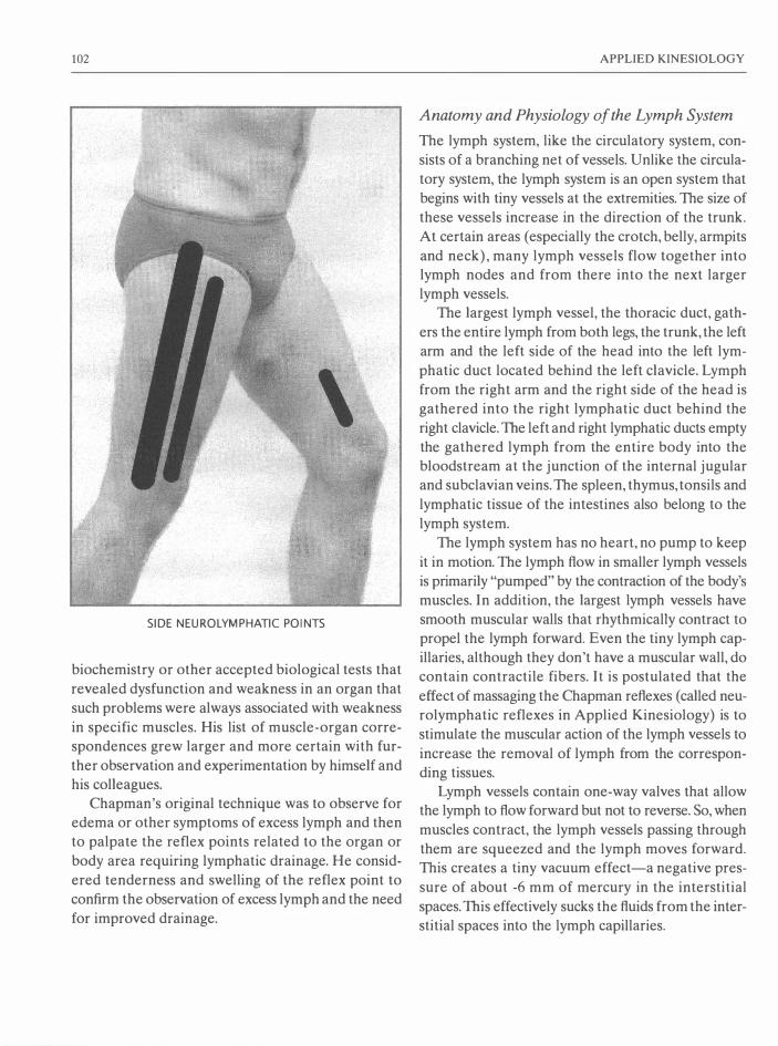
102
SIDE NEUROLYMPHATIC POINTS
biochemistry or other accepted biological tests that
revealed dysfunction and weakness in an organ that
such problems were always associated with weakness
in specific muscles. His list of muscle-organ corre
spondences grew larger and more certain with fur
ther observation and experimentation by himself and
his colleagues.
Chapman's original technique was to observe for
edema or other symptoms of excess lymph and then
to palpate the reflex points related to the organ or
body area requiring lymphatic drainage. He consid
ered tenderness and swelling of the reflex point to
confirm the observation of excess lymph and the need
for improved drainage.
APPLIED KINESIOLOGY
Anatomy and Physiology of the Lymph System
The lymph system, like the circulatory system, con
sists of a branching net of vessels. Unlike the circula
tory system, the lymph system is an open system that
begins with tiny vessels at the extremities. The size of
these vessels increase in the direction of the trunk.
At certain areas (especially the crotch, belly, armpits
and neck), many lymph vessels flow together into
lymph nodes and from there into the next larger
lymph vessels.
The largest lymph vessel, the thoracic duct, gath
ers the entire lymph from both legs, the trunk, the left
arm and the left side of the head into the left lym
phatic duct located behind the left clavicle. Lymph
from the right arm and the right side of the head is
gathered into the right lymphatic duct behind the
right clavicle. The left and right lymphatic ducts empty
the gathered lymph from the entire body into the
bloodstream at the junction of the internal jugular
and subclavian veins. The spleen, thymus, tonsils and
lymphatic tissue of the intestines also belong to the
lymph system.
The lymph system has no heart, no pump to keep
it in motion. The lymph flow in smaller lymph vessels
is primarily "pumped" by the contraction of the body's
muscles. In addition, the largest lymph vessels have
smooth muscular walls that rhythmically contract to
propel the lymph forward. Even the tiny lymph cap
illaries, although they don't have a muscular wall, do
contain contractile fibers. It is postulated that the
effect of massaging the Chapman reflexes (called neu
rolymphatic reflexes in Applied Kinesiology) is to
stimulate the muscular action of the lymph vessels to
increase the removal of lymph from the correspon
ding tissues.
Lymph vessels contain one-way valves that allow
the lymph to flow forward but not to reverse. So, when
muscles contract, the lymph vessels passing through
them are squeezed and the lymph moves forward.
This creates a tiny vacuum effect-a negative pres
sure of about -6 mm of mercury in the interstitial
spaces. This effectively sucks the fluids from the inter
stitial spaces into the lymph capillaries.

DIAGNOSIS AND CORRECTION TECHNIQUES
Trapezius
Subclavius
Supraspinatus Infraspinatus
�\f!��E!I.-�'--"-+-i--- Deltoids
Hamstrings
THE NEUROLYMPHATIC REFLEX POINTS
103

104
Respiration and all activities that raise the abdom
inal pressure (coughing, sneezing, lifting, evacuating
the bowel) pump the lymph from the legs and
abdomen toward the ultimate destination at the base
of the neck. There the lymph empties into the venous
system.
The human body has twice as much lymph as blood
and twice as many lymph vessels as blood vessels. The
blood capillaries carry nutrients to the cells. Fats and
proteins leave the capillaries and bathe the cells. Some
are used by the cells as needed. The unused fats are
too large to return to the (blood) capillaries. They are
received into the lymph capillaries through tiny one
way valves. If the proteins from the blood capillaries
are not used by the cells, they must be absorbed into
the lymph capillaries to be eventually returned to the
blood. Because the blood contains so much protein
already, the protein that leaves the capillaries cannot
return directly to them due to osmotic pressure. It is
estimated that every 24 hours, 50% of the total
amount of protein circulating in the blood exits the
capillaries, is mostly unused by the cells, and is then
reabsorbed back into the lymph system from which
it is delivered back into the venous blood through
ducts near the base of the neck.
Besides transporting water, fats and proteins, the
lymph system is mainly a waste removal and defense
system. In the lymph nodes, bone marrow, thymus
and spleen, lymphocytes (white blood cells) are gen
erated. These agents of the body's defense reside
mainly in the lymph system. White blood cells there
act as "phagocytes," which surround and digest the
filtered waste particles and pathogens in the lymph
nodes.
The waste products of cell metabolism exit the cells
and gather in the interstitial (intercellular) spaces. The
waste fluids then flow by osmotic pressure into the
lymph vessels. When any tissues are infected, the infec
tious material and products of cellular destruction
(pus) are also gathered into the lymph system. There
they are transported to the lymph nodes, which filter
out the tissue remnants, foreign bodies and microor
ganisms to be digested by the white blood cells.
APPLIED KINESIOLOGY
Whenever there is infection in the body, the next
lymph nodes in the direction of the circulation of the
lymph become swollen and tender. In order to pre
vent further spread of infection, the purulent mate
rial is gathered into the lymph nodes and given over
to the white blood cells.
The lymph circulation through the body is slow. It
needs to be slow to have enough time to filter out and
break down large bits of matter that cannot be safely
passed back into the blood. However, if lymphatic
drainage of the tissues is inadequate, lymphatic con
gestion occurs.
Lymphatic Congestion and the Need for Increased Lymphatic Drainage
When the lymph circulation is too slow, the proteins,
fats, waste products of cellular metabolism, and infec
tious material (if present) build up in the interstitial
spaces. This buildup of fluids, which inflates of the
spaces between the cells, is called edema. Edema
moves the cells further away from their capillaries,
which interferes with their proper nutrition. It is with
this disturbance of the lymphatic system that Applied
Kinesiology methods appear to have influence.
Edema stretches the tissues and makes them more
porous. If this has become chronic, it will be more dif
ficult to achieve a lasting correction, as fluids can eas
ily return through the more porous tissues. For this
reason, corrective measures must be applied over a
long period of time to reestablish proper lymph sys
tem function.
Medical authorities agree that excess lymph in the
tissues does not become observable until it has
reached a level 30% above normal. This is likely the
reason why Applied Kinesiology techniques indicate
the need for increased lymphatic activity, although
no edema is observed.
Symptoms of lymphatic congestion include edema
(especially in the extremities), low resistance, infec
tions, and wounds that do not heal. Lymphatic con
gestion may often be seen in the eyes as a "string of
white pearls" around the outer part of the iris. In some
cases there may no apparent symptoms at all.
DIAGNOSIS AND CORRECTION TECHNIQUES
When a lymphatic problem is acute, the reflex
points will be swollen and firm but squishy over the
whole area where the reflex can be stimulated. When
a problem is more chronic, the center of the reflex
point will feel firm but rubbery. When the problem is
very chronic (has existed for a long time), there will be
small, hard, BB sized lumps (in the fat just under the
skin) in the middle of the reflex area.
Chapman's reflex points are located in the inter
costal spaces between the ribs anteriorly where they
meet the sternum and posteriorly where they meet
the vertebrae. There are also some on the lower
abdomen, around the scapula, on the low back and
on the legs. Most of the reflex points are about 3 cm
in diameter. Some, such as those for fascia lata, cover
large areas.
Active lymphatic points are massaged with a firm,
rotary pressure for at least 15 seconds. In certain cases,
such as for the correction of repeated muscle weak
ness (page 123), the points are massaged for 2-4 min
utes or even longer. The points on the anterior side
of the body are generally more tender. The more
chronic a condition, the greater the amount of ten
derness. When the points are very tender, they should
be massaged more gently and for a longer time.
As mentioned, Goodheart correlated Chapman's
reflex points with specific muscles (and organs). Each
muscle was found to have at least one anterior and
one posterior reflex point. Goodheart also discovered
a method to determine when massage of these reflex
points would be useful in strengthening weak-testing
muscles. If therapy localization of the reflex point
makes a previously weak-testing muscle test strong,
massage of the point is indicated. After adequate mas
sage, the muscle should test strong. To confirm the
correction, the patient again therapy-localizes
(touches) the point and the same muscle is again
tested. In Applied Kinesiology, if (after successful
strengthening) the muscle again tests weak with ther
apy localization of the treated reflex point, further
stimulation of the same point is indicated.
In some patients, every muscle in the body will test
weak. A muscle may test strong, then after two or
105
three further tests of the same muscle, it tests weak. In
such patients, it is impossible to find an adequate indi
cator muscle. 1)rpically these people never exercise
and are extremely sedentary, spending most of their
time sitting (likely in front of the TV). These are often
patients with vast quantities of excess lymph in their
bodies. A seeming paradox, these patients are often
dehydrated. Their excess water is not where it needs
to be in the tissues, but rather in the lymph system
diluting the concentrated waste products there. These
patients respond well to the extended massage of neu
rolymphatic reflex points as performed in the cor
rection for repeated muscle failure (page 123). When
most or all muscles test weak, the patient is in the
exhaustion phase of Selye's stress reactions and
urgently requires recuperation (see pages 35-39).
Clearing the body of such extreme amounts of
lymph requires that the patient assist the process
between treatment sessions. One of the best tech
niques for eliminating excess lymph from the whole
body is bouncing on a trampoline. When one bounces
up, one is weightless. When one bounces down, one
effectively has as much as double the normal weight.
Remember bouncing on the family scales as a child,
trying to "weigh" as much as possible? These alter
nate states of "weightless" and "very heavy" act like
a pump upon all the tissues of the body. For those
whose joints cannot withstand jumping on a trampo
line, bouncing while seated on a large gymnastic ball
is an excellent and more gentle alternative.
When ill, most people remain in bed nearly
motionless. Without muscular activity, the lymph gath
ers and the tissues swell. During illness, the lymph is
full of pus and infectious material. Allowing it to
remain and gather in the body is counterproductive to
the goal of returning to health. If able, the ill person
should be moderately active. Taking walks is recom
mended. At the very least, bouncing gently on a gym
nastic ball is advised as an excellent lymphatic system
"pump." Activating the muscles (through muscle test
ing or other activities) followed by massage of the
neurolymphatic reflex points and finished by activat
ing the muscles again will assist the body greatly in

106
eliminating excess lymph from the tissues.
When many different muscles require neurolym
phatic stimulation, there is a generalized lymphatic
problem. This is usually caused by dehydration. Water
is the most common nutritional element needed for
general problems with the lymphatic system. The body
needs water to cleanse itself via the lymphatic sys
tem. Whenever lymphatic problems exist, it is a good
practice to advise the patient to drink at least eight, 8
ounce (2.5 dl) glasses of water each day. The same
effect is not achieved by tea, coffee, cola or any other
kind of drink. For their digestion these other drinks
apparently activate specific metabolic processes that
interfere with the body getting the maximal positive
effects of the water these drinks contain. Imagine tak
ing a shower with cola! Would your body feel clean?
Like your skin, your body also needs regular inner
"showers"-regular intake of pure water with noth
ing added.
It is easy to demonstrate the effect of water. When
ever many muscles test weak or hypertonic, have the
patient drink a large glass of water and then retest the
muscles. Often simply drinking water will make many
or all of them test normotonic. This demonstration
will assist the therapist in convincing the patient of
the need for drinking much more water daily.
It is believed that massage of Chapman's reflex
points stimulates nerves that effect better lymphatic
drainage in the associated organs and body areas. Fol
lowing this reasoning, in Applied Kinesiology, Chap
man's reflex points are called "neurolymphatic
reflexes." The exact anatomy and physiology involved
in these reflexes has yet to be demonstrated. How
ever, it has been demonstrated repeatedly that mas
sage of these points does stimulate lymphatic
drainage. In this case, the "proof is in the pudding."
We don't know exactly what the neurolymphatic
points are or how these reflexes function, but the evi
dence of numerous clinical trials has proven their
effectiveness.
APPLlED KINESIOLOGY
NEUROLYMPHATIC REFLEX POINT TECHNIQUE
Testing to determine if NL massage will strengthen
a weak-testing muscle:
1. Test muscles.
2. When one tests weak, have the patient therapy
localize (touch) one of the neurolymphatic points
associated with the weak-testing muscle. It is often
easier for the patient to touch the anterior point
than the posterior one.
3. While the patient touches the point, retest the
muscle.
4. If the muscle now tests strong, it can be strength-
ened with neurolymphatic massage.
Correction:
Massage both the anterior and posterior neurolym
phatic points for the weak-testing muscle with a firm,
rotary pressure. Very tender points should be mas
saged more gently and for a longer duration. The time
for effective massage may vary from 15 seconds to
several minutes.
Confirm the correction:
1. Retest the muscle. If still weak, and therapy local
ization of the neurolymphatic point makes it again
strong, the point requires further stimulation. If
NL therapy localization no longer strengthens the
muscle, it requires some other kind of strength
ening technique.
2. If the muscle now tests strong, again therapy-local
ize the neurolymphatic point and retest the mus
cle.
3. If the muscle remains strong, the NL massage was
adequate and successful.
4. If the strengthened muscle again weakens with
therapy localization of the NL, further stimula
tion of the same point is required.
5. When therapy localization of the NL no longer
weakens the previously strengthened muscle, the
NL correction is complete.

DIAGNOSIS AND CORRECTION TECHNIQUES
Neurovascular Reflexes
While Goodheart was giving a lecture upon his dis
coveries, a young boy had an acute asthma attack.
When he was placed upon his back, Goodheart
noticed that one leg rotated out much further than
the other. The sartorius muscle tested weak on this
side. Goodheart had already noticed a connection
between the sartorius muscle and the adrenal glands.
Adrenaline is given in cases of acute asthma, so the
connection seemed logical. However, he could not
correct the sartorius muscle with any of his techniques.
Trying to find something that would help, Goodheart
attempted to make an adjustment of the cranial bones.
While doing so, he happened to notice a pulsation
under his fingers upon the posterior fontanel, one of
the "soft spots" on a baby's head. The pulsation was
not the same as his own heart rate nor that of his
patient's. He continued to hold and feel this pulse,
which grew stronger. Then the young patient took a
deep breath, continued to breathe easily, and the leg
turned back in! (Goodheart, You'll Be Better, Chapter
1, p. 78.)
Fascinated by this unexpected success, Goodheart
explored these "vascular reflexes" that had already
been researched and mapped out by the California
chiropractor, Terrence Bennett. Dr. Bennett was a cli
nician and not a theorist. He never attempted to
explain why his reflex points affected vascular circu
lation. Using an x-ray fluoroscope, he observed that
holding specific points caused an increase of blood
flow into specific internal organs, which then swelled
with the extra blood. Fascinated, he spent hundreds
of hours observing the effect of holding his reflex
points. As a result, radiation poisoning is said to have
been cause of his death.
Goodheart found many (but only a portion) of
Bennett's reflex points to be effective in the strength
ening of weak -testing muscles. The points that Good
heart found useful lie mostly upon the head. He also
found (as with Chapman's reflexes) that when one of
Bennett's reflex points strengthened a weak-testing
muscle, it also had an apparent positive effect upon
107
the organ and/or gland associated with the muscle in
Goodheart's own research. This further confirmed his
muscle-organ/gland associations. The Bennett reflexes
used in Applied Kinesiology are called "neurovascu
lar reflexes."
The stimulation of the neurovascular reflexes is
effected by lightly pressing upon specific points upon
the skin and then gently pushing or pulling tangen
tially to the skin ("tugging") in various directions until
a strong pulsation is felt. The examiner experiments
carefully until the precise direction which produces
maxirnun pulsation is detected. This precise direction
of tugging is then gently and motionlessly held for at
least 20 seconds.
The amount of pressure needed to feel the pulse
at the wrist is adequate for stimulation of the neu
rovascular reflex points. The pulsation felt during this
procedure is typically between 70 and 74 beats per
minute and is independent of the rate of heart con
traction. It is speculated that this pulsation is caused
by the contraction of the tiny layer of muscles that
surrounds each of the blood capillaries in the scalp.
This theory has not been conclusively proven, so the
exact physiological basis for this observed pulsation
remains unknown.
Using sensitive thermometers, Goodheart discov
ered that properly tugging on the neurovascular reflex
points causes warming of tissues in areas of the body
far removed from the point. When the stimulation is
adequate, the distant area continues to remain warm
even after the finger contact upon the neurovascular
reflex point is removed. If the examiner changes the
direction of tugging and loses the pulsation (which
can easily happen if he is not fully attentive to the
sensation), the thermal elevation slows and the pos
itive effect of holding and tugging upon the neu
rovascular point may be reversed and lost. Returning
to the correct direction of tugging results in the return
of thermal elevation. When adequate attention is
given to this detail (tugging the neurovascular reflex
point in the direction of maximal pulsation), the
results of using neurovascular reflex point holding
greatly improve.

108
Goodheart's research, however, revealed no pre
dictable pattern to this warming. Different reflex
points sometimes warmed the same areas. The areas
over the corresponding muscles were not specifically
warmed. The patterns of warming have not yet been
plotted. But this research conclusively demonstrates
that holding and correctly tugging the neurovascular
reflex points does affect vascular circulation in remote
areas of the body.
VASCULAR CIRCULATION
FROM THE ARTERIES TO THE VEINS
Arteries (vessels that carry oxygen-rich blood to the
tissues) branch into smaller and smaller arterioles.
Arteries have a heavy muscular coat that gets thin
ner and thinner until it is only one cell thick sur
rounding the tiniest arterioles. The smallest arterioles
branch into the capillaries. Oxygen and nutrients for
the cells diffuse through the somewhat porous walls
of the capillaries. Some of these traverse the ground
substance and enter the cells. Carbon dioxide and
small-sized waste products diffuse from the cells
through the ground substance and back into the cap
illaries. Unused molecularly large nutrients (mostly
protein and fats), and large waste products from the
cells cannot enter the capillaries. These gather in the
interstitial spaces, producing enough pressure to push
them into the lymphatic vessels where they are
processed and eventually delivered back into the
venous blood.
The tiny veins that gather the blood from the cap
illary bed are called venules. The venules converge
into larger and larger veins . Like the arteries, veins
also have a muscular cover, but it is much thinner
than the heavy muscular coat of the arteries. Venous
blood, which carries oxygen-depleted and carbon
dioxide-enriched blood and waste products away from
the tissues, returns to the heart. The heart pumps the
venous blood through the lungs where the carbon
dioxide is expelled and oxygen absorbed. This oxy
gen-rich blood returns to the heart where it is pumped
back into the main artery, the aorta, which branches
into all of the other arteries of the body.
APPLIED KINESIOLOGY
The amount of blood circulating in the tissues is
controlled by the tone of the muscles surrounding the
blood vessels. The tone of the muscles surrounding
arteries and veins is increased by the hormone vaso
pressin, which is produced in the hypothalamus and
stored in the posterior part of the pituitary, from which
it is released into the blood. The tone of the muscles
surrounding arteries and veins is neurally controlled
by the vasocenter of the brain. This is located in the
lower third of the pons and the upper two-thirds of
the medulla oblongata. The upper lateral portion of
the vasocenter sends continual signals for the con
traction of the muscles around all arteries and veins.
The medial and lower portions of the vasocenter send
inhibiting signals to the upper lateral center, which
results in fewer signals with less intensity being sent
to the vascular muscles, thereby creating vasodila
tion. Both the vasodilation and vasoconstriction
effects of the vasocenter are influenced by various
higher centers of the brain.
At the beginning of a branch of an arteriole into
a smaller arteriole (and from the tiniest arteriole into
a capillary) is a circular ring of muscle that controls
the passage of blood. Surprisingly, the vasocenter has
little or no control over these tiny circular muscles.
Some believe that when the tiny circular muscles (and
the tissues of the surrounding area) require oxygen,
the tiny muscles cannot contract effectively. They then
open and allow more oxygen-rich blood to flow in.
Another theory is that the waste products of cell
metabolism act as vasodilators, increasing the blood
circulation to remove the waste products from the
tissues. Also, the presence in the bloodstream of hor
mones such as adrenaline definitely affects vasodila
tion and vasoconstriction, even at the arteriole level.
It is postulated that stimulation of the neurovas
cular reflex points has its effect by reducing the ten
sion in the tiny arteriole ring muscles and thereby
increasing local blood circulation. It is further postu
lated that the reflex effect upon organs is due to the
organ and the particular tissues of the neurovascular
reflex point having a common origin in the embry
onic tissues. While these theories offer a way to con-

DIAGNOSIS AND CORRECTION TECHNIQUES
ceive of how the neurovascular reflex points could
function, the truth is that we just don't know how they
function. But evidence conclusively indicates that
when correctly stimulated, they do both increase local
circulation and circulation far from the point, do make
weak-testing muscles test strong, and do have a pos
itive effect upon the organ or gland related to the
muscle so strengthened.
NEUROVASCULAR REFLEX POINT TECHNIQUE
A. To determine if neurovascular (NV) stimulation
is indicated for the strengthening of a weak-test
ing muscle:
1) Have the patient therapy-localize a NV point
associated with the weak-testing muscle.
2) Repeat the muscle test.
3) If the muscle now tests strong, NV
point-holding is indicated.
In a more complex example, when a muscle first
weakens after it has been muscle-tested several times
repeatedly, therapy-localize the related NY point and
again test the muscle several times. If the muscle tests
strong and remains strong while its NY point is ther
apy-localized, the neurovascular reflex is involved
and requires stimulation for the correction of the
repeated muscle test weakness (see "Repeated Mus
cle Testing," page 123).
B. To locate hidden problems in a normotonic
muscle:
1) Have the patient therapy-localize a NV point
for the normotonic muscle.
2) Retest the muscle. If it now tests weak, NY
holding is indicated.
Correction:
1. Touch the NV(s) with the fingertips and gently
tug the point(s) in various directions until maxi
mal pulsation is achieved.
2. Hold this position of maximal pulsation for at least
20 seconds.
109
Confirming the correction for A & B:
1. Retest the formerly weak-testing muscle (A) or
the formerly normotonic muscle (B).
2. If it now tests strong, challenge the correction by
therapy-localizing the NY point.
3. If the muscle still tests strong, the application of
the NY holding was correct and is complete.
4. If therapy-localizing the NV point makes the
strengthened muscle again test weak, repeat the
same NY point-holding.
5. When therapy localization to the treated NY point
no longer weakens the strengthened muscle, the
correction is complete.
C. When it is suspected that a neurovascular reflex
is involved with a problem that therapy-localized,
but the related neurovascular points do not ther
apy-localize (see "Hidden Problems" later in this
chapter, page 121):
1) Therapy-localize both the area of the
problem and the NY point simultaneously.
2) Test an indicator muscle (that tested weak
with therapy localization of the problem area
alone).
3) If the indicator muscle now restrengthens, the
NV reflex is involved with the problem.
Correction:
1. Touch the NV(s) with the fingertips and gently
tug the point(s) in various directions until maxi
mal pulsation is achieved.
2. Hold this position of maximal pulsation for at least
20 seconds.
Confirming the correction for C:
3. Simultaneously therapy-localize the problem area
and the NY that previously tested weak with such
therapy localization. Test the indicator muscle.
4. If the indicator muscle tests weak, repeat the cor
rection procedure.

1 10 APPLIED KINESIOLOGY
1. Pectoralis major clavicular
Sacrospinalis
Peroneus (tertius, longus and brevis)
Rhomboids (Walther)
2. Pectoralis major sternal
Rhomboids (Leaf)
3. Serratus anticus
Supraspinatus
Subscapularis
Deltoids
4. Rectus abdominis
S. Latissimus dorsi
6. Gluteus medius
Tensor fascia lata
Rectus femoris
Piriformis
7. Hamstrings
Trapezius, middle and lower
8. Sartorius
9. Teres minor
Teres major
Subclavius
10. Trapezius, upper
Pectoralis minor
1 1. Gluteus maxim us
Adductors
12. Iliopsoas
13. Sterno-cleido-mastoideus
The Neurovascular Reflex Points

DIAGNOSIS AND CORRECTION TECHNIQUES
5. If this double therapy localization no longer
weakens the indicator muscle, the correction is
confirmed.
The Meridian System The meridian system is one of the most important
sources of diagnostic information used in Applied
Kinesiology. By comparing and combining the muscle
organ-gland correspondences discovered by George
Goodheart with the organ-gland-meridian corre
spondences of traditional Chinese medicine, muscle
meridian correspondences were discovered. This
became the first bridge between Eastern and West
ern medicine and brought with it many new and very
effective muscle-strengthening techniques.
Before the meridian system was included in AK,
there were four stages of development of techniques
to strengthen weak-testing muscles. First, as already
discussed, Goodheart noticed correspondences
between specific weak-testing muscles and problems
with specific organs and glands. He discovered that
massaging the junction of the weak-testing muscle's
tendon against the bone where it attached often made
a weak-testing muscle test strong and positively
affected the related organ. This "origin-insertion"
procedure was the first technique for strengthening
weak-testing muscles. For the muscles that didn't
1 1 1
respond to this treatment, he prescribed exercises.
These often made the muscles able to lift more
weight, but they still tested weak. As a result of the
failure of this attempt to make the weak-testing mus
cles test strong, Goodheart realized that although
the origin-insertion technique was a valuable break
through discovery, it obviously was not enough.
Goodheart's next breakthrough began when he
experimented with Frank Chapman's reflexes. Good
heart found that massage of Chapman's reflex points
for lymphatic drainage of specific organs and glands
not only positively affected those glands and organs,
but also strengthened the muscles associated with
them. These "neurolymphatic reflex" points became
the second technique for strengthening weak-testing
muscles. With these he was able to strengthen many of
the muscles that his "origin-insertion" technique was
unable to strengthen.
The third discovery involved the correction of
blocks in the natural motion of the cranial and pelvic
bones and the circulation of the cerebrospinal fluid.
This forms an important part of AK diagnosis and
treatment. A few cranial faults were found to corre
spond to specific weak-testing muscles. But most could
not be correlated with specific muscles. The only
example of a cranial fault and muscle correlation that
will be discussed in this text is the opening of the sagit-

112
tal suture as a method to strengthen the rectus abdo
minis muscles (page 193).
Then Goodheart found that Terrence Bennett's
reflex points for increased circulation in organs often
strengthened the weak-testing muscles he had already
associated with those organs. These "neurovascular
reflex" points became the fourth technique for
strengthening weak-testing muscles. Goodheart's list
of correspondences between weak-testing muscles
and organs/glands was rapidly expanding.
The next great breakthrough occurred when Good
heart began to investigate the meridian system. In
Chinese medicine, the "meridians" are considered to
be invisible energy lines that run along the surface
and through the body. Knowledge of the meridian
system was brought to the West from China and Japan
through the technique of acupuncture. The term
"meridian" is a Western term borrowed from sea nav
igation to describe what in Chinese medicine is seen
as a channel for the life energy that the Chinerse call
chi.
The meridians are not made up of any known kind
of tissue. They are anatomically invisible. Despite
their vague nature, over thousands of years, uncount
able Chinese medical practitioners have agreed upon
their location. Meridians have a superficial portion
that lies just under the skin and a deeper extension
that reaches into the inner part of the body and
touches the organ bearing the occidental name of
the meridian.
Most of the acupuncture points lie upon the lines
of the meridians. And they lie upon the superficial part
of the meridian where they are accessible for treat
ment. Upon charts and anatomical dolls, the acupunc
ture points are numbered in the direction of the flow
of the energy. By stimulating the acupuncture points,
one can influence the meridian energy, causing excess
energy to flow away from (sedation) or to a point that
has a deficit of energy (tonification).
The Chinese define a plethora of differentiations of
chi energy. For example, they content that in each
meridian both Yin (female) and Yang (male) ener
gies circulate. Although very important and useful in
APPLIED KINESIOLOGY
Chinese medicine, this and their many further dif
ferentiations of chi do not as yet have useful appli
cation in AK and therefore will not be discussed in
any detail in this text.
For some readers, the Chinese explanations of a
mysterious life-force circulating through invisible
meridians running along the skin are very foreign to
our way of thinking. The existence of meridians and
how they function may sound mystical and appear to
lie beyond experimental proof. Nonetheless, the cor
respondences between meridians and observed con
ditions of the body and mind (general health, glands,
organs, etc.) have been repeatedly documented for
at least three thousand years. Treatments based upon
"balancing" the meridian energies have provided
undeniable benefits for a like period of time.
In general, modern science only accepts the valid
ity of such healing claims when they are repeatable
under identical circumstances. The phenomena con
nected with the meridian system are definitely repeat
able under identical circumstances. However, the
explanations from traditional Chinese medicine as to
why these phenomena occur may seem very unusual
to the Western mind and may require revision at a
later date when we have a greater scientific under
standing of the processes involved. It is my opinion
that the unusual oriental explanations available today
in no way affect the worth of our using the meridian
system concepts for diagnosis and therapy. This should
be held in mind when one begins to consider the phi
losophy and principles of Chinese medicine.
Applied Kinesiology research has documented that
meridian balancing techniques do have a measura
ble effect upon the results of muscle testing and upon
the health of the related organs and glands (Walther,
1988, pp. 203-273).
In Chinese acupuncture theory (more precisely,
the theory of the five-elements), the meridians are
associated with organs and glands. Proponents of Chi
nese medicine believe that when the "yin" or "yang"
energies in a meridian are deficient (or in excess), the
corresponding organs and glands are affected and
may become diseased or otherwise dysfunctional.

DIAGNOSIS AND CORRECTION TECHNIQUES
Even more importantly, practitioners of Chinese med
icine believe, and for centuries have demonstrated,
that their techniques of meridian energy balancing
can positively affect organs and glands; and thereby
influence health in general.
With his unique willingness to consider concepts
and techniques from other fields of healing, Good
heart initiated new research into this ancient Chinese
diagnostic and healing system. For him, the existing
correlations between meridians and organs/glands
were especially fascinating. He found that by using
new-to-him Oriental meridian balancing techniques,
he could strengthen weak-testing muscles. By plac
ing a needle, tapping or simply touching the first seda
tion point, he could make normotonic muscles test
weak. Moreover, the organs and glands associated
with the meridians proved to be the same organs and
glands that his own research associated with specific
muscles. In fact, the meridian system proved to fit
"like a glove" with his already-established system of
correspondences. With this extra piece to the puzzle,
his muscle-organ/gland correspondences became asso
ciated with the meridians.
There are twelve bilateral meridians (and two that
lie on the medial line of the body). Goodheart found
that one of the established meridians corresponded
with almost every muscle he tested. This provided a
simple and useful framework with only fourteen cat
egories for all muscles. According to Chinese medi
cine, every structure and function of the human body
belongs to one or more of these fourteen categories.
In this book, these will be referred to as the fourteen
systems of regulation. Through experiement, Good
heart determined that any technique which improved
the balance of energy in a meridian could often
strengthen the weak-testing muscle associated with
the same organ and gland as the meridian. Conversely,
the techniques he had already developed for strength
ening a weak-testing muscle often helped to balance
the energy of the corresponding meridian.
Until this time, Western science somewhat con
temptuously considered meridians to be merely a
vague and esoteric concept of oriental medicine.
113
When acupuncture was introduced into the West, a
few open-minded Western doctors were successful in
using it to produce anesthesia in their patients. How
ever, no one could explain why it worked. Nonethe
less, every true scientist recognized that there had to
be some "scientific truth" in any such completely
repeatable phenomenon. At that time, there was no
way of relating it logically with Western science. The
anatomy and physiology of muscles, however, are a
researched and accepted area of Western medicine.
Goodheart's discovery of the connection of meridi
ans with muscles created the first bridge of under
standing between oriental and occidental medicine.
In Oriental medicine, the state of health requires
that a normal quantity of energy circulate through all
of the meridians. From the AK viewpoint, an "imbal
anced" meridian may have an excess of energy or it
may have a deficit of energy circulating through it.
When a meridian is in a deficient energy state, some
or all of its associated muscles will test weak. There
fore, through testing muscles, we can swiftly and eas
ily determine energy imbalances in the meridians. The
energy state of the meridian system indicates the
imbalances underlying a patient's problems. This pro
vides the examiner with knowledge of areas of pos
sible dysfunction in the muscles, organs, glands and
functions associated with the meridian. Such knowl
edge is the essential first step toward evaluating those
dysfunctions. Goodheart's discovery and elucidation
of the connection between specific muscles and spe
cific meridians swiftly made the meridian system one
of the most important diagnostic factors in Applied
Kinesiology.
Goodheart acquired his knowledge of the merid
ian system from one early book on the topic,
Acupuncture, Ancient Chinese Art of Healing by the
English physician, Felix Mann. Like Felix Mann,
Goodheart never attempted to differentiate between
the amounts of yin versus yang energy within each
meridian. This finer differentiation, which forms an
important portion of Chinese meridian diagnosis, still
does not exist within Applied Kinesiology.
Within the ICAK-D, the medical doctor Hans

114
Garten is the current authority for Chinese medicine.
In 1991, he and Wolfgang Gerz became the first Ger
man medical doctors to receive the status of AK
diplomat. Hans Garten went to China several times
to study acupuncture. He was in charge of the edu
cation program of the German Medical Acupuncture
Society (DAGfA) for several years. He has devel
oped methods of using muscle testing plus Chinese
herbal remedies to perform detailed diagnosis of pat
terns described in traditional Chinese medicine. In
private communication, he observed that there is "no
AK-way of diagnosing Yin and Yang as they are only
acronyms for a myriad of similar qualities." Even with
out this aspect of Chinese medical diagnosis, AK
applications of meridian knowledge are extremely
useful.
The most commonly used aspects of meridian the
ory in AK are the acupuncture sedation points. When
the sedation point for a meridian is therapy-local
ized, all the associated muscles should test weak. Tap
ping upon this point will usually weaken the
associated muscles for about 10 seconds. This is sim
ple to perform and is the most accurate test for hyper
tonic muscles. Quickly stroking along a meridian in
the opposite direction of its natural flow of energy
should also weaken the associated muscles for about
10 seconds. If touching or tapping upon the corre
sponding sedation point or stroking the meridian
backward does not weaken a strong-testing muscle, it
is in a hypertonic state.
When an organ or gland is dysfunctioning, the cor
responding meridian will be found to be in an imbal
anced energy state. Is such cases, the corresponding
muscles will usually test either weak or hypertonic.
However, an imbalance in meridian energy does not
necessarily mean that the corresponding organ or
gland is already dysfunctioning. The meridian system
is highly sensitive. Meridian diagnosis can reveal
imbalances long before they could manifest as notice
able dysfunction in the related organs and glands.
Thus, it is possible that through diagnosis of the merid
ian system (by means of AK muscle testing), we can
become aware of potential organ and gland dysfunc-
APPLIED KINESIOLOGY
tions before they manifest as illness.
Typically, when meridians become deficient in
energy, their associated muscles that normally test
strong become unexpectedly weak. This weakening
often occurs one to three days before the emergence
of the first symptoms of a simple illness such as a cold.
If effective corrective measures are taken to bring the
meridians back into balance (for example, strength
ening the weak-testing muscles) and steps are taken
to increase body resistance (vitamins, good food, extra
rest and sleep, drinking extra water, immune system
stimulators such as echinacea, etc.), the illness may be
shorter and milder, or may not appear at all. Illnesses
may be thus "nipped in the bud."
Assessment of the meridian system through AK
muscle testing not only provides information about
the meridians but also indicates both the probable
weak-testing muscles and the organs and glands that
may be dysfunctioning. Conversely, when a patient
has a specific organ or gland that is known to be mal
functioning, the energy in the associated meridian
will be out of balance. The testing and strengthening
of all the muscles associated with this meridian will
do much to correct the functioning of the organ or
gland. For specific examples of this process, see the
section, "Case Histories" #2 and 3 on pages 257 and
258.
Oriental meridian-balancing techniques have a
positive effect upon both the weak-testing muscles
and the associated organs and glands. Oriental diag
nostic and correction techniques for the meridian sys
tem are not included in this text. For these, the reader
is directed to the literature and to personal training
in oriental medicine. However, for the examiner who
wishes to experiment with the concepts and tech
niques of oriental medicine, the correspondences
between muscles, meridians, organs and glands (see
pages 244) will provide a useful starting point.
For a summary of recent research on acupuncture
points and the meridian system, see the discussion in
the section on "Biological Medicine and the Systems
of Regulation," pages 59-60).

DIAGNOSIS AND CORRECTION TECHNIQUES 1 15
The Meridians
GOVERNING MERIDIAN
CENTRAL MERIDIAN
GOVERNING, BLADDER MERIDIANS STOMACH MERIDIAN

116 APPLIED KINESIOLOGY
SPLEEN MERIDIAN HEART
SMALL INTESTINE BLADDER

DIA GNOSIS AND CORRECTION TECHNIQUES 117
KIDNE Y CIRCULATION-SEX
TRIPLE HEATER GALL BLADDER

118 APPLIED KINESIOLOGY
GALL BLADDER LIVER
LUNG LARGE INTESTI NE

DIAGNOSIS AND CORRECTION TECHNIQUES
Diagnosis of the Reaction to Substances and Other Stimul i
Muscles may test weak due to a lack of particular
nutrients. Nutrition that can strengthen each muscle
is listed in the section on muscle tests (pages 153-231).
To test if nutrients can strengthen a weak-testing mus
cle (or make a hypertonic muscle normotonic), the
examiner has the patient place some of the nutrient
substance in the mouth, chew it, taste it and retain it
in the mouth. Then the muscle is retested.
Non-toxic substances that are typically ingested
orally should also be tested in the mouth. Substances
that have an odor may be tested nasally by sniffing
them. Smelling nutritional substances that have a dis
tinctive odor will usually produce the same reliable
reaction as tasting them. Controlled studies have
revealed that the testing of nutritional substances by
placing them in the hand is not as reliable. Taste (and
smell) are the only accepted and proven Applied
Kinesiology (ICAK) methods for testing nutritional
and other non-toxic substances. The ICAK-D also
recognizes hand-held testing, but only of energetic
substances such as homeopathic remedies and flower
essences.
Examiners using AK often use prepared organ or
gland tissue for dysfunctions in the same organs (or
glands) of the human body. To test such organ or
gland extracts, herbs, medicines or other healing sub
stances used for improving the function of the organs,
the examiner uses a weak-testing muscle associated
with the ailing organ or gland. The substance that,
when chewed and retained in the mouth, makes the
weak-testing muscle normotonic may aid the func
tion of the associated organ or gland. However, if the
substance strengthens the muscle but brings it into a
hypertonic state, it should not be used!
The exact mechanisms of nutritional or other sub
stance testing are unclear. A weak-testing muscle may
be strengthened within seconds of placing the proper
nutrition into the mouth. Clearly there is not time for
the substance to be absorbed and transmitted to the
brain via the bloodstream. Possibly the route for the
observed change is via the nerves involved in taste.
1 1 9
When we taste foods, we know immediately if we like
them or not. It is likely that when we taste a food sub
stance, the brain knows the chemical content and its
possible benefit for specific organs and glands. Per
haps this recognition causes the energy patterns of
the positively affected regulation systems (including
organs, glands and associated muscles) to immedi
ately improve as observed by muscle testing.
Evidence indicates the existence of another path
way (besides taste) between the mouth and the brain.
Radioactive food was placed in the mouth of rats.
Only several seconds later, they were frozen in liq
uid nitrogen. Then slices of the brain were studied
and found to contain some of the radioactive food.
How it got into the brain so fast remains unexplained.
But this experiment makes it clear that foods placed
in the mouth are present in the brain within seconds.
The nutritional substance that will help a dys
functioning organ will also reduce any tenderness in
the organ or in the related neurolymphatic areas. This
will occur immediately after placing some of the sub
stance in the mouth and tasting it. If more than one
substance strengthens the weak-testing muscles, the
one that (when in the mouth) most reduces the
related tenderness is the one to recommend (Leaf,
1995). But the examiner should also give anything
else clinically needed to support the problem as long
as it doesn't weaken a strong-testing indicator mus
cle or make it hypertonic.
For example, the best AK test result for osteo
porosis may be magnesium. This may reduce pain
more than any other remedy. But rebuilding bones
also requires calcium (calcium oro tate, calcium glu
conate, calcium ascorbate, etc.) and vitamin D3, so
these should be given too. These substances should
be given in a form that is at least tolerated. They
should neither weaken a strong indicator muscle nor
make it hypertonic. The examiner doesn't even need
to test if calcium or vitamin D3 makes a weak-test
ing muscle normotonic or takes the pain away because
magnesium was already found to do so.
Improper nutrition can cause disturbances to the
regulating systems. This can be easily detected with
AK testing. The same procedure may be used to

120
detect negative effects of any substance (or other
stimulus) upon the regulating systems. However, when
an allergic reaction is suspected, or the substance is
somewhat toxic, it should be greatly diluted and/or
presented in only a minute quantity for testing. Highly
toxic substances should not be ingested in any quan
tity. The examiner should follow the old rule, "Pri
mum nihil nocere!" or, The first thing is don't poison
(injure) the patient. The examiner should neither
provoke an allergic reaction nor injure the patient in
any way.
It is a good idea to present stimuli in the way that
the patient normally contacts them. Flowers, perfumes,
auto exhaust or anything with an odor should be
smelled. A face cream, lipstick or other cosmetic
should be freshly applied (and smelled). Music (or
noise) should be listened to. To test the effect of cig
arettes or alcohol, it is best to test the first cigarette
inhalation or drink of the day.
In most cases, substances that have a negative
effect upon one regulating system will create a weak
ening of muscles from all regulating systems. Thus
almost any muscle may be chosen for an indicator
muscle in nutritional testing. However, some sub
stances only have a negative effect upon muscles from
specific regulating systems. For example, excess alco
hol is known to have deleterious effects upon the
liver. To determine if a client's liver has problems with
alcohol, the examiner would be wise to choose an
indicator muscle such as pectoralis major sternal from
the liver regulating system.
Combinations of foods may be similarly tested.
For example, protein and carbohydrates may each
test normotonic when tested separately. However,
they often cause the indicator muscle to test weak or
hypertonic when placed together in the mouth. This
technique can be used to create a personal nutritional
plan defining which foods to eat together at one meal.
Almost any possible stimulus can be similarly
tested. The effect of hot or cold is best tested by direct
application to the area of the body in question. For
example, immediately after a sports injury, the appli
cation of cold (ice) is recommended. After a day or
APPLIED KINESIOLOGY
two the application of warmth is recommended. To
test which will help, TL the injury and test an indica
tor muscle. This should make the indicator muscle
test weak. Now continue the weakening TL and apply
cold to the injury. Retest the indicator muscle. Repeat
this test with the application of warmth. The stimu
lus of cold or warmth that makes the indicator mus
cle normotonic is the correct one to apply.
For further discussion of these topics, see "Chal
lenge" (pages 67-72).
1. Locate an organ that has a known dysfunction.
Palpate the organ and its neurolymphatic points
for tenderness.
2. Test the muscles related to the organ or gland.
Find at least one that tests weak. If none can be
found, TL the organ. If this weakens an indicator
muscle, the TL plus indicator muscle combination
can be used for subsequent tests.
3. Place a nutritional or medicinal substance known
to have a positive effect upon this organ or gland
into the patient's mouth and have her chew and
taste it.
4. Retest the related muscle. If the previously weak
testing muscle now tests strong, retain the substance
in the mouth, TL or tap the sedation point (or apply
another weakening technique), and test again. If
the muscle does not weaken, the substance has
made the muscle hypertonic. Such a substance
should not be taken! If touching the sedation point
makes the muscle test weak, the muscle is now nor
motonic and this nutrition or medicine will be use
ful for correcting the dysfunction in the associated
organ regUlating system.
S. If more than one substance makes the muscle nor
motonic, the one that (when held in the mouth)
most reduces the tenderness in the organ or its
neurolymphatic point is the one to recommend
to the patient. Also give anything else clinically
needed to solve the problem as long as it doesn't
weaken a strong-testing indicator muscle or make
it hypertonic.

DIAGNOSIS AND CORRECTION TECHNIQUES
TESTING FOR POSSmLE POSmvE
EFFECTS OF STIMULI
1. To determine if some stimulus has a positive effect
upon some problem, find a muscle that tests weak
because of the problem.
2. If no muscle can be located that tests weak
because of the problem, use therapy localization
or challenge to provoke the problem. In the fol
lowing test, continue to apply the TL or challenge
that causes an indicator muscle to test weak.
3. Apply the stimulus.
4. Test the previously weak-testing indicator mus
cle. If the indicator muscle only tested weak with
TL or challenge, apply that weakening TL or chal
lenge during this and the following step as well.
5. If the indicator muscle now tests strong, attempt to
weaken it. (Tap the sedation point, pinch the spin
dle cells, stroke the associated meridian backward
or apply the north pole of a magnet to the belly
of the muscle.) If the muscle can be weakened, it
is in a normotonic state. This indicates that the
applied stimulus may be used and will have a pos
itive effect upon the problem tested.
6. If the indictor muscle cannot be weakened, the
stimulus has put it into a hypertonic state. This
stimulus should not be used to correct the tested
problem.
TESTING FOR POSSmLE NEGATIVE EFFECTS OF
N�ON OR OTHER NON-ToXIC SUBSTANCES
1. Consider which regulating system(s) may likely
be affected by the substance in question.
2. Find a normotonic indicator muscle from the reg
ulating system to be tested.
3. Place the nutritional substance into the patient's
mouth and have her chew and taste it. Or, if the
nutritional substance has an odor, have her smell
the substance.
121
4. Retest the indicator muscle. If it now tests weak,
or if it tests strong but cannot be weakened
(hypertonic), then this nutritional substance has
a negative effect upon the regulating system
tested.
H idden Problems Sometimes an organ or gland has an obvious prob
lem, but the associated muscles all test strong. And
the muscles are not hypertonic but rather normotonic,
i.e., they can be weakened by spindle cell pinching,
touching or tapping their sedation points, stroking
the corresponding meridian backward, or placing the
north (south seeking) pole of a magnet upon the
tested muscle's belly. It seems that in such cases, the
body has compensated for the problem. Some "Rob
bing Peter to pay Paul" has occurred and the origi
nal problem is no longer visible via testing of the
related muscles.
Often in such cases, the muscle will test weak when
reflex points to the muscles and/or the ailing organ
or gland are simultaneously therapy-localized. The
exact neurological basis for this seeming compensa
tion is not yet understood. When this procedure
reveals a weakness, it is corrected by applying the
proper type of stimulation for the reflex point
touched. After correction, the muscle may be retested
with the same therapy localization to confirm the cor
rection.
FINDING HIDDEN PROBLEMS
1. Test muscles and perform all needed corrections.
2. Observe, examine and question the patient to
determine any additional problems.
3. Test muscles known to be associated with any dys
functioning organ or gland. If an area of the body
has a problem, but no specific muscle is known to
be associated with that area, have the patient ther
apy-localize the area and test an indicator mus
cle. If the patient has to get into a particular
position to provoke the problem, have the patient
get into this position and touch the problem area.

122
4. If the chosen muscle or indicator muscle in #3 tests
weak, use standard techniques to locate and per
form the needed corrective measures to
strengthen the muscle. If, however, the muscle or
indicator muscle remains strong, use the follow
ing technique to locate the cause of the hidden
problem:
5. Test this muscle (or an indicator muscle) while
stimulating the problem as in #3 while touching reflex points or other associated points, one at a
time.
6. If a point is found that makes the muscle test
weak, the proper corrective technique for this
point will help to correct the cause of the hidden
problem.
Correction:
1. Perform the correction procedure appropriate for
that point.
Confirm the correction:
2. Retest the muscle (or indicator muscle plus TL or
position) while touching the point that made it
test weak.
3. If the muscle now remains strong, the correction
is confirmed.
4. Return to #6 above and repeat the process until
no points can be located which weaken the mus
cle/indicator muscle.
For further techniques to reveal hidden problems, see
the following section on right hemisphere-left hemi
sphere brain activity. Muscles may also only test weak
after multiple tests, after being stretched, or after
other muscles contract first. For these topics, see the
sections on repeated muscle testing (page 125), mus
cle stretch response (page 126), and reactive muscles
(page 131).
To do everything possible to improve the function
of a particular muscle, or to positively influence the
function of the associated organ or gland, the muscle
should be tested in all of the above ways and any
detected imbalances should be corrected.
APPLIED KINESIOLOGY
Activation of the Right and Left Halves of the Brain The two hemispheres of the brain have different tasks.
Hidden problems may emerge and be testable only
when the brain is involved in a one-sided activity. The
mechanisms involved are unknown. The most popu
lar hypothesis is that by "activating" one half of the
brain, the brain is somehow in better touch with the
problem, which then shows up with muscle testing.
When skin sensation is lacking, therapy localiza
tion generally does not function. Goodheart found
that in some cases of paralysis with lack of skin sen
sation, activating one half of the brain made therapy
localization function. In classic Goodheart style, he
took this finding and developed the following impor
tant therapeutic application. He reasoned that if uni
lateral brain activity could produce therapy
localization when it was previously absent, then the
brain-body connection was improved. To check this
hypothesis, he asked a paralyzed patient to attempt
to lift his right leg while reciting the multiplication
tables. Mental math is a left-brain activity. The left
brain controls the right side of the body. When the
man reached 2 x 39, his leg lifted! Furthermore, hum
ming (a right-brain activity) while attempting to lift
the left leg produced movement in the leg after only
two minutes of effort. This technique has helped many
paralyzed individuals to move again.
A problem with the regulating system associated
with a particular organ may be evident, but the asso
ciated muscles may all test strong. Even touching the
reflex points of a muscle associated with the regulat
ing system (as discussed above in the section on hid
den problems) may not weaken the muscle and thus
may not reveal the existence of the suspected prob
lem. When further hidden problems are suspected,
the patient is directed to activate the left hemisphere
of her brain by performing some mathematical activ
ity like reciting the multiplication tables. While she
does so, she again therapy-localizes the various reflex
points for the muscle while the examiner tests it. This
is repeated while she activates the right hemisphere
of the brain by humming an improvised tune (one

DIAGNOSIS AND CORRECTION TECHNIQUES
without words that she thinks up on the spot).
Although neither the therapy localization nor the
humming (or mental math) alone weaken the indica
tor muscle, both together may well do so. When the
combination of humming or mental math plus TL to
a reflex point weakens the indicator muscle, the ther
apy-localized reflex point is then treated. Afterwards,
the muscle is retested with therapy localization to the
reflex point plus the brain hemisphere activity that
caused the muscle to test weak. If the muscle no longer
weakens, the correction is confirmed.
DETECTING HIDDEN PROBLEMS
ThROUGH ACTIVATING
THE RIGHT AND LEFf HALVES OF THE B RAIN:
1. Test muscles and perform all needed corrections.
2. Consider the still-existent problems and deduce
which muscles may be involved.
3. Have the patient therapy-localize reflex points for
the suspect muscle and retest it. If this produces
an active T L, treat the hidden problem as
described in the previous section. If this fails to
produce an active TL, retest the indicator muscle
while performing the activities in 4 and 5 below:
4. TL each of the reflex points for this muscle again
while the patient recites the multiplication tables.
5. TL each of the reflex points for this muscle again
while the patient hums an improvised melody.
6. If the muscle tested weak with either math or
humming, the muscle has a hidden problem.
Correction:
1. Perform the appropriate correction for the ther
apy-localized reflex point.
Confirm the Correction:
1. Have the patient again therapy-localize plus per
form the brain activity that previously weakened
the muscle, and test the muscle again.
123
2. If the muscle no longer weakens with the simul
taneous therapy localization plus brain activity,
the correction is confirmed.
Repeated Musc l e Testing
At times, a muscle will test strong, none of its reflex
points have an active T L, but it fails after repeated
use. This is often a problem with athletes who begin
their sport very well but tire out before they finish.
Goodheart first observed this in a competitive skier
who could not remain in his tightly tucked position
throughout the downhill run. When he muscle-tested
this skier, all of his leg muscles tested strong. Since
the problem only emerged at the end of the race,
Goodheart experimented with testing each muscle
again and again, imitating the constant demand placed
upon the muscles by the sports activity. This is an
example of a basic principle of Applied Kinesiology:
to test the patient in conditions duplicating the situ
ation in his life that causes problems. When he tested
the skier's hamstring muscles, one side remained
strong during repetitive testing, but the other side
weakened after being tested about five times. Good
heart was fascinated and remarked, "I have an
answer, but I haven't asked a question." (Walther,
1984, p. 230.)
Goodheart next looked for something that would
abolish this repetitive muscle test weakening. He had
the client therapy-localize a neurolymphatic point for
the hamstrings and again tested the weak hamstring
muscles repetitively. This time they remained strong.
Note that in this case, the muscle tested strong and
therapy localization of the neurolymphatic points did
not weaken it. The muscle only tested weak after
repeated testing. Therapy localization of the neu
rolymphatic points for that muscle allowed the mus
cle to continue contracting repeatedly without
weakening. Goodheart experimented and found that
prolonged massage of the muscle's neurolymphatic
points caused the muscle to remain strong in subse
quent repeated testing. Based upon this successful
therapeutic finding, he reasoned that the muscle
weakness upon repeated testing was due to the lymph

124
system not functioning properly. He suspected that
this problem is due to lack of adequate lymphatic cir
culation through the muscle tissues.
Goodheart, of course, knew that all muscles con
sist of both fast and slow fibers. Fast fibers can only
contract a few times before they tire. Since in repet
itive muscle testing, the muscle tests strong for the
first few contractions, he reasoned that the fast fibers
must be functioning correctly. The skier's problem
must then logically lie with the slow fibers, which were
failing in their job of sustaining continuous or
repeated contractions over a long duration.
Both the fast and the slow fibers are fueled by glu
cose. The body makes glucose from glycogen, the stor
age form of sugar in the body, as the need arises. Since
the fast fibers stopped contracting, the supply of avail
able glucose (already transformed from glycogen)
appeared to be used up. It is then logical that the red
slow fibers would also not have adequate glucose
available for fuel.
The slow fibers can also use fat for fuel. When they
cease functioning after several contractions of the
muscle, their fat supply must logically be in deficient
supply. The fat they use is delivered to them by the
blood capillaries and the lymph system. The lymph
system is not only a waste transport system. It also
absorbs nutritional fats through the finger-like
"lacteals" of the small intestine and delivers this fat
into the bloodstream, which delivers it to the muscles
for fueling of the slow fibers.
If the lymph is not circulating adequately, the flu
ids that leak out of the blood capillaries plus the waste
products released from the cells build up in the inter
stitial spaces (between the cells). Since in such con
ditions of lymphatic congestion, there is little
movement of the lymph fluids, the cells absorb the
sugars and fats that are immediately next to the cell
wall, and are then not supplied with further fuel.
The fast fibers can contract and thereby give the
muscle strength for a few strong contractions before
they are exhausted. Since for energy, the fast fibers
split sugar quite uneconomically, they run out of fuel
quickly. Even under the best of conditions, white (fast)
APPLIED KINESIOLOGY
fibers cannot be supplied with fuel fast enough to sus
tain repeated strong contractions. Thus, it is natural
that they become exhausted rapidly. The slow fibers
oxidize sugar completely. This very economical
process creates many times the amount of energy
from the same amount of sugar. They can also use
fats for fuel. As long as oxygen and fuel are ade
quately supplied, slow fibers can support repeated
contractions for extended periods of time. However,
if lymph circulation is poor, slow fibers quickly use
up their available fuel. Under these circumstances,
the fuel is not replaced rapidly enough to sustain
repeated contractions. If for these reasons, the red
(slow) fibers cannot continue to contract, the mus
cle unlocks during repeated testing. This is Good
heart's hypothesis of why muscles may weaken with
repeated use.
Another possible explanation lies in the chemistry
within the muscle fibers. The white muscle fibers are
anaerobic and produce the energy needed for con
traction by splitting glucose into lactic acid. When the
lymph system does not remove lactic acid from the
muscle tissues fast enough, they become acidic (their
pH level sinks.) It is the presence of lactic acid in the
muscular tissues that causes the discomfort felt in
fatigued muscles. When the pH level in a muscle is
too low, the muscle fibers cease to contract. This pro
tects the muscle from producing more lactic acid and
further sinking the pH to levels so acidic that the mus
cle fibers could be destroyed. Thus elevation of the
lactic acid levels may also be a reason why a muscle
fails after several initial contractions.
Whether lactic acid buildup or Goodheart's
hypothesis is the explanation for why a muscle fails
after several contractions, inadequate lymphatic cir
culation is the logical cause in both cases. In order to
stimulate the sluggish lymph system in the area of the
muscle, the muscle's associated neurolymphatic reflex
points require lots of stimulation. Extended stimula
tion of the neurolymphatic points (2-4 minutes or
even longer) will give such a muscle the ability to
remain strong upon repeated testing. This may need
to be repeated by the patient at home for several

DIAGNOSIS AND CORRECTION TECHNIQUES
weeks to bring a sluggish lymphatic system into
proper activity.
The Australian Touch for Health teacher Joan
Dewe (co-founder of the Professional Kinesiology
Practitioner system, PKP) related the following story:
In an Australian bar, the men practiced a competitive
sport. They strapped an iron weight onto their shoe.
With this weight they extended their leg as many
times as they could. The strongest man there per
formed 16 extensions. Joan asked to try. They laughed
because a woman wanted to attempt their test of
strength. She asked if they were afraid of being bet
tered by a woman. They let her try. With the weight
on her shoe, she crossed her arms over her chest. This
allowed her to use her fingers surreptitiously to mas
sage under the anterior edge of her rib cage (the neu
rolymphatic areas for the quadriceps muscles
including rectus femoris, which extend the leg). The
men stared with disbelieving eyes and gaping mouths
as she exceeded sixteen repetitions. Through her mas
sage of her quadriceps neurolymphatic reflex areas,
her quadriceps muscles were supplied with fuel and
the waste products of muscular contraction were more
swiftly carried away, allowing her to perform better
than all the men present.
In some cases, the weakness upon multiple testing
is due to poor vascular circulation in the tissues of
the muscle. This is more often the case with muscles
that contain a higher concentration of fast fibers. In
these cases, therapy localization of the neurovascu
lar points will abolish the test weakness encountered
after repetitive muscle tests. And stimulation of the
neurovascular points will eliminate the problem. Some
cases may require both neurolymphatic and neu
rovascular stimulation before the repeated muscle
testing weakness is eliminated.
Aerobic red muscles contract more slowly than
anaerobic white muscles. Mirroring this natural dif
ference in function, muscles that contain a predomi
nance of aerobic red fibers are tested slowly and
rhythmically about 10 times in 10 seconds. Muscles
with a higher concentration of fast fibers contract
faster and should be tested faster. Such anaerobic
125
white muscles should be tested about 20 times in ten
seconds. These tests are performed more rapidly than
normal Applied Kinesiology muscle testing. The
patient clearly does not have adequate time to build
the tension up to a maximum level during each swift
contraction. A sub-maxima/ level of contraction is
adequate for repeated muscle testing.
Many times, lasting correction of repetitive mus
cle weakness requires nutritional supplementation.
Iron is used for muscles that have a predominance of
slow fibers. Pantothenic acid is the choice for pre
dominantly fast-fiber muscles. When patients have
many different muscles that all test weak upon mul
tiple testing, they may not have enough quality nutri
tional fats to fuel their slow red muscle fibers. In such
cases, fatty acids such as oil, evening primrose oil,
sesame oil and/or fish oils are often needed.
In muscles that show weakness during multiple
testing as above, check whether therapy localization
of the neurolymphatic and/or neurovascular points
for the muscle eliminates the weakening. The points
that negate the weakening are those needed for the
application of corrective treatment.
When muscles show weakness in repeated testing,
the application of the above techniques will increase
their capacity for endurance significantly. Anyone can
benefit from increased endurance. This will benefit
the elderly who tire before reaching the top of the
stairs. This can also greatly assist athletes to attain
their highest performance potential.
Repeated Musc le Testing Technique
Test:
1. Test the muscle. If it tests weak, strengthen it
before continuing.
2. Test aerobic (postural) muscles about 10 times in
10 seconds, or
3. Test anaerobic (phasic) muscles about 20 times in
10 seconds.

126
If you don't know which type the muscle is, try both
types of testing.
4. If the muscle unlocks during the repeated testing,
have the patient "insalivate" (chew, mix with saliva
and retain in the mouth) a source of iron, then
pantothenic acid, then fatty acid.
5. Perform the repetitive muscle testing after insali
vation of each substance. If one of these abolishes
the repetitive muscle weakness, recommend that
the client include it in her daily diet. Have her
rinse her mouth and check that the muscle again
weakens with repetitive testing before proceeding.
6. Have the patient touch a neurolymphatic reflex
point for the muscle.
7. While she touches the NL point, perform the
repeated testing again.
8. Have the patient touch a neurovascular reflex
point for the muscle.
9. While she touches the NV point, perform the
repeated testing again.
10. If the muscle can contract ten times during repet
itive muscle testing (with therapy localization of
the NL or NV) without weakening, perform the
following correction.
Correction:
1. If NL contact strengthened the test, deeply rub
all the neurolymphatic points for the muscle for
2-4 minutes.
2. If NV contact strengthened the test, lightly hold
(and tug in the direction of maximal pulsation)
the neurovascular points for the muscle for at least
1 minute.
Confirm the correction:
3. Repeat the repeated muscle test (without touch
ing the reflex points). If the muscle no longer
weakens, the correction was successful.
APPLIED KINESIOLOGY
Fasc ial Release or Chil l and Stretch Techniques
for Muscle Stretch Reaction
Normally, when a muscle is gently stretched, its level
of tone will temporarily increase. For example, in mus
cle testing one applies an increasing force upon the
limb tested, which stretches the neuromuscular spin
dle cells and signals the need for increased tension to
hold the limb steady under the increasing force. When
a muscle tests strong, but tests temporarily weak after
gentle stretching, it is said to have an abnormal stretch
reaction. For example, a ball thrower reaching back
in preparation to throw the ball stretches the muscles
used in the action. This should facilitate them. If the
stretch reaction is present, these muscles will be inhib
ited just when they are most needed, resulting in less
than optimal strength and coordination in the throw
ing motion.
To test for muscle stretch reaction, the muscle is
first tested in the clear. If weak, standard AK tech
niques are applied to strengthen it. When it tests
strong, the muscle is fully extended and given a slight
tug or push toward further extension to stretch the
fibers. The examiner must be careful not to give a
powerful stretch as this will temporarily weaken any
muscle. The muscle is immediately retested. If it tests
weak after this stretching, the muscle has an abnor
mal stretch reaction.
Postural muscles such as the sacrospinalis, ham
strings, and quadriceps that work most of the time
need to be stretched rather slowly to demonstrate the
abnormal stretch reaction. Muscles that are usually
inactive and contract for swift action (phasic muscles)
need to be stretched rather swiftly to reveal an abnor
mal stretch reaction when one is present. When the
muscle type is unknown, test after a fast stretch and
again after a slow stretch.
To correct the abnormal stretch reaction, Good
heart recommends two techniques: the fascial release,
and the chill and stretch (both explained in detail
below). Fascial release seems to be the best choice
when the associated gland or organ has dysfunction.

DIAGNOSIS AND CORRECTION TECHNIQUES
Chill and stretch is indicated when there is pain in or
near the muscle tested.
Around every muscle and organ is a thin, strong,
connective tissue (fascia) sack. The fascia is a slightly
elastic membrane that allows organs and muscles to
slide smoothly against one another. Ideally, the fas
cia sack should be the same length as the muscle and
free to slide smoothly along other tissues. After injury
to a muscle, the fascia may shorten and thicken like
any scar tissue in the body. Injury and certain inflam
matory illnesses can cause the fascia membranes to
stick to one another. This limits the range of motion
of the muscles involved and likely inhibits the free
circulation of blood and lymph fluids.
Nerves and stretch receptors embedded in the fas
cia and in the muscle may cause pain when stretched,
incorrectly signaling the nervous system to react by
contracting and thus further shortening the muscle.
When for any reason a muscle isn't stretched for a
long period of time, the fascia around it shortens. This
structural ly limits the range of muscle lengthening.
Self-applied attempts to stretch it typically produce
only limited success and a lot of discomfort. For this
reason, one who habitually slumps may find it diffi
cult and painful to assume upright posture.
When the fascia around a muscle is shortened or
adheres to other fascial tissues, the function of the
organ or gland associated with the muscle is often
disturbed. Applying the fascial release technique can
help the organ or gland to return to normal function.
For example, the teres minor muscle is associated
with the thyroid gland, which is involved in temper
ature regulation. If teres minor needs and receives
fascial stretch, the body temperature may change
swiftly and measurably. When patients with hypothy
roid activity (which often causes low body tempera
ture) receive fascial stretch to the teres minor, thyroid
activity increases, and a rise in body temperature of
0.5-1.5° C within minutes is usual. This is an exam
ple of how fascial stretching of a muscle can affect
the associated organ or gland.
AK testing confirms that when there is an abnor
mal stretch reaction, the neurolymphatic reflexes are
127
active, indicating the need for increased drainage of
the lymph fluids. Before performing the fascial release
technique, it is advisable to increase the circulation
and drainage of the affected areas. Otherwise the
pressure applied upon the tissues swollen with excess
lymph could stretch and damage the lymph vessels.
For this purpose, the neurovascular and neurolym
phatic reflexes to the affected muscle should be
checked and corrected as necessary. Furthermore, the
tissues should be drained of excess fluids mechani
cally. To do so, the examiner uses the whole surface
of the hand to squeeze the proximal (toward the
heart) end of the muscle firmly for 20-30 seconds. At
the same time, he moves his hand toward the heart
as far as the excursion of the skin allows. As the fluids
are squeezed out, the tissues can be felt to slightly
shrink. Then the examiner moves his hand along the
muscle distally (further away from the heart),
squeezes and moves his hand toward the heart again.
This moves the fluids toward the heart end of the mus
cle. After holding this second hand position for about
10 seconds, he continues to maintain it and also
squeezes the first position again. Then he moves to a
third position more distal than the second and repeats
the process. He continues squeezing the fluids toward
and through the heart end of the muscle until the
entire muscle feels more soft and pliable. Performing
this maneuver first helps to prevent injuring the tis
sues when applying the more aggressive fascial release
technique.
To perform the fascial release, the body is first
brought into a position that gently stretches the mus
cle. The muscle is kept stretched during the whole
procedure. Oil is spread on the skin above the tissues
to be manipulated. To avoid rupturing small blood
and lymph vessels, the examiner begins at the end of
the muscle away from the heart. The examiner presses
his fingers, thumbs, or knuckles into the muscle and
slides with pressure along the muscle toward the
heart. The flat surface of a fingernail or the side of
the thumbnail may be used for a deeper, more aggres
sive effect. The movement is l ike ironing in that it
smooths out the kinks and irregularities in the fascial

128 APPLIED KINESIOLOGY
Draining a Muscle (the Rectus Femoris) in Five Ste ps
STEP 1 STEP 2
STEP 3 STEP 4
STEP 5

DIAGNOSIS AND CORRECTION TECHNIQUES
tissues. The examiner massages, kneads, and slides his
hands along the muscle several times. Often the
smoothing out of the fascial tissues can be felt dur
ing the process. The last step of the treatment is to
stimulate the neurolymphatic and neurovascular
reflexes for the muscle.
The abnormal stretch reaction may also be caused
by palpatory hypertonic muscles. In this case, small,
extremely sensitive "trigger" points may be located
in or near the muscle. Trigger points for specific mus
cles have been well mapped by Travel and Rinzler
(1952, 1976). For illustrations of the location of trigger
points, the reader is referred to this source or to
Walther, 1981 , pp. 171-180 and Gerz, 1996, pp.
132-138. From the patient's report of the areas of
pain, and comparison with illustrations of the areas
of pain related to trigger points, the specific active
THE FACIAL RELEASE TECHNIQUE
129
trigger point may be deduced. Pressure on active trig
ger points produces a predictable pattern of pain near
and sometimes rather distant from the trigger point
itself. Muscles with active trigger points are usually
shortened and overly tense (palpatory hypertonic).
One cannot fully extend such muscles without diffi
culty and pain. If a muscle with active trigger points is
gently tensed and then plucked like the string of a
musical instrument, it will quickly contract. This is
called the "jump sign." Further analysis of trigger
points will not be pursued in this text. Pain upon
extension of a muscle and the jump sign are the diag
nostic indicators for use of the following treatment:
The treatment of hypertonic muscles with trigger
points involves applying cold to the muscle while gen
tly stretching it. The spraying of a chilling fluid for
this technique (as recommended in the AK literature)
should be left to those professionally trained in its
use. Goodheart refers to this technique as "spray and
stretch." Its improper use can cause damage to the
tissues by freezing them. Sliding along the affected
tissue toward the heart with an ice cube (while stretch
ing the muscle) is a relatively safe way to achieve the
same effect. A convenient tool for this purpose can
be made by freezing a wood dowel into a paper cup
full of water (like a popsicle).
The fascia will often lengthen during the applica
tion of the ice. As it does so, the examiner takes up
the slack and continues to extend and stretch the
muscle further. This "chill and stretch" technique is
capable of blocking and eliminating pain, even when
underlying causes have not been corrected. There
fore, this technique should not be used repeatedly.
When pain returns after such treatment, other causes
should be diagnosed and corrected.
After the application of these techniques as
required, stretching a muscle should no longer cause
it to test weak. Combined with manipulation of the
Golgi tendon organs and neuromuscular spindle cells,
the fascial release technique (or "chill and stretch"
as required) can swiftly return full extension to a
shortened muscle. Application of these techniques

130
may remove pain, increase the range of motion and,
of special interest for athletes, dancers, etc., signifi
cantly improve performance.
T HE CHILL AND STRETCH TECHNIQUE
TESTING FOR MUSCLE STRETCH RESPONSE
1 . Test the muscle and if weak, strengthen it.
2. Extend the muscle completely and gently stretch
(extend) it a bit further. Slowly stretch postural
muscles. Quickly stretch phasic muscles. Try both
if you don't know which type the muscle is.
3. Retest the muscle. If it now tests weak, it has an
abnormal stretch response.
4. If the associated gland or organ has a dysfunction,
use the fascial stretch technique.
APPLIED KINESIOLOGY
5. If it is difficult and painful to extend the muscle,
have the patient gently contract the extended mus
cle. Then "pluck" it like the string of a musical
instrument. If the muscle responds by "jumping,"
it needs the chill and stretch technique. (In this
case, pressure on the trigger points will cause a
predictable pattern of pain.)
6. Whether the muscle needs the fascial stretch or
the chill and stretch, first drain it of excess fluids as
outlined below.
DRAINING EXCESS FLUIDS FROM A MUSCLE
1. Grasp the heart-end of a muscle with the whole
surface of one hand (two hands may be used for
large muscles.) Squeeze the muscle firmly (0.5-5
kilos pressure). Begin with light pressure. If this
fails to produce the desired effect (softening of
the muscle after the treatment), begin again with
increased pressure.
2. Without sliding along the skin, move the squeez
ing hand toward the heart as far as the excursion
of the skin allows. Hold and continue squeezing
for 20-30 seconds. Release.
3. Grasp immediately distal (further from the heart)
to the first position, squeeze and push toward the
heart again for 20-30 seconds again.
4. Continue to hold the second position and squeeze
the first position again.
5. Move further distally along the muscle and repeat
the process, pressing the fluids through and out of
the muscle toward the heart. The muscle should
now feel softer and more pliable.
PERFORMING THE FASCIAL RELEASE TECHNIQUE
1. When fascial release is required (see "Muscle
Stretch Response" above), first drain the muscle of
excess fluids.
2. Bring the body into a position that gently stretches
the muscle. Keep the muscle stretched during the
process.

DIAGNOSIS AND CORRECTION TECHNIQUES
3. Put oil on the whole surface of the muscle.
4. Begin at the distal end of the muscle (away from
the heart). Use the fingers, thumb or knuckles to
press in and slide along the tissues of the muscle
toward the heart. Massage, knead and "iron" out
any irregularities in the fascia.
5 . Repeat this process, covering the whole area of
the muscle, always sliding parallel to the fibers of
the muscle.
Confirm the correction:
6. Repeat the muscle stretch. Retest the muscle. The
muscle should no longer weaken.
7. Stimulate the neurolymphatic and neurovascular
reflexes for the muscle.
PERFORMING THE CHILL
AND STRETCH TECHNIQUE
1. Determine if this technique is needed
(see "Muscle Stretch Response" above.)
2. Drain the muscle of excess fluids.
3. Extend and gently stretch the muscle and hold it
in this position.
4. Slide ice slowly across the muscle from its distal
end toward the heart.
5. As the muscle lengthens, continue to extend and
stretch it further.
6. Continue to slide the ice firmly until the whole
surface of the muscle has been covered. Do not
excessively chill the muscle.
Confirm the correction:
7. Retest the muscle stretch response. Stretching
should no longer cause the muscle to test weak.
Range of motion should be better. Pain should be
absent.
8. Cover and warm the muscle.
1 3 1
9. If the pain returns, do not repeat the chill and
stretch technique. Determine and correct the
underlying causes. If the muscle stretch response
returns, there is likely an organ or gland involved
and the muscle will require the fascial release tech
nique.
Reactive Musc les Elevated tonus in a muscle causes a change of tonus
in other muscles. It is physiologically normal that cer
tain muscles are inhibited while other muscles are
activated (reciprocal facilitation and inhibition). When
contraction of one muscle causes the subsequent
weakening of another muscle, an undesirable reac
tive condition exists. In AK, one says that the mus
cles that are inappropriately stimulated to react are
in a "reactive state" or there is a state of "reactive
muscles." The muscle with elevated tonus that is caus
ing the reactivity is called by Bruce Dewe (former
ICAK member) in his PKP program the "reactor."
This is an accurate description but is not generally
accepted in the ICAK. In You 'll Be Better, Goodheart
called this muscle "hypertonic," "over-reactive," "the
first muscle," "primary muscle" or simply "primary."
This gay array of terms has caused much confusion
to students of AK. In this book, the term "primary
muscle" will be used. The primary and the reactive
muscles are called a reactive muscle pair or a reac
tive muscle group if there is more than one reactive
muscle involved. The reactive muscles are reactive to
the primary muscle.
Because reactive muscles may test strong in the
clear, they may be overlooked by an examiner using
AK. When pain exists after a session, or when prob
lems recur (such as a muscle again testing weak) soon
after treatment, an examination for possible reactive
muscles should be performed.
Reactive muscles are often a problem for athletes.
Consider the case of a baseball pitcher. In throwing,
the pectoralis major and anterior deltoid muscles
are the prime movers. In preparation for throwing,
the antagonists tighten to pull the arm back. This
132
lengthens and stretches the prime movers in prepa
ration for their action. As contraction of the antago
nists (teres major, teres minor, trapezius, middle and
posterior deltoids, rhomboids, etc.) becomes com
plete, reciprocal facilitation of the prime movers
occurs. Thus, reaching back should immediately
increase the tension in the muscles used for throw
ing forward. These mechanisms help the muscles to
quickly apply full power. However, if the pectoralis
or deltoid muscles are reactive to the rhomboid mus
cles (located between the shoulder blade and the
spine) or other muscles involved in pulling the arm
back, reaching back (activating the antagonists) will
temporarily weaken the throwing action. Just when
the athlete needs the greatest strength, he does not
have it! A similar case may exist for any ball throwing
or striking action such as in baseball batting, golf
"driving" or playing tennis.
Primary muscles have usually received some form
of prior injury, perhaps due to overexertion, sudden
stretching, sudden contraction, a direct blow, or other
trauma to the muscle. Asking a patient about such
injuries can help the examiner locate possible pri
mary muscles. Having the patient take the position
he was in when an accident occurred can be especially
helpful in identifying suspect primary muscles. The
muscles that were traumatized by an accident are sus
pect primary muscles.
Primary muscles are not only created by accidents.
Any muscle that is held constantly tight tends to
become over-tensed, hard and painful. This continual
tension causes the muscle to become a primary mus
cle and certain other muscles to become reactive to
it. A classic example is the upper trapezius muscles
which lift the shoulders and, when contracting on both
sides at the same time, pull the head back. Most peo
ple carry their heads far forward of the line of grav
ity through their trunk. This improper posture puts
continual stress upon the upper trapezius, which must
hold the head back to prevent it from nodding for
ward. In this poor but typical posture, were the upper
trapezius to relax, the chin would fall upon the chest.
APPLIED KINESIOLOGY
Furthermore, most people raise their shoulders every
time they do anything with their hands. This also
places repeated unnecessary stress upon the upper
trapezius muscles. As a result, at the end of each work
ing day, most people complain of tension and pain in
the upper trapezius muscles. Note that this is not a
natural result of the daily work activities performed.
The extra strain is produced by unnecessary postural
errors.
By reason of poor alignment of the head upon the
spine and excess lifting of the shoulders, the upper
trapezius muscles are continually tightened. This
excess tension causes the upper trapezius muscles to
become primary muscles, which then turn off their
main antagonists, latissimus dorsi. This is one expla
nation for the common experience of examiners using
AK techniques that latissimus dorsi tests weak in
almost every patient and is again weak at the start of
every new session. This problem is easy to demon
strate. After strengthening the latissimus dorsi, the
patient lifts her shoulders up to her ears (which tight
ens the upper trapezius) and then relaxes. Within the
next 10 seconds, her latissimus dorsi is tested. If there
is a primary-reactive relationship between these two
muscles, latissimus dorsi will again test weak. Until
the excess tension in the upper trapezius is eliminated
(by reactive muscle treatment and by improved pat
terns of posture and use), the weakness of latissimus
dorsi will continue to return.
The exact neurophysiology of reactive muscles is
still unknown. It is suspected that the level of nerve
signaling of proprioceptors (neuromuscular spindle
cells or sometimes the Golgi tendon organs) in the
primary muscle is inappropriately high. Thus nor
mal contraction of the primary muscle causes the
proprioceptors to send strong signals into the cen
tral nervous system, which causes the continued inhi
bition (weakening) of the reactive muscles. Since
treatment of the proprioceptors in the primary mus
cle often corrects the reactive muscle condition, this
hypothesis is likely correct.
When looking for reactive muscle groups, first

DIAGNOSIS AND CORRECTION TECHNIQUES
locate a possible primary muscle. A suspect primary
muscle is one that is palpatory hypertonic, painful,
stiff, shortened and/or was involved in an accident.
Next, consider which muscles work in opposition to
it. Reactive muscles are most often the antagonists
to a primary muscle. Synergists can also be reactive
to a primary muscle. Sometimes a muscle on one side
of the body is reactive to the same muscle on the
other side; This is often the case with the upper trapez
ius (located on top of the shoulder between the shoul
der and the neck). In this case, the shoulder will be
elevated on the side with the primary upper trapezius
muscle. An interesting primary-reactive muscle rela
tionship may exist between corresponding muscles
in the two diagonal extremities. For example, the
biceps (the flexor of the arm) on the right side may
be reactive to the biceps femoris (hamstrings, the
flexor of the leg) on the left side. This relationship is
called muscle interlink.
Before testing for reactive muscles, the suspected
primary and reactive muscles must test strong in the
clear. If not, appropriate strengthening techniques
are applied as needed until they do test strong. Next,
the possible primary muscle is tested and the sus
pected reactive muscles are then tested within the
next 10 seconds. If a muscle tests weak immediately
after testing the suspected primary muscle, it is reac
tive to the primary muscle.
Treatment of a reactive muscle most often requires
sedation of the neuromuscular spindle cells located
in the belly of the primary muscle. When the level of
signaling from the neuromuscular spindle cells is too
high, the muscle itself (its synergists and stabilizers)
will be overly facilitated and palpatory hypertonic. It
may also be hypertonic (tests strong but cannot be
weakened). In either case, some or all of its antago
nists and perhaps other muscles will be overly inhib
ited and thus reactive to the overly facilitated main
muscle.
However, when the level of signaling from the
Golgi tendon organs is too high, the muscle itself will
be inhibited. This will facilitate its antagonists, but its
133
synergists and stabilizers may be overly inhibited. In
this case, it is the synergists and stabilizers that are
the suspects for reactivity. Reactivity as a result of the
improper signaling of the Golgi receptors is less com
mon but should be considered, especially when acti
vation of a muscle subsequently causes its synergists
to weaken.
Synergists may also weaken due to dysfunctioning
neuromuscular spindle cells in the prime mover. Per
haps the logical explanation for this unexpected phe
nomenon is the following: When one muscle is
tightening more than is necessary to perform an activ
ity, its synergists (though theoretically facilitated) do
not need to contract to aid in the action and so remain
relatively relaxed. Their job of contraction has been
usurped by the overly tight agonist. So when a muscle
causes one or more of its synergists to become reac
tive, check both the neuromuscular spindle cells and
the Golgi tendon organs to determine which one is
dysfunctioning.
Often therapy-localizing the affected propriocep
tors in the primary muscle will eliminate its effect
upon the reactive muscles. If so, this may be used to
determine the exact location of the involved neuro
muscular spindle cells or Golgi tendon organs requir
ing treatment. To do so (after identifying a
primary-reactive pair of muscles), test the primary
muscle and immediately test the reactive while ther
apy-localizing the belly of the reactive muscle (for
the neuromuscular spindle cells) and then each end
of the muscle (to test the Golgi tendon organs). If
therapy localization to one of these areas of the mus
cle prevents the reactive muscle from weakening, the
proprioceptors that need treatment are located there.
Use the fingertips for more specific therapy localiza
tion to identify the exact position of the problematic
proprioceptor( s).
One can often feel the affected proprioceptors
as fibrous lumps in the muscle. Treatment is applied
to the neuromuscular spindle cells (of the primary
muscle) by squeezing them together in the direction
of the muscle fibers, which are usually parallel to the

1 34
length of the muscle. The Golgi tendon organ treat
ment (less frequently required) is effected by pulling
the Golgi tendon organ away from the belly of the
muscle toward the attachment of the tendon to the
bone at one end of the muscle. Notice that two-point
contact is necessary to push the neuromuscular spin
dle cells together whereas only one point of contact is
needed to pull the Golgi tendon organ away. If the
Golgi tendon organs at both ends of the muscle are
involved, treat first one and then the other end of the
muscle to avoid stretching the neuromuscular spin
dle cells in the belly of the muscle.
Sometimes, years after an injury, reactive muscle
conditions may sti l l cause pain. Treatment of the
affected proprioceptors in the primary muscle wil l
often eliminate such long-standing pains. Proper treat
ment of reactive muscle conditions can also result in
an immediate increase in the possible range of move
ment of the affected body parts. Postural analysis com
parison before and after reactive muscle treatment
will often reveal positive improvements.
Often the muscles that "could have saved you if
they had tightened enough" remain tight (and painful)
even after the injured tissues have healed. This phe
nomenon is known as "muscle memory." In case such
an accident should ever happen again, these primary
muscles are already tensed and ready to protect you.
And they may remain tensed and ready for years after
the original incident !
Here's an example from my own family: At age 67,
my father had an accident body surfing in Santa Mon
ica, California, with high surf at low tide. With tons
of water driving him forward and down, he saw sand
with little water below him. He turned to take the
blow on the back of his shoulder. The impact broke
his shoulder blade as well as three ribs on the oppo
site side of his chest. One year later, his deltoid mus
cles on the side of impact were still extremely hard
and painful. Had his deltoid muscles performed the
superhuman task of lifting his elbow strongly enough
to hold his body off the sand during the accident, he
would theoretically not have broken his shoulder and
APPLIED KINESIOLOGY
ribs. His deltoid muscles "remembered" the incident
and remained tight to protect him from such an acci
dent happening again. The continual tension in the
deltoid muscles caused the rhomboids to be reactive.
This excess tension also perpetuated the pain. As a
result, during the whole year after the incident, he
had continual pain and was unable to l ift his arm
above the level of his shoulder.
In the example of my father's body surfing acci
dent, a single reactive muscle treatment restored the
range of motion of his shoulder joint and eliminated
most of the pain. However, the next day he said, "What
did you do to me?" He complained his shoulder had
about 10% of the same terrible pain he had when the
accident occurred. This seems to be the effect of mus
cle memory being released by the reactive muscle
treatment. Two further reactive muscle corrections of
the same muscles were required to eliminate all of the
pain. This re-emergence of the original pain does not
always occur, but it is a good idea for the examiner to
mention the possibility. The client should know that
it is a positive sign, indicating the release of the orig
inal trauma, and that if it does occur, the muscles may
need another treatment or two before they will remain
in balance and pain-free.
Fluid buildup (edema) in the primary muscle (often
present in the acute stages soon after an injury) may
cause improper signaling from proprioceptors. In such
cases, massage, lymph drainage, or neurolymphatic
massage point stimulation should be employed to
drain the excess fluid from the muscle tissue. Scar tis
sue in a primary muscle may cause improper propri
oceptor signaling in chronic cases long after an injury.
Or poor posture may have allowed the fascial sack
around the muscle to shorten. Attempting proper pos
ture may then stretch the fascial sack again, causing
improper proprioceptor signaling. In these cases, deep
stretching massage techniques (see "Muscle Stretch
Reaction," page 126) may be needed to break up the
adhesions and lengthen the fascial sack.
Very large or very tight muscles may require lots of
neuromuscular spindle cell treatment. As the effect

DIAGNOSIS AND CORRECTION TECHNIQUES
of spindle cell weakening may only last about 15 sec
onds, it is wise to reset the reactives (by testing or
simply having the patient tighten them) after each
10-15 seconds of spindle cell weakening of the pri
mary muscle. The best technique is neuromuscular
spindle cell pinching of the primary muscle, test the
reactive to reset it, and repeat this cycle several times.
This is effective for large muscles like sacrospinalis
primary-abdominals reactive (see "Case Histories"
#1, page 257). For these two groups of muscles, it is
easiest to do the neuromuscular spindle cell weak
ening of sacrospinalis and the tightening of abdomi
nals with the patient lying on the side. Otherwise, the
patient must lie on the stomach for the neuromuscu
lar spindle cell work and roll over each time for
abdominal testing. For abdominal activation on the
side, the patient can press the knees against the exam
iner's leg while the examiner pushes upon the
patient's chest. In the same position, the examiner
can reach over the back and do the neuromuscular
spindle cell pinching.
After the reactive muscle treatment, test the pri
mary muscle and then immediately test the reactive
muscles. If treatment has been appropriate and ade-
135
quate, the former reactive muscles should now remain
strong.
ThE REACTIVE MUSCLE TECHNIQUE
Indication:
Any overly-tight muscles may be primary muscles.
Suspect the existence of reactive muscles when any
strengthened muscles become weak after certain
activities. After the patient has had her weak-testing
muscles strengthened, ask her to walk around, bend
over, climb stairs and especially to perform any activ
ities that she knows may cause her problems. Then
recheck the muscles. If some muscles again test weak
after activity, there is likely one or more primary mus
cles responsible for turning off the reactive muscles
(making them test weak subsequent to the action of
the primary muscles).
Test:
1. Individually test the suspect primary and reactive
muscles. If any test weak, strengthen them.
2. Test the primary and immediately (within 10 sec
onds) test the suspect reactive muscle. If it now
Treatment of the Primary Sacrospinalis Muscle to Reactive-Abdom inals
NEUROMUSCULAR SPINDLE CELL WEA KENING
OF THE SACROSPINALIS
POSITION FOR T ENSING TO RESET THE A BDOMINALS

136
tests weak, the primary-reactive muscle relation
ship exists.
3. Test the primary muscle and immediately test the
reactive muscle while therapy-localizing the belly
of the reactive muscle. If this causes the reactive
muscle to remain strong, a neuromuscular spin
dle cell in this area requires treatment. Therapy
localize smaller areas to determine its exact
position.
4. If no neuromuscular spindle cell can be located,
test the primary muscle and immediately test the
reactive muscle while therapy-localizing the Golgi
tendon organs at each end of the muscle. If this
causes the reactive muscle to remain strong, treat
ment of the Golgi tendon organ is required.
5. If TL to neither neuromuscular spindle cells nor
the Golgi tendon organs causes the reactive mus
cle to remain strong, assume that neuromuscular
spindle cells are at fault.
Correction:
1. Pinch the neuromuscular spindle cells of the pri
mary muscle together to weaken it. If the exact
location could not be determined, pinch them
together over the whole range of the belly of the
APPLIED KINESIOLOGY
muscle. If Golgi tendon organ treatment to weaken
the muscle is required, pull the affected Golgi ten
don organ away from the belly and toward the end
of the muscle. Optional: Test the primary muscle
to confirm that it has been weakened.
2. Immediately test the reactive muscle to activate
it. Or more easily, have the patient tighten the
reactive muscle strongly.
3. Repeat steps 1 and 2 several times.
Confirm the correction:
Perform the test step 2 again. If you have corrected
the primary-reactive relationship, the formerly reac
tive muscle will now test strong.
No list of reactive muscles is complete. Any mus
cle that was active when the body received a trauma
may become involved in a primary-reactive muscle
relationship. The list given below is only a guide to
help the examiner look for possible reactive muscles.
Some muscles that are not illustrated in this book are
listed here for examiners familiar with these addi
tional muscle tests. When looking for primary-reac
tive muscle pairs, also test contralateral muscles
(muscle interlink) and all antagonists and synergists
to the main muscle.

DIAGNOSIS AND CORRECTION TECHNIQUES
PRI MARY M USCLE REQU I R I N G SEDATION
Adductors
Biceps fe moris (hamstrings)
Deltoid, ante rior
Deltoid, ant. mid. & pos.
G l uteus medius
G l uteus maxi m u s
Infras pi natus
I l iopsoas
Lati ss imus dorsi
Pectoral is major clavicular
Pectoral is major sternal
Pectoral is m i n or
Peroneus tert ius
Peroneus longus and brevis
Piriformis
Popl ite us
SUSPECTED REACTIVE M USCLE
Tensor fascia lata, I l iopsoas, Pir iformis,
G l uteus medius and max i m u s
Sacros pinal i s , Quad rice ps, Popl iteus ,
Latissi m u s dors i (contralateral)
Deltoid (poste rior)
Rhomboids, Pectora l i s m ajors and m i nor,
Lat i s s i m u s dors i , Subscapularis
Rectus abdom i n i s (contralate ral), Add u ctors
Sacrospinal is , Pectoral i s major clavicular,
I l iopsoas, Rectus fe moris, Sartori u s , Pir iformis,
Add u ctors, Tensor fascia lata
137
Pectora l is majors, Deltoid (anterior) ,
I nfraspinatus i tse lf (s u pe rior and inferior fi be rs)
Add u ctors, Diaphrag m , G l ute u s maxi m u s ,
Contralateral anterior n e c k flexors (SCM),
Ham stri ngs, Quad ratus l u m boru m , Sacrospina l is
Trapezius (u pper) , Hamstri ngs (contralateral ) ,
Su praspi natus, Delto ids , Levator scapula
Te res major, Teres m i no r,
Lat iss imus dors i , Trapezius (midd le), Deltoid
(pos.), Rhomboids, Supraspi natus, G l uteus
maxi m u s
Su praspinatus, Trapez i u s ( u p p e r and lower) ,
Deltoid (posterior ) , Serratus anticus, Rhomboids
Serratus anticus, Su pras pinatus , Delto ids ,
Trapezius , Rhomboids
Te nsor fascia lata, Ti bial is anterior and posterior,
Peroneus longus and b revis
Peroneus tert i u s , Ti bia l is anterior and poste rior
Sp lenius capitis (contralateral ) , G l uteus medius ,
Tensor fascia lata, Add u ctors, Hamstrings (med ial)
Gastrocnemi us, Quad riceps, Hamstri ngs
( lateral) ,Trapezi u s (upper ) , Rectus abdom i n i s
Quad rice ps, G l uteus medius (contralateral),
Sacrospinal is , Rectus fe mori s , Sartor ius ,
Quadratus l u m borum

138
PRI MARY M USCLE REQU I R I N G SEDATION
Rectus abdom i n i s , lower
Rectu s abdom i n i s , upper
Rectus fe moris
Rhomboid major and m i nor
Sacrospi nal i s
Sartor ius
Ste rnocle idomastoideus (SCM)
Se rratus anticus
Subclavius
Subscapularis
Su pras pi natus
Te nsor fascia lata
Te res major
Te res m i n o r
Trapez i u s , lower
Trapez i u s , m i d d l e
Trapez i u s , u p pe r
APPLIED KINESIOLOGY
SUSPECTED REACTIVE M USCLE
Rectus abdom i n i s , upper
Rectus abdom i n i s , lower
Gastrocne m i u s , Rectus abd o m i n i s , Hamstri ngs,
Sarto r ius , G l uteus max i m u s , Ad ductor magnus
Delto id , Serratus ant icus, Su praspi natus,
Lat iss imus dors i , Pectora l is majors and minor
Trapez i u s (upper, middle and lower)
Rectus abdo m i n i s , G l uteus maxim us, Hamstri ngs
Quad riceps, G l uteus medius and max i m us,
Add uctors, Tensor fascia lata, Ti b ia l is anterior
Peroneus terti us
Neck extensors, Trapezius (u pper), Pectoral is
major, c lavicular is , opposite
Ste rnocle idomastoideus
Rhomboid, Pectora l is major ste rnal ,
Pectora l is m i nor, Trapez ius (midd le)
Ste rnocle idomastoideus
Te res m i nor, Infras p i natus, Delto ids ,
Su praspinatus
Rhomboids, Pecto ral i s m i nor, Te res major,
Te res m i n or, Latiss i m u s dorsi
Adductors, Peroneus tert ius , Ham stri ngs,
G l uteus max i m u s
Pectora l is majors, Deltoid (ante rior) ,
Teres m i nor, I nfras pi natus
Su bscapular is , Lat iss imus dors i , De ltoid ,
Su praspi natus.
Pectora l is majors, Pectoral is m i nor,
Levator scapula , Trapez i u s (upper)
Pectoral is majors, Pectora l is m i nor
Lat iss imus dors i , Biceps, Su bscapularis,
Trapez i u s (upper-contralate ral) , Lat iss imus dors i ,
N e c k flexors (SCM)

DIAGNOSIS AND CORRECTION TECHNIQUES
Exercise
For health and optimal functioning, the body requires
an adequate quantity of the right quality of move
ment. In his AK courses, Goodheart often said that
as long as the muscles of the body test strong in AK
muscle tests, normal daily activities will provide for
adequate fitness. Goodheart himself played tennis for
up to three hours several times per week! With his
level of "normal daily activity" further exercise was
not needed.
However, most of us need more exercise than we
get. The need for exercise for training muscle strength
is becoming an increasingly popular topic in AK. The
following is a short description of how to perform the
type of movements needed for health and for cor
rection of typical postural problems.
BASIC FORMULA FOR AN EXERCISE ROUTINE
1. Warm up
2. Stretch
3. Exercise (whether light aerobic exercise such
as running, or heavy anaerobic exercise
such as weight-lifting)
4. Stretch
5. Cool down
Basic Formula for Exercise
1. If you have or suspect any physical problems,
inquire of a doctor or sports therapist as to which
exercises you may do, and how to perform them
without danger of injury.
2. Warm up your body with light aerobics such as
bicycle riding or easy gymnastics. Wait until you
begin to sweat before continuing.
3. Stretch the muscles you intend to exercise. Do not
bounce your weight upon a stretched muscle but
rather take a position that gently stretches the
139
muscle and places some body weight upon it. Then
wait and breathe deeply until the muscle and other
tissues let go and your extension increases. Be sure
you are warmed up and stretched before per
forming any exercises that require strength.
4. Be sure that the muscles test strong when mus
cle-tested. Exercises produce less benefit, and may
cause damage if they are performed with weak
testing muscles. If you have no one to test you, at
least contract the muscle, rub its neurolymphatic
points, and contract the muscle again before begin
ning with the exercises for this muscle. To
strengthen the abdominals, also pull the sagittal
suture open (see "Rectus Adominis," page 192).
5. Only perform motions that follow the natural
motions of the joints and respect the structures
of the body.
The bones of the neck are designed to bend forward
and backward, lean right and left, and rotate to the
right and the left. Other motions such as circles with
the head should not be performed! Also, do not lean
the head backward while standing. Due to years of
the ubiquitous but incorrect posture of pulling the
head back upon a neck that is slumped forward and
down, the cervical intervertebral discs are thinner on
the back side of the body. Pulling the head back places
extra pressure on the already compromised posterior
portion of these discs. Even when stretching with no
added weight, only perform this movement when on
the hands and knees or bending forward to remove
the weight of the head from pressing upon the thin
portion of these discs.
The lumbar vertebral discs are usually pinched
posteriorly like the cervical discs. To avoid placing
weight upon these already compromised discs, avoid
leaning extremely backward while standing. To stretch
this area, arch the back while on the hands and knees
upon the floor (the yoga posture of the cat). To pro
tect this often-injured area, normal sit-ups should also
be avoided.

140
6. Begin gently and over a period of weeks gradu
ally increase the weights used (if you exercise with
weights) and the number of sets of each exercise.
7. After exercising, again stretch the muscles that
were used. Also thoroughly stretch all shortened
muscles, especially those of the pectoralis group.
8. Gradually cool down (i.e. walk after running).
BASIC FORMULA FOR MUSCLE INJURIES: "RICE"
1. Rest
2. Ice
3. Compression
4. Elevation
If you injure a muscle, first of all rest it. Don't con
tinue with your workout. Place ice (wrapped in a
towel and then in a plastic bag) upon the injured mus
cle. Apply compression such as wrapping it gently in
an elastic bandage to prevent swelling. Elevate it
above the level of your heart. This will also help pre
vent swelling. After a day or so, TL the injured area
and muscle-test an indicator muscle. If this weakens
the indicator muscle, test whether the application of
cold or warmth makes the indicator test strong, and
apply this kind of stimulus. Other possible treatments
can be tested in the same way. Enzymes such as found
in fresh pineapple and in the many digestive and
metabolic enzyme products available without a pre
scription are recommended to dissolve the blood
released into the tissues by the injury. The following
day, when you again start calisthenics involving that
muscle, begin slowly. If such calisthenics do not invoke
pain at the time, and these can be performed without
provoking pain or swelling the following day, the
intensity of the exercises can be gradually increased.
When you proceed to your usual weight-exercise, use
less weight than normal and perform less strenuous
maneuvers. Gradually build up to your normal work
out over a period of several days or weeks as needed
for recovery.
APPLIED KINESIOLOGY
Two TYPES OF EXERCISE: AEROBIC
AND ANAEROBIC
1. Aerobic exercises are easy-to-do exercises that
are repeated for extended periods of time. Light
weight lifting, running, swimming, bicycling, tram
poline springing, and dancing are typical aerobic
exercises. Since many people don't make time for
exercise at all, and exercise is necessary for health,
some English doctors have recommended enthu
siastic love-making as a good aerobic exercise. Aer
obic exercises are performed near but not over a
heart rate of 180 minus your age. If you are out of
shape, lower this number even further. If you are
very fit, your aerobic heart rate may be safely cho
sen a bit higher than 180 minus your age. This type
of exercise uses oxygen to burn fat for fuel. It
results in more slender muscles capable of sus
tained activity. It is wise to build up aerobic fitness
before including anaerobic exercises in your rou
tine. For aerobic fitness, at least 20 minutes of exer
cises in the target heart beat range (180 minus age
for the maximum, 15-20 below this rate for a min
imum) is recommended. An electronic heart mon
itor makes this easy to control. This device makes
one sound when the heart goes over the desired
maximum and another sound when it goes under
the desired minimum. Aerobic exercises can be
performed with good results 4-6 days per week.
2. Anaerobic exercises are strenuous exercises that
one cannot perform for more than 12 repetitions
before exhaustion of the muscles used occurs.
Weight lifting near maximum weights, chin-ups,
dips, and hand-stand push-ups are typical exam
ples. Anaerobic exercises push the heart rate up
higher than aerobic exercises. The body cannot
burn fat with oxygen this rapidly so it breaks down
blood sugar for fuel. This lowers the amount of
sugar circulating in the blood, which may produce
intense hunger. Anaerobic exercise produces
thicker muscles capable of exerting great force for
short periods of time. Rest between anaerobic

DIAGNOSIS AND CORRECTION TECHNIQUES
exercises. Avoid elevating the heart rate exces
sively. Aerobic exercises may be performed the
next day, but one should rest one day to allow the
muscles to recuperate before again performing
anaerobic exercises.
Muscle and Fitness magazine reported research in
which half of the subjects lifted weights but did not
have to lower them. The other subjects only had to
lower the weights but not lift them. Lowering the
weight is called "negative" exercise. Surprisingly, low
ering the weights produced a greater increase in
strength and muscle mass than lifting the weights.
Considering this finding, be sure to lower the weights
slowly to get the greatest benefit from weight lifting,
or from exercises in which you lift your own body
weight.
IMPROVING POSTURE
1. A depressed attitude is reflected in a slumped pos
ture. And a slumped posture predisposes toward
depression. The two mutually influence one
another. Change the underlying attitudes-the
mental posture-and the physical posture may fol
low. But body habit can be strong enough to pull
you back. Change your physical posture, and your
mental/emotional attitudes will change. Specific
mental-emotional states are only achievable from
specific postures. For example, you cannot feel
depressed without collapsing your chest down
ward.
2. Perform exercises to strengthen the muscles
needed to support upright posture.
3. Stretch the muscles whose prior shortening have
pulled the body into poor posture.
4. The procedures of Alexander Technique likely
provide the best training in posture and move
ment. Private sessions with a qualified teacher are
recommended. In such lessons, you will learn to
release the tight muscles at the back of your neck
141
to allow the head to come forward and up, the
back to lengthen and widen.
5. Because AK muscle testing and balancing both
improve posture and change the subjective sense
of what feels right, better posture and patterns of
use are automatically assumed without having to
consciously attend to them.
'TYPICAL POSTURAL DIFFICULTIES
In the upper torso, almost everyone has stronger mus
cles on the front than the back. We are oriented for
ward and down toward our desks, sinks, automobiles,
etc., so naturally the muscles on the front of the chest
are more fit than those on the back. In these and other
activities we mostly perform motions that inwardly
rotate the shoulders. Even sport and fitness enthusi
asts typically do more bench presses than rowing
movements. So they too have stronger pushing mus
cles (pectoralis major clavicularis, pectoralis major
sternal, pectoralis minor, anterior deltoid, middle del
toid, triceps, coracobrachialis, etc.) than pulling mus
cles (rhomboids, trapezius, posterior deltoids,
latissimus dorsi, teres major, teres minor, supraspina
tus, infraspinatus, biceps, etc.). This is easily seen in
the deltoid (shoulder) muscles. The deltoid is typi
cally large in the anterior and middle sections but
quite small in its posterior portion. For various rea
sons, even athletes who consciously exercise their
pulling muscles, tend to ignore the external rotators of
the shoulder. Although latissimus dorsi and teres
major are used in popular pulling motions like chin
ups and rowing, they are also internal rotators of the
shoulder.
As a result of these imbalanced activities, the most
used muscles of the chest (the pectoralis group) and
the inward rotators of the shoulders tend to be com
paratively over-strong and shortened. This is called
"adaptive shortening." Their opponents tend to be
weak, to muscle-test weak and be overly lengthened.
The shortened pectoralis muscles and internal rota
tors pull the shoulders forward and down, rotate them

142
inward and round the back. These changes establish
a new and incorrect norm of balance between these
sets of muscles. The weakness in the external rotators
of the shoulders predisposes them to the common
"rotator cuff" injuries. The rotator cuff muscles hold
the humerus into the shoulder joint. Actually, the
shoulder joint is structurally quite unstable. It is like
a ball pulled onto an open pan by the four rotator
cuff muscles: the supraspinatus, infraspinatus, teres
minor and subscapularis. These little muscles need to
be strong to protect the shoulder from injury. To cor
rect these postural problems, specific exercises are
needed to strengthen, and thereby shorten the neg
lected muscles and also to stretch the incorrectly
shortened muscles.
SPECIFIC EXERCISES FOR THE MOST COMMONLY
NEGLECTED MUSCLES
The following is in no way intended to be a complete
exercise plan but rather a list of specific exercises for
the muscles often ignored and most in need of exer
cise. Performing these exercises as described will
improve posture, prevent injuries and relieve many
pains.
Low back pain is the most common physical diffi
culty that causes discomfort and makes people unable
to work and function well. Typically people have
overly contracted low back muscles combined with
weak abdominal muscles. This produces a "sway
back" with eventual pinching of the discs. Making the
abdominal muscles test strong with AK techniques
will usually temporarily remove the pain. Perform
ing the reactive muscle technique with the primary
sacrospinalis and reactive abdominals will provide a
longer-lasting correction (see page 135). But in most
cases, for lasting freedom from pain, the abdominal
muscles also need to be physically stronger.
Sit- Ups
Rectus abdominis, the muscle that appears like a
"washboard" on the abdomen, is the main abdomi
nal muscle. More than any other muscle, the abdom
inals often not only muscle-test weak but are
APPLIED KINESIOLOGY
functionally weak. To better support the low back and
protect it from pain and disc damage, strong abdom
inal muscles are needed. However, the typical "sit
up" exercises mostly strengthen the iliopsoas muscles,
which connect the inner femur with the lumbar ver
tebrae and the pelvis. These cannot pull the ribs to
the pubic bone like the abdominal muscles do. If iliop
soas is short or overly tense, it pulls the lumbar ver
tebrae forward, creating a hollow low back. Overly
exercising the iliopsoas may increase low back prob
lems. Plus, sit-ups put tremendous pressure on the
intervertebral discs and can damage them and/or
pinch the nerves that exit the vertebral column
between the lumbar vertebrae.
Before exercising the abdominals, first test and cor
rect them with Applied Kinesiology techniques so
that they test strong. Then to exercise the abdominals
safely, lie on the back, bend the knees and sit up only
until the scapulae leave the floor. The function of the
abdominals is only to pull the lower edge of the front
of the rib cage toward the pelvis. When doing sit-ups,
this motion is complete before the low back leaves
the floor. The rest of the normal sit-up motion is
accomplished by the hip flexors such as iliopsoas.
Doing sit-ups this modified way only changes the
angle between all the lumbar vertebrae by a total of
about 3°, thus minimizing any pinching of the discs.
As an easy control, reach with the hands as you sit
up until the hands are beside the knees. Do not sit up
further than this. In order to avoid the typical poor
postural habit of collapsing the chest, some authori
ties recommend doing sit-ups with the shoulders held
back and the shoulder blades pulled together. Begin
with 5-10 repetitions and increase the number as your
strength increases. When doing 25 repetitions becomes
easy, put the hands behind the neck for more resist
ance and decrease the number of repetitions. Do this
exercise both with the feet supported (held down to
the floor) for more lower abdominal activity and
unsupported for more upper abdominal activity.
For the neglected upper body muscles, perform
exercises of the external shoulder rotators and those
that pull the arm and shoulder back (posterior). Take

DIAGNOSIS AND CORRECTION TECHNIQUES
SIT-UPS: BEGINNING POSITION
SIT-UPS: WITH HAND BEHIND HEAD
BEGINNING POSITION
care that the arm is not raised to the front or side
higher than 60° (measured between the body and the
arm) with internal rotation ("hunching" the shoulders
forward). This can cause impinchment syndrome (of
the tendons of supraspinatus, biceps or the bursa of
the joint) and lead to tendinitis in the shoulder. There
is not much space between the head of the humerus
and the acromion of the shoulder joint. External rota
tion of the shoulder (pulling the shoulders back) allows
the tendons and bursa to more easily clear the roof of
the shoulder when the arm is raised.
Most of the external rotators of the shoulder (listed
below) are rather small and functionally not very
SIT-UPS: FINISHING POSITION
SIT-UPS: WITH HAND BEHIND HEAD
FINISHING POSITION
143
strong muscles. Exercise them with small weights (1-6
pounds) and gradually work up to no more than 20
pounds. Lift and lower the weights slowly with
absolutely no jerking movements.
Lying L Flyes
The term, "lying L flyes" refers to the posture in which
the elbow is bent 90° and a movement like flying or
flapping the wings is performed. Lying L flyes are an
excellent exercise for the teres minor, a true external
rotator muscle of the shoulder. To perform them, lie
on a bench on your right side. Support yourself with
your right arm folded under your head or extended

144 APPLIED KINESIOLOGY
Exercise Main Muscles Active
Lying L (b utter)flyes
Sitt i ng or stand i ng L flyes
Bent over flyes
Te res m i nor, infras pi natus
I n fras pi natus, teres m i nor
Lyi ng flyes
Lying s u p raspi natus flyes
Bent rowi ng
Poste rior deltoid, external rotators, m i d d l e trapezius
Posterior de lto id, external rotators, middle trapezius
Su praspi natus, d eltoids
C h i n-u ps stand ing on a stool
Te res major, trapezius, rhomboids, posterior deltoids, biceps
Lat iss imus dorsi, lower trapezius, b iceps
down upon the floor. Choose one of the following
three initial positions:
1. Bend both legs and place your left leg upon your
right leg. Place your head on your folded right arm.
2. Alternatively, you may place your left foot flat on
the bench with the left knee straight up in the air.
In this position, you may rest your right hand over
your right thigh. This is a good position if placing
your arm under your head is uncomfortable.
3. Extend your right arm down below the table onto
the floor on holding on to the leg of the table for
support. You may hook your right foot over the
edge of the bench for increased stability.
LYING L FLYES: WITH THE ARM UNDER THE HEAD
Which ever of these three initial positions you choose
to assume, next bend your left elbow 90° and prop it
upon a firm pillow on your left side. Bring your fore
arm down across your chest. Begin with a very light
weight (-1 pound) in your left hand. Rotate the
weight up as high as it is comfortable to do, keeping
the elbow in place. If you are rehabilitating an injury
to the shoulder, only lift the weight to slightly above
horizontal. Lower the weight and repeat the move
ment for 6 to 8 repetitions. Repeat on the other side.
Do not allow the elbow to slip off to the side or the
back to rotate with the motion. Keep the elbow bent
90° and down upon the side. As your strength
increases, increase the weight but never use more than
1 5 pounds with this exercise.
LYING L FLYES: HOLDING THE THIGH

DIAGNOSIS AND CORRECTION TECHNIQUES
LYING L FLYES
USING THE ARM DOWN FOR SUPPORT
Standing or Sitting L Flyes
Standing or sitting L flyes are an excellent
exercise for the other true external rotator
of the shoulder, the infraspinatus. The pos
terior deltoids are active in this exercise as
well. For this exercise, you will need to sup
port the elbow at a level just below armpit
height. Stand or sit with your chest diago
nal to the support to enable free rotation of
the forearm around the elbow. Hold a light
dumbbell (1-20 pounds maximum) in your
right hand with your right elbow bent 90°.
The angle between your trunk and your
right shoulder should be less than 90° (ori
ented slightly downward from shoulder to
elbow). These adjustments of angle are nec
essary to avoid impinchment at the shoul
der joint. Support your right elbow upon
the back or in the palm of your left hand.
Keep the 90° angle of the elbow and lower
the weight to an angle just below horizon
tal. Gently reverse the direction with no
jerking and raise the weight to the starting
position. Perform 6 to 8 repetitions. Repeat
with the left arm.
STA NDING OR SITTING
L FLYES
BEGINNING POSITION
LYING L FLYES
END POSITION
145
STA NDING OR SITTING
L FLYES
END POSITION

146
Bent-Over F/yes
Bent-over flyes put the main work of contraction into
the posterior deltoids. These are small muscles that
are often ignored in training programs, so go lightly
at first with the weight (1-5 pounds). To perform this
exercise, bend forward at the hips until the back is
near horizontal. Keep the knees gently bent to reduce
BENT-OVER FLYES: BEGINNING POSITION
Lying F/yes
Lying flyes put even more activity into the posterior
deltoids than bent-over flyes. Also, this exercise
includes the first 90° of the motion of the posterior
deltoids that is missed by the bent-over flyes. Strength
in this range of the motion is important in various
sports: An example is the tennis backhand stroke.
Begin in the same lying-on-the-Ieft-side position as
for lying L flyes. Hold a very light dumbbell down
toward the floor in your right hand with your palm
facing inward toward the bench. Begin with 1-4
pounds and work up to no more than 10 pounds. The
posterior deltoid has poor leverage in this position
so don't overload it ! Keep the elbow gently bent
throughout the motion. Slowly lift the weight until
your arm is nearly vertical. Do not roll back as you
lift the weight. Slowly lower the weight and do 6 to 8
repetitions. Repeat with the weight in the left hand.
APPLIED KINESIOLOGY
stress to the low back. Use two dumbbells (or soup
cans or bottles of water, etc.). Start with the arms
hanging downward. Begin to lift the weights by
spreading the arms and continue to lift them to hor
izontal and in line with the ears. Keep the elbows gen
tly bent and rotate the arms externally a bit as you
raise them. When you use heavier weights, support
your chest on a narrow bench.
BENT-OVER FLYES: END POSITION
Lying Supraspinatus Flyes
Lying supraspinatus flyes target the supraspinatus
muscle. Like the lying flyes, this exercise includes the
first 90° of supraspinatus and deltoid motion that most
other exercises of these muscles omit. Performing this
exercise with the arm externally rotated puts more
work into the anterior and middle deltoids. With the
arm internally rotated, the supraspinatus and poste
rior deltoids are more active. To perform this exer
cise, lie in the same position as for the lying L f\yes
and the lying flyes. Use a very light dumbbell. Begin
with the weight held in the nearly straightened arm
down toward the floor. Lift the weight slowly, paral
lel to the bench, first toward your feet and then on
up to about 60° above horizontal. With internal rota
tion, never raise the arm more than 60° above hori
zontal! Slowly lower and repeat for 6 to 8 repetitions.
Repeat with the other arm.

DIAGNOSIS AND CORRECTION TECHNIQUES 1 47
LYING FLYES: BEGINNING POSITION LYING FLYES: END POSITION
LYING SUPRASPINATUS FLYES: BEGINNING POSITION WITH LYING SUPRASPINATUS FLYES: END POSITION WITH INNER
INNER ROTATION ROTATION
LYING SUPRASPINATUS FLYES: BEGINNING POSITION WITH LYING SUPRASPINATUS FLYES: END POSITION WITH OUTER
OUTER ROTATION ROTATION

148
Bent Rowing
Bent rowing is a good exercise for teres major, the
trapezius, the rhomboid, and the posterior deltoid
muscles. The biceps are active too. To perform it, bend
forward at the hips until the back is near horizontal.
Keep the knees gently bent to reduce stress to the
low back. Pull the weight up to the side as if you were
rowing a boat. When you use heavier weights, sup
port your chest on a bench. If you have a narrow
bench, you can row with both arms at once. If your
bench is wide, support yourself with the one hand or
forearm on the bench and row with the other arm.
Reverse sides and repeat. Be sure to pull the shoulder
back as well as the hand toward the shoulder. Doing
this exercise with the elbows wide puts more work
into the teres major muscle.
BENT ROWING: BEGINNING POSITION
APPLIED KINESIOLOGY
BENT ROWING: END POSITION
BENT A LTERNATE ROWING: END POSITION

DIAGNOSIS AND CORRECTION TECHNIQUES
BENT ROWING WITH ONE SUPPORTING ARM BEGINNING
POSITION
When you do this exercise with both arms simulta
neously, squeeze the shoulder blades together when
the weights are maximally elevated. When you lift
each weight alternately, first lift the weight to maximal
elevation with no rotation of the back, and then rotate
the back to lift the weight a bit higher.
Upright rowing is not advised. This exercise places
the shoulder in extreme inward rotation with the arms
held high. Three is the danger of impingement in the
shoulder joint and subsequent tendinitis or bursitis.
You may not feel any pain until hours or days later
and therefore may not know which exercise was the
problem.
BENT ROWING WITH ONE SUPPORTING ARM END
POSITION
AVOID UPRIGHT ROWING!
149

150
Chin- Ups
Chin-ups are too difficult for many people so it is here
advised that you perform them while standing upon
a stool. That way the legs can take some of the weight.
Also, having the feet on a stool keeps the legs bent
at the hips, which is less of a strain upon the low back
than hanging straight.
Allow the arms enough of the weight so that you
reach exhaustion and can't do more than twelve rep
etitions. As long as you have no pain or joint prob
lems, make the full range of the movement. Pull up
from fully extended arms all the way to chest touch
ing the bar. Do chin-ups with your palms facing
toward you and again with your palms facing away.
To change the angle of exercise from vertical to hor
izontal, put your feet on someone's shoulders. This
position allows you to do horizontal rowing without
the strain of leaning over forward. However, this exer
cise requires lots of strength and a very sure grip on
the bar, so it is recommended only for the advanced.
Stretching the Pectoralis Muscles
After performing these and/or other exercises, be sure
to stretch the muscles used. Then stretch the pectoralis
muscles. One easy way is to lie on your back on a nar
row bench or a large exercise ball. Hold two light
dumbbells in your hands and spread your arms wide.
Allow the weight to pull your hands and shoulders
down toward the floor, stretching your pectoralis mus
cles and other inner rotators of the shoulders.
Alternately, you can stand in a doorway with your
upper arm parallel to the floor and your forearm
against the door jamb. Step and lean forward until
the door jamb pulls your arm and shoulder posteri
orly. Remain in this position about 20 seconds. Repeat
with the elbow higher than the shoulder. Repeat both
positions with the other arm.
APPLIED KINESIOLOGY
CHIN-UPS STANDING ON A STOOL,
PALMS FORWARD: BEGINNING POSITION
CHIN-UPS STANDING ON A STOOL,
PALMS FORWARD: END POSITION

DIAGNOSIS AND CORRECTION TECHNIQUES
CHIN-UPS STANDING ON A STOOL,
REVERSE GRIP: BEGINNING POSITION
CHIN-UPS STANDING ON A STOOL,
REVERSE GRIP: END POSITION
STRETCHING THE PECTORALIS MUSCLES
LYING OVER A BALL
STRETCHING THE
PECTORALIS MUSCLES
1 5 1
STANDING IN A DOORWAY
WITH HORIZONTAL ARM
POSITION
STRETCHING THE
PECTORALIS MUSCLES
STANDING IN A DOORWAY
WITH DIAGONAL ARM
POSITION

j {
THIS PAGE INTENTIONALLY LEFT BLANK

* CHAPTER 6
Muscle Tests
This chapter contains abbreviated information about
thirty-three muscles, how to muscle-test, and how
to correct them. All anatomical terms used here are
defined in the "Glossary," (pages 233). Abbreviations
for the vertebral segments: Cervical = C, Thoracic =
T, Lumbar = L, Sacral = S. The origin and insertion describe where the two ends of the muscle attach to
bone, tendons or ligaments. The action is motion pro
duced by contraction of the muscle. The position describes the initial parameters of the muscle test. If
stabilization is required to test the muscle, it is
described. The muscle test describes how the exam
iner applies pressure in the test. The front neurolymphatic, back neurolymphatic and neurovascular points are described and graphically presented. The sedation point is shown graphically. The typical reactive muscles are listed. The meridian and organ/gland cor
respondences for each muscle are given. Nutrition that may strengthen specific muscles is presented. A
discussion of the muscle, and what weakness of the
specific muscle may imply, follows.
153

154
Adductors
Origin: On the anterior and lateral surfaces of the
pubic bone and the ischial tuberosity.
Insertion: Along the whole length of the medial side
of the femur.
Action: Pulls the legs together (hip adduction). The
various adductor muscles are also somewhat active
in medial rotation, hip flexion and hip extension.
Position: Patient lies on the side with the legs straight
and in line with the torso. The examiner lifts the top
leg up from the table and stabilizes it under the knee.
Then examiner lifts the leg that is initially upon the
table up past the midline of the body and toward the
other leg and places his hand on the medial side of
this leg near the knee.
Stabilization: The patient can help stabilize the trunk
by holding onto the edge of the table.
Muscle Test: The examiner presses the lower leg
toward the table. No rotation of the pelvis is allowed.
Front Neurolymphatic: Behind the nipple. To avoid
pain, make contact immediately lateral to the nipple
and then push in carefully toward the ribs. If prefer
able, instruct the patient to massage these points herself.
Back Neurolymphatic: Just distal to the inferior tip
of the shoulder blade.
Neurovascular: On the middle of the lambda suture
located between the back of the ear and the poste
rior fontanelle.
Reactive Muscles: Iliopsoas, piriformis, tensor fascia lata, gluteus medius and maximus.
APPLIED KINESIOLOGY
Meridian: Circulation-sex
Organ/Gland: Reproductive organs and glands. Dur
ing the female climacterium, changes in the hormone
system including the thyroid, pituitary, and adrenal
glands-and problems with the liver and reproduc
tive organs-may also be involved in weakness of the
adductors. Conversely, testing and correcting the
adductors can help to reestablish a better balance of
hormones during the climacterium.
Nutrition: Vitamin E, niacin, zinc.
Discussion: The adductors may also be tested supine.
To do so, the examiner must stabilize one leg while
attempting to abduct the other. The adductors are the
leg muscles a rider uses to grip the horse. The con
stant pressures that riding produces upon the adduc
tors may make them chronically weak, leading to the
"0" legs often seen in cowboys. One-sided weakness
will cause the pelvis on the same side to sink. Weak
ness in the adductors is often the cause of shoulder
pains and "tennis elbow." This may be due to the fact
that tennis playing produces much side-to-side motion
requiring dynamic adductor activity. The neurolym
phatic reflex points for the adductors also stimulate
the lymphatic drainage from the shoulder and arms,
which can help promote healing in these areas.
ADDUCTORS
TESTED SUPINE

MUSCLE TESTS
ADDUCTORS, TESTED LYING ON THE SIDE
ANTERIOR NL POSTERIOR NL NEUROVASCULAR POINT
ADDUCTORS
TESTED STANDING
155
SEDATION POINT,
CIRCULATION-SEX 7

156
Deltoids: Anterior, Middle, and Posterior
Origin: Middle deltoid from the upper surface of the
acromion process. Anterior deltoid from the lateral
third of the clavicle. Posterior deltoid from the lat
eral inferior scapular spine.
Insertion: All three divisions of the deltoid insert
into the deltoid tubercle on the lateral side of the midhumerus.
Action: Anterior deltoid-abduction, flexion and
medial rotation of the humerus.
Middle deltoid-pure abduction of the humerus.
Posterior deltoid-abduction, slight extension and
lateral rotation of the humerus.
Position: With the patient sitting or standing, the
arm is abducted 90° and the elbow is flexed 90°. This
flexion of the elbow allows the examiner to observe
for any rotation of the humerus, which should not be
allowed during the test. For the anterior deltoid, lat
erally rotate the humerus about 45° (which places
the hand higher than the shoulder) and slightly flex
the shoulder (which brings the elbow around toward
the front of the body). For the middle deltoid, the
forearm remains parallel to the floor. For the poste
rior deltoid, position the humerus with a slight internal rotation and slight extension (elbow slightly
posterior ) .
Stabilization: For the anterior deltoid, stabilization
is posterior on the shoulder tested. Stabilization may
not be required for the middle deltoid test. However,
if the patient has the tendency to elevate the shoul
der, the examiner should place his hand over the
shoulder to prevent this. For the posterior deltoid,
stabilization is anterior on the shoulder tested. For all deltoid tests, the scapula must be fixed. If
the muscles that do so (the upper and middle trapez
ius, serratus anticus, pectoralis minor and the rhom
boids) test weak, the examiner must either strengthen
them first or stabilize the scapula during the tests. The
APPLIED KINESIOLOGY
examiner should watch that the patient does not lean
the trunk, rotate the humerus, bend or extend the
elbow or otherwise change position during the tests.
Muscle Test: The examiner makes contact upon the
distal aspect of the humerus near the elbow. For test
ing all three deltoids, the main pressure applied is
adduction. For anterior deltoid, the test pressure is
applied along the line of the forearm in a direction
downward and posterior. For middle deltoid, pres
sure is pure adduction. For the posterior deltoid, the
test pressure is applied along the line of the forearm
in a direction downward and anterior.
Front Neurolymphatic: Between ribs 3 and 4 near
the sternum.
Back Neurolymphatic: Between the transverse
processes of thoracic vertebrae 3 and 4.
Neurovascular: Upon the bregma.
Reactive Muscles: Rhomboids, pectoralis majors
and minor, latissimus dorsi, subscapularis.
Meridian: Lung.
Organ/Gland: Lung.
Nutrition: Vitamin C, RNA, beta-carotene, water.
Discussion: Bilateral weakness in any division of
the deltoids may indicate a vertebral fixation at the
junction of the cervical and thoracic vertebrae. This
may be confirmed by having the patient place one
hand there while the weak-testing deltoid division of
the other arm is again tested. If therapy localization
of this area strengthens the deltoid, the fixation is con
firmed. In this case, chiropractic correction of the fixation may be needed to obtain a lasting correction of
the deltoid.
A lower trapezius bilateral weakness can hide a bilateral weakness in any division of the deltoids. For
this reason, the lower trapezius should be tested and
corrected before testing the deltoids.
Sometimes the anterior deltoid will be palpatory
hypertonic while the middle and posterior divisions

MUSCLE TESTS
test weak. For this problem, the examiner should uti
lize the proprioceptor techniques (neuromuscular
spindle cell or Golgi tendon organ), the fascial
157
release, or the chill and stretch techniques upon the
palpatory hypertonic anterior deltoid as indicated
by the specific case.
ANTERIOR DELTOID
ANTERIOR NL POSTERIOR NL NEUROVASCULAR POINT SEDATION POINT
LUNG 5

158
MIDDLE DELTOID
APPLIED KINESIOLOGY
ANTERIOR DELTOID
ANTERIOR DELTOID,
TESTED STANDING

MUSCLE TESTS 159
POSTERIOR DELTOID
POSTERIOR DELTOID

160
Gluteus Maximus
Origin: On the posterior medial edge of the ilium
where it meets the sacrum and on the dorsal surface
sacrum and lateral margin of the coccyx.
Insertion: Into the lateral posterior superior part of
the femur plus into the iliotibial tract (long flat ten
don/ligament) of the fascia lata.
G LUTEUS MAXIM US
ANTERIOR NL POSTERIOR NL
APPLIED KINESIOLOGY
Action: Extends and helps rotate the thigh laterally.
When the hip is fixed as in standing, the gluteus max
imus extends the pelvis on the thigh.
Position: The patient lies prone, bending his leg at
least 90° and extending his hip (by lifting his leg from
the table) maximally.
Stabilization: The examiner stabilizes the opposite
hip down against the table. Do not allow the patient to straighten the leg (which allows the ham
strings to get involved) or rotate the pelvis
during the test.
Muscle Test: With a hand near the back
of the knee, the examiner pushes the leg
down toward the table (toward flexion of
the hip).
Front Neurolymphatic: A wide stripe
along the whole length of the anterior lat
eral surface of the thigh.
Back Neurolymphatic: In the depressions
between LS and the PSIS.
Neurovascular: On the middle of the
lambda suture located between the back of
the ear and the posterior fontanelle.
NEUROVASCULAR POINT SEDATION POINT,
CIRCULATION-SEX 7

MUSCLE TESTS
Reactive Muscles: Sacrospinalis, pectoralis major
clavicular, iliopsoas, rectus femoris, sartorius, piri
formis, adductors, tensor fascia lata.
Meridian: Circulation-sex.
Organ/Gland: Reproductive organs and glands.
Nutrition: Vitamin E, niacin, zinc.
Discussion: One-sided weakness
of gluteus maximus will cause the
pelvis to sink and rotate forward on
the side of weakness. When gluteus
maximus is weak on both sides,
there will be a sway back and diffi
culty with walking. This double-sided weakness may be the result of a fix
ation of the upper cervical spine. If
gentle rotation of the head and neck
does not free this fixation, it may
require chiropractic correction. Glu
teus maximus is often weak on one
side and palpatory hypertonic on the
other. Strengthen these muscles as
GLUTEUS MAXIMUS
161
usual. However, if the palpatory hypertonic muscle
does not release with the usual techniques, lean the
heel of the hand heavily upon the tight gluteus max
imus until it is felt to relax.
Suspect gluteus maximus weakness when the
patient complains of difficulty climbing stairs. Glu
teus maximus weakness may be associated with a lack of sexual drive and problems with the sexual organs.

162
Gluteus Medius
Origin: A wide attachment of the posterior ilium on
the anterior three-quarters of the iliac crest.
Insertion : Into the lateral surface of the greater
trochanter.
Action: Abduction and medial rotation of the thigh.
Stabilizes the pelvis laterally.
Position: On the side, supine or standing. The leg is
fully extended and held out laterally and slightly pos
terior.
Stabilization: The examiner must see that the pelvis
remains stable. Typically when gluteus medius tests
weak, the patient will attempt to roll toward the side
tested to have fascia lata take over as the prime
mover. When testing supine, the examiner must sta
bilize the opposite ankle.
Muscle Test: Press the leg in medially toward the
other leg. In the supine position, the patient should
lie near the side of the table so that the leg tested can
be positioned a bit lower than the level of the table.
Front Neurolymphatic: Top edge of the pubis sym
physis.
Back Neurolymphatic: In the depressions between L
5 and the PSIS.
APPLIED KINESIOLOGY
Neurovascular: On the posterior part of the pari
etal eminences.
Reactive Muscles: Contralateral rectus abdominis,
adductors.
Meridian: Circulation-sex
Organ/Gland: All the reproductive organs and
glands, male and female.
Nutrition: Vitamin E, niacin, zinc.
Discussion: Weakness in gluteus medius may only
be observable when tested upright with weight on the
opposite leg. When gluteus medius is weak on one
side, the pelvis, shoulder and head will be higher on
the same side. When walking, the opposite pelvis side
will hang down and the leg on this side will swing for
ward more laterally than usual, creating a limp typi
cal of one-sided gluteus medius weakness.
Imbalances of gluteus medius are often associated
with endocrine problems, especially of the reproduc
tive glands. These include menstrual cramps, breast
pains, prostate problems and impotence (see "Case
Histories" #5, page 259).
G LUTEUS MEDIUS, TESTED ON THE SIDE
GLUTEUS MEDIUS G LUTEUS MEDIUS, TESTED SUPINE

MUSCLE TESTS
GLUTEUS MEDIUS
ANTERIOR NL POSTERIOR NL NEUROVASCULAR POINT
163
SEDATION POINT,
CIRCULATION-SEX 7

164
Hamstrings: Medial and Lateral
Origin: On the ischial tuberosity (sit bones).
Insertion: The medial hamstring (Semitendinosus)
inserts into the lateral and posterior surfaces of the
tibia just below the knee. The lateral hamstring
(Biceps femoris) inserts into the lateral and posterior
sides of the fibula just below the knee.
Action: Both hamstrings work together to flex the
knee, and to extend and adduct the thigh.
Position: The patient lies prone. The knee is flexed
no more than 60° up from the table. The greater flex
ion of the knee, the greater the tendency for the ham
strings to cramp when tested.
Stabilization: The examiner grasps the bellies of the
hamstrings and leans his body weight upon them to
hold the thigh down upon the table and to help pre
vent the hamstrings from cramping during the test.
Muscle Test: With hand contact on the leg just prox
imal to the heel (but not on the Achilles tendon), the
examiner presses the leg down toward the table
(toward extending the knee).
Front Neurolymphatic: Proximal to the lesser
trochanter of the femur.
Back Neurolymphatic: In the depression between
the transverse processes of L-S and the PSIS.
Neurovascular: On the sagittal suture 2.5 cm ante
rior to the lambda.
APPLIED KINESIOLOGY
Reactive Muscles: Sacrospinalis, contralateral latis
simus dorsi, quadriceps, popliteus.
Meridian: Large Intestine.
Organ: Rectum.
Nutrition: Vitamin E and F. If cramping of the ham
strings is a problem, calcium or nutrients that work
with calcium such as magnesium and betaine
hydrochloride may be needed.
Discussion: Because the hamstrings are pelvic and
knee stabilizers, they should be checked whenever
there are problems with the pelvis or knees. The
hamstrings are the main posterior stabilizers of the
pelvis. When they are weak, the pelvis will tilt for
ward, increasing the curve of the low back (lumbar
lordosis) . The medial hamstrings can be somewhat
isolated by pressing the leg toward extension and slightly lateral. The lateral hamstrings can be some
what isolated by pressing the leg toward extension
and slightly medial.
The hamstrings make a convenient indicator mus
cle when the patient is lying prone. With the knee only
slightly bent, the tendency for hamstrings to cramp
is reduced. Alternately, an assistant can provide pres
sure upon the belly of hamstrings to prevent it from cramping during testing.
HAMSTRINGS

MUSCLE TESTS
ANTERIOR NL POSTERIOR NL
HAMSTRINGS
NEUROVASCULAR POINT
165
SEDATION POINT, LARGE
INTESTINE 2
166
Iliopsoas
Origin: Psoas on the anterior side of the transverse
processes Ll-L5, lateral side of the vertebral bodies
and discs from T12 to L5. Iliacus on the upper two
thirds of the inner side of the ilium (the largest mus
cle-to-bone attachment in the body), to the ala (wings)
of the sacrum and to the anterior sacroiliac, lum
bosacral and iliolumbar ligaments.
Insertion: Psoas and iliacus both insert into the lesser
trochanter of the femur.
Action: Psoas and iliacus have the same insertion
and nearly the same action. They flex the thigh, weakly
rotate it laterally and abduct it slightly. When the leg
is fixed, one-sided contraction leans the back later
ally. Iliopsoas, here considered functionally as one
muscle, is the most active muscle in sitting up from a supine posture. It is the primary muscle that brings
the leg forward when walking or running.
Position: The patient lies on the back, lifts one leg
up (flexes the hip), abducts the leg slightly and rotates
the leg laterally as much as possible. Observe that the
lateral rotation is from the hip and not only from the
ankle. Walther suggests lifting the leg up high and
abducting it out wide to better isolate the iliacus from
the synergistic psoas.
Stabilization: The examiner must fixate the oppo
site hip to prevent the body from rotating toward the
side tested.
Muscle Test: The examiner pushes the leg down
toward the table (extension of the hip) and out to the
side (abduction). The direction should be diagonal,
APPLIED KINESIOLOGY
between the direction of contraction of rectus femoris
and the adductors. The examiner needs to be gentle
and avoid applying excess pressure when performing
this test.
Front Neurolymphatic: 2.5 cm above the navel and
2.5 cm to each side.
Back Neurolymphatic: In the space between the
transverse processes of T12 & Ll.
Neurovascular: In little indentations 4 cm to each
side of the occipital protuberance, the little bump at
the back base of the skull.
Reactive Muscles: Adductors, diaphragm, gluteus
maximus, contralateral anterior neck flexors, ham
strings, quadratus lumborum, sacrospinalis.
Meridian: Kidney.
Organ/Gland: Kidney.
Nutrition: Vitamins A, E, water.
Discussion: The iliopsoas is an important postural
muscle pair, active in maintaining upright posture.
Excess bilateral tension increases the lumbar "sway
back" curve. Bilateral weakness causes a loss of the
normal lumbar curve. One-sided weakness causes the foot to turn in or the hip to sink on the weak-testing
side. Conversely, walking with one or both feet turned
in can make iliopsoas test weak. Iliopsoas supports
the sacroiliac. When iliopsoas is too tight, it can jam
the sacrum and ilium together in a fixation. When
iliopsoas is too loose, the sacroiliac is unstable and
can go out of alignment (subluxate). Iliopsoas func
tion should be examined in any low back pain or disc
problems.

MUSCLE TESTS 167
ILIOPSOAS, TEST BEGINNING POSITION
ILIOPSOAS, TESTED STANDING

168 APPLIED KINESIOLOGY
ILIACUS, END POSITION WHEN IT TESTS WEAK

MUSCLE TESTS
PSOAS, END POSITION WHEN IT TESTS WEAK
ANTERIOR NL POSTERIOR NL NEUROVASCULAR POINT
169
SEDATION POINT,
KIDNEY 1
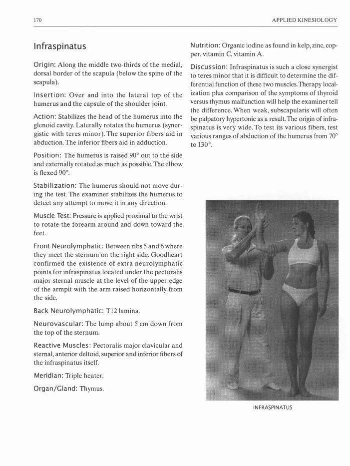
170
Infraspinatus
Origin: Along the middle two-thirds of the medial,
dorsal border of the scapula (below the spine of the
scapula).
I nsertion: Over and into the lateral top of the
humerus and the capsule of the shoulder joint.
Action: Stabilizes the head of the humerus into the glenoid cavity. Laterally rotates the humerus (syner
gistic with teres minor). The superior fibers aid in
abduction. The inferior fibers aid in adduction.
Position: The humerus is raised 90° out to the side
and externally rotated as much as possible. The elbow
is flexed 90°.
Stabilization: The humerus should not move dur
ing the test. The examiner stabilizes the humerus to
detect any attempt to move it in any direction.
Muscle Test: Pressure is applied proximal to the wrist
to rotate the forearm around and down toward the
feet.
Front Neurolymphatic: Between ribs 5 and 6 where
they meet the sternum on the right side. Goodheart
confirmed the existence of extra neurolymphatic
points for infraspinatus located under the pectoralis major sternal muscle at the level of the upper edge
of the armpit with the arm raised horizontally from
the side.
Back Neurolymphatic: T12 larnina.
Neurovascular: The lump about 5 em down from
the top of the sternum.
Reactive Muscles: Pectoralis major clavicular and
sternal, anterior deltoid, superior and inferior fibers of
the infraspinatus itself.
Meridian: Triple heater.
Organ/Gland: Thymus.
APPLIED KINESIOLOGY
Nutrition: Organic iodine as found in kelp, zinc, cop
per, vitamin C, vitamin A.
Discussion: Infraspinatus is such a close synergist
to teres minor that it is difficult to determine the dif
ferential function of these two muscles. Therapy local
ization plus comparison of the symptoms of thyroid
versus thymus malfunction will help the examiner tell
the difference. When weak, subscapularis will often
be palpatory hypertonic as a result. The origin of infraspinatus is very wide. To test its various fibers, test
various ranges of abduction of the humerus from 70°
to 130°.
INFRASPINATUS

MUSCLE TESTS
ANTERIOR NL POSTERIOR NL
INFRASPINATUS
NEUROVASCULAR POINT
171
SEDATION POINT,
TRIPLE HEATER 10

172
Latissimus Dorsi
Origin: The muscle is attached by a broad, flat, sheet
tendon from the spinous processes of the lower six
thoracic vertebrae and all the lumbar vertebrae plus
the posterior crest of the ilium, the lower 3 to 4 ribs,
plus an attachment to the bottom tip of the scapula.
Insertion: Into the medial side of the humerus just
under the shoulder joint.
Action: Pulls the arm (humerus) down and in, rotates
the arm medially, pulls the shoulder blade downward
and inward.
Position: With the elbow fully extended, the patient
rotates the arm inward (medially) and holds the arm
slightly away from the side.
Stabilization: The examiner stabilizes with a hand
on top of the shoulder tested to prevent the patient
from leaning sideways or lifting the shoulder.
Muscle Test: The examiner abducts and slightly flexes
the arm (pulls the arm away from the side and slightly
toward the front).
Front Neurolymphatic: In the junction of rib and
cartilage in the depression between ribs 7 and 8,
directly below the nipple on the left side only.
ANTERIOR NL POSTERIOR NL
APPLIED KINESIOLOGY
Back Neurolymphatic: In the intertransverse space
between T7-8 (usually only on the left).
Neurovascular: Just above the squamosal suture on
the parietal bone, slightly posterior to the vertical
auricular line (about two finger-widths above and
slightly behind each ear).
Reactive Muscles: Upper trapezius, contralateral
hamstrings, supraspinatus, deltoids, levator scapula.
Meridian: Spleen
Organ/Gland: Pancreas
Nutrition: Vitamins A and F, betaine, zinc, selenium
and chromium.
Discussion: These "wing" muscles are active in all
rowing motions and especially in movements that pull
the arms down (caudal) and in to the sides (adduc
tion) like chin-ups. Medially rotating the arm opti
mally aligns the origin and insertion of latissimus dorsi
for testing. However, this position makes it possible
to bend the arm, allowing biceps to aid (synergize) in
the action. For this reason, it is absolutely necessary
for the examiner to make sure that the elbow remain
completely extended during the test. If the patient
can hyperextend the elbow (most common in women),
the elbow must remain hyperextended during the test.
NEUROVASCULAR POINT
SEDATION POINT,
SPLEEN 5

MUSCLE TESTS
To prevent unwanted bending of the elbow, instruct
the patient to imagine clamping a newspaper between the upper arm and the side of the chest. During the
test, the examiner must avoid accidentally touching
(challenging) the alarm points along the radial artery
of the wrist.
Latissimus dorsi often test weak secondary to
hypertonic upper trapezius on the same side. Until
the hypertonicity of the upper trapezius is adequately
reduced, the latissimus dorsi weakness will return;
LATISSIMUS DORSI
173
even after successful strengthening techniques have
been applied (see "Reactive Muscles," page 133).
Latissimus dorsi is a good indicator of blood sugar
imbalances and digestive problems. It often tests weak
by reason of some malfunction in its associated gland,
the pancreas. When so, a left-side weakness often indi
cates problems with insulin production. A right-side
weakness often correlates with an imbalance in pan
creas enzyme production.
LATISSIMUS DORSI,
TESTED FALSE
LATISSIMUS DORSI,
TESTED WITH THE ELBOW
H YPEREXTENDED

174
Pectoralis Major Clavicular
Origin: On the anterior surface of the sternal half of
the clavicle.
Insertion: Into the anterior surface of the humerus,
just below the shoulder joint.
Action: Flexes the shoulder and adducts the humerus
horizontally toward the opposite shoulder (brings the arm up and in). It is the main flexor of the shoulder.
Position: The elbow must remain extended. The
patient holds her fully straightened arm directly to
the front of the trunk. The arm is maximally rotated
inward, which points the thumb toward the feet. The
shoulder is in a relaxed position and not elevated.
Stabilization: The examiner stabilizes the opposite
shoulder to prevent the trunk from rotating toward the arm tested.
Muscle Test: To avoid causing pain, the examiner
uses a full open-hand contact proximal to the patient's
wrist. Pressure is then applied laterally and slightly
inferior.
Front Neurolymphatic: Under the left breast in the
arc between ribs 6 and 7 from the sternum to the
mammillary line (usually only on the left).
Back Neurolymphatic: Between T6-7 near the lami
nae (usually only on the left) .
Neurovascular: Bennett's "emotional reflex" points
located directly above the irises on the frontal bone
eminences.
Reactive Muscles: Teres major, teres minor, latis
simus dorsi, rhomboids, latissimus dorsi, middle
trapezius, posterior deltoid, supraspinatus, gluteus
maximus.
Meridian: Stomach.
Organ/Gland: Stomach.
APPLIED KINESIOLOGY
Nutrition: Vitamin B complex, especially B12, copper, zinc, betaine hydrochloride.
Discussion: Pectoralis major clavicular is related
to emotional disturbances, which are known to cause
problems with the stomach. Weakness when tested
bilaterally (but strength on one or both sides when
tested individually) may indicate a lack of adequate
hydrochloric acid production in the stomach. This may be due to zinc deficiency. This may not show
until a bilateral weakness of the lower trapezius is
corrected. Many examiners only test this muscle
bilaterally.
PECTORALIS MAJOR CLAVICULAR,
TESTED STANDING

MUSCLE TESTS
PECTORALIS MAJOR CLAVICULAR
NEUROVASCULAR
POINT
SEDATION POINT,
STOMACH 45
175
ANTERIOR NL
POSTERIOR NL

176
Pectoralis Major Sternal
Origin: Along the side of the sternum to the 7th rib.
Insertion: Into the anterior lateral surface of the
humerus below the shoulder joint (distal to the inser
tion of the pectoralis major clavicular).
Action: Pulls the arm down and in (caudal and
medial).
Position: The elbow must remain extended. The
patient holds her fully straightened arm directly to
the front of the trunk. The arm is maximally rotated
inward, which points the thumb toward the feet.
Stabilization: The examiner stabilizes the opposite
shoulder or hip to prevent the trunk from rotating
toward the arm tested. If the hip is stabilized, the
abdominal muscles must be able to fix the chest to the pelvis.
Muscle Test: To avoid causing pain, the examiner
uses a full open hand contact proximal to the patient's
wrist. Pressure is applied at a 45° angle up and away
(cranial and lateral). The exact direction is best
applied along a line from the middle of the origin to
the middle of the insertion.
Front Neurolymphatic: Under the right breast in
the arc between ribs 5 and 6 from the mammillary
line to the sternum.
Back Neurolymphatic: Between T5-6 near the lam
inae, usually on the right side.
Neurovascular: Usually on the natural hairline about
4 cm above the frontal eminences, vertically above
the outside edges of the eyes.
Reactive Muscles: Rhomboids, upper and lower
trapezius, posterior deltoid, serratus anticus,
supraspinatus.
APPLIED KINESIOLOGY
Meridian: Liver.
Organ/Gland: Liver.
Nutrition: Vitamin A, bile salts, choline, inositol,
methionine, taurine. (See "Case Histories" #4, page
259.)
Discussion: Pectoralis major sternal makes an excel
lent indicator muscle for liver function. Sometimes
weakness is found in individuals who have a fear of
light. In such cases, vitamin A is helpful with both the
weakness and the fear of light. When pectoralis major
sternal tests weak, the rhomboids will secondarily
become hypertonic. Strengthen pectoralis first and,
if need be, apply muscle stretch techniques or pro
prioceptor manipulation to reduce the tension in the
rhomboids.
PECTORALIS MAJOR STERNAL, TESTED STANDING

MUSCLE TESTS
PECTORALIS MAJOR STERNAL
NEUROVASCULAR
POINT
SEDATION POINT,
LIVER 2
177
ANTERIOR NL
POSTERIOR NL
178
Pectoralis Minor
Origin: From ribs 3,4 and 5 near where the ribs turn
into cartilage before meeting the sternum.
Insertion: Into the coracoid process of the scapula.
Action: Pulls the coracoid process anterior, medial
and inferior, flexing the shoulder joint. Helps with the
act of forced inspiration.
Position: The patient lies supine.
Stabilization: Walther and Frost: Hold opposite
shoulder down upon the table. Leaf: Stabilize the
shoulder on the side tested. The patient's abdominal
muscles must be able to fix the rib cage to the hips. If the patient's abdominal muscles test weak and can
not be strengthened, the examiner must stabilize the
rib cage.
Muscle Test: Walther has the patient lift the shoulder
up off the table. The examiner presses the shoulder
down toward the table (posterior, lateral and caudal)
following the arc of the motion that the shoulder took
in attaining the test position. Leaf recommends that
the patient adduct the straightened arm to align with
the opposite anterior superior iliac spine (front of the
opposite hip). The examiner then elevates (abducts)
the arm. Neither test isolates the pectoralis minor
from the pectoralis majors (clavicular and sternal divi
sions). Palpation is the most important diagnostic pro
cedure for pectoralis minor.
Discussion with Gerz led me to design a more
effective test for pectoralis minor. The client lies
supine with the arm straightened, abducted 120°, and
extended at the shoulder as far as the weight of the
arm will pull it down toward the floor. If the arm
hangs below the level of the examiner's table, the pec
toralis majors will be removed from synergizing in
the test. In this position, the patient flexes the shoul
der up off the table. More specifically, she flexes the
pectoralis minor by pulling the coracoid process ante
rior, medial and inferior. The examiner presses the
shoulder down toward the table exactly opposing the
APPLIED KINESIOLOGY
arc of the motion produced by the contraction of pec
toralis minor (posterior, lateral and caudal). If the
arm cannot hang down lower than the level of the
table (which is very often the case), this test cannot
be performed because the pectoralis majors and
minor are shortened and held too tight. This obser
vation is in itself an important diagnostic result.
Front Neurolymphatic: Just above the xiphoid
process on the sternum.
Back Neurolymphatic: None recognized in AK.
Research by other schools of kinesiology recommends
T8-9, 9-10, 10-11 between where the ribs meet the
vertebrae (Dewe, 1989).
Neurovascular: None recognized in AK. Dewe found
that the same neurovascular point as used for the
upper trapezius works for the pectoralis minor. This point is located above the cheekbone where the tem
poral and sphenoidal bones meet, about 3 cm poste
rior to the outer comer of the eye.
Reactive Muscles: Serratus anticus, supraspinatus,
deltoid, upper trapezius.
Meridian: No meridian is recognized in AK. Dewe's
research correlates pectoralis minor with the stom
ach meridian.
Organ/Gland: Stomach?
Nutrition: RNA, niacin, niacinamide, B-complex vita
mins, zinc, water, herbs and other substances that pos
itively influence the flow of lymph.
Discussion: Pectoralis minor is difficult to isolate
for testing because pectoralis major sternal (synergist) usually assists it in its function. The pectoralis
minor is an important anterior shoulder stabilizer. It
is the only shoulder muscle that does not attach to
the humerus. This makes it difficult to test because
there are no long levers upon which to apply direct
pressure. Gerz considers pectoralis minor to be the
most important (and least tested) muscle in Applied
Kinesiology (Gerz, 1996, p. 50). Pectoralis minor is
most often shortened and tight. This may be felt by

MUSCLE TESTS
palpating the muscle perpendicular to the direction
of its fibers. When it is shortened, the shoulders are
pulled forward and inward (anterior and medial), pro
ducing a very common "slumped" postural appear
ance. A bundle of nerves and veins passes under the
pectoralis minor muscle. When the muscle is tight,
these get squeezed between the pectoralis minor and
the rib cage. This may cause pain and stiffness in the
shoulder joint. The thoracic duct and the right lym
phatic duct empty the lymph gathered from the entire
body into the junction between the subclavian and
internal juggler veins. They both rise in an arch under
the clavicle and return caudal before entering the
veins. The pectoralis minor does not pass directly over
these ducts. But all the structures of the neck are
invested in connected fascia. The pull exerted upon
the facial structures of the neck by a tight pectoralis
minor can squeeze the lymphatic ducts, limiting the
flow of lymph and thereby blocking the entire lymph
drainage of the body.
Pectoralis minor is seldom found to be undertoned.
This is because almost everyone has more developed
muscles on the anterior side of their bodies than the
posterior side. Just about everyone orients themselves toward their desks, steering wheels, etc. This gives
their "pushing" muscles lots of activity. Few people,
even weight lifters, give equal exercise to the pulling
179
and rowing muscles of the back. This can be easily
seen in the comparative size of the large and strong
anterior deltoids (shoulder flexors) and the typically
small and weak posterior deltoids (shoulder extensors). As a result, all the pectoralis muscles become
overly trained, shortened and tight compared to their
antagonists. For further discussion of this topic, see
the section "Exercise" on page 14l. Pectoralis minor may be shortened in response to
a bilateral weakness in the middle or lower trapezius muscles. If these muscles test weak, they must be
strengthened first before reassessing pectoralis minor.
Middle trapezius may be bilaterally weak due to a
fixation of the thoracic and lumbar vertebrae, which
requires chiropractic correction before any correc
tion of pectoralis minor will hold.
To lengthen a shortened pectoralis minor, use pro
prioceptor manipulation of the neuromuscular spin
dle cells and Golgi tendon organs, fascial stretch
technique, "chill and stretch," and reactive muscle
work as indicated for the specific case.
For lasting correction of shortened pectoralis minor
muscles, training to increase the strength of the shoul
der extensors (teres minor, teres major, latissimus
dorsi, trapezius, rhomboids, posterior deltoids, etc.)
is usually required.
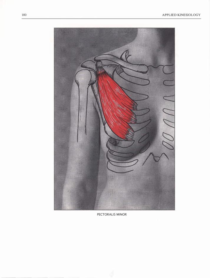
180 APPLIED KINESIOLOGY
PECTORALIS MINOR

MUSCLE TESTS
PECTORALIS MINOR (WALTHER)
PECTORALIS MINOR (FROSn
ANTERIOR NL POSTERIOR NL
181
PECTORALIS MINOR (LEAF, G ERZ)
NEUROVASCULAR POINT
SEDATION POINT,
STOMACH 4S

182
Peroneus Longus and Brevis
Origin-Longus: On the lateral head of the tibia, and
on the head and upper two-thirds of the lateral sur
face of the fibula and on the intermuscular septa.
Brevis: On the distal two-thirds of the fibula on the
lateral surface plus on the intermuscular septa.
Insertion-Longus: Wraps around the lateral side of
the foot and affixes to the lateral side of the proxi
mal end of the first metatarsal and the lateral side of
the adjacent bone, the medial cuneiform.
Brevis: Into the lateral side of the proximal end of
the 5th metatarsal.
Action: Flexes the sole of the foot (plantar flexion)
and turns the foot outward. Also provides lateral stabilization to the ankle.
Position: The foot must be completely straightened
(plantar flexed) and then turned out laterally
(everted) as far as possible. With maximal plantar
flexion, the range of motion to the side is small, per
haps 10-30°. The patient will attempt to extend the
toes and dorsiflex the foot. If so, instruct him not to
do so and start the test again. It may take several tries
to get the test parameters right.
Stabilization: Upon the medial side of the leg just
proximal to the ankle.
Muscle Test: With contact on the lateral side of the
foot (and not against the toes), the examiner applies
pressure to turn the foot back inward (in the direc
tion of inversion).
Front Neurolymphatic: On the inferior edge of the pubic bone.
Back Neurolymphatic: Between L5 and the PSIS.
Neurovascular: On the frontal bone eminences.
APPLIED KINESIOLOGY
Reactive Muscles: Tensor facia lata, tibialis anterior
and posterior, peroneus tertius.
Meridian: Bladder.
Organ: Urinary bladder.
Nutrition: Calcium, potassium, B complex, avoid
foods containing oxalic acid (coffee, cranberries,
plums, etc.) . If the patient cannot hold the breath longer than 30 seconds, repeat the tests (peroneus
and holding the breath) with thiamine chewed and
held in his or her mouth.
Discussion: Peroneus tertius, longus and brevis all
stabilize the ankle laterally. As the heel leaves the
ground, peroneus tertius is first active. When walk
ing, if the peroneus tertius is weak, the heel and ankle
will jerk laterally as the heel leaves the ground. As
the heel rises high from the ground, peroneus tertius
is inhibited and peroneus longus and brevis become
active.
The peroneus brevis has the same action as the
longus and is tested at the same time. Peroneus longus
and brevis test weak in many patients. Many begin
ning examiners fail to find weakness in peroneus
because of allowing dorsal flexion of the foot (in the
peroneus longus and brevis test) and extension of the
toes in either test. A screening test for peroneus weak
ness is to have the patient lie supine with knees
directly up (no outward rotation at the hips) and hang
the feet over the edge of the table. If the foot rotates
in (inverts), peroneus will likely test weak. Recurrent
spraining of the ankle is often due to weak-testing
peroneus muscles. Peroneus longus and brevis are
often reactive to the peroneus tertius, especially after
peroneus tertius has been injured by spraining the
ankle. Correcting this reactivity may be an important
step in preventing further ankle sprains. (See "Case
Histories" # 2, page 257.)

MUSCLE TESTS
PERONEUS LONGUS AND BREVIS
PERONEUS LONGUS AND BREVIS
ANTERIOR NL POSTERIOR NL NEUROVASCULAR POINT
183
INSERTION OF THE
PERONEUS LONGUS
SEDATION POINT,
BLADDER 65

184
Peroneus Tertius
Origin: On the lower one-third to one-half of the
anterior surface of the fibula and the intermuscular
septum.
Insertion: Into the dorsal surface
of the proximal end of the 5th
metatarsal (the bone in the foot from which the little toe continues.)
Action: Flexes the foot dorsally and
everts the foot.
Position: The patient lies supine (or
sits). The foot is dorsiflexed and
everted maximally. The toes are
flexed to prevent extension of the
toes.
Stabilization: The examiner stabi
lizes the leg above the malleoli
(ankle bones). This is easier to do
when the patient lies with the heel
upon the table.
Muscle Test: Pressure is applied
with a soft, broad contact upon the
dorsal side of the 5th metatarsal in
a direction toward plantar flexion
and inversion.
Reactive Muscles: Tensor fascia
lata, tibialis anterior and posterior,
peroneus longus and brevis.
APPLIED KINESIOLOGY
PERONEUS TERTIUS
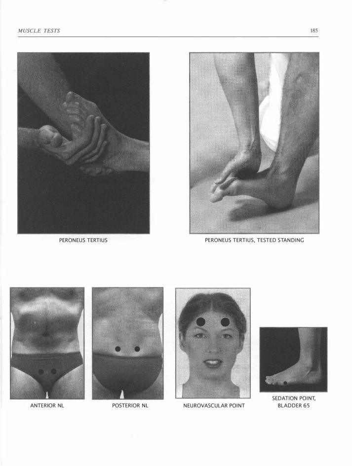
MUSCLE TESTS
PERONEUS TERTIUS
ANTERIOR NL POSTERIOR NL
185
PERONEUS TERTIUS, TESTED STANDING
NEUROVASCULAR POINT
SEDATION POINT,
BLADDER 65

186
Piriformis
Origin: On the anterior surface of the sacrum
between and lateral to the anterior sacral foramen
and onto the capsule of the sacroiliac joint.
Insertion: Into the superior border of the greater
trochanter.
Action: Piriformis helps hold the head of the femur into the acetabulum (hip joint). With the hip extended
or flexed less than 90°, piriformis laterally rotates the
thigh. With the hip flexed more than 90°, piriformis
medially rotates the thigh.
Position: The patient lies supine with the knee flexed
90° and the hip flexed less than 90°. The thigh is
rotated outward (laterally). Piriformis may also be
tested with the patient prone and the knee flexed 90° and the thigh rotated outwardly.
Stabilization: The examiner stabilizes above the knee
to prevent any medial or lateral motion of the thigh.
When using piriformis as an indicator muscle, the
examiner may stabilize the knee against his chest.
This leaves his other hand free for TL or challenge.
Muscle Test: Supine or prone. The examiner presses
above the ankle to rotate the thigh inwardly. The
examiner stabilizes the thigh near the knee to pre
vent motion of the thigh during the test.
Front Neurolymphatic: On the superior aspect of
the pubic bone.
Back Neurolymphatic: Between PSIS and the L5
spinous process.
Neurovascular: On the parietal eminence, above
and posterior to the ear.
Reactive Muscles: Contralateral splenius capitis,
gluteus medius, tensor fascia lata, adductors, medial
hamstrings.
APPLIED KINESIOLOGY
Meridian: Circulation-sex.
Organ/Gland: Reproductive organs and glands.
Nutrition: Vitamin E, vitamin A, niacin, zinc.
Discussion: Like iliopsoas, piriformis extends across
the sacroiliac junction. These two muscles are there
fore involved in stabilizing this important joint. Piri
formis is involved in most problems of the pelvis and
lumbar spine. When piriformis tests weak on both
sides, there may be severe structural problems in the
pelvis. However, it is more common to find bilateral
weakness in piriformis in chronic cases of urogenital
problems. This may take the form of sexual problems
including pain on intercourse in females and male
impotence. In such cases, if piriformis can be strength
ened on both sides, the urogenital problems can usu
ally be successfully treated. When piriformis is weak
on one side only, the other side is very often extremely
palpatory hypertonic. This can cause the ilium to be
pulled and fixed against the sacrum on the hypertonic
side. Sciatic nerve pain is often on the side of the pal
patory hypertonic piriformis. In such cases of one
sided weakness, strengthen piriformis on the weak
side first. If necessary, use standard Applied Kinesi
ology techniques (fascial release, chill and stretch,
proprioceptor manipulation, reactive muscles) for the palpatory hypertonic side. This may free the iliosacral
fixation without the need for chiropractic correction.
Piriformis is often used as an indicator muscle. To
do so, the examiner stands to the side of the supine
patient and tests the leg that is toward him. He sta
bilizes the patient's knee against his chest with the
thigh rotated laterally. This allows him to have one
hand free for therapy localization or challenge tests.
To perform the test, he places his hand above the
patient's ankle and pulls the patient's lower leg in an attempt to medially rotate the thigh.

MUSCLE TESTS 187
PIRIFORMIS

188 APPLIED KINESIOLOGY
PIRIFORMIS, TESTED PRONE

MUSCLE TESTS
PIRIFORMIS AS AN INDICATOR MUSCLE
ANTERIOR NL POSTERIOR NL
189
PIRIFORMIS, TESTED ON THE HANDS AND KNEES
NEUROVASCULAR POINT
SEDATION POINT,
CIRCULATION-SEX 7

190
Popliteus
Origin: On the lateral distal end of the femur, on the
posterior horn of the lateral meniscus, and on the
head of the fibula.
Insertion: Into the posterior surface of the proximal
end of the tibia.
Action: With the femur fixed, the popliteus rotates
the tibia medially. With the tibia fixed, the popliteus
rotates the femur laterally on the tibia. As the heel
strikes the ground in walking or running, the popli
teus contracts, which bends the knee and thus creates
a shock absorber effect. When the knee is flexed,
popliteus withdraws the lateral meniscus. Popliteus
stabilizes the knee posteriorly.
Position: The patient lies prone or supine. The knee
and the ankle are both flexed 90°. The tibia is rotated
medially on the femur.
Stabilization: The patient must be able to fix the
ankle and there must be no pain or problems in the
foot, ankle or knee. The patient must also fix the femur
to prevent rotation during the test. The examiner sta
bilizes the lateral aspect of the heel.
Muscle Test: Pressure is applied with a broad, soft,
hand contact on the medial and under side of the foot
near (but not upon) the toes. The opposite hand pro
vides counter-pressure on the heel. The effort is to
laterally rotate the tibia. Rotation of the foot may
take place even without popliteus weakness. The
actual motion during the test is very small. The indi
cation of weakness of the popliteus is lateral rotation
APPLIED KINESIOLOGY
of the tibia during the test. This can be most easily
detected by watching the head of the tibia (the tibial
tubercle) and the head of the fibula. Since the fibula
is attached to the tibia, they rotate simultaneously. If
during the test, the tibial tubercle rotates and causes
the head of the fibula to no longer protrude so far lat
erally, the popliteus tested weak.
Front Neu rolymphatic: In the intercostal space between ribs 5 and 6 from the mammillary line to the
sternum on the right side.
Back Neurolymphatic: Between the laminae of tho
racic vertebrae 5 and 6 on the right.
Neurovascular: On the medial aspect of the knee
(over the meniscus).
Reactive Muscles: Gastrocnemius, lateral ham
strings, quadriceps and upper trapezius.
Meridian: Gall Bladder.
Organ/Gland: Gall Bladder.
Nutrition: Vitamin A, betaine, essential fatty acids
(vitamin F), gall salts.
Discussion: When popliteus is weak, the knee in
standing is typically unstable and most often hyper
extended (or bent more than usual). When the popli
teus is weak, the lateral meniscus often has problems. If the popliteus tests weak bilaterally, therapy-local
ize the mid-cervical vertebrae. If this strengthens the
popliteus, there is a mid-cervical fixation that may
require chiropractic correction. Hyperextension
injuries to the knee strain the popliteus.

MUSCLE TESTS
POPLITEUS
ANTERIOR NL POSTERIOR NL
POPLITEUS TEST,
BEGINNING POSITION
191
POPLITEUS TEST,
END POSITION WHEN IT TESTS WEAK
NEUROVASCULAR POINT
SEDATION POINT,
GALL BLADDER 38

192
Rectus Abdominis Group
Origin: From the symphysis pubis and the pubic crest.
Insertion: Into the cartilage of the 5th, 6th and 7th
ribs near the sternum an on the lateral aspect of the
xiphoid process.
Action: The rectus abdominis connects the ribs and
the pelvis, thus holding abdominal organs in. With the
help of gluteus maximus and the hamstrings, the rec
tus abdominis keeps the pelvis from tilting forward
(anteriorly).
Position: The patient sits with extended legs, torso
at 90° to the legs. Patient crosses arms upon the chest
(with hands closed and elbows away from the chest
to avoid TL to any active points). If any low back pain exists, the test should be performed with the knees
and hips bent.
Stabilization: Patient's leg flexors must fix the hips
to the legs. If they cannot, they must be strengthened
before this test can be performed. Examiner must
hold the patient's legs down toward the table.
Muscle Test: Examiner places one hand on the ster
num and pushes the upper body away from the legs. Alternately, using one arm, the examiner places his
elbow on one of the patient's shoulders and his hand
on the other shoulder. During the test, a strong iliop
soas can hold the lumbar spine forward, giving the
impression of strong abdominals. The examiner should
observe carefully if the patient can stabilize the ribs
toward the pubis. Any separation of the ribs from the
pubis indicates weak-testing abdominals.
APPLIED KINESIOLOGY
Front Neurolymphatic: A line about 10 cm long on
the medial thigh just above the knee. Massaging this
area produces an unpleasant electric shock sensation.
It is advised to stand toward the feet and out of the
reach of your patient to avoid being accidentally
struck.
Back Neurolymphatic: In the space between L5 and
the PSIS.
Neurovascular: Where the vertical auricular line
crosses the parietal eminences.
Reactive Muscles: Quadriceps, contralateral glu
teus medius, sacrospinalis, rectus femoris, sartorius,
quadratus lumborum, between sections of rectus
abdominis itselt
Meridian: Small intestine.
Organ/Gland: Small intestine.
Nutrition: Vitamin E, coenzyme Q10.
Discussion: If the patient has a history of low back
disc problems, test with the knees bent to put less
stress on the discs. To support the patient, the exam
iner must lean weight against the thighs near the
knees to keep the patient's feet upon the table.
When the rectus abdominis tests weak, it cannot
provide support to the pelvis, which then tips forward
(tilts anteriorly), creating an excessive lordosis (hol
low low back). This places a great stress upon the discs
between the lumbar vertebrae.
When the patient has any chronic disturbances in
the small intestine, the rectus abdominis will typically
test very weak, and the neurolymphatic reflexes will
be extremely tender. In such cases, dietary correc
tions, and other lymphatic corrections including the

MUSCLE TESTS
OPENING THE SAGITTAL SUTURE TO
STRENGTHEN
THE RECTUS ABDOMINIS
193
drinking of more water will be needed before the cor
rection of the abdominis will hold.
Weak-testing abdominal muscles are found in con
junction with a jamming together of the sagittal
suture. If this suture remains jammed, any correction
of the abdominals will not hold. To test if the suture
is involved with weakness of the abdominals, the
patient TLs the suture while the examiner repeats the muscle test. If the rectus (or other) abdominis
muscles now test strong, the sagittal suture is involved.
Correcting this jamming of the sagittal suture is one
of the simplest and safest of all cranial fault correc
tions. Using the fingertips on each side of the suture,
the examiner pulls it apart as if trying to pull open
the top back of the patient's head to the right and
left. This is performed three or four times, and then
the abdominal muscles are retested to confirm the correction.
Many times, the abdominals (which are spinal flex
ors) test weak as a result of excess tension in their
main antagonist, sacrospinalis (a spinal extensor).
After strengthening the abdominals, check if tensing
the sacrospinalis makes them again weak. If so, apply the reactive muscle technique to lower the tension
in the sacrospinalis and free the abdominals from its
influence (page 135).
The various divisions of the rectus abdominis are
individually enervated and may be reactive to one
another. When the back is rounded (straightening the
lumbar curve), the lowest division is tested. Begin
ning from this rounded posture, a step-wise straight
ening of the back before testing tests the various upper divisions sequentially from lower to higher.
For instructions as to how to safely exercise the
abdominals, see the "Exercise" on pages 142-143).

194 APPLIED KINESIOLOGY
RECTUS ABDOMINIS

MUSCLE TESTS
RECTUS ABDOMINIS,
WITH STRAIGHT LEGS
ANTERIOR NL POSTERIOR NL
RECTUS ABDOMINIS,
WITH BENT LEGS
195
NEUROVASCULAR POINT
SEDATION POINT,
SMALL INTESTINE 8
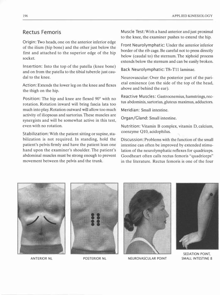
196
Rectus Femoris
Origin: Tho heads, one on the anterior inferior edge
of the ilium (hip bone) and the other just below the
first and attached to the superior edge of the hip
socket.
Insertion: Into the top of the patella (knee bone)
and on from the patella to the tibial tubercle just cau
dal to the knee.
Action: Extends the lower leg on the knee and flexes
the thigh on the hip.
Position: The hip and knee are flexed 90° with no
rotation. Rotation inward will bring fascia lata too
much into play. Rotation outward will allow too much
activity of iliopsoas and sartorius. These muscles are
synergists and will be somewhat active in this test,
even with no rotation.
Stabilization: With the patient sitting or supine, sta
bilization is not required. In standing, hold the
patient's pelvis firmly and have the patient lean one
hand upon the examiner's shoulder. The patient's
abdominal muscles must be strong enough to prevent
movement between the pelvis and the trunk.
ANTERIOR NL POSTERIOR NL
APPLIED KINESIOLOGY
Muscle Test: With a hand anterior and just proximal
to the knee, the examiner pushes to extend the hip.
Front Neurolymphatic: Under the anterior inferior
border of the rib cage. Be careful not to press directly
below (caudal to) the sternum. The xiphoid process
extends below the sternum and can be easily broken.
Back Neurolymphatic: T8-T11 laminae.
Neurovascular: Over the posterior part of the pari
etal eminence (on the side of the top of the head,
above and behind the ear).
Reactive Muscles: Gastrocnemius, hamstrings, rec
tus abdominis, sartorius, gluteus maximus, adductors.
Meridian: Small intestine.
Organ/Gland: Small intestine.
Nutrition: Vitamin B complex, vitamin D, calcium,
coenzyme QlO, acidophil us. 'I
Discussion: Problems with the function of the small
intestine can often be improved by extended stimu
lation of the neurolymphatic reflexes for quadriceps.
Goodheart often calls rectus femoris "quadriceps"
in the literature. Rectus femoris is one of the four
NEUROVASCULAR POINT
SEDATION POINT,
SMALL INTESTINE 8

MUSCLE TESTS
quadriceps muscles, which all have the same reflex
points. For a discussion of the use of the neurolym
phatic reflexes to allow quadriceps to continue to con
tract at full power for extended lengths of time (useful
to athletes!) see the section on repeated muscle test
ing (pages 123).
197
In many athletic injuries, quadriceps is often found
to be involved in reactive muscle relationship, either
as the primary or a reactive muscle. Correcting this
reactive muscle relationship can help the athlete to
return more quickly and without pain to his or her
sport.
RECTUS FEMORIS
RECTUS FEMORIS, TESTED PUSHING RECTUS FEMORIS, TESTED PULLING

198
Rhomboid Major
Origin: Both Walther and Leaf: from the spinous
processes of T2-5. Other authorities say TI-5.
Insertion: Into the medial margin of the scapula.
Rhomboid Minor
Origin: Walther and Leaf-from the spinous processes
of C7 and Tl . Other authorities say C6 and C7.
Insert ion: Into the medial margin of the scapula above rhomboid major.
Action of both: Adduction and elevation of the
medial border of the scapula.
Position: The standing or seated patient adducts and
elevates the scapula. If the patient cannot elevate the
medial border of the scapula, the middle trapezius is
being recruited to perform the adduction. The arm,
which is used as a lever for testing the rhomboids,
should be in a neutral or slightly externally rotated
position.
Stabilization: The patient must be able to hold her
arm down against her side. The examiner should sta
bilize the top of the shoulder on the side tested to
prevent the patient from tipping toward this side dur
ing the test.
Muscle Test: The examiner pulls the elbow to abduct
it away from the body. Movement of the elbow away
from the body does not, in itself, indicate rhomboid
weakness. The examiner should observe for any
abduction and/or sinking of the scapula, which is the
true indication of rhomboid weakness.
APPLIED KINESIOLOGY
Front Neurolymphatic : On the left side between
ribs 6 and 7 from the mammillary line to the sternum.
Back Neurolymphatic: Between T6-7 by the lami
nae on the left side.
Neurovascular: Walther lists the frontal bone emi
nences (used for pectoralis major clavicularis-the
stomach meridian). Leaf lists the points above and
more lateral to the frontal bone eminences (used for
pectoralis major sternal-the liver meridian).
Reactive Muscles: Deltoid, serratus anticus,
supraspinatus, latissimus dorsi, upper, middle and
lower trapezius, coracobrachialis, pectoralis majors
and minor.
Meridian: Liver (Stomach).
Organ/Gland: Liver (Stomach).
Nutrition: Vitamin A, choline, inositol, methionine.
Discussion: Goodheart has associated the rhom
boids with both the liver and the stomach meridians.
The rhomboids seem to respond to neurovascular and
neurolymphatic stimulation of the reflexes for both
the stomach and the liver. Therefore, if the desired
effect is not achieved with treatment of the neu
rolymphatic reflex points on the left side, use those
on the right as well.
When serratus anticus is weak, the antagonistic
rhomboids will often become shortened and hyper
tonic. This is often observed in patients who have pain
between the scapulae. In such cases, attempting to get
the rhomboids to relax will usually not succeed until
serratus anticus is first strengthened. Although antag
onistic to the rhomboids in the motion of the scapula,
serratus anticus is synergistic to the rhomboids in their
combined action of fixing the medial border of the
scapula down upon the rib cage.

MUSCLE TESTS 199
RHOMBOIDS, END POSITION WHEN THEY TEST WEAK

200
Leaf comments that weakness of the rhomboids allows the scapula to excessively rotate during ele
vation of the arm to the front. This can stretch the
suprascapular nerve and thereby weaken the infra
spinatus muscle.
The rhomboids are often involved as the primary
or the reactive muscle in combination with various
other shoulder muscles. Since in normal posture,
there is little space between the medial edge of the scapula and the vertebral column, pinching the neu
romuscular spindle cells of the rhomboids (as
required to sedate a primary muscle) is difficult to
perform. To make more room for such treatment, the
examiner stands behind the patient and reaches
under the armpit and then grasps the superior aspect
ANTERIOR NL POSTERIOR NL
APPLIED KINESIOLOGY
of the shoulder. Then he levers the patient's arm ante
riorly, which pulls the scapula away from the verte
bral column. This lengthens the rhomboids, producing
more space for the neuromuscular spindle cell
manipulation.
When the rhomboids are overly tight, they inhibit
the ability to extend the arms (and shoulders) ante
riorly, as one would to give or receive a hug. Inter
estingly, in my work as a psychologist, I have often
observed a correlation between hypertonic rhom
boids and an inability to reach out-a fear of open
ing oneself to another person. This is just an anecdotal
observation and does not (yet) fonn a part of Applied
Kinesiology.
NEUROVASCULAR POINT
SEDATION POINT, LIVER 2
AND STOMACH 45

MUSCLE TESTS 201
RHOMBOIDS, BEGINNING POSITION
RHOMBOIDS, PINCHING THE NEUROMUSCULAR SPINDLE CELLS

202
Sacrospinalis Group
Origin: (Many separate muscles) upon the sacrum,
iliac crest, spinous processes, transverse processes
and ribs.
Insertion: Into the ribs, spinous processes, transverse
processes and occiput.
Action: Extension, rotation and lateral flexion of the vertebral column, lateral motion of the pelvis.
Position: The patient lies prone and lifts one shoul
der up from the table without using the hands.
Stabilization: The opposite hip is held down on the
table.
Muscle Test: The examiner pushes the raised shoul
der down toward the table.
Front Neurolymphatic: On the anterior surface of
the pubic bone and 2.5 cm lateral to each side of the
navel.
ANTERIOR N L POSTERIOR N L
APPLIED KINESIOLOGY
Back Neurolymphatic: L2, over the transverse
processes.
Neurovascular: Frontal bone eminences.
Reactive Muscles: Gluteus maximus, hamstrings,
abdominis.
Meridian: Bladder.
Organ: Urinary bladder.
Nutrition: Vitamins A, C, P, E and calcium.
Discussion: To get a general idea of the function of
sacrospinalis, have the patient lean directly to each
side (laterally flex the spine) with no tilt of the hips
and no rotation of the spine. The patient will not be
able to lean as far toward the side of sacrospinalis
weakness.
N EUROVASCULAR POINT
SEDATION POI NT,
BLADDER 65
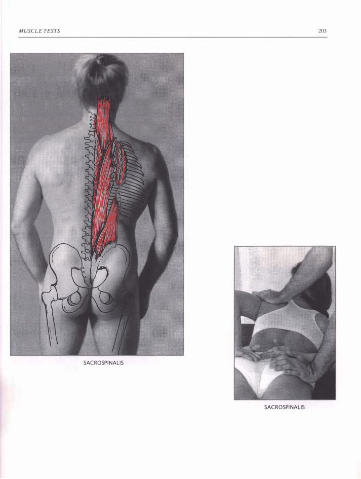
MUSCLE TESTS 203
SACROSPI NAllS
SACROSPI NAllS

204
Sartorius
Origin: On the front top of the hip bone (anterior
superior iliac spine).
Insertion: On the medial side of the shinbone (tibia)
just below the knee.
Action: Sartorius extends over two joints and flexes
the knee and the hip. It rotates the thighs laterally. It stabilizes the medial side of the knee and, when
the knee is flexed, it rotates the tibia medially. When
the leg is fixed, sartorius flexes the hip upon the thigh
and stabilizes the pelvis anteriorly.
Position: The client lies supine, slightly flexes the
knee and the hip joints, and rotates the thigh later
ally (knee out).
Stabilization: Normally not required. If the client
slips on the table, she may hold on to the sides of the
table.
Muscle Test: With one hand, the examiner grasps
the leg above the ankle and pulls to extend the knee.
ANTERIOR NL POSTERIOR NL
APPLIED KINESIOLOGY
The other hand pushes the knee in the direction of
hip extension, thigh adduction and medial rotation.
Both hands must act at the same time. Experiment
with the exact direction of pressure to produce max
imal contraction as observed in the contraction of
the sartorius muscle.
Front Neurolymphatic: 5 cm above the navel and
2.5 cm to each side.
Back Neurolymphatic: T11, 12 near the lamina.
Neurovascular: Posterior fontanelle (lambda).
Reactive Muscles: Quadriceps and anterior tibialis.
Meridian: Circulation-sex and sometimes triple
heater.
Gland: Adrenal.
Nutrition: Vitamin C, pantothenic acid, adrenal extracts, tyrosine, B6, B12, folic acid.
Discussion: The sartorius is the longest muscle in
the body. It is an anterior pelvic stabilizer. Weak
ness in sartorius can cause the ilium on the side of
NEUROVASCULAR POINT
SEDATION POINT,
CIRCULATION-SEX 7

MUSCLE TESTS
weakness to sub luxate posteriorly and can result in
"knock knees." In these conditions, the sartorius' con
tinual effort to stabilize the problem joints will make
the origin and/or insertion of sartorius very tender.
All functions and imbalances associated with the
adrenals (low blood pressure, hypoglycemia or low
blood sugar, hyper insulin production, allergies, and
asthma) can influence sartorius function. Infections
SARTORIUS, TESTED SITTING
205
and adrenal exhaustion influence sartorius. Con
versely, balancing the sartorius can positively influ
ence the function of the adrenals including all the
above-listed adrenal-related problems.
If the client is strong, the examiner may need to
use rotation of his or her pelvis to deliver the force
needed for the test. Sartorius often reveals weakness
only when the patient is tested in sitting position.
SARTORIUS

206
Serratus Anticus
Origin: The lateral superior surfaces of the first 8 or
9 ribs.
Insertion: Underneath the vertebral edge of the
scapula.
Action: Pulls the scapula laterally and rotates it to
place the shoulder joint more vertical. Holds the vertebral border of the scapula down upon the rib cage
(as does its synergists in this activity, rhomboids and
middle trapezius) . With the scapula fixed, serratus
anticus lifts the ribs and assists in forced breathing.
Position: The straightened arm is flexed at the shoul
der to 100°-150° (various angles to test the various
fibers of this muscle) and abducted about 30°.
Stabilization: Not generally needed for this test.
Muscle Test: The examiner presses the lateral edge
of the inferior tip of the scapula medially and feels
the medial edge with his fingers. With his other hand,
he pulls down upon the forearm, proximal to the
patient's wrist. If the scapula moves medially and the
medial edge flares away from the rib cage, serratus
anticus tested weak. The examiner must place his
hand upon the scapula and/or observe the scapula for
movement during the test. Most effective is for the
examiner to press upon the lateral edge of the scapula
during the test. If the scapula moves medially, the ser
ratus anticus tested weak.
Front Neurolymphatic: Between ribs 3, 4 and 5 near
where they meet the sternum.
Back Neurolymphatic: T3, 4 and 5 laminae.
APPLIED KINESIOLOGY
Neurovascular: Anterior fontanelle (bregma).
Reactive Muscles: Rhomboid, pectoralis major ster
nal, pectoralis minor, middle trapezius.
Meridian: Lung.
Organ/Gland: Lung.
Nutrition: Vitamin C, water, beta-carotene.
Discussion: The test determines if serratus can stabilize the scapula. The arm is used as a lever. No
movement should occur at the shoulder joint during
the test. The deltoid and supraspinatus must be strong
in order to test the serratus. When serratus anticus is
weak, pushing with straight arms to the front is diffi
cult. If a weight is held straight out to the front, the
vertebral (medial) edge of the scapula will flare up
away from the back.
SERRATUS ANTICUS, THUMB GRIP

MUSCLE TESTS
SERRATUS ANTI CUS
ANTERIOR NL POSTERIOR NL NEUROVASCULAR POINT
207
SERRATUS ANTICUS
SEDATION POINT,
LUNG 5

208
Sternocleidomastoideus: Neck Flexors
Origin: Sternal head: on the anterior surface of the
manubrium (top-front of the sternum).
Clavicular head: upon the upper surface of the
medial half of the clavicle.
I nsertion: Upon the lateral surface of the mastoid process and the superior nuchal line of the occiput.
Action: One-sided contraction pulls the head toward
the shoulder on the same side as the muscle and rotates the head toward the opposite side. Bilateral
contraction flexes the head upon the neck.
Position: The patient lies upon the back. The arms
are flexed and lifted above the head to avoid press
ing on the table during the test. To test bilaterally, the
patient flexes (lifts) the head from the table. To test
unilaterally, the patient fully rotates the head away
from the side to be tested and lifts it from the table.
The examiner places his other hand upon the table
under the head to catch it in case of weakness.
Stabilization: Tested supine, the table provides the
necessary stabilization. If this test is performed stand
ing, the examiner must provide stabilization with one
hand upon the upper back. When testing in this posi
tion, as soon as the muscle demonstrates any weakness, the test pressure must immediately cease. Great
care must be observed to never push the head back
and down, which could damage the neck.
Muscle Test: Bilateral: Using a broad area of con
tact, the examiner places his hand upon the middle
of the forehead of the patient and presses posterior,
carefully following the arc of the anatomical move
ment. Under no conditions should the pressure be
straight down toward the table, which could injure
the intervertebral discs. In the unilateral test, the
examiner places his hand upon the parietal bone. The
exact area of contact is defined by how far the patient
can rotate his head. The direction of pressure is pos
terior and slightly lateral, in a curve following the
APPLIED KINESIOLOGY
anatomical arc of motion produced by the contrac
tion of the muscle.
Front Neurolymphatic: 1st intercostal space, about
9 cm from the sternum
Back Neurolymphatic: C2 lamina.
Neurovascular: Ramus of the jaw below the zygoma.
Reactive Muscles: Neck extensors, upper trapezius, pectoralis major clavicularis, opposite sternocleido
mas to ide us.
Meridian: Stomach.
Organ: Sinuses, lymphatic drainage of the head.
Nutrition: B6, B3 (niacinamide or niacin), organic
iodine, herbal and homeopathic remedies for the
sinuses, especially nosodes and lymph drainage reme
dies. Leaf recommends a 5-to-1 ratio of niacinamide or niacin to B6 as being most effective.
Discussion: The sternocleidomastoideus (SCM) is
actually a group of several associated muscles. The
contraction of the sterno-cleido-mastoideus is easily
visible, springing out of the neck during the muscle
test. Although quite useful as a general test, the bilat
eral test does not adequately isolate the SCM. In this test, the scalenes and several other neck flexor mus
cles are also active. To best isolate the SCM, the head should be maximally rotated, and the SCM of each
side tested independently. The examiner observes for
any attempt to reduce the angle of rotation during
the test. This usually indicates a weakness of the SCM
and an attempt to recruit the scaienes, which are most
active at 10° of rotation. Inability to remain in the full
rotation or pain upon doing so should be considered
a sign of muscle weakness and treated without put
ting the patient into unnecessary stress or pain that
could be caused by testing. Pressure is applied in the
direction of the tangent of the arc produced by the
anatomical motion of contraction of the SCM. Apply
ing test pressure with a broad contact of the palm
avoids possible tugging on the skin, which can pro
duce a false weakness of the muscle test.

MUSCLE TESTS
Patients with chronic sinus conditions or problems
in the upper vertebrae of the neck may have
extremely weak-testing neck flexors. In such cases,
this test is only to be performed with very gentle test
pressure.
The neck flexors are functionally much weaker
than their opponents, the neck extensors. Due in part
to the postural habit of holding the head far forward
of the line of gravity, the neck extensors rarely relax.
This often creates a pattern of (chronically tight) pri
mary muscle-neck extensors to (chronically weak,
inhibited) reactive muscle-neck flexors. If this condi
tion exists, treatments to strengthen the neck flexors
will only have temporary success until the treatment
for reactive muscles is applied. To test for such reactivity, the neck flexors must first test strong or be tem
porarily strengthened, for example, by massage of the
appropriate neurolymphatic reflex points. Then the
neck extensors are contracted bilaterally. If the patient
is supine, this may be easily accomplished by having
the patient firmly pull the head back down upon the
table.
Immediately following this contraction of the neck
extensors, the neck flexors are bilaterally tested. If
they now test weak, treatment for reactive muscles is indicated. In this case, such treatment consists of neu
romuscular spindle cell pinching of the belly on the
upper trapezius muscles and other neck extensors
located parallel to the spine in the neck, parallel to
the contracting length of the muscles. Areas of pal
pable hardness are repeatedly and deeply pinched.
Support must be given to the forehead so the neck
extensors are not contracted against the force
required for the neuromuscular spindle cell pinching treatment. This may be accomplished by having the
patient sit next to the table, place his elbows upon the
table, and his forehead upon the heels of his hands.
Alternatively, he can lie prone with his forehead upon
his forearm or a hard pillow. It is important to pinch
deeply into the muscle and not only upon the skin.
Then the neck muscles are tested bilaterally to reset
them. This process is repeated several times, focusing
upon the areas of hardness and/or pain in the upper
209
trapezius muscles and other neck extensors. After
effective treatment, contraction of the neck exten
sors should no longer weaken the neck flexors.
Reactive conditions may also exist between uni
lateral SCM and upper trapezius muscles and also
between the SCM muscles on each side. A notewor
thy reactive condition may also be observed between
the reactive SCM on one side and the contralateral
psoas muscle. The author observed and treated this
condition in a European soccer player. After hitting
the ball very hard with one side of his head, he found
that the SCM on that side became hypertonic. Sub
sequently, the insertion of the psoas on the opposite
side of his body became extremely tender to the touch
and painful when he stood or ran. Not only was the
contralateral psoas insertion tender, it also tested
weak on this side. The inhibition of the contralateral
psoas muscle, created by the hypertonic SCM, also
caused him to be out of balance and unstable when
upright. Reactive muscle treatment directed toward
the hypertonic SCM eliminated the pain and
improved his balance and coordination.
Notice in this case how the activity of the unilat
eral SCM nearly mirrors the activity of the con
tralateral psoas. The contraction of the right SCM
pulls the head forward and pulls the right ear toward
the midline. The contraction of the left psoas lifts the left leg forward and pulls it toward the midline.
Though the whole trunk lies between them, so con
sidered, these two muscles may be seen as having a
related and opposing function. This is an example of
the complex relationship often observed between
muscle pairs involved in reactive conditions.
Reactive muscle treatment should be long-lasting. However, the postural problem that caused the
chronic contraction of the neck extensors needs to
be corrected, for example, by lessons in Alexander
Technique (training in optimal posture and move
ment of the body). Or, if the cause of the muscular
imbalance was a trauma, emotional stress release
methods may need to be performed. If these steps
are skipped and the reactivity is only treated manually,
the reactive condition with the associated patterns of

210
tension, pain and possible malfunction of the gait
mechanism is likely to recur.
Malfunction of the neck flexors often causes
peripheral nerve and vascular entrapments in the
shoulders and arms. When this is suspected, have the
patient lie supine and test the function of various arm
muscles that should test strong. Then retest the same
muscles with the head elevated and rotated to con
tract the neck flexors. If the muscles now test weak, have the patient expand and lift the rib cage. If this
does not cause a strengthening of the formerly weak
testing muscles, a nerve entrapment has been located.
These may be corrected with AK procedures such as
chill and stretch or fascial release, carefully applied
to the SCM.
The SCM is particularly interesting because it has
double enervation from C2-C3 and from the tenth
cranial nerve. The only other muscle in the body that
has double enervation is the upper trapezius, which
is the major neck extensor. These two muscles have
a finely attuned simultaneous interaction without
which the fine coordination of the walking (gait) pat
tern would not be possible. Goodheart suggests that
the double enervation of these muscles is necessary
for the correct function of the gait coordination. In
walking or running, there should be a well-orchestrated facilitation and inhibition of the next flexors
and extensors. When the left leg steps forward, the
right shoulder moves forward. In order to keep the
head facing forward, the right SCM as well as the left
upper trapezius (and other left neck extensors) are
inhibited. This is the normal walking or gait coordi
nation. Goodheart devised a method to test for the
normal facilitation and inhibition of these muscles:
APPLIED KINESIOLOGY
The patient steps forward on his right foot and puts
much of his weight upon it. Some weight is also upon
the ball of the left foot with the heel raised as would
be the case when taking a step in walking. In this posi
tion, the SCM and upper trapezius muscles are tested.
If the gait mechanism is intact, in this position the
left SCM and the right upper trapezius should be
inhibited and therefore test weak. Since the upper
trapezius is typically in a state of chronic tension, the
weakness produced in this test may be difficult to
ascertain.
Unilateral weakness of the SCM and/or upper
trapezius muscles can produce a lateral tilt of the
head, which may cause occipital or upper cervical
subluxation and improper stimulation of the head
on-neck reflexes, with consequent disruption of the
equilibrium mechanism. In cases of dizziness, this
possible cause should be considered.
In my work as a psychologist, I have observed that
bilateral weakness of the neck flexors is often cou
pled with a weakened ability to effectively overcome
obstacles and push one's will through against the
opposition of others. These muscles must be strong
in order to "butt heads" effectively, as observed in
dominance rituals in the animal kingdom. Weak-test
ing neck flexors may be associated with being a
"pushover." These are only anecdotal observations
and do not (yet) form an official part of Applied
Kinesiology.
STERNO-CLEIDO-MASTOIDEUS,
BILATERALLY TESTED

MUSCLE TESTS
STERNO -CLEIDO -MASTOIDEUS. UNILATERALLY TESTED
ANTERIOR NL POSTERIOR NL NEUROVASCULAR POINT
21 1
SEDATION POINT,
STOMACH 4 5

212
Subclavius
Origin: On the top of the first rib and its cartilage.
Insertion: Subclavius has a wide insertion under the
middle of the clavicle.
Action: Pulls the clavicle anterior and interior and
pulls it medially toward the sternal-clavicular joint.
Position: Walther says that this muscle is not directly
testable. He tests indirectly by having the client TL
subclavius while an indicator muscle is tested. Good
heart, Leaf and Gerz use the test devised by Alan
Beardall with the arms held straight up to the ears.
Stabilization: If both arms are tested at once, no
stabilization is needed. If only one arm is tested, the
examiner stabilizes upon the trapezius on the side
tested with fingers or thumb upon the clavicle. This has the advantage of allowing the examiner to feel
if the subclavius is active during the test.
Muscle Test: The examiner applies pressure proxi
mal to the wrist to bring the arm away from the head.
The slightest motion of the clavicle indicates weak
ness of the subclavius.
Front Neurolymphatic: At the junction of the clav
icle, sternum and first rib. Also just up from the bot
tom of the sternum (Dewe).
Back Neurolymphatic: One inch to each side of T!
(on the lamina). Also TlO-ll, two inches to each side
of the spine (Dewe).
Neurovascular: Two finger-widths in front of the
ear on a line just above where the top of the ear
attaches to the head.
Reactive Muscles: Upper trapezius.
Meridian: None recognized in AK. Dewe's research
says heart.
APPLIED KINESIOLOGY
Organ: Heart?
Nutrition: Vitamin E, B-complex, C, heart, magne
sium.
Discussion: Subclavius is usually involved in the
"frozen shoulder" syndrome. In such cases, the client
must lean sideways to lift the arm above horizontal.
In 30% of such cases, strengthening of the subclav
ius immediately improves the ability to elevate the
shoulder. AI( recommends treatment applied directly
to the muscle such as proprioceptor manipulation or
fascial release.
SUBCLAVIUS, BILATERALLY TESTED
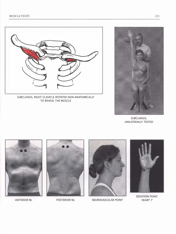
MUSCLE TESTS
SUBCLAVIUS, RIGHT CLAVICLE ROTATED NON-ANATOMICALLY
TO REVEAL THE MUSCLE
ANTERIOR NL POSTERIOR NL NEUROVASCULAR POINT
SUBCLAVIUS,
UNILATERALLY TESTED
213
SEDATION POINT,
HEART 7

214
Su bscapu lari s
Orig in: On nearly the whole inner surface of the
scapula.
I nsertion: Into the anterior surface of the humerus
at the level of the shoulder joint and into the infe
rior portion of the shoulder joint capsule.
Action: Rotates the humerus inwardly. Assists in
adduction of the shoulder. Also pulls the head of the
humerus forward and down when the arm is raised,
which stabilizes the head of the humerus in the gle
noid cavity.
Position: The humerus is at 90° to the side of the
trunk. The elbow is bent 90°. The humerus is rotated
medially, which brings the hand down toward the hip
as far as possible without lifting the shoulder. The test may be performed standing, sitting, prone or
supine.
Stabil ization: The examiner stabilizes the humerus
near the elbow to detect any attempt to move the
upper arm, which should remain motionless during
the test.
• •
ANTERIOR NL POSTERIOR NL
APPLIED KINESIOLOGY
Muscle Test: The examiner presses proximal to the
wrist to laterally rotate the forearm around the
humerus and up toward the head.
Front Neurolymphatic: Between ribs 2 and 3 where
they meet the sternum.
Back Neurolymphatic: Between the transverse
processes of T2-3.
Neurovascular: Anterior fontanelle (bregma).
Reactive Muscles: Teres minor, infraspinatus, del
toids, supraspinatus.
Meridian: Heart.
Organ/Gland: Heart.
Nutrition: Vitamin E, vitamins B2, B3, vitamin C,
carnitine.
Discussion: When exercise does not increase the
heart rate as expected, subscapularis often tests weak.
In this case, neurolymphatic stimulation will
strengthen subscapularis and correct the heart's
response to exercise. "Fluttering of the heart" con
tractions, breast and shoulder pains, and dizziness
NEUROVASCULAR POINT
SEDATION POINT,
HEART 7
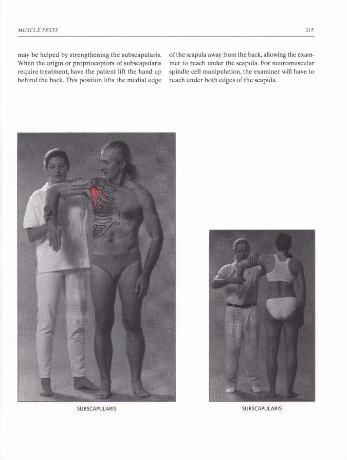
MUSCLE TESTS
may be helped by strengthening the subscapularis.
When the origin or proprioceptors of subscapularis
require treatment, have the patient lift the hand up
behind the back. This position lifts the medial edge
SUBSCAPULARIS
215
of the scapula away from the back, allowing the exam
iner to reach under the scapula. For neuromuscular
spindle cell manipulation, the examiner will have to
reach under both edges of the scapula.
SUBSCAPULARIS

216
Supraspinatus
Origin: The inner two-thirds of the supraspinatus
fossa, the part of the scapula above its "spine" ridge.
Insertion: Into the lateral surface of the head of the
humerus at the level of the shoulder joint.
Action: Abducts the arm (directly to the side and
slightly forward). Also holds the head of the humerus
into the scapula (stabilizes the shoulder joint) .
Supraspinatus can never fully relax or the shoulder
would dislocate.
Position: The patient brings the arm 15°-20° to the
side and slightly forward. The palm is oriented toward
the groin.
Stabilization: Upon the same or the opposite shoul
der. Same side stabilization is preferred, allowing the
examiner to feel the contraction of supraspinatus to
make sure it is active during the test.
Muscle Test: The examiner pushes upon the arm
proximal to the wrist in a direction caudal and medial
to adduct the arm (down and in toward the groin).
Front Neurolymphatic: A line from just inside the
shoulder joint down the anterior side of the shoulder in the depression between the anterior deltoid and
pectoralis muscles, and slightly under the edge of the
anterior deltoid.
Back Neurolymphatic: Under the edge of the
occiput where it meets the neck, about 4 cm to the
right and left of the atlas (posterior to the transverse
processes of the atlas).
APPLIED KINESIOLOGY
Neurovascular: Above the eyes on the little bony
mounts of the forehead (frontal bone eminences).
Reactive Muscles: Rhomboids, pectoralis minor,
teres major, teres minor, latissimus dorsi.
Meridian: Central (conception vessel).
Organ: Brain.
Nutrition: RNA, protein, amino acids, choline.
Discussion: Since the deltoid is active (synergistic)
in this test, it is important to not allow the arm past
20° of elevation which is where the deltoid has a
greater mechanical advantage and takes over most
of the work of lifting the arm. Learning difficulties,
especially in children, may be associated with a weak
ness of supraspinatus. Check for imbalances (such as
reactive muscles) between supraspinatus, subscapu
laris and the deltoid muscles.
SUPRASPINATUS,
TESTED STANDING

MUSCLE TESTS 217
• •
Anterior N L
POSTERIOR N L
SUPRASPINATUS
N e u rovas c u lar Po i nt

218
Tensor Fascia Lata
Origin: On the anterior lateral border of the ilium.
I nsertion: Into the middle one-third of the iliotibial
tract, the long tendinous ligament that connects the
outer hip (iliac crest) with the tibia.
Action: Flexes, abducts and medially rotates the thigh.
Pulls upon the iliotibial band, which stabilizes the knee against bending laterally.
Position: The patient lies supine with the knee com
pletely extended. The leg to be tested is slightly
abducted, medially rotated as much as possible and
slightly flexed at the hip. The correct position for the
test will cause the patient to feel strong contraction
in the fascia lata.
Stabilization: With the patient lying on the back, the
examiner can stabilize the opposite ankle. With the
patient standing, the examiner should grasp around
the patient's hips and clamp the hips to the exam
iner's chest. The patient should further place a hand
upon the examiner's shoulder and/or the other hand
upon a table for further stabilization.
APPLIED KINESIOLOGY
Muscle Test: With contact proximal to the ankle, the
examiner pushes the leg toward adduction and exten
sion of the hip (toward the other leg).
Front Neurolymphatic: These are the largest neu
rolymphatic areas in the body, extending in a wide
band on the sides of both legs from the hips to the
knees.
Back Neurolymphatic: Two triangular areas from
the hips up along the lumbar vertebrae. While stim
ulating these areas, the examiner must avoid press
ing too firmly upon the kidneys.
Neurovascular: On the posterior portion of the pari
etal eminence.
Reactive Muscles: Adductors, peroneus tertius, glu
teus maxim us, hamstrings.
Meridian: Large intestine.
Organ/Gland: Large intestine.
Nutrition: Acidophilus, fenugreek, comfrey, folic
acid, and vitamin D. If bilaterally weak, suspect iron
deficiency.

MUSCLE TESTS
Discussion: Fascia lata is often related to colon prob
lems. When so, there is often severe lymph conges
tion. In such cases, stimulation of the neurolymphatic
areas for the fascia lata and the colon can be helpful.
Such stimulation may need to be continued by the patient as "homework."
The function of the large intestine can be stimu
lated by massage of the neurolymphatic areas for fascia lata on the sides of the thighs. The direction of
muscular contraction and thus of the contents of the
large intestine is ascending on the right abdomen,
then transverse behind the stomach, and then
descending on the left side of the abdomen. Follow
ing this direction with the corresponding neurolym
phatic massage may be most effective in stimulating
a lazy or constipated intestine. Notice that when one
is standing, the reflexes on the thigh are located in
the opposite direction to the movement of the colon,
i.e., the ascending colon goes down (caudal along) the
right thigh. If the knees are pulled up to the chest, the
reflexes exactly follow the colon. To do so, use the fin
gers or knuckles to actively massage the neurolym
phatic reflexes for fascia lata. Following the direction
of the colon, begin on the side of the right leg just
219
above the hip and massage vigorously down to below
the knee. Then continue at the side of the left knee
and massage up (in a direction cranial) to the left hip.
Repeat this several times.
I visited a friend whose small dog had just had sur
gery for a blocked colon. She was worried that he
might not survive because two days after the surgery
he was listless and had not yet had a bowel move
ment. I moved his legs against his resistance to acti
vate the fascia lata muscles. Then I massaged his
neurolymphatic reflexes as described above. Finally,
I moved his legs again against his resistance to acti
vate the muscles. Within minutes he had a bowel
movement. Later in the day he was back out in the
garden digging vast holes, his favorite activity.
Bilaterally weak fascia lata muscles are often a sign
of iron deficiency anemia. Goodheart postulates that
if the lymph system is not draining properly, the pres
sure of the excess lymph can interfere with the pro
duction of red blood cells. Getting the excess lymph
removed has first priority (see "Neurolymphatic
Reflexes" on pages 100-106). Then such cases of ane
mia often respond well to homeopathic dosages of
iron.

220 APPLIED KINESIOLOGY
TENSOR FASCIA LATA

MUSCLE TESTS
TENSOR FASCIA LATA
ANTERIOR N L POSTERIOR N L
TENSOR FASCIA LATA,
TESTED STANDING
221
N EUROVASCU LAR POI NT
SEDATION POINT,
LARGE INTESTIN E 2

222
Teres Major
Origin: From the lower one-third of the dorsal-axil
lary side of the scapula.
Insertion: Into the anterior surface of the humerus,
just distal to the shoulder joint.
Action: Adducts and rotates the humerus inward
(medially).
Position: The patient places the wrist upon the back
of the hip with the arm bent 90°. The elbow is brought
as far back (posterior) as possible.
Stabilization: With the patient prone, the posterior
side of the opposite shoulder must be held down
against the table. With the patient standing, the ante
rior side of the same shoulder must be stabilized. For
bilateral testing (prone), no stabilization is required.
Muscle Test: The examiner presses the elbow in the
direction of abduction and flexion (in an arc out and
around the body). For bilateral testing, the examiner
crosses his arms.
Front Neurolymphatic: Under the clavicle between
ribs 2 and 3 about 7 cm from the midline of the ster-
nUID.
Back Neurolymphatic: In the intertransverse space
between 1'2-3.
APPLIED KINESIOLOGY
Neurovascular: Two finger-widths in front of the
ear on a line just above where the top of the ear
attaches to the head.
Reactive Muscles: Pectoralis major clavicular and
sternal, anterior deltoid, teres minor, infraspinatus.
Meridian: Governing.
Organ: Spine.
Nutrition: When there is excessive sweating, organic
iodine as found in kelp. Organic minerals. When the
patient has difficulty tasting foods, zinc. Acid-alka
line balance may need evaluation.
Discussion: If the rhomboids or trapezius muscles
are weak-testing, the scapula will move laterally dur
ing the test, making a strong teres major appear to
test weak. To determine if teres major or teres minor
is responsible for a "frozen shoulder," have the patient attempt to raise the arm with it rotated medi
ally (teres major) or rotated laterally (teres minor).
The rotation that produces the greatest difficulty in
raising the arm defines which muscle is responsible.
Often bilateral weakness indicates a vertebral fixa
tion of the thoracic spine. If stretching and twisting
do not help, this fixation may require chiropractic
adjustment.
ANTERIOR NL POSTERIOR NL NEUROVASCULAR POINT

MUSCLE TESTS
TERES MAJOR
TERES MAJOR,
BILATERALLY TESTED
TERES MAJOR,
UNILATERAL, PRONE
223

224
Teres Minor
Origin: The middle one-third of the dorsal axillary
border of the shoulder blade.
Insertion: Below the shoulder joint on the posterior
surface of the humerus (distal to the insertion of teres
major).
Action: Rotates the humerus laterally. Holds the
head of the humerus into the glenoid cavity.
Position: With the elbow near the side, the arm is
bent 90° at the elbow. The forearm is rotated later
ally (away from the body to the side).
Stabilization: The examiner stabilizes the elbow to
prevent any other motion besides rotation of the
humerus. The patient's trapezius and rhomboids must
be able to fix the scapula during the test.
Muscle Test: The examiner uses an open hand to
press proximal to the wrist. The forearm is used as a
lever to rotate the humerus inwardly.
Front Neurolymphatic: Between ribs 2 and 3 where
they meet the sternum.
Back Neurolymphatic: T3 lamina.
Neurovascular: Two finger-widths in front of the ear on a line just above where the top of the ear
attaches to the head. Also at the junction of the first
rib, clavicle and sternum.
Reactive Muscles: Subscapularis, latissimus dorsi,
deltoid, supraspinatus.
Meridian: Triple heater.
APPLIED KINESIOLOGY
Gland: Thyroid.
Nutrition: Organic iodine as found in kelp and other
seaweeds, tyrosine.
Discussion: Teres minor holds the head of the
humerus in the shoulder joint when the arm is raised.
If it doesn't perform this important task, the shoul
der may become "frozen" (see "Teres major"). Com
monly, teres minor requires the fascial release treatment, which often produces an increase of body
temperature (related to thyroid function). Infections,
problems with digestion, unexpected weight loss or
gain, and crying without reason (all possibly related
to thyroid function) may be associated with weak
ness in teres minor.
TERES MINOR

MUSCLE TESTS
ANTERIOR N L POSTERIOR N L
TERES MINOR
N EUROVASCULAR POINT
225
S EDATION POINT,
TRIPLE H EATER 10

226
Trapezius , Lower
Origin: Spinous processes from the thoracic verte
brae 6 to 12.
Insertion: Into the middle one-third of the spine of
the scapula.
Action: Rotates the scapula and stabilizes its inferior aspect. Draws the lateral superior (acromial) tip
of the scapula back. Bilateral contraction extends the
spine (helps maintain erect posture).
Position: Prone or standing. The arm is elevated to
the side to align it with the central fibers of the lower
trapezius (about 150°). The arm is rotated externally
so the thumb points posteriorly. The head is straight
ahead or rotated toward the side of the test.
Stabilization: In the prone test, the examiner stabi
lizes the opposite hip to prevent rotation of the trunk.
In the standing test, the examiner stabilizes the ante
rior side of the shoulder on the same side as the test.
If the trunk is not stable in the standing test, the
patient should stand with the back against a wall.
ANTERIOR NL POSTERIOR N L
APPLIED KINESIOLOGY
Muscle Test: The arm is pushed in a direction that
brings it in front of the face. The examiner may push
anywhere from the wrist to above the elbow, depend
ing upon the desired amount of applied force. The
posterior shoulder muscles must be able to fix the
position of the humerus upon the scapula. The test is
the fixation between the scapula and the spine. This
must be observed or felt during the test.
Front Neurolymphatic, Back Neurolymphatic , Neurovascular: All reflex points are the same as for
the middle trapezius.
Reactive Muscles: Pectoralis major clavicular and
sternal, pectoralis minor, upper trapezius and levator
scapula.
Meridian, Organ/Gland, Nutrition: Same as for
middle trapezius.
Discussion: Bilateral weakness of the lower trapez
ius often indicates a fixation at the juncture of the
thoracic and lumbar spine. This may require chiro
practic correction. When the fixation is freed, the
lower trapezius should test strong.
N EUROVASCU LAR POINT
SEDATION POINT,
SPLEEN 5

MUSCLE TESTS 227
LOWER TRAPEZIUS
LOWER TRAPEZIUS

228
Trapezius, Middle
Origin: Spinous processes of thoracic vertebrae 1-5.
Insertion: Into the acromial process and the spine
of the scapula.
Action: Adducts and slightly elevates the scapula,
retracts the acromial process.
Position: Prone or standing. The patient lifts the arm
laterally to 90° from the body. The arm is laterally
rotated (thumb posterior) to place the synergistic
rhomboids at a disadvantage. If the examiner presses
proximal to the wrist, this angle of rotation also pre
vents bending at the elbow.
Stabilization: In the prone test, the examiner stabi
lizes the opposite shoulder to prevent rotation of the
trunk. In the standing test, the examiner stabilizes the
anterior side of the shoulder on the same side as the
test. If the trunk is not stable in the standing test, the
patient should stand with the back against a wall.
Muscle Test: The arm is pushed anteriorly. The exam
iner may push anywhere from the wrist to above the
elbow, depending upon the desired amount of force.
The posterior shoulder muscles must be able to fix the position of the
humerus upon the scapula. The test is the fixation between the scapula and
the spine. This must be observed or felt
for during the test.
Front Neurolymphatic: Same as latis
simus dorsi: between ribs 7 and 8 on
the left side under the nipple.
APPLIED KINESIOLOGY
Back Neurolymphatic: Between 17-8 near the lam
inae on the left.
Neurovascular: One inch superior to the posterior
fontanelle (lambda).
Reactive Muscles: Pectoralis major clavicular and
sternal, pectoralis minor.
Meridian: Spleen.
Organ-Gland: Spleen.
Nutrition: Vitamin C, calcium.
Discussion: Just because the arm comes forward
during the test does not confirm a weakness of the
middle trapezius. This may be due to the shoulder
muscles not adequately fixing the shoulder joint.
When middle trapezius is weak, the scapula will move
away from the spine, and the vertebral edge of the
scapula may flare up from the back. When the mid
dle trapezius is weak on both sides, there is usually a
vitamin C deficiency. To ascertain if this is the case,
have the patient eat a slice of orange or other vita
min C-containing food and retest the middle trapez
ius. If now strong, a vitamin C deficiency is indicated.
MIDDLE TRAPEZIUS, TESTED STANDING

MUSCLE TESTS
MIDDLE TRAPEZIUS
ANTERIOR NL POSTERIOR N L N EUROVASCULAR POINT
229
SEDATION POINT,
SPLEEN 5

230
Trapezius , Upper
Origin: From the base of the skull to the 7th cervi
cal vertebra spinous process.
Insertion: Into the lateral one-third of the clavicle
and the acromion process of the scapula.
Action: Rotates the scapula so the shoulder joint
socket faces upward. When acting with the other por
tions of the trapezius, it pulls the scapula in medially.
With reversed origin and insertion, it pulls the head
forward and rotates the head away from the side of
contraction. Bilateral contraction against resistance
pulls the head back upon the neck.
Position: The patient raises one shoulder and leans
the ear toward the shoulder. The head is slightly
rotated away from the side tested. The shoulder
should remain a few inches away from the ear to
avoid over-shortening and thereby locking the mus
cle. Experience shows that if the patient is allowed to
bring the shoulder and ear together, upper trapezius
almost never tests weak.
Stabilization: Test pressure provides adequate
stabilization.
ANTERIOR N L POSTERIOR N L
APPLIED KINESIOLOGY
Muscle Test: The examiner uses both hands to pull
the shoulder and head apart.
Front Neurolymphatic: A-7 cm-Iong line between
the anterior and middle deltoids.
Back Neurolymphatic: Under the base of the skull
over the posterior arch of the atlas.
Neurovascular: Above the cheekbone, 2-3 cm
behind the outside edge of the eye, where the tem
poral and sphenoid bones meet.
Reactive Muscles: Latissimus dorsi, biceps, sub
scapularis, contralateral upper trapezius, neck flexors.
Meridian: Kidney.
Organ: Eye and ear.
Nutrition: Vitamins A, B, F, G, calcium and water.
Discussion: The upper trapezius is often involved
in certain eye problems, especially strabismus. Hearing
loss can sometimes be corrected (though more in the
lower ranges than the higher) by testing and correct
ing the upper trapezius. Neurolymphatic (and some
times neurovascular) stimulation has the best effect
upon related eye and ear problems. Dehydration can
N EUROVASCULAR POINT
SEDATION POINT,
KIDNEY 1

MUSCLE TESTS
cause pain and tension in the upper trapezius. It is a
good first measure for excess tension in upper trapez
ius to drink two glasses of water and wait 20 minutes
to assess the effect.
Excess upper trapezius tension is extremely com
mon. It is often found to be a primary muscle, caus
ing continued weakness in its antagonist, latissimus
dorsi. In the "fright" response, the head is pulled back
UPPER TRAPEZI US
231
and the shoulders lifted, mainly by the upper trapez
ius muscles. This instinctual response protects the
medulla (located at the lower posterior part of the
cranium) . Heartbeat and respiration are controlled
from the medulla. As a direct blow to the medulla
could cause death, instinctual protection of this area
is important to survival. In patients with continual
fear, tension in this area is more or less constant.
UPPER TRAPEZIUS

THIS PAGE INTENTIONALLY LEFT BLANK

APPENDICES
I. Glossary of Anatomical Terminology
In defining the origin and insertion of muscles upon
bones, I have not always used the correct anatomical
nomenclature. In order to do so, extensive definitions
of the topography of bones would have been neces
sary. Instead of this, I have described their physical
location upon bones and the relation to known struc
tures. For the correct scientific nomenclature of the
origins and insertions of muscles, the reader is referred
to anatomical texts (Walther, 1981, 1988; Leaf, 1995;
Platzer, 1991) .
I also attempted to describe the positions and
motions for the muscle tests with everyday vocabu
lary such as "to the front, to the side, rotate inwardly,
pull the arm back," etc. This proved to require excess
verbiage and could still be unclear. For example, "up"
can mean different directions, depending on the posi
tion of the body. I was able to be more clear and use
fewer words by mostly using the correct anatomical
terminology. For those unfamiliar with anatomical
terms, a brief list of definitions is given here:
ANATOMICAL POSmONS AND DIRECTIONS
Lateral-Of or pertaining to the side. Notice that
the lateral side of the hand is the little finger side,
even though the hand can invert. A lateral rotation
of the arm rotates the thumb over the little finger
to a position with the thumb pointing away from the
body. A lateral rotation is also referred to as an
external rotation.
Medial-Toward the middle. A medial rotation is the
same as an internal rotation.
Internal-An internal (or medial) rotation of the leg
brings the toes inward toward the medial line of the
body.
External-An external (or lateral) rotation of the leg
brings the toes outward away from the medial line of
the body.
Anterior-Situated on the front side (in human
anatomy).
Posterior-Situated on the back side (in human
anatomy).
Cephalic (Cranial)-Situated or directed toward the
head.
Caudal-In the direction of the tail end of the trunk
or torso.
Superior-Situated on the upper aspect (toward the
head).
Inferior-Situated on the lower aspect (toward the
feet).
Dorsal-The vertebral side.
Ventral-The belly side.
Axillary-Toward the armpit.
Proximal-Near to, toward the middle.
Distal-Away from the center, toward the extremi
ties. Thus the distal joint of a finger is the one with
the fingernail.
Prone-Lying upon the belly. In standing posture, the
pronated hand has the palm posterior.
Supine-Lying upon the back.
233
234
Flexes-Closes the joint, pulls the attached bones
together.
Extends-Opens the joint, pulls the attached bones
apart.
Adduct-Pull or push in medially.
Abduct-Pull or push out laterally.
Eversion-To turn outward. Used here to describe
the abduction of the straightened foot and ankle
caused by the contraction of peroneus longus and
brevis.
Inversion-To turn inward. Inversion is only used in
this book to describe the direction of the pressure
applied to test peroneus longus and brevis-in the
direction of adduction of the straightened foot and
ankle.
ThE RIB CAGE, SHOULDER, ARM AND HAND
Sternum-The breast bone.
Intercostal-Between the ribs.
Mammillary line-A vertical line intersecting the nip
ple.
Xiphoid process-The small delicate bone situated
caudal to the sternum.
Scapula-Shoulder blade.
Coracoid process-The anterior wing of the scapula
felt on the anterior superior side of the shoulder.
Acromion-The posterior wing of the shoulder
blade that is palpable as the bone on the posterior
side of the shoulder. The acromion extends into a
long ridge (called the spine) in a direction slightly
caudal across the posterior of the scapula toward
the vertebral column.
Glenoid cavity-The shoulder socket in the scapula
between the acromion and the coracoid process.
Clavicle-The collar bone. The clavicle lies between
the acromion and the sternum.
APPLIED KINESIOLOGY
Humerus-The upper arm bone.
Radius-The forearm bone on the thumb side.
Ulna-The forearm bone on the little finger side.
Carpals-The bones that form the base of the hand
at the wrist.
Metacarpals-The bones that form the body of the
hand.
Digits-The bones of the fingers (and toes).
ThE HEAD
Cranium-The skull.
Frontal bone-The bone that forms the forehead and
extends one-third of the distance over the forehead
toward the back of the head.
Zygomatic bone-The cheek bone.
Temporal bone-The bone under and around the ear.
Sphenoid bone-The bone between the eye and the
ear.
Parietal bone-The bone above and behind the sphe
noid. The left and right parietal bones meet at the
sagittal suture on the top of the head.
Parietal eminence-The ridge on the superior, pos
terior, lateral aspect of the cranium (on the side of
the top of the head, above and behind the ear).
Sagittal suture-The junction of the parietal bones
running from the apex (top-center) to the top of the
back of the head.
Squamosal suture-The junction of the temporal and
the parietal bones.
Bregma-The anterior fontanelle (the soft spot on a
baby's skull) where the frontal bone meets both pari
etal bones. This is found by placing the base of one's
own hand between the eyes at the root of the nose.
Then where the middle finger reaches upon the top
of the head is the location of the bregma.

APPENDICES
Lambda-The posterior fontanelle where the occiput
and both parietal bones meet.
Lambda suture-The junction of the occiput and each
parietal bone.
Mastoid process-The portion of the occipital bone
directly behind the ear lobe.
Occiput-:-The bone in the back and underside of the
back of the head.
Occipital condyles-The two knobs on the inferior
occiput that sit into the two depressions (facies artic
ularis superior) in the atlas (the first vertebra) .
Vertical auricular line-An imaginary line arising ver
tically from the hole in the ear to the top of the head.
THE VERTEBRAE, PELVIS AND LEGS
Atlas-The first vertebra upon which the head sits.
The condyles of the occiput rest in two depressions
on the superior aspect of the atlas. The movement of
the cranium upon the atlas is a tiny rocking move
ment, forward and backward, sometimes called "the
atlas rock."
Axis-The second vertebra. The atlas rotates upon
the axis.
Cervical-The seven vertebrae of the neck.
Thoracic-The twelve vertebrae with ribs attached.
Lumbar-The five lowest and most massive
vertebrae.
Sacrum-The "keystone of the pelvic arch." The ver
tebral column rests upon the sacrum, which hangs on
strong short ligaments between the two Ilia, the two
pelvic or hip bones.
Coccyx-The tail bone at the caudal end of the
sacrum.
Ilia-Plural for ilium, the pelvic bones.
Iliac crest-The edge of the "pelvic bowl," palpable
as the top and front edges of the pelvis.
235
Iliac spine-Four small, pointed, raised areas of the
pelvis.
PSIS-The posterior superior iliac spine. The top edge
of the pelvis across the back from the side to where it
meets the sacrum and fifth lumbar vertebra.
Ischial tuberosity-The "sit bones" at the base of the
pelvis.
Symphysis pubis-The area in the front center of the
pelvis where the two pelvic bones meet caudal to the
navel.
Pubic crest-The superior ridge on each side of the
symphysis pubis.
Tuberculum pubicum-The two protuberances 2-3
cm to the right and left of the center of the pubic
bone.
Acetabulum-The hip socket into which the head of
the femur inserts.
Femur-The thigh bone.
The greater trochanter-The knob at the superior
lateral side of the femur, which can be felt on the side
of the leg at the height of the pubic bone.
The lessor trochanter-The small knob on the medial
side of the femur just below the hip socket, upon
which the iliopsoas muscles attach.
Patella-The flat bone of the knee-the "knee cap."
Meniscus-The cartilage discs within the medial and
lateral sides of the knee joint.
Tibia-The shin bone in the lower leg.
Fibula-The smaller, more lateral, lower leg bone.
Malleoli-The lower heads of the tibia and fibula, the
ankle bones.
Achilles tendon-The large tendon connecting the
calf muscles to the posterior aspect of the heel.
Calcaneus-The heel bone.

236
Tarsals-The bones of the mid-foot.
Metatarsals-The last bones of the foot to which the
toes are attached.
Digits-The bones of the toes (and hands).
ThE STRUcruRES OF A VERTEBRA
Spinous process-The most posterior part of a ver
tebra. The spinous process is the part that can be seen
and felt sticking out of the back from the vertebral
column.
Transverse process-The two side "wings" of the
vertebrae.
Vertebral lamina-The area on the posterior curve
of the vertebrae between the spinous process and the
transverse process. They are located in the groove
about 2.5 cm directly to the side of the spinous
process.
Vertebral bodies-The weight-bearing portion of the
vertebrae.
Intervertebral discs-The gel-filled cartilage cush
ion between the vertebral bodies of two adjacent ver
tebrae.
Vocabulary
A
Actin-Actin and myosin filaments are chemicals in
muscle fibers that slide together under the stimulus
from motor nerves. This is the basis of muscular con
traction.
Active-An active reflex point, acupuncture point or
other area upon the body is one that will therapy
localize (will change the results of muscle testing).
Acupressure-Acupuncture with only pressure
applied to the surface of the skin, without needles or
penetration of the skin by any object.
APPLIED KINESIOLOGY
Acupuncture-An oriental technique of placing nee
dles in chosen meridian points to move the Chi
energy.
Adjustment-A rearranging of the structures of the
body. Adjustment usually refers to chiropractic
realigning of bones that are out of alignment.
Aerobic-Requiring oxygen.
Afferent nerve-Transmits nerve impulses from struc
tures of the body to the spinal cord.
Agonist-The prime mover, which is the main muscle
involved in a movement.
Anaerobic-Taking place in an absence of oxygen.
Anatomy-The study of the structures of living
beings.
Antagonist-The opponent of the agonist. A muscle
that works against the main muscle involved in a
movement.
Applied Kinesiology (AK)-A system for evaluat
ing and correcting bodily function that uses muscle
testing as its main tool of evaluation.
Arteriole-The smallest arteries.
Artery-Blood vessels that distribute blood from the
heart to the body.
Avulsion-A tearing away. Used in this book to
describe the partial tearing away of a tendon from
the periosteum of the bone to which the tendon is
attached.
B
BB-A spherical ball about 3 mm in diameter.
Bennett's reflexes-Points used to stimulate vascu
lar circulation in chosen body areas. Called in AK
"neurovascular reflex points."
Bilateral muscle weakness-A muscle that tests weak
on both sides of the body. When possible, the muscles
APPENDICES
on both sides of the body are tested simultaneously
to determine if bilateral muscle weakness exists.
Biological medicine-A new movement in medicine
and biology that integrates the insights of quantum
and chaos theories to view and treat the patient as a
whole, including his nutrition, posture, social situa
tion and all the various areas of his life that affect his
health. In biological medicine, not the cell but rather
the relationship of the ground substance, nerves and
cells is seen as the basic building block of the body.
Biology-The scientific study of life and living mat
ter, including all of its forms and processes.
Biomechanics-The mechanics involved in the pos
ture and movement of living beings.
c
Calibration-The adjusting of a device so that accu
rate measurements can be made. The initial muscle
tests and pre-tests often performed by examiners can
be considered an act of calibrating the muscle test
reactions of the patient's body. Subsequent to such
calibration, the results of further tests will be more
accurate.
Capillaries-The smallest blood vessels, located
between the arterioles and the venules.
Central meridian-The meridian that flows directly
up the anterior midline of the body. Also called the
conception vessel.
Central nervous system-The brain and spinal cord.
Cerebrospinal fluid-The fluid that circulates through
the spinal column and around the brain, providing
shock cushioning and delivering various chemicals to
the brain.
Challenge-The Applied Kinesiology technique of
measuring the response of an indicator muscle to
some external stimulus.
Challenging a correction-The Applied Kinesiology
237
technique utilized after a successful correction to
check if the correction is complete. Usually this chal
lenge consists of reapplying the stimulus that previ
ously made the normotonic muscle weak, or the
weak-testing muscle strong, after the correction has
been made. If the stimulus can no longer change the
strength of the indicator muscle, the correction was
performed adequately.
Chapman's reflexes-The neurolymphatic reflex
points that, when massaged, activate the drainage of
lymphatic fluids from specific organs and body areas
(and strengthen the associated weak-testing muscles).
Chemical challenge-Applying nutrition, medicine
or other chemicals to the patient and observing the
effect upon the results of muscle testing.
Chi-The Chinese name for life energy. The energy
that flows through the meridians.
Chiropractic-A technique of improving health and
overall functioning by improving the structural align
ment of the body.
Circulation-sex-A meridian without an organ name.
The function of this meridian is the circulation of bod
ily fluids. The circulation-sex meridian is associated
with the adrenal glands and with all the reproductive
organs and glands.
Compensation-The body's reaction of adjustment
to a chronic problem. This may mask and effectively
hide the original problem, which will no longer show
up in muscle testing without special techniques (see
"Hidden Problems").
Conception vessel-Another name for the central
meridian.
Condyle-The rounded end of a bone that makes up
part of a joint.
Connective tissue-The unstretchable tissues that
give the body structural stability. This includes liga
ments, tendons, blood vessels, and all types of fascia

238
in the body. Every organ contains connective tissue
that gives the organ its internal and external structure.
Cranial fault-A misalignment of the bones of the
cranium.
Cranium-The skull. All the various bones that make
up the head except for the jaw.
D
Dehydration-A state in which the body does not
have adequate water for optimal functioning.
Double therapy localization-An accepted Applied
Kinesiology technique for locating the cause of a
problem. If therapy localization of a problem area
makes an indicator muscle test weak, any other point
therapy-localized simultaneously that makes the
weakened muscle then test strong is considered to be
involved as a cause (and to indicate a possible cor
rection) of the problem.
Dysfunction-Not functioning correctly.
E
Edema-Excess fluid in the tissues.
Efferent nerve-A nerve that transmits nerve
impulses from the spinal cord to other structures of
the body. In this text, the efferent nerves described
transmit nerve impulses from the spinal cord to the
muscles.
Endocrine-A gland that produces hormones and
releases them into the bloodstream.
Exocrine-A gland that produces and releases chem
icals onto the internal or external surfaces of the body.
F
Facilitation-Literally "aiding." When a muscle con
tracts, its synergists and stabilizers are automatically
contracted (facilitated) at the same time.
Fascial release-A technique to stretch and smooth
fascial tissues.
APPLIED KINESIOLOGY
Fast fibers-The white anaerobic "twitch" phasic mus
cle fibers that enzymatically split glucose for energy.
They contract very rapidly and tire quickly.
Five-element theory-The Chinese principle of the
five phases of life (active growth, maximal activity,
balance, decline, rest) used in Chinese philosophy to
order and understand all growth processes. Under
standing and use of this principle reveals the func
tional unity within all phenomena.
Fixation-The locking together of two or more ver
tebrae maintained by unnaturally high tension in the
little intervertebral muscles. Fixations may also occur
between other structures of the body.
Fixation, patient-In many muscle tests, the patient
must be able to hold (fix) certain body parts motion
less as a platform for the testing of specific muscles.
Flaccid-Used here to describe a muscle lacking in
most or all tone, usually as a result of not receiving
adequate motor impulses to contract.
Flexor digitorum longum-The muscle on the ante
rior side of the lower leg that pulls back the toes supe
riorly, flexing the foot.
Fluoroscope-An x-ray machine used in the early
part of this century.
Foramen-An opening, a hole through a bone such
as the sacrum.
G
Gland-An organ that produces fluids which are
delivered inside or upon the surface of the body.
Glucose-The form of sugar that the body uses as a
source of energy. As the available glucose is used up,
glycogen is transformed into glucose to replace it.
Glycogen-The body's storage form of sugar.
Golgi tendon organ-Proprioceptors in the junction
where muscle becomes tendon (before connecting to
bone). Golgi tendon organs measure the tension in a

APPENDICES
muscle. When the tension increases too rapidly or
becomes too intense, they signal for the muscle to
relax which protects the muscle (and other attached
structures) from possible injury.
Governing vessel-The meridian that runs up the
spine, over the head and down to the area between
the nose and upper lip. It is the partner to the central
meridian (conception vessel).
Ground substance-The viscous fluid-gel and the con
nective tissue that surrounds and interconnects all
cells. The ground substance forms a network for infor
mation transfer throughout the body and determines
which chemicals enter and exit the cells.
H
Hidden problem-A problem that (when therapy
localized or challenged) does not weaken a normo
tonic indicator muscle. This may be a muscle that
tests strong in the clear because of compensation.
The hidden muscle problem will only be revealed if
some other particular stimulus (such as touching a
reflex point, performing an activity with only one
side of the brain, etc.) is presented while the muscle
is tested.
Hormone-Information-carrying chemicals produced
in endocrine glands and distributed through the blood.
Even in very dilute concentrations, hormones in the
blood influence the metabolism of specific tissues in
characteristic ways.
Hypertonic-Refers to a muscle that will not test
weak, even when weakened with techniques such as
spindle cell pinching or TL to its sedation point. See
also Palpatory hypertonic.
Hypotonic-Refers to a muscle that has less than nor
mal tone. See Palpatory hypotonic.
ICAK-The International College of Applied Kine
siology, founded by Dr. George Goodheart and his
colleagues in 1974.
239
ICAK-D-The German branch of the ICAK.
IMAK-The International Medical Society for
Applied Kinesiology (Austria/Germany).
In the clear-Without the application of any extra
stimulus.
In the clear muscle testing-Testing muscles in and
of themselves with neither therapy localization nor
challenge.
Indicator muscle testing-Using a muscle that tests
strong in the clear as an indicator for some other
applied stimulus.
Inhibition-The blocking or holding back of one
physiological process by another. In muscle function,
when a muscle is active, it lowers the tone of (inhibits)
its antagonists.
Insertion-The attachment of a muscle to the bone
that moves when the muscle contracts.
Intercostal-Between the ribs.
Interstitial-Between the cells.
Joint-The moveable articulation between two neigh
boring bones.
K
Kinesiology, traditional-The science of human and
animal posture and movement-also referred to as
biomechanics.
L
Lacteals-The small intestine's four million small,
finger-like protrusions that extend into and greatly
increase its inner surface area. In each lacteal are cap
illaries and lymph vessels that absorb digested nutri
tional substances.
Ligament-A strong connective tissue band provid
ing structural stability between bones.

240
Ligament stretch reaction-The pathological condi
tion revealed by an indicator muscle testing weak
after the gentle stretching of a ligament.
Locking-When a muscle tests strong, one can feel
it holding the joint motionless (locking the joint).
Lymph-The fluid that gathers between the cells con
sisting of the blood plasma that was exuded from the
capillaries and waste products from the cells. The
lymph also transports fats from the lacteals of the
small intestine. There is about twice as much lymph
in the body as blood.
Lymphatic drainage-The natural job of the lymph
system is to drain lymph from between the cells and
return it to the bloodstream. When the lymph system
isn't doing its job well enough, techniques that assist
the lymph to better drain may be applied.
M
Mechanoreceptors-Nerve receptors that measure
changes that occur inside (proprioceptors) and out
side the body.
Mental challenge-Applying a thought or an emo
tion as a challenge in muscle testing.
Meridian-Energy channels just under the skin that
guide the Chi through the body.
Metabolism-All of the chemical and physical
processes within an organism that make energy avail
able and produce, maintain or destroy substance.
Modality-A mode, a method, a technique.
Motor nerve-An afferent nerve that conducts the
signals from the central nervous system to the mus
cles, stimulating the muscles to contract.
Muscle-The meaty parts of the body that contain
tissues that contract, making human posture and
movement possible.
Muscle fibers-The strand-like structures that con
tain the chemicals actin and myosin that pull together
APPLIED KINESIOLOGY
(contract) in a muscle under the influence of a signal
from a motor nerve.
Muscle proprioceptors-Neuromuscular spindle cells
and Golgi tendon organs.
Muscle stretch response-The reaction when a nor
motonic muscle tests temporarily weak after it is
extended and then stretched a bit more. In such a
case, it requires fascial release or the chill and stretch
technique.
Muscle tone-The continual, gentle, unconscious con
traction of a muscle active in maintaining the posture
of the body.
Myosin-Partner of actin, the two chemicals that
make up the contracting fibers of a muscle.
N
Nervous system-The brain, spinal cord, motor and
sensory nerves and nerve receptors.
Neurologic disorganization-A state of confusion in
the nervous system and body in which observed ele
ments of body language, including the results of mus
cle testing, are confused and yield conflicting results.
Neurology-The study of nerves and the structures
and functions of the whole nervous system.
Neurolymphatic reflex-Massage points that activate
lymphatic drainage in specific organs and body areas.
They are also used to make weak-testing muscles test
strong.
Neuromuscular spindle cell-Nerve receptors con
centrated in the belly of all muscles that measure the
length of the muscle fibers. When quickly stretched
(as in the knee-jerk reflex), neuromuscular spindle
cells signal for the muscle to contract swiftly. Slow
stretching of the neuromuscular spindle cells (as hap
pens in muscle testing) causes a slow increase in the
contraction of the muscle to oppose the lengthening
produced by the patient's and the examiner's increas
ing pressure during the muscle test.

APPENDICES
Neuron-A single nerve cell.
N europhysiology-The function of nerves.
Neurovascular reflex points-Points, mostly on the
head, that when gently held and tugged stimulate local
and remote increase of vascular circulation. They are
also used to make weak-testing muscles test strong.
Normotonic-A muscle that tests strong and can be
weakened by standard means such as TL to its seda
tion point.
o
Ocular lock-A test for neurological disorganization
in which looking in a specific direction or making a
specific movement of the eyes makes a normotonic
indicator muscle test weak.
Organ-A single structure of the body consisting of
connective tissue covering and containing groups of
cells that have specific functions.
Origin-The end of a muscle that is attached to the
bone that does not move during contraction of the
muscle.
Origin-insertion technique-Heavy massage of the
two extreme ends of the tendons of a muscle down
upon the bone where they are attached. This was
Goodheart's first method to strengthen weak-testing
muscles.
Osmosis-Diffusion through a semi-permeable mem
brane.
Osmotic pressure-The pressure produced when one
substance is in higher concentration on one side of a
semi-permeable membrane. If the holes in the mem
brane are large enough, the substance passes through
in the direction of lesser concentration.
Osteopathy-A medical therapeutic system with
emphasis upon the manipulation of bones and mus
cles to produce structural balance and resultant
improvement of organic function.
241
p
Palpation-Examination by touch, used especially to
diagnose disease and the state of tissues in general.
Palpatory hypertonic-The state of a muscle (or other
tissue) that has too much tone, feels hard, and may
be painful to the touch.
Palpatory hypotonic-The state of a muscle (or other
tissue) that has too little tone, and feels soft and
mushy to the touch.
Palpatory normotonic-The state of a muscle (or
other tissue) that has optimal tone and feels firm but
not hard to the touch.
Periosteum-The skin-like tissue that surrounds all
bones.
pH-A measure of the hydrogen ion concentration
and thus of the acidity of a solution.
Phagocyte-White blood cells that digest foreign par
ticles, cell remnants and microorganisms.
Phasic muscles-Muscles that make only quick bursts
of activity, have a high ratio of fast fibers, and many
neuromuscular spindle cells, exhibit high coordina
tion, and tire quickly.
Physiology-The study of the function of the struc
tures of the body.
Postural analysis-The assessment of the structural
alignment of the human body. This is usually done
with the subject standing and compared to a "plumb
bob" (a weight on a string) and to horizontal lines.
Primary muscle-A muscle that, because of excess
tone, inappropriately inhibits other muscles.
Prime mover-The agonist, the main muscle involved
in an action.
Proprioceptors-Nerve receptors that measure stim
uli within the body.

242
Protein-A large family of organic chemicals con
structed from amino acids that make up the bulk of
the substance of the body.
Purulent-Pus-containing.
R
Reactive muscles-Muscles that test weak after acti
vation of other "primary muscles" (often the antag
onists), which have too much tone.
Rebound challenge-An Applied Kinesiology tech
nique of pressing a vertebra, pelvic or cranial bone,
releasing it, and then testing the effect with an indi
cator muscle. A rebound challenge in the direction
needed for correction will temporarily weaken an
indicator muscle.
Reciprocal facilitation and inhibition-This occurs
naturally due to the action of the muscle proprio
ceptors. When the agonist acts, its synergists and sta
bilizers are automatically facilitated while its
antagonists are inhibited.
Recruiting-In AK muscle testing, when a muscle
tests weak, the patient will often (consciously or
unconsciously) change the parameters of the test to
bring in (recruit) other muscles to take over the job of
contraction.
Regulating System-See System of ground regulation.
Repeated muscle testing-The testing of a muscle
several times in rapid succession. This tests the
endurance of the muscle-the capacity for sustained
muscle use.
5
Sedation point-An acupuncture point upon a merid
ian that, when touched, should cause all muscles asso
ciated with the same meridian to test weak.
APPLIED KINESIOLOGY
Sensory nerves-Nerves that connect nerve recep
tors with the central nervous system, bringing in infor
mation about what is going on in or upon the body.
Slow fibers-Red postural aerobic muscle fibers that
contract comparatively slowly and are capable of long
or repeated contractions without tiring.
Spindle cell-See Neuromuscular spindle cell.
Stabilize-To fix in place. In muscle testing, it is often
necessary for the examiner to stabilize parts of the
patient's body so they cannot move.
Stabilizer-A muscle that holds parts of the body
motionless, fixing them in place as a platform for other
muscles to work.
Strabismus-The condition in which the two eyes do
not look in the same direction.
Strong-testing-In early Applied Kinesiology, a
strong-testing muscle was defined as one that can
resist the pressure applied in muscle testing. In mod
ern AK, this is often further differentiated into nor
motonic or hypertonic.
Structural balance-The proper alignment of the
bones and tissues of the body. The goal of chiroprac
tic and osteopathy.
Structural challenge-A stimulus (consisting of some
body movement such as walking or pressing upon an
area of the body) whose effect is examined by muscle
testing an indicator muscle.
Sub-clinical-Too small or too weak to have a observ
able effect.
Subluxation-The misalignment of a bone.
Switching-See Neurologic disorganization.
Synergist-A muscle that assists the agonist and is
automatically facilitated by the action of the agonist.

APPENDICES
System of ground regulation-The interconnected
system of nerve endings, ground substance and cells
responsible for the control of most bodily processes.
T
Temporomandibular joint-The jaw hinge.
Tendon-A strong connective tissue band connect
ing muscle to bone.
Therapy localization-The effect of touch (usually
provided by the patient) upon a body area whose
effect may be measured with muscle testing. A "touch
localization challenge."
Tonic muscles-Postural muscles that make long
steady contractions, have a high ratio of slow fibers
and few neuromuscular spindle cells, exhibit low coor
dination, and can work for long periods of time with
out tiring.
Triad of health-The basic principle of chiropractic.
Structural, chemical and mental factors form the three
243
sides of the triad, affecting each other in health and
disease.
Triple heater-The yang partner of the yin circula
tion-sex meridian. The triple heater has a connection
with the thyroid and thymus glands and has the func
tion of the distribution of bodily warmth. When a
patient has cold hands or feet, meridian therapy to
the triple heater is often indicated.
v
Vascular system-The system of arteries, capillaries
and veins that the blood flows through.
Vein-Blood vessels that return blood from the body
to the heart.
Venules-The smallest blood veins.
w
Weak-testing-In Applied Kinesiology, a weak-test
ing muscle is one that cannot resist the pressure
applied in muscle testing.

244
II. Corre s pondences Of M e rid ians, Muscles, And Organs/Glan d s
Meridian
Central
Governing
Stomach
Spleen
Heart
Small Intestine
Bladder
Kidney
Ci rcu lation-Sex
Triple heater
Gall Bladder
Liver
Lung
Large Intestine
Muscle
Supraspinatus
Teres major
Pectoralis major clavicular
Pectoralis minor (Dewe)
Sternocleidomastoideus
Latissimus dorsi
Trapezius (middle & lower)
Subscapularis
Subclavius (Dewe)
Rectus femoris
Rectus abdominis
Peroneus tertius
Peroneus longus and brevis
Sacrospinalis
Iliopsoas
Trapezius (upper)
Gluteus medius
Adductors
Gluteus maximus
Piriformis
Sartorius (& Triple heater)
Teres minor
Infraspinatus
Popliteus
Pectoralis major sternal
Rhomboid major and minor
Serratus anticus
Deltoids (ant., med., & posterior)
Tensor fascia lata
Hamstrings
Organ/Gland
Brain
Spine
Stomach
Stomach
Stomach
Pancreas
Spleen
Heart
Heart
Small Intestine
Small Intestine
Urinary Bladder
Urinary Bladder
Urinary Bladder
Kidney
Eye and Ear
APPLIED KINESIOLOGY
(Ovaries) Reproductive Organs and Glands
Reproductive Organs and Glands
Reproductive Organs and Glands
(Prostate) Reproductive Organs and Glands
Adrenals (esp. the medulla portion)
Thyroid
Thymus
Gall Bladder
Liver
Liver/Stomach
Lung
Lung
Large Intestine
Rectum

APPENDICES
III. Sedation Points
H 7- Subclavius, Subscapularis
CX 7- Gluteus maximus & medius Adductors, Piriformis, Sartoris
Sp 5- Latissimus dorsi, middle and lower Trapezius
Lu 5- Serratus anterior, anterior middle and posterior Deltoids
K 1- Iliopsoas, upper Trapezius
LV 2- Pectoralis major sternalis Rhomboids
SI 8- Rectus abdominis, Rectus femoris
St 45- Pectoralis major c1avicularis & sternalis, Pectoralis minor, SCM
U 2- Tensor Fascia Lata , Hamstrings
GB 38- Popliteus
245

246
IV. Ste p-By-Ste p Plan for Conduct ing a Ses s ion with AK Tech niques
(using the techniques described in this book)
Obviously, an AK session could include a vastly
greater range of techniques and a more complex
methodology than are found in the following direc
tions for conducting a session. I designed this simpli
fied treatment plan to include only the techniques
described in this book.
1. (If qualified to do so) take a case history. Consider
the results of lab tests. Inquire about the present
ing problem. Observe the body language.
2. Perform the pretests and any pretest corrections
necessary. The individual page numbers given
below indicate where to find more complete
descriptions of the pretests:
A. Dehydration (p. 79)
Examiner and patient both drink a glass of water.
B. Does the Muscle Test Strong? (page 79)
Ask, explain, show, "Press as hard as you can,"
provide isometric resistance to maximal con
traction. Increase examiner pressure 2-5%.
If strong, ok. If weak, strengthen it or choose
and test another muscle.
C. Can the Muscle Be Weakened? (page 82)
Pinch the neuromuscular spindle cells, TL or
tap the sedation point, stroke the meridian back
ward or place the north pole of a magnet upon
the belly of the muscle and retest (weak).
D. Is the Individual Muscle Hypertonic? (page 83)
Spindle cell or Golgi tendon organ correction
to the muscle itself, "Be here with me now, relax,
breathe and be testable," contract in opposite
APPLIED KINESIOLOGY
direction while in extreme contraction and
extension
Or: Contract the muscle in the opposite direction
while it is in a position of extreme contraction
and extreme extension.
Or: Drink water.
If you find a case of bilateral hypertonic mus
cles or general hypertonicity, and you wish to
treat it now, see the section on "General Hyper
tonicity," pages ***)
E. Ocular Lock (page 85)
Test eye rotation clockwise, counterclockwise.
Locate the weak-testing direction.
F. Kidney 2 7s? (page 86)
TL each Kidney 27 plus navel, both K 27s, cross
K 27s and muscle test each. If any pair of points
test weak, massage them both for 20 seconds.
Check that any positive TL and ocular lock has
been eliminated.
G. Auxiliary K 2 7s (page 89)
TL lateral to the transverse processes ofTll
(or Ll-2). If weak, massage point plus navel.
H. Conception Vessel 24, Governing Vessel 27
(page 89)
TL and test CV 24 and GV 27. If either test
weak, press CV 24 and CV 2 (top of pubic
bone). Then hold navel and GV 1 (tip of coc
cyx). Or massage the CV 24 and/or GV 27 that
had a positive TL.
3. Identify all muscles that may have a connection
with the problem. This connection may be due to
location of the problem (e.g., all leg and hip mus
cles for problems with the knee), or by the
organ/gland associations (e.g., muscles associated

APPENDICES
with the bladder meridian for urinary bladder
problems).
4. Identify, if possible, the priority muscle to work
with first.
For structural problems (e.g. muscular or joint pains),
it is a good idea to test and correct as needed all the
muscles surrounding or affecting the area. If the struc
tural problem therapy-localizes, use double therapy
localization to the problem and to associated mus
cles to identify the priority muscle with which to start.
For a palpatory hypertonic painful muscle, begin by
strengthening all its antagonists. Then check if the
hypertonic muscle itself is a primary muscle weak
ening other (reactive) muscles. If so, clear this condi
tion with the reactive muscle technique.
For a dysfunction in an organ (or gland), use double
therapy localization of the organ and the muscles
associated with the organ to locate the priority mus
cle to investigate and correct. If no muscle can be
found that (when touched) eliminates the first TL to
the organ, double therapy-localize the organ and
another organ. If TL to the second organ eliminates
the TL of the first, the presenting problem is a sec
ondary result of a problem in the second organ. In
this case, therapy localization to the second organ will
likely weaken an indicator muscle. If so, again use
double therapy localization to find the priority mus
cle that eliminates an active TL to the second organ.
5. If the muscle tests weak, have the patient taste a
tiny amount of each of the various nutrients asso
ciated with the muscle/organ and retest the mus
cle. If any of them make the muscle test strong,
advise the patient to eat them at a later time. Have
the patient wash out her mouth with water after
each nutrient is tested. The muscle should again
test weak for further testing.
6. If the muscle tests weak, next TL the 0/1 (origin
and insertion), the belly of the muscle for neuro-
247
muscular spindle cells, the junction of the muscle
and its tendons for Golgi tendon organs, the NL
(neurolymphatic reflex points), and the NV (neu
rovascular reflex points), and apply corrective
techniques to the one(s) that made the muscle test
strong.
7. After correcting, TL the same point again and
retest the muscle. If it is again weak, repeat the
treatment.
8. If (or when) the muscle tests strong, TL any points
(I/O, spindles, Golgis, NL or NV) not yet treated
and test the muscle again. If any make the mus
cle test weak, treat them until they cease to do so.
9. If the muscle does not test weak initially and no
points can be found that make it test weak (or if
you suspect that there is more to do with this mus
cle), use the activation of the right and left halves
of the brain (page 122) to locate what needs to be
done and do it.
10. Perform repeated muscle testing. If this weakens
the muscle, check the related nutrition. Then check
whether TL of the NL or NV points eliminates
the repeated muscle test weakness. Massage the
neurolymphatic reflex points for 2-5 minutes or
hold and tug the neurovascular points for about
1 minute as required. Retest to confirm that
repeated muscle testing no longer weakens the
muscle.
1 1 . Completely extend and then further stretch the
muscle. If this weakens the muscle, extend and
pluck it like a string instrument. If the muscle has
pain when extending it, twitches when plucked
(and has extremely tender "trigger" points), it
needs the chill and stretch technique (page 131).
This is more often the case with structural prob
lems. If there is no twitch reaction, employ the fas
cial release technique (page 130). This is most
often needed for problems of dysfunction in the

248
related organs. After application of the chosen
corrective technique, confirm that stretching no
longer weakens the muscle.
12. Consider which muscles could be primary mus
cles to the muscle tested above (hereafter referred
to as the "main muscle") . Test one of them and
immediately test the main muscle. Repeat this test
with the other suspect primary muscles. If the main
muscle tests weak after testing a suspect primary
muscle, neuromuscular spindle cell weaken the
primary muscle and immediately test the main
muscle strongly to reset it . Retest the primary
muscle and then the main muscle to confirm the
correction. Also test if the main muscle is a pri
mary muscle to other (reactive) muscles. If so, cor-
APPLIED KINESIOLOGY
rect this condition with the reactive muscle tech
nique here briefly described. Refer to the text on
pages 131-138 for a more detailed description of
the treatment of reactive muscles.
13. For structural problems, test for and correct the
next priority muscle. If no further muscle shows
as a priority, and the problem still exists, test and
correct all the muscles in the area of, or related
to, the problem.
For organic problems, therapy-localize the organ. If
it still therapy-localizes, repeat the process (with the
next priority muscle). If the problem no longer ther
apy-localizes, but the problem still exists, continue as
above with the other muscles associated with the
organ.

APPENDICES
v. Applied Kines io logy Techniques of Examination and Diagnos is
This section describes A K techniques used i n a ses
sion by the doctor, chiropractor or other therapist
with a license to diagnose. As many of these tech
niques are not otherwise mentioned in this book, this
will give the reader a foretaste of methods that go
beyond the scope of this introductory book.
When examining a patient to determine the prob
able causes of his health problem, a professional
examiner using AK investigates the interaction of all
three sides of the triad of health (structural, chemi
cal, mental). The primary assumption is that the body
and all of its systems and functions form an integrated,
interacting whole. Based upon this assumption, and
assuming that the primary cause of the problem is
not readily detected, the examination procedure is to
first analyze and correct possible secondary problems
and compensations in order to reveal the primary
cause of the health problem.
The work is a combination of various tasks begin
ning with the search for bodily malfunctions. Then
the therapeutic options are tested to determine the
optimal treatment for the problem(s) found. When
this is found, it is carried out. Finally the correction
is challenged to see if it was performed successfully. If
the health problem is not eliminated, or if it recurs
(during the examination or later), the process is con
tinued and the area of search is expanded. When the
primary causes have been located and suitably
treated, the health problem should be permanently
corrected.
An AK examination does not depend only upon
the symptoms reported by the patient. The observa
tions and tests made by the examiner often reveal
potential health disturbances before symptoms of ill
ness emerge. Corrective measures may then be imple
mented before tissue pathology and illness develop.
This is true preventive health care.
AK techniques of examination do not replace
more standard diagnostic procedures. Moreover, the
249
examiner should be familiar with traditional diag
nostic procedures such as feeling the tissues (palpa
tion) and laboratory tests such as blood and urine
analysis. When a patient needs such tests, the examiner
should perform them or refer the patient to the appro
priate specialist or lab. The results of AK testing
should in general concur with the results of other
diagnostic procedures. When a patient has a health
problem that has not progressed far enough for him
to notice, or far enough to produce identifying symp
toms, his doctor cannot be faulted for failing to per
form the specific diagnostic tests that could reveal the
problem. On the other hand, muscle testing has turned
up problems that neither the patient, his examiner
using AK nor his doctor suspected. For example, heart
problems have been revealed by muscle testing before
symptoms become extreme enough for the patient to
notice. Once muscle testing results indicate the pos
sibility of the existence of heart problems, the nature
and extent of the problems can be confirmed by stan
dard medical diagnostic tests (Omura, 1979).
Sometimes lab testing will locate problems missed
by muscle testing. Conversely, muscle testing may find
subclinical problems missed by lab tests. Since each
may locate problems missed by the other, it is wise
(whenever practical) to perform both kinesiologic
muscle testing and standard laboratory tests and com
pare the results.
To diagnose, the examiner will ask about the
patient's symptoms and also observe characteristics
of the patient's body including color, odor, skin and
hair quality, etc. In fact, to really master his trade, he
needs to be able to recognize and interpret more sub
tle factors of "body language" such as tone of voice,
posture, movements, breathing, coloration, etc. The
examiner should give careful consideration to all fac
tors of body language that can be either observed,
or revealed by testing. For such examination of body
language, the examiner may touch the patient or oth
erwise present a stimulus (challenge) and observe
the effect this has upon manual muscle testing. The
examiner may ask the patient to touch herself upon

250
specific spots (therapy localization) and check the
effect with muscle testing.
Originally in AK, the three most important tech
niques for evaluating the state of health of patients
during the first examination were postural analysis,
palpation of the temporal sphenoidal line, and eval
uation of the meridian system. In AK, the muscles,
organs and all bodily functions are grouped into four
teen "regulating systems" related to the fourteen main
APPLIED KINESIOLOGY
meridians of oriental medicine. Examiners originally
used all three of these diagnostic methods early in an
examination because they evaluate the body function
as a whole and often provide direct indicators of the
bodily areas involved in health problems. While pos
tural analysis and evaluation of the meridian system
are still commonly used, the palpation of the tempo
ral-sphenoidal line is seldom employed in AK today.

APPENDICES
VI. Additional Tips for Correcting Weak-Test ing Muscles
Test, treat, test
For a correction technique such as massage of the
neurolymphatic points to be effective in strengthen
ing a weak-testing muscle, the muscle must be acti
vated immediately before and after application of the
strengthening technique (i.e., by testing or otherwise
contracting it). If simply massaging the neurolym
phatic points would strengthen the associated mus
cles, a whole-body massage would make all muscles
test strong. Experience shows that this is not the case.
Many muscles have the same neurolymphatic points.
And yet, even though one of these muscles has been
strengthened by massaging its neurolymphatic points,
another muscle that shares the same points may still
test weak. When the other muscle is tested and then
the same neurolymphatic points are rubbed again,
the effect of this treatment is connected with this mus
cle and usually it too will subsequently test strong.
It appears necessary to activate a specific muscle's
neural circuits before Applied Kinesiology correc
tion techniques will have the desired effect upon the
function of the muscle. It appears that the corrective
effort will only strengthen a muscle if that muscle's
circuit has been freshly awakened by contracting the
muscle. The energy freed by the correction seems to
become coupled with the muscle and its circuit by
again testing the muscle after the correction is applied.
This second muscle test after the corrective effort
"locks in" (and confirms) the correction. The effec
tive formula is: "Test, treat, test."
LOCATING ACTIVE REFLEX POINTS
The position of many reflex points (and other treat
ment points such as the origin and insertion of mus
cles) is often difficult to determine from the
description in words, drawings or pictures in a book.
How can one determine the exact position of the var
ious treatment points used in Applied Kinesiology?
251
When a muscle tests weak, reflex points that (when
properly stimulated) will make it test strong are said
to be "active." This means that they will therapy-local
ize. Similarly, a muscle that tests strong but has hid
den (subclinical) problems will often test weak when
the points needing treatment are touched. This phe
nomenon may be used to locate the precise position
of an active reflex point. To do so, the patient (or the
examiner) may use the whole palm of the hand to
therapy-localize the area. The results are more
dependable when the patient touches the area to be
tested. But if the point lies where the patient cannot
reach (like between the shoulder blades), or if the
examiner wants to save time by not having to explain
and demonstrate where the patient should touch, TL
(actually a challenge) by the examiner will usually
give the same results. If this touch causes the weak
testing muscle to restrengthen (or the normotonic
muscle to weaken), touch with the fingertips to locate
the precise area that therapy-localizes. Voila ! That is
the exact location of the treatment point. This tech
nique will give the examiner the needed "hands-on"
experience. With practice and attention, finding the
exact location of treatment points will become sec
ond nature.
CHALLENGE ALL CORRECTIONS
Whenever a weak-testing muscle has been strength
ened, challenge the correction by therapy localizing
the point that was used to strengthen the muscle. If
this causes the muscle to again test weak, repeat the
correction.
Challenge all the work done in a session as well.
After a complete session, have the patient get up and
move. If any activity caused problems before (such
as climbing stairs), have the patient perform that activ
ity. Do whatever is necessary to attempt to cause the
problems to return. Then retest all the muscles that
were found to test weak during the session. Any weak
nesses that have returned need to be more deeply
explored and treated. Reactive muscles are often the
cause of these recurring weaknesses. If so, find and

252
correct them. Provoke the recurrence of problems
while the patient is still in the office and correct them
so they cannot return later.
"UNSOLVABLE" PROBLEMS
Some weak-testing muscles will not restrengthen with
the techniques given in this book. A muscle may test
weak because of a subluxation in a joint, a fixation in
APPLIED KINESIOLOGY
the vertebral column, a cranial fault, a damaged nerve,
a brain tumor or many other causes not discussed in
this text. If none of the techniques described here suc
ceed in strengthening a weak-testing muscle, advise
the patient to seek the assistance of an examiner qual
ified in AK techniques, a medical doctor or other qual
ified health professional.

APPENDICES
VII. How to Im prove and Maintai n Opti mal Health
GENERAL HEALTH TIPs FOR THE ThERAPIST TO TELL TO HIS PATIENTS
Health depends upon regular repeating cycles of bio
chemical activity in your body. You can assist the
process by living on a regular schedule. Make a reg
ular daily time schedule for sleep and meals.
Eat approximately the same amount of similar
kinds of food at the same time each day. This helps
your body to prepare the proper amount and type of
digestive enzymes to digest your food well.
Eat a big nourishing breakfast, a good solid lunch,
and a small dinner. Oriental medicine states that the
stomach meridian has its highest energy at 8 AM and
its lowest energy at 8 PM. This means that your stom
ach is most capable of digesting food in the morning
and least capable in the evening.
Eat the highest-quality, freshest, untreated foods
possible. If vegetables are cooked, steam or stir-fry
them rather than boil. Otherwise their minerals are
lost in the boiling water. Do not overcook vegeta
bles. They should be crisp and crunch when you bite
them. Avoid prepared and refined foods such as
white flour and sugar, pasta, fast food, etc. Butter is
more healthy than margarine, which is made from
hardened oils. Avoid all hardened oils. Do not over
heat oils. Some research indicates that overheated
oils stick to the lining of blood vessels. Buy good
quality oils in small quantities and store them closed,
cool and in the dark. Virgin olive oil is excellent, but
be sure the quality is good. Eat at least a tablespoon
of flax seed oil daily. Buy it fresh and keep it refrig
erated!
Do not eat raw foods or excess amounts of food
for dinner. Otherwise, these will ferment in the gut
during the night. Separate proteins and carbohydrates
whenever possible. Proteins travel through the intes
tines very slowly. Carbohydrates move through intes-
253
tines very rapidly. When eaten together (meat with
potatoes, cheese with bread), the carbohydrates are
forced to move slowly with the protein and therefore
have time to ferment. Fermentation of carbohydrates
in the intestines produces gas, alcohol, ketones, and
other toxic chemicals.
Vary your diet. Each food has its own mixture of
vitamins, minerals and trace elements that your body
needs. Learn to enjoy various grains (buckwheat, mil
let, oats, quinoa, amaranth, etc.) instead of only eating
rice and wheat products.
Many people muscle-test as sensitive to wheat and
milk products. If you do, avoid these. Avoid all foods
(and other substances) that have a negative effect
upon your body as revealed by muscle testing.
Do not make a habit of repeatedly grabbing some
thing and throwing it in your mouth between other
activities. However, if your blood sugar falls (hypo
glycemia), take one nourishing snack between each
meal. The hypoglycemic person eats too much sugar,
her blood sugar rises quickly, her pancreas overreacts
and produces more than enough insulin to bring the
blood sugar down, the blood sugar falls too low, crav
ing for sweets occurs, she eats sweets again and the
cycle repeats. Other symptoms of hypoglycemia
include hunger that suddenly arises and cannot be
controlled, bad moods, lack of concentration, dizzi
ness, attacks of hunger soon after eating and possi
ble coffee addiction. If this cycle of eating sugar, which
produces high and then low blood sugar is not bro
ken, there may come a time when the pancreas gets
exhausted and cannot make the needed insulin any
more. Then the blood sugar will rise and not come
down. The result is that excess blood sugar is excreted
in the urine, which draws a great amount of water and
minerals out of the body, producing excessive thirst.
This illness is called diabetes. Do not snack on sweets.
These make the hypoglycemic problem worse. A
hearty vegetable soup makes an excellent snack. A
mixture of various whole grains cooked in water
makes an excellent breakfast for the hypoglycemic

254
person. Each type of grain digests and turns to sugar
at a different rate, keeping the blood sugar level rel
atively constant for several hours.
Eat consciously. Taste the foods and chew them
very well before swallowing. Do not read, watch TV,
discuss or mentally involve yourself with any stress
while eating. Pay attention to your food and invite it,
with gratitude, to become you.
Replace nutritional factors missing in your diet.
Chew at least a small amount of each supplement you
take. This alerts your body, via taste, to prepare for
assimilating and utilizing the nutrients.
Whenever possible, when taking nutritional sup
plements, take them with natural foods that contain
the same nutrients. The natural factors that make the
nutrients more biologically active are in the natural
sources of the same nutrients. For example, when tak
ing extra vitamin C, eat an orange or drink some
fresh-squeezed diluted lemon juice. Eat some of the
white inside peel the citrus as well. The biotlavonoids
that help make veins, skin and other tissues elastic
are concentrated there. In a study, rats were fed a diet
lacking vitamin C until their bones got soft. Then one
half received powdered chemical vitamin C. The other
half received dried orange juice with a known amount
of vitamin C content. Both vitamin C sources elimi
nated the symptoms of vitamin C deficiency. But to
have the same clinical effect, the amount of chemical
vitamin C had to be twenty times as great as the vita
min C in the orange juice powder. This demonstrates
how much more effective vitamin C is when admin
istered with the various other chemicals with which
it is found in nature. According to Helmut Heine, vita
min C is the most important vitamin for furthering
the health and proper functioning of the ground sub
stance.
Maintain a good acid-base balance. Buy pH paper
with a range of 5.5-8 from a pharmacy. Before drink
ing or brushing your teeth, test the pH value of your
morning saliva. The color of the pH paper wet with
saliva compared to a scale provided with the paper
reveals the pH value. 7.0 is neutral. Lower numbers
APPLIED KINESIOLOGY
are acidic. Higher numbers are basic. Ideally the
morning pH of the saliva should be between 6.6 and
7.2. Typically, most people are too acidic. Degenerative
diseases (rheumatism, arthritis, tumors, etc.) typically
begin only in a body that is too acidic. If you are too
acidic, you may use an alkali powder (also from the
pharmacy). Check the formulation of the powder.
Some companies use aluminum, nickel or other toxic
substances in their alkali powder. Do not take such
poisons! If you have difficulties with milk products,
avoid mixtures that contain lactose or milk powder. If
your pharmacy doesn't have a good formulation, have
them make up the following recipe:
Sodium bicarbonate 15.0
Potassium bicarbonate 2.5
Calcium phosphate 3.5
Calcium citrate 3.5
Magnesium citrate 0.5
When you find a good product, take a spoonful in
water before sleeping until your morning pH is in the
desired range. Do not take it near mealtime as it will
neutralize the hydrochloric acid that the stomach
releases for digesting food. Also, do not depend upon
such an alkali powder. Use it for a limited time to
break out of the excess acid condition. Then keep
your pH in the desired range by changing your diet.
In general, fruits and vegetables make the system
more alkaline. Grains, sugars and meats make the sys
tem more acidic. The only grains that are not acid
producing are quinoa and millet. Potatoes, as long as
you have no allergies to the nightshade family of
plants, are one of the most alkaline-producing of
foods. Get a good book that lists the acid or alkaline
reactions of foods and experiment with your diet until
you know how to eat to preserve a good pH balance.
Drink lots of pure water with minimal mineral con
tent. Mineral waters with a high content of mineral
salts are only recommended for the treatment of spe
cific ailments. Drunk regularly, such mineral water
can leave mineral salts in your joints like they do in
pipes. Get your minerals in organic form from foods.

A PPENDICES
A reverse osmosis and/or carbon block water filter is
recommended.
Breathe lots of fresh air. Take a run in the woods.
At least regularly open the windows and breathe
deeply several times. When you exercise, you natu
rally breathe more deeply. In the morning, there are
less pollutants and less ozone in the air. For this rea
son, it is advised to exercise early in the morning.
Get sunlight on your skin (before 10 AM and after
3 PM to avoid excess UV radiation). In sunlight, vita
min D is produced in the oils excreted by your skin.
To be of use to the body, these oils need to be reab
sorbed by the skin. For this reason, do not shower for
at least 20-30 minutes after sunning.
Wear sunglasses when the sun shines. Ultraviolet
light can permanently damage the lenses of your eyes.
Eliminate the foci (of infections or prior infections
in the teeth, tonsils, appendix, sinuses, gall bladder,
intestines, or anywhere else in the body). Use muscle
testing to determine effective methods of purification
for your individual case (homeopathic remedies,
nosodes, nutrition, colonics, etc.). If the infection can
not be cleared with less aggressive methods, the
affected organ or structure may need to be removed
surgically. But let this always be a last choice after
natural remedies and methods have all been tried.
Correct the energy blockages that are caused by
scars. Use neural therapy, application of the light of
a laser, Bach Rescue Cream®, APM Cream®, or
Ionen Salbe Forte® as muscle testing reveals best for
your individual case.
Clear out candida overgrowths and fetid matter
from the intestines. Excess candida depresses the func
tion of the immune system. It is important not to fast
or go on a candida diet without taking an antifungal
substance. If starved, candida may change into an
invasive form, penetrate the intestinal wall, and via
the bloodstream invade other tissues. Use muscle test
ing to determine which antifungal substance such as
lapacho (pau d'arco), cat's claw (unicaria tormen
tosa), grapefruit seed extract, nystatin or a systemic
anti-fungal drug works best for you (makes the hyper-
255
tonic rectus femoris normotonic), see pages 95-96).
Candida feeds mainly upon sugars, al l carbohy
drates (pasta, grains, bread, fruits, etc.), and milk prod
ucts. Fermented products (vinegars, sourdough, soy
sauce, etc.) should also be avoided. For an anti-can
dida diet, eat only non-starchy vegetables, nuts, seeds
(sunflower, poppy, sesame, etc.), and meats. While on
this diet, eat high quantities of the natural intestinal
flora Lactobacillus acidophilus and bifidis. After the
diet, continue to eat less of the foods that feed can
dida. Antibiotics destroy the natural intestinal flora
thereby allowing the candida to proliferate freely. If
antibiotics must be taken, take an antifungal sub
stance simultaneously. Replace the natural intestinal
flora for at least a few weeks beginning on the last
few days of taking the antibiotics.
Get adequate sleep (7-8 hours). The hours before
midnight are important so go to sleep no later than
11 PM. Twenty-minute naps can refresh and increase
alertness and energy. When you take naps, you need
fewer total hours of sleep per day.
Keep the body warm and the head cool. Thermal
chilling or overheating is a major stress factor for the
body.
Do warm-ups, stretching, aerobic and anaerobic
exercises.
Get rid of excess fat. You can do so by either eat
ing less or exercising more. If you eat less, be sure you
are getting adequate quantities of the nutrients your
body needs daily. A good multi-vitamin, mineral, trace
element supplement is recommended.
Do not do things that poison your body (smoking,
drinking excess alcohol, drugs, breathing poisonous
vapors, using hair dyes that contain lead, etc.) .
Use natural herbs, vitamins, minerals, trace ele
ments, enzymes, homeopathic remedies, nosodes,
essences, etc., as determined by muscle testing for cor
recting various imbalances.
Reduce stress. Balance mental stress with physi
cal stress such as exercise. Balance intellectual stress
with creative stress (such as playing music). Practice
forgiveness and gratitude. Perform good deeds. Praise

256
yourself for the things you do right. Carefully choose
what you expect as this will tend to come true. Be joy
ful and laugh lots. Laughter is biochemically health
promoting.
When you do get i l l , allow your body to fight
against the illness in its natural way. Your body needs
to fight against infectious agents to strengthen the
APPLIED KINESIOLOGY
immune system. People who get and naturally over
come infections a few times each year have a reduced
rate of cancer. Use no medications that when taken
over a long period of time can cause damage and
other diseases. Use allopathic chemical medicines for
short periods of time only, and only as a last resort
for severe or life-threatening illnesses.
A PPENDICES
VIII. Case H i stories
The following cases were treated successfully using
only the techniques described in this book:
1. A secretary complained of pain in the low back.
The low back pain typically occurred while sitting
at her desk. Palpation of the low back revealed
hypertonic sacrospinalis muscles. Following Good
heart's "swing-door" model, the hypertonic mus
cles were not treated. Their opponents, the rectus
abdominis, were tested and found weak. Strength
ening the rectus abdominis caused the back pain
to cease. The patient was asked to stand, walk, arch
her back, etc. This caused the pain to return and
the rectus abdominis to again test weak.
The return of the pain in the low back and weakness
in the rectus abdominis after various motions implied
the existence of a primary-reactive muscle relation
ship. Rectus abdominis was again strengthened. After
arching her back (contracting the sacrospinalis), rec
tus abdominis again tested weak. This confirmed the
primary-sacrospinalis to reactive-rectus abdominis
muscle relationship.
The patient lay upon the side. Her sacrospinalis
muscles (from about T8 to LS) were neuromuscular
spindle cell weakened. Since such a large area of the
back was involved, the neuromuscular spindle cell
weakening took a few minutes to complete. Efforts
were concentrated upon the areas of the sacrospinalis
that felt especially tight. Since the weakening effect
only lasts a short time, the patient was instructed to
tighten her abdominal muscles about every ten to fif
teen seconds during the treatment. (This technique
with these two muscles is described in detail and illus
trated in the section on reactive muscles, page 13S).
After this treatment, rectus abdominis could no longer
be weakened by tightening of the sacrospinalis, and
the pain did not recur during the treatment session.
Postural training in how to sit and work at a desk cor
rectly (Alexander Technique) was given to prevent
recurrences of the problem.
257
2. A patient had a long history of repeatedly sprain
ing her ankle and of having recurring bladder infec
tions. During one sprain of the ankle, the peroneus
tertius suffered partial separation of tendon from
its insertion upon the dorsal surface of the base of
the Sth metatarsal. Since peroneus is associated
with the bladder meridian and the urinary blad
der, a possible connection between the ankle
sprains and the bladder infections was suspected.
The patient had experience with kinesiology
through Touch for Health and other non-AK kine
siology courses. Peroneus, when tested as described
in Touch for Health, always tested strong over a
period of one year. Touch for Health testing of the
alarm points revealed no excess of energy in the
bladder meridian. And no reactive conditions with
peroneus were detected. Using Touch for Health
techniques, there seemed to be nothing to do for
this organic problem.
The recurring bladder infection began again. Testing
peroneus tertius without the toes being pulled back
(which is allowed in Touch for Health and which
recruits assistance from the flexor digitorum longum)
revealed no weakness. However, repeated muscle test
ing with the toes flexed quickly produced weakness
in peroneus tertius. When the lower margin of the
pubic bone (the anterior neurolymphatic point for
peroneus) was therapy-localized, peroneus tertius
remained strong even after repeated testing. The
patient was asked for permission to massage this sen
sitive and personal area. Contact was made at the
upper inner thigh. Excursion of the skin allowed con
tact with the underside of the pubic bone without
pressing upon the labia. The area was then massaged,
which was painful to the patient. This was repeated
on the other side. The two points on the underside of
the pubic bone that were massaged (the inferior sur
face of the tuberculum pubicum) lie about 4-8 cm
apart. No attempt was made to massage under the
center of the pubic bone. Prolonged massage of the
anterior neurolymphatic reflex points on both sides

258
(and the posterior reflexes) eliminated the repeated
muscle weakness. This had to be performed three
times at an interval of two days each before the
repeated muscle testing weakness ceased to recur.
Therapy localization to both the origin and inser
tion of peroneus tertius was positive. Deep, heavy
massage was painful and eliminated the TL to the ori
gin and insertion. As the ankle had not been sprained
for several months, it was decided that the peroneus
could be stretched without danger of further damag
ing injured tissues. Muscle stretch reactions were pres
ent in peroneus tertius. Stretching and plucking
peroneus tertius produced no jump sign. Application
of the fascial release technique was exquisitely painful
to the patient. After this treatment, stretching the per
oneus tertius no longer caused weakness. This single
treatment did not have to be repeated. Peroneus ter
tius remained free of muscle stretch reactions during
subsequent sessions. No primary muscles were found
to weaken peroneus tertius. After this treatment, none
of the various types of tests (single, hidden, repeated,
muscle stretch, reactive) caused weakness of peroneus
tertius.
Peroneus longus and brevis tested strong. Careful
observation revealed that the patient was not per
forming the test properly. It required several attempts
for the patient to achieve the proper test position
without pulling the toes back or flexing the foot on
the ankle. When the proper test position was achieved
(foot extension and eversion), peroneus longus and
brevis tested extremely weak.
Therapy localization of the underside of the pubic
bone (the anterior neurolymphatic reflex point for
peroneus) caused peroneus longus and brevis to test
strong. After massage of the anterior and posterior
neurolymphatic points (the same ones as for peroneus
tertius), peroneus longus and brevis tested strong.
Repeated testing again weakened peroneus longus
and brevis. Prolonged stimulation of the neurolym
phatic reflex points allowed repeated testing to be
performed without weakening.
TL of the neurovascular points and of the origins
APPLIED KINESIOLOGY
and insertions caused no weakening of the peroneus
longus and brevis.
Peroneus tertius was found to be a primary mus
cle to reactive peroneus longus and brevis. Neuro
muscular spindle cell pinching of the belly of peroneus
tertius was exquisitely painful to the patient. Imme
diately after this treatment, peroneus longus and bre
vis were reset by testing them strongly. After this,
contraction of peroneus tertius caused no subsequent
weakening of peroneus longus and brevis.
After this work with peroneus tertius, longus and
brevis, the bladder infection ceased without any other
sort of treatment. This was surprising as the patient
usually had to take strong antibiotics to stop her blad
der infections. In the one and one-half years since this
treatment, the bladder infection has not occurred
again.
3. A professional tennis player stated that he felt that
he couldn't hit the ball as hard as he believed he
should be able to do. Further questioning revealed
that he couldn't perform a single chin-up. He was
able to make the chin-up motion with great weights
when pushing with his elbows upon an exercise
machine. But when grasping the bar above his head,
he had not the strength to lift himself.
Latissimus dorsi (the main muscle involved in the
chin-up motion) tested strong but became extremely
weak when he grasped anything with his hand. Spe
cific testing revealed that when he pulled his thumb
across his palm (activating opponens pollicis, the mus
cle that connects the entire radial side of the first
metacarpal bone of the thumb to the wrist), latissimus
dorsi and pectoralis major sternal became subse
quently weak. This confirmed the primary- opponens
pollicis to reactive-latissimus dorsi and pectoralis
major sternal muscle relationships.
Opponens pollicis was weakened by pinching its
neuromuscular spindle cells. Latissimus dorsi and
pectoralis major sternal were reset immediately by
testing them strongly. This was performed on both
sides of the body. After this treatment, the client

A PPENDICES
could perform chin-ups for the first time in his life
and could hit the tennis ball more strongly.
4. A young mother had a weak-testing pectoralis
major sternal. The muscle strengthening tech
niques in this book including manipulation of the
neuromuscular spindle cells and Golgi tendon
organs plus various meridian balancing techniques
(acupressure, etc.) all failed to strengthen the mus
cle. Pectoralis major sternal is associated with the
liver meridian. The main nutrition for this muscle
and meridian is vitamin A. A source of vitamin A
(some parsley, which was growing in the window
box) was placed in her mouth and she was
instructed to chew it and keep it in her mouth.
Immediately, the weak-testing pectoralis major
sternal tested fully strong. During the next ses
sion, testing revealed that cod liver oil made the
pectoralis major sternal muscle normotonic. She
was advised to include a spoonful of cod liver oil
in her diet daily. She did so and her pectoralis
major sternal was no longer found to test weak
during subsequent sessions.
5. A patient had painful menstrual cramps. Therapy
localization of the uterus weakened an indicator
muscle. Double-handed therapy localization of
the uterus and the gluteus medius muscle
restrengthened the indicator muscle. Double TL
of the uterus and of the other circulation-sex merid
ian muscles (associated with the reproductive
organs and glands) did not have the restrengthen
ing effect. Therefore, corrective efforts were con
fined to the gluteus medius muscle.
The gluteus medius muscle tested weak. A small
quantity of vitamin E oil placed in her mouth
returned gluteus medius to testing strong. She was
advised to include vitamin E in her daily diet. After
rinsing her mouth, gluteus medius again tested weak.
Therapy localization of the pubic bone (the ante
rior neurolymphatic reflex point for gluteus medius)
made gluteus medius test strong. Neurolymphatic
reflex point massage of this anterior point and the
259
posterior point was performed. Gluteus medius then
tested strong. Subsequent therapy localization of the
neurolymphatic reflex point did not weaken gluteus
medius.
Therapy localization of the neurovascular point
for gluteus medius caused it to again test weak. Neu
rovascular point-holding was performed. TL of the
neurovascular point again weakened the gluteus
medius. Neurovascular point holding was again per
formed with great care to find the exact direction of
tugging that caused the greatest pulsation. After this,
TL of the neurovascular point no longer weakened
the gluteus medius.
Therapy localization of the origin and insertion of
gluteus medius both weakened the gluteus medius.
Heavy massage of these areas removed this positive
therapy localization.
Repeated testing of gluteus medius caused weak
ness. Prolonged stimulation of the neurolymphatic
reflexes removed this finding.
Stretching of the gluteus medius caused weakness.
Extending and plucking it caused no jump sign nor
were there active trigger points. Therefore the fas
cial release technique was employed. This caused the
patient great pain. Afterwards, stretching the muscle
had no further weakening effect.
No primary muscle could be found to weaken the
gluteus medius. No muscles were found to be reac
tive to gluteus medius.
After this extensive work on gluteus medius, the
menstrual cramping ceased and did not return during
this menstrual cycle.
6. When babies begin to eat solid food, many suffer
from colic. The gas, diarrhoea and obvious suffer
ing with crying and sleeplessness are common
symptoms. When the family doctor is asked about
these symptoms, many mothers receive the answer,
"Ah, this is colic. It is normal. It will go away in
about six months to one year."
The examiner visited the mother with a baby suffer
ing from colic. All the foods that the baby had been

260
eating were placed upon the table. The mother was
muscle-tested and an easily testable normotonic mus
cle was located. With the baby in her lap and skin
contact between the mother and child, a bite of one
kind of food was placed in the baby's mouth. The
mother was again muscle-tested. This was repeated
with all of the foods. When a food in the baby's mouth
caused the mother's muscle to test weak, the baby
was given a drink of water and the mother's indica
tor muscle-tested to ensure that it again tested strong
before proceeding.
APPLIED KINESIOLOGY
After the testing, the mother was advised to not
give her baby the foods that had tested weak. She did
so and the colic disappeared and did not reappear.
7. During a holiday in France, one of the group
brushed her bare leg against a bush of stinging
nettles. Touching the spot provided an active TL.
Leaves from plants growing nearby were gathered
and placed one at a time upon the spot. The active
TL was abolished by contact with plantain. A mash
of plantain leaves rubbed upon the area soothed
the itch and reduced the swelling.

A PPENDICES
IX. B ib l iog raphy
Athenstaedt, H., Pyroelectric and piezoelectric prop
erties of vertebrates, Annals of the New York Acad
emy of Science 238, 1974, pp. 221-242.
Bergsmann, 0., Bioelektrische Phiinomene und Reg
ulation in der Komplementiirmedizin, Wien, Aus
tria Facultas Universitiitsverlag, 1994. ' .
Bergsmann, 0., Grundsystem, Regulation und Regu
lationsstOrung in der Praxis der Rehabilitation, in
A. Pischinger, Das System der Grundregulation,
eighth edition, Heidelberg, Germany, Haug Ver
lag, 1990, pp. 89-139.
Bergsmann, 0., and R. Bergsmann, Projektion
ssymptome, Wien, Facultas Universitatsverlag, 1988.
Chaldni, Ernst, Entdeckungen uber die Theorie des
Klanges, Leipzig, 1787.
Dewe, Bruce M.D. and Dewe, Joan, Professional Kine
siology Practice 1, Queensland, Australia, Inter
national College of Specialized Kinesiology and
Natural Therapies, 1989.
Frost, Robert, Ph.D. , Grundlagen der Applied Kine
siology, Freiburg, Germany, VAK, 1998.
Gerz, Wolfgang, M.D., Bio-logische Priiparate fur
Diagnose und Therapie in der naturheilkundlichen
Praxis, Miinchen, AKSE Verlag, 1997.
Gerz, Wolfgang, M.D., Hilfe durch Naturheilweisen,
Ein Ratgeber fUr den Sport und das tiigliche Leben,
Oberhaching, Germany, Sportinform Verlag, 1989.
Gerz, Wolfgang, M.D., Lehrbuch der Applied Kinesi
ology (AK) in der naturheilkundlichen Praxis,
Miinchen, AKSE Verlag, 1996.
Gleditsch, J.M. , Mundakupunktur, second edition,
Schorndorfr, WBV Biologische-Medizinische
Verlag, 1981 .
261
Goodheart, George J., Jr., D.c., Applied Kinesiology,
thirteenth edition, Detroit, Self-published, 1977.
Goodheart, George J., Jr. , D.C. , "The interosseous
vertebral holographic subluxation, Part II," Chiro
Econ, Vol 29, No 2 (Sept/Oct 1986).
Goodheart, George J., Jr., D.c., You'll Be Better, The
Story of Applied Kinesiology, Geneva, Ohio, USA
44041 , AK Printing, year not given.
Hauss, W. H. : Die unspezifische Mesenchymreaktion
(UMR.) Das essentielle Erignis der in den Indus
triestaaten hiiufigsten Erkrankungen, in: Perfusion
9 (1994) , pp. 312-322.
Heine, Hartmut, Lehrbuch der biologischen Medizin,
Stuttgart, Hippokrates Verlag, 1991 , 1997.
Heine, Hartmut, Neurogene Entzundung als Basis
chronischer Schmerzen, New York, Plenum Pub
lishing Company, 1990: pp. 127-159.
Horrigan, Joseph, and Robinson, Jerry, The 7-Minute
Rotator Cuff Solution, Los Angeles, Health for
Life, 1991 .
Jenny, Hans, Cymatics, Vol . II , Switzerland, Basilus,
1974.
Kellner, G., "Wundheilung-Mikrowunde (Nadelstich)
- chirurgischer Laser - Laser-Regulationsthera
pie," in: Deutsche Zeitschrift fUr Akupunktur, 22,
1979, pp. 86-95.
Kendall, H.o. / Kendall, F.P. / McCreary, E.K., Mus
cles - Testing and Function, Baltimore: Williams & Wilkins, first edition 1949; third edition 1983.
Klinghardt, Dietrich: Lehrbuch der Psycho-Kinesi
ologie, Freiburg: Bauer, 1996.
Leaf, David w., D.C., Applied Kinesiology Flowchart
Manual, Published privately and available from
the author: David D. Leaf, 159 Samoset St., Ply
mouth, MA, USA 02360, 1995.

262
Leaf, David W. D.c., "Nutrient testing evaluation,"
in: Proceedings of Summer meeting, Santa Mon
ica, California, ICAK, 1985.
Lebowitz, Michael, and Steele, Mark, Correcting
chronic health problems. A doctor's manual,
Reprint: Mtinchen, AKSE, 1989.
Lippert, Herbert, Lehrbuch Anatomie, Mtinchen
Wien-Baltirnore, Urban & Schwarzenberg Verlag,
1990.
Mandelbrot, Benoit B. , The Fractal Geometry of
Nature, W. H. Freeman, 1982.
Mann, Felix, Acupuncture. Ancient Chinese art of heal
ing, London: Heinemann Medical Books.
Maturana, Huberto, and Varela, Franzisco: Der Baum
der Erkienntnis, Mtinchen, Goldmann, 1987.
Omura, Yoshiaki, "Acupuncture and Related
Unorthodox Methods of Diagnosis and Treatment:
. . . Applied Kinesiology," ACUPUNCTURE & ELECTRO-THERAPEUT. RES., [NT. J., 1979, 4:
pp. 69-89.
Pischinger, A. , Das System der Grundregulation.
Grundlagen fUr eine ganzheitsbiologische Theorie
der Medizin, Heidelberg, Haug, fourth edition 1975.
Platzer, Werner, Taschenatlas der Anatomie fur
Studium und Praxis, Band 1: Bewegungsapparat,
Stuttgart/New York, Thieme, 1991 .
Popp, A., Neue Horizonte in der Medizin, Heidelberg,
Haug, second edition 1987.
Prigogine, I., Vom Sein zum Werden, Mtinchen and
Ztirich, Piper, 1979.
Schaffler, Arne, M.D., and Schmidt, Sabine, M .D.
(Hrsg.), Mensch, Korper, Krankheit; Anatomie,
Physiologie, Krankheitsbilder; Lehrbuch und Atlas
fUr die Berufe im Gesundheitswesen, Neckarsulm,
Germany, Jungjohann Verlag, 1993.
APPLIED KINESIOLOGY
Schmidt, Walter, D.c., Common Glandular Dysfunc
tions in the General Practice, Applied Kinesiology
Study Program, Chapel Hill, North Carolina, 1981 .
Schnering, H.G., "Die Krtimmung chemischer Struk
turen," in: Nova acta Leopoldina NF, 65, 1991, pp.
89-103.
Schumann, w.o. , "Uber die strahlungslosen Eigen
schwingungen einer lei tend en Kugel, die von einer
Luftschicht und einer lonospharenhtille umgeben
ist," in: ZeitschriJt fUr Naturforschung 7a, 1954, pp.
149-154.
Selye, Hans, EinfUhrung in die Lehre vom Adaption
ssyndrom, Stuttgart, Thieme, 1952.
Selye, Hans, Stress Without Distress, New York, Signet,
1974.
Stecco, L., La manipolazione neuroconnettivale,
Roma, Marrapese, 1996.
Travel, Janet, "Myofascial trigger points-clinical
view," in: Advances in pain research and therapy,
Vol. I , editors Bonica, J.J. , and Albe-Fessard, D.,
New York, Raven, 1976.
Travel, Janet, und Rinzler, S.H., "The myofascial gen
esis of pain," in: Post-graduate medicine, Vol. II,
Number 5 , May 1952.
Virchow, R., Die Cellularpathologie in ihrer Bedeu
tung auf physische und pathologische Geweb
slehre, Berlin, Hirschwald, 1858.
Voll, R., Topographische Lage der MefJpunkte der
Elektroakupunktur, Uelzen, Germany, Med. Lit.
Verlagsanstalt, 3 Bande, 1973-1976.
Walther, David S., D.C., Applied Kinesiology Synop
sis, Pueblo, Colorado, Systems DC, 1988.
Walther, David S., D.c., Applied Kinesiology, Volume
1, Basic Procedures and Muscle Testing, Pueblo,
Colorado, Systems DC, 1981 .

A PPENDICES
Wiener, N., Kybernetik-Regelung und Nachrich
tunubermittlung im Lebewesen und in der Mas
chine, Dusseldorf, Econ-Verlag, 1963.
Yokochi, Rohen and Weinreb, Photographische
Anatomie des Menschen, Stuttgart-New York,
Schattauer Verlag, 1992.
263
Zerlauth, B., etc., "Histologie der Akupunkturpunkte,"
in: Deutsche ZeitschriJt fUr Akupunktur, 35, 1992,
pp. 34-38.
Zink, Christoph (Bearb.), Pschyrembel Klinisches
Worterbuch, Berlin-New York, Walter de Gruyter
Verlag, 1990.

264
x. Contact Add resses
For Products, Services, and Information Used
and Recommended by ICAK Members
ORTHOMOLECU LAR (N UTRITIONAL) PRODUCTS
Biotics Research Corp.
PO Box 36888
Houston, TX 77236
800-231-5777, 281-344-0725,
www.bioticsresearch.com
Their products include a series of neo-natal glan
dulars (as close as legally possible to live-cell glandu
lars in the US), vegetable-source minerals, and a line
of digestive, proteolytic and systemic enzymes. They
specialize in double-blind-tested delivery systems so
nutrients really get absorbed well. They have produced
products only for health professionals for 26 years.
D.G. Labs, Inc.
24887 Avenue Rockefeller
Valencia, CA 91355
661 -702-0436, Fax 661-775-5932, PIN 200510
www.dglabs.com
D. G. Labs makes extremely pure, all-natural nutri
tional supplements with no fillers added and packaged
in vegetarian capsules. Prices are low for the quality
deLivered. My personaL favorites are their opes, coen
zyme QlO, and Super Blue Stuff; an all-naturaL, anti
inflammatory, pain-relief gel that is effective for most
pains including arthritis, fibromyalgia, joint and nerve
pain.
Ecological Formulas and Cardiovascular Research
1061-B Shary Circle
Concord, CA 94518
925-827-2636, 800-351-9429, 800-888-4585,
Fax 925-676-9231
technical information 800-952-0432
APPLIED KINESIOLOGY
Ecological Formulas is a 20-year-oLd company with
160 products. They have performed much ground
breaking medicaL research that has been used in for
mulating their products.
Generally, when a patient compLains of a sore throat,
a primary-care medical doctor doesn't attempt to dis
tinguish between a viraL or a bacteriaL infection. Since
standard laboratory cuLture tests take time and are
costLy, he typically just prescribes an antibiotic. But by
overusing a medicine that may not be indicated, the
doctor contributes to the probLem of antibacterial resist
ance. Therapists using AK can use EcoLogicaL For
muLa's Monolaurin (effective against viraL infections)
and Nutricillin (a natural antibiotic formuLa) to swiftLy
make a differential diagnosis and then prescribe the
correct one as indicated by muscle testing. This line of
attack is recommended for cases of recurring sore
throats or ear infections, etc.
In controlled studies, Lauric acid has been found to
solubilize the Lipid enveLope of capsid viraL membranes,
thus aiding in their destruction. MonoLauren is a
patented ester of Lauric acid, found naturally in mother's
milk, which help infants to have immunity to a wide
range of viruses.
EcologicaL Formulas aLso formuLates products to
address many rare conditions including Meniere's syn
drome. Their wide range of specific formuLas may at
Last bring heLp to your patients when nothing else has.
Metagenics
100 Avenida La Pata
San Clemente, CA 92673
800-692-9400, 949-369-3355, Fax 949-366-2859
www.metagenics.com
Metagenics uses the results of its staff's originaL
medical research to create efficacious nutraceutical
formulations and "medicaL foods " for a wide variety
of common health probLems. Metagenics coupLes the
best of traditional knowledge of the impact of nutri
tion and botanicaL medicines on health with the Lat
est research in the nutrient modulation of gene
expression, focused on heaLthy aging. They suppLy

APPENDICES
third-party assaying of purity and potency and third
party research data on their formulations plus excel
lent educational support material for patients.
Nutri-West Home Office, 307-358-5066
Fax 307-358-9208, 800-443-3333
www.nutri-west.com
The owner and operator is Dr. Paul White, an orig
inal ICAK diplomate. Call the home office for the near
est Nutri-West distributor.
Nutri-West products are known for their quality,
purity, and efficacy. They won a FDA blue ribbon
award for GMP (Good Manufacturing Practices). As
a spokesman and formulator for Nutri-West, Dr. John
Brimhall has taught thousands of chiropractors and
AK-inclined practitioners his successful protocol. He
has developed a line of products that have passed rig
orous muscle testing and field tests for efficacy, includ
ing the top-selling Nutri-West product, DSF (De-Stress
Formula) which includes adrenal glandular.
Nutri-West South, 800-343-0754
South 99 White Bridge Rd, Ste. 202
Nashville, TN 37205-1450
615-352-4455, 615-352-4491
Ask for Mary for AK seminar information for the
Southern US states of Alabama, Georgia, Tennessee,
Louisiana, Mississippi and Arkansas.
Pure Encapsulations, Inc., 978-443-1999
490 Boston Post Rd.
Sudbury, MA 01776
http://www.pureencapsulations.com
PE is a trusted source of pure individual nutrients
for AK testing. It is one of the only companies to pro
vide complete disclosure of all ingredients. Their vita
mins and amino acids are synthetically produced. They
produce what are perhaps the most hypo-allergenic
supplements available.
Standard Process Inc., 1200 W. Royal Lee Dr.,
Palmyra, WI 53156, 414-495-2122,
414-495-2512, 800-848-5061
265
Standard Process Inc. has made whole-food (ani
mal and vegetable source) nutritional supplement prod
ucts for over 70 years. They find foods that are high in
naturally occurring nutrients and combine them and
concentrate them. They have products that target spe
cific organs and organ systems and specific nutritional
compounds. Most of their ingredients are grown on
their own 1 000 acres of farms and carefully extracted
in-house. Standard Process has long been a favorite
of George Goodheart. Calcium Lactate, Immuplex
and Catalyn are their most popular products with chi
ropractors.
Thorne Research, Inc., 25820 Highway 2 West,
Dover, ID 83825, 208-263-1337, 208-265-2488,
[email protected], http://www.thorne.com
Thorne products are known for their purity and
good acceptance by hypersensitive patients. They now
supply their products in vegetarian capsules.
TPCS, 660 W. Baker Street, Suite 229,
Costa Mesa, CA 92626, 800-838-8727,
714-760-1475, [email protected], www.health
tpcs.com
This is John Thie's company, the makers of Iosol,
a liquid organic iodine used especially for female prob
lems.
Zenith Advanced Health Systems, International,
Inc., PO Box 1739,
Corvallis, OR 97339, 800-547-2741 ,
[email protected], www.zenith4theplanet.com
Zenith offers complete nutritional systems, chela ted
minerals, unique oxygen-supplying products, and a
line of cutting-edge adaptogens. They make Standard
Formula, my personal favorite multi-vitamin-mineral
trace element-herbal complex. If you say you want to
be a "member, " you will get a 20% discount.

266
IN EUROPE, THE FOLLOWING
ARE THE BEST SOURCES OF AK TEST KITS
Bio-Apotheque
Frauenstr. 17, 80469 Miinchen
Tel. 011 49 89-225630, Fax 01 1 49 89-2289300.
They supply AK Test sets with orthomolecular sub
stances, Pure Encapsulations products, imported nutri
tional supplements and medicines. The small-sized
bottles make this set good for travel.
Fa. Centropa
Waltherstr. 27, 80337 Miinchen
Tel 089 544229-0, Fax 089 54422988.
They provide AK test sets with large size samples.
TABLES AND TOOLS
Foot Levelers, Inc.
518 Pocahontas Ave NE
Roanoke, VA 24027
800-553-4860.
They do shoe inserts, etc. Goodheart uses them.
Impac
PO Box 535, Salem, OR 97308
503-581-3239, 503-364-7754
www.impacinc.net
lmpac makes adjusting tools (the "Arthrostem " and
various percussors) for osteopaths.
HesscoIWessco
2344 Hwy 33/POB 170
Saukville, WI 53080
909-676-81 16.
They distribute therapy tables.
MANUFACTURERS OF TABLES
Lloyd, Zenith, Hill, Kyro.
Kyro's DC101
A good, strong AK table with variable height.
APPLIED KINESIOLOGY
DIAGNOSTIC LABS
Metametrix, Inc.
5000 Peachtree Ind. Blvd., Ste. 1 10
Norcross, G A 30071
770-446-5483 ext. 325.
Great Smokies Diagnostic Lab, Inc.
63 Zillicoa St.
Asheville, NC 28801-1074
828-253-0621 .
Diagnostechs
6620 S. 192nd Place, Ste. 1104
Kent, WA 98032-1948
425-251-0596.
WEB SITES
http://www.akse.de (German and English)
This site was initiated by Wolfgang Gerz, M. D. ,
leAK diplomate and former president of the leAK-D.
The author is the webmaster for the English side of
this site. Here may be found charts, graphics, articles
by leading AK authors, links to other AK sites, an AK
"chat room, " sources of AK books, upcoming AK
courses and other useful tools. This is a place to learn
of the advancements in AK made by diplomates and
other members of European and world-wide leAK
groups.
In many European countries, chiropractic is not a
recognized profession. Those who practice AK there
are mostly medical doctors, dentists and other licensed
therapists. The medical doctors there have critically
analyzed AK and developed methods of using AK
muscle testing for accurate medical diagnosis.
Although these and other breakthroughs made in
Europe have so far been ignored by the leAK in
America, it is highly recommended that those inter
ested in AK make themselves aware of these advance
ments to the field through this site. leAK members,
world-wide, are invited to submit their papers with
abstracts for addition to this site.

APPENDICES
http://www.icakusa.com
The official web site of the International College of
Applied Kinesiology for North A merica. This is the
central source for information about A K.
www.icak.com
A semi-official site afthe ICAK- USA.
. ICAK CHAPTER CONTACTS 2000-2001
(CONTACT THE USA CENTRAL OFFICE FOR CURRENT UPDATES)
For meeting, membership and publication
information, please contact:
ICAK-USA, Central Office
Terry Kay Underwood, executive director
6405 Metcalf Ave., Suite 503
Shawnee Mission, KS 66202-3929
Phone: 913-384-5336, Fax: 913-384-5112
Email: [email protected]
(Andria Dibbern, membership services /
publications coordinator)
Website: http://www.icakusa.com
ICAK-AUSTRALASIA
Richard P. Cheyne, D.C
2 Champion St., Porirua East
Wellington, New Zealand
Phone: 6442377711 , Fax: 6442377711
Email: [email protected]
ICAK-BENELUX
Geert Drenth, D.o.
Heuveneindewig 6
Zonhoven, 3520, Belgium
Phone: 32 11812075, Fax: 32 1 1812075
Email: [email protected]
ICAK-CANADA
Zeya Alikhan
241 Hastings St. N, Box 417
Bancroft, Ontario KOL 1 CO, Canada
Phone: 613-332-3030, Fax: 613-332-1937
Email: [email protected]
ICAK-DAGAK
Hans Garten, MED, DIBAK
Nederlingerstr. 35, Munchen, 80638, Germany
Phone: 49 891595951 , Fax: 49 891596161
Email: [email protected]
ICAK-DEUTSCHLAND
Karl Kienle, M.D., DIBAK
Munzstr. 17, Schongau, D-86956, Germany
Phone: 49 8861 900583, Fax: 49 8861 900584
Email: [email protected]
IMAK Buro Deutschland,
International Medical Society
267
for Applied Kinesiology, German office
Lanzenhaarerstr. 2, 82041 Oberhaching, Germany
Phone: 49 89-66665389
Fax: 49 89-66665388
IMAK, International Medical Society
for Applied Kinesiology
Harald Stossier, MED
St.-Vieter-Str. 34, Klagenfurt, A-9020, Austria
Phone: 43 463 585635, Fax: 43 463 514222
Email: [email protected]
ICAK-FRANCE (non-official),
Richard Meldener, D.C, DIBAK
49 Rue Des Mathurins, 75008 Paris, France
Phone: 33 142658720, Fax: 33 142663767
ICAK-ITALY
Marcello Caso, D.C
Centro Chiropractico, Salus Via Clara, Maffei, 14A
Bergamo 24121 Italy
Phone: 39 035 222 959, Fax: 39 035 222 959
Email: [email protected] (Antonio Gil, D.C)

268
ICAK-RUSSIA
Lyudmila Vasilyeva
Stroiteley 98, Novokuznetsk, 654006, Russia
Fax: 7 3843469821
Email: [email protected]
ICAK-SCANDINAVIA
Patrick Wennergren, D.e., DIBAK
Kirropracetur Centrum, Kungsgatan 1 OB
Goteberg, 411 19, Sweden
Phone: 46 317116600
Email: [email protected]
ICAK-SWITZERLAND
Michel Barras, D.e.
Ch. De Mornex 7, Lausanne CH -1003, Switzerland
Phone: 41 213232325,
Fax: 41 213232509
Email: [email protected]
APPLIED KINESIOLOGY
ICAK-UK
Tracy S. Gates, D.O., M.R.o., DIBAK
Doneechka Clinic, Mill Straight, Southwater
West Sussex, RHI 3 7EY, England
Phone: 44 1403 734321
Fax: 44 1403 732689
Email: [email protected]
FRACTALS
The Julia set fractals in this book were created with
the formula: NEW z = OLD z l\ 2 + c where c =
-0.69000 + 0,32000i and i = -V-I Everyone who has a computer and an internet
connection can make their own fractal diagrams. The
site I used to make these fractals is no longer on line.
Those who want to learn more about fractals or make
their own are advised to do an internet search on
fractals.

I N DEX
A
Actin 23, 236, 240
Acupressure 236, 240
Acupuncture 4, 40, 91, 236, 242
Adjustment 32, 106, 222, 236
Aerobic 15, 16, 124, 125,139, 140,141, 236, 242,
255
Afferent nerve 21, 23, 24, 26, 29, 236
Agonist 6, 8, 9, 17, 25-29, 33-35, 84, 132, 133, 179,
193, 236, 239, 241 , 242
Anaerobic 16, 124-126, 140, 141, 236, 238, 255
Anatomy 3, 6, 15, 43, 51 , 101, 104, 233, 236
Antagonist 6-9, 17, 24-29, 33-35,
Applied Kinesiology (story of) 1-11
B
Bennett, Terrence 3,9, 107, 1 12,174, 236
Bennett's reflexes - See Neurovascular reflex.
Bilateral muscle weakness 156, 166, 174, 178, 186,
190, 210, 218, 221 , 225, 236
Biological medicine 40, 51-61 , 237
Biomechanics 1 , 3, 237, 239
Brahe, Tycho 41
C
Calibration 79, 90, 237
Capillaries 16, 20, 47, 51 , 53, 55, 101 , 102, 106, 124,
237, 239, 240, 243
Central meridian 86, 237, 239
Central nervous system 3, 17-22, 32-34, 53, 54, 64,
84, 132, 237, 240, 242
Cerebrospinal fluid 1 1 , 1 1 1 , 237
Challenge 2, 6, 8, 35, 38, 39, 66-79, 82, 90, 94-97,
109, 120, 121, 186, 237, 239-242, 249, 251
Chemical 72, 237
�ental 68, 71, 240
Rebound 69-71 , 242
Structural 66, 69, 242
Challenging a correction 237
Chaos theory 39-50, 55
Chapman, Frank 4, 9, 71,101-103, 107, 1 1 1 , 237
Chapman's reflexes-See Neurolymphatic
reflex.
Chi 10, 1 1 1 , 1 12, 236, 240
Chiropractic 3-6, 68, 69, 85, 89, 94, 100, 156, 161,
179, 186, 190, 222, 226, 236, 237, 242, 243, 266
Circulation-sex 154, 161, 162, 186, 205, 244
Compensation 1 1 , 34, 75, 94, 121, 237, 239, 249
Conception vessel (see also central meridian) 86,
216, 237, 239
Condyle 235, 237
Connective tissue 22, 51-53, 57-61 , 127, 237, 239,
241 , 243
Cranial fault 10, 70, 71 , 90, 1 1 1 , 193, 238, 252
Cranium 18, 19, 71 , 231 , 234, 235, 238
D
da Vinci, Leonardo 3
Dehydration 76, 79, 84, 90, 104, 230, 238, 246
Dewe, Bruce 131, 179, 212, 244, 261
Dewe, Joan 125, 261
Diamond, John 65, 66
Dizziness 210, 215, 253
Double therapy localization 1 1 1 , 238
Dysfunction 2, 4, 10, 26, 27, 28-39, 69, 73, 101,
1 13-1 15, 1 19, 121, 126, 130, 133, 238, 247, 262
E
Edema 79, 101 , 102, 134, 238
Efferent nerve 21-25, 29, 32, 58, 238
269

270
Endocrine 18, 19, 53, 162, 238, 239
F Facilitation 34, 131 , 132, 210, 238, 242
Fascial release 126-131 , 157, 186, 210, 212, 224,
238, 240, 247, 258, 259
Fast fibers 15-17, 124, 125 , 238, 241
Five-element theory 1 12, 238
Fixation 68-70, 156, 161, 166, 179, 186, 190, 222,
226, 228, 238, 252
Flaccid 6, 17, 238
Fluoroscope 9, 107, 238
Foramen 1 1 , 22, 186, 238
G
Galilei, Galileo 41 , 42
Garten, Hans 83, 1 14, 267
Gerz, Wolfgang 13, 36-39, 61, 68-78, 82, 89, 93-96,
1 14, 129, 178, 212, 261, 266
Gland 9-1 1 , 18-20, 38, 39, 42, 52, 53, 57, 60, 72, 75,
93-95, 97, 106, 107, 109, 1 1 1-1 14, 1 19-122, 126,
127, 129, 131 , 153, 154, 173, 238, 239, 243, 244,
246, 247, 259, 262, 264, 265
Gleditsch, Jochen 43, 261
Glucose 16, 124, 238
Glycogen 124, 238
Golgi tendon organ 22, 29-33, 96, 99, 129, 132,
133, 136, 157, 179, 238, 240, 246, 247, 259
Goodheart, George 1 , 3-11 , 27, 31 , 33, 39, 50, 60,
67, 68, 73-77, 84, 94, 95, 99-102, 106-108, 1 1 1-
1 13, 122-124, 126, 129, 131 , 139, 170, 196, 198,
210, 212, 219, 239, 241 , 257, 261, 265, 266
Governing vessel (Governing meridian) 86, 87,
89, 93, 222, 239, 244, 246
Ground substance 51-61 , 73, 108, 237, 239, 243,
254
H
Hauss, W. H. 60, 261
Hidden problem 1 1 , 76, 109, 121-123, 237, 239
Hormone 19, 20, 40, 52, 108, 154, 238, 239
Hypertonic 6-1 , 17, 26-30, 32, 33, 37, 38, 61, 65, 67,
71-74, 78, 82-84, 91 , 93-97, 104, 1 14, 1 19-121,
128, 131 , 133, 156, 157, 161, 170, 173, 176, 186,
APPLIED KINESIOLOGY
198, 200, 209, 239, 241 , 242, 246, 247, 255, 257
Hypotonic 6, 8, 17, 28, 33, 83, 84, 239, 241
ICAK-The International College of Applied
Kinesiology 12, 13, 73, 93, 94, 1 19, 131, 239, 262,
264-268
ICAK-D-The International College of Applied
Kinesiology, Germany 13, 61, 73, 93, 114, 119,
239, 266, 267
In the clear 2, 66, 69, 79, 80, 90, 126, 131, 133, 239
Indicator muscle 2, 26, 28-30, 32, 37, 61, 66, 67,
69-75, 77-89, 91-94, 96, 100, 103, 1 10, 1 1 1 , 1 19-
123, 140, 164, 176, 186, 212, 237-242, 247, 259,
260
Inhibition 25, 34, 131, 132, 209, 210, 239, 242
Insertion 4, 17, 63, 64, 80, 99, 100, 1 1 1 , 153, 205,
209, 233, 239, 241 , 247, 251 , 257, 258, 259
Intercostal 103, 190, 208, 234, 239
Interstitial 101 , 102, 108, 124, 239
J Jenny, Hans 46, 261
Joint 1 , 3, 6, 15 , 18, 22, 25, 33, 34, 36, 64, 67, 69, 80,
84, 91, 103, 134, 139, 142-144, 149, 150, 179, 186,
205, 224, 228, 233-235, 237, 239, 240, 243, 247,
252, 255, 264
Julia, Gaston 48, 268
K
Kepler, Johannes 41, 42
Kinesiology, traditional 1-3
Copernicus, Nikolaus 41 , 42
L
Leaf, David 13, 85, 1 19, 178, 198, 200, 208, 212,
233, 261, 262
Lebowitz, Michael 95, 262
Ligament 15, 22, 52, 64, 81 , 153, 159, 166, 218, 235,
237, 239, 240
Ligament stretch reaction 240
Lymph 4, 9, 1 1 , 22, 39, 43, 45, 52, 53, 74, 75, 79, 80,
100-104, 108, 1 1 1 , 124, 127, 134, 178, 179, 208,
219, 239, 240

INDEX
Lymphatic drainage 4, 9, 101, 102, 104, 1 1 1 , 154,
208, 240
M Mandelbrot, Benoit 47, 48, 262
Mann, Felix 1 13
Maturana, Huberto 33, 262
Mayr, F. X. 8
Mechanoreceptors 21, 22, 240
Meridian 8, 9, 1 1 , 37, 43, 60, 67, 76, 82, 85-87, 91 ,
94-96, 1 1 1 -114, 121, 153, 236, 237, 239, 240, 242,
243, 244, 246, 247, 250, 253, 257, 259
Metabolism 18, 52, 102, 108, 239, 240
Motor nerves-See also Efferent nerve. 21, 24,
29, 33, 52, 64, 236, 240
Muscle fiber 15, 16, 22-29, 66, 124, 125,134, 236,
238, 240, 242
Muscle stretch response 122, 130, 131 , 240
Muscle tone 6, 8, 9, 17, 18, 22, 30, 34, 35, 83, 240
Myosin 15, 23, 236, 240
N
Nervous system 1-3, 17-23, 32, 33, 50-54, 56, 57,
64-66, 79, 81 , 84, 127, 132, 237, 240, 242
Neurologic disorganization 65, 84, 85, 90-93, 95,
240, 242
Neurology 4, 51 , 240
Neurolymphatic reflex 9, 39, 43, 79, 99, 101 , 103,
104, 1 1 1 , 124-127, 154, 192, 196, 198, 208, 219,
237, 240, 247, 257-259
Neuromuscular spindle cell 22-35, 82-84, 91 , 96,
121, 126, 129, 132-136, 157, 179, 200, 209, 215,
239-243, 246-248, 257, 258
Neurophysiology 3, 17, 43, 56, 132, 241
Neurovascular reflex 10, 43, 106-1 12, 126, 129,
131, 236, 241 , 247
Newton, Isaac 39-46, 50, 58
o Ocular lock 72, 85-88, 90, 92, 241 , 246
Origin 4, 17
Origin-insertion technique 4, 99, 100, 1 1 1 , 241
Osmosis 241 , 255
Osmotic pressure 102, 241
Osteopathy 5, 67, 71 , 241 , 242
p Palmer, D. 94
Palpation 6, 9, 26, 178, 241, 249, 250, 257
Paralysis 122
Phasic muscles 23, 126, 130, 241
Physiology 3, 6, 12, 15 , 17, 19, 43, 50, 68, 71 , 84,
101, 104, 1 13, 132, 241
Pischinger, Alfred 40, 56, 57, 59, 60, 65, 66, 261 ,
262
Popp, Fritz 55, 255 , 262
Postural analysis 134, 241 , 250
271
Primary muscles 28, 29, 32, 131-136, 166, 200, 209,
231 , 241 , 242, 247, 248, 258, 259
Prime mover - See Agonist.
Proprioceptors 22, 29, 31 , 34, 72, 74, 80, 83, 84,
132-134, 215, 238, 240-242
Protein 27, 31 , 49, 51 , 52, 58, 59, 102, 108, 120, 216
242, 253 '
Q Quantum theory 43-49
R
Ragland sign 38
Reactive muscles 3, 26, 28, 30, 32, 35, 94, 122, 131-
136, 142, 153, 173, 179, 186, 193, 197, 200, 209,
216, 242, 247, 248, 251 , 257
Rebound challenge - See Challenge, rebound.
Recruiting 198, 208, 242, 257
Repeated muscle testing 103, 109, 122-126, 197,
242, 247, 257, 258
S
Schmidt, Walter 262
Sensory nerve - See also afferent nerve 21 32
53, 54, 64, 240, 242 ' ,
Selye, Hans 35-39, 56, 73, 103, 262
Slow fibers 15 , 16, 17, 124, 125, 242, 243
Spindle cell - See Neuromuscular spindle cell.
Stabilize 17, 80, 81 , 90, 91 , 204, 206, 242
Stabilizer 17, 24, 25, 28, 133, 186, 238, 242
Strabismus 230, 242
Structural balance 4, 6, 17, 241
Subclinical 94, 249, 251

272
Subluxation 68, 69, 71 , 74, 75, 89, 210, 242, 252,
261
Switching - See Neurologic disorganization
Synergist 17, 24, 25, 29, 63, 133, 136, 198, 238, 242,
System of ground regulation 40, 51 , 52, 54, 56, 57,
59, 61, 64, 66, 243
T
Temporomandibular joint 36, 243
Tendon 1 , 4, 15, 17, 18, 21, 22, 29-31, 52, 56, 58, 60,
64, 84, 96, 99, 1 1 1 , 134, 143, 153, 235-242, 247,
257, 259
Therapy localization 2, 8, 1 1 , 26, 30, 32, 61, 66,
73-76, 79, 89, 90, 94, 103, 104, 109, 1 1 1 , 121-125,
133, 156, 170, 186, 238, 239, 243, 247, 250, 258,
259
I N D EX O F TH E MAI N M USC LES
D I SC U SS E D I N TH IS TEXT
Adductors 154
Deltoids (Anterior, Middle and Posterior) 156
Gluteus maxim us 160
Gluteus medius 162
Hamstrings 164
Iliopsoas 166
Infraspinatus 170
Latissimus dorsi 172
Pectoralis major clavicular 174
Pectoralis major sternal 176
Pectoralis minor 177
Peroneus longus and brevis 182
Peroneus tertius 184
Piriformis 186
Popliteus 190
Rectus abdominis 192
APPLIED KINESIOLOGY
Tonic muscles 24
Touch for Health 10, 63, 93, 125, 257
Triad of health 4, 5, 1 1 , 67, 72, 90, 243, 249
Triple heater 94, 95, 170, 205, 224, 243, 244
U
Utt, Richard 6, 84
V Varela, Franzisco 33, 262
Vascular system 19, 101, 108, 179, 243
W
Walther, David 12, 13, 49, 70
Water 16, 18, 37, 40, 51 , 58-60, 76, 79, 84, 90, 91,
94, 102-104, 1 14, 129, 134, 146, 156, 166, 178, 193,
206, 230, 231, 238, 245, 247, 253-255, 260
Rectus femoris 196
Rhomboid major and minor 198
Sacrospinalis 202
Sartorius 204
Serratus anticus 206
Sterno-Cleido-Mastoideus 208
Subclavius 212
Subscapularis 214
Supraspinatus 216
Tensor fascia lata 218
Teres major 222
Teres minor 224
Trapezius, lower 226
Trapezius, middle 228
Trapezius, upper 230

ABOUT TH E AUTHOR
Robert Frost (born 1950) received degrees in Psychology, Biology, and
Physics from the University of California at Santa Cruz. There he also stud
ied dance and body awareness in action with John Graham. In 1970, he stud
ied traditional kinesiology with Andre Bernard (New York University). Three
years of training with Patrick Macdonald in London qualified him as a teacher
of Alexander Technique (posture and correct patterns of use). Eight years
of practical work in a medical clinic in Basel, Switzerland gave him an excel
lent opportunity to do extensive research using various healing methods
with medical patients. To increase his professional effectiveness, he returned
to London for 250 hours of training in Neurolinguistic Programming with
Richard Bandler, Robert Dilts and others. His published works include his
ground-breaking scientific research on gemstones, quartz crystals (1989) and
woods (1994) and their effects upon human neurological functioning. In 1991
he received his doctorate in Psychology from City University, Los Angeles,
for his combined studies in psychology and kinesiology. In 1998 he joined,
studies with, and is creating courses combining psychology and AK for the
ICAK-D (International College of Applied Kinesiology- Germany).
Besides these scientific activities, Dr. Frost is a goldsmith (specializing in
Goddess symbols), songwriter, singer and a musician. His completed CDs
to date are "Love Your Woman Tenderly" (1996) and "Songs of Goddess"
(2000). Woven into many of his songs is the theme of helping men to better
relate to, understand, respect, and love women.
From 1980-2000 he worked mostly as a therapist and teacher of various
aspects of kinesiology in Switzerland, Germany and Spain. He now resides
in Carlsbad, California. Throughout all his work runs the same theme: to
enable people to achieve fulfilling relationships, health, success and happiness.
He calls his professional work with clients "Optimal Performance" and pro
vides supplies for therapists through his company "Tools of Transformation".