Embed Size (px)
Citation preview


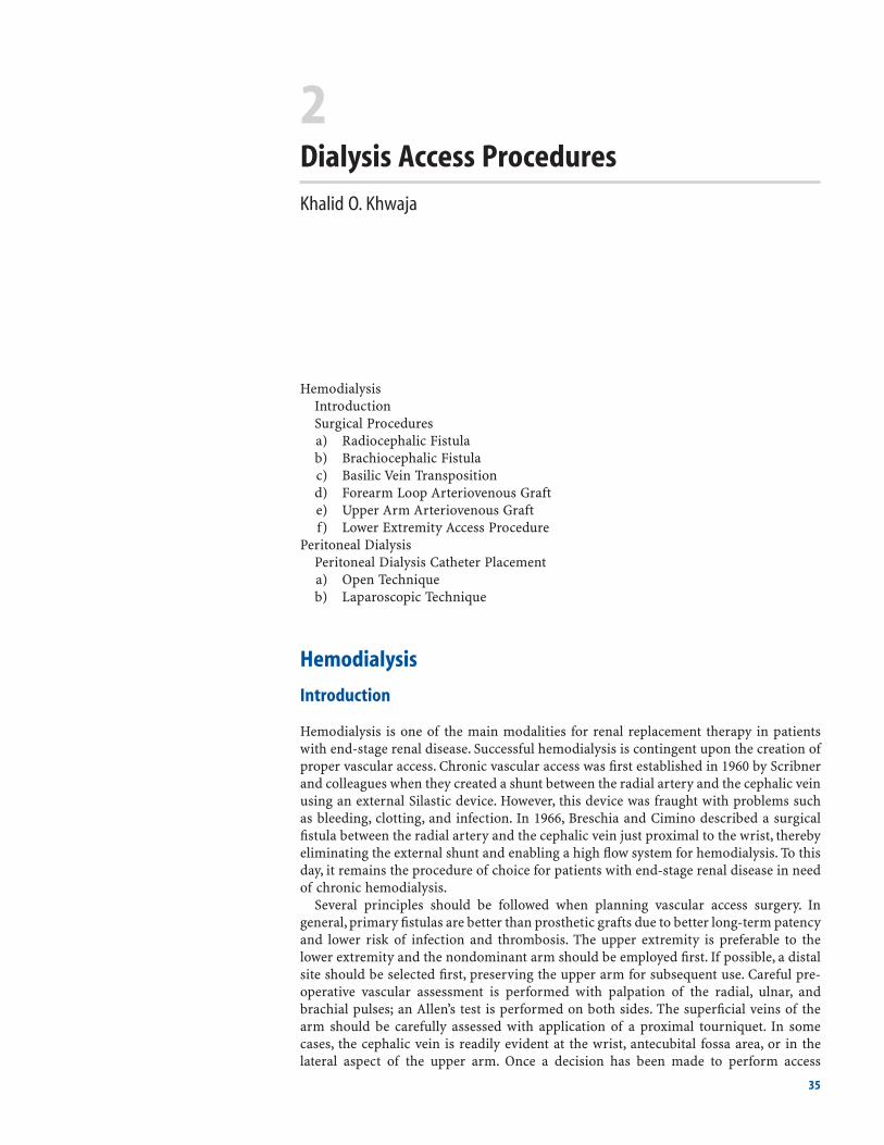
Atlas of Organ Transplantation

Abhinav Humar, Arthur J. Matas and William D. Payne
Atlas of OrganTransplantationWith 438 Figures

Abhinav Humar, MD, Arthur J. Matas, MD, William D. Payne, MD,FRCS FACS FACS
Department of Surgery Department of Surgery Department of SurgeryDivision of Division of Division of
Transplantation Transplantation TransplantationUniversity of Minnesota University of Minnesota University of MinnesotaMinneapolis, MN Minneapolis, MN Minneapolis, MNUSA USA USA
Cover photograph by author.
British Library Cataloguing in Publication Data
Atlas of organ transplantation1. Transplantation of organs, tissues, etc. – AtlasesI. Humar, Abhinav II. Matas, Arthur J. III. Payne, William D.617.9′54
ISBN-13: 9781846283147ISBN-10: 1846283140
Library of Congress Control Number: 2005938494
ISBN-10: 1-84628-314-0 e-ISBN 1-84628-316-7 Printed on acid-free paperISBN-13: 978-1-84628-314-7
© Springer-Verlag London Limited 2006
The software disk accompanying this book and all material contained on it is supplied without any warrantyof any kind. The publisher accepts no liability for personal injury incurred through use or misuse of the disk.
Apart from any fair dealing for the purposes of research or private study, or criticism or review, as permittedunder the Copyright, Designs and Patents Act 1988, this publication may only be reproduced, stored or trans-mitted, in any form or by any means, with the prior permission in writing of the publishers, or in the case ofreprographic reproduction in accordance with the terms of licences issued by the Copyright Licensing Agency.Enquiries concerning reproduction outside those terms should be sent to the publishers.
The use of registered names, trademarks, etc. in this publication does not imply, even in the absence of a specificstatement, that such names are exempt from the relevant laws and regulations and therefore free for generaluse.
Product liability: The publisher can give no guarantee for information about drug dosage and applicationthereof contained in this book. In every individual case the respective user must check its accuracy by con-sulting other pharmaceutical literature.
Printed in the United States of America (BS/KYO)
9 8 7 6 5 4 3 2 1
Springer Science+Business Mediaspringer.com

Preface
The field of organ transplantation has undergone remarkable changes in the last decade.The growing numbers of agents available for immunosuppression have played asignificant role in the advancement of this field. However, just as important has been thedevelopment of surgical innovations in the field. This includes not only the developmentof new surgical procedures, but also modifications of the existing ones. This has involvedall areas of organ transplantation including deceased-donor procurement techniques,living-donor transplantation, and transplantation of the individual organs includingkidney, liver, pancreas, and intestine. Examples include procurement from non–heart-beating donors; living-donor transplants involving the liver, pancreas, or intestine;laparoscopic donor nephrectomy; split-liver transplants; and multivisceral transplants.All of these represent new, innovative procedures that are being performed on a regularbasis only in the last few years. Given these recent dramatic changes in the surgical faceof transplantation, we felt it was time for a surgical atlas of transplantation that high-lighted these recent developments.
The aim of this book is to provide the reader with a comprehensive, pictorial step-by-step account of abdominal organ transplant procedures performed by contemporarytransplant surgeons. Emphasis has been placed on newer procedures or procedures thathave undergone significant modifications. It is recognized that there are many well-accepted techniques for the same procedure, with each having potential merit. While itis impossible to present all of these variations, an attempt has been made to describe thecommon variations in surgical technique.
Innovations in imaging have allowed us to organize this atlas in a format that pro-vides the reader with the most clear and realistic view of the operative procedures.Schematic diagrams are included to complement high-quality intraoperative photo-graphs, allowing readers to clearly visualize the course of the operative procedure. Aunique feature of this atlas is a digital video file of the major operative procedures, whichprovides the reader with the closest possible experience to being present in the operat-ing suite. It is hoped that this format will provide the reader with a clear visual andwritten description of all major abdominal transplant procedures performed by themodern transplant surgeon.
v

The editors gratefully acknowledge the contributing authors for their hard work in thepreparation of individual chapters or sections of chapters. The help of Dr. Khalid Khwajawas greatly appreciated in the design of the illustrations used in this atlas. The videosthat accompany this atlas would not have been made possible without the expertise andhard work of Dr. Bradley Linden, Eric Carolan, and Jake Gotler.
Acknowledgments
vii
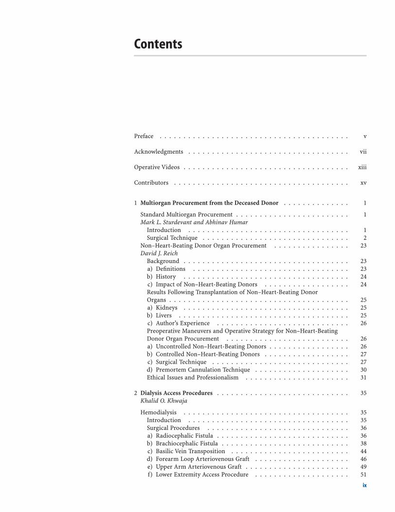
Preface . . . . . . . . . . . . . . . . . . . . . . . . . . . . . . . . . . . . . . . . v
Acknowledgments . . . . . . . . . . . . . . . . . . . . . . . . . . . . . . . . . . vii
Operative Videos . . . . . . . . . . . . . . . . . . . . . . . . . . . . . . . . . . . xiii
Contributors . . . . . . . . . . . . . . . . . . . . . . . . . . . . . . . . . . . . . xv
1 Multiorgan Procurement from the Deceased Donor . . . . . . . . . . . . . . 1
Standard Multiorgan Procurement . . . . . . . . . . . . . . . . . . . . . . . . 1Mark L. Sturdevant and Abhinav Humar
Introduction . . . . . . . . . . . . . . . . . . . . . . . . . . . . . . . . . . 1Surgical Technique . . . . . . . . . . . . . . . . . . . . . . . . . . . . . . . 2
Non–Heart-Beating Donor Organ Procurement . . . . . . . . . . . . . . . . 23David J. Reich
Background . . . . . . . . . . . . . . . . . . . . . . . . . . . . . . . . . . . 23a) Definitions . . . . . . . . . . . . . . . . . . . . . . . . . . . . . . . . . 23b) History . . . . . . . . . . . . . . . . . . . . . . . . . . . . . . . . . . . 24c) Impact of Non–Heart-Beating Donors . . . . . . . . . . . . . . . . . . 24Results Following Transplantation of Non–Heart-Beating Donor Organs . . . . . . . . . . . . . . . . . . . . . . . . . . . . . . . . . . . . . . 25a) Kidneys . . . . . . . . . . . . . . . . . . . . . . . . . . . . . . . . . . . 25b) Livers . . . . . . . . . . . . . . . . . . . . . . . . . . . . . . . . . . . . 25c) Author’s Experience . . . . . . . . . . . . . . . . . . . . . . . . . . . . 26Preoperative Maneuvers and Operative Strategy for Non–Heart-Beating Donor Organ Procurement . . . . . . . . . . . . . . . . . . . . . . . . . . 26a) Uncontrolled Non–Heart-Beating Donors . . . . . . . . . . . . . . . . . 26b) Controlled Non–Heart-Beating Donors . . . . . . . . . . . . . . . . . . 27c) Surgical Technique . . . . . . . . . . . . . . . . . . . . . . . . . . . . . 27d) Premortem Cannulation Technique . . . . . . . . . . . . . . . . . . . . 30Ethical Issues and Professionalism . . . . . . . . . . . . . . . . . . . . . . 31
2 Dialysis Access Procedures . . . . . . . . . . . . . . . . . . . . . . . . . . . . 35Khalid O. Khwaja
Hemodialysis . . . . . . . . . . . . . . . . . . . . . . . . . . . . . . . . . . . 35Introduction . . . . . . . . . . . . . . . . . . . . . . . . . . . . . . . . . . 35Surgical Procedures . . . . . . . . . . . . . . . . . . . . . . . . . . . . . . 36a) Radiocephalic Fistula . . . . . . . . . . . . . . . . . . . . . . . . . . . . 36b) Brachiocephalic Fistula . . . . . . . . . . . . . . . . . . . . . . . . . . . 38c) Basilic Vein Transposition . . . . . . . . . . . . . . . . . . . . . . . . . 44d) Forearm Loop Arteriovenous Graft . . . . . . . . . . . . . . . . . . . . 46e) Upper Arm Arteriovenous Graft . . . . . . . . . . . . . . . . . . . . . . 49f) Lower Extremity Access Procedure . . . . . . . . . . . . . . . . . . . . 51
Contents
ix

x Contents
Peritoneal Dialysis . . . . . . . . . . . . . . . . . . . . . . . . . . . . . . . . 54Peritoneal Dialysis Catheter Placement . . . . . . . . . . . . . . . . . . . . 54a) Open Technique . . . . . . . . . . . . . . . . . . . . . . . . . . . . . . . 54b) Laparoscopic Technique . . . . . . . . . . . . . . . . . . . . . . . . . . 56
3 Nephrectomy from a Living Donor . . . . . . . . . . . . . . . . . . . . . . . 59Raja Kandaswamy and Abhinav Humar
Introduction . . . . . . . . . . . . . . . . . . . . . . . . . . . . . . . . . . . . 59Open Nephrectomy . . . . . . . . . . . . . . . . . . . . . . . . . . . . . . . . 61Laparoscopic Donor Nephrectomy . . . . . . . . . . . . . . . . . . . . . . . . 65a) Introduction . . . . . . . . . . . . . . . . . . . . . . . . . . . . . . . . . . 65b) Preoperative Preparation . . . . . . . . . . . . . . . . . . . . . . . . . . . 66c) Hand-Assisted Laparoscopic Nephrectomy . . . . . . . . . . . . . . . . . 67d) Pure Laparoscopic Technique . . . . . . . . . . . . . . . . . . . . . . . . . 79e) Robotic-Assisted Technique . . . . . . . . . . . . . . . . . . . . . . . . . . 80
4 Kidney Transplantation . . . . . . . . . . . . . . . . . . . . . . . . . . . . . . 91Abhinav Humar and Arthur J. Matas
Introduction . . . . . . . . . . . . . . . . . . . . . . . . . . . . . . . . . . . . 91Benching the Kidney from a Deceased Donor . . . . . . . . . . . . . . . . . . 93a) Procedure . . . . . . . . . . . . . . . . . . . . . . . . . . . . . . . . . . . 94Adult Kidney Transplant . . . . . . . . . . . . . . . . . . . . . . . . . . . . . 99a) Operative Procedure . . . . . . . . . . . . . . . . . . . . . . . . . . . . . . 100Surgical Variations . . . . . . . . . . . . . . . . . . . . . . . . . . . . . . . . 116a) Pediatric Recipient Kidney Transplant . . . . . . . . . . . . . . . . . . . . 116b) Pediatric Donor En-Bloc Kidneys . . . . . . . . . . . . . . . . . . . . . . 119c) Multiple Renal Arteries . . . . . . . . . . . . . . . . . . . . . . . . . . . . 120Surgical Complications and Their Management . . . . . . . . . . . . . . . . 122a) Hemorrhage . . . . . . . . . . . . . . . . . . . . . . . . . . . . . . . . . . 123b) Renal Artery Thrombosis . . . . . . . . . . . . . . . . . . . . . . . . . . . 123c) Renal Artery Stenosis . . . . . . . . . . . . . . . . . . . . . . . . . . . . . 123d) Recipient Arterial Complications . . . . . . . . . . . . . . . . . . . . . . . 125e) Renal Vein Thrombosis . . . . . . . . . . . . . . . . . . . . . . . . . . . . 126f) Venous Thromboembolism . . . . . . . . . . . . . . . . . . . . . . . . . . 126g) Aneurysms and Fistulas . . . . . . . . . . . . . . . . . . . . . . . . . . . . 126h) Urologic Complications . . . . . . . . . . . . . . . . . . . . . . . . . . . . 127i) Lymphoceles . . . . . . . . . . . . . . . . . . . . . . . . . . . . . . . . . . 128j) Wound Complications . . . . . . . . . . . . . . . . . . . . . . . . . . . . . 129
5 Pancreas Transplantation . . . . . . . . . . . . . . . . . . . . . . . . . . . . . 133Abhinav Humar, Khalid O. Khwaja, and David E.R. Sutherland
Standard Procedure . . . . . . . . . . . . . . . . . . . . . . . . . . . . . . . . 133a) Pretransplant Evaluation . . . . . . . . . . . . . . . . . . . . . . . . . . . 134b) Surgical Technique . . . . . . . . . . . . . . . . . . . . . . . . . . . . . . 134c) Postoperative Care . . . . . . . . . . . . . . . . . . . . . . . . . . . . . . . 134d) Results . . . . . . . . . . . . . . . . . . . . . . . . . . . . . . . . . . . . . 135e) Islet Cell Transplantation . . . . . . . . . . . . . . . . . . . . . . . . . . . 135Benching the Pancreas from a Deceased Donor . . . . . . . . . . . . . . . . . 135a) Surgical Procedure . . . . . . . . . . . . . . . . . . . . . . . . . . . . . . 136Simultaneous Pancreas Kidney (SPK): Systemic/Enteric Drainage . . . . . . . 145a) Surgical Procedure . . . . . . . . . . . . . . . . . . . . . . . . . . . . . . 146Simultaneous Pancreas Kidney: Portal/Enteric Drainage . . . . . . . . . . . . 154A. Osama Gaber and Hosein Shokouh-Amiria) Surgical Procedure . . . . . . . . . . . . . . . . . . . . . . . . . . . . . . 156

Contents xi
Isolated Pancreas Transplant: Bladder Drainage . . . . . . . . . . . . . . . . 166a) Surgical Procedure . . . . . . . . . . . . . . . . . . . . . . . . . . . . . . 168Pancreas Transplant from a Living Donor . . . . . . . . . . . . . . . . . . . . 178Miguel Tan, Raja Kandaswamy, and Rainer W.G. Gruessnera) Donor Evaluation . . . . . . . . . . . . . . . . . . . . . . . . . . . . . . . 179b) Surgical Procedure . . . . . . . . . . . . . . . . . . . . . . . . . . . . . . 179Surgical Complications After Pancreas Transplant . . . . . . . . . . . . . . . 192a) Thrombosis . . . . . . . . . . . . . . . . . . . . . . . . . . . . . . . . . . 192b) Hemorrhage . . . . . . . . . . . . . . . . . . . . . . . . . . . . . . . . . . 193c) Pancreatitis . . . . . . . . . . . . . . . . . . . . . . . . . . . . . . . . . . 193d) Urologic Complications . . . . . . . . . . . . . . . . . . . . . . . . . . . . 193e) Infections . . . . . . . . . . . . . . . . . . . . . . . . . . . . . . . . . . . 193f) Leaks . . . . . . . . . . . . . . . . . . . . . . . . . . . . . . . . . . . . . . 194g) Minimizing Surgical Complications . . . . . . . . . . . . . . . . . . . . . 194
6 Liver Transplantation . . . . . . . . . . . . . . . . . . . . . . . . . . . . . . . 197Abhinav Humar and William D. Payne
Standard Procedure . . . . . . . . . . . . . . . . . . . . . . . . . . . . . . . . 197a) Preoperative Evaluation . . . . . . . . . . . . . . . . . . . . . . . . . . . . 198b) Surgical Procedure . . . . . . . . . . . . . . . . . . . . . . . . . . . . . . 199c) Postoperative Care . . . . . . . . . . . . . . . . . . . . . . . . . . . . . . . 200d) Results . . . . . . . . . . . . . . . . . . . . . . . . . . . . . . . . . . . . . 201Benching the Liver from a Deceased Donor . . . . . . . . . . . . . . . . . . . 201a) Surgical Procedure . . . . . . . . . . . . . . . . . . . . . . . . . . . . . . 202Adult Cadaver Liver Transplant . . . . . . . . . . . . . . . . . . . . . . . . . 207a) Surgical Procedure . . . . . . . . . . . . . . . . . . . . . . . . . . . . . . 207Deceased Donor Split-Liver Transplant: Adult/Pediatric Recipients . . . . . . 235Hasan Yersiz and John F. Renza) Surgical Procedure . . . . . . . . . . . . . . . . . . . . . . . . . . . . . . 236Deceased Donor Split-Liver Transplant: Adult/Adult Recipients . . . . . . . . 242a) Selection Criteria . . . . . . . . . . . . . . . . . . . . . . . . . . . . . . . 243b) Technical Aspects . . . . . . . . . . . . . . . . . . . . . . . . . . . . . . . 243c) Ethics of Splitting . . . . . . . . . . . . . . . . . . . . . . . . . . . . . . . 243d) Split Potential . . . . . . . . . . . . . . . . . . . . . . . . . . . . . . . . . 244e) Surgical Procedure . . . . . . . . . . . . . . . . . . . . . . . . . . . . . . 244Living-Donor Liver Transplant: Pediatric Recipient . . . . . . . . . . . . . . . 251a) Donor Procedure . . . . . . . . . . . . . . . . . . . . . . . . . . . . . . . 253b) Recipient Procedure . . . . . . . . . . . . . . . . . . . . . . . . . . . . . . 261Living-Donor Liver Transplant: Adult Recipient . . . . . . . . . . . . . . . . . 264a) Donor Procedure . . . . . . . . . . . . . . . . . . . . . . . . . . . . . . . 267b) Recipient Procedure . . . . . . . . . . . . . . . . . . . . . . . . . . . . . . 283Surgical Complications After Liver Transplant . . . . . . . . . . . . . . . . . 290a) Hemorrhage . . . . . . . . . . . . . . . . . . . . . . . . . . . . . . . . . . 290b) Hepatic Artery Complications . . . . . . . . . . . . . . . . . . . . . . . . 290c) Portal Vein Complications . . . . . . . . . . . . . . . . . . . . . . . . . . 290d) Biliary Complications . . . . . . . . . . . . . . . . . . . . . . . . . . . . . 290e) Wound Complications . . . . . . . . . . . . . . . . . . . . . . . . . . . . . 291f) Primary Nonfunction . . . . . . . . . . . . . . . . . . . . . . . . . . . . . 291
7 Intestinal and Multivisceral Transplantation . . . . . . . . . . . . . . . . . . 293Thomas M. Fishbein and Cal S. Matsumoto
Isolated Intestine Transplant . . . . . . . . . . . . . . . . . . . . . . . . . . . 294a) Donor Procedure . . . . . . . . . . . . . . . . . . . . . . . . . . . . . . . 294b) Back-Table Procedure . . . . . . . . . . . . . . . . . . . . . . . . . . . . . 297c) Recipient Procedure . . . . . . . . . . . . . . . . . . . . . . . . . . . . . . 300

xii Contents
Combined Liver–Intestine Transplant . . . . . . . . . . . . . . . . . . . . . . 310a) Donor Procedure . . . . . . . . . . . . . . . . . . . . . . . . . . . . . . . 310b) Back-Table Procedure . . . . . . . . . . . . . . . . . . . . . . . . . . . . . 312c) Recipient Procedure . . . . . . . . . . . . . . . . . . . . . . . . . . . . . . 314Multivisceral Transplant . . . . . . . . . . . . . . . . . . . . . . . . . . . . . 318a) Donor Procedure . . . . . . . . . . . . . . . . . . . . . . . . . . . . . . . 318b) Recipient Procedure . . . . . . . . . . . . . . . . . . . . . . . . . . . . . . 319Living-Donor Small-Bowel Transplant . . . . . . . . . . . . . . . . . . . . . . 324a) Surgical Procedure . . . . . . . . . . . . . . . . . . . . . . . . . . . . . . 325Perioperative Management . . . . . . . . . . . . . . . . . . . . . . . . . . . . 330
Index . . . . . . . . . . . . . . . . . . . . . . . . . . . . . . . . . . . . . . . . . 333

Deceased Donor
1. Multiorgan Procurement
Kidney
2. Brachiocephalic Arteriovenous Fistula3. Hand-Assisted Laparoscopic Donor Nephrectomy4. Benching Kidney from Deceased Donor5. Adult Kidney Transplant (Two Arteries)6. Pediatric Kidney Transplant
Pancreas Transplant
7. Benching Pancreas from Deceased Donor8. Pancreas Transplant Alone: Systemic/Bladder Drainage9. Simultaneous Pancreas Kidney (SPK) – Portal/Enteric Drainage
Contribution by: Osoma Gaber, Hosein Shokouh-Amiri10. Laparoscopic Distal Pancreatectomy for Living-Donor Pancreas Transplant
Contribution by: Miguel Tan, Raja Kandaswamy, Rainer W.G. Gruessner
Liver Transplant
11. Benching Liver from Deceased Donor12. Deceased Donor Liver Transplant – Adult Recipient13. In-Situ Split of the Deceased Donor Liver for Adult/Pediatric Recipients
Contribution by: Hasan Yersiz, John Renz, Ronald Busutil14. In-Situ Split of the Deceased Donor Liver for Two Adult Recipients15. Adult to Adult Living-Donor Liver Transplant Using the Right Lobe – Donor
Procedure16. Adult to Adult Living-Donor Liver Transplant Using the Right Lobe – Recipient
Procedure
Intestinal Transplant
17. Living-Donor Intestinal Transplant
Operative Videos
xiii

Contributors
A. Osama Gaber, MD, FACSMethodist Transplant InstituteUniversity of TennesseeMemphis, TN, USA
Thomas M. Fishbein, MDTransplant InstituteGeorgetown University HospitalWashington, DC, USA
Rainer W.G. Gruessner, MD, PhDDepartment of SurgeryDivision of TransplantationUniversity of MinnesotaMinneapolis, MN, USA
Abhinav Humar, MD, FRCSDepartment of SurgeryDivision of TransplantationUniversity of MinnesotaMinneapolis, MN, USA
Raja Kandaswamy, MBBSDepartment of SurgeryDivision of TransplantationUniversity of MinnesotaMinneapolis, MN, USA
Khalid O. Khwaja, MDTransplant CenterBeth Israel Deaconess Medical CenterBoston, MA, USA
Arthur J. Matas, MD, FACSDepartment of SurgeryDivision of TransplantationUniversity of MinnesotaMinneapolis, MN, USA
Cal S. Matsumoto, MDGeorgetown University HospitalWashington, DC, USA
William D. Payne, MD, FACSDepartment of SurgeryDivision of TransplantationUniversity of MinnesotaMinneapolis, MN, USA
David J. Reich, MD, FACSLiver Transplant ProgramDepartment of SurgeryAlbert Einstein Medical CenterPhiladelphia, PA, USA
John F. Renz, MD, PhDCenter for Liver DiseaseNew York Presbyterian HospitalNew York, NY, USA
Hosein Shokouh-Amiri, MD, FACSLive Donor and Pediatric Liver
TransplantationMethodist Transplant InstituteUniversity of TennesseeMemphis, TN, USA
Mark L. Sturdevant, MDDepartment of SurgeryDivision of TransplantationUniversity of MinnesotaMinneapolis, MN, USA
David E.R. Sutherland, MD, PhDDepartment of SurgeryDivision of TransplantationUniversity of MinnesotaMinneapolis, MN, USA
Miquel Tan, MDDivision of TransplantationDepartment of SurgeryJohns Hopkins HospitalBaltimore, MD, USA
Hasan Yersiz, MDDepartment of Hepatobiliary and Liver
Transplant SurgeryDumont-UCLA Transplant CenterDavid Geffen School of Medicine at
UCLAUniversity of California, Los AngelesLos Angeles, CA, USA
xv

1Multiorgan Procurement from theDeceased Donor
Standard Multiorgan ProcurementMark L. Sturdevant and Abhinav Humar
IntroductionOperative Technique
Non–Heart-Beating Donor Organ ProcurementDavid J. Reich
Backgrounda) Definitionsb) Historyc) Impact of Non–Heart-Beating DonorsResults Following Transplantation of Non–Heart-Beating Donor Organsa) Kidneysb) Liversc) Author’s ExperiencePreoperative Maneuvers and Operative Strategy for Non–Heart-Beating DonorOrgan Procurementa) Uncontrolled Non–Heart-Beating Donorsb) Controlled Non–Heart-Beating Donorsc) Surgical Techniqued) Premortem Cannulation TechniqueEthical Issues and Professionalism
Standard Multiorgan Procurement
Mark L. Sturdevant and Abhinav Humar
Introduction
Organ procurement for transplantation was first accomplished by the Soviet surgeon YuYu Voronoy, who performed the first human kidney transplant on April 3, 1933. Thedonor was a 60-year-old man who died on admission to the hospital from a traumaticbrain injury; the kidney was removed 6 hours postmortem and transplanted into thethigh of a 26-year-old woman with acute renal failure from mercury poisoning. The allo-graft did produce several milliliters of urine before the patient died 2 days after trans-plantation. The first attempt at liver transplantation, on March 1, 1963, by Thomas Starzl,was possible only after successful liver procurement from a child who had died aftercardiac surgery, but was left on the heart-lung machine to allow for procurement.
1
2 Atlas of Organ Transplantation
Kidney transplantation in the 1950s and 1960s was primarily from live donors.However, in 1966 the concept of brain death was established in France by Guy Alexandre, who described the removal of kidneys from “heart-beating” cadavers withsubsequent transplantation. In the United States, public support for this concept wasoverwhelming and led to the Harvard Ad Hoc Committee report in 1968 that outlinedthe criteria for brain death determination. The donor pool increased markedly afterthese policies entered clinical practice.
Advancement to the modern-day status of deceased-donor organ procurement wasaided in large part by work done in organ preservation. The combined efforts of Dr. JohnNajarian and Dr. Folkert Belzer at the University of California–San Francisco (UCSF),starting in 1966, aimed at decreasing ischemia times and adding organ preservatives toincrease organ viability. Prior to the acceptance of brain death, Belzer procured kidneysfrom non–heart-beating donors in the greater San Francisco area and emergently trans-ported the organs to Najarian at Moffitt Hospital, who would have simultaneously startedthe recipient operation. Advances in organ preservation along with the acceptance ofdonation after brain death resulted in a more systematic, semielective kidney procure-ment, which resulted in organ delivery to recipients almost anywhere. Within 5 years aportable perfusion machine had been developed, and on December 24, 1971, a deceased-donor kidney procured in San Francisco was hand delivered by Belzer to transplantsurgeon Hans Dicke in the Netherlands with a cold ischemia time of 37 hours. The trans-planted kidney had excellent function 17 years later when the recipient died of a rup-tured cerebral aneurysm.
The technique of multiple-organ procurement (kidney, liver, pancreas, small bowel)was first described by Starzl and his colleagues in 1984. Nakazato and his colleagues in1992 described the technique of total abdominal evisceration with ex vivo dissection.Most centers have now added their own modifications to these pioneering techniquesand differ primarily in their degree of in vivo dissection. Some centers perform exten-sive dissection of the organs to be recovered prior to flushing the organs with preserva-tive solution. Other centers prefer to flush the organs early, remove the abdominalcontents “en bloc”, and perform the separation and dissection of the individual organson the back table. Each technique has its potential advantages and disadvantages.Regardless of personal technique and preference, it is paramount that the transplantsurgeon develops a systematic approach to safely procure the liver, pancreas, andkidneys, even in the unstable donor.
Surgical Technique
1. Incision and exposure: An incision extending from the sternal notch to the pubis,which is cruciated at the level of the umbilicus (Figure 1.1), provides maximal exposurefor multiorgan procurements. The abdominal flaps can be folded back and held in placewith sharp towel clips (Figure 1.2). This provides excellent exposure of the abdominalorgans, without the need for a retractor. Only a sternal retractor is needed if the thoracicorgans are to be procured. Sternotomy and division of the pericardium allows for exam-ination of the heart while division of the ligamentous attachments to the liver allows forcomplete examination of the liver. A thorough abdominal exploration is then quicklyperformed to rule out contraindications to procurement such as malignancy or intraab-dominal sepsis.

Multiorgan Procurement from the Deceased Donor 3
Figure 1.1
Figure 1.2

4 Atlas of Organ Transplantation
Figure 1.3
Figure 1.4
2. A Cattel-Braasch maneuver extending across the midline, with complete mobiliza-tion of the distal small bowel, right colon, and duodenum (Figure 1.3), allows foridentification of the distal aorta, iliac bifurcation, and the distal inferior vena cava (IVC)(Figure 1.4).

Multiorgan Procurement from the Deceased Donor 5
Figure 1.5
3. Division of the inferior mesenteric artery (black arrow) aids in the dissection ofthe distal aorta (yellow arrow and lines), which is then encircled with two umbilical tapes(Figure 1.5). This will be the site for later aortic cannulation for flushing (broken blackline). The inferior vena cava sits just to the right (blue arrow).
6 Atlas of Organ Transplantation
Figure 1.6
Figure 1.7
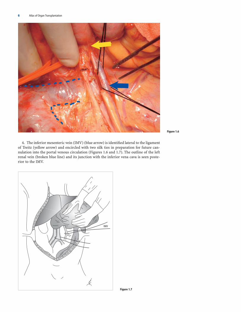
4. The inferior mesenteric vein (IMV) (blue arrow) is identified lateral to the ligamentof Treitz (yellow arrow) and encircled with two silk ties in preparation for future can-nulation into the portal venous circulation (Figures 1.6 and 1.7). The outline of the leftrenal vein (broken blue line) and its junction with the inferior vena cava is seen poste-rior to the IMV.

Multiorgan Procurement from the Deceased Donor 7
Figure 1.8
Figure 1.9
5. The third portion of the duodenum is retracted cephalad, and the superior mesen-teric artery (SMA, yellow broken line) is identified, dissected free, and encircled with avessel loop (Figures 1.8 and 1.9). This allows for occlusion of the SMA later, at the timeof flushing. This limits the incidence of overperfusion injury to the pancreas. The leftrenal vein (broken blue line) is seen just inferior to the SMA, and the inferior mesen-teric vein (blue arrow) is just lateral.

8 Atlas of Organ Transplantation
Figure 1.10
Figure 1.11
6. In preparation to obtain control of the supraceliac aorta, the left triangular ligamentof the liver is divided and the gastrohepatic ligament is examined and divided if no aber-rant left hepatic artery is noted (Figure 1.10).If one is noted it will need to be preserved.Theright diaphragmatic crus is divided,and the supraceliac aorta is identified and mobilized.
7. The supraceliac aorta is encircled with an umbilical tape (Figure 1.11). In the un-stable donor, cold perfusion may be done at this point.

Multiorgan Procurement from the Deceased Donor 9
Figure 1.13
Figure 1.12
8. The portal triad is now examined, and if present, an aberrant right hepatic arterywill be palpated on the right edge of the porta hepatis, posterior to the bile duct andlateral to the portal vein. The course of an accessory or replaced right (black arrow) isshown in broken lines (Figure 1.12).
9. The common hepatic artery is identified and traced to the celiac axis, allowing forvisualization and limited dissection of the splenic artery and gastroduodenal artery. Thehepatic artery does not need to be completely dissected out at this time – rather, just theorigin of the above branches need to be identified (Figure 1.13).
10. Limited dissection of the common bile duct (CBD, yellow arrow), which lies justto the right and anterior to the main portal vein (broken blue lines) is performed followed by ligation of the distal CBD just proximal to the pancreas. The CBD is then

10 Atlas of Organ Transplantation
Figure 1.14 Figure 1.15
Figure 1.16 Figure 1.17
transected with a scalpel and the proximal end is left open (Figure 1.14). The gallblad-der is incised and flushed with saline to clear the CBD of retained bile (Figure 1.15).
11. Systemic heparinization (300 units/kg) is performed. The distal aorta is ligatedand a 24-French (F) aortic cannula (red arrow) is placed at this site. The IMV is ligatedand a 14-F perfusion cannula (blue arrow) is placed (Figures 1.16 and 1.17). Alterna-tively, the portal vein can be cannulated directly for portal flushing.

Multiorgan Procurement from the Deceased Donor 11
12. The supraceliac aorta is ligated or clamped and cold preservation fluid is infusedvia the two cannulas. The suprahepatic IVC is transected at its entrance to the rightatrium in order to vent the perfusate (Figure 1.18). If heart procurement is also per-formed, the thoracic surgeon will incise the IVC just cephalad to the diaphragm. Inadults, 5 L of preservation fluid is flushed via the aortic and inferior mesenteric vein can-nulas (3 L and 2 L, respectively). The superior mesenteric artery is occluded after 1 L toavoid overperfusion of the pancreas. Slushed ice is placed into the peritoneal and peri-cardial cavities to complete the cooling process (Figure 1.19). Thoracic organ procure-ment occurs once flushing has been initiated.
Figure 1.18
Figure 1.19

12 Atlas of Organ Transplantation
Figure 1.20
Figure 1.21
13. After the organs have been adequately flushed and the thoracic organs removed,attention is turned to the intraabdominal organs, starting first with the liver.Donor hepatectomy is started by dividing the diaphragm and ligamentous attachmentsto mobilize the liver (Figure 1.20).
14. The common hepatic artery is fully dissected and traced to the celiac axis wherean aortic cuff is fashioned after the left gastric and splenic arteries are divided (Figure1.21). A fine suture is placed on the distal splenic artery to aid in its identification near

Multiorgan Procurement from the Deceased Donor 13
the pancreas. The gastroduodenal artery (GDA) is ligated distally near the pancreas andtransected, leaving the proximal end open and available for possible reconstruction purposes.
15. Portal vein transection (blue arrow) completes division of the porta hepatis(Figures 1.22 and 1.23). If the pancreas is being procured, at least 1.5 cm of portal veinshould be left with the pancreas graft.
Figure 1.22
Figure 1.23

14 Atlas of Organ Transplantation
16. Transection of the infrahepatic IVC is performed cephalad to the confluences ofthe renal veins. The liver, along with a rim of diaphragm, is now removed (Figure 1.24).
17. The pancreas is removed next. At any juncture prior to cold perfusion, the nasogastric tube is advanced into the duodenum and 500 mL of amphotericin solution
Figure 1.24

Multiorgan Procurement from the Deceased Donor 15
Figure 1.25
(50 mg/L) is delivered into the second portion of the duodenum. After the donor hepa-tectomy, the gastrocolic ligament is divided along the edge of the greater curvature ofthe stomach extending laterally to the spleen (Figure 1.25). This maneuver brings theentire pancreas into view for examination.

16 Atlas of Organ Transplantation
Figure 1.27
Figure 1.26
18. The ligamentous attachments to the spleen are divided next. The spleen along withthe pancreatic body and tail can now be carefully mobilized from the retroperitoneum(Figures 1.26 and 1.27).
19. The first portion of the duodenum is circumferentially dissected and a mechani-cal stapler is used to divide the duodenum from the stomach (broken blue line) just distal

Multiorgan Procurement from the Deceased Donor 17
Figure 1.28
Figure 1.29
to the pylorus (blue arrow) (Figure 1.28). The stapler is also used to divide the duode-num just distal to the ligament of Treitz (Figure 1.29). The root of the transverse meso-colon, which runs along the anterior aspect of the pancreas is then divided using serialligatures.

18 Atlas of Organ Transplantation
Figure 1.30
Figure 1.31
20. The IMV is ligated after the cannula is removed. The small bowel mesentery,which includes the superior mesenteric artery (SMA, broken red lines) and superiormesenteric vein (SMV, broken blue lines), is transected with a mechanical stapler (Figure 1.30).
21. The proximal superior mesenteric artery (broken red lines), which was previouslyencircled with a vessel loop, is now easily identified and transected flush with the aorta(Figure 1.31).

Multiorgan Procurement from the Deceased Donor 19
Figure 1.32
22. The pancreas is then removed en bloc with the duodenum and spleen (Figure1.32).
23. The two kidneys are removed next en bloc with the cava and aorta. The lateralattachments to the kidney are divided and the ureters are traced caudally, and transecteddistally near their entrance into the bladder (Figure 1.33).
Figure 1.33

20 Atlas of Organ Transplantation
Figure 1.34
Figure 1.35
24. The aorta and inferior vena cava (IVC) are divided at their bifurcations.The uretersand these vessels are retracted cephalad and anteriorly, along with the two kidneys (solidblue lines), and dissection proceeds along their posterior aspects, anterior to the surfaceof the vertebral bodies and psoas muscle (broken black lines) (Figures 1.34 and 1.35).

Multiorgan Procurement from the Deceased Donor 21
25. The kidneys are removed en bloc by transecting the aorta just proximal to thesuperior mesenteric artery takeoff. The IVC had been previously transected just abovethe renal veins during the donor hepatectomy. The kidneys are placed en bloc in coldpreservation solution and are separated on the back table. Via an anterior approach, theleft renal vein is identified and divided at its junction with the inferior vena cava (blackarrow) (Figures 1.36 and 1.37).
Figure 1.36
Figure 1.37

22 Atlas of Organ Transplantation
Figure 1.38
26. The aortic wall is then divided longitudinally down its center aspect, which allowsfor inspection of the renal artery orifices (red arrows) from within the aortic lumen(Figure 1.38).

Multiorgan Procurement from the Deceased Donor 23
27. The posterior aortic wall is then divided between the renal artery orifices. Thiscompletes division of the right and left kidneys (Figure 1.39).
28. Finally, the iliac artery and veins are removed, to be used for possible vascularreconstructions in the pancreas and liver transplant procedures. Vessels are taken from the left and right side, starting at the common iliac vessel and extending to the distalexternal iliac vessels. These conduits are packaged with the pancreas and liver grafts.The deceased donor is then closed with a large suture.
Non–Heart-Beating Donor Organ Procurement
David J. Reich
Background
a) Definitions
The non–heart-beating donor (NHBD), also referred to as the donation after cardiacdeath (DCD) donor, is one type of expanded criteria donor that is increasingly being uti-lized by organ procurement organizations (OPOs) and transplant centers across theUnited States, Europe, and Asia to successfully boost the number of deceased donors anddecrease the dire shortage of transplantable organs.1 An NHBD death is characterizedby irreversible absence of circulation, in contrast to heart-beating donor death, defined
Figure 1.39

24 Atlas of Organ Transplantation
by irreversible cessation of all brain functions. Organ ischemia is minimized in the brain-dead donor because circulatory arrest typically occurs concurrently with perfusion ofpreservation solution and rapid core cooling. The NHBDs are less than ideal because theorgans suffer ischemia during the prolonged periods between circulatory dysfunction,circulatory arrest, and subsequent perfusion and cooling. Furthermore, the surgical pro-cedure for NHBD organ recovery, the main focus of this section, is demanding andrushed.
It is important to differentiate controlled from uncontrolled NHBDs. UncontrolledNHBDs sustain circulatory arrest and either fail to respond to cardiopulmonary resus-citation or are declared dead on arrival at the hospital. Uncontrolled NHBD death isunplanned, so the organs suffer protracted ischemia prior to recovery. Although kidneystolerate a short period of the resultant warm ischemia, transplantation of extrarenalorgans from uncontrolled NHBDs carries a much greater risk. In contrast, controlledNHBDs undergo circulatory arrest following planned withdrawal of life support, mostoften in the operating room, with a donor surgical team readily available. ControlledNHBDs suffer terminal illness, usually a severe neurologic injury without the possibil-ity of meaningful recovery or survival. Controlled NHBDs provide organs that areexposed to significantly less ischemic damage than those of uncontrolled NHBDs and ingeneral, offer superior posttransplant function when compared with uncontrolledNHBDs.
b) History
The first human kidney, liver, and heart transplants, in 1958, 1963, and 1967, respectively,were performed using organs recovered from uncontrolled NHBDs. During the earliestyears of transplantation through the 1960s, determination of death required heartbeatcessation. However, modern critical care, respirators and cardiopulmonary resuscitationmade it possible to reestablish or maintain cardiopulmonary function even in the faceof irreversible coma, leading to the 1968 landmark criteria for determination of braindeath (Harvard neurologic definition and criteria for death).2 These were endorsed bythe major medical and legal professional associations in the United States and Europe,and for the following 25 years virtually all organ donation was from brain dead or livingdonors. Such donors are more desirable than NHBDs because the organs are protectedfrom warm ischemic injury and are less prone to poor graft function. Non–heart-beatingdonor organ transplantation was reintroduced by the University of Pittsburgh transplantprogram in 1992.3 The Pittsburgh program and the program in Madison,Wisconsin, werepivotal in initiating controlled NHBD organ transplantation and were the first todescribe results of controlled NHBD kidney and liver transplantation (LTX), both in1995.4,5
c) Impact of Non–Heart-Beating Donors
Over the past decade there has been increasing interest in pursuing controlled NHBDorgan transplantation because results have improved and NHBDs have the potential tobecome a significant source of solid organs. Currently, only approximately 4% ofdeceased donors in the United States are NHBDs.6 However, it has been estimated thatNHBDs could increase the deceased donor pool in the United States by approximately1000 donors per year, a 25% increase.7 Among pediatric cadaver donors, increases of over40% have been estimated.8 In the United States, the number of NHBDs has steadilyincreased from only 42 in 1993, providing 81 organ transplants, to 270 in 2003, provid-ing 549 transplants.6 Some of the reasons that controlled NHBD is gaining acceptanceand becoming more widespread involve increasing reluctance to prolong futile treatmentand artificial life support of terminally injured and ill patients, increasing use of advancedirectives and health care proxies, and encouraging data on survival following NHBDorgan transplantation. Families more frequently request this option in their discussions

Multiorgan Procurement from the Deceased Donor 25
about removal of life support from patients who suffer devastating trauma or critical,irreversible illness. Certain OPOs have developed highly successful NHBD programs; in2003, 15% of the deceased donors made possible by the Gift of Life Donor Program wereNHBDs (51/344).9 Strong OPO initiatives are crucial for increasing NHBDs. The OPOsmust develop NHBD protocols; educate physicians, nurses, and other health careproviders; engender enthusiasm among the OPO and transplant teams; recruit sup-port from regional hospitals; and concentrate on public relations.7,10
Results Following Transplantation of Non–Heart-Beating Donor Organs
a) Kidneys
Non–heart-beating donors have provided for transplantation of kidneys, livers, somepancreata, and some lungs.6 The majority of experience with NHBD organs is withkidney transplantation. One-year graft survival rates after uncontrolled NHBD kidneytransplantation have been reported at 79% to 86%, similar to results of controlled NHBDkidney transplantation, which provides 1-year graft survival rates of 82% to 86%.11 Com-parison of 708 NHBD and 97,990 heart-beating deceased-donor renal transplants in theUnited States reveals that NHBD organ recipients had nearly twice the incidence ofdelayed graft function (DGF) (42% vs. 23%), but 1- and 5-year allograft survival rateswere comparable between groups (83% vs. 87% and 61% vs. 65%, respectively).12 Thus,in spite of temporary problems early on, renal grafts from NHBDs ultimately functionas well as grafts from heart-beating donors.12,13
b) Livers
Several single-center experiences with NHBD LTX have been published.14–19 Currently,uncontrolled NHBD LTX provides 1-year graft survival rates of only 17% to 55%.4,11,19 Inmarked contrast, the reports on controlled NHBD LTX indicate patient and graft sur-vival rates (76% to 90% and 72% to 90% at 1 year, respectively) that are comparable tothose following heartbeating cadaver donor LTX.1,16–18 At one institution, long-termNHBD graft loss was higher (54% vs. 81%), but half the graft losses were due to patientdeath with normal liver function.16 Non–heart-beating donor LTX has not resulted in anincreased incidence of primary nonfunction (0% to 11%) or hepatic artery thrombosis(0% to 5%).1,16,18 The University of Pennsylvania group was the first to report a higherrisk of major biliary complications in NHBD liver recipients (33% vs. 10%), mostlyischemic type biliary strictures and/or bile cast syndrome.17 The group cautioned thatbiliary epithelium is particularly prone to ischemia-reperfusion injury, and it advocatesusing younger NHBDs and minimizing ischemia times. Although other groups have notexperienced as high an incidence of biliary complications, it has become increasinglyclear that NHBD livers are prone to ischemic-type biliary strictures, particularly thoseexposed to longer ischemia times. The Miami and the author’s groups have shown thatLTX can be safely performed using older, controlled NHBDs,1,18 although others adviseagainst this.16,17 Controlled NHBD LTX may be safely performed in the face of true warmischemia time – the interval between significant (variably defined) hypotension orhypoxemia, and initiation of perfusion – up to and perhaps somewhat beyond 20 to 30minutes. Total warm ischemia time – the interval between stopping of mechanical ven-tilation and initiation of perfusion – may extend up to and perhaps somewhat beyond30 to 45 minutes.1
Results of controlled NHBD LTX reported by single centers experienced in this fieldare superior to the results provided by one group’s analysis of the United Network ofOrgan Sharing (UNOS) database, grouping together controlled and uncontrolled NHBDLTXs in the United States.20 Comparison of 144 NHBD and 26,856 heart-beating donorLTXs reveals 1-year patient and graft survival rates that are lower for the NHBD cohort(80% vs. 85% and 70% vs. 80%, respectively), although the difference is not significant

26 Atlas of Organ Transplantation
when comparing controlled NHBD grafts to heart-beating donor grafts. The NHBD LTXrecipients required retransplantation more frequently (14% vs. 8%) and had moreprimary nonfunction (12% vs. 6%). Early retransplantation after NHBD LTX was morelikely with donors over age 60, cold ischemia times longer than 8 hours, and recipientson life support prior to transplantation.
c) Author’s Experience
At the Albert Einstein Medical Center in Philadelphia, we recently provided long-termoutcomes for 22 controlled NHBD LTXs performed at our institution through 2002.1 Todate, we have performed 30 controlled NHBD LTXs using grafts procured by our team.Controlled NHBDs have provided for 7% of all LTXs (30/442) that we performed overthe past 9 years. Causes of neurologic injury were trauma (n = 14), cerebral vascular accident (n = 8), and anoxia (n = 8). One third of the donors (10/30) were more than 50 years old; donor age ranged from 11 to 66 years. The interval between develop-ment of significant hypotension, defined as a drop in mean arterial pressure below 50 mm Hg, and the initiation of perfusion, was less than 26 minutes in all cases. Thelongest interval between stopping of mechanical ventilation and initiation of perfusionwas 35 minutes. All donors arrested within 27 minutes of discontinuing ventilation. Per-fusion was always initiated within 2 to 6 minutes of incision. Mean follow-up for the group is 39 months. Patient and graft survival rates for the 30 NHBD LTXs are thesame, and comparison with the survival curves for heart-beating deceased donor LTXsthat we performed during the same time period reveals no significant differences. Bothpatient and graft survival rates at 1 and 2 years post–NHBD LTX are 90% and 85%,respectively.
Of the nine deaths thus far, one or perhaps two are primarily related to the allograftscoming from NHBDs; one patient succumbed to severe primary nonfunction and anotherpatient to ischemic type biliary strictures. The latter patient also had severe medianarcuate ligament syndrome,which very likely caused profound biliary ischemia.15 In addi-tion to the one primary nonfunction mentioned above (3%), there was one instance ofreperfusion syndrome and another instance of poor early graft function that resolvedwithin days. There was no case of early hepatic artery thrombosis. Ischemic type biliarystrictures developed in four patients (13%); one resolved with percutaneous drainage,onesuccumbed as mentioned above, and two will likely require retransplantation. Notably, asignificant number of the recipients developed transaminitis [mean peak alanine amino-transferase (ALT) was 1119 IU] and/or cholestasis (mean peak total bilirubin was 8mg/dL), but in all cases the ALT quickly normalized and hyperbilirubinemia peakedwithin 3 weeks post-LTX and then completely resolved.Although we do not generally useT-tubes, it is our practice to place them in NHBD liver recipients to facilitate evaluationof cholestasis, should it occur.
Preoperative Maneuvers and Operative Strategy for Non–Heart-BeatingDonor Organ Procurement
a) Uncontrolled Non–Heart-Beating Donors
Various postmortem measures have been used to increase the yield of transplantableorgans from uncontrolled NHBDs, including cardiopulmonary resuscitation (CPR), per-fusion of preservation solution using femoral vessel cannulation, and core cooling viaperitoneal catheterization prior to organ retrieval.11,19 Some protocols involve noncon-sensual measures, such as CPR and cardiopulmonary bypass, and are therefore poten-tially ethically problematic, even though organ procurement is aborted if consent fordonation is not obtained. Madrid, Spain, has an established uncontrolled NHBD organprocurement program that is based on such nonconsensual initial measures.19

Multiorgan Procurement from the Deceased Donor 27
b) Controlled Non–Heart-Beating Donors
Potential controlled NHBDs are identified by referral of dying patients to the OPO.Non–heart-beating donor management should be consistent with ethically sound pro-tocols. In our OPO, heparin (but not phentolamine) is administered to the donor,21 as isdone by the Pittsburgh group4; the Madison group administers both agents.5 Communi-cation between the NHBD surgeon and the operating room nursing staff about conductof the operation prior to withdrawal of support facilitates cooperation and speedinessof the recovery. Upon withdrawal of support a preprinted flow sheet (see example inFigure 1.40) should be filled out by a coordinator in the operating room, documentinghemodynamic measurements every minute and the times of discontinuation of mechan-ical ventilation, declaration, waiting period, incision, and perfusion. After the procure-ment, careful assessment of the information on the flow sheet is critical for appraisingthe ischemic injury.
When deciding on whether to transplant a procured liver, there needs to be greatemphasis on assessing the various ischemia time intervals. We particularly care aboutthe interval between development of significant hypotension and perfusion. With alllivers that we have decided to use thus far, this interval has been less than 26 minutesand no other group has routinely used NHBD livers exposed to longer true warmischemia times; the safe limit remains unknown. If the patient remains alive 60 minutesafter withdrawal of support, then organ procurement is aborted and the patient isreturned to a ward for continued comfort care; in the rare instances that this occurredin our OPO, the patient always expired within the next few hours.
c) Surgical Technique
1. Most surgeons who procure NHBD organs use some modification of the super-rapid technique described by the Pittsburgh group.4,14,17,18,22 The donor surgery should beperformed by a surgeon who is experienced in rapid procurement from controlled
Figure 1.40

28 Atlas of Organ Transplantation
Figure 1.41
NHBDs. Ideally, patients undergo withdrawal of support in the operating room. Other-wise, transporting the NHBD to the operating room after declaration of death mayexclude subsequent LTX because of excessive hepatic ischemia. To minimize operatingand ischemic times, the potential donor should be prepared and draped prior to with-drawal of support. Instruments required for rapid entry and aortic cannulation shouldbe chosen, including a scalpel, a pair of Kocher clamps, a moist towel, Metzenbaum scis-sors, a right-angle clamp, a moist umbilical tape, two Kelly clamps, a sternal saw, andabdominal and sternal retractors (Figure 1.41). As well, the cannula and tubing shouldbe flushed and placed on the field, maintaining the containers of preservation solutionin an ice bucket to prevent warming.
2. Following the above preparatory maneuvers the surgical team should exit the oper-ating room and wait until potential procurement, to avoid conflict of interest duringwithdrawal of support and declaration of death. Postmortem, a midline laparotomy isperformed. Upward traction on two Kocher clamps placed on each side of the umbili-cus expedites rapid entry without injury to the viscera. A large scalpel is used to inciseall layers of the abdominal wall.
3. A moist towel is used to retract the small intestine to the right while the sigmoidcolon is retracted to the left.Although not pulsatile, the aorta is easily palpated just aboveits bifurcation on the left side of the vertebral column. Metzenbaum scissors are used toclear the retroperitoneum over a small segment of distal aorta in preparation for can-nulation. There is no need to dissect out the inferior mesenteric artery. A right-angleclamp is used to pass a moist umbilical tape around the distal aorta, which will be usedto secure the cannula. Distally, the aorta is clamped with a Kelly clamp. Next, the cannula
Multiorgan Procurement from the Deceased Donor 29
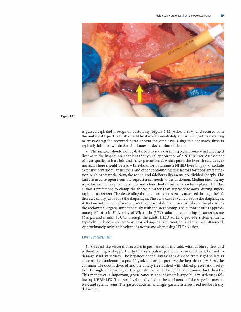
is passed cephalad through an aortotomy (Figure 1.42, yellow arrow) and secured withthe umbilical tape. The flush should be started immediately at this point, without waitingto cross-clamp the proximal aorta or vent the vena cava. Using this approach, flush istypically initiated within 2 to 3 minutes of declaration of death.
4. The surgeon should not be disturbed to see a dark, purple, and somewhat engorgedliver at initial inspection, as this is the typical appearance of a NHBD liver. Assessmentof liver quality is best left until after perfusion, at which point the liver should appearnormal. There should be a low threshold for obtaining a NHBD liver biopsy to excludeextensive centrilobular necrosis and other confounding risk factors for poor graft func-tion, such as steatosis. Next, the round and falciform ligaments are divided sharply. Theknife is used to open from the suprasternal notch to the abdomen. Median sternotomyis performed with a pneumatic saw and a Finochietto sternal retractor is placed. It is thisauthor’s preference to clamp the thoracic rather than supraceliac aorta during super-rapid procurement. The descending thoracic aorta can be easily accessed through the leftthoracic cavity just above the diaphragm. The vena cava is vented above the diaphragm.A Balfour retractor is placed across the upper abdomen. Ice slush should be placed onthe abdominal organs simultaneously with the sternotomy. The author infuses approxi-mately 5 L of cold University of Wisconsin (UW) solution, containing dexamethasone 16 mg/L and insulin 40 U/L; through the adult NHBD aorta to provide a clear effluent,typically 1 L before sternotomy, cross-clamping, and venting, and then 4 L afterward.Approximately twice this volume is necessary when using HTK solution.
Liver Procurement
5. Since all the visceral dissection is performed in the cold, without blood flow andwithout having had opportunity to assess pulses, particular care must be taken not todamage vital structures. The hepatoduodenal ligament is divided from right to left asclose to the duodenum as possible, taking care to preserve the hepatic artery. First, thecommon bile duct is divided and the biliary tree flushed with chilled preservation solu-tion through an opening in the gallbladder and through the common duct directly.This maneuver is important, given concern about ischemic-type biliary strictures fol-lowing NHBD LTX. The portal vein is divided at the confluence of the superior mesen-teric and splenic veins. The gastroduodenal and right gastric arteries need not be clearlydelineated.
Figure 1.42

30 Atlas of Organ Transplantation
6. The left lateral segment of the liver is elevated by dividing the left triangular liga-ment. It is safest to assume that there is a replaced or accessory left hepatic artery arisingfrom the left gastric artery. Therefore, the lesser omentum and left gastric artery shouldbe separated from the lesser curvature at the level of the stomach. The splenic artery isdivided to the left of the midline, far from the celiac axis, and then dissected toward theaorta, so that it can be rotated to the right for exposure of the superior mesenteric artery,which lies deep to it. Unless the plan is to procure the NHBD pancreas, discussed below,the head of the pancreas should be taken with the liver to avoid transecting an aberrantright hepatic artery and to expedite organ extraction time. After a Kocher’s maneuver,the duodenum and pancreatic head are elevated and retracted caudally to expose thesuperior mesenteric artery which is then dissected down to the aorta. Care is taken notto transect an accessory or replaced right hepatic artery by avoiding dissection on theright side of the superior mesenteric artery. Rather than taking extra time to search fora right branch, it is safest to assume that one exists and to take a common patch of supe-rior mesenteric and celiac arteries with the liver. An aortotomy is performed betweenthe superior mesenteric and right renal arteries and extended to provide the arterialpatch.
7. The left diaphragm is then divided down toward the Carrel patch. The suprahep-atic inferior vena cava is divided. The right diaphragm is then divided down to the upperpole of the right kidney. The infrahepatic inferior vena cava is then transected just abovethe renal veins. The liver is extricated and immediate back-table portal flush with 1 L ofchilled UW solution is performed. The liver is then packaged for transport to the trans-plant center.
Kidney Procurement
8. Bilateral nephrectomies are then performed. The kidneys may be kept en bloc for machine perfusion, or separated and sent directly to the recipient centers. Eventhough NHBD organ procurement is a rushed procedure, it is still crucial to performcomplete donor exploration in an effort to discern the possibility of unrecognized malig-nancy. It is also particularly important with NHBD grafts to perform back-table graftinspection and trimming well in advance of the recipient surgery, to ensure that it is safeto transplant the organ(s) and to allow adequate time for arterial reconstruction, ifnecessary.
Pancreas Procurement
9. Adding whole-organ pancreatectomy to hepatectomy during a super-rapid re-covery carries risk for transecting an aberrant right hepatic artery because there is noopportunity to palpate arterial pulsations in the NHBD. Meticulous in situ dissection insearch of a right branch can significantly increase extraction time. Therefore, the NHBDliver is typically removed with the pancreatic head to avoid injuring an aberrant righthepatic artery. We do not routinely procure NHBD whole pancreata when procuringNHBD livers unless there is favorable donor body habitus and other issues are optimal,such as warm ischemia time.23 Alternatively, the liver and pancreas may be removed enbloc.
d) Premortem Cannulation Technique
The group at Madison, Wisconsin, performs consensual, preextubation (premortem)femoral vessel cannulation.5 The NHBDs are typically taken to the operating room beforewithdrawal of life support. Right femoral artery and right femoral vein cannulae areinserted under local anesthesia. After cessation of respiration, lack of a monitored arte-rial pulse, declaration of death, and an additional wait time of 5 minutes, cold UW solu-tion is infused into the femoral artery cannula. The femoral vein cannula is opened to

Multiorgan Procurement from the Deceased Donor 31
gravity to decompress the venous system. Median sternotomy and midline abdominalincisions are made and the intraabdominal organs are removed en bloc or separately.Portal flush and organ separation are performed on the back table.
Ethical Issues and Professionalism
Certainly, NHBD organ procurement honors the donor’s wishes, brings some comfort tothe family, and benefits the recipient. It is appropriate to provide a brief overview of theethics of NHBD organ procurement and transplantation because surgeons must befamiliar with the following basic principles before working with NHBDs: individualsmay not be killed for their organs or killed as a result of the removal of their organs (the“dead donor” rule), patients must not be jeopardized in order to facilitate organ pro-curement, euthanasia is prohibited, informed consent and respect for family wishes mustnot be violated, and the autonomous right of patients to refuse treatment must beupheld.3,11,24–29 It is imperative to ensure that there is no conflict of interest between theduty to provide optimal patient care and the desire to recover organs for transplanta-tion.26,27,29 Specifically, the rationale for withdrawal of life support and the determinationof death must be extricable from the decision to recover organs. Therefore, the patientcare and organ donor teams need to be completely separate.
Several issues related to the ethics of NHBD organ procurement remain sources ofdebate. Interventions that improve the chance of successful donation rather than directlybenefiting the donor are permitted, as long as they are consensual and do not hastendeath or harm the donor, and medications routinely provided for patient comfort arepermitted even if they might hasten death.28 However, there are differing views regard-ing use of anticoagulants, vasodilators, narcotics, and intravascular cannulae placed pre-mortem.27 Another issue that is debated is whether determination of NHBD deathrequires loss of cardiac electrical activity or if absence of heart sounds, pulse, and bloodpressure are sufficient criteria, just as they are for patients who are not organ donors.3,11
Ultimately, the donor hospital and care team have the responsibility for defining anddeclaring patient death.Another important ethical question that impacts upon the warmischemic time endured by NHBD organs relates to the duration of the waiting periodused to assure irreversible death. Autoresuscitation after 1 minute of pulselessness hasnot been reported in the literature.26 However, different wait times from the determina-tion of death to organ procurement have been prescribed by various groups, rangingfrom 2 to 10 minutes11,25–28; most groups wait 5 minutes.
Conclusion
Ideally, patients who are brain dead, or will likely soon become so, should donate organsaccording to brain death protocols because the yield of transplantable organs fromNHBDs and outcomes of NHBD organ transplantation are generally not as favorable aswith heart-beating donors. Nonetheless, NHBD organ transplantation has become highlysuccessful, bringing full circle the history of organ donation. The surge in enthusiasmabout NHBDs has been fueled by several important organizations, including the Depart-ment of Health and Human Services of the United States federal government, the Insti-tute of Medicine (IOM) of the National Academy of Science, UNOS, and variousprofessional societies such as the Society of Critical Care Medicine.6,11,24,28 These groupshave endorsed NHBD organ procurement as an ethically effective solution for the organshortage, have recommended standardized NHBD protocols, and have arrangedsignificant funding for education and research about NHBD organ transplantation. Dis-covery of effective cytoprotective agents that could be administered to NHBDs and/orNHBD organ recipients to protect against ischemic organ injury would further expandthe ability to transplant NHBD organs. Surgeons who recover organs from NHBDs mustbe familiar with rapid procurement techniques, as described above.

32 Atlas of Organ Transplantation
Standard Multiorgan Procurement: Selected Readings
Abu-Elmagd K, Fung J, Bueno J, et al. Logistics and technique for procurement of intestinal, pancreatic, andhepatic grafts from the same donor. Ann Surg 2000;232(5):680–687.
Boggi U,Vistoli F, Del Chiaro M, et al. A simplified technique for the en bloc procurement of abdominal organsthat is suitable for pancreas and small-bowel transplantation. Surgery 2004;135(6):629–641.
D’Alessandro AM, Southard JH, Love RB, Belzer FO. Organ preservation. Surg Clin North Am 1994;74(5):1083–1095.
Delgado DH, Rao V, Ross HJ. Donor management in cardiac transplantation. Can J Cardiol 2002;18(11):1217–1223.
Dunn DL, Morel P, Schlumpf R, et al. Evidence that combined procurement of pancreas and liver grafts doesnot affect transplant outcome. Transplantation 1991;51(1):150–157.
Kootstra G, Kievit J, Nederstigt A. Organ donors: heartbeating and non-heartbeating. World J Surg 2002;26(2):181–184.
Ojo AO, Heinrichs D, Emond JC, et al. Organ donation and utilization in the USA. Am J Transplant 2004;4(suppl9):27–37.
St. Peter SD, Imber CJ, Friend PJ. Liver and kidney preservation by perfusion. Lancet 2002;16;359(9306):604–613.
Starzl TE, Miller C, Broznick B, Makowka L.An improved technique for multiple organ harvesting. Surg GynecolObstet 1987;165(4):343–348.
Van Buren CT, Barakat O. Organ donation and retrieval. Surg Clin North Am 1994;74(5):1055–1081.Van der Werf WJ, D’Alessandro AM, Hoffmann RM, Knechtle SJ. Procurement, preservation, and transport of
cadaver kidneys. Surg Clin North Am 1998;78(1):41–54.
Non–Heart-Beating Donor: References
1. Reich DJ, Manzarbeitia CY. Non-heart-beating donor liver transplantation. In: Busuttil RW, KlintmalmGB, eds. Transplantation of the Liver. Philadelphia: WB Saunders, 2005:529–543.
2. A definition of irreversible coma: report of the Ad Hoc Committee of the Harvard Medical School toExamine the Definition of Brain Death. JAMA 1968;205:337–340.
3. DeVita MA,Vukmir R, Snyder JV, et al. Procuring organs from a non-heart-beating cadaver: a case report.Kennedy Institute of Ethics Journal 1993;3:371–385.
4. Casavilla A, Ramirez C, Shapiro R, et al. Experience with liver and kidney allografts from non-heart-beating donors. Transplantation 1995;59:197–203.
5. D’Alessandro AM, Hoffmann RM, Knechtle SJ, et al. Successful extrarenal transplantation from non-heart-beating donors. Transplantation 1995;59:977–982.
6. United Network of Organ Sharing. Donation After Cardiac Death: A Reference Guide. Richmond, VA:UNOS, 2004.
7. Edwards JM, Hasz RD, Robertson VM. Non-heart-beating organ donation: process and review. AACNClinical Issues 1999;10:293–300.
8. Koogler T, Costarino AT Jr. The potential benefits of the pediatric nonheartbeating organ donor. Pedi-atrics 1998;101:1049–1052.
9. Gift of Life Donor Program. 2003 Annual Report. Philadelphia: Gift of Life Donor Program, Inc., 2004.10. Reiner M, Cornell D, Howard RJ. Development of a successful non-heart-beating organ donation program.
Prog Transplant 2003;13:225–231.11. Institute of Medicine, National Academy of Sciences. Non-Heart-Beating Organ Transplantation: Practice
and Protocols. Washington, DC: National Academy Press, 2000.12. Rudich SM, Kaplan B, Magee JC, et al. Renal transplantations performed using non-heart-beating organ
donors: going back to the future. Transplantation 2002;74:1715–1720.13. Cho YW, Terasaki PI, Cecka M, et al. Transplantation of kidneys from donors whose hearts have stopped
beating. N Engl J Med 1998;338:221–225.14. Reich DJ, Munoz SJ, Rothstein KD, et al. Controlled non-heart-beating donor liver transplantation: a suc-
cessful single center experience, with topic update. Transplantation 2000;70:1159–1166.15. Manzarbeitia CY, Ortiz JA, Jeon H, et al. Long-term outcome of controlled non-heartbeating donor liver
transplantation. Transplantation 2004;78:211–215.16. D’Alessandro AM, Hoffmann RM, Knechtle SJ, et al. Liver transplantation from controlled non-heart-
beating donors. Surgery 2000;128:579–588.17. Abt PL, Crawford MD, Desai NM, et al. Liver transplantation from controlled and uncontrolled non-heart-
beating donors: an increased incidence of biliary complications. Transplantation 2003;75:1659–1663.18. Fukumori T, Kato T, Levi D, et al. Use of older controlled non-heart-beating donors for liver transplanta-
tion. Transplantation 2003;75:1171–1174.19. Otero A, Gomez-Gutierrez M, Suarez F, et al. Liver transplantation from Maastricht category 2 non-heart-
beating donors. Transplantation 2003;76:1068–1073.20. Abt PL, Desai NM, Crawford MD, et al. Survival following liver transplantation from non-heart-beating
donors. Ann Surg 2004;239:87–92.

Multiorgan Procurement from the Deceased Donor 33
21. Gift of Life Donor Program. Asystolic Cadaveric Organ Recovery Procedures Following Patient and/orFamily Directed Withdrawal of Life Support. Philadelphia: Gift of Life Donor Program, Inc., 1998.
22. Olson L, Davi R, Barnhart J, et al. Non-heart-beating cadaver donor hepatectomy “the operative proce-dure”. Clin Transplantation 1999;13:98–103.
23. Jeon H, Ortiz JA, Manzarbeitia CY, et al. Combined liver and pancreas procurement from a controllednon-heart-beating donor with aberrant hepatic arterial anatomy. Transplantation 2002;74:1636–1639.
24. Institute of Medicine, National Academy of Sciences. Non-Heart-Beating Organ Transplantation: Medicaland Ethical Issues in Procurement. Washington, DC: National Academy Press, 1997.
25. Koostra G. The asystolic, or non-heartbeating, donor. Transplantation 1997;63:917–921.26. Whetstine L, Bowman K, Hawryluck L. Pro/con ethics debate: is nonheart-beating organ donation ethi-
cally acceptable? Critical Care 2000;6:192–195.27. Bell MD. Non-heart beating organ donation: old procurement strategy – new ethical problems. J Med
Ethics 2003;29:176–181.28. Recommendations for nonheartbeating organ donation. A position paper by the Ethics Committee,
American College of Critical Care Medicine, Society of Critical Care Medicine. Crit Care Med 2002;29:1826–1831.
29. Arnold RM, Younger SJ. Time is of the essence: the pressing need for comprehensive non-heart-beatingcadaveric donation policies. Transplant Proc 1995;27:2913–2921.
2Dialysis Access ProceduresKhalid O. Khwaja
HemodialysisIntroductionSurgical Proceduresa) Radiocephalic Fistulab) Brachiocephalic Fistulac) Basilic Vein Transpositiond) Forearm Loop Arteriovenous Grafte) Upper Arm Arteriovenous Graftf) Lower Extremity Access Procedure
Peritoneal DialysisPeritoneal Dialysis Catheter Placementa) Open Techniqueb) Laparoscopic Technique
Hemodialysis
Introduction
Hemodialysis is one of the main modalities for renal replacement therapy in patientswith end-stage renal disease. Successful hemodialysis is contingent upon the creation ofproper vascular access. Chronic vascular access was first established in 1960 by Scribnerand colleagues when they created a shunt between the radial artery and the cephalic veinusing an external Silastic device. However, this device was fraught with problems suchas bleeding, clotting, and infection. In 1966, Breschia and Cimino described a surgicalfistula between the radial artery and the cephalic vein just proximal to the wrist, therebyeliminating the external shunt and enabling a high flow system for hemodialysis. To thisday, it remains the procedure of choice for patients with end-stage renal disease in needof chronic hemodialysis.
Several principles should be followed when planning vascular access surgery. Ingeneral, primary fistulas are better than prosthetic grafts due to better long-term patencyand lower risk of infection and thrombosis. The upper extremity is preferable to thelower extremity and the nondominant arm should be employed first. If possible, a distalsite should be selected first, preserving the upper arm for subsequent use. Careful pre-operative vascular assessment is performed with palpation of the radial, ulnar, andbrachial pulses; an Allen’s test is performed on both sides. The superficial veins of thearm should be carefully assessed with application of a proximal tourniquet. In somecases, the cephalic vein is readily evident at the wrist, antecubital fossa area, or in thelateral aspect of the upper arm. Once a decision has been made to perform access
35

36 Atlas of Organ Transplantation
surgery, no venipunctures or blood pressure monitoring should be performed in thatarm. If no superficial veins are apparent, the venous system may be assessed by ultra-sound examination of the arm. Both the cephalic and basilic systems are interrogated,as well as the deep venous system and the central veins. Patients with suspected centralvenous stenosis or prior catheters inserted on the ipsilateral side, or with abnormalfindings on ultrasound, may be assessed by conventional venography. If central stenosesare found, they should be corrected by endovascular techniques preoperatively, or analternate site for access should be sought.
Vascular access should be established prior to the actual need for hemodialysis,thereby avoiding temporary external catheters, which have a higher risk of infection andare also associated with central venous stenosis. This chapter discusses the technique forsome common permanent access procedures.
Surgical Procedures
a) Radiocephalic Fistula
The radiocephalic fistula, as described by Breschia and Cimino, is the procedure ofchoice. A suitable cephalic vein just proximal to the wrist is identified preoperatively anda negative Allen’s test confirmed. Although up to a third of these fistulas fail to mature,the long-term patency is excellent with as many as half of them still functioning 5 to 10years after creation.
1. The whole arm, including the axilla, is prepped and draped. As no prosthetic deviceis being employed, antibiotics are not a requisite. A vertical incision is fashioned justproximal to the flexion crease of the wrist between the radial artery and the cephalicvein (black arrow). Some surgeons prefer to make an incision over the anatomic snuffbox more distally and use the deep branch of the radial artery to create the fistula (greyarrow) (Figure 2.1).
Figure 2.1

Dialysis Access Procedures 37
2. The incision is carried through the subcutaneous tissue, and then medial andlateral flaps are raised. Both the cephalic vein and radial artery are very superficiallylocated and can readily be exposed (Figure 2.2a). The cephalic vein is mobilized as farproximally and distally as possible and any large branches ligated. The radial artery isalso mobilized for a short distance. Once mobilization is complete, both the cephalic veinand radial artery are placed adjacent to each other in a side-by-side fashion. This can beaccomplished by placing a vessel loop proximally and distally, with each loop incorpo-rating the artery and vein. By tightening up on the loop, the two vessels are broughttogether (Figure 2.2b). If necessary, systemic heparin can now be administered. Controlof the vessels can be achieved by tightening up on the vessel loops or by using small vas-cular clamps (Figure 2.2c).
3. A corresponding venotomy and arteriotomy are made in the cephalic vein andradial artery (Figure 2.3a). The arteriotomy should be limited to 6 or 7 mm to prevent a
Figure 2.2
Figure 2.3

38 Atlas of Organ Transplantation
steal syndrome. A side-to-side anastomosis is then constructed using fine, nonab-sorbable monofilament suture (Figures 2.3b and 2.3c). One technique is to sew the pos-terior wall from within the lumen, then running the suture anteriorly to complete theanastomosis. Prior to reperfusion, the anastomosis can be probed through an openingcreated in the distal cephalic vein (Figure 2.3d). The probe is passed sequentially up thecephalic vein and the radial artery. The distal cephalic vein is then ligated and the arte-rial clamps released to perfuse the fistula (Figure 2.3e).
4. Alternatively, an end-to-side or end-to-end anastomoses between the radial arteryand cephalic vein can be created by dividing the vein initially (Figure 2.4a) and thenanastomosing it to the artery (Figure 2.4b).
A radiocephalic fistula usually requires 8 to 12 weeks to mature. Sometimes a second procedure is required to ligate a side branch or angioplasty an area of proximalstenosis.
Figure 2.4
b) Brachiocephalic Fistula
If a suitable cephalic vein at the wrist is not present, then a more proximal brachio-cephalic fistula may be created. In some instances, the cephalic vein might be deep inthe upper arm and not clinically visible. If it is seen on ultrasound examination and hasa diameter of 3 mm or more, then it may be suitable for fistula creation. Brachiocephalicfistulas have a primary failure rate of close to 10% and good long-term patency.

Dialysis Access Procedures 39
Figure 2.5
Figure 2.6
1. A preoperative evaluation is performed as described for the radiocephalic fistula.The whole arm is prepped from the wrist to the axilla. This procedure can be performedunder local anesthetic with sedation. The elbow is examined to mark the course of thecephalic vein (red arrow) and palpated to locate the position of the brachial artery(broken red line), which lies adjacent to the basilic vein (yellow arrow) (Figure 2.5).
2. Both the brachial artery and cephalic vein can be isolated through a transverse inci-sion either above (Figure 2.6a) or below (Figure 2.6b) the antecubital crease. The cephalicvein may require some mobilization to reach the brachial artery and may need to be dissected distally to the upper part of the forearm to gain adequate length for it to reach the artery. Frequently, an antecubital or median cubital vein (blue arrow) canbe found that communicates with the cephalic vein, and this vein can be used for the

40 Atlas of Organ Transplantation
Figure 2.7
Figure 2.8
anastomosis with the distal cephalic vein being ligated (Figure 2.6b). It usually easilyreaches the brachial artery (red arrow) (Figure 2.7)
3. Once an adequate length of vein is dissected free, it is divided. An end-to-side anas-tomosis is then constructed using fine, nonabsorbable monofilament suture (Figure 2.8).If the cephalic vein is found to be unsuitable, then the basilic vein can be used throughthe same approach and transposed so it runs in a more superficial course (describednext). If none of these veins is suitable, a loop forearm graft can be created through thesame incision. A primary brachiocephalic fistula matures in 8 to 12 weeks.

Dialysis Access Procedures 41
4. Sometimes the cephalic vein at the level of the elbow and above is of adequate size,but too deep to simply anastomose directly to the brachial artery. If the vein is deep, itmay be difficult to cannulate for subsequent dialysis. In this case the vein can be dis-sected for some length above the elbow, and then brought through a tunnel created justbelow the skin to transpose the vein to a more superficial location. This is done by firstisolating the vein either just above or below the antecubital fossa and then dissecting itfurther proximally as much as possible through the transverse incision. The vein isdivided as far distally as possible. A longitudinal counterincision is then made higher inthe arm and the same vein is isolated and mobilized into this incision (Figure 2.9). Atthis point it is helpful to gently dilate the vein (large blue arrow) with saline solution toensure that there is no twisting. The vein can be marked with a pen to maintain this ori-entation. One is now ready to create the superficial tunnel for the vein (small bluearrows).
Figure 2.9

5. A subcutaneous tunnel is created for the vein using a tunneler or other blunt instru-ment (Figure 2.10). The tunnel should start just above the brachial artery and end at theproximal extent of the upper arm incision.
6. The cephalic vein is brought through the tunnel, making sure that the vein doesnot twist as it is being pulled through. The end of the vein (blue arrow) is positionedjust above the brachial artery (yellow arrow), in preparation for the anastomosis (Figure 2.11).
42 Atlas of Organ Transplantation
Figure 2.10
Figure 2.11
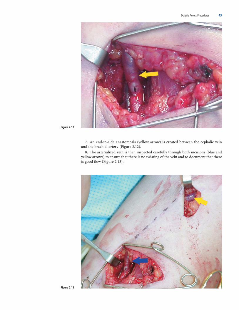
Dialysis Access Procedures 43
Figure 2.12
Figure 2.13
7. An end-to-side anastomosis (yellow arrow) is created between the cephalic veinand the brachial artery (Figure 2.12).
8. The arterialized vein is then inspected carefully through both incisions (blue andyellow arrows) to ensure that there is no twisting of the vein and to document that thereis good flow (Figure 2.13).

44 Atlas of Organ Transplantation
c) Basilic Vein Transposition
As the basilic vein in the upper arm runs deep to the fascia, it is protected from traumarelated to venepuncture. It may be transposed to a more superficial location for dialysisaccess. This is a suitable option when the cephalic vein is inadequate for fistula creation.The primary patency for this fistula is about 60% to 70% and the long-term patency issimilar to that of a graft.
1. The procedure can be performed with a general anesthetic or local anesthetic withsedation, though the former is preferred due to the extensive dissection involved. Thewhole arm is prepped, including the axilla. The incision is begun at the antecubital creasein a vertical fashion just medial to the brachial artery pulse (Figure 2.14a). The incision is carried through the subcutaneous tissue; the fascia is incised and the brachialartery exposed. The median nerve (Figure 2.15, green arrow) lies medial to the brachialartery (yellow arrow) at this location and should be identified and preserved (Figure2.14b). Proceeding slightly more medial and still deep to the fascia, the basilic vein (bluearrow) is visualized and traced proximally, all the way to its junction with the axillaryvein (Figure 2.15). At some point, usually in the proximal third of the arm, the medial
Figure 2.14
Figure 2.15
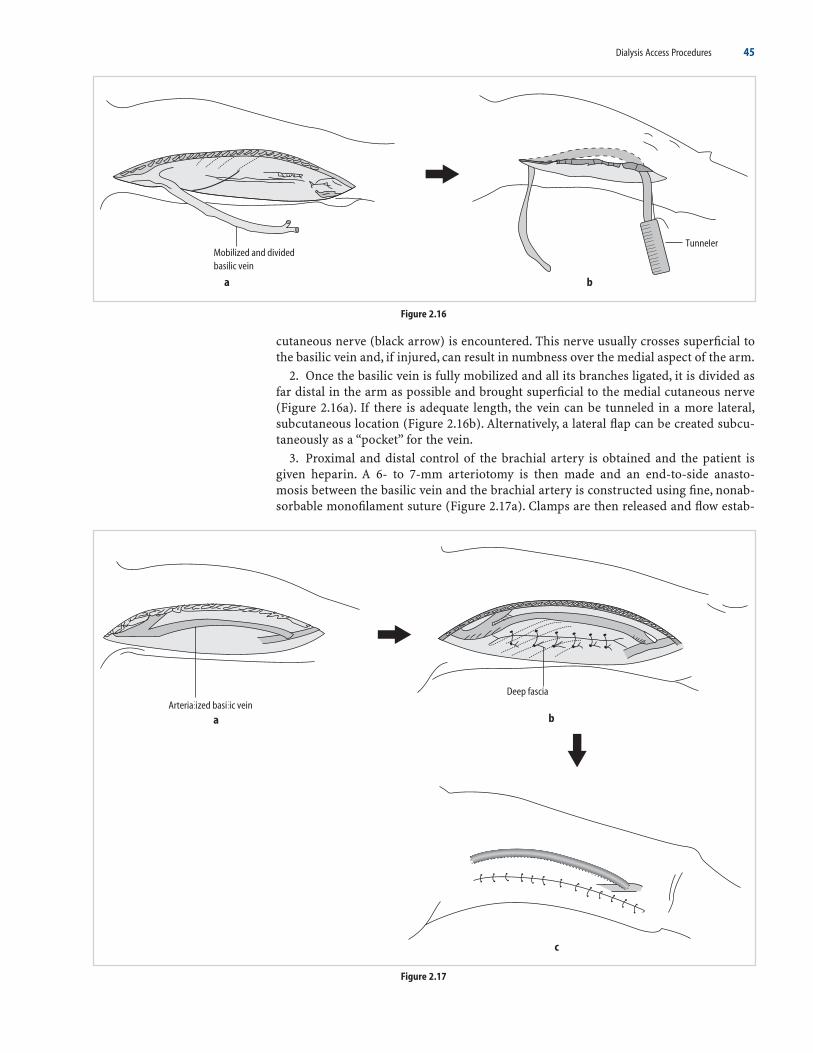
Dialysis Access Procedures 45
Figure 2.16
Figure 2.17
cutaneous nerve (black arrow) is encountered. This nerve usually crosses superficial tothe basilic vein and, if injured, can result in numbness over the medial aspect of the arm.
2. Once the basilic vein is fully mobilized and all its branches ligated, it is divided asfar distal in the arm as possible and brought superficial to the medial cutaneous nerve(Figure 2.16a). If there is adequate length, the vein can be tunneled in a more lateral,subcutaneous location (Figure 2.16b). Alternatively, a lateral flap can be created subcu-taneously as a “pocket” for the vein.
3. Proximal and distal control of the brachial artery is obtained and the patient isgiven heparin. A 6- to 7-mm arteriotomy is then made and an end-to-side anasto-mosis between the basilic vein and the brachial artery is constructed using fine, nonab-sorbable monofilament suture (Figure 2.17a). Clamps are then released and flow estab-
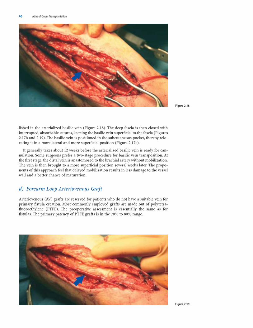
46 Atlas of Organ Transplantation
lished in the arterialized basilic vein (Figure 2.18). The deep fascia is then closed with interrupted, absorbable sutures, keeping the basilic vein superficial to the fascia (Figures2.17b and 2.19). The basilic vein is positioned in the subcutaneous pocket, thereby relo-cating it in a more lateral and more superficial position (Figure 2.17c).
It generally takes about 12 weeks before the arterialized basilic vein is ready for can-nulation. Some surgeons prefer a two-stage procedure for basilic vein transposition. Atthe first stage, the distal vein is anastomosed to the brachial artery without mobilization.The vein is then brought to a more superficial position several weeks later. The propo-nents of this approach feel that delayed mobilization results in less damage to the vesselwall and a better chance of maturation.
d) Forearm Loop Arteriovenous Graft
Arteriovenous (AV) grafts are reserved for patients who do not have a suitable vein forprimary fistula creation. Most commonly employed grafts are made out of polytetra-fluoroethylene (PTFE). The preoperative assessment is essentially the same as forfistulas. The primary patency of PTFE grafts is in the 70% to 80% range.
Figure 2.18
Figure 2.19

Dialysis Access Procedures 47
1. The procedure is usually performed under a local anesthetic with sedation. Pre-operative antibiotics are utilized. A transverse incision is fashioned below the antecu-bital crease (Figure 2.20a). The antecubital, cephalic, or basilic veins may be exposed inthe subcutaneous layer (Figure 2.20b). Any one of these veins can be used for outflow. Ifthese are not suitable, the graft can be drained into the deep brachial vein.
2. If a suitable superficial vein is identified, it is gently looped and preserved. Thebicipital aponeurosis is then opened (Figure 2.21a). The brachial artery (red arrow), withits concomitant veins can be exposed deep to the aponeurosis (Figures 2.21b and 2.22).Care must be taken to avoid injury to the median nerve, which courses slightly deeperand medial to the artery.
Figure 2.20
Figure 2.21
Figure 2.22
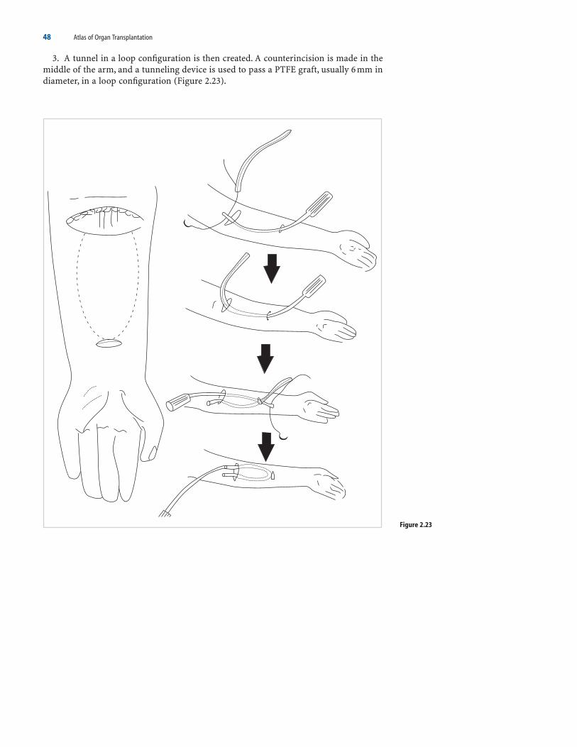
48 Atlas of Organ Transplantation
Figure 2.23
3. A tunnel in a loop configuration is then created. A counterincision is made in themiddle of the arm, and a tunneling device is used to pass a PTFE graft, usually 6 mm indiameter, in a loop configuration (Figure 2.23).

Dialysis Access Procedures 49
4. Once the graft has been tunneled, the patient can be heparinized. The arterial andvenous anastomosis are performed, both in an end-to-side fashion. For the venous end,the graft can be cut at a bias to increase the diameter of the anastomosis and decreasethe chances of venous stenosis (Figure 2.24a). For the arterial end, large anastomoses are avoided to decrease the chance of developing steal syndrome (Figure 2.24b).
5. The skin is closed over the completed anastomosis (Figure 2.25). It is unnecessaryto close the deep fascia. The radial artery pulse is checked prior to completing the oper-ation. Another option for a forearm graft is to place a straight graft with inflow from theradial artery at the wrist and outflow to one of the veins in the antecubital fossa.
Figure 2.24
Figure 2.25

50 Atlas of Organ Transplantation
Figure 2.26
e) Upper Arm Arteriovenous Graft
An upper arm or brachial artery to axillary vein graft is possible in most patients,provided there is no central stenosis and no arterial disease. The early patency is excel-lent with a failure rate of only 0% to 3%. However, complications associated with the use of prosthetic material are inherent in this procedure, such as thrombosis and infection.
1. The preoperative assessment is as already described. The procedure is performedunder local or general anesthesia. The whole arm including the axilla is prepped. Thebrachial artery can be exposed above the antecubital crease through a transverse inci-sion. The axillary vein is usually exposed through a vertical or transverse incision madein the axilla (Figure 2.26a, c). The axillary incision is continued through the subcuta-neous tissue and the fascia is opened. The axillary vein can be readily exposed or, alter-natively, the brachial vein or basilic vein can be exposed more distally (Figure 2.26b).These two vessels coalesce to form the axillary vein. Any of these veins, depending ontheir caliber, may be used for outflow.

Figure 2.27
Dialysis Access Procedures 51
2. Using a tunneling device, a 6-mm PTFE graft is placed subcutaneously between thetwo incisions (Figure 2.27a, b). The graft should be directed lateral, as this is more comfortable for the patient during dialysis. Once the graft has been tunneled in its loca-tion, the patient may be heparinized. The venous end of the graft is cut at a bias toincrease the diameter. An end-to-side anastomosis is then performed using a fine,monofilament, nonabsorbable suture. The arterial anastomosis is then made in a similarfashion, limiting the arteriotomy to 6 or 7 mm to avoid steal syndrome. The venousclamps are first released followed by the arterial clamps and flow in the graft established(Figure 2.27c).
f) Lower Extremity Access Procedure
Use of the lower extremity for permanent dialysis access should be limited to those sit-uations in which all options involving the upper extremity have been exhausted or arenot possible. These procedures unfortunately have a high risk of failure, poor long-termpatency rates, high risk of infection, and poor tolerance by patients. Options include aprimary arteriovenous fistula utilizing the saphenous vein as the conduit and thesuperficial femoral artery as the source of inflow. Alternatively, a prosthetic graft can be used with anastomosis to the femoral artery and saphenous vein.

52 Atlas of Organ Transplantation
1. For a primary arteriovenous fistula, the saphenous vein is completely dissected outfrom its junction with the main femoral vein to a level in the mid to distal thigh (Figure2.28a). All branches are divided and the distal few centimeters of the vein is completelyfreed so that it can be brought close to the superficial femoral artery for anastomosis.The vein is ligated and divided distally. The superficial femoral artery is isolated at thislevel for an adequate length to allow for easy anastomosis. The vein is then brought tolie close to the artery. An end-to-side anastomosis is fashioned (Figure 2.28b).
Figure 2.28

Dialysis Access Procedures 53
Figure 2.29
Figure 2.30
2. If the saphenous vein is not of useable quality for an adequate length, then a pros-thetic graft can be used. A horizontal or vertical incision is made in the femoral region,directly over where the saphenous vein likely enters the main femoral vein. The artery,which lies just lateral to the femoral vein, is similarly dissected out. The graft is broughtthrough a tunnel, similar to as with a looped graft procedure in the forearm. The endsof the graft are anastomosed to the corresponding artery and vein (Figures 2.29 and2.30).

54 Atlas of Organ Transplantation
Peritoneal Dialysis
Peritoneal dialysis has been in wide usage since the 1960s. For some patients it is aneffective means of renal replacement therapy. Several modalities of peritoneal dialysisare now available, but the principle remains the same – fluid and solutes are exchangedvia the peritoneum.
As with hemodialysis, careful preoperative planning is required. Peritoneal dialysismay not be a viable option in patients with multiple prior abdominal surgeries asintraabdominal adhesions may preclude effective exchange of the dialysate. The patientmust be competent enough to manage the machine and catheter at home. It is a usefuloption in patients who have difficult vascular access.
Peritoneal Dialysis Catheter Placement
a) Open Technique
In 1968, Tenckhoff developed a silicone catheter that is still used for peritoneal dialysis.Several modifications of this catheter are currently available. Most of the catheters havetwo Dacron cuffs, one that is implanted just above the peritoneum and the other in asubcutaneous location. Both these cuffs create an inflammatory reaction with subse-quent fibrosis and adhesion, preventing bacterial ingrowth from the skin.
1. Typically, either a vertical or transverse incision is made in a paramedian locationbelow the umbilicus (Figure 2.31). If the patient is a renal transplant candidate, the inci-sion should be made on the left, saving the right side for the kidney.
Figure 2.31

Dialysis Access Procedures 55
Figure 2.32
Figure 2.33
2. The incision is carried through the subcutaneous tissue and the anterior rectussheath incised transversely (Figure 2.32a). The fibers of the rectus muscle are then splitbluntly to expose the transversalis fascia and peritoneum (Figure 2.32b). An opening ismade in the peritoneum and a purse-string suture placed around the opening (Figure2.32c).
3. The catheter is then flushed and threaded over an inserting stylet. The catheter(Figure 2.33a) can then be gently fed through the peritoneal opening into the peritonealcavity, directing it caudad (Figure 2.33b). Care must be taken to avoid injury to under-lying viscera during this maneuver. The purse-string suture is tied, securing the peri-toneum around the catheter.

56 Atlas of Organ Transplantation
4. The first cuff is seated above the peritoneum within the body of the rectus muscle.Part of the suture may be used to directly transfix the Dacron cuff. A lateral tunnel isthen created in a subcutaneous space and a small, sharp instrument passed through thetunnel into the original incision (Figure 2.33c); the free end of the catheter is graspedand pulled through. The anterior rectus sheath is then closed with nonabsorbablesutures. The second Dacron cuff is seated 1 or 2 cm away from the skin exit site to preventerosion through the skin (Figure 2.34a). It is usually not necessary to suture the catheterto the skin. In fact, this may lead to irritation and infection of the catheter site. The skinincision is closed in standard fashion (Figure 2.34b). Prior to leaving the operating room,the catheter is tested by instilling it with saline solution and then checking for drainageby placing the catheter in a dependent position.
b) Laparoscopic Technique
The advantage of the laparoscopic method is that the catheter can be placed under directvisualization, thereby reducing the risk of visceral injury and optimizing placement.Also, with this technique, the catheter may be used right away; the open techniquerequires a 1 to 2-week wait. The disadvantage is that a general anesthetic is required.
Figure 2.34

Dialysis Access Procedures 57
1. The whole abdomen is prepped. Usually two 5-mm trocars are adequate (Figure2.35a). Some surgeons prefer a third port as well. A 5-mm port is placed in the supraum-bilical area either using the open technique or with a closed technique using a Veressneedle. Pneumoperitoneum is established with CO2 insufflation. A second 5-mm port isthen placed in a paramedian position below the umbilicus. The peritoneal cavity is care-fully inspected for any adhesions or other abnormalities. Adhesiolysis is performed ifneeded. The Silastic peritoneal dialysis catheter is then threaded over its thin insertingstylet (Figure 2.35b). The 5-mm port in the left paramedian area is then removed andthe catheter with stylet is inserted through this opening under direct laparoscopic visu-alization (Figure 2.35c). The stylet and catheter are carefully advanced toward the supra-pubic area and the catheter threaded into the peritoneal cavity (Figure 2.35d). Thecatheter is pushed into the abdomen until the Dacron cuff is visible. This is then seated
Figure 2.35

58 Atlas of Organ Transplantation
just above the peritoneum. The stylet is then completely withdrawn leaving the catheterin place, with its tip directed into the pelvis (blue arrow) (Figure 2.36).
2. A small incision is then created laterally for the catheter exit site. Using a longgrasping instrument, a subcutaneous tunnel is created between this incision and theparamedian incision (Figure 2.37a). The end of the catheter is grasped and pulled out,situating the second cuff about 1 or 2 cm away from the skin exit site (Figure 2.37b). Thesubcutaneous tissue and skin are closed at the paramedian incision but no deeperstitches are necessary. The pneumoperitoneum is released and catheter function is testedby instilling 500 cc to 1 L of normal saline and then letting it flow out by dependentdrainage. The initial port site is then closed in standard fashion (Figure 2.37c).
Selected Readings
1. Akoh JA, Sinha S, et al. A 5-year audit of hemodialysis access. Int J Clin Pract 2005;59(7):847–851.2. Brescia MJ, et al. Chronic hemodialysis using venipuncture and a surgically created arteriovenous fistula.
N Engl J Med 1966;275:1089.3. Crabtree JH, Fishman A. A laparoscopic method for optimal peritoneal dialysis access. Am Surg
2005;71(2):135–143.4. Diaz-Buxo JA. Access and continuous flow peritoneal dialysis. Perit Dial Int 2005;25(suppl 3):S102–104.5. Gray RJ, Sands JJ, eds. Dialysis Access: A Multidisciplinary Approach. Philadelphia: Lippincott Williams
& Wilkins, 2002.6. Kawecka A, Debska-Slizien A, Prajs J, et al. Remarks on surgical strategy in creating vascular access for
hemodialysis: 18 years of one center’s experience. Ann Vasc Surg 2005;19(4):590–598.7. Keuter XH, van der Sande FM, Kessels AG, de Haan MW, Hoeks AP, Tordoir JH. Excellent performance of
one-stage brachial-basilic arteriovenous fistula. Nephrol Dial Transplant 2005;20(10):2168–2171.8. Lin PH, Bush RL, Nguyen L, Guerrero MA, Chen C, Lumsden AB. Anastomotic strategies to improve
hemodialysis access patency – a review. Vasc Endovasc Surg 2005;39(2):135–142.9. National Kidney Foundation. Kidney Disease Outcomes Quality Initiative (NKF K/DOQI). Clinical prac-
tice guidelines for vascular access. Am J Kidney Dis 2001;37(suppl 1):s137–s181.10. Ramage IJ, Bailie A, Tyerman KS, McColl JH, Pollard SG, Fitzpatrick MM. Vascular access survival in chil-
dren and young adults receiving long-term hemodialysis. Am J Kidney Dis 2005;45(4):708–714.11. Striker GE, Tenckhoff H. A transcutaneous prosthesis for prolonged access to the peritoneal cavity.
Surgery 1971;69:70.12. Wilson SE, ed. Vascular Access: Principles and Practice. St Louis: Mosby, 1996.
Figure 2.36
Figure 2.37

3Nephrectomy from a Living DonorRaja Kandaswamy and Abhinav Humar
IntroductionOpen NephrectomyLaparoscopic Donor Nephrectomya) Introductionb) Preoperative Preparationc) Hand-Assisted Laparoscopic Nephrectomyd) Pure Laparoscopic Techniquee) Robotic-Assisted Technique
Introduction
The kidney, the first organ to be used for living-donor transplants, is the most commontype of organ donated by living donors today. In the Unites States, the number of livingkidney donors now outnumbers the number of deceased kidney donors. Initially it wasfelt that only close family members could be potential donors. However, it is well recog-nized now that there does not need to be any direct relationship between the donor andrecipient to achieve a highly successful outcome.Any healthy person is a potential kidneydonor, including relatives, coworkers, friends, and acquaintances.
A living-donor kidney transplant offers significant advantage over its deceased-donorcounterpart. Living-donor kidney recipients enjoy improved long-term success, avoid aprolonged wait, and are able to plan the timing of their transplant in advance. Moreover,they have a significantly decreased incidence of delayed graft function and increasedpotential for human leukocyte antigen (HLA) matching. As a result, living-donor trans-plants generally have better short- and long-term results, as compared with deceased-donor transplants. Of course, the risks to the living donor must be acceptably low. Thedonor must be fully aware of potential risks and must freely give informed consent. Butas long as these conditions are met, the search for a living donor should not be restrictedto immediate family members. Results with living, unrelated donors are comparable tothose with living, related (non–HLA-identical) donors.
The preoperative evaluation of individuals who present as possible kidney donors isa crucial part of the living-donor kidney transplant process. A complete and thoroughevaluation is important in minimizing the risks of the procedure for the potential donorand optimizing donor safety – the underlying principle of any living-donor procedure.The predonation evaluation of the potential donor is also critical in ensuring a success-ful outcome for the recipient. The evaluation process may vary slightly depending onindividual center preference. But the steps of the process essentially remain the same,and can be divided into three major parts: (1) medical evaluation, (2) surgical or radio-logic evaluation, and (3) psychosocial evaluation. The potential donor should satisfy each
59
60 Atlas of Organ Transplantation
of the three parts of this evaluation process before being considered an acceptable donor.The evaluation process, at the same time as being complete, should not be overly cum-bersome for the donor. It should be streamlined and efficient, invasive tests should beminimized, and it should be cost-effective. Members of a multidisciplinary team need tobe involved in the evaluation process, including a nephrologist, surgeon, psychologist,social worker, and transplant coordinator.
One main goal of the preoperative medical evaluation is to ensure that the donor doesnot have underlying medical problems that would significantly increase the risk associ-ated with a general anesthetic and a major operative procedure. This part of the evalu-ation is not too dissimilar from the evaluation for any person undergoing a major generalsurgical procedure. The other important part of the medical evaluation serves to iden-tify and exclude any possibility of chronic kidney disease or kidney dysfunction in thepotential donor. The medical evaluation includes the screening for viral pathogens thatcould potentially be significantly harmful when transmitted to the recipient. The nextpart of the evaluation looks at the surgical anatomy of the kidney including the bloodvessels supplying the kidney. Present-day noninvasive imaging is sufficient to provideadequate preoperative information, eliminating the need for invasive tests such asangiograms. The final part of the evaluation looks at the psychosocial status of the donor,ensuring that the donor is mentally fit to donate, understands the associated risks, andis able to give informed consent without coercion.
The operative procedure for removal of a kidney for living-donor kidney transplanthas changed dramatically in the last 5 years. Laparoscopic techniques have becomeincreasingly more common, and at many centers, laparoscopic donor nephrectomy isnow the preferred method for removal of the kidney. Advantages for the donor includea quicker recovery, a shorter hospital stay, and decreased postoperative pain. It is likelythat laparoscopic removal of the kidney will become the standard of care at almost allcenters over the next few years.
The early postoperative care of donors is very similar to that of anyone undergoing amajor intraabdominal procedure. Fluid and electrolytes are monitored closely, as is theurine output. Pain and other postoperative symptoms such as nausea should be ade-quately controlled. Donors should be encouraged to ambulate early to minimize the risk of pulmonary complications or deep venous thrombosis. Donors should also be moni-tored closely for any surgical complications including postoperative bleeding, woundinfections, or bowel complications.
Most donors undergoing laparoscopic nephrectomy stay in the hospital for 2 to 3 daysafter the procedure. The early recovery period (i.e., the first 2 weeks after surgery)involves gradual return to activities of daily living. At 2 weeks, most donors are able totolerate mild exercise in addition to regular activities. They should wait 4 to 6 weeks before engaging in any strenuous activity or heavy lifting.
Studies have shown that laparoscopic donors experience less pain than open donors,as indicated by a decreased use of pain medicines (narcotics) after surgery. In addition,a shorter hospital stay and quicker return to full activity and work have been docu-mented. Early to intermediate follow-up (up to 5 years) of laparoscopic donors hasrevealed no increase in the risk of complications, as compared with open donors.However, long-term follow-up information (i.e., 10, 20, and 30 years) is lacking. Becauselaparoscopic nephrectomy involves working through the abdominal cavity (in contrastto open donor nephrectomy, which is a retroperitoneal procedure), there may be a small risk of long-term intestinal obstruction due to adhesions within the abdomen. Thisrisk remains theoretical; in fact, with other types of laparoscopic procedures in the last20 years, no increased risk of intestinal obstruction has been consistently documented.In the short term, however, laparoscopic donors may experience bowel dysfunction(nausea, bloating, irregular bowel movements) during the first few days after surgery.Such dysfunction usually responds to a combination of bowel rest and laxatives.

Nephrectomy from a Living Donor 61
Open Nephrectomy
1. The patient is positioned with the right or left flank (depending on which kidneyis being removed) elevated and exposed. The incision begins posteriorly at the tip of the12th rib and curves anteriorly in the direction of the umbilicus, stopping at the lateralborder of the rectus muscle (black arrow) (Figure 3.1).
Figure 3.1

62 Atlas of Organ Transplantation
2. The three muscular layers of the lateral abdominal wall are divided. Sometimes asmall portion of the tip of the 12th rib may need to be removed. Deep to the muscularlayers, the peritoneum is identified and retracted medially to visualize the retroperi-toneal space. The ureter (black arrow) is identified below the lower pole of the kidney,running on the anterior surface of the psoas muscle. It is mobilized down to the pelvis,taking care not to remove the surrounding periureteral tissue. Once the ureter is mobi-lized, Gerota’s fascia is incised and the kidney is separated from the surrounding perire-nal fat (Figure 3.2).
Figure 3.2

Nephrectomy from a Living Donor 63
Figure 3.3
3. The renal vein is identified and mobilized proximally. For the left kidney, the veinis mobilized to the point where it lies anterior to the aorta (Figure 3.3). The gonadal vein,adrenal vein, and any lumbar veins will need to be ligated and divided. The right renalvein, however, usually has no branches draining into it. The vein on the right side istraced proximally to its junction with the inferior vena cava.

64 Atlas of Organ Transplantation
4. The renal artery is identified next, usually lying posterior and superior to the lowerborder of the renal vein. It is useful to retract the kidney medially and carry out the dis-section along the posterior aspect of the hilum. The renal artery is mobilized to its junc-tion with the aorta (Figure 3.4).
5. The ureter is divided distally. At this point one can easily check to ensure that thekidney is producing urine. If the function of the kidney appears adequate, the donor isgiven intravenous heparin. Vascular clamps are applied on the renal artery at its junc-tion with the aorta, and on the renal vein where it runs anterior to the aorta (Figure 3.5).These structures are divided. The kidney is removed and sent to the recipient team.
Figure 3.4
Figure 3.5

Nephrectomy from a Living Donor 65
6. The vascular stumps are then oversewn with a nonabsorbable suture (Figure 3.6).After checking for hemostasis, closure is initiated. The abdominal wall is closed in layers.
Laparoscopic Donor Nephrectomy
a) Introduction
Nephrectomy from a living donor, which has been performed since the 1960s using theopen technique, has been associated with a relatively long incision along the flank, ofteninvolves removal of a portion of the 12th rib, and may be associated with significant painand significant loss of time from work and other activities. The recovery period is usually6 weeks and sometimes longer. However, long-term results with open donor nephrec-tomy in terms of quality and longevity of life have been excellent.
The short-term disability caused by open donor nephrectomy was the impetus to seek an alternative – a less invasive method that would result in a smaller incision,less pain, and faster recovery. Even though laparoscopic nephrectomy for disease hadbeen performed as early as 1991, laparoscopic donor nephrectomy was first performedin the United States in 1995. Early concerns about an increased incidence of delayed graftfunction and graft thrombosis were quickly dispelled as experience in the field grew. Cur-rently, over 60% of living donors in the United States are performed using the laparo-scopic technique.
Laparoscopic donor nephrectomy can be performed using a variety of techniques.These include hand-assisted, pure laparoscopic, robotic hand–assisted, and robotic purelaparoscopic. The main advantages of the hand-assisted techniques are that they areeasier to adapt for a surgeon with little experience in advanced laparoscopy, they providean added safety margin in case of emergencies, and they provide tactile feedback whileoperating. The disadvantages of the hand-assisted techniques include a slightly largerincision than would be required to deliver the kidney, and restricted areas where theincision can be placed to facilitate reaching to the kidney. Usually, upper midline inci-sions are used for a hand-assisted left nephrectomy, although some surgeons do thisusing a lower midline or even a Pfannenstiel incision. Pure laparoscopic technique, whichis most commonly employed by advanced laparoscopic surgeons, offers the advantageof placing the incision below the belt line, where it is better concealed. Also, the size ofthe incision is usually smaller since the kidney is delivered using a bag. Theoretically,the pain would be expected to be lower, since upper midline incisions are more painfulthan lower incisions in the abdomen. In the last few years, the addition of the surgicalrobot to assist in donor nephrectomy led to the use of this technique by some centers.Again, as with traditional laparoscopy, this can be done either with or without handassistance. The advantages of using a robotic system are as follows:
1. A 360-degree articulating wrist for the operating arms, providing greater flexibilityand maneuverability;
2. Tremor-free operation due to a stabilization system built in;
Figure 3.6
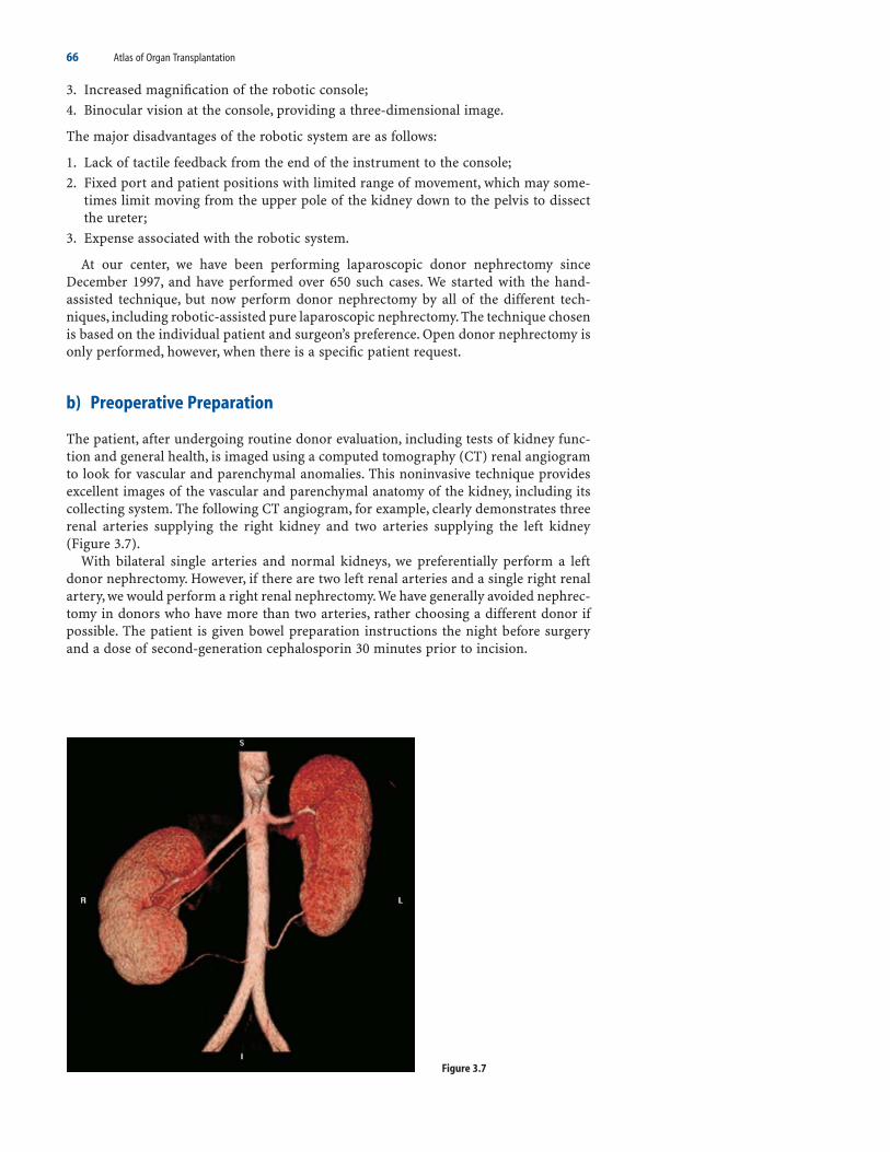
66 Atlas of Organ Transplantation
3. Increased magnification of the robotic console;4. Binocular vision at the console, providing a three-dimensional image.
The major disadvantages of the robotic system are as follows:
1. Lack of tactile feedback from the end of the instrument to the console;2. Fixed port and patient positions with limited range of movement, which may some-
times limit moving from the upper pole of the kidney down to the pelvis to dissectthe ureter;
3. Expense associated with the robotic system.
At our center, we have been performing laparoscopic donor nephrectomy sinceDecember 1997, and have performed over 650 such cases. We started with the hand-assisted technique, but now perform donor nephrectomy by all of the different tech-niques, including robotic-assisted pure laparoscopic nephrectomy. The technique chosenis based on the individual patient and surgeon’s preference. Open donor nephrectomy isonly performed, however, when there is a specific patient request.
b) Preoperative Preparation
The patient, after undergoing routine donor evaluation, including tests of kidney func-tion and general health, is imaged using a computed tomography (CT) renal angiogramto look for vascular and parenchymal anomalies. This noninvasive technique providesexcellent images of the vascular and parenchymal anatomy of the kidney, including itscollecting system. The following CT angiogram, for example, clearly demonstrates threerenal arteries supplying the right kidney and two arteries supplying the left kidney(Figure 3.7).
With bilateral single arteries and normal kidneys, we preferentially perform a leftdonor nephrectomy. However, if there are two left renal arteries and a single right renalartery, we would perform a right renal nephrectomy.We have generally avoided nephrec-tomy in donors who have more than two arteries, rather choosing a different donor ifpossible. The patient is given bowel preparation instructions the night before surgeryand a dose of second-generation cephalosporin 30 minutes prior to incision.
Figure 3.7

Nephrectomy from a Living Donor 67
Positioning is done similarly for all the different techniques of laparoscopic nephrec-tomy, with the patient in a lateral position. The kidney rest is elevated; the table isretroflexed to open up the angle between the rib cage and the iliac crest. The patient issecured and placed with a bean bag and tapes as needed (Figure 3.8). The table can thenbe rotated from side to side to move the patient from a pure lateral to an almost supineposition, as needed.
c) Hand-Assisted Laparoscopic Nephrectomy
1. A 6- to 7.5-cm incision (depending on surgeon hand size) is made in the midline[above the umbilicus for a left nephrectomy (Figure 3.9), below or around the umbilicus
Figure 3.8
Figure 3.9

68 Atlas of Organ Transplantation
for a right nephrectomy (Figure 3.10)]. Any one of a number of different hand portdevices can be used. With the hand inside the abdomen, the other ports required areinserted. For a left nephrectomy this includes one 10- to 12-mm port just slightly infe-rior and lateral to the umbilicus (camera port) and a second port about 8 cm lateral tothe first one (dissecting port). On the right side, an additional 5-mm port is inserted forthe liver retractor.
2. With the hand in the abdomen, pneumoperitoneum is achieved. The hand may beremoved and inserted as necessary (Figure 3.11). It is useful to insert a small gauzesponge into the abdomen to help wipe fluid and blood. Care should be taken to keep anaccurate count of sponges inserted.
Figure 3.10
Figure 3.11

Nephrectomy from a Living Donor 69
3. The left colon is mobilized extensively, starting at the splenocolic ligament andextending inferiorly to the pelvic brim. The colon is reflected medially to visualize theretroperitoneal space and the kidney enclosed in Gerota’s fascia (Figure 3.12).
Figure 3.12

70 Atlas of Organ Transplantation
Figure 3.13
4. The ureter and gonadal vein are identified at the level of the pelvic brim just whereit crosses the iliac artery (yellow arrow). Often these two structures are close together,and the two can be encircled and maintained together in one bundle (blue arrow) (Figure3.13) to help preserve the vascular tissue adjacent to the ureter.
5. The gonadal vein (blue arrow) is divided at this location (Figure 3.14). The ureter/gonadal bundle proximal to this site is then dissected further upward (broken blue line)toward the kidney.
Figure 3.14

Nephrectomy from a Living Donor 71
6. On the left side, the gonadal vein serves as a useful landmark to locate the renalvein. Following the gonadal vein superiorly will lead to its junction with the renal vein(yellow arrow) (Figure 3.15). On the right side, the gonadal vein drains into the inferiorvena cava, not the right renal vein. However, locating the anterior surface of the cava onthis side will allow one to identify its junction with the right renal vein.
7. The renal vein is then dissected and mobilized circumferentially, staying well awayfrom the hilum (Figure 3.16). On the right there are usually no branches, but on the leftthe adrenal (yellow arrow), gonadal (blue arrow), and lumbar branches need to beidentified and divided.
Figure 3.15
Figure 3.16

72 Atlas of Organ Transplantation
Figure 3.17
8. With the vein dissected free circumferentially, it is mobilized further proximally foradequate length. Usually freeing the left vein to the point where it passes anterior to theaorta is sufficient (Figure 3.17). On the right, the vein is mobilized to its junction withthe inferior vena cava.
9. With the vein displaced in a cephalad direction (blue arrows), the location of therenal artery (red arrow) can be seen and palpated. The artery is identified by dividingthe ganglion tissue anterior to it (Figure 3.18).
Figure 3.18

Nephrectomy from a Living Donor 73
10. The renal artery is then traced proximally to its junction with the aorta (Figure3.19).
11. With the vascular and ureteral dissection complete, mobilization of the kidney isperformed (Figure 3.20). This can be done in a stepwise fashion starting with the supe-rior and medial attachments to the adrenal gland, mobilizing the upper pole and upperlateral aspect of the kidney, proceeding from the inferior pole along the lateral aspect ofthe kidney, and then reflecting the kidney medially to free it along its posterior aspect.
Figure 3.19
Figure 3.20

Figure 3.21
74 Atlas of Organ Transplantation
12. The adrenal gland (yellow arrow) is identified first and carefully separated fromthe upper medial aspect of the kidney (blue line) (Figure 3.21). This area tends to bequite vascular and this step is best accomplished with an instrument such as a harmonicscalpel.

Nephrectomy from a Living Donor 75
13. The kidney is then completely mobilized from the upper pole to the lower polealong its lateral aspect. Care should be taken to preserve the triangle of tissue betweenthe ureter and lower pole of the kidney, as extensive dissection here can disrupt the bloodsupply to the ureter (Figure 3.22).
Figure 3.22
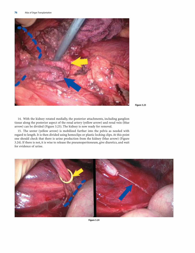
76 Atlas of Organ Transplantation
14. With the kidney rotated medially, the posterior attachments, including gangliontissue along the posterior aspect of the renal artery (yellow arrow) and renal vein (bluearrow) can be divided (Figure 3.23). The kidney is now ready for removal.
15. The ureter (yellow arrow) is mobilized further into the pelvis as needed withregard to length. It is then divided using hemoclips or plastic locking clips. At this pointone should check that there is urine production from the kidney (blue arrow) (Figure3.24). If there is not, it is wise to release the pneumoperitoneum, give diuretics, and waitfor evidence of urine.
Figure 3.23
Figure 3.24

Nephrectomy from a Living Donor 77
16. The donor is given heparin in preparation for removal of the kidney. The kidneyis grasped with one hand and retracted out laterally to stretch out the renal artery andvein. Retracting the vein cephalad gives excellent exposure at this point to the renalartery at the point of its junction with the aorta (Figure 3.25).
17. The artery can now be divided using either locking clips or a vascular stapler(Figure 3.26). It is useful at this point to insert the stapler through the camera port andplace the camera in the dissection port – this gives the straightest approach for thestapler to reach the artery. Our preference is to use a noncutting stapler to get maximallength on the artery. The staple line can be reinforced with a hemoclip, but we do notuse hemoclips in isolation for fear of dislodgment.
Figure 3.25
Figure 3.26

78 Atlas of Organ Transplantation
18. The vein is divided with a stapler also, and the kidney removed through the hard-port site (Figure 3.27). The kidney is then flushed.
19. A careful check is then made for hemostasis. Areas to inspect specifically includethe vascular stumps (blue arrow), the adrenal bed (yellow arrow), and the distal stumpof the divided ureter (Figure 3.28).
Figure 3.27
Figure 3.28

Nephrectomy from a Living Donor 79
Figure 3.29
Figure 3.30
20. All incisions, including the 10- to 12-mm port sites are closed. The appearance ofthe incisions at 2 weeks postdonation is shown (Figure 3.29).
d) Pure Laparoscopic Technique
The pure laparoscopic technique varies in port placements as shown. Ports 1 or 2 can act as a camera port while the other two are dissecting ports (Figure 3.30). The Pfannenstiel incision is low and small. Details of the dissection are discussed in the following section.
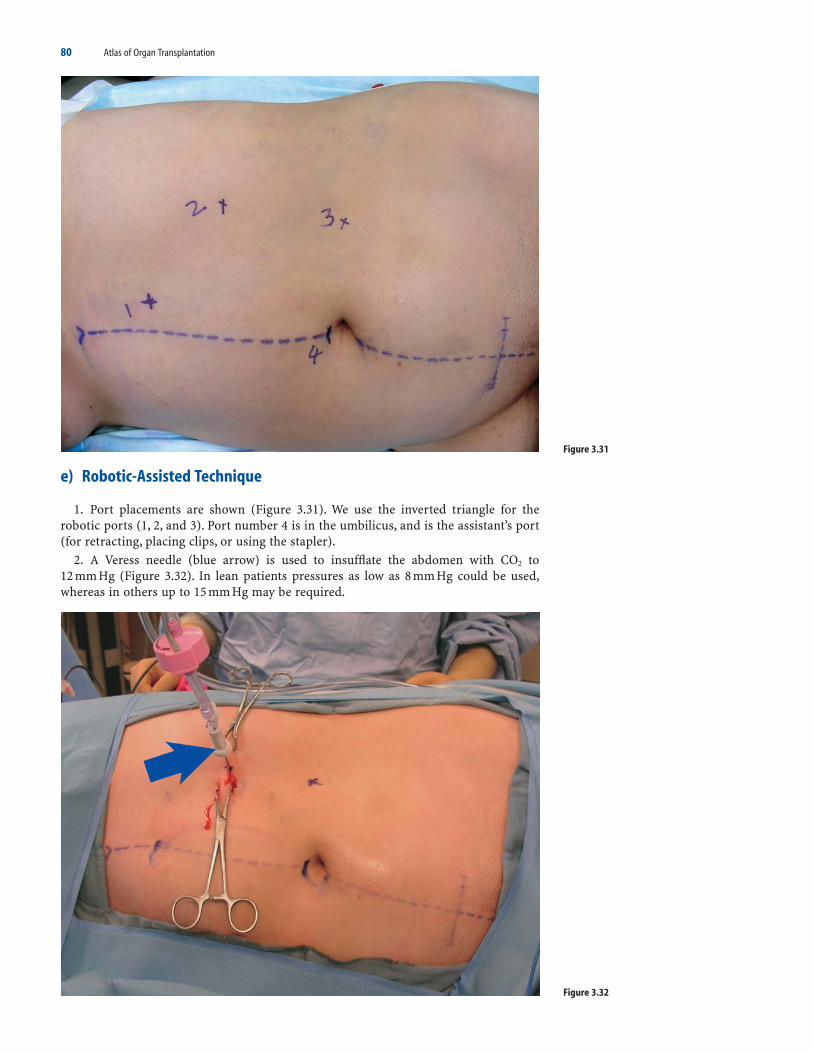
80 Atlas of Organ Transplantation
Figure 3.31
Figure 3.32
e) Robotic-Assisted Technique
1. Port placements are shown (Figure 3.31). We use the inverted triangle for therobotic ports (1, 2, and 3). Port number 4 is in the umbilicus, and is the assistant’s port(for retracting, placing clips, or using the stapler).
2. A Veress needle (blue arrow) is used to insufflate the abdomen with CO2 to 12 mm Hg (Figure 3.32). In lean patients pressures as low as 8 mm Hg could be used,whereas in others up to 15 mm Hg may be required.

Figure 3.33
Figure 3.34
Nephrectomy from a Living Donor 81
3. A 10- to 12-mm nonbladed trocar is introduced at site 2. The 0-degree scope isinserted through the trocar to provide visual assistance as the fascia and peritoneum arepenetrated. This helps minimize the risk of injury to the bowel. Subsequently two 7-mmports (DaVinci ports) are places at sites 1 and 3. These are inserted under visual guid-ance from the inside. Finally, a 10- to 12-mm bladed trocar is inserted through theumbilicus (Figure 3.33)
4. The robotic system is docked in position and the 0-degree camera is inserted usingvisual guidance. The forceps in the left port and the hook in the right port are insertedto position of function (Figure 3.34). The assistant sits next to the patient with easy accessto port 4. In robotic hand–assisted cases the right hand of the assistant is insertedthrough a midline handport. In pure laparoscopic robotic cases the assistant helps usingthe umbilical port (yellow arrow). The surgeon is ready to sit at the console and begin.The surgeon should have undergone the standard training offered by the manufacturer

82 Atlas of Organ Transplantation
and should be certified to operate the system. The surgeon should sit at a comfortableheight and operate with user-friendly ergonomics (Figure 3.35).
5. The first step in dissection of the left side is mobilization of the left colon by takingdown the line of Toldt from the splenic flexure to the sigmoid colon. This is done usinga Harmonic scalpel or electrocautery. Once the colon is reflected medially, Gerota’s fasciais identified and traced medially to the renal vein (Figure 3.36).
Figure 3.35
Figure 3.36

Nephrectomy from a Living Donor 83
Figure 3.37
6. The renal vein is dissected well away from the hilum. This will expose the adrenaland the gonadal veins (Figure 3.37). Fine dissection around the vein is done using hookelectrocautery taking care to avoid heat injury to the vasculature.
7. The venous branches of the renal vein including gonadal (yellow arrow, Figure3.38), adrenal, and one or more lumbar veins can be divided between hemoclips,or, if less than 7 mm in diameter, can be divided with an electrocautery device.
Figure 3.38

Figure 3.39
84 Atlas of Organ Transplantation
8. Cephalad displacement of the renal vein usually exposes the renal artery. Thickganglionic tissue anterior to the artery will have to be divided to get a good view of it.The artery is dissected circumferentially to its junction with the aorta (yellow arrow,Figure 3.39).
9. Once the branches of the renal vein have been divided, circumferential dissectionproximally to get adequate length is carried out.
10. Having completed the vascular dissection from the anterior side, the kidneyshould now be released from its superior and medial attachments. The adrenal gland isseparated from the kidney. This step could be complicated by small bleeding due tovenous hypertension of the adrenal gland. The Harmonic scalpel may help minimizeblood loss.

Nephrectomy from a Living Donor 85
11. Dissection is then carried out around the upper pole (blue arrow). It is preferable,whenever possible, to free the upper pole first before the lower pole is dissected (Figure3.40). This prevents the kidney from retracting upward and making the upper pole dis-section more difficult.
12. Next, the inferior medial aspect of the kidney is dissected just lateral to thegonadal vein. The ureter (blue arrow) is usually identified in this position. It is then dis-sected down to its crossing of the iliac artery, taking care not to damage the periureteralvessels (Figure 3.41).
Figure 3.40
Figure 3.41

86 Atlas of Organ Transplantation
Figure 3.42
13. The inferior pole is then freed lateral to the ureter, and dissection is carried alongto the lateral and posterior attachments of kidney to facilitate a posterior view. Once the posterior attachments are released, the kidney can be slipped medially to expose thevessels from the posterior side. This ensures complete release of all attachments of thekidney except the vessels (blue arrow) and the ureter (Figure 3.42). The kidney is nowready for removal.

Nephrectomy from a Living Donor 87
Figure 3.43
Figure 3.44
14. Next, the ureter is divided after clipping distally. A locking clip can be used forthe purpose (Figure 3.43). The cut end of the ureter should be inspected for urine pro-duction. If urine output is absent or low, then letting down the pneumoperitoneum andwaiting a few minutes may help. Forced diuresis is facilitated throughout the case usingmannitol and furosemide.
15. At this point the pneumoperitoneum is released and a 6-cm Pfannenstiel incisionis made.The fascial incision is midline.An endoscopic bag is inserted through a peritonealpurse string. The bag is deployed and the kidney and ureter placed in it (Figure 3.44).

Figure 3.45
88 Atlas of Organ Transplantation
16. The only structures outside the bag are the renal artery and vein. After adminis-tration of 70 U/kg heparin, the renal artery is double clipped with locking clips anddivided distally (Figure 3.45).
17. Then the vein is divided using a 35-mm articulating endovascular linear cuttingstapler. Care should be taken to avoid catching any unwanted structures in the stapler(e.g., superior mesenteric artery). At the same time the stapler should be pushed farenough proximally to get maximal length on the vein. Alternatively, a noncutting staplercould be used to get slightly more length. This is our routine on the right side. Prota-mine is given to reverse the heparin once the vessels are ligated.
18. The kidney is now completely enclosed by turning the mouth of the bag upward.The mouth of the bag is then closed and it is withdrawn through the incision after thepneumoperitoneum is released and the peritoneotomy extended manually.
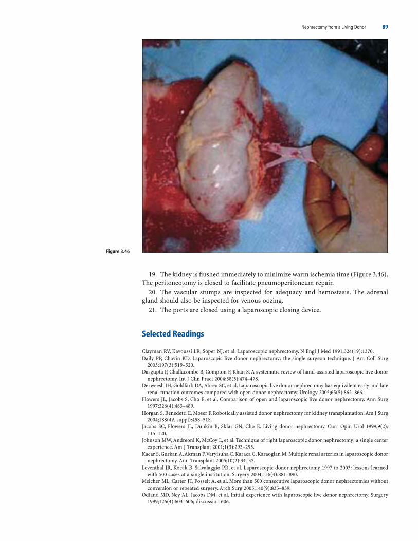
Nephrectomy from a Living Donor 89
19. The kidney is flushed immediately to minimize warm ischemia time (Figure 3.46).The peritoneotomy is closed to facilitate pneumoperitoneum repair.
20. The vascular stumps are inspected for adequacy and hemostasis. The adrenalgland should also be inspected for venous oozing.
21. The ports are closed using a laparoscopic closing device.
Selected Readings
Clayman RV, Kavoussi LR, Soper NJ, et al. Laparoscopic nephrectomy. N Engl J Med 1991;324(19):1370.Daily PP, Chavin KD. Laparoscopic live donor nephrectomy: the single surgeon technique. J Am Coll Surg
2003;197(3):519–520.Dasgupta P, Challacombe B, Compton F, Khan S. A systematic review of hand-assisted laparoscopic live donor
nephrectomy. Int J Clin Pract 2004;58(5):474–478.Derweesh IH, Goldfarb DA, Abreu SC, et al. Laparoscopic live donor nephrectomy has equivalent early and late
renal function outcomes compared with open donor nephrectomy. Urology 2005;65(5):862–866.Flowers JL, Jacobs S, Cho E, et al. Comparison of open and laparoscopic live donor nephrectomy. Ann Surg
1997;226(4):483–489.Horgan S, Benedetti E, Moser F. Robotically assisted donor nephrectomy for kidney transplantation. Am J Surg
2004;188(4A suppl):45S–51S.Jacobs SC, Flowers JL, Dunkin B, Sklar GN, Cho E. Living donor nephrectomy. Curr Opin Urol 1999;9(2):
115–120.Johnson MW, Andreoni K, McCoy L, et al. Technique of right laparoscopic donor nephrectomy: a single center
experience. Am J Transplant 2001;1(3):293–295.Kacar S, Gurkan A,Akman F,Varylsuha C, Karaca C, Karaoglan M. Multiple renal arteries in laparoscopic donor
nephrectomy. Ann Transplant 2005;10(2):34–37.Leventhal JR, Kocak B, Salvalaggio PR, et al. Laparoscopic donor nephrectomy 1997 to 2003: lessons learned
with 500 cases at a single institution. Surgery 2004;136(4):881–890.Melcher ML, Carter JT, Posselt A, et al. More than 500 consecutive laparoscopic donor nephrectomies without
conversion or repeated surgery. Arch Surg 2005;140(9):835–839.Odland MD, Ney AL, Jacobs DM, et al. Initial experience with laparoscopic live donor nephrectomy. Surgery
1999;126(4):603–606; discussion 606.
Figure 3.46

90 Atlas of Organ Transplantation
Oyen O, Andersen M, Mathisen L, et al. Laparoscopic versus open living-donor nephrectomy: experiences from a prospective, randomized, single-center study focusing on donor safety. Transplantation 2005;79(9):1236–1240.
Pietrabissa A, Boggi U, Moretto C, Ghilli M, Mosca F. Laparoscopic and hand-assisted laparoscopic live donornephrectomy. Semin Laparosc Surg 2001;8(2):161–167.
Rajab A, Mahoney JE, Henry ML, et al. Hand-assisted laparoscopic versus open nephrectomies in living donors.Can J Surg 2005;48(2):123–130.
Ruiz-Deya G, Cheng S, Palmer E, Thomas R, Slakey D. Open donor, laparoscopic donor and hand assistedlaparoscopic donor nephrectomy: a comparison of outcomes. J Urol 2001;166(4):1270–1273.
Schostak M, Wloch H, Muller M, et al. Living donor nephrectomy in an open technique; a long-term analysisof donor outcome. Transplant Proc 2003;35(6):2096–2098.
Terasaki PI, Cecka JM, Gjertson DW, Takemoto S. High survival rates of kidney transplants from spousal andliving unrelated donors. N Engl J Med 1995;333:333–336.
Tooher RL, Rao MM, Scott DF, et al. A systematic review of laparoscopic live-donor nephrectomy. Transplan-tation 2004;78(3):404–414.

4Kidney TransplantationAbhinav Humar and Arthur J. Matas
IntroductionBenching the Kidney from a Deceased Donora) ProcedureAdult Kidney Transplanta) Operative ProcedureSurgical Variationsa) Pediatric Recipient Kidney Transplantb) Pediatric Donor En-Bloc Kidneysc) Multiple Renal ArteriesSurgical Complications and Their Managementa) Hemorrhageb) Renal Artery Thrombosisc) Renal Artery Stenosisd) Recipient Arterial Complicationse) Renal Vein Thrombosisf) Venous Thromboembolismg) Aneurysms and Fistulash) Urologic Complicationsi) Lymphocelesj) Wound Complications
Introduction
In the last 35 years, few fields of medicine have undergone the rapid advances that havebeen seen with kidney transplantation. From the development of the surgical techniquesnecessary for transplantation at the beginning of the century, to the dawn of moderntransplantation with the introduction of immunosuppressants in the late 1950s, and toits current status as the treatment of choice for end-stage renal disease (ESRD), renaltransplantation has enjoyed remarkable progress. The surgical techniques for organtransplantation, including methods of vascular anastomosis, were developed in animalmodels by Carrel and Guthrie in the early 1900s. The first clinical deceased renal trans-plant was performed in 1933 by the Ukrainian surgeon Voronoy, with unsuccessfulresults secondary to the immunologic barrier. In the 1950s these obstacles were cir-cumvented by performing the procedure between identical twins. The era of modernrenal transplantation began with the introduction of the immunosuppressive agent azathioprine, and renal transplantation was established as a viable option for the treat-ment of ESRD.
91

92 Atlas of Organ Transplantation
For the majority of individuals with ESRD, transplantation results in superior survival,improved quality of life,and lower costs as compared with chronic dialysis.There are veryfew absolute contraindications and so most patients with ESRD should be considered aspotential candidates. The surgery and general anesthesia, however, impose a significantcardiovascular stress. The subsequent lifelong chemical immunosuppression is also asso-ciated with considerable morbidity. Therefore, evaluation of a potential recipient mustfocus on identifying risk factors that could be minimized or may even contraindicate atransplant.
The preoperative evaluation can be divided into four phases: medical, surgical,immunologic, and psychosocial. The medical evaluation begins with a complete historyand physical examination. Mortality after transplantation is just as likely to be due tounderlying cardiovascular disease as to infectious and neoplastic complications ofimmunosuppression. Any history of congestive heart failure, angina, myocardial infarc-tion, or stroke should be elicited. Patients with symptoms suggestive of cardiovasculardisease or significant risk factors (e.g., diabetes, age over 50, previous cardiac events)should undergo further cardiac evaluation. Any problems identified should be treatedappropriately (medically or surgically). Patients with suspected cerebrovascular diseaseshould undergo evaluation with carotid duplex Doppler studies.
Untreated malignancy and active infection are absolute contraindications to trans-plantation because of the requisite lifelong immunosuppression. Following curativetreatment of malignancy, an interval of 2 to 5 years is recommended prior to trans-plantation. This recommendation is influenced by the type of malignancy, with longerobservation periods for neoplasms such as melanoma or breast cancer and shorterperiods for carcinoma in situ or low-grade malignancies such as basal cell carcinoma ofthe skin. Chronic infections such as osteomyelitis or endocarditis must be fully treated.Other areas of the medical evaluation should concentrate on gastrointestinal problemssuch as peptic ulcer disease, symptomatic cholelithiasis, and hepatitis.
The surgical evaluation should concentrate on identifying vascular or urologic abnor-malities that may affect transplantation. Evidence of vascular disease that is revealed bythe history (e.g., claudication or rest pain) or the physical examination (e.g., diminishedor absent pulse, bruit) should be evaluated further by Doppler studies or angiography.Severe aortoiliac disease may make transplantation technically impossible; one option inthese patients is a revascularization procedure such as an aorto-bifemoral graft prior tothe transplant. Areas of significant stenosis proximal to the planned site of implantationmay need preoperative balloon angioplasty or stenting. Urologic evaluation should ruleout chronic infection in the native kidney,which may require nephrectomy pretransplant.Other indications for nephrectomy include very large polycystic kidneys, significantreflux,and uncontrollable renal vascular hypertension.Children especially require a com-plete genitourinary tract examination to evaluate reflux and bladder outlet obstruction.
An assessment of the patient’s immunologic status involves determining blood type, tissue type [human leukocyte antigens (HLAs) A, B, DR], and the presence of anycytotoxic antibodies against HLAs (because of prior transplants, blood transfusions, orpregnancies).
A psychosocial evaluation is necessary to ensure that patients understand the natureof the transplant procedure, with its attendant risk. They must be capable of followingthe medical regimen after the transplant. Patients who have not been compliant withtheir medical regimen in the past must demonstrate a willingness and capability to doso, before they undergo the transplant.
Living donors are preferred over deceased donors. Recipients of living-donor organsenjoy improved long-term success, avoid a prolonged wait, and are able to plan thetiming of their transplant in advance. Moreover, they have a significantly decreased inci-dence of delayed graft function. All of these advantages contribute to a lower incidenceof early acute rejection and to improved graft and patient survival rates. While there issignificant benefit for the recipient, there is no physical benefit for the living donor, onlythe potential for harm. Therefore, it is paramount that the risks of donation be accept-ably low, and that the donor is fully aware of the potential risks and has freely giveninformed consent.

Kidney Transplantation 93
The surgical technique for renal transplantation has changed very little from the orig-inal pelvic operation described in 1951. The most common approach today is the stan-dard pelvic operation, with retroperitoneal placement of the kidney, allowing easy accessfor percutaneous renal biopsy. Usually, the right iliac fossa is chosen because of the moresuperficial location of the iliac vein on this side. The procedure involves two vascularanastomoses (renal artery and vein) and the ureter to bladder anastomosis.
The initial postoperative care is not unlike that of other surgical patients. Fluid andelectrolyte status, vital signs, central venous pressure, and urine output are carefullymonitored. Special issues include immunosuppression and monitoring for transplant-related surgical and medical complications unique to these patients. Potential surgicalcomplications specific to the kidney transplant include renal artery or vein thrombosis,ureteral leak, and ureteral stricture.
Outcomes after kidney transplantation have steadily improved over the past threedecades, thanks to improvement in maintenance immunosuppression, antirejectiontherapy, organ retrieval techniques, perioperative care, and treatment of posttransplantinfectious complications. Most centers now report patient survival rates exceeding 95%during the first posttransplant year for all recipients. Transplants from living donors havea clear advantage over those from deceased donors; reported 5-year patient survival ratesafter living and deceased donor transplants are approximately 90% and 80%, respec-tively. Compared with dialysis, the survival advantage after a transplant is probablygreatest for diabetics.
Benching the Kidney from a Deceased Donor
Kidneys from a deceased donor are generally procured with a significant amount of thesurrounding tissue including the surrounding perineal fat, the adrenal gland, and por-tions of the aorta and inferior vena cava (IVC). The two kidneys may also be procured enbloc. This wide dissection minimizes the risk of damage to the renal hilum at the time ofprocurement. Therefore, the first step of the deceased-donor kidney transplant procedureis to adequately prepare the graft to render it suitable for transplant. The basic stepsinvolved are inspection of the organ, dissection of the artery and vein, and removal of thesurrounding perirenal fat.
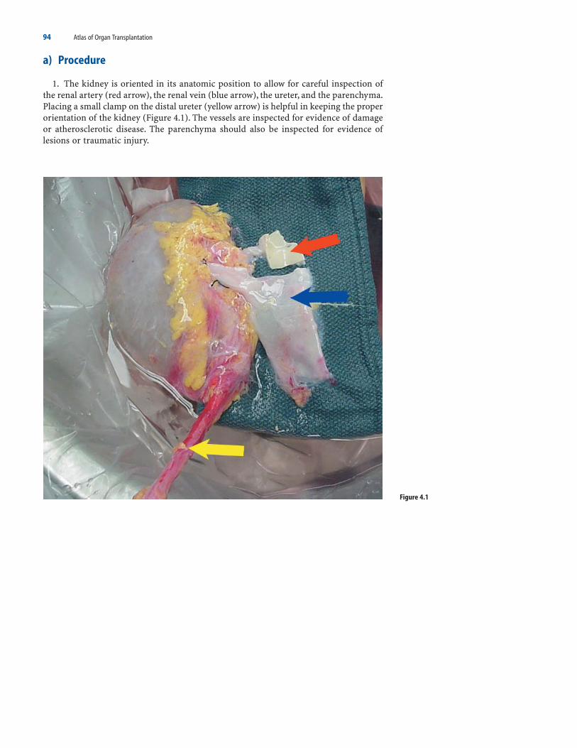
94 Atlas of Organ Transplantation
a) Procedure
1. The kidney is oriented in its anatomic position to allow for careful inspection ofthe renal artery (red arrow), the renal vein (blue arrow), the ureter, and the parenchyma.Placing a small clamp on the distal ureter (yellow arrow) is helpful in keeping the properorientation of the kidney (Figure 4.1). The vessels are inspected for evidence of damageor atherosclerotic disease. The parenchyma should also be inspected for evidence oflesions or traumatic injury.
Figure 4.1

Figure 4.2
Kidney Transplantation 95
2. The renal artery is dissected free from the surrounding hilar tissue and an appro-priate sized aortic patch is fashioned (Figure 4.2).

96 Atlas of Organ Transplantation
3. The renal vein is then similarly dissected for an adequate length. Branches includ-ing the adrenal, gonadal, and lumbar all need to be divided and ligated. These branchesare usually present on the left renal vein, but the right renal vein usually has no branches.For a left kidney, there is usually adequate length so that the inferior vena cava does notneed to be included (Figure 4.3).
Figure 4.3

Kidney Transplantation 97
4. With a right kidney, the right renal vein may sometimes be short. If so, it can belengthened by using a segment of the IVC, transecting it transversely superior and infe-rior to the renal vein, and then closing the edges of the transected IVC using a finerunning vascular suture (Figures 4.4 and 4.5).
Figure 4.4
Figure 4.5

Figure 4.6
98 Atlas of Organ Transplantation
5. The surrounding perineal fat is then removed from the kidney, taking care not toinjure the capsule. This is started from the medial aspect of the upper pole, where the adrenal gland is identified and removed. The dissection continues along the upperpole and then the lateral aspect of the kidney (gray arrows). Care should be taken notto carry the dissection too extensively in the lower pole, as this may devascularize theureter. The fat (black arrow) in the triangle formed by the lower pole, the hilar vessels,and the ureter (Figure 4.6) should not be separated from the kidney, to preserve bloodsupply to the ureter.
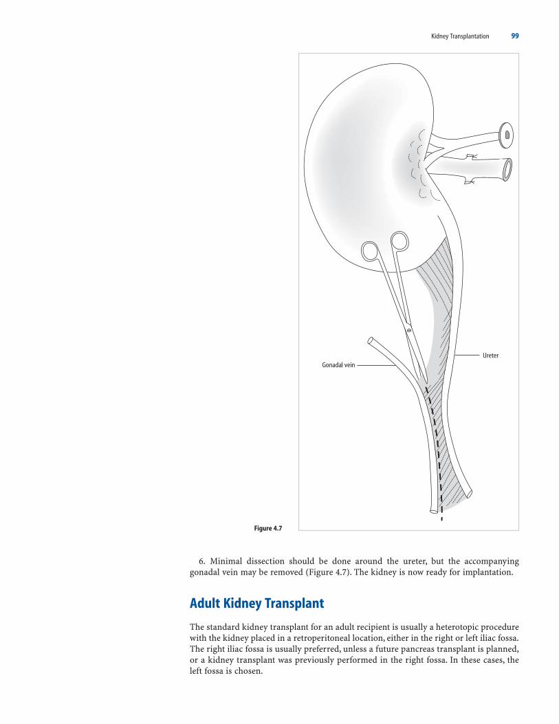
Kidney Transplantation 99
6. Minimal dissection should be done around the ureter, but the accompanyinggonadal vein may be removed (Figure 4.7). The kidney is now ready for implantation.
Adult Kidney Transplant
The standard kidney transplant for an adult recipient is usually a heterotopic procedurewith the kidney placed in a retroperitoneal location, either in the right or left iliac fossa.The right iliac fossa is usually preferred, unless a future pancreas transplant is planned,or a kidney transplant was previously performed in the right fossa. In these cases, theleft fossa is chosen.
Figure 4.7

100 Atlas of Organ Transplantation
In certain situations, an intraperitoneal placement of the kidney may be the betteroption. If the procedure is performed with a simultaneous pancreas transplant, then thekidney is best placed intraperitoneally, on the opposite side of the pancreas. In very smallpediatric patients (usually <20 kg), an intraperitoneal position usually allows for morespace and anastomosis to larger vessels for inflow and outflow. Lastly, in the recipientwith multiple previous transplants, or with other previous procedures on both the rightand left iliac vessels, an intraperitoneal location allows one to stay away from the site ofprevious surgery, and use the distal aorta and IVC for graft implantation if necessary.
Regardless of the location of the kidney, for adult recipients the vascular anastomosesare generally performed to the iliac vessels. For arterial inflow, the common iliac artery,internal iliac artery,or external iliac artery may be used.Generally,the external iliac arteryis preferred, but if it is diseased or small in caliber, then there should be no hesitation inusing the common iliac artery. Ultimately, the best option is to place the kidney in thefossa prior to selecting a site for the anastomosis, and choose the site where the renalartery will lie most naturally without significant tension, redundancy, and angulation.
The iliac vein is used for venous outflow – again most commonly the external iliac vein.However, the optimal site should be chosen based on the position of the kidney. Thebranches of the iliac vein are more variable than the artery. There may be one dominantinternal iliac vein or several smaller ones. If the graft renal vein is short, or the recipientiliac vein deep, then the branches of the iliac vein may be divided. This allows the vein tobe brought up into a more superficial position, making the anastomoses simpler.
Urinary continuity is restored by connecting the transplant ureter to the bladder. Thismay be done with or without a stent. Several different techniques have been describedfor performing this anastomosis. The key goals are to ensure that the ureter is not undertension or too redundant, and that it is well vascularized and perfused at its most distalposition where the anastomosis will be performed. It is advisable to create some formof tunnel for the distal aspect of the transplant ureter to diminish urinary reflux.
a) Operative Procedure
1. With the patient in the supine position, a “hockey-stick” – shaped incision is madein the lower quadrant (purple line) (Figure 4.8). The incision starts in the midline, oneor two finger widths above the pubic bone (solid black line). The incision extends later-ally, gently curving upward until the lateral edge of the rectus muscle is reached (brokenred line). The incision is then extended superiorly along the lateral edge of the rectus.
Figure 4.8

Kidney Transplantation 101
2. The incision is deepened to the fascia. Starting medially, and going laterally, theanterior sheet of the rectus muscle is incised (Figure 4.9). At the lateral edge of the rectusmuscle, the fascial incision is carried superiorly just lateral to the edge of the rectusmuscle (broken line) (Figure 4.10). This is where the muscular layers of the lateralabdominal wall start to form the anterior and posterior sheets of the rectus muscle. Rel-atively little actual muscle needs to be divided with this approach. Medially it is usuallynot necessary to divide the rectus muscle, but mobilizing it down to its attachment tothe pubic bone helps in later exposure of the bladder.
Figure 4.9
Figure 4.10

102 Atlas of Organ Transplantation
3. The inferior epigastric vessels are encountered in the middle to lower portion ofthe incision once the fascia is incised (broken blue line) (Figure 4.11). These vessels aredivided between ligatures.
4. As the incision is deepened, the next structure to be encountered is the round lig-ament in females and the spermatic cord in males (broken yellow lines) (Figure 4.12).The round ligament can be divided, but the spermatic cord should be preserved and avessel loop passed around it.
Figure 4.11
Figure 4.12

Kidney Transplantation 103
5. The peritoneum is then gently reflected medially toward the midline using onehand to retract the peritoneum (broken blue line) and the cautery to divide loose con-nections. This allows visualization of the retroperitoneal space, with the underlyingpsoas muscle (broken yellow line) (Figure 4.13).
Figure 4.13

Figure 4.14
104 Atlas of Organ Transplantation
6. A self-retaining retractor is inserted at this point. An outline of the iliac vessels(black arrow) can be seen at the base of the wound with the genitofemoral nerve (yellowarrow) laterally and the native ureter (blue arrow) medially (Figure 4.14). Where theureter enters the pelvis is a good landmark to locate the bifurcation of the common iliacartery into its internal and external branches. Care should be taken to ensure that theblades of the retractor do not impinge and occlude the iliac vessels proximally.

Kidney Transplantation 105
7. The external iliac artery is dissected free and mobilized for an adequate length toallow for subsequent clamp placement and anastomosis. Lymphatics (yellow arrow)overlying the artery should be carefully ligated to diminish the risk of lymphocele formation (Figure 4.15).
8. The external iliac vein (blue arrow), usually lying just medial to the artery (yellowarrow), is subsequently isolated. If the iliac vein is deep, or the transplant renal vein short,the internal iliac venous branches can be divided to place the vein in a more superficial
Figure 4.15

106 Atlas of Organ Transplantation
Figure 4.16
Figure 4.17
position. It is usually a good idea once the vascular dissection is finished to place thekidney in the iliac fossa to determine the best location to perform the venous and arte-rial anastomosis. These locations can be marked with a marking pen (broken purpleline) (Figure 4.16).
9. Usually the venous anastomosis is performed first. Clamps are placed proxi-mally and distally on the vein and an appropriate-sized venectomy is made. There areseveral ways to perform the actual anastomosis. One technique is to place four sutures(Figure 4.17): one at the upper end of the anastomosis, one at the lower end (solid lines),and one each on the two sides (broken lines).
10. The four sutures are tied and the superior stitch is sewn circumferentially aroundthe anastomosis, with the primary surgeon suturing his/her side and the assistant the

Kidney Transplantation 107
Figure 4.18
Figure 4.19
other side (Figure 4.18). In this manner the renal vein is anastomosed to the iliac veinin an end-to-side fashion (blue arrow) (Figure 4.19).

Figure 4.20
108 Atlas of Organ Transplantation
11. The arterial anastomosis can be performed in a similar fashion after clamping the artery and making an appropriate sized arteriotomy. If there is no aortic cuff presenton the artery (as with a living donor transplant), then a small circular arteriotomy ismade on the iliac vessel (yellow arrow) (Figure 4.20). A vascular punch device, as shown

Figure 4.21
Kidney Transplantation 109
(Figure 4.21), may be useful for this step. The clamps are removed after this anastomo-sis, and the kidney allowed to reperfuse.

110 Atlas of Organ Transplantation
Figure 4.22
12. If there is a patch present around the renal artery, then a straight arteriotomy canbe made on the recipient vessel, and the patch sewn to the arterial wall (Figure 4.22).
13. There are several methods to perform the ureter to bladder anastomosis includ-ing the Ledbetter-Pollitano, Litch, and anterior one-stitch techniques. The latter two tech-niques are subsequently described. The distal ureter is adequately prepared by trimmingto an appropriate length, ligating the accompanying ureteral vessels (black arrows), andspatulating the distal end (Figure 4.23).
Figure 4.23

Kidney Transplantation 111
Figure 4.24
14. The retractors are repositioned to expose the bladder (Figure 4.24). It is helpfulto fill the bladder and clamp the urinary catheter at this point. An incision is made

112 Atlas of Organ Transplantation
through the detrusor muscle, exposing the underlying mucosa, but not going through it(black arrow) (Figure 4.25).
Figure 4.25

Kidney Transplantation 113
15. The detrusor muscle is then gently separated from the underlying mucosa (Figure 4.26). This can be done bluntly with gauze (blue arrow). This step is useful for creation of the subsequent tunnel, which will cover the distal part of the ureter.
16. With the Litch technique, an incision is made in the bladder mucosa and the distalspatulated end of the ureter is anastomosed for the bladder mucosa using a fine runningabsorbable suture (Figure 4.27). Triangulating the anastomosis is a useful technique here.One suture is placed in the heel, and one at each of the two distal corners. The heel stitchis then tied and run toward the tops.
Figure 4.26
Figure 4.27

114 Atlas of Organ Transplantation
17. With an anterior one-stitch technique a double-ended suture is placed in the spatulated end of the ureter with the two needles on the “inside” aspect of the ureter(Figure 4.28a). These two sutures are then brought through a small opening made in thebladder mucosa with the needles exiting the bladder further distally (Figure 4.28b).These sutures are then used to pull the ureter into the bladder. The ureter is then secured to the anterior wall of the bladder by tying the anterior double-ended stitch(Figure 4.28c).
Figure 4.28

Figure 4.30
Kidney Transplantation 115
18. With either technique the detrusor muscle is then closed over the distal 5 to 6 cmof transplant ureter (broken blue line) to prevent reflux (Figures 4.29 and 4.30).
Figure 4.29

116 Atlas of Organ Transplantation
19. The kidney is then placed in a position such that there is no angulation or twisting of the renal artery (yellow arrow) or the renal vein (blue arrow) (Figure 4.31).The incision is then closed by approximating the fascia using a running or interruptedsuture.
Surgical Variations
a) Pediatric Recipient Kidney Transplant
The surgical techniques for kidney transplantation in pediatric recipients, especially inthe older child, are not very different from the techniques described for adult recipients.
Figure 4.31

Kidney Transplantation 117
1. For recipients ≤20 kg, we prefer an intraperitoneal approach using a kidney froman adult-sized living donor. The kidney is placed on the right side, behind the right colon(Figure 4.32). Anastomosis is generally to the distal aorta and cava.
2. The procedure is started with a long midline incision to enter the peritoneal cavity.The right colon (blue arrow) is mobilized and reflected medially to expose the right
Figure 4.32

118 Atlas of Organ Transplantation
Figure 4.33
retroperitoneal area, including the psoas muscle (black arrow) and the distal aorta/proximal iliac vessels (yellow arrow) (Figure 4.33).
3. The infrarenal cava (blue arrow), infrarenal aorta (yellow arrow), and proximal iliacvessels are isolated and encircled (Figure 4.34).
Figure 4.34

Kidney Transplantation 119
Figure 4.35
4. The renal artery is anastomosed to the distal aorta and the renal vein is anasto-mosed to the distal cava (Figure 4.35), in a manner similar to that described for adultrecipients. The bladder anastomoses can also be performed as previously described foradults. The colon is then returned to its proper position, lying anterior to the trans-planted kidney. The incision is then closed.
b) Pediatric Donor En-Bloc Kidneys
Kidney from small pediatric donors (<15 kg) can be used in adult recipients. In this sit-uation, however, both kidneys should be transplanted en bloc into the one recipient.
1. The kidneys are prepared on the back table. The aorta and cava are left intact withthe two kidneys. The proximal ends of the cava and aorta are oversewn (Figure 4.36).The distal ends can then be used to perform the anastomoses to the recipient vessels.
Figure 4.36

Figure 4.37
120 Atlas of Organ Transplantation
2. The anastomoses are performed to the iliac vessels of the recipient. The two ureterscan be anastomosed separately into the recipient bladder (Figure 4.37).
c) Multiple Renal Arteries
Multiple renal arteries are present in 10% to 15% of cases. Many different options arepossible for reconstruction, depending on size of the vessels, location of the vessels,donor source (living vs. deceased), and quality of the recipient vessels.

Kidney Transplantation 121
1. Often the two arteries (yellow and blue arrow) are implanted separately, either tothe external or common iliac vessels (Figure 4.38) . . .
2. . . . or one to the external iliac and one to the internal iliac artery (Figure 4.39).
Figure 4.38
Figure 4.39

122 Atlas of Organ Transplantation
Surgical Complications and Their Management
Surgical complications remain an important potential cause of graft loss after kidneytransplants. They may also result in graft dysfunction that must be differentiated frommedical and immunologic causes of graft dysfunction. The initial presentation of surgi-cal problems may be very similar to nonsurgical problems such as rejection or drug tox-icity. Even if these complications do not affect graft function, they nonetheless mayaccount for significant morbidity in the recipient, or may even result in death. For all ofthese reasons, it is crucial that all persons involved in the postoperative care of kidneytransplant recipients be aware of the potential surgical complications that may occur,thus allowing for rapid diagnosis and initiation of treatment. Common surgical compli-cations include the following.
Figure 4.40
Figure 4.41
3. The two vessels can be reconstructed on the back table so that only one anasto-mosis is required in the recipient. If an aortic patch is present on the two vessels, the twopatches can be sewn together (Figure 4.40).
4. Another option is to connect one vessel to the other in an end-to-side fashion. Asshown in this example with three renal arteries, artery 1 and 2 have been sewn togetherwith a common patch (broken blue line), while artery 3 has been anastomosed to artery1 in an end-to-side fashion (broken yellow line) (Figure 4.41).

Kidney Transplantation 123
a) Hemorrhage
Bleeding is uncommon after kidney transplants; it usually occurs from unligated vesselsin the graft hilum or from the retroperitoneum of the recipient. Risk factors includerecipient obesity, the presence of antiplatelet agents, and the need for anticoagulation. Afalling hematocrit level, hypotension or tachycardia, and significant flank pain should allraise concern regarding the possibility of bleeding. Surgical exploration is seldomrequired, because the bleeding often stops spontaneously. However, ongoing transfusionrequirements, hemodynamic instability, and compression of the kidney by hematomaare all indications for surgical reexploration.
b) Renal Artery Thrombosis
This complication usually occurs early posttransplant; it is an uncommon event, with anincidence of <1%. However, it is a devastating complication, usually resulting in graftloss. Typically, it occurs secondary to a technical problem, such as intimal dissection orkinking or torsion of the vessels. Presentation is with a sudden cessation of urine output.Diagnosis is easily made with color flow Doppler studies. Urgent thrombectomy is indi-cated, but the transplanted kidney has no collateral vessels, and its tolerance of warmischemia is very poor. Therefore, most of these grafts cannot be salvaged and requireremoval.
c) Renal Artery Stenosis
Stenosis of the renal artery, a late complication, is generally much more common thanrenal artery thrombosis. Renal artery stenosis has a reported incidence of 1% to 10%.Most cases are identified within the first few years posttransplant. Recipients may presentwith poorly controlled hypertension, allograft dysfunction, and peripheral edema; phys-ical examination may detect a bruit over the kidney. Doppler studies are a good screen-ing tool with high sensitivity and specificity. A magnetic resonance angiogram orcomputed tomography (CT) angiogram can be performed to confirm the diagnosis.
First-line treatment for symptomatic lesions uses interventional radiologic tech-niques, usually angioplasty with or without a stent. Initial success rates of about 80%have been reported with angioplasty, albeit with a fairly significant recurrence rate. Somelesions are less amenable to angioplasty. For example, anastomotic strictures and longstrictures are less likely to be treated successfully with angioplasty and have a higherrisk of angioplasty complications. Potential complications with any angioplasty proce-dure include rupture or thrombosis of the vessel, which can lead to graft loss. Surgery isreserved for stenoses that do not respond to radiologic techniques. Possible optionsinclude reimplantation of the vessel, patch angioplasty, and surgical bypass. While thesuccess rate with surgery is generally good, it may be technically very difficult becauseof the extensive fibrosis that often develops around the transplanted kidney. Graft lossat the time of attempted surgical correction is a potential risk.

Figure 4.42
124 Atlas of Organ Transplantation
1. If surgery is indicated, one useful technique is to use the internal iliac artery tobypass a proximal stenosis. The surgery should be approached from an intraperitonealroute. The transplant renal artery distal to the stenosis is isolated (broken blue line)(Figure 4.42). The internal iliac artery (broken yellow line) is isolated and mobilized deepinto the pelvis. Here it is divided (black arrow) (Figure 4.43).
Figure 4.43

Kidney Transplantation 125
2. The transplant renal artery is then divided and anastomosed to the divided inter-nal iliac artery (black arrow) (Figures 4.44 and 4.45).
d) Recipient Arterial Complications
Arterial complications that affect the recipient vessels (most commonly the iliac vessels)are much less common, but can be equally devastating. Early events, such as iliac arterythrombosis or intimal dissection, can be limb-threatening as well as potentially lead to
Figure 4.44
Figure 4.45

126 Atlas of Organ Transplantation
graft loss. Late complications, such as pseudoaneurysms or fistulas, can lead tosignificant hemorrhage. Predisposing risk factors include underlying peripheral vascu-lar disease, deep infections, and insulin-dependent diabetes. Iliac artery thrombosisresults in an ischemic extremity, which usually occurs very early postoperatively (attimes, even intraoperatively). If the problem is recognized intraoperatively, it should be immediately resolved (e.g. an intimal dissection is repaired by tacking the intima tothe arterial wall.) At the end of every transplant procedure, a quick vascular exam andinspection of the lower extremities should be performed. If an ischemic extremity isnoted, immediate surgical exploration with balloon thrombectomy is essential to salvagethe limb and prevent long-term sequelae.
Occlusive disease of the iliac artery proximal to the site of renal artery anastomosesmay clinically mimic renal artery stenosis. Occlusive lesions may develop as a result ofprogressive peripheral vascular disease, or they may represent an injury from the vas-cular clamp placed on the artery at the time of the transplant. Patients may present withdeteriorating renal function, hypertension, and buttock claudication. On physical exam-ination of such patients, diminished femoral pulse should make the clinician suspiciousof occlusive disease. This disease tends to be very amenable to treatment with radiologictechniques such as angioplasty and stenting.
e) Renal Vein Thrombosis
This complication, like its arterial counterpart, usually results in graft loss. Causesinclude angulation or kinking of the vein, compression by hematomas or lymphoceles,anastomotic stenosis, and extension of an underlying deep venous thrombosis. Mostcases occur early, usually within the first 10 days posttransplant. Patients may presentwith a tender swollen graft and/or hematuria. Doppler studies are again the best diag-nostic tool. Urgent thrombectomy is indicated, but for most patients, graft salvage is notpossible; graft nephrectomy is usually required. However, graft salvage is possible ifthrombectomy can be achieved soon after the event (usually within 1 hour). Even if graftsalvage is not possible, urgent reexploration should be performed, because such grafts can become very swollen and edematous, with a risk for rupture and subsequent hemorrhage.
f) Venous Thromboembolism
Venous thromboembolic complications that affect the recipient vessels (deep venousthrombosis and pulmonary embolism) are not uncommon. The incidence of deepvenous thrombosis is close to 5%; the incidence of pulmonary embolism is 1%. Usuallytwo peaks in incidence are reported – one early in the postoperative period (likely relatedto operative factors) and a second peak at around 4 weeks (perhaps related to a risinghematocrit level). Risk factors include older recipient age, diabetes, thrombophilic dis-orders, and a history of deep venous thrombosis. For recipients with such risk factors,prophylaxis with low-dose heparin is recommended.
g) Aneurysms and Fistulas
Other potential vascular problems include arteriovenous fistulas and aneurysms. Mostaneurysms that occur posttransplant are pseudoaneurysms, usually resulting frompartial disruption of the arterial anastomosis. Some aneurysms are associated with alocal infection, though this association is more common after pancreas transplants.Patients may be asymptomatic, with the abnormality being noted on a routine ultra-sound exam. However, hypotension and abdominal pain due to rupture of the aneurysmmay also occur. Sometimes, expansion of the aneurysm results in local pressure symp-toms before rupture. Ultrasound is a good screening test, but a further definitive tool,

Kidney Transplantation 127
such as angiography, is necessary – unless the patient presents with a rupture, in whichcase an immediate repair is indicated. The technique of repair depends on the presenceor absence of infection or if the patient presents with massive bleeding. If either infec-tion or massive bleeding is present, graft salvage is usually not possible; instead, graftnephrectomy and repair of the recipient vessels with autogenous vein usually representsthe best option. In a more elective setting without infection, pseudoaneurysm repair andgraft salvage may be possible.
Arteriovenous fistulas may occur in the kidney graft parenchyma after a biopsy. Theyare easily detected by Doppler studies. Asymptomatic fistulas can simply be observed,because most will resolve spontaneously. Fistulas that occur with significant hematuriacan be managed by selective arterial catheterization and embolization.
h) Urologic Complications
Urinary tract complications, manifesting as leakage or obstruction, generally occur in2% to 10% of kidney recipients. The underlying cause is often related to poor bloodsupply and to ischemia of the transplant ureter. Rarely life-threatening, urinary tractcomplications can result in significant morbidity for immunocompromised recipients orin significant, long-term graft damage.
Urine Leaks
Leakage most commonly occurs early. It is usually from the anastomotic site. Causesother than ischemia include undue tension created by a short ureter, and direct surgicalinjury to the ureter (usually at the time of procurement). Presentation is usually early(before the 5th posttransplant week); symptoms include fever, pain, swelling at the graftsite, increased creatinine level, decreased urine output, and cutaneous urinary drainage.Diagnosis can be confirmed with a hippurate renal scan. Early surgical exploration withureteral reimplantation is indicated for very early leaks, for large leaks, or for leaks thatdo not respond to conservative measures. Many leaks, however, may be managed by usingthe principles of drainage and stenting. Drain placement to evacuate a urinoma andurinary tract stenting (usually by percutaneous nephrostomy and stent placement) cansuccessfully manage many urine leaks posttransplant.
Obstruction
Obstruction may present early or late. Early obstruction may be due to edema, bloodclots, hematoma, and kinking. Late obstruction is generally due to scarring and fibrosisfrom chronic ischemia. Presentation is usually with an elevated serum creatinine level.An ultrasound exam, assessing for hydronephrosis, is a good initial tool. A furosemiderenogram is useful in less obvious cases of obstruction; a percutaneous nephrostogramis the most specific test. Initial treatment with percutaneous transluminal dilatation(PTD), followed by internal or external stent placement, has yielded good results. Ifsuch treatment is not successful, then surgical intervention is indicated. For very distalstrictures, the transplanted ureter may be reimplanted into the bladder. If the stricture

128 Atlas of Organ Transplantation
is more proximal, then the native ureter can be used to bypass the obstruction (Figure 4.46).
Hematuria
Another potential urinary complication is hematuria. Mild hematuria is not infrequent.It is usually observed in the first 12 to 24 hours posttransplant. In most patients, itresolves spontaneously. More extensive bleeding may result in retained blood clots and urinary tract obstruction, which is the most common cause of sudden cessation of urine output immediately posttransplant. Continuous bladder irrigation usuallyrestores diuresis, but if not, cystoscopy may be necessary to evacuate the clot and cau-terize the source of the bleeding.
Lymphoceles
The incidence of lymphoceles (fluid collections of lymph that generally result from cutlymphatic vessels in the recipient) is 1% to 15%. Careful ligation of all lymphatics seenat the time of iliac vessel dissection can help minimize the incidence of lymphoceles.Lymphoceles usually do not occur until at least 2 weeks posttransplant. Symptoms, ifpresent, are generally related to the mass effect and compression of nearby structures(e.g., ureter, iliac vein).An ultrasound exam confirms a fluid collection, but percutaneousaspiration may be necessary to rule out other complications such as urinoma, hematoma,or abscess.
Many lymphoceles are clinically asymptomatic; if so, they usually are less than 3 cm,resolve spontaneously over time, and do not require any therapeutic intervention.
Symptomatic lymphoceles require drainage, which can be achieved either by surgeryor by percutaneous radiologic methods. The standard surgical treatment is creation ofa peritoneal window to allow for drainage of the lymphatic fluid into the peritonealcavity, where it can be absorbed. Either a laparoscopic or an open approach can be used.Another option is percutaneous insertion of a drainage catheter, with or without scle-
Figure 4.46

Kidney Transplantation 129
rotherapy; however, this option is associated with some risk of recurrence or infection.A variant approach is to initially drain the lymphoceles percutaneously after insertinga drainage catheter; sclerotherapy is then attempted via this catheter. If the lymphoce-les continue to drain, if they recur, or if they were not amenable to percutaneous drainageinitially, then a laparoscopic or open peritoneal window should be created surgically.
1. Laparoscopically the lymphocele is drained from the inside of the peritoneal cavity(Figure 4.47).
2. The lymphocele is identified from the inside of the peritoneal cavity by its bulging(solid arrows) (Figure 4.48a). Intraoperative ultrasound may be useful to identify theexact location of the lymphocele. A circular rim of peritoneum is excised (Figure 4.48b)to allow for internal drainage of the lymphocele (Figure 4.48c).
Wound Complications
Wound complications are now probably the most common types of complication posttransplant. They usually do not result in graft loss or death, but can result in
Figure 4.47
Figure 4.48

130 Atlas of Organ Transplantation
significant morbidity with prolonged hospitalization or rehospitalization. In certain sit-uations, wound complications may also be associated with inferior graft survival rates.Wound complications can be broadly categorized into infectious and noninfectiouscomplications.
Wound Infections
Wound infections generally occur earlier after a surgical procedure as compared with noninfectious wound complications. The incidence of wound infections afterkidney transplants is about 5%, a figure consistent with that reported in the urologic literature for nontransplant procedures of similar magnitude. A kidney transplant is generally considered a clean-contaminated case: the bladder is opened, and some urineis usually spilled in the operative field. The added stress of immunosuppressive drugs,including prednisone, probably also has some impact on the risk for wound infectionsposttransplant.
Wound infections may be either superficial or deep (i.e., to the fascia). Deep infectionsare generally related to complications such as urinary leaks. Superficial infections, morecommon than deep infections, are related to contamination from skin organisms or fromurine (during the bladder anastomosis). Obesity is probably the biggest risk factor for a wound infection posttransplant. Other risk factors include a urine leak, the need for areoperation, and perhaps the use of the newer, more powerful immunosuppressive medications.
Treatment depends on whether the wound infection is superficial or deep. Deep infec-tions are treated with drainage – either by surgery or by percutaneous drainage – and,usually, antibiotics. Superficial infections are usually treated by opening the surgicalwound and allowing it to heal by secondary intention; antibiotics are usually not neces-sary, unless the recipient has significant cellulitis or systemic symptoms.
Noninfectious Wound Complications
These include early fascial dehiscence and late incisional hernias. Recipient obesity isthe most significant risk factor. These will generally require surgical repair.
Selected Readings
1. Adams J, Gudemann C, Tonshoff B, Mehls O, Wiesel M. Renal transplantation in small children – a com-parison between surgical procedures. Eur Urol 2001;40(5):552–556.
2. Ali-El-Dein B, Osman Y, Shokeir AA, Shehab El-Dein AB, Sheashaa H, Ghoneim MA. Multiple arteries inlive donor renal transplantation: surgical aspects and outcomes. J Urol 2003;169(6):2013–2017.
3. Bay WH, Hebert LA. The living donor in kidney transplantation. Ann Intern Med 1987;106:719–727.4. Bitker MO, Benoit G. Surgical aspects of kidney transplantation in France in 1997. Eur Urol 1998;34(1):
1–5.5. Breza J, Navratil P. Renal transplantation in adults. BJU Int 1999;84(2):216–223.6. Calne RY, Alexondre GP, Murray JE. A study of the effects of drugs in prolonged survival of homologous
renal transplants in dogs. Ann NY Acad Sci 1962;99:743–749.7. Carrel A. The transplantation of organs. NY Med J 1914;99:839–851.8. Cecka JM, Terasaki PI. The UNOS scientific renal transplant registry. Clin Transplant 1992;6:1–16.9. Doehn C, Fornara P, Fricke L, Jocham D. Laparoscopic fenestration of posttransplant lymphoceles. Surg
Endosc 2002;16(4):690–695.10. Greneir N, Douws C, Morel D, et al. Detection of vascular complications in renal allografts with color
Doppler flow imaging. Radiology 1991;178:217–223.11. Guthrie CC. Blood vessel surgery and its applications. New York: Longmans Green, 1912.12. Hakim NS, Benedetti E, Pirenne J, et al. Complications of ureterovesical anastomosis in kidney transplant
patients: the Minnesota experience. Clin Transplant 1994;8:504–507.13. Hamilton DNH, Reid WA. Yu Yu Voronoy and the first human kidney allograft. Surg Gynecol Obstet
1984;159:289–294 .14. Johnson EM, Remucal M, Pandian K, et al. Complications and risks of renal donation. J Am Soc Nephrol
1996;7(9):1934.

Kidney Transplantation 131
15. Jones JW, Hunter DR, Matas AJ. Successful percutaneous treatment of ureteral stenosis after renal trans-plantation. Transplant Proc 1993;25:1038.
16. Kinnaert P, Hall M, Janssen F, Vereerstraeten P, Toussaint C, Van Geertruyden J. Ureteral stenosis after kidney transplantation: true incidence and long-term follow-up after surgical correction. J Urol1985;133:17–20.
17. Makowka L, Sher L. Handbook of Organ Transplantation. Georgetown, TX: R.G. Landes, 1995.18. Matas AJ, Tellis VA, Karwa GL. Comparison of post-transplant urologic complications following
extravesical ureteroneocystostomy by a “single-stitch” or “mucosal” anastomosis. Clin Transplant1987;1:159–163.
19. Merrill JP, Murray JE, Harrison JH. Successful homotransplantation of the human kidney between iden-tical twins. JAMA 1956;160:277–282.
20. Murray JE, Merrill JP, Harrison JH, et al. Prolonged survival of human-kidney homografts by immuno-suppressive drug therapy. N Engl J Med 1963;268:1315–1323.
21. Osman Y, Shokeir A, Ali-el-Dein B, et al. Vascular complications after liver donor renal transplantation:study of risk factors and effects on graft and patient survival. J Urol 2003;169(3):859–862.
22. Plainfosse MC, Menoyo Calonge V, Beylouve-Mainard C, Glotz D, Duboust. Vascular complications in theadult kidney transplant recipient. J Clin Ultrasound 1992;20:517–527.
23. Shoskes DA, Hanbury D, Cranston D, Morris PJ. Urological complications in 1,000 consecutive renal trans-plant recipients. J Urol 1995;153(1):18–21.
24. Suthanthiran M, Strom TB. Renal transplantation. N Engl J Med 1994;331:36.25. Thomalla JV, Lingeman JE, Leapman SB, Filo RS. The manifestation and management of late
urological complications in renal transplant recipients: use of the urological armamentarium. J Urol1985;134:944–948.
26. Zinke H, Woods JE, Aguila JJ. Experience with lymphoceles after renal transplantation. Surgery 1973;77:444–450.

5Pancreas TransplantationAbhinav Humar, Khalid O. Khwaja, and David E.R. Sutherland
Standard Procedurea) Pretransplant Evaluationb) Surgical Techniquec) Postoperative Cared) Resultse) Islet Cell TransplantationBenching the Pancreas from a Deceased Donora) Surgical ProcedureSimultaneous Pancreas Kidney (SPK): Systemic/Enteric Drainagea) Surgical ProcedureSimultaneous Pancreas Kidney: Portal/Enteric DrainageA. Osama Gaber and Hosein Shokouh-Amiria) Surgical ProcedureIsolated Pancreas Transplant: Bladder Drainagea) Surgical ProcedurePancreas Transplant from a Living DonorMiquel Tan, Raja Kandaswamy, and Rainer W.G. Gruessnera) Donor Evaluationb) Surgical ProcedureSurgical Complications After Pancreas Transplanta) Thrombosisb) Hemorrhagec) Pancreatitisd) Urological Complicationse) Infectionsf) Leaksg) Minimizing Surgical Complications
Standard Procedure
The world’s first clinical pancreas transplant was performed at the University ofMinnesota on December 16, 1966, to treat a uremic diabetic patient. Since that time,nearly 15,000 pancreas transplants have been performed around the world, the major-ity in the United States.
A successful pancreas transplant can establish normoglycemia and insulin independ-ence in diabetic recipients. It also has the potential to halt progression of some second-ary complications of diabetes. No current method of exogenous insulin administrationcan produce a euglycemic, insulin-independent state akin to that achievable with a technically successful pancreas graft. Pancreas transplants are performed to improve the
133

134 Atlas of Organ Transplantation
quality of life over that achieved by the alternative treatment – exogenous insulin admin-istration. But as a treatment for type 1 diabetes, pancreas transplants have not yetachieved widespread application in all diabetics because the operative procedure is asso-ciated with complications, albeit of increasingly lower incidence, coupled with long-termside effects of immunosuppression, which together may exceed the complications due todiabetes. Thus, pancreas transplants are preferentially performed in diabetic patientswith renal failure who are also candidates for a kidney transplant, and who would requireimmunosuppression to prevent rejection of the kidney. A pancreas transplant alone isappropriate for diabetics whose day-to-day quality of life is so poor from a managementstandpoint (e.g., labile serum glucose with ketoacidosis or hypoglycemic episodes, pro-gression of severe diabetic retinopathy, nephropathy, neuropathy, or enteropathy) thatchronic immunosuppression is justified to achieve insulin independence.
a) Pretransplant Evaluation
Pancreas transplant procedures may be divided into three major recipient categories:(1)simultaneous pancreas-kidney (SPK) transplant; (2) pancreas transplant after akidney transplant (PAK); and (3) pancreas transplant alone (PTA). All uremic type 1 dia-betics who are candidates for a kidney transplant should be considered potential candi-dates for a pancreas transplant. The benefits of the pancreas transplant (e.g., insulinindependence, protection of the new kidney from recurrent disease) should be weighed,on an individual basis, against the risks of the procedure (surgical complications,increased incidence of infection, malignancy, etc.).
The pretransplant evaluation does not differ substantially from that which is under-taken for diabetic kidney transplant recipients. Examination of the cardiovascularsystem is most important because significant coronary artery disease may be presentwithout symptoms. Noninvasive testing may not identify such disease, so coronaryangiography is performed routinely. In PTA candidates, detailed neurologic, ophthal-mologic, metabolic, and renal function testing may be needed to assess the degree ofprogression of secondary complications. Once patients are placed on a waiting list, theirmedical condition should be reassessed yearly or more frequently.
b) Surgical Technique
The initial preparation of the donor pancreas is a crucial component of a successfultransplant. Examination at this time is often the best and only way to confirm suitabil-ity of the organ for transplantation. If sclerotic, calcific, or markedly discolored, the pan-creas should not be used. Prior to implantation, a surgical procedure is undertaken toremove the spleen and any excess duodenum, ligate blood vessels at the root of themesentery, and perform a vascular reconstruction to connect the donor superior mesen-teric and splenic arteries, most commonly using a reversed segment of donor iliac arteryas a “Y graft.” The pancreas graft is then implanted via an anastomosis of the afore-mentioned arterial graft to the recipient common iliac artery and a venous anastomo-sis of the donor portal vein to the recipient iliac vein or superior mesenteric vein, usingan intraperitoneal approach. Generally, the kidney is placed in the left iliac fossa and thepancreas in the right iliac fossa during SPK transplantation. Once the pancreas is revas-cularized, a drainage procedure must be performed to handle the pancreatic exocrinesecretions. Options include anastomosing the donor duodenum to the recipient bladderor to the small bowel, the latter either in continuity or to a Roux-en-Y limb.
c) Postoperative Care
In general, pancreas transplant recipients do not require intensive care monitoring inthe postoperative period. Laboratory values – serum glucose, hemoglobin, electrolytes,and amylase – are monitored daily, the former more frequently if normoglycemia is notimmediately achieved. Nasogastric suction and intravenous fluids are continued for thefirst several days until bowel function returns. In the early postoperative period, regular

Pancreas Transplantation 135
insulin is infused to maintain plasma glucose levels at less than 150 mg/dL, becausechronic hyperglycemia may be detrimental to beta cells. In recipients who undergobladder drainage, a urinary catheter is left in place for 10 to 14 days. At most centers,some form of prophylaxis is instituted against bacterial, fungal, and viral infections. Inaddition, many centers routinely institute some form of prophylaxis against venous orarterial thrombosis of the allograft.
One crucial aspect of posttransplant care is monitoring for acute rejection and com-plications (both surgical and medical) or the pancreas graft. Rejection episodes may beidentified by an increase in serum creatinine (in SPK recipients), a decrease in urinaryamylase (in recipients with bladder drainage), an increase in serum amylase, and, in thelater stages, an increase in serum glucose levels. In cases where a kidney transplantbiopsy would not be helpful (PAK or PTA), a percutaneous biopsy of the pancreas canbe obtained under ultrasound or computed tomography (CT) guidance or via cystoscopyin bladder-drained allografts.
d) Results
The International Pancreas Transplant Registry (IPTR) maintains data on pancreastransplants. Analyses of IPTR data have been published yearly since the mid-1980s. Theresults (particularly as measured by long-term insulin independence) have continuallyimproved over time. Patient survival rates are not significantly different between thethree main recipient categories and are >90% at 3 years posttransplant. Most deaths arefrom preexisting cardiovascular disease; the mortality risk of a pancreas transplant perse is extremely low (e.g., patient survival at 1 year for PTA recipients is >95%). Pancreasgraft survival rates at 1 year remain higher in the SPK (85%) than in the PAK categoryand PTA (80%) categories, according to IPTR data. The differences, in part, are due tothe decreased ability to monitor for rejection episodes in enteric-drained solitary pan-creas transplant recipients. With improving immunosuppressive protocols and thedecreasing incidence of acute rejection, the difference between the three categories hasbeen steadily decreasing since the late 1990s.
e) Islet Cell Transplantation
Islet cell transplantation involves the extraction of islets of Langerhans from organdonors through a complex purification process. These are the cells responsible for theproduction of insulin. These cells are then injected into the recipient, usually into theportal vein. They then engraft into the parenchyma of the liver and secrete insulin. Ifsuccessful, islet cell transplants can provide excellent moment-to-moment control ofblood glucose, without the need for a major surgical procedure necessary with a wholeorgan transplant. The problem to date, however, has been very poor long-term resultswith these types of transplants. More recently, however, some centers have reportedencouraging results with islet transplants, with insulin independence rates similar tothose seen with pancreas transplants. Several modifications have allowed for theseimproved results including improvements in the isolation and preparation of the isletsand immunosuppressive regimens that do not involve the use of steroids.
Benching the Pancreas from a Deceased Donor
Careful benching of the pancreas graft is a crucial part of the operative procedure, andin some ways may be the most important part of it. Preparation of the organ begins withthe multiorgan procurement from the deceased donor, as has been described previously.Minimal handling of the pancreas during the procurement is important to minimize anytrauma to the organ. After the organ is removed, the period of cold ischemia to the graftshould be minimized, as there is good evidence to suggest that the incidence of surgicalcomplications after transplant increases with prolonged (>20 hours) preservation time.
The basic steps involved in bench preparation of the cadaver pancreas prior to implan-tation are as follows:

Figure 5.2
136 Atlas of Organ Transplantation
Figure 5.1
• Careful inspection of the organ for quality and evidence of trauma• Removal of the spleen• Removal of excess fat and surrounding tissues from the body and tail• Isolation of the splenic and superior mesenteric arteries and the portal vein, with
removal of excess ganglion tissue from the posterior aspect of the pancreas• Removal of excess duodenum• Ligation of vessels at the root of the mesentery• Placement of a Y graft on the two arterial vessels
a) Surgical Procedure
1. Bench preparation of the pancreas graft begins with a careful evaluation of theappearance and feel of the pancreas. Grafts with significant fatty infiltration into theparenchyma, or areas of traumatic injury, or a hard fibrotic feel to palpation should notbe used. A suitable (Figure 5.1) vs. an unsuitable pancreas (Figure 5.2) for transplant areshown.

Pancreas Transplantation 137
2. Splenectomy should be performed by dividing the splenic artery (red arrow) andvein close to the splenic hilum (Figures 5.3 and 5.4). The pancreatic parenchyma cansometimes extend far into the splenic hilum (broken blue line) (Figure 5.4), and careshould be taken not to injure the tail of the pancreas.
Figure 5.3
Figure 5.4

138 Atlas of Organ Transplantation
Figure 5.5
3. Excess tissue along the superior and inferior aspect of the body of the pancreas isthen ligated.Along the superior border of the pancreas, care should be taken to not injurethe splenic artery (solid black arrow) (Figure 5.5). The orifice of the splenic artery isvisualized at the medial aspect of the upper border of the body, and tagged with a stitchfor easy identification. At the inferior border of the body, the medial aspect is markedby the inferior mesenteric vein (blue arrow), which should be ligated (Figure 5.6).
Figure 5.6

Pancreas Transplantation 139
4. The head of the pancreas is then approached from its posterior aspect. Thecommon bile duct (green arrow) is identified and suture ligated. The portal vein (bluearrow) is identified and dissected free for a short distance to allow for easier anastomo-sis in the recipient (Figure 5.7).
5. Similarly, the superior mesenteric artery (yellow arrow) is mobilized by dissectingand ligating the surrounding ganglion tissue (Figure 5.8). Care should be taken not to carry the dissection too close to the pancreatic parenchyma, for fear of injuring the
Figure 5.7
Figure 5.8

140 Atlas of Organ Transplantation
inferior pancreaticoduodenal artery. Similarly, excess tissue around the origin of thesplenic artery (red arrow) is ligated (Figure 5.9).
6. The pancreas is then approached from its anterior aspect. Lymphatic and gangliontissue along the superior aspect of the head are ligated. The proximal duodenal cuff staple line (which should be distal to the pylorus) is inverted with a running non-absorbable suture (Figure 5.10). Similarly, the long duodenal-jejunal segment distally isdissected off the pancreas. If a bladder-drained graft is planned, the duodenal cuff shouldbe kept short to minimize metabolic losses. The excess bowel can then be divided and
Figure 5.9
Figure 5.10

Pancreas Transplantation 141
discarded using a stapling device; the staple line is oversewn (Figure 5.11). Alternatively,if the exocrine drainage anastomosis is to be done using a stapling device, the distalsegment can be left long to allow for introduction of the stapler. (SMA, superior mesen-teric artery; SMV, superior mesenteric vein.)
7. Again, looking at the pancreas from its anterior aspect, redundant tissue in the areaof the root of the transverse mesocolon is ligated. Finally the root of the mesentery, whichcontains the superior mesenteric artery and vein (SMA and SMV) (black and grayarrows) is either serially ligated or divided with a vascular stapler (Figure 5.12). Thestaple line is then oversewn.
Figure 5.11
Figure 5.12

142 Atlas of Organ Transplantation
Figure 5.13
Figure 5.14
8. Preparations are now made to place the arterial Y graft onto the two arterial vesselsto create one arterial orifice for anastomosis in the recipient. The distance between thesuperior mesenteric and splenic arteries is noted, and the donor iliac artery Y graft istrimmed to appropriate length (Figures 5.13 and 5.14).
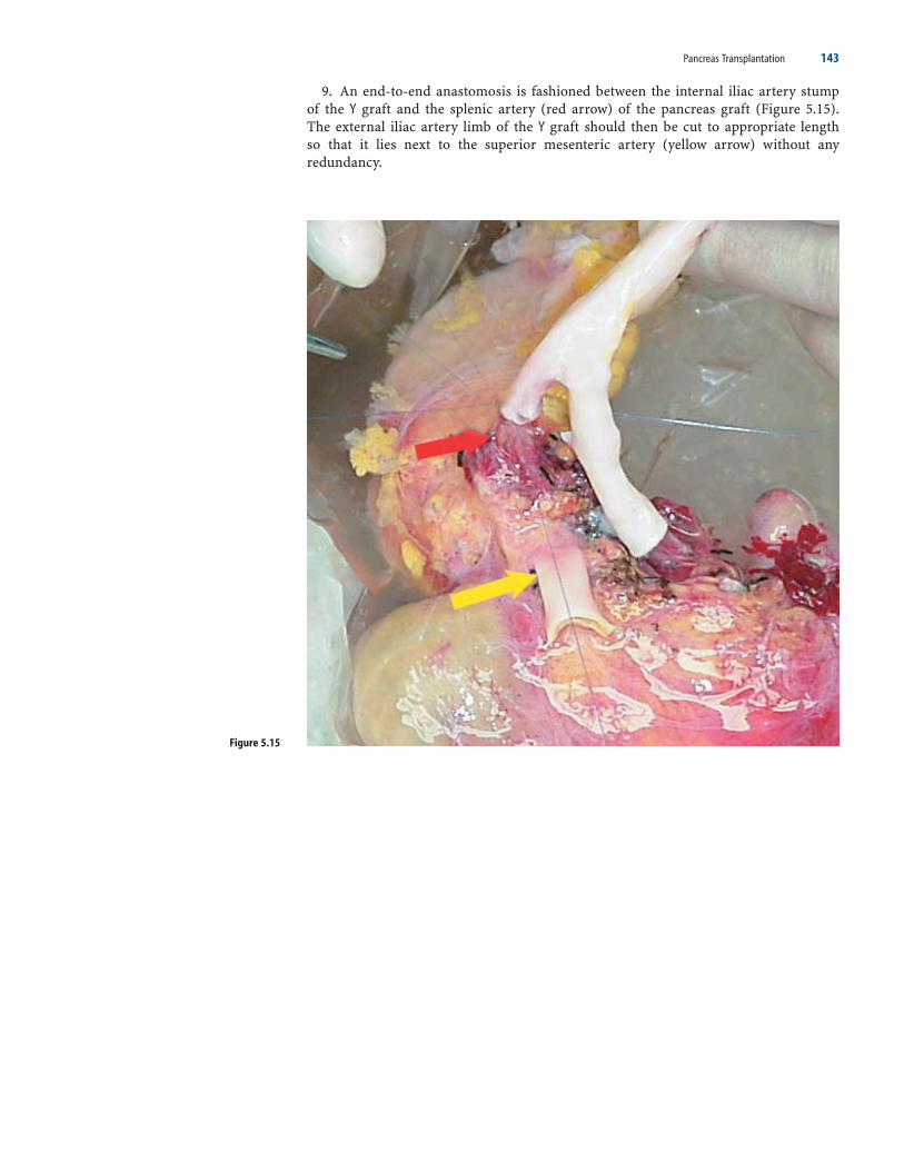
Pancreas Transplantation 143
9. An end-to-end anastomosis is fashioned between the internal iliac artery stump of the Y graft and the splenic artery (red arrow) of the pancreas graft (Figure 5.15).The external iliac artery limb of the Y graft should then be cut to appropriate length so that it lies next to the superior mesenteric artery (yellow arrow) without any redundancy.
Figure 5.15

Figure 5.16
Figure 5.17
144 Atlas of Organ Transplantation
10. The end of the external iliac artery of the Y graft is then anastomosed to the superior mesenteric artery (yellow arrow), again in an end-to-end fashion (Figure 5.16).
11. The pancreas graft is now ready for implantation (Figure 5.17).

Pancreas Transplantation 145
Simultaneous Pancreas Kidney (SPK):Systemic/Enteric Drainage
A pancreas transplant is most commonly performed in conjunction with a simultane-ous kidney transplant (Figure 5.18). The pancreas is often drained into the systemic circulation, usually by connecting the portal vein of the graft to the iliac vein ofthe recipient. This drains the endocrine secretions into the systemic circulation. Theexocrine secretions can then be managed by connecting the donor duodenum to therecipient bladder or the bowel. Alternatively, the portal vein of the graft can be drainedinto the portal circulation, by connecting to the superior mesenteric vein or a large trib-utary of the vein. Exocrine secretions are then drained into the recipient’s bowel, as thepancreas will generally not reach the bladder when drained into the portal circulation.
Figure 5.18

146 Atlas of Organ Transplantation
a) Surgical Procedure
1. The initial part of the operation usually involves implantation of the kidney. If thereis concern regarding prolonged ischemia of the pancreas graft, then this organ can beimplanted first, followed by the kidney. In either case, a long midline incision is per-formed, and the kidney is placed on the left-sided iliac vessels (Figures 5.19 and 5.20).
Figure 5.19
Figure 5.20

Pancreas Transplantation 147
2. The sigmoid colon is returned to its position, where it drapes over and covers thekidney (broken blue lines) (Figure 5.21).
3. The retractors are then repositioned in preparation for the dissection of the right-sided iliac vessels. The cecum and right colon are mobilized to the left (solid arrows) toexpose the right iliac vessels and to make space for the pancreas graft (Figure 5.22).
Figure 5.21
Figure 5.22

148 Atlas of Organ Transplantation
4. A good length of the common iliac, external iliac, and internal iliac arteries is dis-sected out in preparation for the arterial anastomosis (Figure 5.23). The distal commoniliac artery is the preferred site for implantation of the artery (blue mark).
5. The right iliac vein is then completely mobilized (broken blue line), and all thepelvic branches (yellow arrow) of this vein should be divided to allow for the vein to be
Figure 5.23

Pancreas Transplantation 149
in a more superficial location (Figures 5.24 and 5.25). Dividing the branches also allowsthe vein to be lateral to the artery, which is the correct orientation when the pancreasgraft is oriented such that the duodenum is pointing toward the recipient’s pelvis. It iswise to suture ligate the stumps of these branches from the main right iliac vein (Figure5.25) to prevent ligature slippage and troublesome bleeding.
Figure 5.24
Figure 5.25
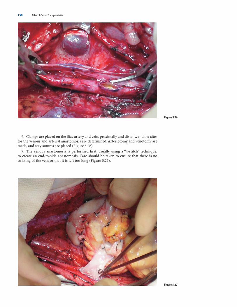
150 Atlas of Organ Transplantation
Figure 5.26
6. Clamps are placed on the iliac artery and vein, proximally and distally, and the sitesfor the venous and arterial anastomosis are determined. Arteriotomy and venotomy aremade, and stay sutures are placed (Figure 5.26).
7. The venous anastomosis is performed first, usually using a “4-stitch” technique,to create an end-to-side anastomosis. Care should be taken to ensure that there is notwisting of the vein or that it is left too long (Figure 5.27).
Figure 5.27

Pancreas Transplantation 151
8. The arterial Y graft is then trimmed to appropriate length and anastomosed to thecommon iliac artery, again using a “4-stitch” technique (Figures 5.28 and 5.29).
Figure 5.28
Figure 5.29

Figure 5.30
152 Atlas of Organ Transplantation
9. The clamps are removed and the pancreas allowed to reperfuse (Figure 5.30). Thegraft should be inspected carefully at this point to control any bleeding.
10. The exocrine secretions can then be drained by connecting the donor duodenumto the bladder or bowel of the recipient. The latter is generally preferred because of a

Pancreas Transplantation 153
lower incidence of metabolic and urinary tract problems. However, if there is anyconcern regarding the donor duodenum, or if ischemia time has been long, a bladderanastomosis is always safer. The bowel anastomosis can be performed either to a Roux-en-Y limb or just simply to the side of the bowel. The site should be fairly proximal toprevent future diarrhea. However, it is more important to choose the portion of the bowelthat reaches the donor duodenum without tension. The bowel anastomosis can be per-formed in a hand-sewn fashion or with a stapler. With a hand-sewn technique, a two-layer anastomosis is done, using a nonabsorbable suture for the outer layer and anabsorbable suture for the inner layer. Both layers can be performed with a simple runningsuture technique. The outer layer of the back wall is placed first after carefully aligningthe donor duodenum (blue arrow) and recipient small bowel (yellow arrow) (Figure5.31).
Figure 5.31

154 Atlas of Organ Transplantation
11. The duodenum and side of the bowel are opened for a corresponding length. Theinner layer of the back wall is performed. The anastomosis is completed by performingthe two layers of the anterior wall (Figure 5.32).
Simultaneous Pancreas Kidney: Portal/Enteric Drainage
A. Osama Gaber and Hosein Shokouh-Amiri
The major differences between the portal pancreas operation and other pancreas trans-plant procedures are related to the location of the portal anastomosis in the recipient,the orientation of the pancreas, and the need to traverse the small bowel mesentery forthe arterial anastomosis. The location and size of the portal vein are of utmost impor-tance, as they determine the venous outflow from the transplant pancreas. We havelearned through experience that in some patients, portal anastomosis of the donor portalvein is not advisable because of the small size of the portal tributaries. In cases wherethe superior mesenteric vein or its branches are small or too deeply located in a thick-ened mesentery, it is preferable to use systemic placement of the gland to the interiorvena cava (IVC) or the iliac veins. The orientation of the gland is also important as thedonor-to-recipient anastomosis has to be constructed parallel to the axis of the vein toprevent distorting the recipient vein if one attempts to construct a perfectly verticalanastomosis. Since the bowel anastomosis fixes the vein in position, the location of thatanastomosis should be chosen so as not to rotate the venous anastomosis, which couldincrease the risk of venous thrombosis. Finally, the arterial anastomosis site should bechosen in such a way as to prevent redundancy of the arterial conduit, particularly inthe donor external and internal iliac limbs. Because the pancreas is a low-flow organ,these vascular considerations are important in preventing arterial or venous thrombo-sis. In addition, since the majority of portal pancreas transplants are drained enterically,there is no need to keep the duodenal segment short, and a generous duodenal segmenthelps increase the total amount of flow into the gland and reduces the thrombosis risk.
Figure 5.32

Pancreas Transplantation 155
The operation is generally performed by an intraperitoneal approach through amidline incision (Figure 5.33). In rare cases with extensive intraperitoneal adhesions,a retroperitoneal dissection of the superior mesenteric vein can be performed and thena peritoneal window is constructed to ensure utilization of the peritoneal surface in absorption of potential pancreatic surface leaks and prevent retroperitoneal fluid collections.
Figure 5.33

156 Atlas of Organ Transplantation
a) Surgical Procedure
Bench Preparation of the Pancreas
Preparation of the pancreas for the portal procedure follows the same steps as in otherpancreas transplant procedures: closure of the bowel ends over the staple lines, removalof retroperitoneal tissue from around the superior mesenteric artery, ascertaining hemo-stasis at the cut edge of the mesenteric edge overlying the uncinate process, dissectionof the portal vein to ensure adequate length for anastomosis, and construction of thearterial conduit for a single donor-recipient anastomosis. The donor portal vein shouldbe redundant over the pancreatic surface to prevent tethering of the venous anastomo-sis following gland perfusion. The arterial conduit is usually constructed with a segmentof the common iliac and its external and internal branches.Whenever possible, the exter-nal and internal iliac branches of the arterial conduit should be shortened such as to tra-verse the majority of the distance between the pancreatic blood vessels and the recipientiliac artery by the straight, and less likely to kink, common iliac artery of the donor(Figure 5.34).
Figure 5.34

Pancreas Transplantation 157
Recipient Operation
1. With the patient in the supine position, a midline incision extending from thexyphoid to the pubic symphysis is made (Figure 5.35). It is generally necessary to dividethe falciform ligament of the liver. A thorough abdominal exploration is then performedto ascertain the absence of anatomic or pathologic anomalies that could affect the procedure.
2. The transverse colon is then reflected upward and out of the incision and wrappedin warm moist towels on the skin surface. Abdominal retractors are then placed on bothsides of the wound. Bowel loops are packed in the left lateral gutter and inferiorly in the pelvis, stretching the mesenteric root. This exposes the recipients’ duodenal loop,ligament of Trietz, and the uncinate process. The superior mesenteric vein (blue arrow)with its tributaries can be seen under the thin peritoneal covering of the root of themesentery (Figure 5.36).
Figure 5.35
Figure 5.36

158 Atlas of Organ Transplantation
3. The peritoneum overlying the vessels is cut with the scissors or the electrocauterymaking sure to continuously separate the superior mesenteric vein (blue arrow) by gentlyteasing it away from the peritoneal membrane to prevent its injury (Figure 5.37). Thedissection is carried in the root of the mesentery until the vein is well exposed for a dis-tance of about 4 to 5 inches and the venous tributaries are dissected. Using minimal trac-tion, the posterior aspect of the vein is exposed to ensure the absence of any tributariesthat could cause bleeding during the anastomosis. The main vein and all its tributariesare then encircled with thin vessel loops.
4. It is important during the dissection around the vein (blue arrow) to look for lym-phatic tributaries and ligate larger lymph channels whenever possible to avoid the devel-opment of postoperative lymph leaks (Figure 5.38). Small venous tributaries can be
Figure 5.37
Figure 5.38

Pancreas Transplantation 159
ligated on the vein side and electrocoagulated on the mesenteric side. Care should beexercised to avoid electrocoagulating both ends, so as to prevent postoperative bleedingfrom the small branches. Following completion of the dissection it is not uncommon forthe vein to be in spasm and have a reduced diameter. In such cases, it is best to removeall vessel loops, proceed with dissection of the iliac vessels, and decide at a later pointwhether the vein size permits portal anastomosis.
5. Exposure of the iliac vessels is generally done on the right side except in caseswhere a prior kidney has been done on the right; here one could use the left side. Thepacking of the bowel loops is removed and the retractor is adjusted to provide maximumexposure on the right side. Mobilization of the right colon and the small intestinalmesentery is undertaken (Figure 5.39). The right colon and intestines are then wrappedin a large towel and reflected upward and to the left. The sigmoid colon is also coveredby moist towels and retracted to the left.
Figure 5.39

Figure 5.40
160 Atlas of Organ Transplantation
6. The retroperitoneum is then opened after identification of the ureter and thegonadal vessels. These structures are gently swept medially and dissection is carried ontop of the common iliac artery. The common iliac artery (yellow arrow) is exposed fromthe aorta to the beginning of the external iliac (Figure 5.40). Gentle palpation ensuresthat there are no hard calcifications that could prevent performing a safe anastomosis.In patients with calcification, the external iliac could be dissected, or alternatively onecould use the recipient aorta. Since occlusion for the arterial anastomosis is generallydone by a large side-biting C-clamp, one has to dissect enough of the common iliac veinto ensure safe placement of the vascular clamp.
7. The bowel loops are then brought back into their anatomic position and the bowelsare packed as during the portal vein dissection, maintaining the right-sided traction toavoid repositioning retractors between the venous and arterial anastomosis.

Pancreas Transplantation 161
8. Once the decision is made (based on the size of the portal tributary) to proceedwith portal placement, the next step is to decide on the location, size, and direction ofthe anastomosis. We prefer to place the anastomosis distal to a large tributary. Our pre-ference is based on a theoretical concern that clotting of the venous anastomosis couldpropagate upward into the portal circulation with catastrophic results on bowel venousdrainage. Although this has never been reported, we still favor having flow proximal tothe anastomotic site. The recipient superior mesenteric vein (blue arrow) is controlledeither by traction on the vessel loops or by small bulldog clamps (Figure 5.41). An ante-rior venotomy is then performed, taking care to avoid injury of the back wall of the softvessel, particularly if a posterior branch necessitates placement of a posterior clamp.
Figure 5.41

162 Atlas of Organ Transplantation
9. The allograft pancreas is then wrapped in two white towels with ice slush overly-ing its surface. Venous anastomosis (blue arrow) is performed using 6.0 Prolene in arunning fashion (Figure 5.42). The surgeon’s side is completed first, and then the assis-tant has the benefit of the traction on the back wall afforded by ligation of the back wallstitch in the middle of the anastomosis.
10. Once both sides of the venous anastomosis are completed, a bulldog clamp isplaced on the donor portal vein and recipient’s portal flow is restored (Figure 5.43). Thevenous anastomosis (blue arrow) is inspected and any suture line bleeding is dealt with.
Figure 5.42
Figure 5.43

Pancreas Transplantation 163
11. The site of the anastomosis on the iliac artery is then visualized by elevation ofthe right colon and the root of the mesentery. The arterial conduit (blue arrow) is alsovisualized, and the point at which the conduit traverses the mesentery is chosen so as to achieve the shortest, most vertical path for the conduit to the artery. The electro-cautery is used to incise the mesentery, and a curved vascular clamp is passed on top ofthe recipient iliac artery through the root of the mesentery. The arterial conduit is thenadjusted to remove any possible rotation, and the donor common iliac artery is passedthough the defect in the root of the mesentery to the site of the arterial anastomosis(Figure 5.44).
12. The right colon and small intestine are again wrapped in a moist towel and moved upward and to the left, exposing the dissected retroperitoneum. The recipientiliac artery is then occluded with a large C-clamp. The arterial conduit (blue arrow) isthen shortened to appropriate length and beveled for the anastomosis. The recipientarteriotomy (yellow arrow) is created with a No. 11 blade and fashioned with a 4.8-mmaortic punch. A 5.0 Prolene suture is used to create the end-to-side arterial anastomosis
Figure 5.44

164 Atlas of Organ Transplantation
Figure 5.45
(Figure 5.45). Once the anastomosis is completed, the bowel is brought down to anatomicposition, the portal anastomosis is exposed, and the bulldog clamp occluding the donorportal vein is removed, allowing, venous revascularization of the graft. In the absence ofvenous bleeding, the mesentery is lifted upward, allowing exposure of the arterial anas-tomosis, and the arterial clamp is removed, completing revascularization of the trans-plant pancreas. A quick inspection of the pancreas is necessary at this point to clampand ligate any bleeders from the surface. Once hemostasis is achieved, the gland iswrapped in a warm towel and bowel anastomosis is started.
13. Enteric drainage is to a Roux-en-Y limb or a side-to-side anastomosis of the trans-plant duodenum to an adjoining bowel loop. Regardless of the technique used, the recip-ient bowel loop used in the anastomosis should be redundant enough to avoid rotationof the pancreas. We prefer hand-sewn, double layer (silk and PDS) bowel anastomosis(Figure 5.46).
Figure 5.46

Pancreas Transplantation 165
14. After placement of the posterior silk layer, bowel occlusion clamps are placed onthe recipient bowel and the electrocautery is used to open both the recipient bowel anddonor duodenum. Once the duodenal segment is opened, contents are cultured and thesegment irrigated copiously with antibiotics. Following completion of the bowel anas-tomosis (Figure 5.47), the abdominal cavity is irrigated and all contaminated instru-ments are handed off the field. If a Roux-en-Y limb has been used, the side-to-side bowelanastomosis is completed prior to handing off the contaminated instruments. In thesecases it is important to close the mesenteric gap created by mobilizing the bowel for theintestinal anastomosis to prevent internal herniation. Once bowel work is completed andfollowing abdominal irrigation, the spleen is excised followed by a final careful inspec-tion of the pancreas to ensure appropriate hemostasis.
15. When a kidney transplant is to be performed, the retractors are then moved toallow maximum exposure on the left side. We prefer creating a retroperitoneal pocketby dissecting the peritoneum off the abdominal wall muscles. This allows completeclosure of the retroperitoneal space around the kidney at the time of closure. In largerindividuals, however, accessing the vessels from behind the sigmoid colon mesenteryachieves good exposure of the iliac vessels for kidney placement.
Figure 5.47

166 Atlas of Organ Transplantation
Technical Points
1. If possible, we prefer to wrap the pancreas with the omentum (Figure 5.48). Thisrequires mobilizing some of the omentum off the transverse colon but serves to keepbowel loops separable from the pancreas in the case of reoperative therapy.
2. We attempt to bring the tail of the pancreas up to the abdominal wall by stitchingsome of the ligated splenic vessels to the peritoneum on one side of the incision. Onehas to be careful, however, of pancreatic rotation, and one should favor allowing the pan-creas to choose its own axis.
Acknowledgments
The authors acknowledge the expert assistance of Dr. Mohammad Shokouh-Amiri forpreparation of the illustrations and Mrs. Shaherah Rankins-Amos for assistance withmanuscript preparation.
Isolated Pancreas Transplant: Bladder Drainage
Isolated pancreas transplants can be either in the form of a pancreas after kidney transplant (PAK) or a pancreas transplant alone (PTA). A PAK procedure is usually performed once the recipient has adequately recovered from the kidney transplant. Apancreas transplant alone (PTA) may be performed for the diabetic patient without renal failure but with significant problems with glucose control (e.g., ketoacidosis,hypogylcemic unawareness, etc.) or evidence of secondary organ damage (i.e., neuro-pathy). The majority of isolated pancreas grafts are drained into the bladder. This hasthe advantage of allowing for urine amylase monitoring, which can act as a surrogatemarker for rejection – a decrease in urine amylase values indicating pancreatic graft dys-function. Drainage to the bowel is, however, more physiologic and not associated withproblems such as urethritis, hematuria, dehydration, and acidosis. As rejection ratesdecrease, enteric drainage is becoming a more widely used option, even for PTA and PAK transplants.
Figure 5.48

Pancreas Transplantation 167
An intraperitoneal approach is preferred, with a midline incision in the lowerabdomen. The preference is to place the pancreas in the right iliac fossa, with the headand duodenum pointing downwards (Figure 5.49). One then has the option of eitherbladder or enteric drainage. If the right iliac vessels have already been used, the pancreascan be placed on the left side, or higher in the abdomen with portal/enteric drainage.
Figure 5.49

168 Atlas of Organ Transplantation
a) Surgical Procedure
1. The start of the procedure is exactly as described for the pancreas portion of thesimultaneous pancreas kidney transplant. After a midline incision, the cecum is mobi-lized and the iliac vessels are identified. The external iliac artery, internal iliac artery, andcommon iliac artery (broken yellow line) are all isolated and encircled. The arterial anastomosis is ideally placed on the common iliac artery (Figure 5.50).
2. The iliac vein is completely mobilized with division and suture ligation of allhypogastric branches (Figure 5.51). This allows the iliac vein to be placed in a moresuperficial location lateral to the iliac artery, which is the correct orientation of the portalvein and Y graft of the pancreas graft when it is placed in its position with the duode-nal cuff next to the recipient bladder. This only applies if the pancreas is being placedon the right side. If the pancreas is to be placed on the left side, the iliac vein should bepositioned medial to the iliac artery.
Figure 5.50
Figure 5.51

Figure 5.53
Pancreas Transplantation 169
3. Clamps are placed proximally and distally on the iliac artery and vein. Appropri-ate marks are made for the position of the arteriotomy and venotomy. The venotomyshould be placed just distal to the arteriotomy site, and if possible, the two should notoverlap in position (Figures 5.52 and 5.53).
Figure 5.52

Figure 5.54
170 Atlas of Organ Transplantation
4. The venous anastomosis is usually performed first, creating an end-to-side anastomosis between the end of the portal vein and the side of the iliac vein (Figure 5.54).

Pancreas Transplantation 171
5. The Y graft is trimmed to appropriate length so that there is not undue redundancy,and the arterial anastomosis is performed, again in and end-to-side fashion (Figure5.55).
Figure 5.55

172 Atlas of Organ Transplantation
6. The clamps are removed, and the graft (broken blue line) is allowed to perfuse(Figures 5.56 and 5.57). Bleeding points are controlled using hemoclips or suture liga-tures. The graft is inspected to make sure hemostasis is adequate and the anastomosislies adequately, before proceeding with the bladder anastomosis.
Figure 5.57
Figure 5.56

Pancreas Transplantation 173
7. The bladder anastomosis is fashioned between the dome of the bladder (brokenblue line) and the antimesenteric border of the duodenum (broken yellow line) (Figure5.58). A two-layer hand-sewn anastomosis is fashioned using continuous nonabsorbable
Figure 5.58

Figure 5.59
174 Atlas of Organ Transplantation
suture for the outer layer, and continuous absorbable suture for the inner layer. The posterior outer layer is performed first using a running nonabsorbable suture. Appro-priate-sized openings are then made in the donor duodenum and recipient bladder(Figure 5.59).
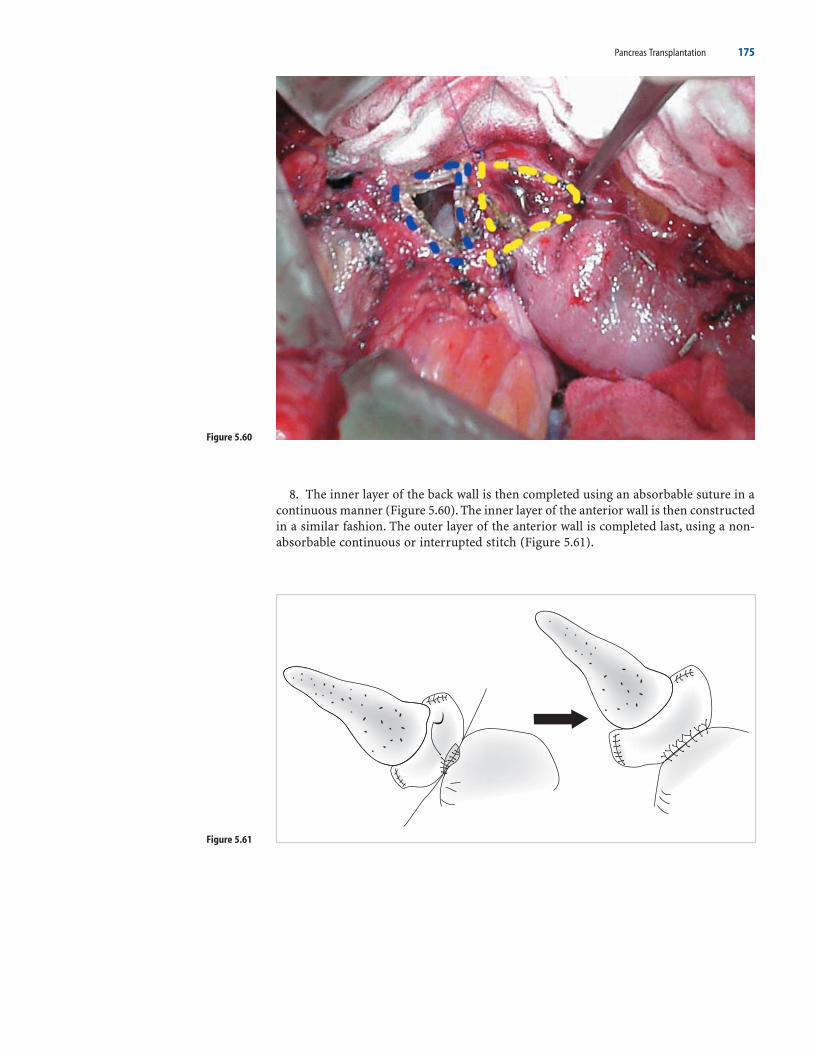
Pancreas Transplantation 175
8. The inner layer of the back wall is then completed using an absorbable suture in acontinuous manner (Figure 5.60). The inner layer of the anterior wall is then constructedin a similar fashion. The outer layer of the anterior wall is completed last, using a non-absorbable continuous or interrupted stitch (Figure 5.61).
Figure 5.60
Figure 5.61

Figure 5.62
176 Atlas of Organ Transplantation
9. Alternatively, this anastomosis can be done using a luminal stapling device. A cys-totomy is made in the dome of the bladder and this is used to insert the anvil of thestapler into the posterior aspect of the bladder (Figure 5.62).

Pancreas Transplantation 177
10. The stapler is inserted via the end of the distal duodenum of the graft, and thepin forced through the center of the antimesenteric border (Figure 5.63). It is connectedto the anvil and fired. Excess donor duodenum is then trimmed using a linear staplingdevice.
Figure 5.63

178 Atlas of Organ Transplantation
11. The staple line is oversewn for hemostasis, and the bladder is closed in two layers(Figure 5.64).
Pancreas Transplant from a Living Donor
Miquel Tan, Raja Kandaswamy, and Rainer W.G. Gruessner
Although the pancreas was the first extrarenal organ to be successfully used from livingdonors (LDs), of the greater than 18,000 pancreas transplants performed since the 1960s,less than 1% have come from live donation. Several reasons account for the underuse ofthis resource, among them the potential morbidity of a distal pancreatectomy in an oth-erwise healthy donor and a higher technical failure rate compared to deceased donortransplants. In selected cases, however, LD pancreas transplantation may be an appro-priate option. This includes high panel reactive antibody (PRA) recipients who areunlikely to receive a cadaver organ or uremic diabetics on the simultaneous cadaver pan-creas-kidney (SPK) waiting list. This procedure may be performed either for an isolatedpancreas transplant, or combined with a nephrectomy for an SPK transplant.
Figure 5.64

Pancreas Transplantation 179
a) Donor Evaluation
It is crucial that all potential donors undergo a thorough preoperative evaluation. Thepurpose is twofold: first, to ensure that the donor is healthy enough to undergo the oper-ation safely, and second, to ensure that the reduced pancreatic mass would be sufficientto maintain a normal metabolic state. The general health of the donor is best ascertainedby a detailed history and physical examination, looking for evidence of cardiovascular,pulmonary, renal, and major gastrointestinal disease. This part of the workup is no dif-ferent from the evaluation for any other general surgical patient undergoing a pancre-atic resection.
Endocrinologic evaluation includes family history. Potential donors should be at least10 years older than the age of onset of diabetes in the recipient (and the onset of dia-betes in the recipient must have been at least 10 years pretransplant). In addition, forsibling donors, no family member other than the recipient should be diabetic. Whenthese two criteria are met, donors are at no greater risk for diabetes than the generalpopulation, even if they are human leukocyte antigen (HLA) identical with the recipi-ent. Potential donors should be asked about their alcohol intake. Female donors who havehad previous pregnancies should be asked about gestational diabetes.
Metabolic studies are done to try to estimate islet cell mass and reserve in the poten-tial donor. Tests that can be done include measurement of the hemoglobin A1c (shouldbe <6%), an oral glucose tolerance test (OGTT), insulin secretion in response to an intra-venous glucose tolerance test (IVGTT), and an arginine stimulation test.
Lastly, some radiologic assessment of the vascular anatomy should be done. This stepis more important if the kidney is to be procured also, as the vascular anatomy of thesplenic artery is fairly constant in comparison to the renal arteries. An invasive arteri-ogram is no longer necessary to obtain this information as current CT or magnetic res-onance imaging (MRI) provides adequate information with regard to the renal andsplenic vessels.
The surgical procedure for removal of the pancreas with or without a kidney can bedone either as an open or laparoscopic procedure. Initially the removal of the distal pan-creas from a living donor was done via an open operative procedure. While this can bedone safely, it does involve a significant recovery time for the donor. The advent oflaparoscopic technology provides a viable alternative to the traditional open approach.This has been demonstrated most clearly with laparoscopic donor nephrectomy, whichhas rapidly become the procedure of choice for kidney donation because of reduced hos-pital stay and more rapid convalescence. Cosmetically, it is more appealing to potentialdonors compared to the traditional flank incision required for open nephrectomy.Laparoscopic nephrectomy is equivalent to the open procedure in terms of donor safetyand quality of allograft. Laparoscopic techniques have rapidly been applied to otherorgan systems including the pancreas. Laparoscopic distal pancreatectomies have beendescribed for treatment of a variety of pathologic states and appear to be safe with the additional benefit of reduced hospital costs and accelerated postoperative recovery.The technique of open and laparoscopic live donor pancreatectomy is subsequentlydescribed.
b) Surgical Procedure
Open Technique
1. A midline incision is preferred. If the kidney is to be removed, it is generallyremoved first. The right or left kidney can be used, depending on the vascular anatomy.The preference is to use the left kidney (because of concurrent partial mobilization ofthe pancreas, greater length of the renal vein, and lack of liver mobilization). The spleno-colic ligament should be preserved, if possible, as it may carry collateral blood vesselsto the spleen. The kidney is removed once the dissection is complete.

Figure 5.65
180 Atlas of Organ Transplantation
2. Procurement of the distal pancreas begins by dividing the gastrocolic ligament,starting medially and advancing laterally to the margin of the spleen (Figure 5.65). Careshould be taken to preserve the gastroepiploic artery and the short gastric vessels inorder to diminish the likelihood of devascularizing the spleen. The retroperitonealattachments of the spleen are not disturbed, and the spleen is not mobilized. Thestomach can then be retracted superiorly, and the inferior margin of the distal pancreasis mobilized.

Pancreas Transplantation 181
3. A peritoneal incision is made over the tail of the pancreas at its junction with thespleen. The pancreas is gently dissected off the splenic surface. The splenic vessels arethen identified, and the main trunks of both the splenic artery and vein divided proxi-mal to the splenic branches (again, preserving collateral blood vessels supplying thespleen) (Figure 5.66).
Figure 5.66

182 Atlas of Organ Transplantation
4. The superior margin of the pancreas is then mobilized, retaining the splenic arteryand vein in continuity with the body and tail of the pancreas. As the pancreas is elevated from its bed and retracted medially, the confluence of the inferior mesenteryvein (IMV) as it joins the splenic vein can be seen. The location of this confluence varies;it can be very close to the superior mesenteric vein (SMV)–splenic vein junction. Oncethe IMV is divided, the pancreas can be mobilized further to the surgical neck. The portalvein is identified at the confluence of the SMV and splenic vein. As the pancreas con-tinues to be rotated medially, the splenic artery is isolated at its origin off the celiac trunkand encircled with a vessel loop (Figure 5.67).
Figure 5.67

Pancreas Transplantation 183
5. The avascular plane between the pancreas and portal vein is bluntly dissected todefine the narrowest portion of the pancreas (i.e., the neck) (Figure 5.68). This will bethe site for division of the pancreas (broken blue line) (Figure 5.69), lying just anteriorto the portal vein at the point of junction with the splenic vein (blue arrow).
Figure 5.68
Figure 5.69

184 Atlas of Organ Transplantation
Figure 5.70
6. The pancreatic neck can then be divided using multiple 4.0 silk ligatures. Both endsof the pancreatic duct should be identified. The proximal end can be oversewn, and thedistal duct tacked with a fine suture for identification. At this point, the segmental pan-creas graft is ready for removal. Clamps are placed on the splenic artery and the splenicvein at its junction with the portal vein (Figure 5.70). After dividing the vessels andremoving the graft, the vascular stumps are oversewn closed.

Pancreas Transplantation 185
7. The recipient operation is not very different from its deceased donor counterpart.A lower abdominal midline incision is used. If a kidney graft is to be implanted, it shouldbe placed on the left side. The pancreas graft should be placed on the right side if pos-sible, because of the more superficial location of the vessels on this side. In preparationfor the pancreas graft implantation, all branches of the iliac vein, including the hypogas-tric, should be divided (allowing the vein to lie in a more superficial location). To allowfor an “artery-lateral-to-the-vein” alignment (opposite of positioning for deceased donortransplant), it is useful to divide the internal iliac artery. Doing so gives a better lie tothe graft after it is implanted and decreases the chances of the graft artery kinking. Thedonor splenic artery can then be anastomosed end-to-side to the external or commoniliac artery (yellow arrow); the splenic vein is anastomosed end to side to the iliac vein(blue arrow) (Figure 5.71).
Figure 5.71

Figure 5.72
186 Atlas of Organ Transplantation
8. Pancreatic exocrine secretions can be managed by a number of techniques. Ifthe pancreatic duct is of adequate size, a direct anastomosis between the duct andbladder mucosa can be constructed with interrupted 7.0 absorbable sutures (Figure5.72). If the duct is small and the diameter of the cut edge of the pancreas is small, thenan invagination technique can be used. The pancreas graft is invaginated into the bladderusing one internal layer of 4.0 absorbable sutures and one external layer of interrupted4.0 nonabsorbable sutures. Doing so obviates the need for a tedious duct-to-mucosaanastomosis, but it could create problems from exposure of pancreatic tissue to urine.The pancreatic duct may also be drained enterally, using a Roux-en-Y limb of distalbowel.
Laparoscopic Technique
1. After induction of general anesthesia, the patient is placed in a modified rightlateral decubitus position to allow the patient to be rotated from a left-side-up position(for the nephrectomy) to a supine position (for the distal pancreatectomy). For patientsundergoing a distal pancreatectomy alone, the patient is kept in the supine position.

Pancreas Transplantation 187
Figure 5.73
If a left nephrectomy is to be performed at the same time, the patient is placed in rightlateral decubitus position. The operative table is then flexed to 45 degrees to open up theleft subcostal space to facilitate dissection of the kidney. A 6-cm midline incision is thenmade either supra- or periumbilically depending on the patient’s body habitus. A donorwith a longer torso would require the incision to be placed in a supraumbilical positionso that the surgeon’s hand can reach both the kidney and pancreas. A Gelport® (AppliedMedical, Rancho Santa Margarita, CA) or HandPort® (Smith and Nephew Inc., Andover,MA) device is placed in the midline incision to allow for hand assistance. Using stan-dard laparoscopic equipment, a 12-mm port is placed at the level of the umbilicus alongthe lateral edge of the left rectus for insertion of a 30-degree camera. After pneu-moperitoneum is established, a second 12-mm port is inserted in the left mid-abdomenin the plane of the anterior axillary line (Figure 5.73). This allows insertion of ultrasonicshears, laparoscopic scissors, and any other needed instrumentation. The surgeon andassistant stand on the patient’s right side during the nephrectomy (Figure 5.74). Theassistant moves to the patient’s left side during the pancreatectomy. Mobilization of theleft colon, ureter, and left gonadal vein, as well as dissection of the kidney is describedin detail elsewhere.
Figure 5.74

188 Atlas of Organ Transplantation
2. After removal of the kidney, the donor is then rotated to a supine position withreverse Trendelenburg to help move the transverse colon away from the operative field.The partially dissected inferior margin of the pancreas is mobilized using ultrasonicshears and electrocautery. The IMV is identified, ligated with hemoclips, and dividednear its insertion into the splenic vein. The tail of the pancreas is then encircled bymaking a hole in the avascular plane along the superior margin of the pancreatic tailand passing a finger along the underside and posterior aspect of the pancreas. The tailis encircled with a vessel loop to facilitate atraumatic anterior retraction of the pancreas(Figure 5.75). The posterior surface of the pancreas is then freed from its retroperitoneal
Figure 5.75

Pancreas Transplantation 189
attachments using electrocautery. The splenic artery and vein are identified in the hilumof the spleen and individually ligated and divided using a 35-mm vascular stapler, allow-ing the pancreas to be lifted from its bed and retracted medially (broken blue line)(Figure 5.76). Care should be taken not to disturb the short gastric vessels and right gas-troepiploic artery, as this constitutes the main remaining blood supply to the spleen.
3. With the splenic vessels divided distally and the pancreas retracted medially,its posterior aspect can be visualized. The splenic vein (blue arrow) and the splenic artery (yellow arrow) can be seen running along the posterior aspect of the pancreas(Figure 5.77).
Figure 5.76
Figure 5.77

190 Atlas of Organ Transplantation
4. The splenic vein is mobilized circumferentially near its junction with the superiormesenteric vein (blue arrow). Likewise, the splenic artery is mobilized just as it branchesoff the celiac axis (yellow arrow) (Figure 5.78).
5. Heparin (30 units/kg) is administered intravenously prior to ligation of the vessels.Two clips are applied to the splenic artery just distal to the celiac axis (yellow arrow)(Figure 5.79). The splenic artery is then divided with laparoscopic scissors. The splenicvein can be divided at its junction with the SMV, either using clips or a stapling device(blue arrow).
Figure 5.78
Figure 5.79

Figure 5.80
Pancreas Transplantation 191
6. A linear cutting stapler device is used to transect the pancreatic neck (Figure 5.80).The distal pancreatic segment is then extracted by hand through the midline incisionand passed off to the recipient team to be flushed immediately with cold University ofWisconsin solution.
7. The staple line (green arrow) of the pancreatic remnant is carefully inspected(Figure 5.81) and oversewn intracorporeally with 4–0 polypropylene suture to achievehemostasis and prevent leakage of the pancreatic duct.The vascular stumps of the splenicartery (yellow arrow) and vein (blue arrow) are carefully inspected to make sure there isno bleeding. After irrigation, a drain is placed near the pancreatic remnant. The spleenshould be reexamined at this point to assess its viability. Closure is then performed.
Figure 5.81
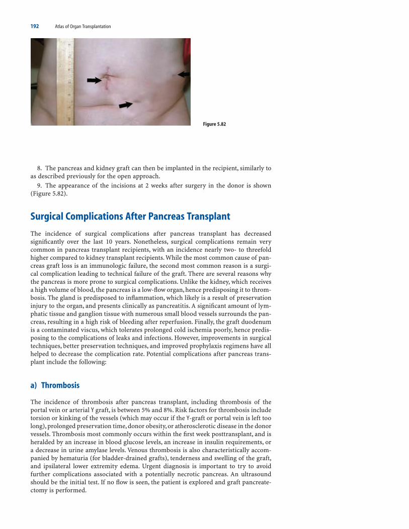
192 Atlas of Organ Transplantation
8. The pancreas and kidney graft can then be implanted in the recipient, similarly toas described previously for the open approach.
9. The appearance of the incisions at 2 weeks after surgery in the donor is shown(Figure 5.82).
Surgical Complications After Pancreas Transplant
The incidence of surgical complications after pancreas transplant has decreasedsignificantly over the last 10 years. Nonetheless, surgical complications remain verycommon in pancreas transplant recipients, with an incidence nearly two- to threefoldhigher compared to kidney transplant recipients. While the most common cause of pan-creas graft loss is an immunologic failure, the second most common reason is a surgi-cal complication leading to technical failure of the graft. There are several reasons whythe pancreas is more prone to surgical complications. Unlike the kidney, which receivesa high volume of blood, the pancreas is a low-flow organ, hence predisposing it to throm-bosis. The gland is predisposed to inflammation, which likely is a result of preservationinjury to the organ, and presents clinically as pancreatitis. A significant amount of lym-phatic tissue and ganglion tissue with numerous small blood vessels surrounds the pan-creas, resulting in a high risk of bleeding after reperfusion. Finally, the graft duodenumis a contaminated viscus, which tolerates prolonged cold ischemia poorly, hence predis-posing to the complications of leaks and infections. However, improvements in surgicaltechniques, better preservation techniques, and improved prophylaxis regimens have allhelped to decrease the complication rate. Potential complications after pancreas trans-plant include the following:
a) Thrombosis
The incidence of thrombosis after pancreas transplant, including thrombosis of theportal vein or arterial Y graft, is between 5% and 8%. Risk factors for thrombosis includetorsion or kinking of the vessels (which may occur if the Y-graft or portal vein is left toolong), prolonged preservation time, donor obesity, or atherosclerotic disease in the donorvessels. Thrombosis most commonly occurs within the first week posttransplant, and isheralded by an increase in blood glucose levels, an increase in insulin requirements, ora decrease in urine amylase levels. Venous thrombosis is also characteristically accom-panied by hematuria (for bladder-drained grafts), tenderness and swelling of the graft,and ipsilateral lower extremity edema. Urgent diagnosis is important to try to avoidfurther complications associated with a potentially necrotic pancreas. An ultrasoundshould be the initial test. If no flow is seen, the patient is explored and graft pancreate-ctomy is performed.
Figure 5.82

Pancreas Transplantation 193
b) Hemorrhage
Postoperative bleeding may be minimized by meticulous intraoperative control of bleed-ing sites. Although hemorrhage may be exacerbated by anticoagulants and antiplateletdrugs, their benefits seem to outweigh the risks. Bleeding is a much less significant cause(<1%) of graft loss than is thrombosis (5% to 8%). Significant bleeding is treated byimmediate reexploration.
c) Pancreatitis
Most cases of graft pancreatitis occur early on, are self-limited, and are probably due toischemic preservation injury. Clinical manifestations may include graft tenderness, fever,and hyperamylasemia. Treatment consists of intravenous (IV) fluid replacement andfasting the recipient. Sometimes early pancreatitis may be very severe, with partialnecrosis of the graft; this is usually an indication for graft pancreatectomy. Graft pan-creatitis can also occur late (months or years after transplant) and may be caused byreflux into the allograft duct in recipients with bladder drainage, direct trauma to thepancreas, or cytomegalovirus (CMV) infection. Reflux is treated by urinary catheterdrainage, and occasionally conversion to enteric drainage; CMV infections are treatedwith ganciclovir.
d) Urologic Complications
Urologic complications are almost exclusively limited to recipients who undergo bladderdrainage. Hematuria is not uncommon in the first several months posttransplant, but itis usually transient and self-limiting. Bladder calculi may develop, due to exposed suturesor staples along the duodenocystostomy, which may serve as a nidus for stone forma-tion. Recurrent urinary tract infections commonly occur concurrently. Treatment con-sists of cystoscopy with removal of the sutures or staples. Urinary leaks are mostcommonly from the proximal duodenal cuff or the duodenal anastomosis to the bladderand typically occur during the first several weeks posttransplant. Small leaks can be suc-cessfully managed by prolonged (for at least 2 weeks) Foley catheter drainage; largerleaks require surgical intervention. Other urinary complications include chronic refrac-tory metabolic acidosis due to bicarbonate loss, persistent and recurrent urinary tractinfections, and urethritis. Along with recurrent hematuria, these complications are themajor indications for converting patients from bladder drainage to enteric drainage ofexocrine secretions. Eventually, 15% to 20% of recipients who undergo initial bladderdrainage require conversion to enteric drainage. For this reason, the recent trend hasbeen to perform enteric drainage at the time of the transplant. Enteric drainage is asso-ciated with significantly fewer urinary tract infections and urologic complications, butthis approach obviates the use of urinary amylase determinations, which are a sensitiveindicator of pancreatic allograft rejection.
e) Infections
Infections remain a significant problem after pancreas transplantation. Most commonare superficial wound infections. Deep intraabdominal infections may also be seen andare often related to graft complications such as leaks. Thanks to appropriate periopera-tive antimicrobial regimens (for prophylaxis against gram-positive bacteria, gram-negative bacteria, and yeast), the incidence of significant infections has decreased,although it remains approximately 10% and is associated with significant morbidity andmortality. Thus, should serious intraabdominal infection occur, whether or not associ-ated with the above-described complications, reexploration and graft removal must bestrongly considered, with concurrent reduction in immunosuppressive drug therapy.

194 Atlas of Organ Transplantation
f) Leaks
A leak may occur from the graft duodenum to bladder or bowel anastomosis. It may alsooccur from the proximal or distal duodenal cuff. The incidence is between 3% and 5%,and does not differ significantly between bladder- and enteric-drained grafts. Clinicalpresentation, however, may be quite different in these two scenarios. Patients withenteric-drained grafts and a leak generally present with severe pain and generalized peri-tonitis, as the enteric contents are leaking into their abdominal cavity. With significantintraabdominal contamination, graft salvage is usually not possible and removal of thegraft is the best option. With bladder drainage, leaks do not usually present with suchsevere symptoms, and graft salvage is usually possible.
g) Minimizing Surgical Complications
Several steps can be taken to minimize surgical complications after pancreas transplant.Broadly, these steps can be separated into the following three categories:
1. Proper donor and recipient selection2. Meticulous surgical techniques3. Careful postoperative care
Donor selection plays a very important role in minimizing surgical complications.Donors with a body mass index greater than 30 generally have a pancreas with significantfatty infiltration. This predisposes to bleeding and deep infections after transplant. Coldischemia of the graft should be minimized as much as possible, as prolonged preserva-tion is associated with an increased risk for leaks and thrombosis. Ultimately, carefulexamination of the pancreas at the time of procurement and during the benching processis the best indicator of whether a pancreas should be used or not. Careful benching ofthe cadaver pancreas also plays a major role in minimizing complications. The spleenshould be removed without damaging the tail of the pancreas. Large clumps of fat andlymphatic tissue should not be left on the gland, as they often are poorly perfused post-transplant, and serve as a nidus for infection. Care should be taken not to devascularizethe proximal and distal ends of the graft duodenum. Finally, careful attention should bepaid to the arterial Y graft, ensuring that it is placed properly on the vessels withoutsignificant redundancy.
The care of the recipient early after transplant also may impact on surgical compli-cations. Use of low-dose heparin or some other variant is likely helpful in diminishingthe incidence of graft thrombosis. While the risk of bleeding is increased with the useof anticoagulation, this rarely is severe enough to result in loss of the graft. Lastly, theroutine use of antibacterial and antifungal prophylaxis for a short time after transplantlikely plays a role in diminishing the incidence of posttransplant infections.
Selected Readings
1. Boggi U, Vistoli F, Signori S, et al. A technique for retroperitoneal pancreas transplantation with portal-enteric drainage. Transplantation 2005;79(9):1137–1142.
2. Gaber AO, Shokouh-Amiri H, Grewal HP, Britt LG. A technique for portal pancreatic transplantation withenteric drainage. Surg Gynecol Obstet 1993;177(4):417–419.
3 Gaber AO, Shokouh-Amiri MH, Hathaway DK, et al. Results of pancreas transplantation with portal venousand enteric drainage. Ann Surg. 1995;221(6):613–622; discussion 622–624.
4. Gruessner RWG, Kandaswamy R, Denny R. Laparoscopic simultaneous nephrectomy and distal pancre-atectomy from a live donor. J Am Coll Surg 2001;193(3):333.
5. Gruessner RWG, Kendall DM, Drangstveit MB, Gruessner AC, Sutherland DER. Simultaneous pancreas-kidney transplantation from live donors. Ann Surg 1997;226(4):471.
6. Gruessner RWG, Leone JP, Sutherland DER. Combined kidney and pancreas transplants from livingdonors. Transplant Proc 1998;30:282.

Pancreas Transplantation 195
7. Gruessner AC, Sutherland DER. Pancreas transplant outcomes for United States (US) and non-US casesas reported to the United Network for Organ Sharing (UNOS) and the International Pancreas TransplantRegistry (IPTR) as of October 2002. Clin Transplant 2002:41.
8 Gruessner AC, Sutherland DE. Pancreas transplant outcomes for United States (US) and non-US cases asreported to the United Network for Organ Sharing (UNOS) and the International Pancreas TransplantRegistry (IPTR) as of June 2004. Clin Transplant 2005;19(4):433–455.
9. Gruessner RWG, Sutherland DER. Simultaneous kidney and segmental pancreas transplants from livingrelated donors – the first two successful cases. Transplantation 1996;61:1265.
10. Gruessner RWG, Sutherland DER, Drangstveit MB, Bland BJ, Gruessner AC. Pancreas transplants fromliving donors: short- and-long-term outcome. Transplant Proc 2001;33:819.
11. Jacobs SC, Cho E, Foster C, Liao P, Bartlett ST. Laparoscopic donor nephrectomy: the University ofMaryland 6-year experience. J Urol 2004;171(1):47.
12. Kercher KW, Heniford BT, Matthews BD, et al. Laparoscopic versus open nephrectomy in 210 consecutivepatients: outcomes, cost, and changes in practice patterns. Surg Endosc 2003;17(12):1889.
13. Leventhal JR, Deeik RK, Joehl RJ, et al. Laparoscopic live donor nephrectomy – is it safe? Transplantation2000;70(4):602.
14. Martin X, Petruzzo P, Dawahra M, et al. Effects of portal versus systemic venous drainage in kidney-pancreas recipients. Transplant Int 2000;13(1):64–68.
15. Philosophe B, Farney AC, Schweitzer EJ, et al. Superiority of portal venous drainage over systemic venousdrainage in pancreas transplantation: a retrospective study. Ann Surg 2001;234(5):689–696.
16. Reddy KS, Stratta RJ, Shokouh-Amiri MH, Alloway R, Egidi MF, Gaber AO. Surgical complications afterpancreas transplantation with portal-enteric drainage. J Am Coll Surg 1999;189(3):305–313.
17. Schweitzer EJ, Wilson J, Jacobs S, et al. Increased rates of donation with laparoscopic donor nephrectomy.Ann Surg 2000;232(3):392.
18. Sharara AI, Dandan IS, Khalifeh M. Living-related donor transplantation other than kidney. TransplantProc 2001;33:2745.
19. Stratta RJ, Gaber AO, Shokouh-Amiri MH, et al. A prospective comparison of systemic-bladder versusportal-enteric drainage in vascularized pancreas transplantation. Surgery 2000;127(2):217–226.
20. Sutherland DER, Najarian JS, Gruessner RWG. Living versus cadaver pancreas transplants. TransplantProc 1998;30:2264.
21. Tan M, Kandaswamy R, Gruessner RWG. Laparoscopic donor distal pancreatectomy for living donor pancreas transplantation. Am J Transplant 2005;5(8):1966–1970.
22. Troppmann C, Gruessner AC, Sutherland DE, Gruessner RW. [Organ donation by living donors in isolated pancreas and simultaneous pancreas-kidney transplantation]. Zentralbl Chir 1999;124(8):734.
23. Ueno T, Oka M, Nishihara K, et al. Laparoscopic distal pancreatectomy with preservation of the spleen.Surg Laparosc Endosc Percutan Tech 1999;9(4):290.
24. Vezakis A, Davides D, Larvin M, McMahon MJ. Laparoscopic surgery combined with preservation of thespleen for distal pancreatic tumors. Surg Endosc 1999;13.

6Liver TransplantationAbhinav Humar and William D. Payne
Standard Procedurea) Preoperative Evaluationb) Surgical Procedurec) Postoperative Cared) ResultsBenching the Liver from a Deceased Donora) Surgical ProcedureAdult Cadaver Liver Transplanta) Surgical ProcedureDeceased Donor Split-Liver Transplant: Adult/Pediatric RecipientsHasan Yersiz and John F. Renza) Surgical ProcedureDeceased Donor Split-Liver Transplant: Adult/Adult Recipientsa) Selection Criteriab) Technical Aspectsc) Ethics of Splittingd) Split Potentiale) Surgical ProcedureLiving-Donor Liver Transplant: Pediatric Recipienta) Donor Procedureb) Recipient ProcedureLiving-Donor Liver Transplant: Adult Recipienta) Donor Procedureb) Recipient ProcedureSurgical Complications After Liver Transplanta) Hemorrhageb) Hepatic Artery Complicationsc) Portal Vein Complicationsd) Biliary Complicationse) Wound Complicationsf) Primary Nonfunction
Standard Procedure
The field of liver transplantation has undergone remarkable advances in the last twodecades. An essentially experimental procedure in the early 1980s, a liver transplant isnow the treatment of choice for patients with acute and chronic liver failure. Patient sur-vival at 1 year posttransplant has increased from 30% in the early 1980s to more than
197

198 Atlas of Organ Transplantation
85% at present. The major reasons for this dramatic increase include refined surgicaland preservation techniques, better immunosuppressive protocols, more effective treat-ment of infections, and improved care during the critical perioperative period.Yet a livertransplant remains a major undertaking, with the potential for complications affectingevery major organ system.
The history of liver transplantation began with experimental transplants performedin dogs in the late 1950s. The first liver transplant attempted in humans was in 1963 byThomas Starzl. The recipient was a 3-year-old boy with biliary atresia, who unfortunatelydied of hemorrhage. The first successful liver transplant was in 1967, again by Starzl. Yet,for the next 10 years, liver transplants remained essentially experimental, with survivalrates well below 50%. Still, advances in the surgical procedure and in anesthetic man-agement continued to be made during that time. The major breakthrough for the fieldcame in the early 1980s, with the introduction and clinical use of the immunosuppres-sive agent cyclosporine. Patient survival dramatically improved, and liver transplant wassoon being recognized as a viable therapeutic option for patients with liver failure.Resultscontinued to improve through the 1980s, thanks to ongoing improvements in immuno-suppression, in critical care management, in surgical technique, and in preservation solu-tions. The late 1980s and 1990s saw a dramatic increase in the number of liver transplants.However, there was an even greater increase in the number of patients waiting for a trans-plant, which in turn increased waiting times, as well as mortality rates while waiting.
The longer time and higher mortality rates on the deceased-donor liver waiting listled to the development of innovative surgical techniques, such as split-liver transplantsand living-donor liver transplants. Initially, these new techniques were mainly appliedto pediatric patients because of the difficulty associated with finding appropriate size-matched organs for them. However, as the number of adults on the waiting list grew,these techniques started to be applied for adult recipients as well. The use of living-donorliver transplants progressed at an even more rapid pace in countries where the conceptof deceased-donor organ donation was not widely accepted.
a) Preoperative Evaluation
A liver transplant is indicated for liver failure, whether acute or chronic. Liver failure issignaled by a number of clinical symptoms (e.g., ascites, variceal bleeding, hepaticencephalopathy, malnutrition) and by biochemical liver test results that suggest impairedhepatic synthetic function (e.g., hypoalbuminemia, hyperbilirubinemia, coagulopathy).The cause of liver failure often influences its presentation. For example, patients withacute liver failure generally have hepatic encephalopathy and coagulopathy, whereaspatients with chronic liver disease most commonly have ascites, gastrointestinal bleed-ing, and malnutrition.
A host of diseases are potentially treatable by a liver transplant. Broadly, they can becategorized as acute or chronic, and then subdivided by the cause of the liver disease.Chronic liver diseases account for the majority of liver transplants today. The mostcommon cause in North America is chronic hepatitis, usually due to hepatitis C, lesscommonly to hepatitis B. Chronic alcohol abuse accelerates the process, especially withhepatitis C. Chronic hepatitis may also result from autoimmune causes, primarily inwomen; it can present either acutely over months or insidiously over years. Alcohol oftenplays a role in end-stage liver disease secondary to hepatitis C, but it may also lead toliver failure in the absence of that viral infection.
Cholestatic disorders (primary biliary cirrhosis, primary sclerosing cholangitis, andbiliary atresia) also account for a significant percentage of transplant candidates withchronic liver disease. A variety of metabolic diseases can result in progressive, chronicliver injury and cirrhosis, including hereditary hemochromatosis, α1-antitrypsindeficiency, and Wilson’s disease.
Hepatocellular carcinoma (HCC) may be a complication of cirrhosis from any cause,most commonly with hepatitis B, hepatitis C, hemochromatosis, and tyrosinemia.Patients with HCC may have stable liver disease, but are not candidates for hepatic resec-

Liver Transplantation 199
tion because of the underlying cirrhosis; they are best treated with a liver transplant.The best transplant candidates are those with a single lesion less than 5 cm in size, orwith no more than three lesions, the largest no greater than 3 cm in size. Beyond this,the tumor recurrence rates after transplant may be quite high.
Acute liver disease, more commonly termed fulminant hepatic failure, is defined asthe development of hepatic encephalopathy and profound coagulopathy shortly after theonset of symptoms, such as jaundice, in patients without preexisting liver disease. Themost common causes include acetaminophen overdose, acute hepatitis B, various drugsand hepatotoxins, and Wilson’s disease; often, however, no cause is identified. Treatmentconsists of appropriate critical care support, giving patients time for spontaneous recov-ery. The prognosis for spontaneous recovery depends on the patient’s age (those youngerthan 10 and older than 40 years have a poor prognosis), the underlying cause, and theseverity of liver injury (as indicated by degree of hepatic encephalopathy, coagulopathy,and kidney dysfunction).
The simple presence of chronic liver disease with established cirrhosis is not an indi-cation for a transplant. Some patients have very well compensated cirrhosis with a lowexpectant mortality. Patients with decompensated cirrhosis, however, have a poor prog-nosis without transplant. The signs and symptoms of decompensated cirrhosis includehepatic encephalopathy, ascites, spontaneous bacterial peritonitis, portal hypertensivebleeding, hepatorenal syndrome, and severe debilitation with muscle wasting.
Once it is decided that a liver transplant is clinically indicated, it is important to ensurethat there are no contraindications to transplant.There are no specific age limits for recip-ients; their mean age is steadily increasing. Patients must have adequate cardiac and pul-monary function. Coronary artery disease is uncommon in liver transplant candidates,but those with cirrhosis may develop significant hypoxia and pulmonary hypertension;those with severe hypoxemia or with right atrial pressures greater than 60 mm Hg rarelysurvive a liver transplant. Other contraindications, as with other types of transplants,include uncontrolled systemic infection and malignancy. Those HCC patients withmetastatic disease, obvious vascular invasion, or significant tumor burden are not goodtransplant candidates. Patients with other types of extrahepatic malignancy should bedeferred for at least 2 years after completing curative therapy before a transplant isattempted. Currently, the most common contraindication to a liver transplant is ongoingsubstance abuse. Before considering patients for a transplant, most centers require a doc-umented period of abstinence, demonstration of compliant behavior, and willingness topursue a chemical dependency program.
b) Surgical Procedure
The surgical procedure is divided into three phases: pre-anhepatic, anhepatic, and post-anhepatic. The pre-anhepatic phase involves mobilizing the recipient’s diseased liver inpreparation for its removal. The basic steps include isolating the supra- and infrahep-atic vena cava, portal vein, and hepatic artery, and then dividing the bile duct. Givenexisting coagulopathy and portal hypertension, the recipient hepatectomy may be themost difficult part of the procedure. The anesthesia team must be prepared to deal withexcessive blood loss.
Once the above structures have been isolated, vascular clamps are applied. The recip-ient’s liver is removed, thus beginning the anhepatic phase. This phase is characterizedby decreased venous return to the heart because of occlusion of the inferior vena cavaand portal vein. Some centers routinely employ a venovenous bypass (VVB) systemduring this time: blood is drawn from the lower body and bowels via a cannula in thecommon femoral vein and portal vein, and returned through a central venous cannulain the upper body. Potential advantages of bypass include improved hemodynamic sta-bility, reduction of bleeding from an engorged portal system, and avoidance of elevatedvenous pressure in the renal veins. However, many centers do not routinely use VVB orselectively reserve it for patients who demonstrate hemodynamic instability with a trialof caval clamping.

200 Atlas of Organ Transplantation
With the recipient liver removed, the donor liver is anastomosed to the appropriatestructures to place it in an orthotopic position. The suprahepatic caval anastomosis isperformed first, followed by the infrahepatic cava and the portal vein. The portal andcaval clamps may be removed at this time. The new liver is then allowed to reperfuse.Either before or after this step, the hepatic artery may be anastomosed.
With the clamps removed and the new liver reperfused, the post-anhepatic phasebegins, often characterized by marked changes in the recipient’s status. The most dra-matic changes in hemodynamic parameters usually occur on reperfusion, namelyhypotension and the potential for serious arrhythmia. Severe coagulopathy may alsodevelop because of the release of natural anticoagulants from the ischemic liver orbecause of active fibrinolysis. Both ε-aminocaproic acid and aprotinin have been usedprophylactically to prevent fibrinolysis and decrease transfusion requirements. Elec-trolyte abnormalities, most commonly hyperkalemia and hypercalcemia, are often seenafter reperfusion; they are usually transient and respond well to treatment with calciumchloride and sodium bicarbonate. After reperfusion, the final anastomosis is performed,establishing biliary drainage. The recipient’s remaining common bile duct (choledo-choduodenostomy) or a loop of bowel (choledochojejunostomy) may be used.
Several variations of the standard operation have been described.With the “piggybacktechnique,” the recipient’s inferior vena cava is preserved, the infrahepatic donor cava isoversewn, and the suprahepatic cava is anastomosed to the confluence of the recipienthepatic veins. With this technique, the recipient’s cava does not have to be completelycross-clamped during anastomosis—thus allowing blood from the lower body to returnto the heart uninterrupted, without the need for VVB. The piggyback technique has manypotential advantages, including improved hemodynamic stability, improved kidney per-fusion, and avoidance of the complications possible with VVB. However, no good ran-domized studies have yet demonstrated the superiority of one technique over the other.
Another important variation of the standard operation is a partial transplant, eithera living-donor transplant or a deceased-donor split-liver transplant. Both have devel-oped in response to the donor shortage, and are gaining in popularity. Usually, in living-donor liver transplants for pediatric recipients, the left lateral segment or left lobe isused; for adult recipients, the right lobe is used. Split-liver transplants from deceaseddonors involve dividing the donor liver into two segments, each of which is subsequentlytransplanted.
c) Postoperative Care
The immediate postoperative care for liver recipients involves (1) stabilizing the majororgan systems (e.g., cardiovascular, pulmonary, renal); (2) evaluating graft function andachieving adequate immunosuppression; and (3) monitoring and treating complicationsdirectly and indirectly related to the transplant. This initial care should generally be per-formed in an intensive care unit (ICU) setting, because recipients usually requiremechanical ventilatory support for the first 12 to 24 hours. The goal is to maintain ade-quate oxygen saturation, acid–base equilibrium, and stable hemodynamics. Continuoushemodynamic monitoring is important to ensure adequate perfusion of the graft andvital organs. Hemodynamic instability early posttransplant is usually due to fluid imbal-ance, but ongoing bleeding must first be ruled out. Instability may also be secondary tothe myocardial dysfunction that is often seen early in the reperfusion phase, but thatmay persist into the early postoperative period. Such dysfunction is characterized bydecreased compliance and contractility of the ventricles. The usual treatment is to opti-mize preload and afterload and to use inotropic agents such as dopamine or dobuta-mine if necessary.
Fluid management, electrolyte status, and kidney function require frequent evalua-tion. Most liver recipients have an increased extravascular volume but a reducedintravascular volume. Attention should be given to the potassium, calcium, magnesium,phosphate, and glucose levels. Potassium may be elevated because of poor kidney func-tion, a residual perfusion effect, or medications. Diuretics may be required to remove

Liver Transplantation 201
excess fluid acquired intraoperatively, but they may result in hypokalemia. Magnesiumlevels should be kept above 2 mg/dL to prevent seizures, and phosphate levels between2 and 5 mg/dL for proper support of the respiratory and alimentary tracts. Markedhyperglycemia, which may be secondary to steroids, should be treated with insulin.Hypoglycemia is often an indication of poor hepatic function.
A crucial aspect of postoperative care is to repeatedly evaluate graft function. In fact,doing so begins intraoperatively, soon after the liver is reperfused. Signs of hepatic func-tion include good texture and good color of the graft, evidence of bile production, andrestoration of hemodynamic stability. Postoperatively, hepatic function can be assessedusing clinical signs and laboratory values. Patients who rapidly awaken from anesthesiaand whose mental status progressively improves likely have a well-functioning graft.Laboratory indicators of good graft function include normalization of the coagulationprofile, resolution of hypoglycemia and hyperbilirubinemia, and clearance of serumlactate. Adequate urine production and good output of bile through the biliary tube (ifpresent) are also indicators of good graft function. Serum transaminase levels willusually rise during the first 48 to 72 hours posttransplant secondary to preservationinjury, and then should fall rapidly over the next 24 to 48 hours.
Another important aspect of postoperative care is to monitor for any surgical andmedical complications. The incidence of complications tends to be high after liver trans-plants, especially in patients who were severely debilitated pretransplant. Surgical com-plications related directly to the operation include postoperative hemorrhage andproblems with the anastomoses.
d) Results
Patient and graft survival rates after liver transplants have improved significantly sincethe mid-1990s, with many centers now reporting graft survival rates of about 85% at 1year. The main factors affecting short-term (within the first year posttransplant) patientand graft survival are the medical condition of the patient at the time of transplant andthe development of early postoperative surgical complications. Severely debilitatedpatients, with numerous comorbid conditions such as kidney dysfunction, coagulopa-thy, and malnutrition, have a significantly higher risk of early posttransplant mortality.Such patients are more likely to develop surgical and medical complications (especiallyinfections) and are unable to tolerate them. The national U.S. data shows that for 2001,patient survival at 1 year was 86.4%, and graft survival 80.2%.
Long-term survival rates (after the first year posttransplant) depend more on the causeof the underlying liver disease and on the presence or absence in the recipient of riskfactors for other medical problems (especially cardiovascular risk factors). Generally,from 1 to 10 years posttransplant, survival curves slowly decline. Roughly half of thedeaths in this time period are due to events not related to the underlying liver disease(e.g., myocardial infarctions, cerebrovascular accidents, and trauma). The other half ofthe deaths, however, are related to complications either of the underlying liver disease(e.g., recurrence) or of immunosuppression (e.g., infection or malignancy).
The original cause of liver failure has a definite impact on long-term survival rates.Primary biliary cirrhosis in adults and biliary atresia in children are generally associatedwith a better long-term outcome,because recurrence of these diseases in the transplantedliver is rare. But recipients with HCC or hepatitis C usually have poorer long-termoutcome, because these diseases may recur posttransplant.
Benching the Liver from a Deceased Donor
Similar to the kidney or pancreas, benchwork of the cadaver liver is an important partof the operative procedure. The basic steps involved include careful inspection of theliver, removal of attached diaphragm, dissection of the infra- and suprahepatic cava, anddissection of the hilar structures (common bile duct, portal vein, and hepatic artery).

202 Atlas of Organ Transplantation
a) Surgical Procedure
1. All aspects of the liver are carefully inspected to evaluate for steatosis and areas oftrauma (Figure 6.1). Biopsy may be warranted if the appearance is abnormal.
2. Starting first with the posterior aspect of the liver, the retrohepatic cava (brokenblue line) is visualized and excess retroperitoneal tissue is removed. It helps to have theassistant place his/her finger in the cava during this step (Figure 6.2). The adrenal vein(blue arrow) and any lumbar veins are identified and ligated, and the adrenal gland(broken yellow line) is removed.
Figure 6.1
Figure 6.2

Liver Transplantation 203
3. Diaphragm attached to the right and left lobe is dissected free and discarded.Phrenic vein branches (yellow arrow) should be identified and ligated (Figure 6.3).
4. The cava can be inspected and probed from the inside to ensure that all phrenicbranches (black arrow) and the adrenal vein (gray arrow) have been ligated (Figure 6.4).
Figure 6.3
Figure 6.4

204 Atlas of Organ Transplantation
5. The hilum is inspected and the common bile duct, hepatic artery, and portal veinidentified (Figures 6.5 and 6.6). The portal vein (blue arrow) is dissected free distally inthe hilum until the bifurcation is seen. The arterial blood supply (yellow arrow) isinspected for variations. An aortic patch is made and excess tissue is removed from thepatch to the gastroduodenal artery (GDA). Branches not going to the liver are oversewn.
Figure 6.5
Figure 6.6

Liver Transplantation 205
Figure 6.7
6. An accessory left hepatic artery (HA) usually arises from the left gastric artery, abranch of the celiac axis (Figure 6.7). Generally no reconstruction is required. The acces-sory left HA is traced from its origin to the liver, and branches not going to the liver areligated.
7. An accessory right HA usually arises from the superior mesenteric artery (SMA),and passes behind the common bile duct. It should be dissected free from its origin todistally until there is an adequate length. It can then be reconstructed by mobilizing itand anastomosing to either the stump of the splenic artery (SA) (Figure 6.8b), or the
Figure 6.8

206 Atlas of Organ Transplantation
gastroduodenal artery (GDA) (Figure 6.8a), or by anastomosing the common hepaticartery itself onto the stump of the SMA (Figure 6.8c).
8. If a caval replacement technique is planned, the inferior vena cava is left open atboth ends (Figure 6.9a). If a standard piggyback technique is planned, the infrahepaticcava is closed with a running stitch (Figure 6.9b). A small corner of this closure is leftopen to allow for later flushing of the liver once the anastomoses are completed. If a side-to-side cavaplasty is planned for outflow reconstruction, both the supra- and infrahep-atic caval openings are closed and a venotomy is made in the middle of the cava, alongits posterior aspect (Figure 6.9c).
Figure 6.9

Liver Transplantation 207
Adult Cadaver Liver Transplant
a) Surgical Procedure
1. The patient is placed in the supine position with both arms abducted. A bilateralsubcostal incision, which can be extended in the midline toward the xiphoid, is made(Figure 6.10). One of the groins (black arrow) should also be prepped if systemic bypassis later contemplated. The axillary vein can be used to shunt the blood back to the heartwhen on bypass, but our preference is to cannulate the internal jugular vein percuta-neously (gray arrow).
Figure 6.10

208 Atlas of Organ Transplantation
2. The operation begins with the removal of the diseased liver. The teres ligament isdivided between ligatures; the liver is retracted inferiorly and the falciform ligament(blue arrows) divided with the electrocautery (Figure 6.11).
3. The left triangular ligament is divided (broken black line), stopping medially at theleft hepatic vein (black arrow) (Figure 6.12).
Figure 6.11
Figure 6.12

Liver Transplantation 209
4. The gastrohepatic ligament is divided from the edge of the hepatic hilum superi-orly to the edge of the left hepatic vein (Figures 6.13 and 6.14). Care should be taken toappropriately ligate an accessory left hepatic artery if present.
Figure 6.13
Figure 6.14

210 Atlas of Organ Transplantation
5. The self-retaining retractors can then be inserted to allow for wide exposure of theupper quadrant (Figure 6.15). Good retraction of the rib cage is an essential part of theprocedure. An upper body retractor fixed to both sides of the bed provides good retrac-tion of the rib cage, even in obese patients (Figure 6.16).
Figure 6.15
Figure 6.16

Liver Transplantation 211
6. The hilar dissection is then commenced, starting high in the hilum, close to theliver (broken blue line) (Figure 6.17). Because of portal hypertension, there may besignificant bleeding from small vessels and other tissues. The cautery and careful liga-tion are important to try to minimize bleeding.
Figure 6.17

212 Atlas of Organ Transplantation
7. The common hepatic artery is isolated first, and traced distally to the left and rightbranches (Figure 6.18). These are individually divided to allow for later creation of apatch for the arterial anastomosis (Figure 6.19).
Figure 6.18
Figure 6.19

Liver Transplantation 213
8. If the artery is small at this level, it should be traced further proximally beyond the gastroduodenal artery (yellow arrow) (Figure 6.20). The gastroduodenal artery canthen be divided (broken yellow line) and a patch created for anastomosis using this bifurcation.
Figure 6.20

214 Atlas of Organ Transplantation
9. The common bile duct (CBD) (broken yellow line) is dissected free in the right edgeof the hepatoduodenal ligament. The proximal end is ligated and the duct is divided,preserving some length on the distal portion that will be later used for anastomosis tothe transplant duct (Figures 6.21 and 6.22). The distal end of the duct will require finesuture ligation of the accompanying biliary veins to control troublesome bleeding.
Figure 6.21
Figure 6.22

Liver Transplantation 215
10. The portal vein is then identified lying in between and deep to the course of thecommon hepatic artery and the common bile duct. Once the portal vein is isolated andencircled (Figure 6.23a), all other structures in the hilum at that level (lymphatics, nodes,nerves) can be divided between ligatures or with cautery (Figure 6.23b). The portal veinis traced distally until the bifurcation is identified (Figure 6.24).
Figure 6.24
Figure 6.23

216 Atlas of Organ Transplantation
Figure 6.25
11. If a portal bypass is to be performed at this point, the portal vein (broken blueline) is clamped proximally with a vascular clamp, and divided as far distally as pos-sible. The distal stump is oversewn or suture ligated (Figure 6.25).
12. A cannula is then placed in the portal vein and secured in place with a tie, whichis tied around the distal portal vein (Figure 6.26) and then secured to the cannula.
Figure 6.26

Liver Transplantation 217
13. Systemic bypass can be performed by placing a cannula through the greatersaphenous vein into the common iliac vein (Figure 6.27). This can be done through agroin incision.
Figure 6.27

218 Atlas of Organ Transplantation
14. Mobilization of the liver is continued. The infrahepatic cava (broken blue line) isvisualized by division of the peritoneum anterior to it with electrocautery (Figures 6.28and 6.29). It can then be encircled (Figure 6.29). The adrenal branch (yellow arrow), ifseen, should be divided when a caval replacement procedure is planned.
Figure 6.28
Figure 6.29

Liver Transplantation 219
15. The liver is then approached from the left side. With the left lobe retracted overto the right, dissection is continued from the infrahepatic cava superiorly along themargin of the left side of the inferior vena cava (Figure 6.30). Superiorly the dissectionshould extend to where the left phrenic vein enters the cava.
16. The left lobe is returned to its place and dissection is carried out further alongthe falciform ligament until the anterior aspect of the three hepatic veins is clearly visu-alized (Figure 6.31).
Figure 6.31
Figure 6.30

220 Atlas of Organ Transplantation
17. The liver and the right lobe are now rotated to the left, allowing for visualizationand division of the right triangular ligament (Figure 6.32).
Figure 6.32

Liver Transplantation 221
18. Continuing to rotate the liver to the left, the entire right lobe along its bare areais mobilized from the retroperitoneum (Figure 6.33). Care should be taken to stay closeto the surface of the liver to avoid bleeding from enlarged retroperitoneal veins. The rightlateral aspect of the retrohepatic cava should now be visible.
Figure 6.33

222 Atlas of Organ Transplantation
19. The suprahepatic cava can now be encircled and a plastic tubing or umbilical tapepassed around it (Figure 6.34).
Figure 6.34

Liver Transplantation 223
Figure 6.35
20. Finally, the posterior aspect of the cava is completely freed from its tissue attachments. The adrenal vein, if not already divided, should be ligated and divided(Figure 6.35).
21. Clamps are then placed on the supra- and infrahepatic cava; these structures aredivided and the liver is removed (Figure 6.36). With the liver removed, the retroperi-toneal area can be inspected for hemostasis. Once this is reasonably controlled, implan-tation of the graft can begin.
Figure 6.36

224 Atlas of Organ Transplantation
22. The suprahepatic caval anastomosis is then performed. The back wall is per-formed first using a vertical mattress type of suturing technique (Figure 6.37a), followedby an “over and over” stitch for the anterior wall (Figure 6.37b).
23. The infrahepatic caval anastomosis is performed next in a similar fashion (Figure6.38a). A small corner in the anterior wall should be left open to wash blood and storagesolution out of the liver at the “flushing” phase (Figure 6.38b).
Figure 6.37
Figure 6.38

Liver Transplantation 225
24. The hepatic arterial anastomosis can be performed next, or after the portal anas-tomosis and reperfusion of the liver are complete. The donor’s proper hepatic artery canbe sewn in an end-to-end fashion to the recipient’s hepatic artery at the level of the rightand left bifurcation, or at the level of the gastroduodenal artery. A bifurcation point isused for the arterial anastomosis so that the vessel can be opened up to create a patcharound the main arterial vessel (Figure 6.39).
Figure 6.39

226 Atlas of Organ Transplantation
25. Finally the portal venous anastomosis is performed in an end-to-end continuousfashion. The ends are tied with an “air” knot (black arrow) of about 2 cm – this providesa “growth factor” for the vein, preventing narrowing when the vein fills with blood(Figure 6.40).
Figure 6.40

Liver Transplantation 227
26. The graft is now ready to be flushed. The portal clamp is opened and 300 to 600 cc of blood is allowed to flush out via the small opening at the infrahepatic cavalanastomosis (Figure 6.41). The opening for the flush is then closed, and all clamps are removed to allow the liver to perfuse with arterial and portal venous blood.
Figure 6.41

228 Atlas of Organ Transplantation
Figure 6.42
27. After any major bleeding points are controlled, the final anastomosis (thecommon bile duct, CBD) can be performed. The gallbladder is removed first. The donorCBD is anastomosed in an end-to-end fashion to the recipient CBD using interruptedabsorbable sutures. The back wall is completed first, followed by the arterial wall. It ishelpful to do this anastomosis over an internal stent (Figure 6.42).
28. Alternatively, if the recipient’s distal common bile duct cannot be used (e.g., scle-rosing cholangitis in recipient), biliary continuity can be restored with a hepaticoje-junostomy to a Roux-en-Y bowel loop. The loop itself should be about 40 to 50 cm inlength and should be brought up through the transverse mesocolon, such that it lies nextto the donor bile duct without any tension (Figure 6.43).
Figure 6.43

Liver Transplantation 229
29. The bile duct is then sewn to a small opening created in the bowel using inter-rupted absorbable suture. The back wall is done first, followed by the anterior wall. It ishelpful to place an external stent through this anastomosis, which is brought out throughthe Roux loop and then through the anterior abdominal wall (Figure 6.44).
Figure 6.44

230 Atlas of Organ Transplantation
Standard “Piggyback” Technique
1. In a standard “piggyback” technique the retrohepatic cava of the recipient is pre-served. The liver is mobilized completely off the underlying cava, leaving it attached byonly the three main hepatic veins. This is done by first retracting the left lobe over to theright and mobilizing the caudate lobe (green arrow) from the cava (blue arrow) by divi-sion of any venous branches (Figures 6.45 and 6.46). It is easiest to start inferiorly andproceed superiorly, stopping at the lower border of the left hepatic vein (yellow arrow).
Figure 6.45
Figure 6.46

Liver Transplantation 231
2. The liver (broken yellow line) is then retracted superiorly and to the left (yellowarrow), and the process is repeated from the right side (Figures 6.47 and 6.48). In thismanner the liver is completely separated from the retrohepatic cava, leaving it attachedby only the three main hepatic veins.
Figure 6.47
Figure 6.48

232 Atlas of Organ Transplantation
3. A clamp is then placed across the cava just distal to the junction with the threehepatic veins (Figure 6.49a). The cava is only partially occluded and venous return con-tinues to the heart. The orifices of the three hepatic veins are then opened up togetherto create one common orifice (Figure 6.49b).
4. The suprahepatic cava of the donor is then sewn to the common orifice of the recip-ient hepatic veins (black arrow) (Figure 6.50a). Note the donor infrahepatic cava hasalready been sutured closed, except for a small opening in the corner to allow for laterflushing (Figure 6.50b).
Figure 6.50
Figure 6.49

Liver Transplantation 233
Side-to-Side Cavaplasty
1. In this modification of the piggyback procedure, the three hepatic veins of therecipient are simply sutured or stapled shut (Figure 6.51) and the diseased liver isremoved by dividing the three hepatic veins beyond the staple line (broken yellow line)(Figure 6.52).
Figure 6.51
Figure 6.52

234 Atlas of Organ Transplantation
Figure 6.53
2. The supra- and infrahepatic cava of the donor liver are sutured closed. A venotomyis made in the posterior aspect of the donor retrohepatic cava (Figure 6.53).
3. A corresponding venotomy is made on the anterior aspect of the recipient’s retro-hepatic cava, after application of a partially occluding clamp (Figure 6.54).
Figure 6.54

Liver Transplantation 235
4. The anastomosis is then completed in a side-to-side fashion. This can be done byplacing four corner sutures (Figure 6.55), and then sewing in a circumferential manner.
Deceased Donor Split-Liver Transplant:Adult/Pediatric Recipients
Hasan Yersiz and John F. Renz
One method to increase the number of liver transplants is to split the liver from adeceased donor into two grafts, which are then transplanted into two recipients. Thus, awhole adult liver from such a donor can be divided into two functioning grafts. The vastmajority of split-liver transplants (SLTs) have been between one adult and one pediatricrecipient. Usually the liver is split into a smaller portion (the left lateral segment, whichcan be transplanted into a pediatric recipient), and a larger portion (the extended rightlobe, which can be transplanted into a normal-sized adult recipient). Results for pedi-atric recipients with SLTs have been very good, almost equivalent to those seen withwhole-liver transplants. Benefits have included significant decreases in waiting-listtimes, decreased waiting-list mortality, and lower utilization of living donors.
Split-liver transplantation is an attractive alternative to living donation. Paramount tothe success of SLT is careful donor and recipient selection. The SLT techniques have beenrestricted to ideal donors who are young, stable, and have had a relatively short periodof hospitalization. The authors prefer the in situ technique over ex vivo dissection, as insitu dissection reduces cold ischemia, enhances identification of biliary and vascularstructures, and reduces hemorrhage upon graft reperfusion. Comparison of living-donor, in situ, and ex vivo split-liver pediatric grafts confirmed a higher incidence ofprimary nonfunction among grafts prepared ex vivo versus in situ or living-donor techniques.
The technique described has been applied by our donor recovery teams at outsidefacilities during routine donor procurements without specialized equipment and
Figure 6.55

236 Atlas of Organ Transplantation
simultaneous with additional organ (heart, lung, pancreas, kidney) procurements. Addi-tional time is required for the procedure but this has not been an excessive burden pro-vided adequate communication has occurred between donor teams. Furthermore,split-liver grafts have been successfully exported and shared between centers.
The current challenge within the transplant community is the formulation of pu-blic policy that will realize the greatest potential from a critically limited donor pool.Split-liver transplantation is an integral mechanism to achieve this goal; however, it is atechnically demanding endeavor that requires additional logistic as well as personnelsupport. Broader application of split-liver transplantation, including extension of SLT totwo adults from one adult cadaver graft, will only be realized after the liberalization of allocation policy as well as the provision of incentives for clinicians who choose toinvest in this effort, since benefit will be derived by all potential candidates awaiting livertransplantation.
a) Surgical Procedure
1. Any technical description of split-liver transplantation must begin with acknowl-edgment of the anatomic classification of the liver described by Couinaud and refinedby Bismuth. This anatomic system has been universally accepted by the transplant com-munities of Europe, Asia, and North America as the reference system for describingpartial-organ liver grafts (Figure 6.56). This classification divides the liver into eightfunctional units, termed “segments.” Individual segments each receive a hilar pediclecontaining a portal venous branch, hepatic arterial branch, and a bile duct radicle withsegmental drainage by individual venous branches. Hepatic parenchymal transectioncorresponds to “scissurae” or connective tissue planes that separate individual liver seg-ments thereby reducing intraoperative blood loss and postresection parenchymalischemia.
Figure 6.56

Liver Transplantation 237
2. The technique described herein creates a Couinaud segment II/III graft, termed aleft lateral segment graft, that produces an approximately 250-cc graft for pediatric recip-ients, and a Couinaud segment I, IV–VIII “trisegment” graft of approximately 1100 cc fortransplantation of adult recipients (Figure 6.57).
3. Prior to the performance of SLT, the standard procedures of abdominal organ pro-curement including supraceliac and infrarenal aortic dissection as well as cannulationof the inferior mesenteric vein should be completed such that if the donor were tobecome unstable, the SLT could be aborted with rapid progression to aortic cannulation,cross-clamping, and organ cold perfusion. The dissection is initiated with division ofthe falci-form ligament and carried to the level of the diaphragm with identification ofthe hepatic veins. The left hepatic vein is isolated and encircled with a vessel loop (Figure 6.58).
Figure 6.57
Figure 6.58

238 Atlas of Organ Transplantation
Figure 6.59
4. Preparation of the left lateral segment graft begins with hilar dissection at the baseof the round ligament with isolation of the left hepatic artery and left portal vein branch.The left hepatic artery (yellow arrow) is encircled and dissection is carried throughoutits entire length. Particular attention is devoted to the preservation of segment IV pen-etrating arteries (blue arrow) (Figure 6.59). If the segment IV artery arises high off theleft hepatic artery and appears to provide significant arterial inflow, the authors recom-mend reanastomosis of the segment IV artery to the gastroduodenal origin.

Liver Transplantation 239
Figure 6.60
5. Portal venous branches to segment IV are ligated and divided lateral to the umbil-ical fissure to isolate the entire left portal vein (Figure 6.60); however, portal venousbranches to segment I are preserved as they originate from the main, not left, portal vein.
6. With total vascular control of the left lateral segment achieved, parenchymal tran-section is initiated with electrocautery by scoring the liver surface just to the right of thefalciform ligament (Figure 6.61). The anterior liver parenchyma is divided by electro-cautery between the left lateral segment and segment IV and carried to 1 cm above theleft bile duct in the umbilical fissure. Small penetrating vessels and biliary radicles aresuture ligated as required.
Figure 6.61

240 Atlas of Organ Transplantation
7. The remaining left hilar plate and bile duct are sharply transected with scissorsclose to the surface so as to preserve biliary drainage to segment IV. Intraoperativecholangiogram can be added to define anatomy. Upon completion of the dissection, theleft lateral segment is separated from the remaining parenchyma with its own vascularpedicle and venous drainage (Figure 6.62). Organ procurement continues with perfusionand cooling of the donor organs.
8. Following perfusion, the left hepatic artery, the left portal vein, and the left hepaticveins are divided and the left bile duct is flushed with preservation solution prior tostorage of the graft in cold solution (Figure 6.63).
Figure 6.62
Figure 6.63

Liver Transplantation 241
9. The right graft is removed in the usual fashion and stored in cold preservative solution (Figure 6.64).
10. Transplantation of the left lateral segment graft involves preservation of the nativeinferior vena cava. The right hepatic vein entrance to the vena cava is suture ligated asare the smaller accessory hepatic veins and the septum dividing the left, middle, and
Figure 6.64

242 Atlas of Organ Transplantation
right hepatic vein openings to the cava is divided to form a large common trunk for lefthepatic venous anastomosis. Anastomosis of the portal vein is achieved either throughan end-to-end or end-to-side fashion while the hepatic artery is anastomosed to thecommon hepatic artery or infrarenal aorta by an iliac artery interposition graft. Biliarydrainage is achieved by a duct-to-duct or Roux-en-Y hepaticojejunostomy (Figure 6.65b).Transplantation of the extended right graft is done by standard techniques (Figure6.65a).
Deceased Donor Split-Liver Transplant:Adult/Adult Recipients
The vast majority of SLTs have been performed between an adult and a pediatric recip-ient. The benefits for pediatric recipients have been tremendous, with a significantdecrease in waiting times and mortality rates. Splitting an adult liver for pediatric recip-ients has no negative impact on the adult donor pool, but it does not increase it either. Yet adults now account for 96% of patients dying on the waiting list; in 1988,they accounted for only 70%. If SLTs are to have a significant impact on waiting-list time
Figure 6.65

Liver Transplantation 243
and mortality, they must be performed so that the resulting two grafts can also be usedin two adult recipients.
Division of the liver at the falciform ligament will generate a left lateral segment thatwould be inadequate liver volume for the majority of adult recipients. Transection in the midplane of the liver divides it into the anatomic right lobe (60% of the liver) andthe left lobe (40% of the liver); this will usually generate grafts of sufficient size for twoadult recipients. The minimum amount of liver mass needed to sustain life immediatelyposttransplant is unclear. Some experience with living donor liver transplants suggeststhat a graft weight to recipient weight (GW/RW) ratio of 0.8% is the minimum. Fordeceased donors, the minimum amount of liver mass may also be influenced by suchfactors as donor hemodynamic stability and cold ischemic time.
a) Selection Criteria
Proper recipient and donor selection are crucial in ensuring a good outcome. A GW/RWratio of close to 0.8% should likely be the minimum when selecting appropriate recipi-ents. Graft size is not the only criterion in selecting donors and recipients. Donors shouldbe medically ideal to minimize the risks of primary nonfunction, especially for left loberecipients.Young, hemodynamically stable donors with normal liver function test resultsshould be selected; with such donors, primary nonfunction for the recipients should beuncommon. Cold ischemic time should be minimized as much as possible in all SLTdonors. For this reason, it is preferable to do the actual transection of the parenchymain situ in the donor. Performing the split on the back table could add up to 2 to 3 hoursof cold ischemia. Also, there is likely to be some warming of the liver on the back table,even if the split is being performed in a cold ice bath of University of Wisconsin solu-tion. Performing the split in situ also has other advantages. Significantly less bleedingoccurs when the organs are reperfused. Additionally, the two liver grafts can be assessedin the donor immediately after parenchymal transection and before vascular interrup-tion, to ensure adequate perfusion and viability.
b) Technical Aspects
Several technical points need emphasis regarding the donor operation, which is verysimilar to a right lobe liver procurement from a living donor. The transection planeshould stay to the right of the middle hepatic vein, so that this structure is retained withthe left lobe. Segment IV makes up a crucial part of the left lobe, and hence the middlehepatic vein should be preserved with the left lobe to ensure that there is no congestion.Loss of the middle hepatic vein usually does not significantly affect drainage of segmentV and VIII in the right lobe graft, as these segments drain adequately via the right hepaticvein. Regarding the dissection in the hilum, our preference has been to leave the fulllength of the hilar structures intact with the left lobe. The right-sided hilar structuresare usually larger than the left-sided structures. Therefore, leaving the main vessels intactwith the left lobe makes that transplant easier. One crucial technical point for the recip-ient operation is ensuring adequate venous outflow of the grafts to prevent congestion.Preserving the cava with the right lobe graft helps to maximize outflow by preservingall inferior hepatic veins. This also allows for back-table reconstruction of any segmentV and VIII veins draining from the right lobe to the middle hepatic vein.
c) Ethics of Splitting
Surgical complications are probably more common in SLT (vs. whole graft) recipients,related to the cut surface of the liver, smaller vessels for anastomosis, and more compli-cated biliary reconstruction. Therefore, one important aspect of the recipient selection

244 Atlas of Organ Transplantation
process is adequately informing the potential recipient of the splitting procedure andobtaining informed consent.
d) Split Potential
More data are needed to better define donor and recipient selection criteria, which arecrucial to success. It is difficult to estimate how much impact adult SLTs will have on thedonor pool. About 25% of all deceased donors in the United States are between 15 and35 years of age. If many of these livers could be used for splits, the number of liver trans-plants could potentially increase by 20% to 25%, or by close to 1000. With better preser-vation techniques, more livers may be amenable to splitting. In the near future, this technique will likely become part of every major liver transplant center’s repertoire, toprovide the maximum advantage for their candidates on the waiting list.
e) Surgical Procedure
1. When splitting for two adults, the liver is split in its midplane, separating theanatomic right lobe (segments V,VI,VII, and VIII) from the anatomic left lobe (segmentsI, II, II, and IV). The cava and common bile duct are maintained with the right lobe graft,while the main portal vein and common hepatic artery are preserved with the left lobegraft (Figure 6.66).
Figure 6.66

Liver Transplantation 245
2. The liver is mobilized by division of the falciform and left ligaments. The commontrunk of the left and middle hepatic vein (blue arrows) is encircled with an umbilicaltape (Figure 6.67).
Figure 6.67

246 Atlas of Organ Transplantation
3. The left lobe including the caudate lobe is completely mobilized off the underlyingcava (Figures 6.68 and 6.69).
Figure 6.68
Figure 6.69

Liver Transplantation 247
4. The right hepatic artery (yellow arrow) and right portal vein are dissected free andmobilized (the gallbladder is removed first). The left hilar plate is taken down and theleft hepatic duct (green arrow) in its extrahepatic portion is encircled (Figure 6.70). Theline of planned transection through the hilum is (blue broken line) through the originof the right portal vein and right hepatic artery, and at the junction of the left hepaticduct with the common hepatic duct.
Figure 6.70

248 Atlas of Organ Transplantation
5. A line for parenchymal transection is drawn just to the right of the estimated courseof the middle hepatic vein (blue arrow). The umbilical tape is passed around the left lobeto suspend it upward. Liver transection is then performed in situ (Figure 6.71).
6. After the parenchymal transection is complete, the two halves of the liver areinspected to make sure there is no significant vascular compromise of either portion(Figure 6.72). All organs are flushed with cold preservation solution in the usual manner.
Figure 6.71
Figure 6.72

Liver Transplantation 249
Figure 6.73
7. Final separation is done on the back table. The following structures are divided tocompletely separate the left and right lobes: the common junction of the middle and lefthepatic vein (broken black line), the left hepatic duct at the junction with the right,the right hepatic artery, and the right portal vein close to its origin (broken blue line)(Figure 6.73).
8. Any large hepatic veins draining segment V or VIII (yellow arrow) into the middlehepatic vein are reconstructed with an appropriate sized vein graft. The right lobe is thenready for implantation (Figure 6.74).
Figure 6.74

250 Atlas of Organ Transplantation
9. With the left lobe graft, the vascular stumps of the right portal vein and the righthepatic artery are oversewn; the graft is then ready for implantation. The outflow of thegraft is based on the combined orifices of the middle and left hepatic veins (yellowarrow) (Figure 6.75).
Figure 6.75

Liver Transplantation 251
10. The grafts can then be implanted, usually with a piggyback procedure, though theright lobe can also be placed with a caval replacement procedure (Figure 6.76).
Living-Donor Liver Transplant: Pediatric Recipient
The greatest advantage of a living-donor liver transplant (LDLT) is that the patient avoidsthe waiting time associated with deceased donor organs. Over 15,000 people are nowwaiting for liver transplants in the U.S., but only 4500 transplants are performed everyyear. Roughly 25% of the candidates will die of their liver disease before having thechance to undergo a transplant. For those who do end up receiving a transplant from adeceased donor, the waiting time can be significant, resulting in severe debilitation. Withan LDLT, this waiting time can be bypassed, allowing the transplant to be performedbefore the recipient’s health deteriorates further. In areas of the world where deceaseddonor transplants are not performed, the advantages of LDLT are even more obvious.
A partial hepatectomy in an otherwise healthy donor is a significant undertaking, soall potential donors must be very carefully evaluated. Detailed medical screening mustensure that the donor is medically healthy, radiologic evaluation must ensure that theanatomy of the donor’s liver is suitable, and a psychosocial evaluation is done to ensurethat the donor is mentally fit and not being coerced. The decision to donate should bemade entirely by the potential donor after careful consideration of the risks and ofthe potential complications, with no coercion from any individual.
Figure 6.76

252 Atlas of Organ Transplantation
For a pediatric recipient, usually the left lateral segment of the donor’s liver (about25% of the total liver) is removed. For a larger child, sometimes the left lobe is used. Theoperative procedure involves isolating the blood vessels supplying the portion of the liverto be removed, transecting the hepatic parenchyma, and then removing the portion tobe transplanted. The operative procedure is in part very similar to that described foradult/pediatric cadaveric in-situ splits. The outflow of the graft is based on the lefthepatic vein, while the inflow is based on the left hepatic artery and left portal vein(Figure 6.77).
The overall incidence of complications after living-donor liver donation ranges from5% to 30%. There is also a small risk (<0.5%) of death. Bile duct problems are the mostworrisome complication after donor surgery. Bile may leak from the cut surface of theliver or from the site where the bile duct is divided. That site may later become strictured.Generally, bile leaks resolve spontaneously with simple drainage. Strictures and some-times bile leaks may require an endoscopic procedure and stenting. If the above measuresfail, a reoperation may be required. Intraabdominal infections developing in donors areusually related to a biliary problem. Other complications after donor surgery may include
Figure 6.77

Liver Transplantation 253
Figure 6.78
incisional problems such as infections and hernias. The risk of deep venous thrombosisand pulmonary embolism is the same as for other major abdominal procedures.
a) Donor Procedure
1. The abdomen is opened through a bilateral subcostal incision. The falciform liga-ment and left triangular ligaments are divided until the anterior aspect of the left hepaticvein (black arrow) is seen (Figures 6.78 and 6.79).
Figure 6.79

254 Atlas of Organ Transplantation
2. The left lateral segment is lifted up slightly and the gastrohepatic ligament isdivided from the hilum posteriorly to the left hepatic vein superiorly (Figure 6.80).
3. The left hepatic vein (black arrow) can now be encircled with an umbilical tape(Figure 6.81).
Figure 6.80
Figure 6.81

Liver Transplantation 255
Figure 6.82
4. The bridge of liver tissue (black arrow) overlying the ligamentum teres and con-necting the left lateral segment to segment IV is divided (Figure 6.82); this can usuallybe done with electrocautery.
5. The left hepatic artery (yellow arrow) is identified at the base of the ligamentumteres and traced distally to where it enters the liver, and proximally to the segment IVbranch (Figure 6.83).
Figure 6.83
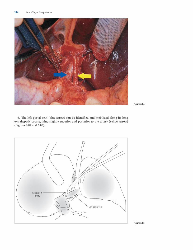
256 Atlas of Organ Transplantation
6. The left portal vein (blue arrow) can be identified and mobilized along its longextrahepatic course, lying slightly superior and posterior to the artery (yellow arrow)(Figures 6.84 and 6.85).
Figure 6.84
Figure 6.85

Liver Transplantation 257
7. The falciform ligament is pulled to the left during this part, and “feedback vessels”(black arrow) of the portal vein going to segment IV can be identified (Figure 6.86a).These should be divided with suture ligation (Figures 6.86b and 6.87).
Figure 6.86
Figure 6.87

258 Atlas of Organ Transplantation
8. The umbilical tape previously passed around the left hepatic vein can be nowbrought around the left lateral segment itself, passing in the ligamentum groove, andanteriorly excluding the artery and vein going to the left lateral segment. Pulling on thetwo ends of the umbilical tape lifts up the portion of liver to be transected, giving a goodguide for the plane of transection (Figure 6.88). This is known as the “hanging technique.”
Figure 6.88
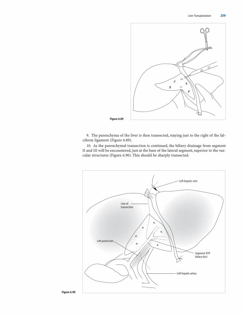
Liver Transplantation 259
Figure 6.89
9. The parenchyma of the liver is then transected, staying just to the right of the fal-ciform ligament (Figure 6.89).
10. As the parenchymal transection is continued, the biliary drainage from segmentII and III will be encountered, just at the base of the lateral segment, superior to the vas-cular structures (Figure 6.90). This should be sharply transected.
Figure 6.90

260 Atlas of Organ Transplantation
Figure 6.91
Figure 6.92
11. When the recipient team is ready, the graft can be removed by dividing the vas-cular structures (Figure 6.91). Clamps can be placed proximally on the left portal veinand left hepatic artery. The left hepatic vein is easiest taken with a laparoscopic staplingdevice.
12. The graft is flushed and given to the recipient team (Figure 6.92). The cut surfaceof the donor should be carefully inspected for any evidence of bleeding or bile leak. Oncethis is satisfactory, closure is performed.

Liver Transplantation 261
b) Recipient Procedure
1. A standard caval-preserving hepatectomy is performed while the donor team isprocuring the left lateral segment graft.
2. Creation of a large, patulous, hepatic venous anastomosis is important to maximizeoutflow of the graft. This is best performed by isolating the cava between two clamps asshown. A large triangular opening is created on the anterior surface of the cava, usuallyat the site of the left and middle hepatic vein orifices (Figure 6.93).
Figure 6.93
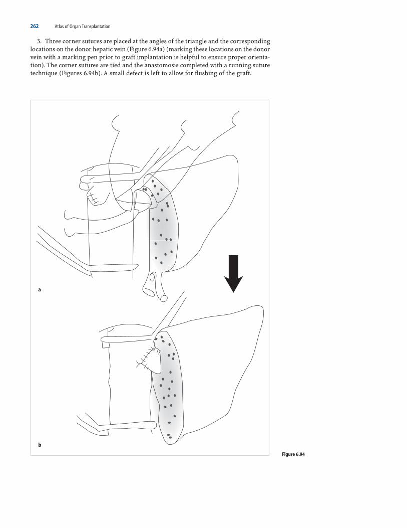
262 Atlas of Organ Transplantation
3. Three corner sutures are placed at the angles of the triangle and the correspondinglocations on the donor hepatic vein (Figure 6.94a) (marking these locations on the donorvein with a marking pen prior to graft implantation is helpful to ensure proper orienta-tion). The corner sutures are tied and the anastomosis completed with a running suturetechnique (Figures 6.94b). A small defect is left to allow for flushing of the graft.
Figure 6.94

Liver Transplantation 263
4. The portal venous anastomosis is completed next. Usually this can be done withoutthe use of a jump graft. Correct orientation of the donor and recipient vein is importantto ensure unobstructed flow.
5. The graft is then flushed to remove preservative solution from the graft. Afterflushing, the defect in the hepatic venous anastomosis is closed and the clamps removed.
6. The hepatic arterial anastomosis is completed next. Again, this can usually be donewithout the use of a “jump” graft (Figure 6.95). An operating microscope or high-powered loupes and fine interrupted sutures are helpful in creating this anastomosis.
Figure 6.95

264 Atlas of Organ Transplantation
7. A roux-en-Y loop is then created for the biliary anastomosis. The loop is broughtup through a retrocolic opening, and placed adjacent to the bile duct(s), so that a tension-free anastomosis can be performed (Figure 6.96a). It is helpful to perform this anasto-mosis over a small feeding tube, which can be used as an external stent (Figure 6.96b).
Living-Donor Liver Transplant: Adult Recipient
The use of living donors for liver transplant has been of tremendous benefit for pedi-atric recipients and has resulted in a significant decrease in the waiting-list mortalityrate for these patients. However, more than 90% of patients on the waiting list today areadults; the majority of deaths pretransplant occur in adults. Therefore, to have maximumimpact on the waiting list, living donors also need to be utilized for adult recipients. Forsuch recipients, a larger piece of the liver is required. The right lobe, the left lobe, andtwo left lobes from two donors have all been reported as potential grafts for adult recip-ients. However, most centers employ the right lobe of the donor for this procedure.
The preoperative evaluation of the potential living donor is an essential part of thetransplant process. A thorough medical evaluation is done to ensure that there are norisk factors that would contraindicate donation. This involves ruling out major diseaseprocesses that may involve the extrahepatic systems such as cardiac, respiratory, or renal.Biochemical markers of liver function and injury such as bilirubin, transaminases, andinternational normalized ratio (INR) need to be performed.Viral pathogens such as hep-atitis B and C need to be screened for.
Removal of the right lobe is a substantially bigger operative procedure than removalof the left lateral segment. A much larger volume of the liver is to be removed. There-fore, an important part of the preoperative evaluation is to make an accurate volumemeasurement of the donor’s liver. The purpose of this is twofold: (1) to make sure thatthe recipient is receiving enough liver tissue to sustain life, and (2) to make sure that thedonor has adequate residual liver volume to sustain life. For the recipient, one shouldtarget a GW/RW ratio of at least 0.8%. The donor should have at least 30% of his or hertotal liver volume left behind at the end of surgery.
Figure 6.96

Liver Transplantation 265
Besides the volumetric evaluation preoperatively, many centers also routinely employa liver biopsy for the evaluation of a potential right lobe donor. The purpose of this is torule out steatosis, and occult diseases of the liver. Other centers biopsy on a selectivebasis, depending on the presence or absence of risk factors such as obesity, elevatedtriglycerides or cholesterol levels, and abnormal imaging studies or liver function tests.
The choice of surgical procedure for adult living-donor transplant varies from centerto center, but most prefer the right lobe. Inflow is based on the right hepatic artery andright portal vein, while outflow is based on the right hepatic vein. Outflow issues tend to be more prominent with right lobe vs. left lateral segment grafts. Various methodshave been tried to improve outflow for these grafts including taking the middle hepaticvein with the right lobe, and reconstructing large venous tributaries using a vasculargraft.
The greatest concern with this procedure, in fact with any living-donor procedure, isthe risk of death to the donor. Unfortunately, there is some risk of mortality associatedwith any surgical procedure, but the magnitude of risk associated with this procedurehas been difficult to determine. As of January 31, 2003, 1158 adult LDLTs (ALDLTs) hadbeen performed in the U.S. and reported to UNOS (United Network for Organ Sharing).There had been three associated donor deaths (0.26%). Of these three deaths, two(0.17%) were early in the postoperative period and hence directly related to the surgery.The third death was a suicide at 2 years postdonation and likely not related to the dona-tion surgery. The number of deaths worldwide, however, is more difficult to determine,as there is no mandatory reporting. By some reports, there have been 14 or 15 deathsworldwide. Most centers cite a 0.5% risk of mortality associated with this procedure,though the cited rate may vary from 0.1% to 1.0% at different centers.
A number of different complications are possible, and have been reported in donors.The vast majority of these complications tend to occur in the early postoperative period(usually within 1 month postdonation). The reported incidence of complications in thedonor varies in the literature, but likely is in the 10% to 20% range. The most commonlyreported major complication involves the biliary system; a bile leak or stricture has beenreported in roughly 5% of the donors. Other reported complications include the needfor nonautologous blood, the need for a reoperation, and a major postoperative infec-tion. Overall, the risk of complications for right-lobe donors is felt to be significantlyhigher compared to that for left lateral segment or left-lobe donors.
While there is no obvious benefit for donors, there are advantages for recipients ofliving-donor transplants. The potential waiting-list mortality associated with waiting fora deceased donor is avoided. Additionally, patients can be transplanted before theydevelop far advanced liver disease associated with marked overall decompensation. Thedisadvantage for living-donor recipients, at least for adult recipients, is that there aresome data to suggest that outcomes may be inferior compared to whole-liver transplants,especially if the patients are matched for severity of liver disease. Living-donor recipi-ents also have been noted to have a higher incidence of surgical complications post-transplant as compared with whole-liver recipients. Partial grafts have smaller vessels,more complicated biliary reconstructions, and a cut surface, all of which make for a technically more challenging procedure and a higher incidence of surgical complica-tions. Most centers have reported a 15% to 20% incidence of early bile leaks after trans-plant and a 15% to 20% incidence of late biliary strictures. Those figures are significantlyhigher than generally reported for whole-liver recipients.
Certain subgroups of recipients especially may do worse after living-donor trans-plants. Critically ill adult recipients with advanced liver failure, high MELD (Model for End-Stage Liver Disease) scores, and numerous secondary complications have generallybeen reported to have very poor outcomes with this procedure. Such recipients haveminimal functional reserve and are probably ill-equipped to manage the lower hepato-cyte mass and the higher complication rate associated with partial transplants.
The recipient operation with LDLTs is not greatly different from whole-organdeceased-donor liver transplants. The hepatectomy is performed in a similar fashion, butthe recipient cava should be preserved in all such cases, because the graft will generallyonly have a single hepatic vein for outflow. This hepatic vein is then anastomosed directly

266 Atlas of Organ Transplantation
to the recipient’s preserved vena cava (Figure 6.97). Outflow can be augmented by includ-ing the middle hepatic vein with the graft or by reconstructing large segment V or VIII tributaries to the middle hepatic vein using a vascular conduit. Inflow to the graftcan be done by reconnecting the corresponding hepatic arterial and portal venousbranches. Finally biliary reconstruction is performed either by a duct-to-duct techniqueor with a hepaticojejunostomy.
Figure 6.97

Figure 6.99
Liver Transplantation 267
a) Donor Procedure
1. A bilateral subcostal incision is made. The falciform ligament is divided and thencontinued posteriorly until the anterior wall of the right hepatic vein (blue arrow) isvisualized (Figure 6.98). The groove between the right and middle hepatic veins shouldbe defined (Figure 6.99).
Figure 6.98

268 Atlas of Organ Transplantation
Figure 6.100
2. The right lobe of the liver is completely mobilized by division of the right trian-gular ligament and freeing of the bare area from the underlying retroperitoneum (Figure6.100). The right lateral border of the retrohepatic cava should be seen.
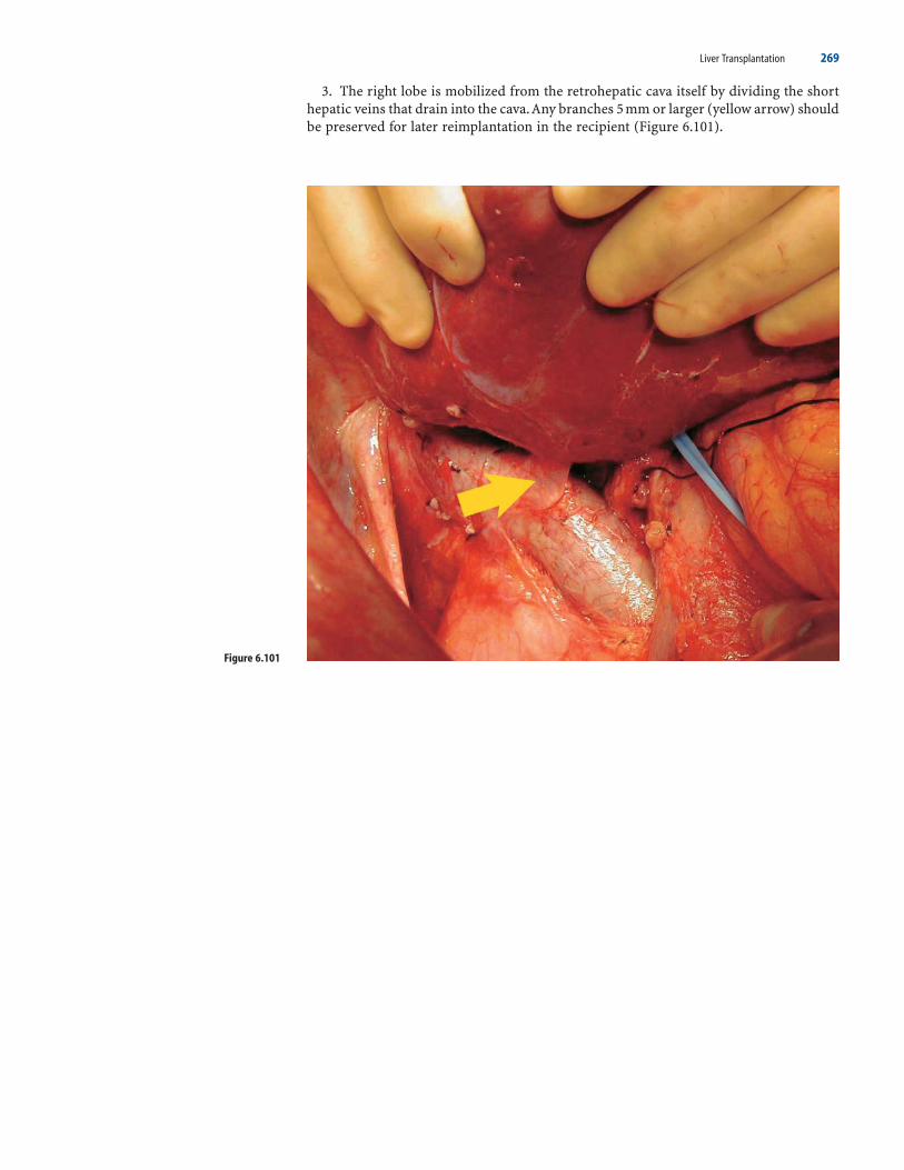
Liver Transplantation 269
Figure 6.101
3. The right lobe is mobilized from the retrohepatic cava itself by dividing the shorthepatic veins that drain into the cava.Any branches 5 mm or larger (yellow arrow) shouldbe preserved for later reimplantation in the recipient (Figure 6.101).

Figure 6.103
270 Atlas of Organ Transplantation
4. The undersurface of the right hepatic vein marks the superior extent of the dis-section. The hepatocaval ligament (blue arrow) is encountered just inferior to the righthepatic vein (Figures 6.102 and 6.103). This band of tissue attaching the posterior rightlobe to the inferior vena cava (broken blue line) is variable in its composition. It maycontain a sizable vein, may be composed of liver parenchyma, or may consist of justfibrous tissue. It should be divided between clamps and suture ligated in case there is avein within it.
Figure 6.102

Liver Transplantation 271
Figure 6.104
Figure 6.105
5. It should now be possible to encircle the right hepatic vein (blue arrow) and passan umbilical tape around it (Figures 6.104 and 6.105).

272 Atlas of Organ Transplantation
6. Attention is now turned toward the hilar dissection. A cholecystectomy is per-formed. The right hilar plate is taken down and the junction of the right and left hepaticducts (green arrow) is defined (Figure 6.106).
7. The right hepatic artery (yellow arrow) is identified close to where it enters intothe liver (Figure 6.107). It is then traced proximally to the point where it usually ispassing behind the common bile duct (CBD) (broken green line). It usually should not be dissected much proximal to this point so as not to risk causing vascular injury tothe CBD.
Figure 6.106
Figure 6.107

Liver Transplantation 273
Figure 6.108
Figure 6.109
8. The right portal vein is then identified. It is easiest to locate this vein by dissectingalong the posterior right edge of the hepatoduodenal ligament, behind the right hepaticartery (yellow arrow). The junction with the left portal vein (blue arrow) is clearlydefined so as to not compromise the latter when the right portal vein is eventuallydivided (Figure 6.108). There are usually one or two small branches from the right portalvein going to the caudate lobe. It is important to divide these veins (green arrow) to max-imize the length of the right portal vein.
9. With the right hepatic artery (HA) and right portal vein (PV) retracted inferiorly,the right hepatic duct (green arrow) can be encircled with a right-angle instrument anda radiologic marker of some form passed around it (Figure 6.109).

274 Atlas of Organ Transplantation
10. An intraoperative cholangiogram is now performed to delineate the biliaryanatomy. In the first cholangiogram, the right anterior and posterior sectoral ducts jointo form a common trunk, which then joins the left hepatic duct (Figure 6.110). This sit-uation should result in a single bile duct (green arrow) to the right lobe graft. In thesecond cholangiogram, the right posterior sectoral duct drains into the left hepatic duct(Figure 6.111). This will result in two separate bile ducts (green arrows) to reanastomosein the right lobe graft.
Figure 6.110
Figure 6.111

Liver Transplantation 275
11. Intraoperative ultrasound is then performed to outline the course of the middlehepatic vein (MHV) (Figure 6.112). This is correlated with the preoperative imagingstudy (Figure 6.113). We prefer to perform the parenchymal transection just to the rightof the MHV, leaving that structure with the donor. Others have described doing theparenchymal transection to the left of the MHV, leaving that structure with the rightlobe graft.
Figure 6.112
Figure 6.113
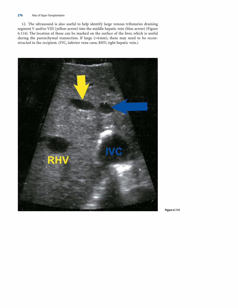
276 Atlas of Organ Transplantation
12. The ultrasound is also useful to help identify large venous tributaries drainingsegment V and/or VIII (yellow arrow) into the middle hepatic vein (blue arrow) (Figure6.114). The location of these can be marked on the surface of the liver, which is usefulduring the parenchymal transection. If large (>4 mm), these may need to be recon-structed in the recipient. (IVC, inferior vena cava; RHV, right hepatic vein.)
Figure 6.114

Liver Transplantation 277
13. The umbilical tape previously passed around the right hepatic vein is passedaround the entire right lobe (Figure 6.115a). The tape is passed inside the three righthilar structures (artery, vein, and hepatic duct) (blue arrows) (Figure 6.115b). The twoends of the umbilical tape can then be pulled up (black arrows), acting as a sling that helps to direct the line of parenchymal transection (Figure 6.115c). This is knownas the “hanging” technique (Figure 6.116).
Figure 6.115
Figure 6.116
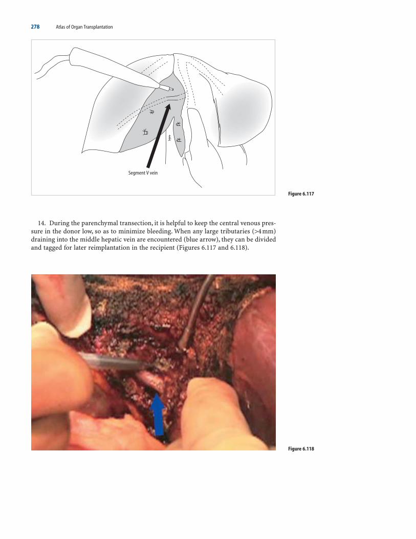
278 Atlas of Organ Transplantation
14. During the parenchymal transection, it is helpful to keep the central venous pres-sure in the donor low, so as to minimize bleeding. When any large tributaries (>4 mm)draining into the middle hepatic vein are encountered (blue arrow), they can be dividedand tagged for later reimplantation in the recipient (Figures 6.117 and 6.118).
Figure 6.117
Figure 6.118

Liver Transplantation 279
Figure 6.119
15. The parenchymal transection is completed, leaving the right lobe attached onlyby its vascular and biliary attachment (Figure 6.119).
16. The right hepatic duct is divided at the determined site based on the cholan-giogram. The corresponding biliary stump on the left side is closed, taking care not tonarrow the common hepatic duct (Figure 6.120).
Figure 6.120

280 Atlas of Organ Transplantation
17. We prefer to wait at this point until the recipient team is ready to receive the liver.This also allows for close inspection of the two pieces of the liver to ensure that they areboth adequately perfused (Figure 6.121).
Figure 6.121

Liver Transplantation 281
18. When the recipient team is ready, the vascular structures of the right graft aredivided. The right hepatic artery is taken first with a vascular clamp. The right portalvein is divided next, either using a vascular clamp or a linear stapling device for proxi-mal control. Regardless of how control is achieved, it is important that the stump of theright portal vein is closed in an anterior to posterior fashion, rather than a side-to-sidemanner. This ensures that there is no narrowing at the junction between the left andmain portal veins (Figures 6.122 and 6.123).
Figure 6.122
Figure 6.123

282 Atlas of Organ Transplantation
19. The right hepatic vein (blue arrow) is easiest to divide using a laparoscopic sta-pling device (Figures 6.124 and 6.125).
Figure 6.124
Figure 6.125

Liver Transplantation 283
20. The right lobe is removed and given to the recipient team. The cut surface of theleft lobe is carefully inspected for any evidence of bile leak or bleeding (Figure 6.126).The falciform ligament is reattached using a running suture to prevent torsion of theremaining left lobe. We prefer to leave a drain in place, but many centers do not. Theincision is then closed.
b) Recipient Procedure
1. The graft is flushed with preservative solution and weighed. If there are anysegment V or VIII veins that will need to be reimplanted (based on size being ≥5 mm),then a vein graft should be anastomosed onto them in an end-to-end fashion (yellowarrow) (Figure 6.127). Options include using cryopreserved vein, recipient’s saphenousor superficial femoral vein, or using a segment of vein from the resected liver (such asthe left portal vein). A saphenous vein graft has been used in this case.
Figure 6.126
Figure 6.127

284 Atlas of Organ Transplantation
2. A standard inferior vena cava–sparing hepatectomy is done in the recipient; theliver is mobilized off the underlying cava and all short hepatic veins are divided (Figure6.128).
Figure 6.128

Liver Transplantation 285
3. The orifices of the three hepatic veins are stapled shut (Figure 6.129), and the liveris removed completely (Figure 6.130).
Figure 6.129
Figure 6.130
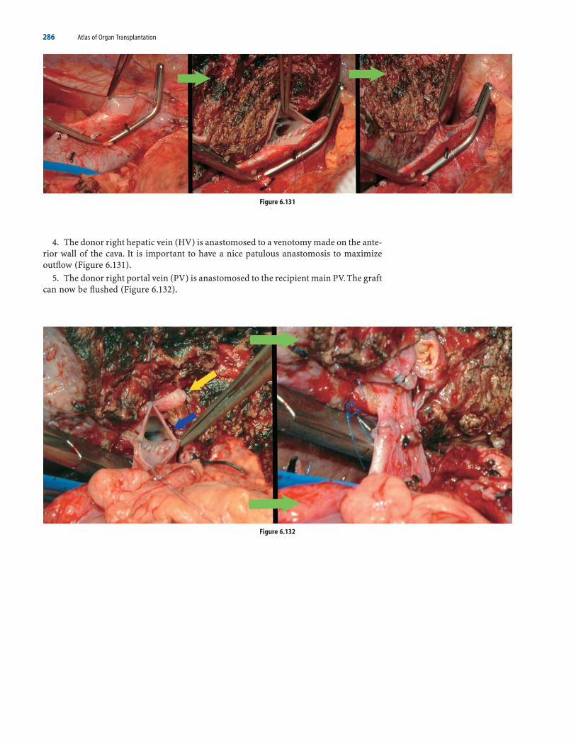
286 Atlas of Organ Transplantation
4. The donor right hepatic vein (HV) is anastomosed to a venotomy made on the ante-rior wall of the cava. It is important to have a nice patulous anastomosis to maximizeoutflow (Figure 6.131).
5. The donor right portal vein (PV) is anastomosed to the recipient main PV. The graftcan now be flushed (Figure 6.132).
Figure 6.131
Figure 6.132

Liver Transplantation 287
6. Any middle hepatic vein tributaries that had been reconstructed can be reim-planted. This can be done by anastomosing the end of the vein graft to the recipient cava(Figures 6.133 and 6.134).
Figure 6.133
Figure 6.134

288 Atlas of Organ Transplantation
7. The donor right hepatic artery (HA) is anastomosed to the recipient right HA usingfine (7.0) interrupted sutures (Figures 6.135 and 6.136).
8. Finally, the bile ducts are reconstructed. If the recipient bile duct reaches the donorbile duct without tension, a duct-to-duct anastomosis can be done (yellow arrow). Even
Figure 6.135
Figure 6.136

Liver Transplantation 289
when there are two ducts in the donor graft, as long as the recipient ducts have beendivided high in the hilum, a duct-to-duct anastomosis is possible to the native right and left hepatic ducts (Figure 6.137). It is helpful to perform this anastomosis over aninternal stent.
9. Alternatively, a Roux-en-Y loop can be used if it is not possible to use the recipi-ent’s own ducts. Using an external biliary stent is helpful when performing this anasto-mosis, and allows for easy imaging afterward (Figure 6.138).
Figure 6.137
Figure 6.138

290 Atlas of Organ Transplantation
Surgical Complications After Liver Transplant
The incidence of surgical complications after liver transplant remains significantlyhigher than that after kidney transplant. This is not surprising given the increased com-plexity and magnitude of the operative procedure and, often, the more critical conditionof the recipients. Most surgical complications occur early posttransplant, usually withinthe first few weeks.
Risk factors vary depending on the exact complication, but generally partial-livertransplants (i.e., living-donor and deceased-donor splits) have a higher technical com-plication rate than whole-liver transplants. Recipients who are significantly debilitatedat the time of their transplant and critically ill, also tend to have increased complicationspostoperatively, especially infection related.While numerous potential complications arepossible after transplant, surgical complications related directly to the operation mostcommonly include postoperative hemorrhage and problems with either the vascular orthe biliary anastomoses. Common complications include the following:
a) Hemorrhage
Bleeding is common in the postoperative period and is usually multifactorial. It may becompounded by an underlying coagulopathy resulting from deficits in coagulation,fibrinolysis, and platelet function. Blood loss should be monitored via the abdominaldrains; hemoglobin and central venous pressure should be measured serially. If bleedingpersists despite correction of coagulation deficiencies, an exploratory relaparotomyshould be performed.
b) Hepatic Artery Complications
The incidence of hepatic arterial vascular complications is reportedly 5% to 10%. Throm-bosis is the most common early event; stenosis and pseudoaneurysm formation occurlater. Hepatic artery thrombosis (HAT) has a reported incidence of about 3% in adultsand about 5% to 10% in children. Partial-liver transplant recipients have a higher inci-dence than whole-liver recipients. After HAT, liver recipients may be asymptomatic, orthey may develop severe liver failure secondary to extensive necrosis. Doppler ultrasoundevaluation is the initial investigation of choice, with more than 90% sensitivity andspecificity. Urgent exploration with thrombectomy and revision of the anastomosis areindicated; if the diagnosis is made early, up to 70% of grafts can be salvaged. If hepaticnecrosis is extensive, a retransplant is indicated. A HAT also may present in a less dra-matic fashion. Thrombosis may render the common bile duct ischemic, resulting in alocalized or diffuse bile leak from the anastomosis, or a more chronic, diffuse biliarystricture.
c) Portal Vein Complications
Thrombosis of the portal vein is far less frequent compared to its arterial counterpart.Liver dysfunction, tense ascites, and variceal bleeding may occur. Doppler evaluationshould establish the diagnosis. If thrombosis is diagnosed early, operative thrombectomyand revision of the anastomosis may be successful. If thrombosis occurs late, liver func-tion is usually preserved, due to the presence of collaterals; a retransplant is then unnec-essary, and attention is diverted toward relieving the left-sided portal hypertension.
d) Biliary Complications
Biliary complications continue to be significant, occurring in 15% to 35% of liver trans-plants. Such complications manifest either as leaks or as obstructions. Again, partial-
Liver Transplantation 291
liver transplant recipients seem to be at higher risk, for both leaks and strictures. Leakstend to occur early postoperatively and often require surgical repair, while obstructionsusually occur later and can be managed with radiologic or endoscopic techniques. Clin-ical symptoms of a bile leak include fever, abdominal pain, and peritoneal irritation.Ultrasound may demonstrate a fluid collection; cholangiography is required for diag-nosis. Some leaks may be successfully managed by endoscopic placement of a biliarystent. If the leak does not respond to stent placement or if the patient is systemically ill,a relaparotomy is warranted.
Biliary stricture occurs later in the postoperative period and is most common at theanastomotic site, likely related to local ischemia. Strictures usually manifest as cholan-gitis, cholestasis, or both processes. Initial treatment involves balloon dilatation or stentplacement across the site of stricture, or both. If these initial options fail, surgical revi-sion is required.
e) Wound Complications
Common problems related to the wound are infection, hematoma, and seroma. Woundhematomas can result from the presence of huge collateral veins in the abdominal wall.Wound infections usually present after postoperative day 5. Treatment consists ofopening the wound, changing the dressings, and allowing healing by secondary inten-tion. If significant cellulitis or systemic symptoms are present, intravenous (IV) antibi-otics should be administered. Late wound complications generally consist of incisionalhernias, which may present at any time after transplant. Repair is warranted if the patientis symptomatic.
f) Primary Nonfunction
Primary nonfunction is a devastating complication, and the attendant mortality rate is>80% without a retransplant. By definition, this syndrome results from poor or nohepatic function from the time of the transplant procedure. The incidence in mostcenters is about 3% to 5%. Donor factors associated with primary nonfunction includeadvanced age, increased fat content of the donor liver, longer donor hospital stay beforeorgan procurement, prolonged cold ischemia time, and reduced-size grafts. Conditionsthat may mimic primary nonfunction must be ruled out, such as HAT, accelerated acuterejection, and severe infection. Intravenous prostaglandin E1 has some useful effect andshould be administered to recipients with suspected primary nonfunction. They shouldbe listed for an urgent retransplant.
Similar to primary nonfunction is small-for-size syndrome, seen in recipients ofpartial-liver transplants. Presentation is with liver dysfunction, manifesting as cholesta-sis, coagulopathy, and failure to thrive. It is believed that portal hyperperfusion causesdamage to the relatively small graft. Some studies have suggested that splenic artery lig-ation or portal shunts may be beneficial in this scenario, but experience is limited.
Selected Readings
Bismuth H. Surgical anatomy and anatomical surgery of the liver. World J Surg 1982;6:3–9.Brown R Jr, Russo M, Lai M, et al. A survey of liver transplantation from living adult donors in the United
States. N Engl J Med 348(9):818–825.Brown A, Williams R. Long-term postoperative care. In: Maddrey WC, Schiff ER, Sorrell MF, eds. Transplanta-
tion of the Liver. Lippincott Williams & Wilkins, 2001.Couinaud C. Le Foie: Etudes Anatomiques et Chirurgicales. Paris: Masson, 1957.Everson GT, Kam I. Immediate postoperative care. In: Maddrey WC, Schiff ER, Sorrell MF, eds. Transplantation
of the Liver. Philadelphia: Lippincott Williams & Wilkins, 2001.Fan ST, Lo CM, Liu CL. Technical REF1inement in adult-to-adult living donor liver transplantation using right
lobe graft. Ann Surg 2000;231(1):126–131.

292 Atlas of Organ Transplantation
Humar A, Gruessner R. Critical care of the liver transplant recipient. In: Intensive Care Medicine, 4th ed. RippeJM, Irwin RS, Fink MP, Cerra FB, eds. Philadelphia: Lippincott-Raven, 1998.
Icoz G, Kilic M, Zeytunlu M, et al. Biliary reconstructions and complications encountered in 50 consecutiveright-lobe living donor liver transplantations. Liver Transplant 2003;9(6):575–580.
Kim-Schluger L, Florman S, Schiano T, et al. Quality of life after lobectomy for adult liver transplantation.Transplantation 73(10):1593–1597.
Kiuchi T, Inomata Y, Uemoto S, et al. Living-donor liver transplantation in Kyoto. Clin Transplant 1997;191–198.Lee T, et al. Split-liver transplantation using the left lateral segment: a collaborative sharing experience between
two distant centers. Am J Transplant 2005;5:1646–1651.Marcos A, Fisher RA, Ham JM, et al. Right lobe living donor liver transplantation. Transplantation 1999;
68(6):798–803.Nashan B, Lueck R, Becker T, Schlitt MH, Grannas G, Klempnauer J. Outcome of split liver transplantation using
ex-situ or in-situ splits from cadaver donors. Joint meeting of the International Liver Transplantation Society,European Liver Transplantation Association, and Liver Intensive Care Group of Europe, Berlin, Germany, July11–13, 2001.
Neuhaus P, Blumhardt G, Bechstein WO, Steffen R, Platz KP, Keck H. Technique and results of biliary recon-struction using side-to-side choledococholedochostomy in 300 orthotopic liver transplants. Ann Surg1994;219(4):426–434.
Renz JF, Busuttil RW. Adult-to-adult living-donor liver transplantation: a critical analysis. Semin Liver Dis2000;20(4):411–424.
Reyes J, Gerber D, Mazariegos GV, et al. Split-liver transplantation: a comparison of ex vivo and in situ tech-niques. J Pediatr Surg 2000;35:283–289; discussion 289–290.
Rogiers X, Malago M, Gawad K, et al. In situ splitting of cadaveric livers. The ultimate expansion of a limiteddonor pool. Ann Surg 1996;224:331–334.
Shah SA, Grant DR, Greig PD, et al. Analysis and outcomes of right lobe hepatectomy in 101 consecutive livingdonors. Am J Transplant 2005;5(11):2764–2769.
Strong RW, Lynch SV, Ong TN, et al. Successful liver transplantation from a living donor to her son. N Engl JMed 1990;322:1505–1507.
Surman OS. The ethics of partial liver donation. N Engl J Med 346(14):1038.Trotter J, Talamantes M, McClure M, et al. Right hepatic lobe donation for living donor liver transplantation:
impact on donor quality of life. Liver Transplant 7(6):485–493.Trotter JF, Wachs M, Everson GT, Kam I. Adult-to-adult transplantation of the right hepatic lobe from a living
donor. N Engl J Med 2002;346(14):1074–1082.Umeshita K, Fujiwara K, Kiyosawa K, et al. Japanese Liver Transplantation Society. Operative morbidity of living
liver donors in Japan. Lancet 2003;362(9385):687–690.Wachs ME, Bak TE, Karrer FM, et al. Adult living donor liver transplantation using a right hepatic lobe. Trans-
plantation 1998;66,1313–1316.Washburn K, et al. Split-liver transplantation: results of statewide usage of the right trisegmental graft. Am J
Transplant 2005;5:1652–1659.Wiesner RH. Current indications, contraindications, and timing for liver transplantation. In: Busutill RW,
Klintmalm GB, eds. Transplantation of the Liver. Philadelphia: WB Saunders, 1996.Yersiz H, et al. One-hundred in situ split-liver transplantations: a single center experience. Ann Surg 2003;
238(4):496–505.Zimmerman MA, Trotter JF. Living donor liver transplantation in patients with hepatitis C. Liver Transplant
9(11): S52–S57.

7Intestinal and MultivisceralTransplantationThomas M. Fishbein and Cal S. Matsumoto
Isolated Intestine Transplanta) Donor Procedureb) Back-Table Procedurec) Recipient ProcedureCombined Liver–Intestine Transplanta) Donor Procedureb) Back-Table Procedurec) Recipient ProcedureMultivisceral Transplanta) Donor Procedureb) Recipient ProcedureLiving-Donor Small-Bowel Transplanta) Surgical ProcedurePerioperative Management
Intestinal transplantation has evolved from an experimental procedure with limitedsuccess to standard of care for patients with intestinal and parenteral nutrition failure,achieving outcomes commensurate with other solid-organ transplants.1,2 Paramount toachieving the success of intestinal and multivisceral organ transplant procedures hasbeen the refinement of donor organ procurement and transplantation techniques.3 Earlygraft failures and deaths due to technical and donor-related complications have beenminimized using the techniques described here, leaving the current challenge largely ofoptimizing immunologic and infection management strategies.4,5 Techniques depictedhere are those of choice in our experience, while mention is made of alternative tech-niques that may be preferred by others. Because patients requiring intestinal transplan-tation often present with multiple organ failures and require multiorgan transplantation,modification of these techniques may be required on an individual basis. Addition ofrenal allotransplantation, inclusion of the colon in a small-bowel or multivisceral graft,and modified multivisceral transplantation with preservation of the native liver are someof the common modifications. Intimate description of all possible techniques is beyondthe scope of the chapter.
The three most common types of allografts involving the small intestine are isolatedintestinal transplantation, combined liver–small bowel transplantation, and multivis-ceral transplantation. These are described in turn, detailing our commonly preferredmethods for both donor and recipient surgical techniques.
293

294 Atlas of Organ Transplantation
Isolated Intestine Transplant
a) Donor Procedure
1. The standard sternum to pubis incision is made, taking care not to injure the bowelupon entering the abdomen. On many occasions, due to either the nature of injury orthe preoperative state of the donor, the intestine may be dilated or edematous and mayclosely abut the abdominal wall. A nasogastric tube should decompress the stomach andupper gastrointestinal tract. We additionally prefer to decontaminate the allograftthrough instillation of antibiotics into the lumen through the indwelling nasogastrictube. Immediately upon entering the abdomen, another dose of selective gut decontam-inant may be administered, and passed through the pylorus into the small intestinalgraft.
A broad Kocher and Cattell maneuver are each performed, and the aorta and inferiorvena cava are exposed behind the intestinal mesentery up to the superior mesentericartery (Figure 7.1).
2. The ligament of Treitz is broadly mobilized from the retroperitoneum and aorta,mobilizing the distal duodenum and proximal jejunum. The abdominal aorta is encir-cled distally for eventual aortic cannulation and flush. The gastrocolic omentum is
Figure 7.1

Intestinal and Multivisceral Transplantation 295
Figure 7.2
Figure 7.3
divided, and the left colon is mobilized (red arrow) (Figure 7.2). The mesentery of thecolon is then dissected, exposing right, middle and left colic vessels. Exposure of the colicvessels (blue arrow) facilitates rapid removal of the colon immediately prior to hepar-inization and cannulation.
3. A medial visceral rotation mobilizes the tail of the pancreas (yellow arrow) withthe spleen (blue arrow), exposing the left side of the aorta and origin of the mesentericvessels (Figure 7.3). As with standard pancreas procurement, the pylorus is transectedwith a linear stapler, leaving sufficient length to invert the staple line with another layerof suture. The stomach is rotated laterally out of the abdominal field after ligation of theleft gastric artery, separation of the stomach from the greater omentum, and division ofthe short gastric vessels to the spleen. The use of the inferior mesenteric vein (IMV) forflush during intestinal procurement is discouraged, as high flow and pressure in the IMVflush may decrease intestinal outflow through the superior mesenteric vein. In multivis-ceral graft procurement, care must be taken to avoid injury to the left gastric artery, whileattachments of the stomach to the colon via the greater omentum are still divided.
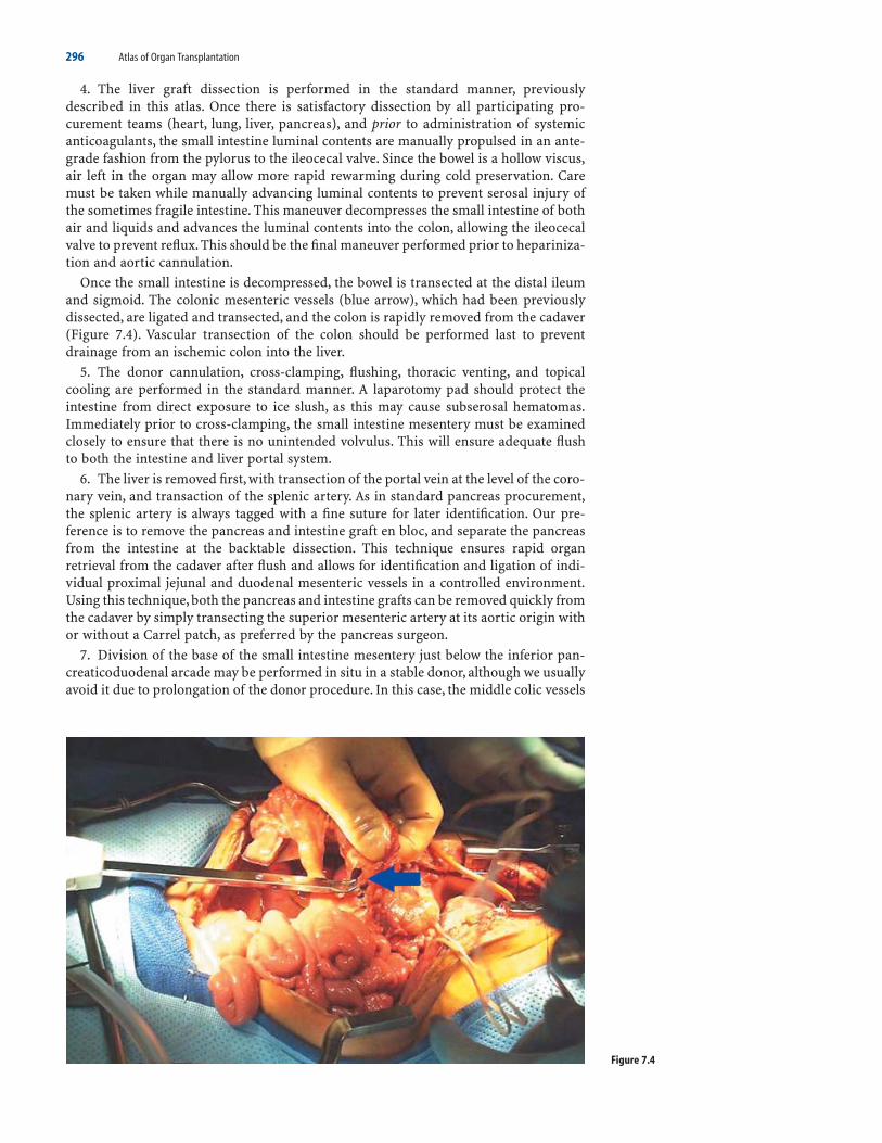
296 Atlas of Organ Transplantation
4. The liver graft dissection is performed in the standard manner, previouslydescribed in this atlas. Once there is satisfactory dissection by all participating pro-curement teams (heart, lung, liver, pancreas), and prior to administration of systemicanticoagulants, the small intestine luminal contents are manually propulsed in an ante-grade fashion from the pylorus to the ileocecal valve. Since the bowel is a hollow viscus,air left in the organ may allow more rapid rewarming during cold preservation. Caremust be taken while manually advancing luminal contents to prevent serosal injury ofthe sometimes fragile intestine. This maneuver decompresses the small intestine of bothair and liquids and advances the luminal contents into the colon, allowing the ileocecalvalve to prevent reflux. This should be the final maneuver performed prior to hepariniza-tion and aortic cannulation.
Once the small intestine is decompressed, the bowel is transected at the distal ileumand sigmoid. The colonic mesenteric vessels (blue arrow), which had been previouslydissected, are ligated and transected, and the colon is rapidly removed from the cadaver(Figure 7.4). Vascular transection of the colon should be performed last to preventdrainage from an ischemic colon into the liver.
5. The donor cannulation, cross-clamping, flushing, thoracic venting, and topicalcooling are performed in the standard manner. A laparotomy pad should protect theintestine from direct exposure to ice slush, as this may cause subserosal hematomas.Immediately prior to cross-clamping, the small intestine mesentery must be examinedclosely to ensure that there is no unintended volvulus. This will ensure adequate flushto both the intestine and liver portal system.
6. The liver is removed first, with transection of the portal vein at the level of the coro-nary vein, and transaction of the splenic artery. As in standard pancreas procurement,the splenic artery is always tagged with a fine suture for later identification. Our pre-ference is to remove the pancreas and intestine graft en bloc, and separate the pancreasfrom the intestine at the backtable dissection. This technique ensures rapid organretrieval from the cadaver after flush and allows for identification and ligation of indi-vidual proximal jejunal and duodenal mesenteric vessels in a controlled environment.Using this technique, both the pancreas and intestine grafts can be removed quickly fromthe cadaver by simply transecting the superior mesenteric artery at its aortic origin withor without a Carrel patch, as preferred by the pancreas surgeon.
7. Division of the base of the small intestine mesentery just below the inferior pan-creaticoduodenal arcade may be performed in situ in a stable donor, although we usuallyavoid it due to prolongation of the donor procedure. In this case, the middle colic vessels
Figure 7.4

Intestinal and Multivisceral Transplantation 297
are divided, and the ligament of Treitz is widely mobilized from over the aorta and theinferior margin of the pancreas. The mesentery may be isolated by placing the entiresmall bowel over a moist laparotomy pad after removal of the colon. The jejunum maybe divided with a stapling device approximately 10 cm distal to the ligament, and thesuperior mesenteric vein is identified laterally in the mesentery. Fine ligation of all lym-phatic structures is performed, isolating the superior mesenteric vein and artery. Theartery will usually give off a proximal jejunal arcade that requires division. Additionally,care must be taken not to injure the main jejunal vein draining the proximal allograft,as this usually runs posterior to the artery, joining the ileal vein to form the superiormesenteric high in the mesentery.
The isolated intestine donor operation historically precluded the use of the pancreasfrom the same donor; however, with close coordination between liver, pancreas, andintestinal procurement teams, successful procurement of all of these individual organscan be safely achieved.10 The notable exception is the case in which the donor has areplaced right hepatic artery arising low on the superior mesenteric artery.
b) Back-Table Procedure
1. In the event that the pancreas is not being transplanted, the back-table dissectionbegins at the level of the portal vein. Stay sutures are placed on the portal vein and cuffof superior mesenteric artery (Figure 7.5)
Figure 7.5

298 Atlas of Organ Transplantation
2. The portal vein dissection begins at the level of transection. Small pancreaticvenous tributaries are identified and ligated. Care should be taken to preserve the splenicvein (Figure 7.6), as this provides a useful conduit for flush of preservation solution fromthe allograft at the time of reperfusion. (SMV, superior mesenteric vein; IMV, inferiormesenteric vein.)
Figure 7.6
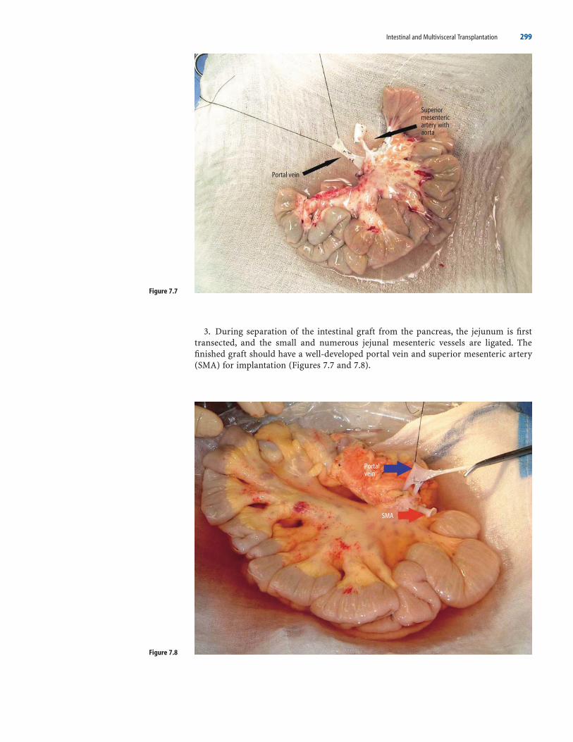
Intestinal and Multivisceral Transplantation 299
3. During separation of the intestinal graft from the pancreas, the jejunum is firsttransected, and the small and numerous jejunal mesenteric vessels are ligated. Thefinished graft should have a well-developed portal vein and superior mesenteric artery(SMA) for implantation (Figures 7.7 and 7.8).
Figure 7.7
Figure 7.8

Figure 7.9
300 Atlas of Organ Transplantation
4. To facilitate the graft blood flush, a small cannula may be placed in the splenic vein(blue arrow) for ease in flush measurement. We prefer the use of a Sundt carotidendarterectomy shunt (red arrow), which is used to allow egress of the preservation solu-tion prior to opening the venous clamp (Figure 7.9). The shunt is then removed, venousoutflow clamp opened, and the splenic stump ligated.
5. If the pancreas is also to be transplanted as a whole-organ allograft, the mesenteryis isolated upon a moist lap pad, and the mesenteric vessels are isolated below the infe-rior pancreaticoduodenal artery and vein, which provide necessary flow to the pancreasallograft. The middle colic vessels and first jejunal arcade usually must be ligated. Careshould be taken not to injure the major jejunal vein, which usually courses behind thesuperior mesenteric artery to join the ileocolic vein high in the base of the small bowelmesentery. Small vascular clamps are placed on the superior mesenteric artery and veinafter the flush, and these vessels are transected. The pancreatic side is individually over-sewn with fine nonabsorbable suture and the bowel graft is again flushed on the backtable. The pancreas and liver are then removed in the usual fashion. In the event that anaberrant right hepatic artery or aberrant common hepatic artery is encountered fromthe SMA, care must be taken to avoid injury to these vessels and the SMA must be divideddistal to the origin of the aberrant vessel. When this is encountered, good-quality iliacor common femoral artery and vein extension grafts are required for implantation ofthe small bowel graft.
c) Recipient Procedure
Isolated intestinal transplantation is usually described by the technique of vascularreconstruction used for venous drainage, being either “mesenteric/portal” or “systemic.”The surgical approach is determined by the underlying recipient intestinal disease, thehealth of the recipient’s liver, and anatomic considerations that may influence thesurgeon.11 This is usually accomplished through a generous midline incision, althoughin babies less than 10 kg we sometimes prefer a transverse incision if one has previouslybeen made.

Intestinal and Multivisceral Transplantation 301
We prefer to use mesenteric drainage for patients in whom the native small bowel isin place and will be removed at the time of transplantation. In cases of extreme shortbowel, the superior mesenteric vessels carry little flow, are usually quite small and maynot accommodate sufficient blood supply for the transplanted bowel; in these cases weusually prefer systemic vascular reconstruction.
Mesenteric Vascular Reconstruction
1. For candidates receiving an isolated intestinal allograft for functional disorders(infantile diarrhea or motility disorder), the native small bowel is usually in place. In thisinstance, blood flow through the mesenteric vessels is preserved, and they are of ade-quate caliber and quality for supply to the graft. Implantation, therefore, can be per-formed using these vessels prepared in the same manner as for isolated intestinal donorprocurement. The jejunum is divided 10 to 20 cm distal to the ligament of Treitz, whichis broadly mobilized. The arcade to the proximal jejunum is preserved. The recipientbowel is suspended by the root of the mesentery after disconnection at the proximaljejunum and colon, enabling fine dissection of the mesenteric root (yellow arrow), andisolation of the superior mesenteric artery and vein (Figure 7.10).
Figure 7.10

302 Atlas of Organ Transplantation
2. If the colon is intact, the middle colic vessels are ligated, the right and transversecolon mobilized, and the left transverse or descending colon divided. We always mobi-lize or remove the splenic flexure of the colon to assure endoscopic access to the ileumfrom below after closure of the stoma. The superior mesenteric vein (SMV, blue arrow)is located lateral to the superior mesenteric artery (SMA, yellow arrow) (Figure 7.11).Dissection of the base of the mesentery is undertaken to skeletonize these vessels. Thesetissues should all be ligated to avoid lymphatic or chylous ascites after transplantation.12
3. Small vascular clamps may be used to control the superior mesenteric artery (SMA)and vein (SMV), dividing them distally and preserving long cuffs for anastomosis(Figure 7.12). The proximal native jejunum should be preserved with the proximaljejunal arcade of the SMA for anastomosis to the donor jejunum.
Figure 7.11
Figure 7.12

Intestinal and Multivisceral Transplantation 303
4. In some cases, placement of iliac or femoral arterial and venous conduits onto thegraft vessels will be required (Figure 7.13). We have found that this facilitates trans-plantation when the pancreas has been procured for transplantation, as the graft mesen-teric vessels will be shorter. They are best sewn in place prior to bringing the allograftinto the field for implantation. Usually, either a short portion of external iliac orsuperficial femoral artery and vein are of appropriate caliber for anastomosis to the supe-rior mesenteric vessels. The weight of the small bowel allograft may lead to tension overthe duodenal sweep and predispose the graft to settle inferiorly toward the pelvis, sowhen using mesenteric reconstruction, attention should be paid to leaving generouslength on the mesenteric vessels. Again, the main SMV should be used above theconfluence of jejunal and ileal branches, which usually is quite close to the duodenalsweep.
In patients with short-bowel syndrome, the superior mesenteric vein sometimescannot be exposed, as the base of the mesentery contracts after multiple prior resections.The venous anastomosis may be performed to the lateral wall of the portal vein in pig-gyback fashion, dissecting this vein free from the posterior porta hepatis, and placingan extension graft of iliac vein onto this in the end-to-side fashion.13 We have only rarelyemployed this technique, and we discourage its routine use, as outcomes with systemicdrainage are equivalent and accomplished with greater ease. However, it may be usefulin cases of inferior vena cava thrombosis, where the alternative is placement of a venousextension graft on the suprarenal inferior vena cava, discussed below.
Figure 7.13

304 Atlas of Organ Transplantation
Figure 7.14
5. The bowel is then brought to the field after extension grafts are placed on the recip-ient vessels, and the base of the mesentery placed in a transverse plane aligning the donorand recipient vessels. The assistant is best positioned with a cold moist lap pad to bothflatten the base of the mesentery as well as prevent bowel loops from entering the field,giving upward traction on the bowel allograft during anastomosis. The vessels are anas-tomosed end to end (Figure 7.14).

Intestinal and Multivisceral Transplantation 305
6. Reperfusion is then accomplished without heparinization. Upon reperfusion, bloodand preservation solution is flushed out (blue arrow) prior to removal of the superiormesenteric vein clamp (Figure 7.15). This is facilitated by the previously placed cannulain the graft splenic vein, or through the superior mesenteric vein anastomosis.
7. The superior mesenteric vein clamp is then removed and venous return is reestab-lished (blue arrow, Figure 7.16).
Figure 7.15
Figure 7.16

Figure 7.17
306 Atlas of Organ Transplantation
8. Fixation of the base of the mesentery transversely and without kinking of thevessels is critical to avoid volvulus or traction on the mesenteric vessels (Figure 7.17).
Systemic Vascular Reconstruction
This technique is more commonly employed for patients with short-bowel syndrome,particularly those with total or near-total loss of jejunum and ileum. In this disease state,the mesenteric vessels are often small in caliber, lacking good inflow. However, this is notalways the case, and when a smaller donor with commensurately small vessels is beingused, mesenteric drainage may still be used.

Intestinal and Multivisceral Transplantation 307
Figure 7.18
1. The recipient operation requires exposure of the infrarenal aorta for anastomosisof the arterial graft (Figure 7.18). The limits of dissection include the left renal veinabove, and the inferior mesenteric artery below.
2. The inferior vena cava and aorta are clamped in preparation for systemic vascularanastomosis (Figure 7.19). The dissection may proceed below the inferior mesentericartery, and this vessel controlled with a fine clip during anastomosis.
Figure 7.19

308 Atlas of Organ Transplantation
3. The arterial graft must be performed in a tension-free manner, as the weight of thebowel filled with secretions can lead to traction from the aorta to the pelvis or lead todecreased flow and thrombosis. The venous anastomosis is end-to-side to the anteriorwall of the vena cava, as with a portocaval shunt (Figure 7.20). This is best accomplishedafter removal of an ellipse of vena cava to avoid narrowing. We prefer aortic inflow andcaval drainage for patients who have demonstrated significant liver cholestasis, fibrosis,or ultrashort-bowel syndrome.14 It is easy to accomplish, and in cases where intestinaltransplantation is indicated due to reversible progressive liver disease, avoids drainageof the bowel graft into the possibly high-pressure portal circulation. The bowel graft is again oriented with the transverse mesentery parallel to the plane of the retroperi-toneum, and sewn either directly to the aorta and vena cava, or as is our preference, toshort extension grafts already placed to these vessels. Prior placement of extension graftsroutinely makes implantation easy, not allowing loops of allograft small intestine toobscure the field of anastomosis. The mesentery is then fixed to the retroperitoneum toavoid internal hernia, volvulus, or traction on the vessels, care being taken to avoid theureters. Systemic drainage has not been shown to yield inferior nutritional results, assome had initially predicted.10,15,16
4. Another technique is required when systemic drainage is being used in a patientwith thrombosis of the inferior vena cava. This is not infrequently encountered, due tomultiple femoral accesses for parenteral nutrition. In this case, ligation and division ofa few low caudate veins allows placement of an extension graft of iliac or femoral veinon the suprarenal vena cava. This is accomplished with placement of a partially occlud-ing clamp, removal of an ellipse of anterior caval wall, and placement of an extensiongraft. The graft is tunneled behind the mobilized duodenum and head of pancreas, tolie next to the infrarenal aortic anastomosis. We have found this easier to accomplishthan piggyback drainage to the portal vein.
Figure 7.20

Intestinal and Multivisceral Transplantation 309
Figure 7.21
5. Enteral continuity is established proximally and distally in conjunction with anileostomy, using either a loop-diverting ileostomy or a proximal ileocolostomy in a“chimney” fashion to provide access for surveillance allograft biopsy (Figure 7.21). Thesestomas also allow future ileostomy closure to be performed without a full laparotomy.Placement of tubes for intestinal access can be accomplished with gastric, jejunal, orcombined tubes. These are placed to avoid prolonged need for nasogastric suction andfacilitate early feeding.

310 Atlas of Organ Transplantation
Combined Liver–Intestine Transplant
a) Donor Procedure
1. Donor management and operation begin as for isolated intestinal transplantation.17
The gallbladder is incised and the biliary tract is flushed with saline. After broad Cattelland Kocher maneuvers, the tail of the pancreas and spleen are mobilized from theretroperitoneum, exposing the left lateral aspect of the aorta. Care is taken to avoid injuryof the superior mesenteric vessels or the celiac axis (Figure 7.22).
The ligament of Treitz is widely mobilized. The proximal duodenum is then dissectedat the level of the pylorus and divided with a stapling device. Care must be taken duringthis process to inspect the gastrohepatic ligament to avoid injury to an aberrant lefthepatic artery if it is present.
2. After advancement of intraluminal contents distally, removal of the colon and sys-temic heparinization, the flush is performed with only aortic cannulation. Suprahepaticcaval venting is performed.
Figure 7.22

Intestinal and Multivisceral Transplantation 311
3. By retracting medially the spleen, pancreas and the small bowel in a cold lap pad,a Carrel patch or circumferential segment of aorta can then easily be taken, containingthe origins of the celiac and superior mesenteric arteries. The plane is anterolateral onthe left side of the aorta, and there is usually extensive mesenteric nervous plexus presentin this tissue. Once this is divided, the aortic patch or segment can be clearly divided.Care must be taken not to injure the takeoff of renal arteries. If the kidneys are not beingused (which is often the case with small pediatric donors), the entire aorta may beremoved with the graft. We remove the entire aorta from iliac bifurcation to proximaldescending aorta in the small pediatric donor; this requires the diaphragm to be split atthe time of extraction. Then, the entire pancreas, spleen, small intestine and liver areremoved en-bloc with intact inferior vena cava and duodenum (Figure 7.23).
Figure 7.23

312 Atlas of Organ Transplantation
b) Back-Table Procedure
1. The back-table procedure requires cleaning of mesenteric and celiac plexus withligation of vascular tissues surrounding these vessels. The celiac axis and superiormesenteric artery are exposed. Eventually the supraceliac aorta will be transected andoversewn (Figure 7.24). The thoracic aorta will then be used as a conduit from the nativeinfrarenal aorta in a reversed interposition fashion.
2. The splenic vein and artery are dissected from the pancreas (Figure 7.25).
Figure 7.24
Figure 7.25

Intestinal and Multivisceral Transplantation 313
3. The pancreas is transected just to the left of the mesenteric vessels, and the splenicvein stump is used to flush preservation solution from the bowel before being ligated. Itis important to oversew or staple the pancreatic remnant, reducing the pancreas glandand leading to a graft with intact duodenum with the pancreatic head. We prefer the useof a vascular stapling device for the pancreas (Figure 7.26). This technique is particu-larly suited to small pediatric donors in whom the pancreas is of sufficient size to warrantreduction.
4. The splenic artery must be ligated, but the gastroduodenal and inferior pancreati-coduodenal arteries are preserved, feeding the head of pancreas and duodenum to betransplanted with the allograft. If the pancreas is diminutive, no reduction is necessary.In infant donors, the small caliber of the aorta at the level of the SMA and celiac arterymakes aortic inflow to the graft preferable. This can be performed using the supraceliac,infrarenal, or a transposed donor thoracic aortic segment as a conduit to the distal donoraorta (see figures in recipient procedure, below). The vena cava of the graft is then pre-pared as for isolated liver transplantation. The cut edge of the mesentery is evaluatedclosely and any small branches are sutured to avoid development of a hematoma in themesentery. The vena cava of the liver should be prepared in the standard fashion.
The adult donor usually contains a larger pancreas gland, and this is more prone toleak if reduced. The entire pancreas may be transplanted with the liver–bowel allograftin order to facilitate use of the duodenal preservation technique. Alternatively, noncom-posite transplantation of the orthotopic or piggyback liver and systemic or mesentericisolated bowel implantation may be chosen, after removal of these two organs from themultiorgan en-bloc cluster that has been procured.
Figure 7.26

314 Atlas of Organ Transplantation
c) Recipient Procedure
1. This is most commonly accomplished using an en-bloc allograft with duodenalpreservation. This technique allows for transplantation without disruption of the biliarysystem and hepatic arterial branches, and may be advantageous in small pediatricpatients with tiny vascular and biliary structures that are otherwise easily injured.11,12,18
The recipient is explored through a midline incision with bilateral subcostal extension,usually placed lower on the abdominal wall than for liver transplantation to provideimproved lower abdominal exposure. Most patients will have prior incisions, and indi-vidualizing the opening may be required. Children have often undergone prior supraum-bilical transverse incisions, which may be utilized.
2. Initial dissection of the liver hilum allows ligation of the hepatic artery andcommon bile duct. The portal vein is then skeletonized and the infrahepatic vena cavais isolated for construction of a portacaval shunt. Thereafter, the liver is dissected fromthe cava as for piggyback liver transplantation.
3. The infrarenal or supraceliac aorta is then exposed for anastomosis. Particularlyamong patients whose disease was inflammatory (i.e., necrotizing enterocolitis) adhe-sions may be dense and vascular, and care must be taken to avoid significant bleedingduring this phase of the operation. Mobilization of the recipient duodenum is importantto gain exposure of the infrarenal aorta. An aortic extension graft (either donor thoracicaorta or a bifurcation iliac artery graft) can now be placed on the recipient infrarenalaorta to facilitate inflow to the graft. Alternatively, some prefer to use supraceliac aorta,with direct anastomosis of the donor supraceliac aorta. This is easiest to expose duringthe anhepatic phase.We have found that smooth bloodless preparation of the site of graftarterial inflow prior to liver explantation is critical to a short warm ischemic implanta-tion procedure and overall technical success.
4. The hepatic veins can now be clamped and the native liver is removed, maintain-ing long cuffs for anastomosis. Because the native foregut is preserved, a portocaval shuntmust be performed to provide venous outflow for these organs after the compositeliver–bowel graft is transplanted.13,19 An end-to-side portocaval shunt is constructedmost efficiently during the anhepatic phase (Figure 7.27).
Figure 7.27
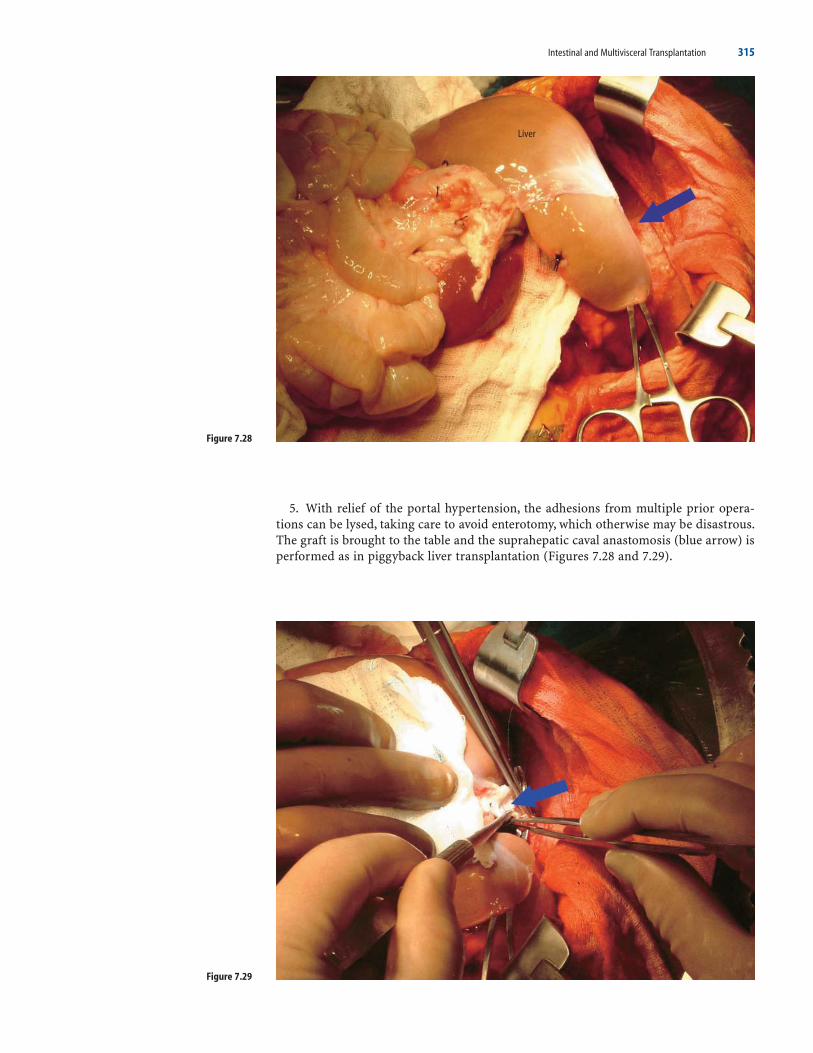
Intestinal and Multivisceral Transplantation 315
5. With relief of the portal hypertension, the adhesions from multiple prior opera-tions can be lysed, taking care to avoid enterotomy, which otherwise may be disastrous.The graft is brought to the table and the suprahepatic caval anastomosis (blue arrow) isperformed as in piggyback liver transplantation (Figures 7.28 and 7.29).
Figure 7.28
Figure 7.29

316 Atlas of Organ Transplantation
6. The Carrel patch or SMA/celiac complex (yellow arrow) can now be anastomosedeither directly to the aorta or more commonly to an interposition conduit of donor tho-racic aorta (blue arrow) (Figures 7.30 and 7.31) or bifurcating iliac artery.
Figure 7.30
Figure 7.31

Figure 7.32
Intestinal and Multivisceral Transplantation 317
7. The stump of the splenic vein may be used to vent preservation solution, and if thisis planned, the surgeon should isolate and prepare the splenic vein orifice during theback-table procedure (prior to transection of the pancreas).20 Cholecystectomy is per-formed in the usual fashion.
8. Enteral continuity is reestablished with jejunojejunostomy after removal of mostof the proximal recipient small bowel, anastomosis being performed distal to the ligament of Treitz of the allograft. Distal anastomosis is accomplished as with isolatedintestinal allografting (Figure 7.32).
9. In some cases, it may be preferable to place separate liver and intestinal allografts,rather than employing the en-bloc allograft technique.21 Severe portal hypertension anddense adhesions may make the aortic exposure required for combined transplantationdifficult. Placement of the liver prior to aortic exposure can facilitate transplantationwhen faced with this difficulty. The piggyback liver transplant is performed, and the iso-lated intestinal transplant procedure with systemic drainage is then performed.

318 Atlas of Organ Transplantation
Multivisceral Transplant
a) Donor Procedure
1. Donor procurement is fundamentally the same as in the liver–intestine procure-ment except that variable amounts of the stomach or colon are preserved with the graft.Recipients with disorders of motility and gastric dysfunction may benefit from inclu-sion of the stomach in the allograft. Recipients with severe chronic pancreatitis usuallyrequire inclusion of the entire pancreas. Tumors of the mesentery sometimes requireremoval of the entire gastroduodenal complex, and patients suffering familial adeno-matous polyposis sometimes have gastric, duodenal, or periampullary polyps requiringremoval. Some children may rarely require concomitant renal transplantation. In thiscase, one or both pediatric kidneys can be kept in continuity with the allograft, witheither the distal or the thoracic allograft aorta used for inflow. The renal arteries andveins are not disrupted from their connections to the aorta and vena cava. The funda-mental technical alterations are preservation of the left gastric artery, careful preser-vation of the gastroepiploic arcade when removing the greater omentum (Figure 7.33),and enteric division of the gastroesophageal junction, rather than the pylorus orjejunum.
Figure 7.33

Figure 7.34
Intestinal and Multivisceral Transplantation 319
2. The abdominal viscera are then removed en bloc with the vena cava and the same Carrel patch or intact aorta (Figure 7.34), as would be the case with liver–intestineprocurement.
b) Recipient Procedure
1. This transplant procedure is different from the previously described ones in thatthe entire splanchnic circulation with accompanying organs is removed; no portocavalshunt or bypass is therefore necessary. The pancreas and spleen, the root of the intes-tinal mesentery, the stomach, and the liver are removed together, preserving vena cavacontinuity. Thus, the recipient operation is similar to the multivisceral donor operationitself. The procedure begins with mobilization of the liver from the retrohepatic venacava to allow piggyback placement of the allograft. No portal dissection is required.

Figure 7.35
320 Atlas of Organ Transplantation
2. Next, the proximal stomach is isolated, allowing for division with a stapling device,preserving the gastroesophageal junction in the recipient, with descending blood supplyfrom the esophagus. This exposes the supraceliac aorta, sometimes allowing the celiacto be identified. However, this is most easily found proceeding from the left with a medialvisceral rotation, leading to isolation of the base of the celiac and superior mesentericarteries from the anterolateral left approach (Figure 7.35). Cattell and Kocher maneu-vers (if the colon is intact) expose the right side of the superior mesenteric artery, andmobilization of the entire base of the intestinal mesentery.
3. The left colon is divided, taking care to preserve the left colic artery and inferiormesenteric arteries, as no collateral flow from the superior mesenteric artery will existafter exenteration.Vascular clamps can then be placed on the base of the celiac and supe-rior mesenteric arteries and they may be transected. This step leaves the patient func-tionally anhepatic, so preparation to remove the organs prior to this step is critical. Thehepatic veins are clamped and divided. The stomach, pancreas, spleen, small bowel, rightcolon, and liver are removed together, preserving the inferior vena cava for piggybackallograft implantation.
4. The hepatic venous piggyback anastomosis is usually performed first, aligning thegraft in proper position.

Intestinal and Multivisceral Transplantation 321
5. Inflow must be brought to both the celiac and superior mesenteric arteries. Twoinflow vascular anastomoses may be used, using the native celiac and superior mesen-teric arteries end-to-end with the same donor structures (our preferred method), usingan iliac bifurcating graft to these vessels from either the supraceliac or infrarenal aortaafter ligation of the native vessels, or through a single anastomosis of infrarenal aorta ofthe donor allogaft end to side to the infrarenal aorta of the recipient (Figure 7.36).
Figure 7.36

322 Atlas of Organ Transplantation
6. Preservation of a cuff of native stomach allows for a two-layer gastrogastric anas-tomosis, rather than an esophagogastric anastomosis. We have encountered no anasto-motic leakage with this technique.22 Pyloroplasty is required to provide gastric emptyingof the denervated stomach. Distal enteral continuity is reestablished with ileostomy con-struction and colon anastomosis as described above (Figures 7.37 and 7.38).
Figure 7.37

Intestinal and Multivisceral Transplantation 323
Figure 7.38

324 Atlas of Organ Transplantation
7. Transplantation of the stomach may result in either gastric stasis or mild dumping,and some form of gastric decompression tube is mandatory, usually with a combinedgastrostomy-jejunostomy tube to allow early feeding into the graft jejunum. Drains areplaced in case of the development of chylous ascites that may emanate from the graftafter a fat-containing diet is resumed (Figure 7.39).
Living-Donor Small-Bowel Transplant
Very few small-bowel transplants have been done to date utilizing living donors. Lessthan 50 cases have been reported to the International Intestinal Transplant Registry. Pri-marily this is because the results with deceased donors are satisfactory, and the issueswith organ shortage are not as great as with liver or kidney transplant. Nonetheless, themortality list on the waiting list is high, especially in pediatric patients. Use of livingdonors has the potential advantages of lowering waiting time, reducing preservationinjury, and allowing for better human leukocyte antigen (HLA) matching.25–28
Various segments of bowel have been reported to have been used for such transplants,but the most experience is with a long segment of the donor terminal ileum as the graftfor the recipient. In this scenario, approximately 150 to 200 cm of distal ileum is used.The graft inflow is based on the distal part of the superior mesenteric artery (SMA); graftoutflow is based on the corresponding part of the superior mesenteric vein (SMV).
As with all living-donor transplants, donor selection is a critical part of the process.Potential donors should be ABO blood group compatible and carefully evaluated for possible cardiopulmonary risk factors. The medical evaluation is not too dissimilar tothe evaluation for a potential kidney donor. Specific to the potential small-bowel donoris radiologic imaging to evaluate the anatomy of the superior mesenteric vessels.This can be done with conventional selective mesenteric angiogram. However, this is an invasive test with some potential risk. The same information can be obtained with a good-quality computed tomography (CT) angiogram with three-dimensional reconstructions.
Figure 7.39

Intestinal and Multivisceral Transplantation 325
a) Surgical Procedure
1. A midline incision is made and the abdominal cavity is explored. The first impor-tant step is to measure the length of bowel and mark the portion to be removed (Figure7.40). The last 20 to 30 cm of terminal ileum, including the ileocecal valve, should be pre-served in the donor. Therefore, starting at the ileocecal valve, 20 cm is measured proxi-mally and the bowel is marked here, which represents the distal margin of resection. Thebowel is then marked 150 to 200 cm proximal to this, which represents the proximalresection margin. One should also ensure that at least 60% of the total small-bowel lengthwill be left in the recipient.
Figure 7.40

Figure 7.41
326 Atlas of Organ Transplantation
2. Using transillumination and palpation, the distal branch of the SMA is identified,and dissected free for 2 to 3 cm. Care should be taken to ensure that this is distal to themajor branches of the SMA that supply the jejunum, proximal ileum, and right colon.The corresponding portion of the superior mesenteric vein is identified next to the arteryand dissected free for a similar length (Figure 7.41).

Intestinal and Multivisceral Transplantation 327
3. Next, the mesentery of the bowel to be removed is scored and divided. This is donein a V-shaped fashion, with the apex corresponding to the mobilized portion of the SMAand SMV, and the two ends corresponding to the proximal and distal resection margins(Figure 7.42).
Figure 7.42

328 Atlas of Organ Transplantation
4. The bowel is divided proximally and distally. The vessels are divided after applica-tion of vascular clamps. The graft is removed and flushed (Figure 7.43).
Figure 7.43

Intestinal and Multivisceral Transplantation 329
5. The two divided ends of the bowel are reapproximated either with a stapler or ina hand-sewn fashion to create an end-to-end anastomosis (Figure 7.44). The defect inthe mesentery is closed, followed by closure of the incision.
6. The recipient operation first involves isolation of the vessels to be used for inflowand outflow of the graft. The infrarenal aorta and inferior vena cava are most commonlyused. End-to-side anastomoses are performed for the vein and artery of the graft (Figure
Figure 7.44

330 Atlas of Organ Transplantation
7.45). Once the bowel is reperfused, intestinal continuity is restored by anastomosing theproximal end to the recipient remnant bowel. The distal end is usually brought out asan ileostomy.
Perioperative Management
Patients require intensive care immediately after transplant. They generally remain intubated in the early postoperative period, although isolated intestinal transplantpatients may be extubated early. In the first 24 hours, the transplanted intestine usuallyproduces minimal stool, but third-space losses can be significant, requiring aggressivehydration. If kidneys are transplanted with the graft, the urine output should be replacedon an hourly basis accordingly. Finally, pulmonary artery catheter pressures or trans-thoracic echocardiography may be employed to help guide fluid management at the discretion of the critical care team. The intestinal vasculature is sensitive to vasocon-strictive agents, particularly α-adrenergic agents, and they should be avoided. Sepsissyndrome that requires intervention usually indicates the need for reoperation due tovascular compromise or perforation. Because the graft is exteriorized with an ileostomy,blood flow to the graft may be assessed with Doppler flow at the bedside, rather thanultrasound duplex, which is made difficult by overlying bowel gas. A bedside nurse mayemploy a fetal Doppler hourly to assure good waveform as would be done for a vascu-larized free flap.
Figure 7.45
Intestinal and Multivisceral Transplantation 331
Electrolyte imbalances should be corrected aggressively with intravenous delivery.The transplanted bowel after the first few days may exhibit calcium and magnesium mal-absorption with high ileostomy outputs, although this usually does not develop untilafter the institution of enteral nutrition. Water, sodium, and bicarbonate may be lost inlarge quantities with high ileostomy outputs. This may lead to a characteristic metabolicacidosis, requiring sodium bicarbonate added to the intravenous or enteral formula forcorrection. Hypomagnesemia potentiates tacrolimus-related neurotoxicity and shouldbe avoided.
Intravenous broad-spectrum antibiotics should be continued early after transplanta-tion or until the first biopsy confirms mucosal integrity of the transplanted bowel. Somepatients will have lost the right of domain of the abdominal cavity and will return to theintensive care unit closed with a prosthetic mesh. Although we carefully match donorand recipient sizes, at times abdominal lavage and staged closure is necessary. A dedi-cated line for parenteral nutrition should be maintained during the intensive care unitstay, as the average time to the achievement of complete enteral nutrition after intestinaltransplantation is approximately 1 month.1,11 We generally institute feeding 5 days aftertransplantation if no reoperation is planned, and early graft function is adequate, judgedby initiation of stomal output.
Conclusion
Intestinal transplantation is a very technically demanding field, requiring intimateknowledge of advanced techniques in both liver transplantation and gastrointestinalsurgery. Some degree of individualization of procedures is required, based on each recip-ient’s gastrointestinal anatomy, function, and vascular complications of parenteral nutrition.Appropriate technical and logistical planning will minimize unnecessary com-plications and keep ischemic times short, and have helped attain the current acceptableoutcomes.
References
1. Grant D, Abu-Elmagd K, Reyes J, et al. 2003 report of the intestine transplant registry: a new era hasdawned. Ann Surg 2005;241(4):607–613.
2. The Intestinal Transplant Registry. http://www.intestinaltransplant.org/.3. Fishbein TM, Gondolesi GE, Kaufman SS. Intestinal transplantation for gut failure. Gastroenterology
2003;124(6):1615–1628.4. Fishbein TM. The current state of intestinal transplantation. Transplantation 2004;27:78(2):175–178.5. Fishbein TM, Matsumoto CS. Regimens for intestinal transplant immunosuppression. Curr Opin Organ
Transplant 2005;10:12–123.6. Abu-Elmagd K, Fung JJ, Bueno J, et al. Logistics and techniques for procurement of intestinal, pancreatic,
and hepatic grafts from the same donor. Ann Surg.7. Fishbein TM, Kaufman SS, Florman SS, et al. Isolated intestinal transplantation: proof of clinical efficacy.
Transplantation 2003;76(4):636–640.8. Reyes J, Bueno J, Kocoshis S, et al. Current status of intestinal transplantation in children. J Pediatr Surg
1998;33(2):243–254.9. Tzakis A, Toso S, Reyes J, et al. Piggyback orthotopic intestinal transplantation. Surg Gynecol Obstet
1991;172(216):605–609.10. Berney T, Kato T, Nishida S, et al. Portal versus systemic venous drainage of small bowel allografts:
comparative assessment of survival, function, rejection and bacterial translocation. J Am Coll Surg2002;195:804–813.
11. Bueno J, Abu-Elmagd K, Mazariegos G, et al. Composite liver-small bowel allografts with preservation ofdonor duodenum and hepatic biliary system in children. J Pediatr Surg 2000;35(2):291–196.
12. Kato T, Romero R, Verzaro R, et al. Inclusion of entire pancreas in the composite liver and intestinal graftin pediatric intestinal transplantation. Pediatr Transplant 1999;3:210–214.
13. Grant D, Wall W, Mimeault R, et al. Successful small bowel/liver transplantation. Lancet 1990;335:181–184.14. Fishbein T, Schiano T, Jaffe D, et al. Isolated intestinal transplantation in adults with nonreconstructible
GI tracts. Transplant Proc 2000;32(6):1231–1232.15. Shaffer D, Diflo T, Love W, et al. Immunological and metabolic effects of caval versus portal drainage in
small bowel transplantation. Surgery 1988;104:518–524.

332 Atlas of Organ Transplantation
16. Reyes J, Mazariegos GV, Bond G, et al. Pediatric intestinal transplantation: historical notes, principles andcontroversies. Pediatr Transplant 2002;6:193–207.
17. Sindhi R, Fox IJ, Hefron T, et al. Procurement and preparation of human isolated small intestinal graftsfor transplantation. Transplantation 1995;60:771–773.
18. Sudan D, Iyer K, Deroover A, et al. A new technique for combined liver/small intestinal transplantation.Transplantation 2001;72(11):1846–1848.
19. Starzl T, Rowe M, Todo S, et al. Transplantation of multiple abdominal viscera. JAMA 1989;261:1449–1457.20. Fishbein T, Facciuto M, Harpaz N, et al. A simple blood flush technique and mannitol promote homody-
namic stability and avoid reperfusion injury in isolated intestinal transplantation. Transplant Proc 2000;32:1313–1314.
21. Fishbein TM, Florman SS, Gondolesi GE, et al. Noncomposite simultaneous liver and intestinal trans-plantation. Transplantation 2003;27:75(4):564–565.
22. Kato T, Tzakis A, Selvaggi G, et al. Surgical techniques used in intestinal transplantation. Curr Opin OrganTransplant 2004;9(2):207–213.
23. Buchman AL, Scolapio J, Fryer J. AGA technical review on short bowel syndrome and intestinal trans-plantation. Gastroenterology 2003;124(4):1111–1134.
24. Reyes J, Mazariegos GV, Bond GM, et al. Pediatric intestinal transplantation: historical notes, principlesand controversies. Pediatr Transplantant 2002;6(3):193–207.
25. Kato T, Ruiz P, Thompson JF, et al. Intestinal and multivisceral transplantation. World J Surg 2002;26(2).26. Benedetti E, Baum C, Cicalese L, et al. Progressive functional adaptation of segmental bowel graft from
living related donor. Transplantation 2001;71(4):569–571.27. Gruessner RW, Sharp HL. Living-related intestinal transplantation: first report of a standardized surgical
technique. Transplantation 1997;64(11):1605–1607.28. Testa G, Holterman M, John E, Kecskes S, Abcarian H, Benedetti E. Combined living donor liver/small
bowel transplantation. Transplantation 2005;79(10):1401–1404.

AA-adrenergic agents, 330Acetaminophen, 199Acidosis, 166Adhesions/adhesiolysis
in dialysis access, 57from inflammatory disease, 314–315in kidney transplant, 155from laparoscopy, 60
Adrenal glands, 74, 84, 89, 202Adrenal vein, 83, 203fAlcohol intake, 179, 198–199Alexandre, Guy, 2Amphotericin solution, 14–15Anastomoses
of aorta, 307–308, 316biliary, 228–229, 264, 288–289in bowel grafts, 153–154, 155f, 164–165, 304,
322, 329in brachiocephalic fistula, 39f, 40–43caval, 224, 232, 235of duodenum to bladder, 173–178end-to-end, 151f, 329end-to-side, 150f, 170–171, 314gastrogastric, 322of hepatic artery, 225, 263, 288of iliac vein, 170in kidney transplant, 106–110, 119–120, 122,
150–151in liver transplant, 200, 204–223, 265–266in pancreas transplant, 134piggyback technique in, 200, 206, 230–232,
251, 303, 315, 317, 320placement options for, 154in PTFE grafting, 48–49in radiocephalic fistula, 37–38of splenic artery, 143, 185in split-liver transplants, 242, 261–264,
286–289strictures of, 125–126of superior mesenteric artery, 143–144of ureters to bladder, 110–115, 119See also specific procedures
Aneurysms, 126–127Angiography, 134, 324Angioplasty of renal artery, 123Anterior one-stitch technique, 114Antibiotics
cephalosporin, 66irrigation with, 165, 294
postoperative, 291, 331preoperative, 47, 66
Anticoagulants, 193–194, 296Antiplatelets, 193–194Antivirals, 193Aorta
anastomosis of, 307–308, 316cannulation and flushing of, 10, 28–29,
296dissection/exposure of, 4–5, 8, 294,
314division of, 20–23infrarenal, 307in nephrectomy, 72f, 75fperfusion of, 10removed with graft, 311, 319thoracic, 312–314, 316
Aortic cuffs, 109–110Aortic patches, 204Aprotinin, 200Arginine stimulation test, 179Arterial conduits, 142–144, 151, 156Arteries
aorta. See Aortaarteriotomy, 169farteriovenous grafts, 46–49brachial, 38–43calcifications in, 160celiac, 320–321epigastric, 102femoral, 30–31gastric, 205, 295, 318gastroduodenal, 9, 13, 204, 213,
313gastroepiploic, 180, 189, 318hepatic. See Hepatic arteryiliac. See Iliac arteryinferior mesenteric, 5, 141radial, 37renal. See Renal arterysplenic. See Splenic arterysuperficial femoral, 51–53superior mesenteric. See Superior
mesenteric arterySee also Anastomoses
Arteriotomy, 169fAscites, 324Assistant’s ports, 80–81Axillary vein, 49–51, 207Azathioprine, 91
BBack table procedures
graft inspection, 30in intestinal transplant, 297–300, 312–313kidney separation, 21in multivisceral transplant, 297–300in pediatric kidney transplant, 119–120portal flushing, 30vs. in situ liver splitting, 243in split-liver grafts, 249, 260vessel reconstruction, 122See also Benching procedures
Basilic vein, 44–46, 50–51Basilic vein transposition, 44–46Benching procedures
care taken in, 194of deceased donor liver, 201–206of deceased donor pancreas, 135–144,
300of kidneys, 21, 93–99, 119in multivisceral transplant, 297–300
Bile leak after transplant, 252, 265, 290–291Biliary tree. See Common bile duct/biliary
treeBiopsies
of preop donor liver, 265of transplanted bowel, 331of transplanted kidney, 135of transplanted pancreas, 135
Bladder, urinaryfor pancreas drainage, 167, 173–178, 193ureteral anastomosis to, 110–115, 119See also Urologic complications
Bleeding. See Hemorrhage; Hemostasis,intraoperative
Blood. See Hemorrhage; Laboratoryevaluations
Blood transfusions, 200Blood vessels. See Arteries; VeinsBowel
anastomosis of, 153–154, 155f, 164–165, 304,322, 329
mobilization, retraction, and protection of,157, 159–160, 163
See also Colon; Small bowel; specific bowelprocedures
Brachial artery, 38–43Brachial vein, 38–43, 50–51Brachiocephalic fistula, 38–43Brain death, 2, 23–24
Index
333
Page numbers followed by f indicate figures.Large page ranges indicate main discussions.

334 Index
CCadaver donor organs. See Organs, donorCalcifications, arterial, 160Calcium, serum, 331Calculi/stones in urinary bladder, 193Camera ports, 67–68, 79, 187Cancer, 30, 92, 198–199, 201, 318
See also specific tumorsCannulation
of aorta, 10, 28–29, 296central venous, 199, 207, 278, 290of femoral vessels, 30–31premortem, 30–31Sundt carotid endarterectomy shunt for, 300for systemic portal bypass, 216–217
Cardiovascular disease, 92, 134, 192Carrel patches, 296, 311, 316, 319Catheters
for arteriovenous grafts, 46–51for peritoneal dialysis, 54–58temporary external for dialysis, 36
Cattell and Kocher maneuvers, 294, 310, 320Cattell-Braasch maneuver, 4Caudate lobe, 246, 273Caval replacement technique, 206, 218, 251Cavaplasty, 206, 233–235Cecum, 147Celiac artery, 320–321Celiac axis, 190, 310, 312, 316, 320Cellulitis, 291Central venous pressure, 199, 207, 278, 290Cephalic vein, 36–43Cephalosporin, 66Cerebrovascular disease, 92Cholangiography, 274, 291Cholecystectomy, 272, 310, 317Cholestasis, 26, 308Cholestatic disorders, 198Cirrhosis, liver
chronic, 198, 201compensated vs. decompensated, 199primary biliary, 201
Cold perfusion/preservative solutionfor aorta, 10, 29for femoral vessels, 30–31for kidney, 21, 89for liver, 240–241, 248, 283, 286in multiorgan procedures, 8for pancreas, 191for small bowel, 305, 328Sundt carotid endarterectomy shunt for, 300vents for, 29, 310, 317
Colonanastomosis of, 153–155, 164–165, 304, 322,
329dissection/exposure of, 295–297, 302left/descending, 69, 295, 320mobilization, retraction, and protection of,
157, 159–160, 163right/ascending, 147, 320sigmoid, 147transverse, 157, 228–229vascular transection of, 296See also Small bowel; specific colon
proceduresCommon bile duct/biliary tree
anastomosis of, 228–229, 264, 288–289cannulation and flushing of, 29
complications of, 252, 264–265, 290–291dissection/exposure of, 9–10division of, 139, 214, 259, 279drainage options for, 242intraoperative cholangiogram of, 274, 291postop complications of, 252, 265, 290
Computed tomography (CT) scanrenal, 66for vessel evaluation, 179, 324
Conflicts of interest, 28, 31Couinaud segment grafts, 237Cyclosporine, 198Cytomegalovirus (CMV), 193
DDeep vein thrombosis (DVT), 126Dehiscence, 130Dehydration, 166Delayed graft function (DGF), 25Detrusor muscle, 111–113, 115Diabetes mellitus
gestational, 179pancreas transplants for, 134, 166, 178
Dialysishemodialysis, 35–53peritoneal, 54–58
Diaphragmremoval from liver graft, 203transection of, 30
Dissecting ports, 67–68, 79Diuretics, 76, 87, 200–201Documentation/flow sheets, 27Donation after cardiac death (DCD), 23Donor organs. See Organs, donorDonors/donor pool
deceased, 1–2evaluation of, 179increase/decrease in, 24–25limitations of, 236, 242–243, 251living intestinal donor, 324–331living kidney donor, 59–60living liver donor, 264living pancreas donor, 179–192living vs. deceased, 92, 324non-heartbeating. See Non-heartbeating
donor (NHBD) organsDopamine/dobutamine, 200Drainage options. See under specific procedures
and organsDrugs/chemotherapies
α-adrenergic agents, 330acetaminophen, 199antibiotics. See Antibioticsanticoagulants, 193–194, 296antiplatelets, 193–194antivirals, 193aprotinin, 200azathioprine, 91cyclosporine, 198diuretics, 76, 87, 200–201dopamine/dobutamine, 200ε-aminocaproic acid, 200furosemide, 87gancyclovir, 193heparin. See Heparin/heparinizationimmunosuppressive agents. See
Immunosuppressioninotropic agents, 200
mannitol, 87prophylactic. See Prophylaxisprostaglandin E1, 291protamine, 88steroids, 201
Duodenal cuffs, 140Duodenum
dissection/exposure of, 310drainage conduit procedure on, 152–154,
164–165, 167, 173–178graft characteristics of, 192mobilization of, 294, 314procurement of, 16–17segment length of, 154
EE-aminocaproic acid, 200Edema, lower extremity, 192Electrolyte status, 200–201, 331En bloc allograft techniques
for pediatric patients, 119–120, 311, 314,319–320
vs. separate allografts, 317Endoscopic bags, 87–88Endo-staplers, 260, 282End-stage renal disease (ESRD), 91–92Epigastric artery, 102Epigastric vein, 102Ergonomics for robotic-assisted laparoscopy,
82Ethics issues
about ischemia times, 31, 280about liver splitting, 243–244in organ procurement, 251
Exercise, postoperative, 60Exposure, surgical. See under specific
proceduresExtension grafts, 303, 308
FFalciform ligament
exposure and division of, 208, 219, 239, 243,245, 257, 267
reattachment of, 283Family histories, 179Fat/lipoid tissues
hepatic, 265pancreatic, 194perirenal, 98–99
Fistulasbrachiocephalic, 38–43as complications, 126–127radiocephalic, 36–38saphenous-femoral, 51–53
Flow sheets, organ donor, 27Flushing. See under specific procedures and
organsForearm loop arteriovenous grafts, 46–49Furosemide, 87
GGallbladder, 10, 310, 317Gancyclovir, 193Gastric artery, 205, 295, 318Gastrocolic ligament, 180Gastroduodenal artery, 9, 13, 204, 213, 313Gastroepiploic artery, 180, 189, 318Gastrohepatic ligament, 209, 254

Index 335
Gastrostomy-jejunostomy tubes, 324Genitofemoral nerve, 104Gestational diabetes, 179Glucose levels, serum, 135, 166, 192, 200–201Glucose tolerance tests, 179Gonadal vein, 70, 83, 99Graft weight to recipient weight (GW/RW)
ratio, 243, 264, 283, 291Grafts
arteriovenous, 36–53Couinaud segment grafts, 237en bloc, 119–120, 311, 314, 317, 319–320extension grafts, 303, 308flushing of, 224, 227, 232, 260, 262–263, 283,
286graft weight to recipient weight (GW/RW)
ratio, 243, 264, 283, 291reperfusion of, 89, 164, 172, 192, 200, 227survival rates of, 25trisegment grafts, 237vein grafts, 249, 283Y types of. See Y graftsSee also specific organs
HHand-assist ports, 67–68, 187Harmonic scalpels, 74, 82Hematomas, 291, 296, 313Hematuria, postop, 128, 166, 192–193Hemoclips, 77–78Hemodialysis
access procedures for, 35–53basilic vein transposition, 44–46forearm loop arteriovenous graft, 46–49lower extremity access procedure,
51–53radiocephalic and brachiocephalic fistula,
36–43upper arm arteriovenous graft, 49–51
history and principles of, 35–36Hemodynamic stability/instability, 200,
243Hemoglobin A1c, 179Hemorrhage
in hepatectomy, 199post liver transplant, 290in nephrectomy or post renal transplant,
122–123post pancreatic transplant, 192–194
Hemostasis, intraoperative, 89, 164–165, 172,191
of liver bed, 223. See also Hilum, hepaticof mesentery, 313with stapling devices, 77–78, 141, 178, 189,
313Heparin/heparinization
reversal of, 87systemic, 10, 77, 190, 296and thromboembolism risk, 126, 194
Hepatectomy, 14, 199, 284–285, 296Hepatic artery
aberrant, accessory, or replaced, 8–9, 30, 205,310
anastomosis of, 225, 263, 288complications of, 290dissection/exposure of, 12, 212–213, 247,
272–273division of, 281
in split-liver transplants, 255, 260thrombosis of, 290
Hepatic veinanastomosis of, 262, 265–266, 286dissection/exposure of, 208–209, 223, 267division of, 260, 282options for middle branch, 275–276preservation in split-liver procurement, 243,
269, 271in split-liver transplants, 243, 245, 250,
252–254three opened into one, 230–231, 242tributaries of, 278, 287
Hepatitis, chronic, 198–199Hepatocaval ligament, 270Hepatocellular carcinoma (HCC), 198–199,
201Hepatoduodenal ligament, 214Hernias, incisional, 291Hilum, hepatic
in combined liver-intestine transplant, 314dissection/exposure of, 201, 204, 209,
211–216, 238–240for split-liver transplant, 243, 247, 272–280
“Hockeystick” incisions, 100Human leukocyte antigen (HLA), 59, 179, 324Hypogastric vein, 185Hypoglycemia/hyperglycemia, 135, 166, 192,
200–201
IIce slush, 29, 296Ileostomy/ileocolostomy, 309, 322, 330Iliac artery
for adult transplant, 100anastomosis of, 104–105, 120–122, 125,
150–151, 163, 185dissection/exposure of, 147–149, 160, 168removal of, 23Y grafts from, 142–144, 151, 156, 171
Iliac fossa, 99–100, 167Iliac vein
for adult kidney transplant, 100anastomosis of, 104–105, 150–151cannulation of, 217dissection/exposure of, 147–149, 168removal of, 23
Immunologic status, 92Immunosuppression
with cyclosporine, 198induced/induction of, 92post islet cell transplantation, 135post kidney transplant, 122post pancreatic transplant, 134, 193
In situ liver splitting, 243, 249, 260Incisional hernias, 291Incisions/ports
appearance two weeks postop, 192dehiscence of, 130in groin, 217for hand-assisted laparoscopy, 67–68, 187for hemodialysis access, 36–39, 44, 47hernias of, 291“hockeystick,” 100for intestinal transplant, 294, 314for isolated pancreas transplant, 167for liver transplant, 207, 267, 314for living donor procurement, 179, 187
for multiple organ procurement, 2, 187for open nephrectomy, 61–62, 65for pediatric kidney transplant, 117–118for pediatric liver transplant, 253for peritoneal dialysis, 54–55Pfannenstiel, 79, 87for “pure” laparoscopic nephrectomy, 65for SPK transplantation, 146, 155, 157
Infectionschronic hepatitis, 198–199cytomegalovirus, 193preoperative, 92prophylaxis against, 135sepsis, 330of surgical wounds, 129–130, 193, 291of urinary tract, 193
Inferior mesenteric artery, 5, 141Inferior mesenteric vein
identification and division of, 18, 138,181–184, 188
used for flushing, 6, 295Inotropic agents, 200Instruments and equipment
endo-staplers, 260, 282gastrostomy-jejunostomy tubes, 324handing-off contaminated, 165harmonic scalpels, 74, 82linear cutting stapler devices, 191loupes, 263luminal stapling devices, 176–178nasogastric tubes, 294operating microscopes, 263self-retaining retractors, 210Sundt carotid endarterectomy shunts, 300for super-rapid procurement, 27–30trocars, 81vascular clamps, 160, 162–163, 260vascular punch devices, 108–109vascular staplers/hemoclips, 77–78, 141, 177,
189, 313Veress needles, 80
Insulin, 133–135International Pancreas Transplant Registry
(IPTR), 135Intestinal transplantation, 293–317, 324–331
combined with liver transplant, 310–317back table procedure in, 312–313procurement procedure in, 310–311recipient procedure in, 314–317
isolated, 294–309back table procedure in, 297–300procurement procedure in, 294–297recipient procedure in, 300–309
living donor, small bowel, 324–331surgical procedure for, 325–330
mesenteric/portal vs. systemic drainage in,300–301
overview of, 293, 324perioperative management of, 330–331See also Multivisceral transplantation
Irrigation with antibiotics, 165, 294Ischemia times
ethics issues in, 31, 280minimization of, 2, 89, 135, 146, 153,
192–194, 243in non-heartbeating donor organs, 24–25,
27prolonged, 291

336 Index
Islets of Langerhanssize and reserve estimate of, 179transplantation of, 135, 179–186
JJejunal vein, 297, 300Jejunojejunostomy, 309, 317Jejunum, 297, 299, 301–302Jugular vein, 207
KKetoacidosis, 166Kidney transplantation, 91–130
benching from a deceased donor, 21, 93–99,119
complications after, 122–130history, indications, and preop evaluation
for, 91–93outcomes of
with non-heartbeating donor organs,25
overview of, 93recipient vascular complications after,
122–127surgical techniques for, 99–122
adult, 99–116with multiple renal arteries, 120–122pediatric, 116–120
KidneysCT scan of, 66end-stage renal disease, 91–92living donor nephrectomy, 59–89mobilization of, 73–76, 84–88in multivisceral transplant, 330perfusion/flushing of, 89placement of, 100, 146, 185procurement of, 19–21, 30transplantation outcomes for, 25vasculature of. See Arteries; VeinsSee also specific kidney procedures
LLaboratory evaluations
for islet cell mass estimation, 179for kidney rejection, 166for liver graft function, 201, 290for organ rejection, 135post pancreas transplant, 134pre liver transplant, 264–265urinary amylase, 135See also specific tests
Laparoscopic techniquesadhesions from, 60for donor nephrectomy, 65–67hand-assisted, 67–79for peritoneal dialysis catheter placement,
56–58“pure,” 79robotic-assisted, 80–89
Leaks/leakagebiliary, 252, 264–265, 290–291urinary, 127vascular. See Hemorrhage
Ligament of Treitz, 6, 17, 228f, 294, 297Ligaments. See specific ligamentsLinear cutting stapler devices, 191Litch technique, 113Liver transplantation (LTX), 197–291
adult cadaver liver transplant, 207–235piggyback technique in, 200, 206,
230–232side-to-side cavaplasty in, 206,
233–235surgical procedure for, 207–229
benching from a deceased donor,201–206
biliary drainage in, 200deceased donor split-liver: adult/adult
recipients, 242–251complications after, 243ethics of, 243–244implant procedure in, 250–251overview of, 242–243procurement procedure in, 244–249selection criteria and technical aspects of,
243split potential in, 244
deceased donor split-liver: adult/pediatricrecipients, 235–242
implant procedure in, 240–242overview of, 235–236procurement procedure in, 236–240
donor risks in, 265, 290ex vivo vs. in situ graft splitting in, 236,
248–249history, indications, contraindications for,
1–2, 199living donor/adult recipient, 264–291
complications after, 290–291donor procedure in, 266–283overview of, 264–266recipient procedure in, 283–289
living donor/pediatric recipient, 251–264anastomoses in, 252f, 261–264complications after, 252–253donor procedure in, 253–260ethics issues in, 243–244, 251overview of, 251–253recipient procedure in, 261–264
outcomes for, 201with non-heartbeating donor organs,
25–26partial transplants, 200preexisting conditions and, 265, 290standard procedure in, 197–201
overview of, 197–198postoperative care, 200–201preoperative evaluation, 198–199surgical procedure, 199–200
three surgical phases of, 199–200waiting list for, 235–236, 242, 251, 265
Liver/liversacute liver disease/fulminant hepatic failure,
199anatomic classification of, 236appearance of, 29, 265back table separation of, 249, 300cirrhosis of, 198–199, 201dissection/exposure of, 204–223, 236–240,
244–249, 277flushing of graft, 224, 227, 232hilum of. See Hilum, hepaticligaments of. See specific ligamentslobes of, 236–237, 242, 244, 246, 251f,
264mobilization of, 268–273
parenchymal transection of, 248–249, 259,278–279
procurement/removal of, 14, 29–30, 296,320
reperfusion of, 200, 227scissurae/connective tissue planes of,
236steatosis of, 265transplantation outcomes for, 25–26vasculature of. See Arteries; VeinsSee also specific liver procedures
Loupes, 263Lower extremity access procedure, 51–53Lymphatic tributaries, 158–159, 297Lymphoceles, 105, 128–129
MMagnesium, serum, 201, 331Magnetic resonance imaging (MRI), 179Malignancy, 30, 92, 198–199, 201, 318
See also specific tumorsMannitol, 87Mesenteric root, 302, 306Mesenteric vascular reconstruction, 301–306,
308Mesentery
defect closure in, 329hemostasis of, 313removal of, 320scoring and division of, 327tumors of, 318
Metabolic acidosis, 331Metabolic disorders, 198Microscopes, operating, 263Mortality rates, 92, 265Multiple organ procurement
incision and exposure for, 4–5overview of, 2, 293, 324procedure for, 318–319See also Multivisceral transplantation
Multivisceral transplantation, 318–324, 323fbenching in, 297–300overview of, 293, 324perioperative management of, 330–331procurement procedure in, 318–319recipient procedure in, 319–324
Musclesdetrusor, 111–113, 115psoas, 103rectus, 100–101
NNasogastric tubes, 294Nephrectomy (living donor), 59–89
indications and evaluation for, 59–60, 91–93,179
laparoscopicadvantages and disadvantages of, 65hand-assisted, 67–79, 186–187overview of, 179preoperative evaluation for, 59–60, 65–67,
179“pure,” 79robotic-assisted, 80–89
open technique, 61–65, 179–186positioning for, 67, 186–187
Nerves, genitofemoral, 104

Index 337
Neuropathies, diabetic, 166Non-heartbeating donor (NHBD) organs
future protocols in, 31preoperative control maneuvers for, 26–27procurement of, 23–31
controlled and uncontrolled, 24definitions and history for, 23–24surgical techniques for, 27–30
transplantation outcomes for, 25–26See also specific organs
OObesity, 123, 130, 192, 194Obstructions. See Strictures/obstructionsOmentum, 166, 294–295Operating microscopes, 263Operating room non-heartbeating organ
donor flow sheets, 27Organ procurement. See Organs, donorOrgan procurement organizations (OPOs), 23Organs, donor
from deceased donors, 1–2vs. living donors, 251
en bloc removal of, 119–120, 311, 314, 317ethics issues about, 31, 243–244, 251grafting suitability of, 134, 136, 160multiple. See Multiple organ procurementfrom non-heartbeating donors. See Non-
heartbeating donor (NHBD) organspediatric. See Pediatric patientspreservation of, 1–2, 10–11, 21, 153, 162,
240–241procurement of, 2–22. See also specific
procedures and organsthoracic, 11transplantation outcomes for. See specific
procedures and organsOutflow issues, 243, 250, 265
PPancreas transplantation, 133–194
after kidney transplant (PAK), 134, 166alone (PTA), 134, 166benching from deceased donor, 135–144
overview of, 135–136procedure for, 136–144
complications afterminimization of, 194surgical, 192–194
drainage options for, 145, 152–154, 167, 186,193
isolated with bladder drainage, 166–178complications after, 166, 193overview of, 166–167surgical procedure for, 168–178
with kidney transplant, 100. See alsoSimultaneous Pancreas Kidney (SPK)transplantation
from living donor, 179–192donor evaluation for, 179laparoscopic procurement procedure for,
186–192open procurement procedure for, 179–184overview of, 178recipient procedure, 185–186
reperfusion and inspection of, 164, 172, 192simultaneous pancreas kidney (SPK),
145–166
standard procedures for, 133–135islet cell transplantation, 135overview and evaluation, 133–134surgical technique, postop care, and
outcomes, 134–135Pancreas/pancreata
back table separation of, 300characteristics of, 192dissection/exposure of, 137, 139–140,
188–189, 295fdivision of, 183–184drainage options for, 145, 152–154, 167, 186grafting suitability of, 134, 136mobilization of, 310in multiorgan transplant, 313omentum wrap of, 166placement of, 134, 166, 167, 185preservation of, 162, 192–194procurement of, 14–16, 30, 296, 320remnant oversew of, 313vasculature of. See Arteries; VeinsSee also specific pancreatic procedures
Pancreatectomylaparoscopic hand-assisted, 186–192open technique, 180–184, 296preoperative evaluation for, 179
Pancreatic duct, 184, 186, 191Pancreatitis
post pancreatic transplant, 192–194as pre-existing condition, 318
Panel reactive antibody (PRA), 178Parenchyma. See specific organsParenteral nutrition, 331Pediatric patients
donor pool for, 24, 235, 243–244en bloc grafting in, 119–120, 311, 314,
319–320kidney transplant in, 116–120liver transplant in, 235–242, 251–264See also specific pediatric procedures
Percutaneous transluminal dilatation (PTD),127–128
Perfusion. See Cold perfusion/preservativesolution
Peritoneal dialysisaccess procedures for, 54–58
incisions for, 54–55, 57–58laparoscopic, 56–58open technique, 54–56
Peritoneal windows, 155Peritoneum, 56–58, 103, 155Pfannenstiel incisions, 79, 87Phosphate, serum, 201Phrenic vein, 203fPiggyback techniques, 200, 206, 230–232, 251,
303, 315, 317, 320Pneumoperitoneum, 68, 76, 87Polyps, gastrointestinal, 318Polytetrafluoroethylene (PTFE) grafts, 46–49Port placements, 67–68, 79–80Portacaval shunts, 314Portal vein, 203f
anastomosis of, 226, 263, 286complications of, 290dissection/exposure of, 139, 156, 184, 204,
273, 297–298division of, 13, 215, 260, 281islet cell injection into, 135
preservation of, 239, 247, 299size of, 154, 156in split-liver transplants, 252f, 256–257systemic portal bypass, 216–217thrombosis/stenosis of, 192, 290
Postoperative careantibiotics in, 291, 331exercise for, 60for intestinal and multivisceral transplant,
330–331for kidney transplant, 93for liver transplant, 200for pancreas transplant, 134
Potassium, serum, 200–201Premortem cannulation technique, 30–31Primary nonfunction, 135, 291Procurement teams, 296–297Prophylaxis
against increased blood transfusion, 200against infection, 135, 193–194against thrombosis, 135
Prostaglandin E1, 291Protamine, 88Psoas muscle, 103Psychosocial evaluation, 92Pulmonary embolism, 126Pyloroplasty, 322
QQuality of life, 134
RRadial artery, 37Radiocephalic fistula, 36–38Rectus muscle, 100–101Rejection episode symptoms, 135, 166, 291Renal artery
anastomosis and placement of, 106–110, 116,119
angioplasty of, 123anomalies of, 66in benching procedure, 93–99dissection/exposure of, 64, 72–73, 77–79,
88multiple, 120–122preservation of, 311recipient complications of, 125–126thrombosis/stenosis of, 99, 123–125
Renal veinanastomosis and placement of, 106–110, 116,
119in benching procedure, 93–99dissection/exposure of, 6–7, 63, 71–72,
77–79, 83, 84, 88length of, 97, 100thrombosis of, 126
Reperfusion of grafts, 89, 164, 172, 192, 200,227
See also Cold perfusion/preservativesolution
Robotic ports, 80–81Round ligament, 102Roux-en-Y grafts, 164–165, 186, 228–229, 242
for biliary drainage, 264, 289
SSaphenous vein, 51–53, 217, 283Scissurae/connective tissue planes, 236

338 Index
Self-retaining retractors, 210Sepsis, 330Serum amylase, 135Serum creatinine, 135Short bowel syndrome, 306, 308Simultaneous Pancreas Kidney (SPK)
transplantation, 145–166kidney placement in, 165overview of, 145portal/enteric drainage, 154–166
arterial anastomoses in, 154benching of pancreas in, 156overview of, 154–155surgical procedure for, 157–166
systemic/enteric drainage, 145–154colon placement in, 147duodenum/bowel anastomosis in,
153–154kidney placement in, 146overview of, 145vascular anastomoses in, 148–151
Small bowelanastomosis of, 153–155, 308, 329dissection/exposure of, 296donor/recipient volume ratios of, 325manual decompression of, 296, 310procurement and preservation of, 297, 305,
320, 328removal of excess (graft), 140–141resection lines for, 325–326See also Colon; specific bowel procedures
Small-for-size syndrome, 291Sodium, serum, 331Spermatic cord, 102Spleen
blood supply to, 180–181, 189dissection/exposure of, 295fmobilization of, 310procurement of, 16, 16–19, 320
Splenectomy, 137Splenic artery
anastomosis of, 9, 143, 185dissection/exposure of, 30, 140, 181–184ligation and division of, 137–138, 189–190,
312–313preservation of, 296
Splenic veinanastomosis of, 185cannulation and flushing of, 300dissection/exposure of, 181–182, 312ligation and division of, 189–190preservation of, 298venting through, 317
Split-liver transplants (SLTs). See under Livertransplantation (LTX)
Starzl, Thomas, 1, 198Steatosis
hepatic, 265pancreatic, 194perirenal, 98–99
Stenosis. See Strictures/obstructionsStents, 228–229, 264, 289, 291Steroids, 201Stomach
decompression of, 324division and removal of, 320gastrogastric anastomosis of, 322See also specific procedures
Stomas, 309, 331Strictures/obstructions
biliary, 252, 265, 290–291ureteral, 127–128vascular, 126–128
Substance abuseof acetaminophen, 199of alcohol, 179, 198–199
Sundt carotid endarterectomy shunts, 300Superficial femoral artery, 51–53Superior mesenteric artery
accessory arteries from, 205–206anastomosis of, 143–144dissection/exposure of, 7, 30, 156, 326ligation and division of, 141, 311, 320–321in living donor, small bowel transplants,
324mobilization of, 139preservation of, 11, 299, 302
Superior mesenteric veinanastomosis of, 161–162dissection/exposure of, 18, 157–159,
158–159, 326identification and division of, 157,
181–184placement choices for, 161preservation of, 302spasm of, 159
Surgeonspositioning of, 187training and certification of, 81–82
Survival rates, 25, 197–198, 201Sutures/stitches, specialized
anterior one-stitch technique, 114Litch technique, 113See also Anastomoses
Systemic portal bypass, 216–217Systemic vascular reconstruction, 306–309
TTeres ligament, 208, 255Thrombosis
post liver transplant, 290post pancreatic transplant, 192prophylaxis against, 135post renal transplant, 123
Transaminase, serum, 201Transaminitis, 26Triangular ligament, 208, 220, 268Trisegment grafts, 237Trocars, 81
UUltrasound
intraoperative, 275–276for pancreatic graft viability, 192postoperative, 290–291, 330
Umbilical tape “hanging technique,” 258,277
United Network of Organ Sharing (UNOS)database, 25
University of Wisconsin solution, 29Upper arm arteriovenous graft, 49–51Uremia, 134, 178Ureters
anastomosed to bladder, 110–115, 119in benching procedure, 93–99division of, 64
mobilization of, 69, 75–76, 85–86obstruction of, 127–128postop leaks from, 127transplantation of, 100
Urethritis, 166Urinary tract infection (UTI), 193Urine amylase, 135, 166, 192, 193Urine leaks after transplant, 127Urine production, 76, 87Urologic complications, 127, 127–128, 166,
192–193
VVascular clamps, 160, 162–163, 260Vascular punch devices, 108–109Vascular staplers/hemoclips, 77–78, 141, 177,
189, 313Vein grafts, 249, 283Veins
adequate outflow through, 243, 250, 265adrenal, 83, 203faxillary, 49–51, 207basilic, 50–51brachial, 50–51central stenoses of, 36cephalic, 36–43epigastric, 102extension grafts from, 303, 308gonadal, 70, 83, 99grafts of, 249, 283hepatic. See Hepatic veinhypogastric, 185iliac. See Iliac veininferior mesenteric. See Inferior mesenteric
veinjejunal, 297, 300jugular, 207phrenic, 203fportal. See Portal veinrenal. See Renal veinsaphenous, 51–53, 217, 283spasm of, 159splenic. See Splenic veinsuperior mesenteric. See Superior
mesenteric veinvena cava. See Vena cavavenotomy, 169f, 206, 234, 261f, 286venous conduits, 303venous thromboembolism, 126See also Anastomoses
Vena cavaanastomosis of, 224, 232, 235, 307–308,
315caval replacement technique, 206, 218,
251cavaplasty, 206, 233–235dissection/exposure of, 202–203, 218–223,
222f, 230–231, 268–270, 294inferior, 71, 97infrahepatic, 202–203, 218–219, 221–223liver transplant modifications to, 206,
313preservation of, 265–266for renal vein lengthening, 97in split-liver transplants, 241–242, 244, 246,
249transection of, 11, 14, 20, 30venting of, 29, 310

Index 339
Venotomy, 169f, 206, 234, 261f, 286Venous thromboembolism, 126Venovenous bypass (VBB) system, 199–200Vents/venting, 29, 310, 317Veress needle, 80Volvulus, 296, 308Voronoy, Yu Yu, 1
WWaiting lists, 235–236, 242, 251, 265, 324Wilson’s disease, 198–199Wound complications
infections, 193, 291post liver transplant, 291post nephrectomy, 129–130
YY grafts
care taken in, 194Roux-en-Y limb of bowel, 164–165, 186,
228–229, 242thrombosis/stenosis of, 192vascular, 142–144, 151, 156, 171


![0521856841 - Neil M. Borden - 3D Angiographic Atlas of Neurovascular Anatomy and Pathology [2006]](https://img.pdfslide.net/doc/110x75/557213e7497959fc0b934c88/0521856841-neil-m-borden-3d-angiographic-atlas-of-neurovascular-anatomy.jpg)