Embed Size (px)
Citation preview

A Problem oriented approach
BENIGN BREAST DISEASE
Dr.B.SELVARAJ MS;Mch;FICS;
�PEDIATRIC SURGEON
�ASSOCIATE PROFESSOR
�MELAKA MANIPAL MEDICAL COLLEGE
�MALAYSIA
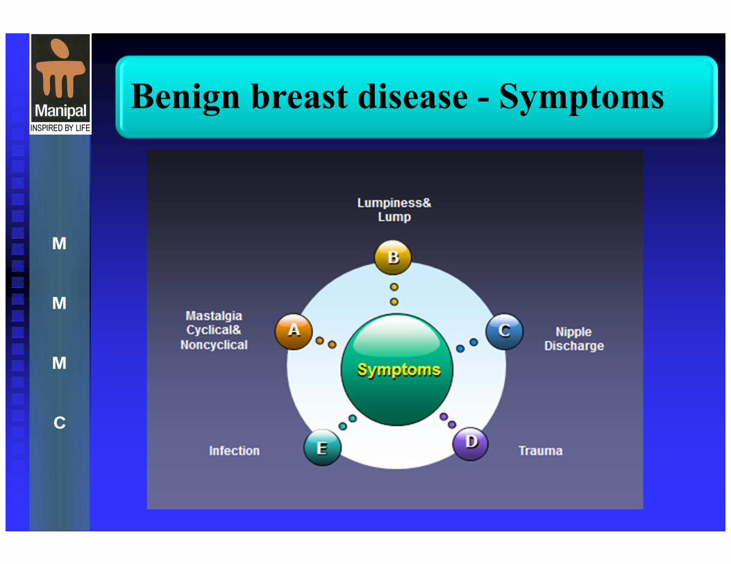
Benign breast disease - Symptoms
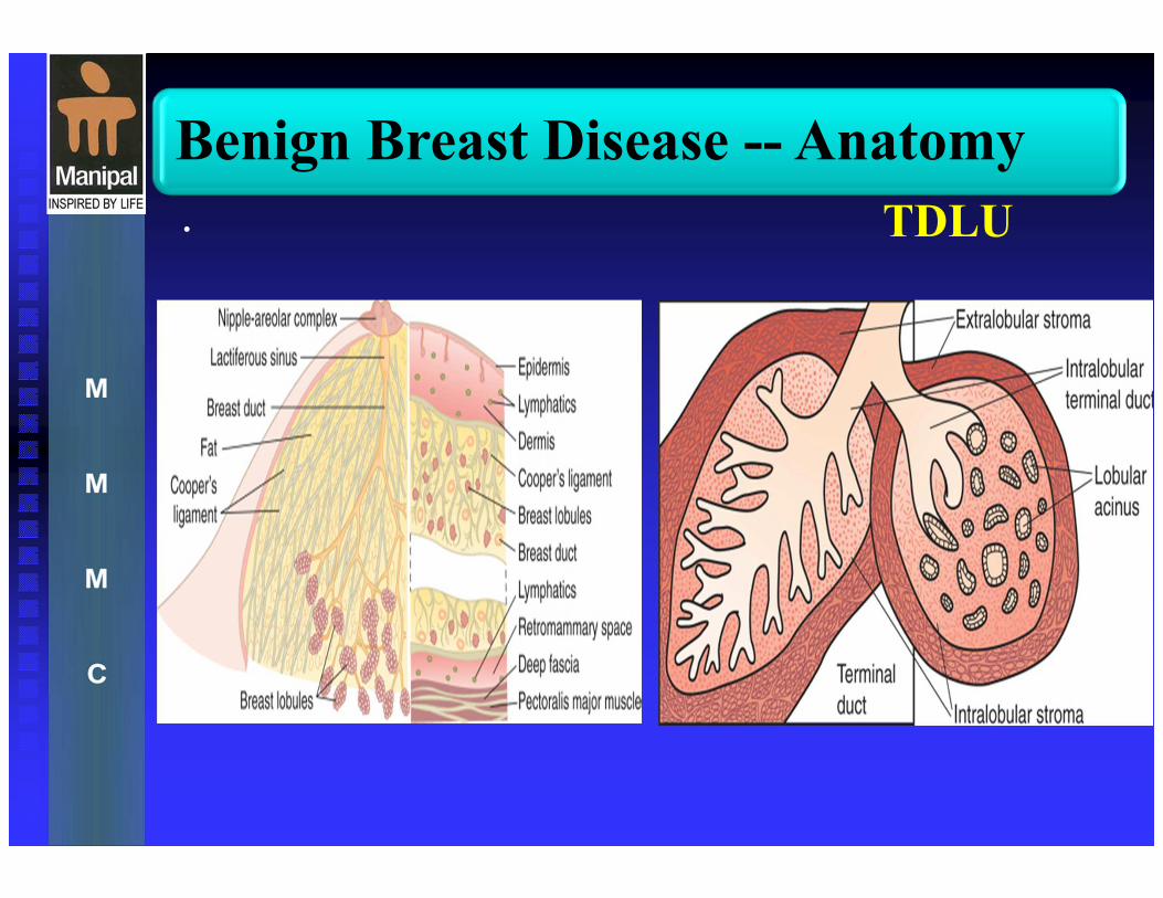
Benign Breast Disease -- Anatomy
• TDLU
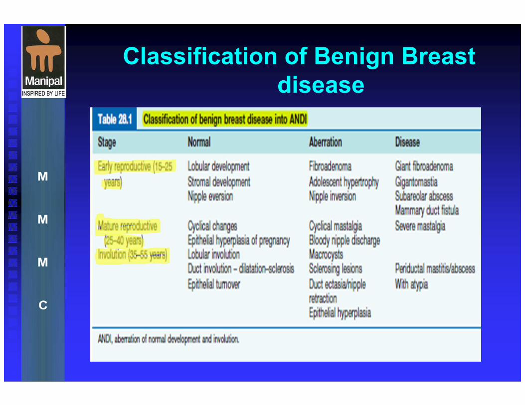
Classification of Benign Breast
disease

Common causes of breast
symptoms

INTRODUCTION
�Breast is host to a spectrum of benign and
malignant diseases.
�Benign breast conditions are practically a
universal phenomena among women.
� It accounts for 80% of clinical presentation
related to the breast.
CONGENITAL & DEVELOPMENTAL
ABNORMALITIES
� Although the normal location of the breast is the anterior thorax, breast tissue with or without a nipple or just nipple and areola alone can occur any where along the milk line
� The milk line is an ectodermal thickening appearing at 6 weeks of gestation running from axilla to the midportion of inguinal ligament
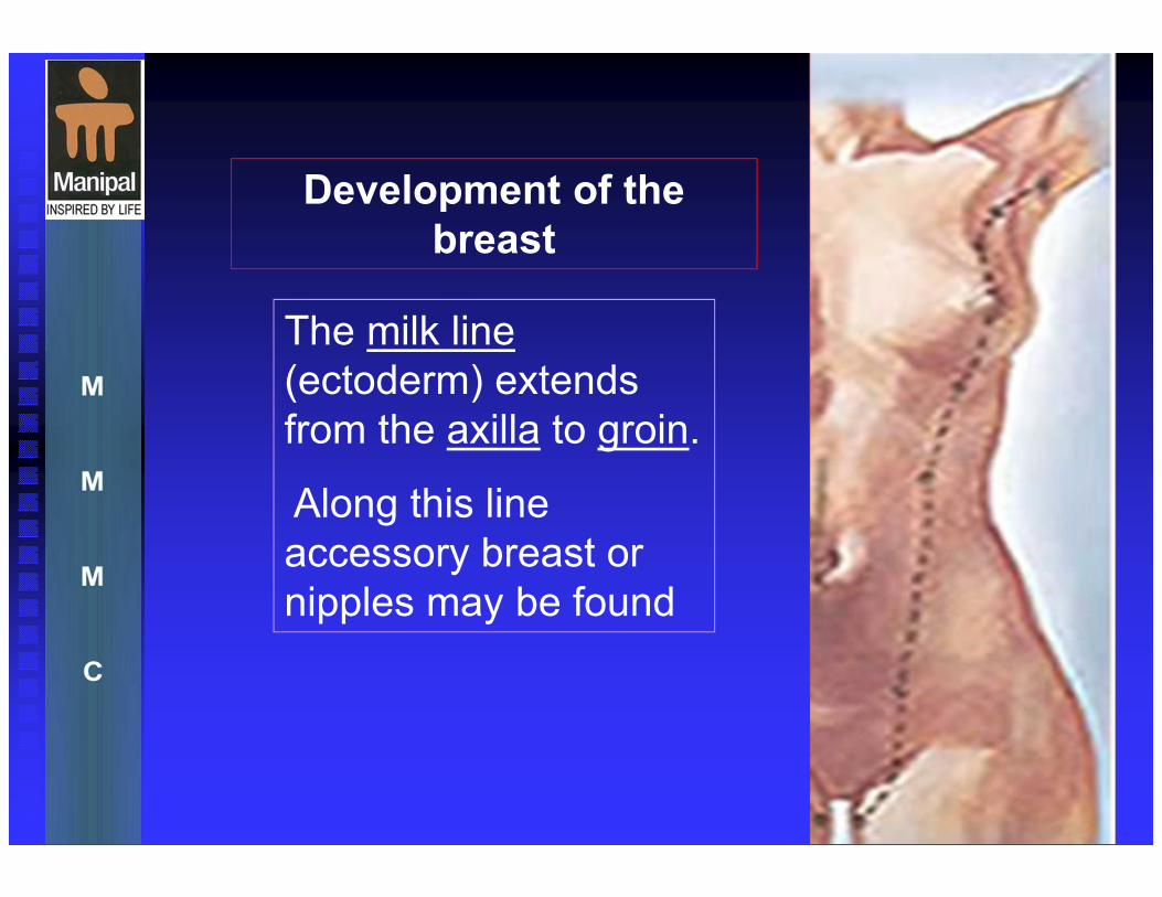
The milk line
(ectoderm) extends
from the axilla to groin.
Along this line
accessory breast or
nipples may be found
Development of the
breast
CONGENITAL & DEVELOPMENTAL
ABNORMALITIES
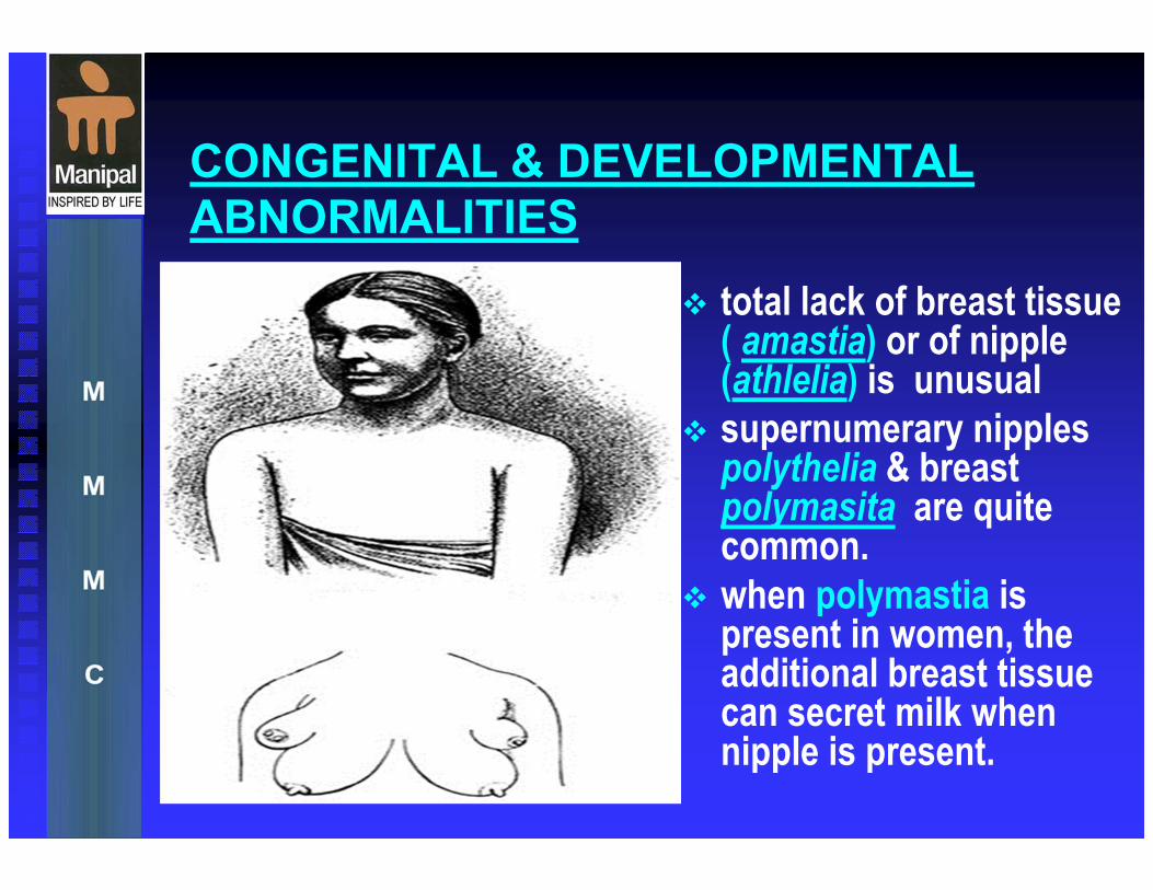
� total lack of breast tissue ( amastia) or of nipple (athlelia) is unusual
� supernumerary nipples polythelia & breast polymasita are quite common.
� when polymastia is present in women, the additional breast tissue can secret milk when nipple is present.
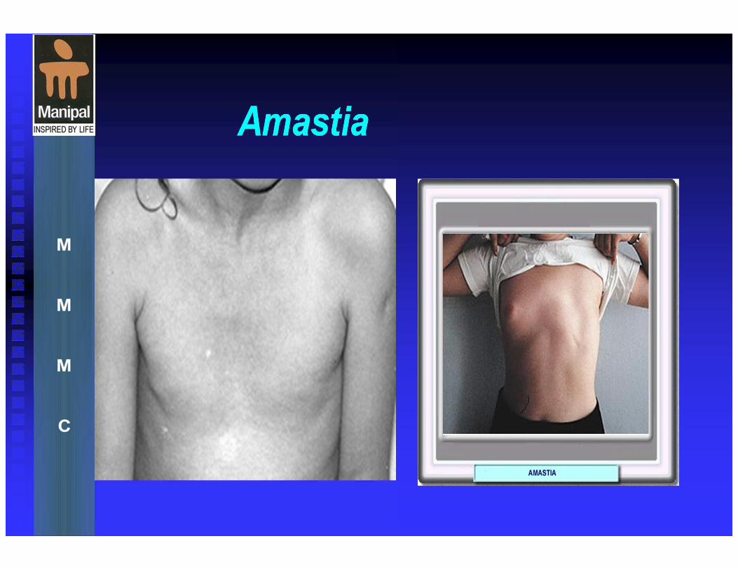
Amastia
� Amastia: A rare condition wherein the normal growth of the breast or nipple does not occur.
� Unilateral amastia (just on one side) is often associated with absence of the pectoral muscles�Poland’s syndrome
� Bilateral amastia (with absence of both breasts) is associated in 40% of cases with multiple congenital anomalies involving other parts of the body as well.
� Amastia is distinguished from amazia wherein the breast tissue is absent, but the nipple is present. Amazia typically is a result of radiation or surgery.
Amastia
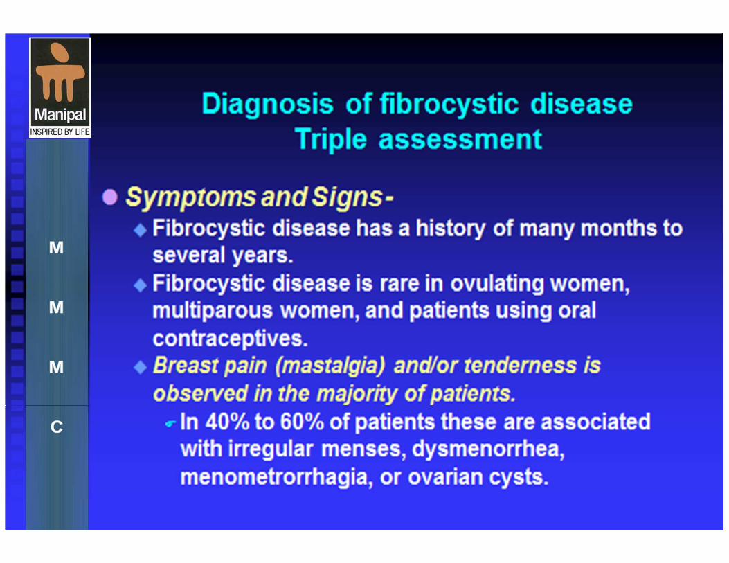
Mastalgia
� Mastalgia is breast pain and is generally
classified as either cyclical (associated with
menstrual periods) or noncyclical
� Breast pain of any type is a rare symptom of
breast cancer , only 7% of breast cancer have
mastalgia as the only symptom.
� Most mastalgia is of minor to moderate severity
and accepted as part of the normal changes that
occur in relation to menstrual cycle.
Mastalgia
�Cyclical mastalgia: begin since average
30 yrs, relieved by menopause, physical
activity can increase the pain, e.g. by
weight lifting and prolonged use of arm.
�Non-cyclical mastalgia: affects older
women (mean age 43), arises from chest
wall� eg: Teitz’s disease, Breast itself or
outside the breast.
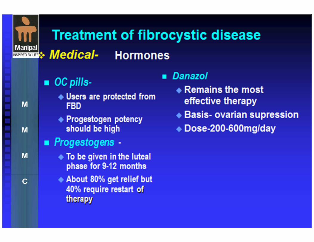
Cyclical Mastalgia - treatment
�Danazol: 200-300 mg daily, slowly reduced
to 100 mg daily or on alternative day, given
on days 14-28 of menstrual cycle.
�Gamma-lineolic acid(evening primerose
oil)
320mgm/day for 3to4 months
�Responses are usually seen within 3
months
�Weight gain, acne and hirsutism

Non Cyclical Mastalgia -
treatment
�More resistant to treatment than cyclical breast
pain
�Hormonal manipulation ineffective
� Symptomatic- analgesics and anti-inflammatory
drugs
� Firm supportive bra
FIBROCYSTIC DISEASE


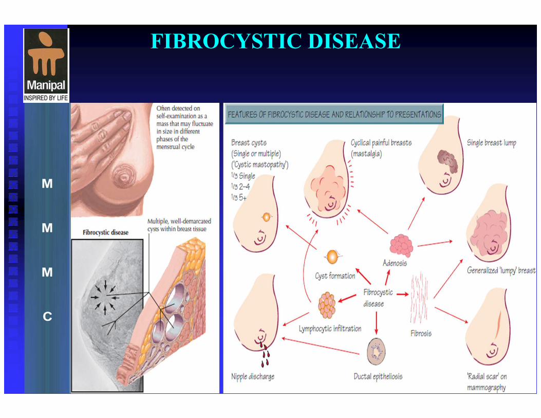
FIBROCYSTIC DISEASE


FAT NECROSIS
� This is traumatic in nature & is met with women with large fatty breast
� Results from injury to breast fat by Trauma, surgery, biopsy4.
� Causes to focal fibrosis and cicatrix formation.
� Early: edema of the fat lobules,increased echogenicity.
� Post surgical scar, hematoma, seroma
FAT NECROSIS
Clinically: The patient develop sever bruising after moderately sever
trauma, When the bruise settles the woman notice swelling which is clinically Impossible to distinguish from carcinoma of the breast because the Irregular mass is often attached to the skin.
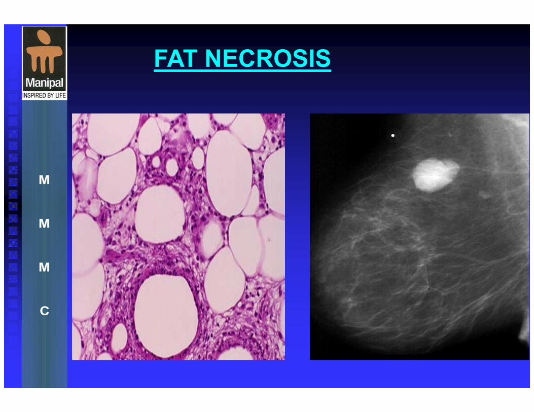
Microscopically a central area of necrotic fat cells are surrounded by a granulomatous reaction consisting of macrophage cells.
FAT NECROSIS
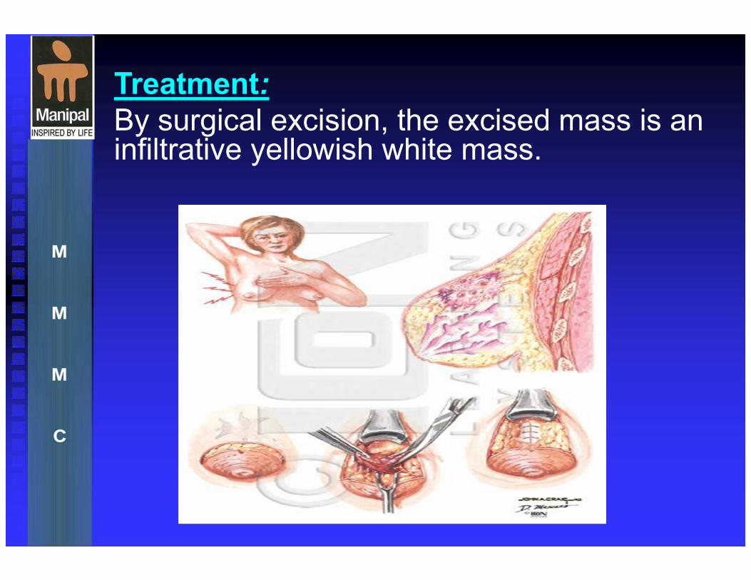
Treatment:
By surgical excision, the excised mass is an infiltrative yellowish white mass.

Duct Ectasia
This condition has several stages of involvement & vanity of names include (plasma- cell mastitis, comedo mastitis, & chronic abscess simulating carcinoma).
It is benign lesion may be virtually impossible to differentiate from carcinoma by it is gross appearance
Duct Ectasia
is a widening of the ducts of the breast, a
condition that occurs most frequently in women in
their 40s and 50s. A thick and sticky discharge,
usually gray to green in color, is the most
common symptom.
Tenderness and redness of the nipple and
surrounding breast tissue may also be present.
Sometimes, scar tissue forms around the
abnormal duct, leading to a lump that may be
initially mistaken for cancer.
Duct Ectasia
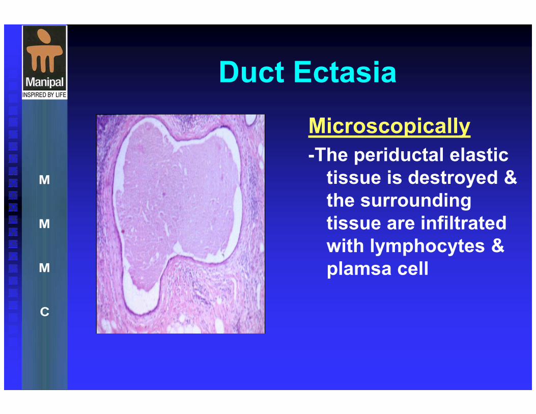
Microscopically
-The periductal elastic
tissue is destroyed &
the surrounding
tissue are infiltrated
with lymphocytes &
plamsa cell
Duct Ectasia
Clinically:- this condition present as solitary or multiple tender swelling in the sub or Peri-areolar region of the breast.
- Nipple retraction, skin adherence, edema & axillary
adenopathy may accompany a hard, diffuse mass within the
breast
- palpation reveals a number of cord like swelling which radiate from the areola.
- the ducts are dilated & contain an inspissated yellow cheesy material that can be expressed like toothpaste from the cut end of a duct.
- occasionally, the inflammatory response are so acute that
skin changes occur & the condition may be mistaken for
a breast abscess.
Duct Ectasia
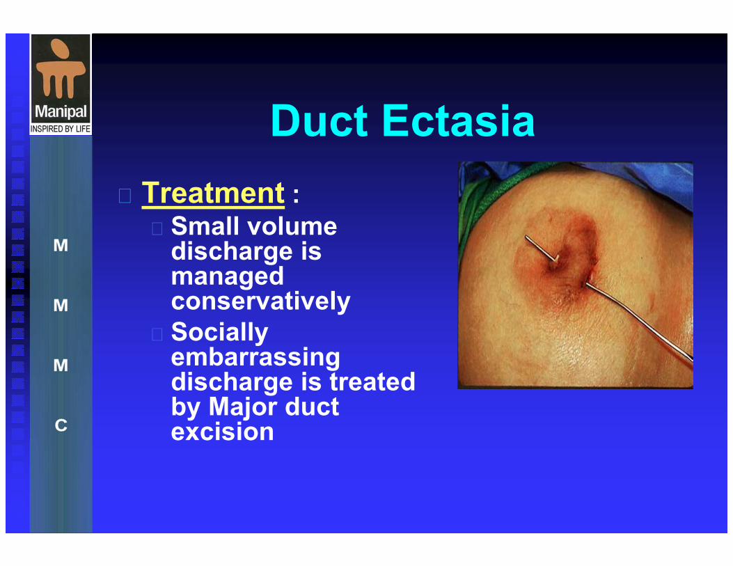
Treatment : Small volume discharge is managed conservatively
Socially embarrassing discharge is treated by Major duct excision
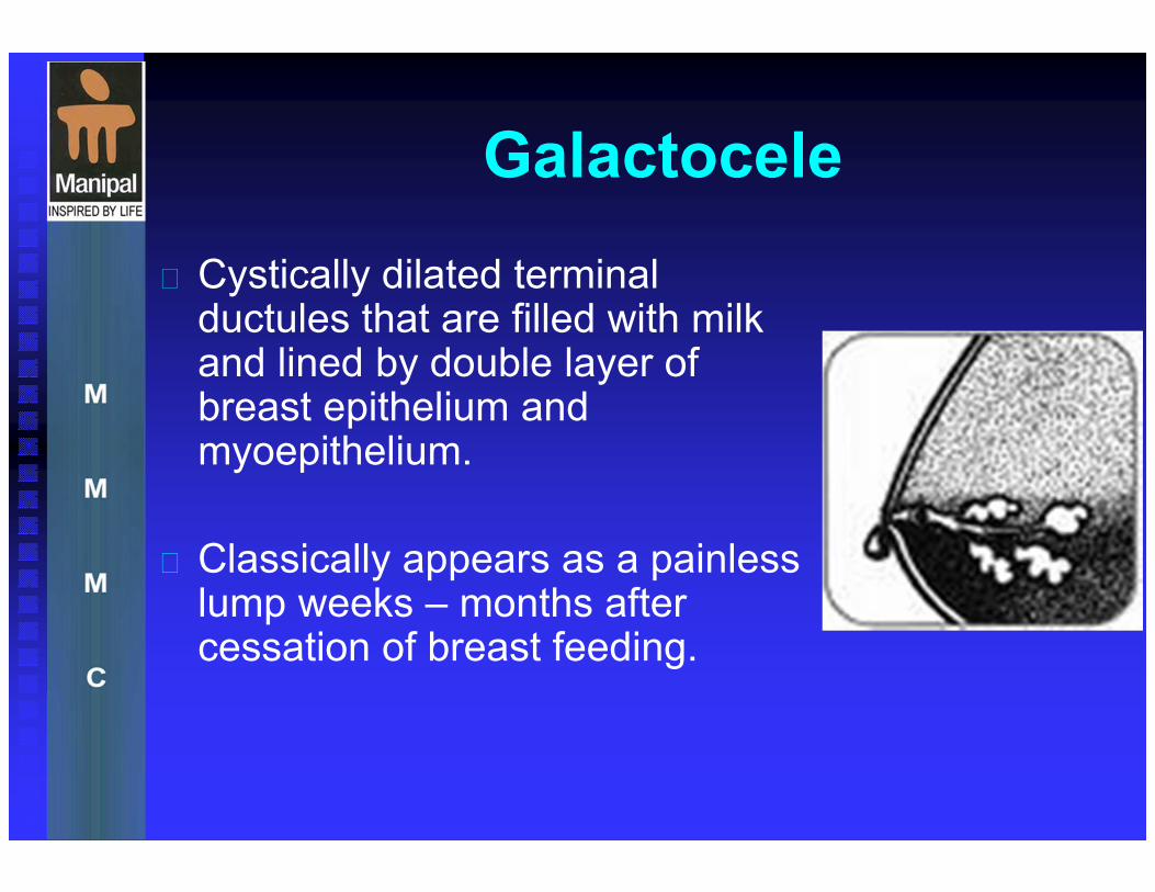
Galactocele
Cystically dilated terminal ductules that are filled with milk and lined by double layer of breast epithelium and myoepithelium.
Classically appears as a painless lump weeks – months after cessation of breast feeding.
GALACTOCELE
It is probably formed by obstruction to a duct
in the puerperium . the milk retained proximal
to the obstruction eventually becomes
cheese-like.
The common complication of this type of
swelling is infection.
The treatment is by surgical excision.

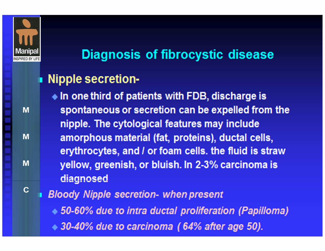
INTRA-DUCTAL PAPILLOMA
This benign lesions of the lactiferous duct wall occur centrally beneath the areola In 75% of cases.
They most commonly produce a bloody nipple discharge, some times associated with Pain
They are solitary proliferation of ductal epithelium
Intraductal papillomas should be treated by excision of a duct as a wedge resection.
Cystosarcoma phyllodes (CSP)
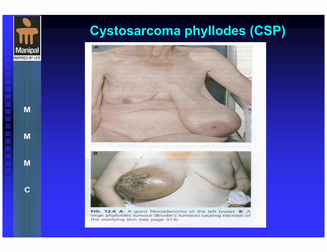
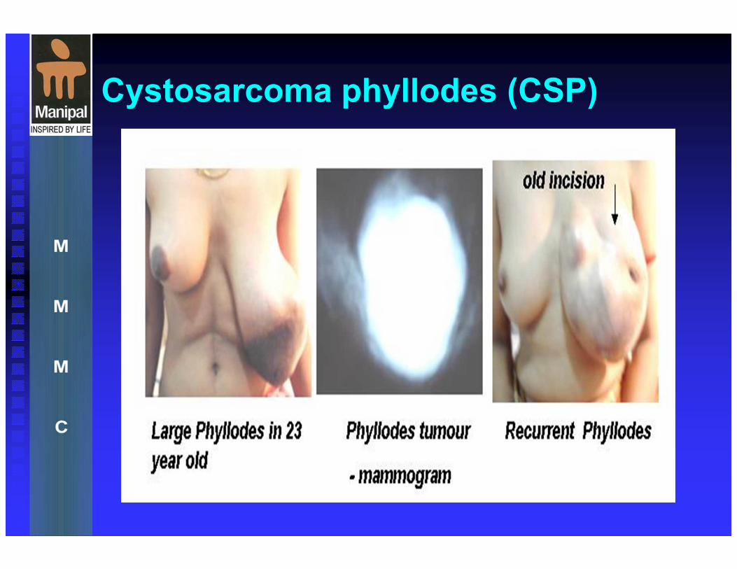
Cystosarcoma phyllodes (CSP) is a rare, predominantly benign tumor that occurs almost exclusively in the female breast. Its name is derived from the Greek words sarcoma, meaning fleshy tumor, and phyllo, meaning leaf.
Grossly, the tumor displays characteristics of a large, malignant sarcoma, takes on a leaflike appearance when sectioned, and displays epithelial cystlike spaces when viewed histologically (hence the name).
Because most tumors are benign, the name may be misleading. Thus, the favored terminology is now phyllodes tumor.
Cystosarcoma phyllodes (CSP)
Pathophysiology of CSP
Pathophysiology:
Phyllodes tumor is the most commonly
occurring nonepithelial neoplasm of the
breast, and it occurs only in the female
breast.
It has a sharply demarcated, smooth texture
and is typically freely movable. It is a
relatively large tumor, and the average size
is 5 cm. However, lesions more than 30 cm
in size have been reported.
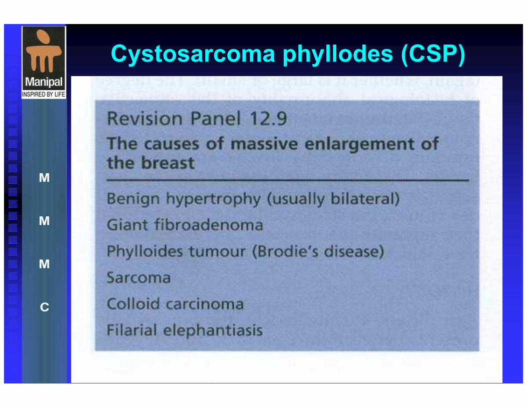
Cystosarcoma phyllodes (CSP)
Cystosarcoma phyllodes (CSP)
TREATMENT of CSP
Surgical Care: In most cases, perform wide local excision with a rim of normal tissue
If the tumor/breast ratio is sufficiently high to preclude a satisfactory cosmetic result by segmental excision
total mastectomy, with or without reconstruction, is an alternative.
More radical procedures generally are not warranted
Perform axillary lymph node dissection only for clinically suspicious nodes. However, virtually all of these nodes are reactive and do not contain malignant cells.

MASTITIS
MASTITIS
Breast mastitis is an infection that commonly affects women who are breast-feeding (especially during the first two months after childbirth) but can occur in all women at any time.
Mastitis is a benign condition that can usually be treated successfully with antibiotics.
Inflammation can be caused by many types of injury including : infectious agents and their toxins,
physical trauma
or chemical irritants

SIGNS AND SYMPTOMS OF MASTITIS
Part or all of the breast is intensely: painful,
hot, tender, red, and swollen.
Some patients can pinpoint a definite area of inflammation, while at other times the entire breast is tender. - feel tired, run down, achy, have chills .feel like flu .
A breastfeeding mother who thinks she has the flu probably has mastitis.
SIGNS AND SYMPTOMS OF
MASTITIS
chills or feel feverish, or temperature 38c or higher. These symptoms suggest an infection.
Feeling progressively worse, the breasts are growing more tender, and the fever is becoming more pronounced.
Other signs of mastitis: cracked or bleeding nipples,
stress or getting run down,
missed feedings or longer intervals between feedings.
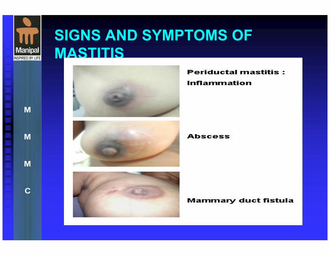
SIGNS AND SYMPTOMS OF
MASTITIS
TREATMENT OF MASTITIS
Mastitis usually requires treatment.Treatment for mastitis may require the following:
Antibiotics are usually prescribed by a physician to help clear up the infection.
Use warm water on the infected area of the breast before breast-feeding to help stimulate let-down (the milk ejection reflex).
Breast-feed or pump frequently, using both breasts. Lactation consultants recommend first breast-feeding from the unaffected breast until let-down (milk ejection reflex) occurs and then switch to the breast with mastitis.
Breast-feed only until the breast is soft.
Apply icy compresses to the breasts after breast-feeding to relieve pain and swelling.
Drink fluids and get enough rest.
Analgesia to control the pain.
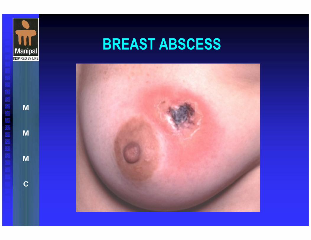
BREAST ABSCESS

BREAST ABSCESS
This condition is usually found during lactation . as role the infecting organism is : staphylococcus aureus, and less commonly streptococcus pyogenes .
the usual mode of infection is via the nipple, the infection being carried by suckling infant in the nasopharynx.
The infection is at first limited to the segment drained by the lactiferous duct but it may subsequently spread to involve other areas of the breast.

BREAST ABSCESS
CAUSES :
Staphylococcus aureus and streptococcal
species are the most common organisms
isolated in puerperal breast abscesses.
Nonpuerperal abscesses typically contain
mixed flora (S aureus, streptococcal
species) and anaerobes.
BREAST ABSCESS
CLINICAL FEATURES
SYMPTOM
Localized breast area edematous,
erythematous, warm, and painful
History of previous breast abscess
Associated symptoms of fever, vomiting,
and spontaneous drainage from the mass or
nipple
May be lactating

BREAST ABSCESS
CLINICAL FEATURES
SIGNS
Localized breast area erythematous, hot, edematous, and extremely painful
Most commonly found in the areolar or periareolar area
Fluctuance of the mass
May have associated fever or axillary lymphadenopathy
Discharge with palpation from nipple or mass
Nipple inversion

Investigations
1-Ultrasound: used to localize the abscess
2. FNAC: used to exclude underlying carcinoma
especially in chronic Breast abscess where the
abscess become encapsulated with a thick
fibrous capsule & the condition can’t be
distinguished from a carcinoma without a biopsy.
3. Needle Aspiration: to confirm presence of pus.
4. Mammogram: to exclude underlying carcinoma.
BREAST ABSCESS MANAGEMENT
1- If the patient present in the cellulitis stage the patient should be treated with an appropriate Antibiotic.
2- Breast rested with feeding on the opposite side only.
3- The milk should be expressed from the healthy segments of the affected breast.
4- Support of the breast
5- Local heat & analgesia to relive the pain.
6- If the infection doesn’t resolve within 48 h, the breast should be incised & drained.
N.B. if antibiotics used in the presence of undrained pus, an Antibioma form. This is a large sterile brawny edematous swelling which takes many weeks to resolve.
BREAST ABSCESS
MANAGEMENT 7.If pus is present at the time of presentation, which can
be confirmed by Needle aspiration, Incision &
Drainage is done which can be achieved by : � Simple Needle Aspiration: using a wide pore needle under local anesthesia.
� Guided drainage: under image control with radiological or ultrasound techniques a tube drain can be inserted & left until the cavity has collapse.
� Surgical drainage: it is the most certain method, not only can all loculi be reached, but also dead tissue can be removed. The cavity is then dressed regularly & left open to heal by 2ry intention.
� Excision of all of the major ducts in case of Periductal Mastitis.
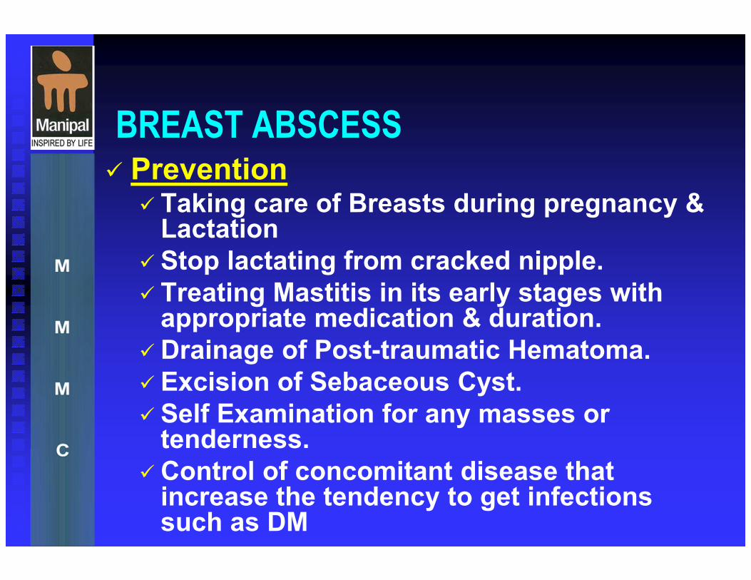
BREAST ABSCESS � Prevention
� Taking care of Breasts during pregnancy & Lactation
� Stop lactating from cracked nipple.
� Treating Mastitis in its early stages with appropriate medication & duration.
�Drainage of Post-traumatic Hematoma.
� Excision of Sebaceous Cyst.
� Self Examination for any masses or tenderness.
�Control of concomitant disease that increase the tendency to get infections such as DM
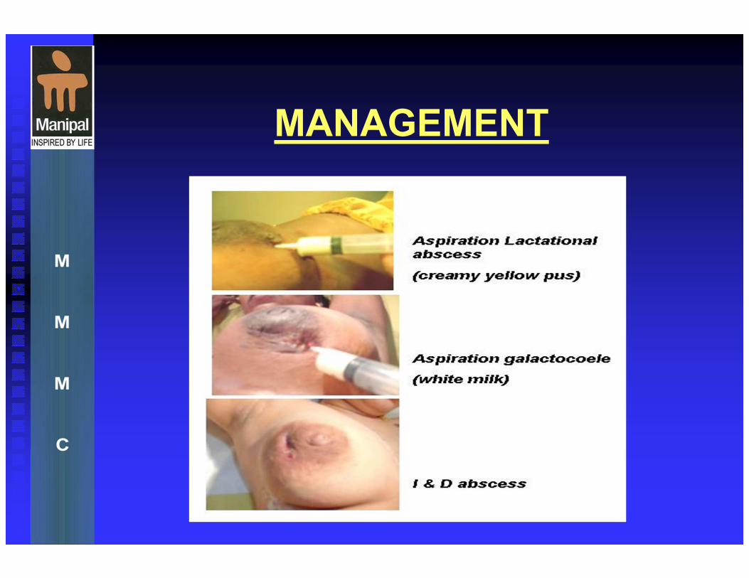
MANAGEMENT

Lactational breast abscess
Usually due to Staph. aureus
Usually peripherally situated
Surgery may be pre-empted by early diagnosis
Attempt aspiration
If no pus - antibiotics
If pus present consider repeated aspiration or incision and drainage
Consider biopsy of cavity wall
Continue breast feeding from opposite breast
No need to suppress lactation
Non-lactational breast abscess
Occur in periareolar tissue
Culture yield - Bacteroides, anaerobic strep, enterococci
Usually manifestation of duct ectasia / periductal mastitis
Occur 30- 60 years , More common in smokers
Often give history of recurrent breast sepsis
Repeated aspiration is the treatment of choice
Metronidazole and flucloxacillin
Drain through small incision if non-resolving
Definitive treatment when quiescent with antibiotic prophylaxis
Usually a major duct excision = Adair's operation
Spontaneous discharge or surgical excision can result in mammary fistula
BREAST ABSCESS
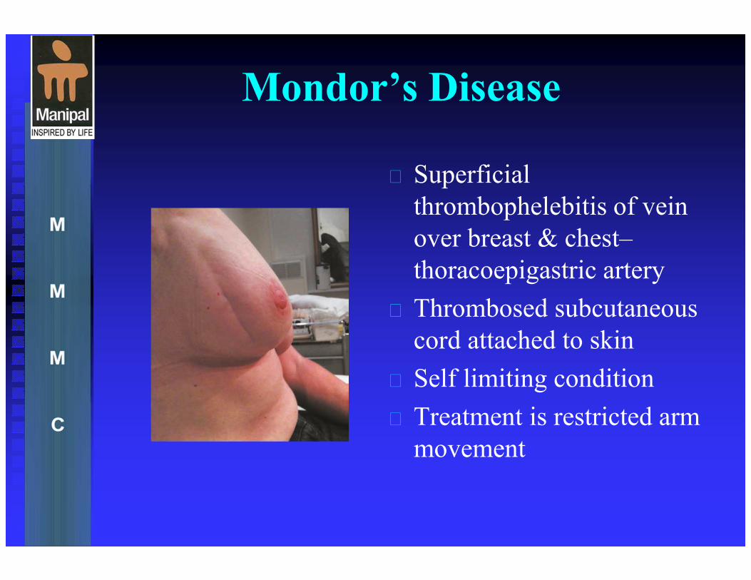
Mondor’s Disease
Superficial
thrombophelebitis of vein
over breast & chest–
thoracoepigastric artery
Thrombosed subcutaneous
cord attached to skin
Self limiting condition
Treatment is restricted arm
movement
Benign breast disease
Video Podcast

CONCLUSION
Benign breast disorders & diseases are common
The aetiopathogenesis is complex and not fully understood
Lump and pain are the most common complaints
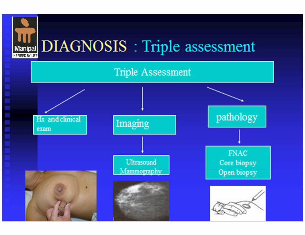
Evaluation is done by Triple assessment
Histological risk factors for future malignancy are relative and not absolute risk factors
Treatment is based on the natural history of clinical problems
Treatment must be tailored to individual needs

EMQ


![1 Estrategiadeempresayrhunr 131116113703 Phpapp01[1]](https://img.pdfslide.net/doc/110x75/577c81001a28abe054ab1236/1-estrategiadeempresayrhunr-131116113703-phpapp011.jpg)