Embed Size (px)
Citation preview

7
Breast Cancer: Radioimmunoscintigraphy and Radioimmunotherapy
Mojtaba Salouti and Zahra Heidari Department of Biology, Faculty of Sciences, Zanjan Branch,
Islamic Azad University, Zanjan, Iran
1. Introduction
Breast cancer is one of the most common diseases and a major public health problem in the
world today. It is responsible for 32% of all cancers and 15% of cancer deaths in women
(Dirisamer et al., 2010). Advances in diagnosis and treatment of breast cancer have led to
decline in mortality. However, just about 60% of patients can now be cured by initial
treatments, but the rest, in spite of receiving palliation with currently available therapy
methods die. Therefore, tremendous amounts of time and efforts are dedicated to search
toward earlier detection and more efficient treatment of this disease. Advances in molecular
cancer biology achieved an increased understanding of the biologic factors that contribute to
breast cancer pathogenesis and progression. This understanding has already led to both
early diagnosis and more effective treatment. Among different imaging and therapy
contribution modalities, nuclear medicine provides an important role to the clinical
management of breast cancer. In this chapter, we highlight the uses, advantages, limitations
and possible improvements of radioimmunoscintigraphy and radioimmunotherapy
techniques as targeted molecular imaging and therapy methods, respectively.
2. Diagnosis of breast cancer
Breast cancer is currently the most common cancer in females and the second highest cause
of cancer death in female population of the world. The majority of breast cancer deaths are
due to metastasis (Costelloe et al., 2009). If breast cancer is detected prior to metastatic
spread, it has considerably better prognosis than the cancer that has already spread.
Therefore, the detection of cancer in an early stage is a major factor in the reduction of
patient mortality and leading to less cancer costs and more successful treatments. The
management of patients with a suspicious lesion includes confirming the diagnosis,
identifying the stage of disease and choosing an appropriate treatment for each individual
patient. Identifying the stage of a disease in any patient is a crucial step, because it
determines the type of treatment that is appropriate for that particular person. Also, it is
necessary to follow patients that need therapeutic procedures to assess their response to the
treatment. There are a number of methods available for the detection of breast cancer. This
section briefly points out some difficulties that encounter with these conventional imaging
www.intechopen.com

12 Chapters on Nuclear Medicine 166
modalities and describes application of nuclear medicine techniques and targeted molecular
imaging in the investigation of patients with breast cancer.
2.1 Mammography
Mammography is a gold standard imaging method for breast cancer screening, detection and diagnosis in women under forty with a relatively high sensitivity in the range of 85-90% (Berg et al., 2004). This method is a national screening program in the United Kingdom (Glasspool & Evans., 2000). Although mammography is an effective imaging tool, it is not without limitations like many other diagnostic modalities. First, the sensitivity of mammography decreases dramatically in young patients due to their dense breast tissues. On the other hand, its ability to detect malignant lesions in young female patients decreases to 68% (Nystrom et al., 2002, Kopans, 1992). As a result, some patients with breast cancer are missed and some others without a malignant tumor undergo unnecessary biopsies due to incorrect mammography findings. Secondly, mammography in patients evaluated following breast surgery or radiotherapy, is unreliable with a false negative rate of 25-45%, because it cannot always differentiate benign from malignant diseases (Fass, 2008). Many efforts have ever been taken to establish new tests to enable us to collect more complete information by non-invasive methods. These efforts minimize the use of breast biopsy in women who do not have breast cancer. Therefore, many other imaging modalities such as ultrasonography (US), magnetic resonance imaging (MRI), diffuse optical tomography (DOT), computed tomography (CT) and measurement of tumor markers in blood serum have been initiated to increase the diagnostic accuracy of mammography.
2.2 Ultrasonography
Ultrasonography is an important adjunct to mammography for both diagnosis and characterization of breast cancer and is routinely used in this role (Fass, 2009). It is non-invasive, easily available, relatively cheap and also recommended for pregnant or lactating women when ionizing radiation may not be recommended (Schueller et al., 2008). It has been found that ultrasonography, when combined with mammography, can prevent up to 22% of unnecessary biopsies (Zonderland, 2000). Although ultrasonography can reliably differentiate a cystic lesion from a solid lesion, it does not provide a high specificity to distinguish benign from malignant lesions (Balleyguier et al., 2008; Sauer et al., 2005). Another disadvantage of this method as a screening tool, when applied to general population, is low sensitivity and specificity and furthermore it is operator-dependent (Liberman, 2000).
2.3 Magnetic Resonance Imaging (MRI)
Breast cancer was one of the first that was examined using MRI (Ross et al., 1982). This method is very useful for further evaluation when mammography and ultrasonography are indeterminate to study the presence or location of a suspect abnormality (Glasspool et al., 2000). However, due to cost reasons, low access and high false positives, MRI is not yet considered as a screening exam for breast cancer except for special cases. MRI of breast cancer is recommended in the repeated screening of patients who have the increased risk of radiation induced DNA mutations (Fass, 2008). MRI is used to screen women with a family history of breast cancer, women with very dense breast tissues or women with silicone implants that can obscure pathology in mammography (Fass, 2008).
www.intechopen.com

Breast Cancer: Radioimmunoscintigraphy and Radioimmunotherapy 167
2.4 Diffuse Optical Tomography (DOT)
While breast cancer is still increasing in frequency, new diagnostic procedures must be available to challenge existing procedures to make diagnosis of breast cancer more accurate and reliable. DOT is one of the most important non-invasive and non-ionizing imaging modalities that are available for breast cancer diagnosis. DOT can be used to locate lesions within breast (Frangioni, 2008; Kepshire et al., 2007; Hawrysz et al, 2000). Despite promising results, there are several factors that limit the wide application of DOT for the imaging of breast tissue in clinic. First, the maximum depth of imaging in breast tissue is less than 15 mm. Secondly, the spatial resolution of DOT is less than one centimeter, that is not as good as mammography, ultrasonography or MRI. Because of these limitations, DOT is not a widely-accepted imaging modality for breast cancer (Kepshire et al., 2007; Hawrysz et al., 2000).
2.5 Computed Tomography (CT)
X-ray computed tomography (introduced into clinical practice in 1972) was the first of modern slice-imaging modalities. CT scan has several advantages including (1) unlimited depth penetration; (2) high spatial resolution; (3) short acquisition time (minutes); (4) moderately cheap and (5) ability of performing anatomical imaging. CT has some limitations including (1) sensitivity of CT scan decreases in early stage of breast cancer, (2) CT scan is associated with radiation exposure and should never be done in pregnant females because of radiation risk to the fetus, (3) CT is not very good at identifying pathology of soft tissues, (4) the dye used in CT is iodine based and is often a cause of allergy and (5) CT probably can not be used for molecular imaging and currently is just used for anatomical and functional imaging (Pysz et al., 2010).
2.6 Measurement of tumor markers in blood
Several biochemical compounds in the serum/plasma may act as the indicators of the
presence, risk or prognosis of cancer. In patients with history of breast cancer, elevated
tumor marker levels may represent cases of tumor relapse. It has been shown that increasing
levels of tumor markers is associated with disease recurrence and may indicate the need for
further investigations. Examples of tumor markers in blood including CA 15.3, CA 27.29,
CA125, CEA and circulating tumor cells. While breast cancer blood marker tests are
promising, they are not absolutely conclusive. When a breast cancer blood marker test
comes back negative, it doesn't necessarily mean that the patient is free and clear of breast
cancer and a positive result does not always mean that the cancer is growing (Merkle et al.,
2009).
2.7 Nuclear medicine
As cited, each imaging method has strengths and weaknesses in terms of sensitivity, specificity, spatial and temporal resolution, contrast and cost. Therefore, the fundamental efforts for introduction and development of new methods for breast cancer diagnosis are requested. Nuclear medicine is defined as a branch of medicine that employs open radioactive sources, commonly referred to radionuclides, in diagnosis and treatment of diseases (Glasspool & Evans, 2000). The application of nuclear medicine techniques to study patients with breast cancer has recently raised its profile particularly in the investigation of indeterminate mammographic lesions and for overcoming limitations of MRI, US, CT and
www.intechopen.com
12 Chapters on Nuclear Medicine 168
DOT techniques (Berghammer et al., 2001). The differences in tumor biology such as blood flow, metabolism, concentration of specific receptors or differences in antigen expression are exploited in order to target radionuclides to the tumor tissue (Glasspool & Evans, 2000). The major advantage of nuclear medicine is that just picomolar concentrations of radiotracers are required to provide a measurable signal (Cook et al., 2003).
2.7.1 Scintimammography
Future developments in nuclear medicine are in the area of development of specialized imaging systems. One example of specialized gamma camera is scintimammography in imaging of breast and axillary nodes. Scintimammography term is given to radionuclide imaging of breast cancer (Schillaci et al., 2007). Scintimammography is performed after injection of the radiopharmaceutical into an arm vein contralateral to the suspected tumor or into a pedal vein and the subsequent imaging with right and left lateral, prone and supine views, covering both breasts and both axillae and later computer acquisition of data (Nguyen et al., 2009; Brem et al., 2005). Most studies evaluating the role of scintimammography, have compared it with mammography, ultrasound and MRI. Its sensitivity has ranged from 62% to 93% with specificity from 79% to 94% (Schillaci & Buscombe, 2004). Its sensitivity is greater for palpable lesions. Current recommendations for the use of scintimammography are (Fass, 2008): 1. As a general adjunct to mammography to differentiate between benign and malignant
breast lesions in patients with palpable masses or suspicious mammograms. 2. In patients referred for biopsy when lesions are considered to have a low probability of
malignancy. 3. In patients with probably benign findings on mammography, but are recommended for
close follow-up (e.g. repeated mammography in 3–6 months). 4. In patients who have dense breast tissues and are considered difficult to evaluate on
mammography. 5. For detection of axillary lymph node metastases in patients with confirmed breast cancer. The main limitations of scintimamography are low sensitivity and high dose of radionuclide that is used for imaging. Currently, for overcoming the cited limitations, the researchers are studying on targeted molecular imaging methods.
3. Treatment of breast cancer
To understand the diagnostic needs for breast cancer, it is important to understand the range of approaches for breast cancer treatment. With the exception of early and late stage disease, almost all breast cancer therapy methods, involve a combination of locoregional and systemic treatments. Locoregional therapy includes surgery and radiotherapy and systemic therapy includes chemotherapy, hormone therapy and targeted therapy. Because the breast cancer, that is metastatic beyond the regional node, is rarely cured, systemic therapy is the primary treatment for metastatic disease with locoregional treatment reserved for symptom control and is used possibly in some patients with limited metastatic disease who may achieve prolonged remissions.
3.1 Surgery
Surgery is the oldest and still the most widely used treatment modality that is available for breast cancer patients and can be accomplished by mastectomy, removal of the entire
www.intechopen.com
Breast Cancer: Radioimmunoscintigraphy and Radioimmunotherapy 169
breast or by breast-conserving surgery often termed lumpectomy of cancer. If the tumor is detected at early stage before spreading, surgery alone might be sufficient to reach complete remission. When the tumor is detected after spreading with distant metastases, surgery is frequently used in combination with radiotherapy or chemotherapy (Keshtgar et al, 2010).
3.2 Radiotherapy
In radiotherapy, also known as radiation therapy, high-energy ionizing radiation such as x or ┛-rays is irradiated to the tumor tissues for killing the cancer cells. Radiation can be delivered via external radiation from a source outside the body directing to the tumor or by an internal radiation source (brachytherapy) which is positioned inside the body adjacent to or inside the tumor. Radiation must affect only cancer cells in the treated area and not normal cells. To treat secondary tumors and stopping growth of any remaining tumor cells, radiation therapy is often used in conjunction with other treatment modalities like chemotherapy and surgery (Luini et al., 2007).
3.3 Chemotherapy
The use of cytotoxic chemotherapy, in both advanced and early stage of breast cancer, has made significant progress in the last decade (Hassan et al., 2010). Chemotherapy is a systemic breast cancer therapy and compared to surgery and radiation therapy, it has one advantage. It is able to eliminate cancer cells throughout the entire body. Chemotherapeutic agents currently approved for treatment of breast cancer are: anthracycline, alkaloids, topoisomerase inhibitors, alkylating agents, and antimetabolites. The important disadvantage of chemotherapy is side effects of drugs such as nausea, vomiting, fatigue, sore mouth, diarrhea, constipation and decreased blood cells count. These side effects can impact patient ability to tolerate treatment, maintain a healthy diet, stay active and enjoy a good quality of life (Hassan et al., 2010).
3.4 Hormone therapy
Hormone therapy, also called "endocrine-based therapy", is a systemic treatment and plays an important role in breast cancer therapy. It is the first type of systemic treatment directed at a specific target, the hormone-dependent cancer cell, and may be referred to "targeted therapy" (Hind et al., 2007). The purpose of hormone therapy is to add, block or remove hormones. There are certain hormones that can attach to breast cancer cells and affect their ability to multiply such as tamoxifen, fareston, arimidex and so on (Jones& Buzdar, 2004). Hormone therapy may be used alone or in combination with radiation therapy. It is rarely used simultaneously, but is often used following chemotherapy. The benefits and side effects of the drugs relate only to the natural effects of the hormone itself and the hormone-cancer cell interaction. For that reason, the typical side effects seen with chemotherapy are not present with hormone or endocrine-based therapies (Hind et al., 2007).
3.5 Immunotherapy
Immunotherapy (also called biological therapy, biotherapy or biological response modifier therapy) of breast cancer uses patient body's natural ability (immune system) to fight the disease and/or to lesson treatment-related side effects (Diss et al., 1997b). The immunotherapy drugs are bound to specific proteins on breast cancer cells to slow or stop
www.intechopen.com
12 Chapters on Nuclear Medicine 170
their growth. Currently, there are at least three FDA-approved antibodies for targeting of breast cancers (Stipsanelli & Valsamaki, 2005; Disis et al., 1994a, 1997b):
• Trastuzumab (Herceptin) that is bound to the extracellular domain of Her2/neu receptor
• Pertuzumab that is bound to a different epitope of Her2/neu receptor
• Bevacizumab that is bound to vascular endothelial growth factor receptors (VEGFRs). Immunotherapy of breast cancer may cause side effects such as fever, chill, pain, weakness, nausea, vomiting, diarrhea, headache, and rash. These side effects generally become less severe after the first treatment. In addition, immunotherapy is a very expensive method for patients.
4. Targeted molecular imaging and therapy
The researchers are trying to improve the methods that can decrease the dose of drugs and transfer them just to the tumor cells with high efficiency and kill the cancer cells without affecting healthy cells. The recent improvements in biology of tumor cells, have introduced new methods in diagnosis and therapy of cancer named targeted molecular imaging and therapy (Sawyers, 2004). The knowledge of the diversities between tumor and normal tissues is the key to identify novel targets for new selective diagnosis and therapeutic strategies (Gasparini et al., 2005). The aim of these methods is to deliver a drug for therapy or a contrast agent for imaging just to the tumor site or cancer cells using different technologies. In these methods, not only the dose of drugs is decreased, but also the damage to the normal tissues is decreased. On the other hand, the probability of the diagnosis and therapy is increased too. In addition, the targeting methods in comparison with other methods have fewer side effects and the patients tolerate less physical and mental hurts. Tumor markers are used for targeting cancer cells in molecular imaging and therapy methods. Tumor marker is objectively measured and evaluated as an indicator of normal biological processes, pathogenic processes or pharmacologic responses to a therapeutic intervention (Meel et al., 2010).
4.1 Targeted molecular imaging
Recent advances in molecular and cellular biology have facilitated the discovery of novel molecular targets on tumor cells such as key molecules involved in proliferation, differentiation, cell death and apoptosis, angiogenesis, invasion and metastasis or associated with cancer cell stemness (William et al., 2008). Molecular imaging is defined as visualization, characterization and measurement of biological processes at the molecular and cellular levels in human beings and other living systems (Pysz et al., 2010). Today, molecular imaging is increasingly used in clinical oncology, because molecular imaging is a non-invasive diagnostic method that allows for more sensitive and specific monitoring of key cancer-related molecular targets in vivo and is expected to play an important role in future cancer diagnosis. Generally, molecular targets can be applied to (Meel et al., 2010; Neves & Brindle, 2006): 1. Detection: screening high-risk groups and large populations of asymptomatic
individuals 2. Diagnosis of cancer in individuals with signs or symptoms 3. As a prognostic indicator 4. Localization of tumor and metastases
www.intechopen.com

Breast Cancer: Radioimmunoscintigraphy and Radioimmunotherapy 171
5. Staging the extent of disease 6. Monitor the clinical course of cancer patients 7. Determine the effectiveness of therapy 8. Early detection of recurrent or metastatic cancer A molecular imaging agent typically consists of a signaling moiety (radionuclide in nuclear medicine) and a functional target. Specific targeting of cancer-associated targets with radiolabeled targeting agents (e.g. antibodies, peptides and non-immunoglobulin proteins) and the use of subsequent visualization systems like gamma camera, SPECT and PET are examples of molecular imaging methods in nuclear medicine.
4.2 Targeted molecular therapy
Major advances in molecular biology, cellular biology and genomics have substantially improved our understanding of cancers. Now, these advances are being translated into therapy. The term targeted therapy ideally connotes the ability to identify a known therapeutic target that is important in the biology of cancer cells and use a specific agent that can treat the disease by modifying the expression or activity of the target in the growth and progression of the cancer. With this approach, only patients with a likelihood of benefit are treated, so the therapeutic index will hopefully be improved. Selecting a biologically active target is usually the first step in molecular imaging research and leads to the design of a molecular imaging agent. Potential targets include proteins, DNA, RNA, carbohydrates and lipids. Several properties of these biomolecules as imaging targets need to be considered that are summarized in the following (Meel et al., 2010): 1. High specificity (i.e. binding specifically to a tumor-associated molecular target with
minimal non-specific binding to non-target molecules). 2. High target binding affinity (low subnanomolar binding is typically desired). 3. Small size (enabling fast distribution to the tumor and quick clearance from the blood
and other compartments). 4. High structural stability for labeling with signaling agents. 5. Available ligands that specifically target these biomolecules.
5. Targeted molecular agents of breast cancer
Molecular probes in nuclear medicine are the proof of principle for targeted molecular imaging and therapy. Molecular nuclear medicine holds the unique potential of being able to find, diagnose and treat diseases as well as to monitor treatment response. Several molecular targeting agents have been approved for clinical uses in nuclear medicine. Many others are under preclinical evaluation with high potential for clinical applications. The molecular targeting agents, that are common in nuclear medicine, are small molecules, oligonucleotides /PNAs, affibodys, peptides and antibodies.
5.1 Small molecules
Several radiolabeled small molecules have been applied for the detection and therapy of breast and prostate tumors and leukemia (Cornelissen & Vallis, 2010). For example, radiolabeled estrogens and estrogen receptor antagonists are bound to the estrogen receptor, a nuclear membrane receptor, which then translocate to the nucleus. 123I- tamoxifen, an estrogen receptor (ER) antagonist, has been used as a diagnostic tool to determine estrogen receptor status in patients with breast or head-and-neck cancer (Wiele et al., 2001). Other
www.intechopen.com

12 Chapters on Nuclear Medicine 172
small-molecule radiopharmaceuticals include 125I-daunorubicin, 111In-folate, 123/131I metaiodo- benzyl-guanidine (MIBG), 125I-iododeoxyuridine and 111In-bleomycin (Jalilian et al., 2006, 2007). Small molecules such as tyrosine kinase inhibitors (TKIs) are less specific than therapeutic monoclonal antibodies (mAbs) (Huang & Armstrong,2004) and some of them can inhibit multiple targets simultaneously including cell receptors or signal transduction pathway proteins leading to a higher risk for toxicity (Xia et al., 2005).
5.2 Oligonucleotides/ PNAs/ MORF
The natural ability of oligonucleotides and the oligonucleotide mimetic peptide nucleic acids (PNAs) and phosphorodiamidate morpholinos (MORF) to anneal with RNA and DNA makes them the appealing vehicles to bring radionuclides in close proximity to the RNA/DNA. Both 125I and 111In have been used to radiolabel oligonucleotides and have been applied successfully to target over-expression of certain genes involved with cancer (Cornelissen & Vallis, 2010). Aptamers are synthetically based DNA or RNA oligonucleotides that are highly stable structures and are considered to have low immunogenicity. They are selected for their ability to bind to a target of interest (Perkins & Missailidis, 2007). Hicke et al. demonstrated that the aptamers cleared quickly from the blood, reaching maximum tumor uptake within 10 min, but then decreasing to approximately 2% by 3 h. However, the rapid clearance from the blood and tissues resulted in highly favorable tumor/blood and kidney ratios but there was considerable additional clearance of the 99mTc through the liver and intestines (Hicke et al., 2006). However, using aptamers as the radiopharmaceuticals needs further improvements.
5.3 Affibody molecules
Affibody molecules are 58 amino-acid, three-helix bundle affinity proteins and are derived
from the β-domain of the five-domain Ig-binding region protein A from Staphylococcus aureus (Orlova et al., 2007). They represent highly specific binders, selected by phage display from a library generated by randomization of 13 amino acids in helix 1 and 2, which are responsible for the Fc-binding site. Recently, affibody molecules have been investigated for tumor targeting purposes both for targeted imaging and therapy. The first generated and used affibody molecule for radionuclide imaging was ZHER2 with a binding affinity of 50 nM to HER2 protein (Capalaet al., 2009). Affibody molecules represent a promising novel class of targeting molecules that can be used as relatively small, high-affinity, cancer-specific ligands and that are well suited for tumor molecular imaging and therapy, providing a possible new route for imaging of tumor-specific receptors (Orlova et al., 2006). Since these structures are derived from a Staphylococcal protein, the potential immunogenicity of these molecules may be a concern (Sharkey & Goldenberg, 2008).
5.4 Peptides
During the past decade, proof of the principle that peptide receptors can be used
successfully for in vivo targeting of human cancers, has been provided (Ferro-Flores et al.,
2010). Peptides used for tumor targeting show some advantages over antibodies. Peptides
are small and show rapid diffusion into the target tissues resulting in rapid
pharmacokinetics (Ferro-Flores et al., 2010). Their fast blood clearance can lead to high
tumor to background ratio shortly after administration of the radiopeptide. In addition, they
www.intechopen.com
Breast Cancer: Radioimmunoscintigraphy and Radioimmunotherapy 173
can tolerate harsh chemical conditions and are easy to be purified and modified. The
common peptides that are used in molecular imaging and therapy of breast cancer are
somatostatin, bombesin, neuropeptide Y (NPY) and vasoactive intestinal peptide (VIP)
(Ferro-Flores et al., 2010). The important disadvantage of peptides is rapid degrading from
blood by endogenous peptidases and proteases (Ferro-Flores et al., 2010).
5.5 Antibodies
Antibodies are now increasingly recognized as important biological agents for the detection and treatment of cancer (Sharkey & Goldenberg, 2006). In the 1970s, polyclonal antibodies were already essential components of medical diagnosis as well as for therapy as antitoxins for the prevention of tetanus and other diseases (Goldsmith & Signore, 2010). The development of monoclonal antibody technology by Kohler and Milstein in the 1970s accelerated the exploitation of the chemo-specificity of antibodies for diagnostic and therapeutic purposes (Goldsmith & Signore, 2010). In the last two decades several different monoclonal antibodies have been approved by the Food and Drug Administration (FDA) for therapeutic purposes and some of these have also been radiolabeled for diagnostic and therapeutic purposes (Xiao et al., 2008). For imaging, it is highly desirable that targeting agents are rapidly excreted from the body. It is also essential that the targeting agent binds rapidly to its target, reducing the time between injection and imaging. The application of monoclonal antibodies for therapy and diagnosis is limited by generation of an immune response known as human anti-mouse antibody (HAMA) response (Salouti et al., 2011). One of the most successful approaches to overcome immunogenicity is ”humanization” of rodent mAbs by genetic engineering (Waldmann&Morris, 2008). A simple approach to make an antibody to be more humanized is the replacement of the constant domains of the antibody with constant domains of a human antibody. The resulting chimeric antibody contains only the variable regions of murine origin and would therefore be expected to be less immunogenic in people. Many chimeric antibodies have been prepared and shown to retain the full antigen binding ability of the parent murine antibody as well as taking on the constant region effecter functions of the human antibody used (Waldmann&Morris, 2008). Humanization of rodent antibodies can be taken further to produce fully humanized antibodies in the form of reshaped, engineered human antibodies, in which much of the variable domain sequences are also replaced by human antibody sequence. In these approaches, the antigen binding loops are derived from the rodent antibody and much of the supporting framework is humanized (Waldmann&Morris, 2008). Targeting agents that have been approved for breast cancer include trastuzumab and pertuzumab directed against human epidermal growth factor receptor 2 (HER2) and bevacizumab, directed against vascular endothelial growth factor (VEGF) (Goldsmith & Signore, 2010). Several other targeting agents are currently under evaluation in preclinical and clinical trials (Carl & Roland, 2001).
5.5.1 Antibody derivatives
The use of mAbs has presented challenges in radionuclide imaging. Because of their large size (molecular weight of ~150 kDa), mAbs penetrate slowly and have long residence time in the blood circulation (days-weeks) due to active recycling by the neonatal Fc receptor, leading to limited tumor-to-normal organs ratio in biodistribution and low contrast images for the detection of biomarkers/tumors (Jonathan et al., 2000). Advances in protein
www.intechopen.com

12 Chapters on Nuclear Medicine 174
engineering have led to a number of alternative constructs for imaging and therapy. These alternatives characterized by smaller size include:
• Fragment antigen binding (Fab): The area around the antibody hinge is more susceptible to proteolysis than the tightly folded domains and thus this is the point at which cleavage usually takes place. Proteolysis above the disulphide bonds in the hinge region with, for example papain, results in Fab fragments which are monovalent for antigen binding (~55 kDa).
• Divalent fragment antigen binding (F(ab´)2): If antibody proteolysis occurs below the hinge disulphide bonds, with enzymes such as pepsin, it results in a divalent F(ab´)2 fragment (~110 kDa).
• Single chain fragment (scFv): This antibody fragment consists only the variable heavy (VH) and the variable light (VL) chain of an antibody tethered together by a flexible linker, attaching the carboxyl terminus of the VL sequence to the amino terminus of the VH sequence (~25 kDa).
• Nanobody: Nanobodies are small antibody fragments (15 kDa) derived from the naturally occurring heavy-chain-only antibodies (Friedman. et al., 2009). These fragments have also been referred to VHH as they consist of a variable domain (VH) of the heavy chain (H) of antibodies (Meel et al., 2010).
When compared to intact IgGs, these antibody fragments have the advantages of faster biodistribution, rapid penetration into the tissues and improved tumor-to-normal organs ratio. These forms (e.g. F(ab')2, Fab or other molecular constructs) achieve maximum tumor accretion more quickly with improved tumor/blood ratio that make earlier visualization, possible (Colcher et al., 1998). The use of fragments of antibodies has also been advocated as a possible means to reduce the immunogenicity of rodent antibodies in human beings. Antibody fragments generated by proteolysis or by genetic engineering, have been tested both in vitro and in vivo. As monovalent binding entities, antibody fragments suffer from relatively low avidity binding. Hence, to increase their binding avidity, they have also been engineered into multivalent constructs include:
• Diabody: ”Diabody” is one of the engineered multivalent constructs. Diabodies can be produced to high levels in the form of stable dimmers (Wu & Yazaki, 2000; Hudson & Kortt, 1999; Lawrence et al., 1998). However, the stability of such dimeric species varies from one antibody to another.
• Tribody: In some cases, particularly with direct fusions of VH and VL, stable trimeric species are produced which have been termed ”tribody” (Todorovskaet al., 2001). Some tribodies show improved avidity for antigens as expected from the increased number of binding sites, although this is not always the case (Todorovskaet al., 2001).
6. Target antigens
In recent years, there has been a significant improvement in the understanding of molecular events and critical pathways involved in breast cancer. The studies have confirmed the feasibility of using radiolabeled antibodies for imaging and therapy of primary and metastatic breast cancer and that a diverse array of molecules can serve as targets for localizing antibodies (Carlos et al., 2006). Theoretically, an ideal target for radionuclide detection and therapy of metastatic breast cancer would be tumor-specific, generously expressed on all the breast cancer cells, not expressed by normal tissues and not released into the blood circulation (Mohammadnejad et al., 2010). For antibody screening of breast
www.intechopen.com

Breast Cancer: Radioimmunoscintigraphy and Radioimmunotherapy 175
cancer, it is essential that the antibody is well characterized with little cross-reactivity to other antigens. The antibodies show cross-reactivity with, e.g. leucocytes, potentially yielding false-positive images. The majority of breast cancer targeting antibody studies have used antibodies against MUC1, CEA, TAG-72 and L6 antigens (Carlos et al., 2006).
6.1 CEA
First described by Gold and Freedman in 1965, CEA was thought to be a specific marker for colon adenocarcinoma. However, subsequent studies demonstrated CEA expression in other human adenocarcinomas including the surface membrane of breast cancer cells (Jonathan et al., 2000). Expression of CEA has been reported in 10% to 95% of breast cancers (Hong et al., 2008, Denardo, 2005). Preliminary studies with 99mTc-labeled CEA antibody appeared to indicate a useful role for this agent in distinguishing between benign and malignant breast lesions in patients with indeterminate mammographic findings (Denardo, 2005). Therapy studies specifically in breast cancer have also been performed with T84.66 (Koppe et al., 2005). T84.66 is a well characterized murine IgG1 antibody with high specificity and affinity for a unique epitope of CEA molecules. A chimeric form of T84.66 (cT84.66) has been also used in clinical studies for the scintigraphic detection of breast cancer and in phase I/II therapy trials and scFv-based anti-CEA constructs are under study (Denardo, 2005). Another mAb that has been classified in this group is NP4. NP-4 belongs to the murine IgG1 subclass and is specific for CEA, reacting with a class III peptide epitope of CEA molecules (Richman & DeNardo, 2001).
6.2 MUC-1
MUC-1 mucins are large, complex glycoproteins that have a polypeptide core with multiple oligosaccharide side chains (Mukhopadhyay et al., 2011). The mature molecule is anchored within the cell surface by a characteristic transmembrane domain, but most of the mucins are expressed extracellular. This polar distribution is lost with neoplastic transformation and increased heterogenous MUC-1 synthesis is a common feature of breast cancer. This glycoprotein is aberrantly over expressed in adenocarcinomas including 80% of breast cancers (Salouti et al., 2011). Targeting, using antibodies directed against the MUC-1 antigen has been tested in patients with breast cancer using various antibodies. BrE-3 is an IgG1 antibody directed against the peptide epitope of the MUC-1 antigen that have been evaluated for RIT (Mohammadnejad et al, 2010). The studies of the application of BrE-3 mAbs in patients with breast cancer showed minimal cross reactivity with normal breast tissues (Howell et al., 1995; Blank et al., 1992). Because the majority of the patients rapidly showed HAMA response against the murine BrE-3 mAb, a humanized version of BrE-3 (hBrE-3) was developed (Kramer et al., 1993). Kramer and colleagues investigated the pharmacokinetics and biodistribution of 111In-MX-DTPA-labeled hBrE-3 in seven patients with metastatic breast cancer (Kramer et al., 1993). hBrE-3 was proved to have a lower immunogenicity compared to murine BrE-3 (only one patient developed a HAMA response) while tumor-targeting properties were preserved (Denardo, 2005). Monoclonal antibody 170H.82 (m170) is a murine IgG1 prepared using a synthetic asialo GM1 terminal disaccharide immunogen related to the Thomsen–Friedenreich disaccharide and selected by reactivity with MUC-1 expressing cancer cell membranes (Jonathan et al., 2000). The results of experimens showed that labeled m170 was effective for imaging of primary and metastatic breast cancer and was able to detect lesions as small as 1 cm in size using SPECT with an overall clinical accuracy of 92%. In particular, mAb 170H.82 has been studied in
www.intechopen.com
12 Chapters on Nuclear Medicine 176
breast cancer patients, with reported a sensitivity and specificity for detecting locoregional soft tissue disease of 90% and 93%, respectively (Denardo, 2005). Because aberrant MUC-1 has provided effective target for breast cancer, gene-engineered antibody fragments (scFv) have been developed to MUC-1 antigen by phage display immunoglobulin gene libraries from mice immunized with MUC-1 peptide core and MCF-7 membranes. Numerous other monoclonal antibodies have been generated against MUC1 and have been used for breast cancer imaging such as HMFG-1 and HMFG-2, SM3, DF3, 12H12, BM2 (formerly called 2El 1), BM7, EBA-1, MA5 and PR81 (Richman & Denardo, 2001; Salouti et al., 2008). Although, not all of these antibodies necessarily react with the same MUC1 determinant, they have all shown the ability to target breast cancer either in animal xenografts or in patients. Thus, these antibodies have been shown to be suitable antibodies for radioimmunoscintigraphy and radioimmunotherapy studies in this cancer type.
6.3 TAG-72
Immunohistochemical and immunocytochemical techniques have demonstrated preferential expression of TAG-72 known as tumor-associated glycoprotein in breast, gastrointestinal and ovarian adenocarcinomas compared to normal tissues (Denardo, 2005). Antibodies against the TAG-72 antigen particularly B72.3 and chB72.3 were evaluated in patients after careful study in human xenograft mouse models (Lorraine et al., 1998). The TAG-72 target antigen reactive with B72.3 pancarcinoma antibody has been used to target and image breast cancer (Denardo, 2005). CC49, a newer antibody to a different epitope of TAG-72, is a murine IgG1 monoclonal antibody that was labeled with Lutitium-177 by Milenic et al. (Milenic et al., 1991). He found that Lutitium-177 was an attractive alternative radionuclide with a lower energy beta emission and longer half life than 90Y. The images demonstrated the activity uptake at the site of tumor as well as in bone marrow.
6.4 L6
L6 cell surface antigen is a 24-kDa surface protein containing 3 hydrophobic transmembrane regions that are followed by a hydrophilic region (Richman & Denardo, 2001). L6 antigen is related to a number of cell surface proteins with similar predicted membrane topology that have been implicated in cell growth. L6 antigen is highly expressed in 50% of breast cancer specimens. Two mAb of this family, L6 mAb and the chimeric version (ChL6) have been studied in clinical trials (Denardo et al., 1994). 131I-Chimeric L6 antibody demonstrated therapeutic promise for patients with breast cancer (John et al., 2009).
6.5 EGFR
Epidermal growth factor receptor (EGFR) is a family of transmembrane growth factor receptor tyrosine kinases involved in regulation of cell proliferation and survival of epithelial cells (Cleator & Heller, 2007). EGFR family includes four receptors: EGFR/ErbB1, HER2/ErbB2, HER3/ErbB3 and HER4/ErbB4. EGFR and HER2 are over expressed in approximately 40% and 25% of breast cancers, respectively and associated with aggressive clinical behavior and poor prognosis. Due to the important roles of EGFR and HER2 receptors in diagnosis and therapy of breast cancer patients, both receptors are discussed in details (Munagalaet al., 2011).
• EGFR receptor EGFR (also known as ErbB1 or HER1) is a 170 kDa transmembrane protein with an intracellular tyrosine-kinase domain. Many epithelial cancers including tumors of the head
www.intechopen.com
Breast Cancer: Radioimmunoscintigraphy and Radioimmunotherapy 177
and neck, breast, colon, lung, kidney, prostate, brain, bladder and pancreas overexpress EGFR. Such overexpression is associated with poor prognosis and this leads to several strategies to block this pathway and improve the outcome. Cetuximab is a chimeric IgG1 monoclonal antibody that competes with an endogenous ligand to bind to the extracellular domain of EGFR (Harris, 2004). On the basis of these findings and those of previous studies, Cetuximab received FDA approval in February 2004, for using in treatment of EGFR-positive metastatic colorectal cancer (Harris, 2004).
• HER2/Neu receptor The HER2/Neu receptor is a member of the EGFR family and another important cancer-related receptor that is over expressed in several human tumors notably on a subset of breast cancer cells (Ross et al., 2004). Trastuzumab (commonly referred to Herceptin) was the first recombinant bivalent humanized mAb targeted against extracellular domain of HER2 reported in 1998, has been approved by FDA and is frequently used clinically in “naked antibody” therapy (Rasaneh et al., 2009). Trastuzumab binds with high affinity to HER2 and leads to internalisation of HER2 receptor and blockage of signal transduction. Unlike chemotherapy, trastuzumab do not have toxic effects such as nausea, vomiting, hair loss and bone marrow toxicity (Munagala, 2011). Pertuzumab, a recombinant humanized monoclonal antibody, binds to extracellular domain II of HER2 receptor and blocks its ability to dimerize with other HER family receptors (HER1, HER2, HER3, HER4) (Untch, 2010). Pertuzumab is the first in a new class of targeted agents known as HER dimerization inhibitors (HDIs). The drug showed promising activity with trastuzumab in the treatment of metastatic breast cancer in a phase II study (Baselga et al., 2007, 2010). The patients treated with trastuzumab have an increased risk of developing cardiac dysfunction (Widakowich et al., 2007). When trastuzumab was conjugated with a radioneclide, the dose of drug was decreased that led to decrease its cardiac toxicity (Harris, 2004). Radioactive anti-HER2/neu rhumAb are considered attractive agents for RIS and RIT of aggressive HER-2/neu-positive breast carcinomas (Munnink, 2009).
6.6 VEGF
Vascular-endothelial growth factor (VEGF) is a proangiogenic growth factor that regulates vascular proliferation and permeability and is an antiapoptotic factor for new blood vessels. VEGF acts via two receptors, VEGFR1 and VEGFR2, which are expressed on the vascular endothelium. VEGFR expression is commonly increased in response to hypoxia, oncogenes and cytokines and its expression is associated with poor prognosis. Bevacizumab (Avastin) is a humanised monoclonal antibody that inhibits angiogenic signaling. In February, 2004, bevacizumab received FDA approval for using in the first line treatment of metastatic colorectal cancer in combination with 5-fluorouracil-based chemotherapy (Munnink, 2009). FDA is still moving toward stripping the cancer drug bevacizumab of its indication for treating advanced breast cancer, but not other cancers (Munnink, 2009).
7. Radioimmunoscintigraphy
It is well known that mammography provides a high sensitivity at the cost of relatively low specificity. Therefore, breast cancer diagnosis requires an adjunctive test to mammography that can increase diagnostic specificity while maintaining a high positive predictive value. Although sestamibi imaging has been introduced as an adjunctive test to mammography, it fails to provide the necessary sensitivity, specificity and predictive values for nonpalpable
www.intechopen.com
12 Chapters on Nuclear Medicine 178
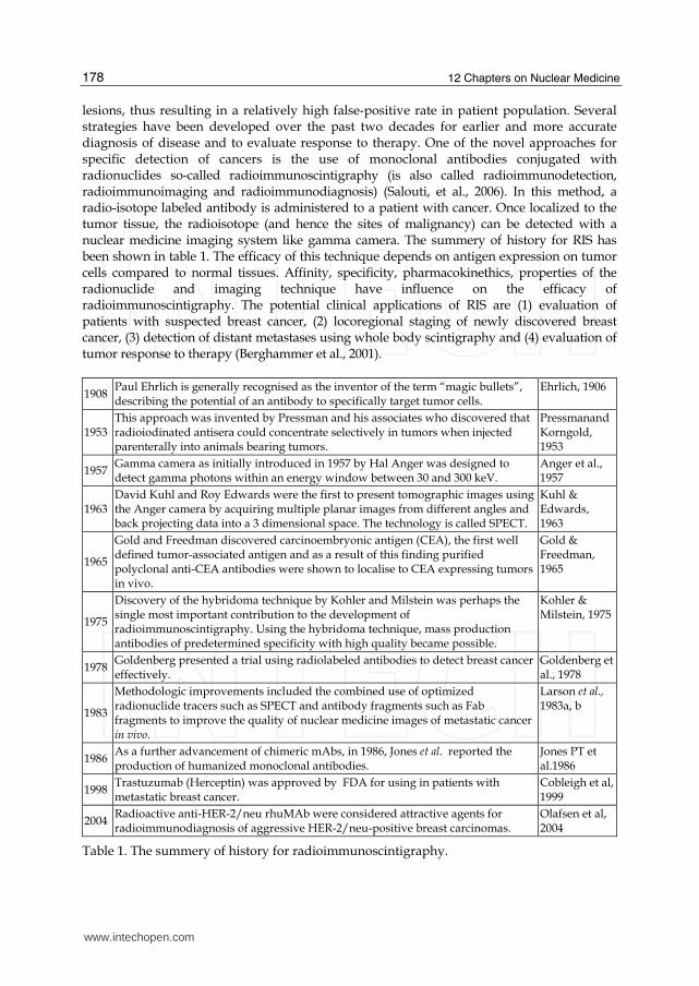
lesions, thus resulting in a relatively high false-positive rate in patient population. Several strategies have been developed over the past two decades for earlier and more accurate diagnosis of disease and to evaluate response to therapy. One of the novel approaches for specific detection of cancers is the use of monoclonal antibodies conjugated with radionuclides so-called radioimmunoscintigraphy (is also called radioimmunodetection, radioimmunoimaging and radioimmunodiagnosis) (Salouti, et al., 2006). In this method, a radio-isotope labeled antibody is administered to a patient with cancer. Once localized to the tumor tissue, the radioisotope (and hence the sites of malignancy) can be detected with a nuclear medicine imaging system like gamma camera. The summery of history for RIS has been shown in table 1. The efficacy of this technique depends on antigen expression on tumor cells compared to normal tissues. Affinity, specificity, pharmacokinethics, properties of the radionuclide and imaging technique have influence on the efficacy of radioimmunoscintigraphy. The potential clinical applications of RIS are (1) evaluation of patients with suspected breast cancer, (2) locoregional staging of newly discovered breast cancer, (3) detection of distant metastases using whole body scintigraphy and (4) evaluation of tumor response to therapy (Berghammer et al., 2001).
1908 Paul Ehrlich is generally recognised as the inventor of the term “magic bullets”, describing the potential of an antibody to specifically target tumor cells.
Ehrlich, 1906
1953 This approach was invented by Pressman and his associates who discovered that radioiodinated antisera could concentrate selectively in tumors when injected parenterally into animals bearing tumors.
Pressmanand Korngold, 1953
1957 Gamma camera as initially introduced in 1957 by Hal Anger was designed to detect gamma photons within an energy window between 30 and 300 keV.
Anger et al., 1957
1963 David Kuhl and Roy Edwards were the first to present tomographic images using the Anger camera by acquiring multiple planar images from different angles and back projecting data into a 3 dimensional space. The technology is called SPECT.
Kuhl & Edwards, 1963
1965
Gold and Freedman discovered carcinoembryonic antigen (CEA), the first well defined tumor-associated antigen and as a result of this finding purified polyclonal anti-CEA antibodies were shown to localise to CEA expressing tumors in vivo.
Gold & Freedman, 1965
1975
Discovery of the hybridoma technique by Kohler and Milstein was perhaps the single most important contribution to the development of radioimmunoscintigraphy. Using the hybridoma technique, mass production antibodies of predetermined specificity with high quality became possible.
Kohler & Milstein, 1975
1978 Goldenberg presented a trial using radiolabeled antibodies to detect breast cancer effectively.
Goldenberg et al., 1978
1983
Methodologic improvements included the combined use of optimized radionuclide tracers such as SPECT and antibody fragments such as Fab fragments to improve the quality of nuclear medicine images of metastatic cancer in vivo.
Larson et al., 1983a, b
1986 As a further advancement of chimeric mAbs, in 1986, Jones et al. reported the production of humanized monoclonal antibodies.
Jones PT et al.1986
1998 Trastuzumab (Herceptin) was approved by FDA for using in patients with metastatic breast cancer.
Cobleigh et al, 1999
2004 Radioactive anti-HER-2/neu rhuMAb were considered attractive agents for radioimmunodiagnosis of aggressive HER-2/neu-positive breast carcinomas.
Olafsen et al, 2004
Table 1. The summery of history for radioimmunoscintigraphy.
www.intechopen.com
Breast Cancer: Radioimmunoscintigraphy and Radioimmunotherapy 179
7.1 Radionuclides for imaging
The most common radionuclides in nuclear medicine are 99mTc, 123I, 67Ga and 111In. Lower energy ┛-rays are readily absorbed in tissues and therefore less useful for external imaging. On the other hand, highest energy ┛-rays cause to decrease the sensitivity of imaging system (Berghammeret al., 2001). Technetium-99m is so far the most commonly used radionuclide in nuclear medicine (Hamoudeh et al., 2008). This is due to the highly interesting physical properties of 99mTc which is advantageous for both effective imaging and patient safety perspectives. Its properties include short half-life (6 h), gamma energy at 140 keV with practically no alpha or beta emissions and latent chemical properties, facilitating thereby the labeling of several types of kits for versatile diagnostic applications and readily available and inexpensive (it is derived as a column elute from a 99Mo/99mTc generator) (Verhaar et al., 2000). It is most often used with smaller antibody forms such as Fab, scFv, diabodies and nanobodies. The gamma-ray emitting radionuclides are commonly used in gamma camera and single photon emission tomography (SPECT). Other groups of diagnostically used radionuclides are ß+ emitters such as 11C, 18F, 13N and 15O (Hamoudeh et al., 2008). The positron emitting radionuclides are used in positron emission tomography (PET). The positive electron travels only a short distance through the tissues and interacts with a free or loosely bound negative electron. The outcome of this interaction is two photons, consisting each of 511 keV energy and being given off in opposite directions (Boswell & Brechbiel, 2007).
7.2 Imaging systems in nuclear medicine
Imaging systems in nclear medicine include gamma camera, single photon emission computed tomography (SPECT) and positron emission tomography (PET).
7.2.1 Gamma camera
Gamma camera is a one-headed, variable-angle diagnostic instrument that is used to image gamma radiation emitting radioisotopes, a technique known as scintigraphy. Gamma camera consists of a scintillation crystal, optically coupled to an array of photomultiplier tubes which converts the ┛-rays into electric signals.
7.2.2 Single photon emission tomography
Single photon emission computed tomography (SPECT) is a sensitive nuclear imaging technique that provides a 3D spatial distribution of single-photon emitting radionuclide within the body (Gomes et al., 2010). Multiple 2D images, also called projections, are acquired from multiple angles around the patient and subsequently reconstructed using the reconstruction imaging methods to generate cross-sectional images of the internal distribution of the injected molecules (Wernick, & Aarsvold,2004). Because of the isotropic emission of ┛-rays, a geometric collimation is needed to restrict the travelling direction of the emitted ┛-rays from the body, through the use of lead collimators. In clinical systems, collimators typically have many parallel holes that produce no magnification. The photons that travel in other directions than those specified by the aperture of collimator are absorbed and do not contribute to the image, which reduces the detection efficiency and sensitivity of SPECT as compared to PET (Madsen, 2007). SPECT have several advantages including (1) whole body imaging, (2) quantitative molecular imaging and (3) can be combined with CT for preparing anatomical information. The disadvantages of this method are radiation exposure, low spatial resoulation (0.3-1 mm, 12-15 mm3) and long acquisition time (Pysz et al., 2010).
www.intechopen.com
12 Chapters on Nuclear Medicine 180
7.2.3 Positron emission tomography (PET)
Positron emission tomography is a nuclear medicine technique that produces high resolution tomographic imaging through the detection of high energy photon pairs emitted during positron decay (Costelloeet al., 2009). This method was initially developed in 1960s, but has largely been used as a research tool. However, PET can provide useful information for clinical practice. The images generated by PET represent the metabolic activity of the underlying tissues and can therefore distinguish benign from malignant lesions on the basis of differences in metabolic activity. Similarly, it can identify recurrent diseases in areas in which conventional scans are difficult to interpret because of prior treatment (Costelloeet al., 2009). PET represents the most advanced imaging technique, because it not only allows a three-dimensional image reconstruction, but also it can quantify the activity uptake (Fass, 2008). It combines the highest degree of sensitivity with a resolution of currently, 5-7 mm. The principal applied radionuclide for PET is Fluor-18 (18F) which is known for its ideal half-life to manage (1.83 h). The development of radiopharmaceutical [2-18F]-2-fluoro-2-deoxyglucose (18FDG) has been so far an important progress for PET imaging in oncology (Berghammer et al., 2001). 18FDG acts as a glucose analogue allowing for the visualization of glucose consumption, a metabolic process being massively enhanced in many malignancies (Einat & Moshe, 2010). PET has several advantages include (1) unlimited depth penetration; (2) whole body imaging possible, (4) quantitative molecular imaging and (5) can be combined with CT or MRI for anatomical information (Pysz et al., 2010). The disadvantage of PET is that it requires a conveniently located and expensive cyclotron and radiochemistry facilities to produce the short-half life isotopes and to incorporate these into suitable probe molecules (Fass, 2008).
8. Radioimmunotherapy
The first theory on the existence of proteins with specific binding capabilities to pathogenic organisms, thus acting as "magic bullet"’, was postulated at the end of the 19th century by the german pathologist Paul Ehrlich (Enelich, 1906). He was the first to recognise antibodies for their ability to differentiate between normal cells and transformed malignant cells. He specifically introduced immunotherapy as a potential treatment modality for targeting and treating tumors. After it had been recognized in 1950 that proteins could be labeled with 131I without significantly altering their immunological specificity (Eisen & Keston, 1950), Pressman and Korngold tested the tumor-targeting potential of a 131I-labeled rabbit antiserum in rats bearing osteosarcoma and confirmed preferential antibody uptake in the tumor xenografts (Pressman & Korngold, 1953). The first clinical trial investigated the therapeutic efficacy of radiolabeled antibodies was performed in the 1950s by Beierwaltes, who treated fourteen patients with metastatic melanoma with 131I-labeled rabbit antibodies and reported a pathologically confirmed remission in one patient (Beierwaltes, 1974). In 1965, Gold and Freedman discovered carcinoembryonic antigen (CEA), the first well defined tumor-associated antigen. The purified polyclonal anti-CEA antibodies were shown to localize to CEA expressing tumors in vivo (Gold & Freedman, 1965). In the late 1970s, Goldenberg and colleagues successfully targeted colon cancer in patients using a polyclonal goat anti-CEA antiserum (Goldenberg et al., 1978). Nowadays, CEA has not only become one of the most extensively used tumor markers in clinical oncology, but also due to its pronounced expression in various carcinomas, it is one of the most targeted antigens in RIT. In 1975, Köhler and Milstein reformed the field of radioimmunotargeting as they introduced the hybridoma
www.intechopen.com

Breast Cancer: Radioimmunoscintigraphy and Radioimmunotherapy 181
technology, a method that made it possible to produce large quantities of monoclonal antibodies with high purity and reproducibility (Kohler & Milstein, 1975). Since then, numerous antigen-antibody systems have been established and several of the antibodies have been taken to clinical trials. Radioimmunotherapy is a method of selectively delivering radionuclides with toxic emissions to cancer cells, while reducing the dose to normal tissues. Using mAbs labeled with radionuclides has two major advantages over the application of mAbs conjugated with either drugs or toxins. Firstly, tumor cells not expressing the target antigen can still be sterilized by the so-called crossfire phenomenon, i.e., radiation energy emitted by radionuclides bound to antibodies targeting adjacent tumor cells. Secondly, radionuclides are not subject to multidrug resistance. Although promising, RIT has been less effective for solid tumors, in part because they are less radiosensitive. However, early micrometatasis of breast cancer have been demonstrated to be radiosensitive (Koppe et al., 2005). On the other hand, an advantage of RIT is that it can target small metastatic lesions that are undetected by conventional scanning and would otherwise remain untreated. In addition, RIT is able to target multiple metastases throughout the body in a single treatment.
8.1 Radionuclides for treatment
The selection of a radionuclide for cancer treatments depends on several parameters including: 1. Physical parameters: The type of radiation emitted by the radionuclide, required
energy necessary for therapy and half-life of the radionuclide are physical parameters that must be considered. The type of radiation and the content of its energy are important factors that determine what radionuclide is suitable for killing of single disseminated cells, small metastases or large cancer tissues. The physical half-life of the radionuclides should preferably be in the same order of magnitude as the biological half-life. A too long physical half-life increases the necessary amount of radionuclide to be delivered to the tumor cells to allow the reasonable amounts of decays before excretion. A shorter physical half-life, on the other hand, will not give enough time for the targeting process to take place. It seems reasonable to assume that the most suitable physical half-lives ranges from a few hours up to some days when targeting of disseminated cells is considered (Boswell & Brechbiel, 2007).
2. Chemical parameters: The chemical parameters are as follow: achievable specific activity, stability of the radionuclide/antibody complex after labeling and that the labeling procedure must not interfere with the immunological activity of the antibody.
3. Biological parameters: tumor type, size and location, antibody kinetics, antigen density and heterogeneity and antigenicity are the most important biological parameters.
4. Other parameters: radioisotope availability and its cost. Three main categories of radionuclides have been investigated for their potential therapeutic characterisation in radioimmunotherapy including ┚-particle emitters, ┙-particle emitters and auger electron cascades.
1. ┚-particle emitters
So far, the vast majority of preclinical and clinical studies have been made to use ┚-emitting radionuclides such as 131I, 90Y, 186Re and 188Re. These radionuclides have a tissue range of about several millimeters. This can create a ‘‘cross fire’’ effect, so that antigen or receptor negative cells in a tumor can also be treated. High energy B-particles are not efficient for killing of single disseminated cells or small metastases. So, ┚-particle therapy is preferred for large tumors (Boswell & Brechbiel, 2007).
www.intechopen.com
12 Chapters on Nuclear Medicine 182
2. ┙-particle emitters
Radionuclides emitting ┙-particles such as 225Ac (half-life 10 days), 211At (half-life 7.2 h), 212Bi (half-life 60.55 min) and 213Bi (half-life 45.6 min) are options for treatment of small tumor nests or single disseminated tumor cells. Alfa particle emitting radionuclides are short ranged, high-energy helium nuclei with a high linear energy transfer (LET). As a consequence, ┙-emitters have a high relative biological effectiveness (RBE) which means, if nuclear localisation is possible, fewer radionuclides per cell are needed (Fass, 2009).
3. Auger electron cascades
Auger-electrons, discovered by Lise Meitner in 1922 and by Pierre Auger in 1923, are formed when the vacancy created in an inner shell is filled with an electron from a higher energy level after electron capture. Auger-electrons have high LET Like ┙-particles (Cornelissen & Vallis, 2010). Auger-electron emitters, like 125I, deposit a concentrated amount of energy in even shorter distances than ┙-emitters. This means that these radionuclides need to be located in the vicinity of the tumor cell nucleus to be effective. For this reason, antibodies labeled with auger-emitting radionuclides need to target the entire tumor cell population for efficient therapy (Cornelissen & Vallis, 2010).
9. Improving the properties of antibody in radioimmunoscintigraphy and radioimmunotherapy
Several investigations using different radionuclides, engineered antibodies and methods to increase antibody accumulation and penetration are currently being evaluated and have so far shown promising results. The main properties of antibodies that have been manipulated to optimize efficacy are size, immunogenicity, affinity and avidity.
9.1 Size Size is a factor that impacts the circulation time of antibodies. IgG antibodies are large proteins with a molecular weight of 150 kDa which limits the diffusion of the antibodies from the blood into the tumor, resulting in a heterogeneous intratumoral distribution. Furthermore, IgG antibodies are characterized by a long circulatory half-life in plasma for three to four days. Due to this slow clearance from the blood, tumor-to-background ratio is usually low. The primary concern for using radionuclide labeled IgG is that it remains in the blood for an extended period of time which continually exposes the highly sensitive red marrow to radiation resulting in dose-limiting myelosuppression. While intact mAbs are primarily catabolized by the liver and spleen, mAb fragments are mainly excreted via the kidneys, thereby increasing uptake in the kidneys and lead to increase consequently the kidney absorbed radiation dose. (Koppe et al., 2005). If radiometals are used as the radiolabel, they will accumulate in the hepatic parenchyma. The large size of an antibody impacts its ability to move through a tumor mass. The smaller forms of antibodies such as F(ab')2 or Fab fragments and more recently, molecularly engineered antibody subfragments with more favorable pharmacokinetic properties, are removed more rapidly from the blood, thereby improving tumor/blood ratio. There have been reports of improved therapeutic responses using smaller-sized antibodies, but these smaller entities frequently are cleared from the blood by renal filtration and as a result many radionuclides (eg, radiometals) become trapped in a higher concentration in the kidneys than in the tumor. Changes in the molecular size/structure of the IgG can also alter the normal tissue distribution, shifting uptake from the liver to the kidneys (Sharkey & Goldenberg, 2009). Reducing the size of
www.intechopen.com
Breast Cancer: Radioimmunoscintigraphy and Radioimmunotherapy 183
antibody below the filtration threshold of kidneys (70 kDa), increases renal excretion and therefore decreases toxicity to this organ. Hence, being smaller molecules, antibodies are more suited to RIT and RIS with short circulation time, lower absolute localisation to the tumor and rapid excretion by the kidneys (Yazaki et al., 2001).
9.2 Immunogenicity
The first mAbs being investigated for RIS and RIT were murine antibodies which can provoke an immune response in human beings. HAMA inactivate and eliminate murine antibodies after repeated administration. The formation of antibody-HAMA complexes also leads to the allergic-like HAMA response. In this way, therapeutic benefit of murine mAbs is limited by their side effect profile, short serum half-life and inability to trigger human immune effector functions. In order to reduce the immunogenicity of antibodies, chimeric antibodies were designed by combining constant domains of human antibodies with variable regions of murine antibodies (Carlssona et al., 2003). However, chimeric mAbs minimize the immunogenic content, trigger the immunologic efficiency and allow a prolonged serum half-life in comparison with murine mAbs. As a further advancement of chimeric mAbs, in 1986, Jones first reported the production of humanized monoclonal antibodies (Jones et al., 1986). Humanized antibodies are almost completely of human origin with only the complementarity determining regions (CDRs) being murine. To completely avoid the risk of immunogenicity, further developments have led to the production of fully human antibodies that contain 100% human proteins. For the development of fully human mAbs, phage display technology and genetically engineered mice are the key techniques that have been widely used to link genotype and phenotype. Immunosuppressive agents have been investigated to reduce HAMA. Low-dose cyclosporin, as used with a highly immunogenic antibody, was unable to significantly reduce HAMA following murine CC49 delivery. Thus, cyclosporin may have some efficacy in reducing immunogenicity of murine antibodies in patients, but does not appear to be sufficient to permit administration of multiple doses in all patients (Pagel, 2009).
9.3 Affinity and avidity
The antibody affinity is a measure of the strength of binding of an individual antibody binding site to a single antigenic site. This can be considered as the sum of all the non-covalent interactions between antibody and antigen involved in the binding reaction. However, antibody molecules usually have more than one binding site and many antigens contain more than one antigenic site and therefore multivalent binding may be possible. The strength with which a multivalent antibody binds to an antigen, is termed avidity. Although high affinity is a requirement for good tumor localization, there seems to be a point at which further increases in affinity do not increase uptake at the target site (Schlomet al., 1992; Colcher et al., 1988). Indeed, reduced tumor uptake and limitations on penetration of antibody into the tumor tissue can result from the increasing of antibody affinity (Dearling & Pedley, 2007).
10. Technical limitations of radioimmunoscintigraphy and radioimmunotherapy
Although this conceptually simple technique has been investigated and refined for almost 50
years, it still has inherent limitations. In the present part, the problems of imaging and therapy
of breast cancer by radioimmunoscintigraphy and radioimmnotherapy methods are discussed.
www.intechopen.com
12 Chapters on Nuclear Medicine 184
10.1 Antibody
Monoclonal antibodies have inherent limitations for application in targeting methods of imaging and therapy that can be cited in followings:
• mAbs are large molecules and so have difficulty in penetrating to large tumor masses especially in the early stages of the malignancies (Sergides et al., 1999).
• mAbs are currently not entirely sensitive for malignant tissues. For example, mAb B72.3 recognizes TAG-72 and has been used extensively for the detection of several malignancies including breast, lung, ovary and colorectal (Granowska et al., 1991).
10.2 Antigen
Tumors produce a chaotic vascular system in which blood flow is slow and can be interrupted or even reversed. The ratio of tissue cells to vascular support is lower than most normal tissues. These effects create areas of tumor hypoxia which are relatively resistant to radiation therapy and therefore reduce the efficacy of RIT (Sergides et al., 1999). Some antigens of breast cancer are epithelial surface antigens lying on the inner surfaces of cells, thus being exposed to the circulation only by neoplastic architectural disruption. Other factors influencing the suitability of antigens for tumor targeting are internalization and shedding into the bloodstream. Unfortunately, the majority of identified antigens in human tumors represent tumor-associated antigens, not only present on the tumor tissues, but also detectable on normal tissues (Sergides et al., 1999).
10.3 Background radioactivity
A high background level of radiation due to the presence of radioactivity in normal tissues
reduces the tumor/background ratio which reduces the success of diagnosis and therapy.
Tumor/background ratio may be diminished by the following factors: (1) the relative long
circulation time of nonlocalized murine immunoglobulin in human beings; (2) binding of
radiolabeled antibodies to antigens, released by the tumor in the blood pool; (3) the presence
of free radionuclides and the subsequent accumulation in kidneys, bladder and other
tissues, (4) non-specific uptake antibody by binding Fc fragment of antibody to normal
tissues; (5) phagocytosis of murine immunoglobulin especially in the liver; (6) the presence
of immune complexes due to the reaction of labelled antibody with Fc cell surface receptors,
if non-fragmented immunoglobulins are used and (7) unconjugation of radio-isotope from
antibody in the patient body (Sergides et al., 1999).
11. Improving in radioimmunoscintigraphy and radioimmunotherapy techniques
A variety of methods have been developed to counter the inherent flaws in RIS and RIT techniques. The following section discusses the methods for overcoming these problems.
11.1 Pre-targeting
The pretargeting procedure was developed from the concept that the targeting antibody should be separated from the targeting radionuclide through the use of a bispecific antibody (Chang et al., 2002). An alternative approach to improve tumor:blood ratio for RIS is the use of pre-targeting strategies. The pre-targeting strategies have led to significant improvements in T/B ratio and better diagnostic imaging (Dearling & pedley, 2007). Also, pretargeting has been
www.intechopen.com
Breast Cancer: Radioimmunoscintigraphy and Radioimmunotherapy 185
applied successfully for radioimmunoimaging, because the pretargeted antibody is nontoxic. High doses can be administered to saturate antigenic sites at the tumor. Pretargeting strategies for RIT have been applied to achieve higher intratumor concentration of isotope than achieved by conventional RIT (Kraeber–Bodere et al., 2009). The simplest form of pre-targeting is to use a second antibody reagent to clear blood background activity, hence improving the signal:noise ratio and the quality of the image. Immune complexes formed by a second antibody are rapidly removed from the circulation by the reticuloendothelial system, particularly in the liver. In the alternative approach, the administration of antibody and radiolabel are separated. Antibody is allowed to localize to the tumor and sufficient time is allowed for antibody clearance from the blood and non-target tissues (Dearling & pedley, 2007). Radioisotope is then injected separately in a form which can be readily captured by the tumor bound antibody. The approaches using streptavidin/biotin binding systems raised much interest, because the affinity of streptavidin for biotin is exceptionally high (Gruaz-Guyon et al., 2005). In this strategy, the high affinity of the avidin:biotin system is used to capture radiolabeled small molecules from the blood as a two-step imaging method. Antibody-avidin conjugate is injected and allowed to be localized to the tumor and cleared from the blood (Dearling & pedley, 2007). Radioactive avidin is then injected which localizes the tumor by taking advantage of the high affinity and specificity of avidin for biotin (Roland et al., 2010, Dearling & pedley, 2007).
11.2 Dose fractionation
Dose fractionation has been proposed as a method to improve the therapeutic effect of radioimmunotherapy (Dearling & pedley, 2007, Denardo et al., 2002). Fractionated radioimmunotherapy may improve therapeutic outcome by decreasing heterogeneity of the dose delivered to the tumor and by decreasing hematologic toxicity, thereby allowing an increased amount of radionuclide that can be administered (Linden et al,. 2005) . A variety of fractionation regimens have been developed and the studies have reported both against and in its favour (Goel et al., 2001; Buchsbaum et al., 1995; Pedley et al., 1993; Schlom et al.,1990; Beaumier et al., 1991). This technique has several advantages including more uniform distribution of mAb and radiation dose, patient-specific radionuclide and radiation dose, control toxicity by titration of an individual patient, reduced toxicity, increased tumor radiation and efficacy and prolongation of tumor response (Violet, 2008). This technique has some disadvantages including lower radiation dose rate, complex strategy to implement, treatment interruption, increased cost and potential delay in tumor regression (Dearling & pedley, 2007).
11.3 Delayed imaging
Kinetic differences in specific and non-specific uptake of radioactivity provide an opportunity to image at a time when the T/B ratio is optimal. Background radioactivity falls with time due to excretion and decay of the radioisotope. Tumor radioactivity also falls with time, but not as fast as the background. The optimum time for RIS is dependent on the selected antibody and radioisotope and detection method (Sergides et al., 1999).
11.4 Background subtraction
If the background is labeled using a non-specific antibody, the background signal can then be subtracted from the results of RIS (Sergides et al., 1999; Goldenberg et al., 1978). It should
www.intechopen.com
12 Chapters on Nuclear Medicine 186
be remembered that this technique leads to increase the total dose of radioactivity in the patient body that leads to undesirable side effects.
11.5 Pre-scouting
In the latter application, RIS is performed as a scouting procedure prior to RIT to enable the confirmation of tumor targeting and the estimation of radiation dose delivered to both tumors and normal tissues. For this purpose, the radioimmunoconjugates used for RIS and RIT should demonstrate a similar biodistribution and therefore radionuclides with comparable chemical properties have to be chosen (Dearling & pedley, 2007). When using 131I for RIT, both 123I and 131I can be used for a RIS scouting procedure. When using 90Y for RIT, 111In can be used to represent this pure beta-emitter (Dearling & pedley, 2007). Finally, when aiming the therapeutic use of 186Re or 188Re, either these radionuclides themselves can be used for an imaging procedure or 99mTc (Pontus et al., 2004).
11.6 Increasing the dose delivery
The result of experiments indicated that increasing the dose of antibody will increase the amount of radioactivity in the tumor (Begent et al., 1987). On the other hand, this technique has side effects due to HAMA response against the dramatic rise in dose of administered antibody that limits the application of this technique (Dearling & pedley, 2007).
11.7 Combined imaging systems
The resolution of latest-generation of PET and SPECT cameras has been improved and the combination of these systems with an integrated CT (and also MRI) has led to a much better interpretation of the data (Dearling & pedley, 2007). In addition, the indirect combination of nuclear imaging and optical imaging systems can also serve shared purposes (Munnink et al, 2009).
11.8 Second antibody
The background counts can be reduced by removing radioactivity from the circulation. This can be achieved by the use of a second antibody active against the RIS antibody. In this technique, after the binding time of original antibody to tumor tissue, the second antibody administers to the patient (Sergides et al., 1999). The second antibody binds to the radioactive antibody that remains in the blood stream. The reticuloendothelial system uptakes antibody-antibody complex and leads to concentrate radioactivity in liver and spleen. This technique leads to increase in T/B ratio without adverse effects. The only disadvantage of this method is limitation in the detection of metastases at liver and spleen (Sergides et al., 1999).
11.9 Improving the internalization of the radiolabeled mAb
One significant factor that influences the absorbed radiation dose to the tumor is the fate of the radiolabel after internalization of the radiolabeled mAb into the tumor cells. In case of internalisation, the radionuclides will come closer to the critical radiation target, i.e. nuclear DNA. Internalization of the antibody depends on various factors including the antibody, the targeted antigen and the tumor cells. On the other hand, internalisation might be disadvantageous, if it leads to quick degradation of the targeting agent followed by diffusion and elimination of the radionuclide (Boswell et al., 2007). Most antibodies including those
www.intechopen.com

Breast Cancer: Radioimmunoscintigraphy and Radioimmunotherapy 187
that target antigens located on the surface of the tumor cells such as anti-CEA antibodies, are eventually catabolized. Intracellular degradation of the targeting agent can be prevented by e.g. dextranation (Bue et al., 2000). Radiolabeling of antibodies with 90Y or 177Lu is performed by linking these radionuclides to chelators (DTPA or DOTA) which are chemical moieties that complex free metal ions. These chelators are conjugated to the antibodies and subsequently to the radiolabel. After catabolization of mAbs labeled with 90Y- or 177Lu-DTPA/-DOTA, the catabolic products are conjugated to, in most cases, lysine (e.g. 90Y- or 177Lu-DOTA-lysine). Whereas radioiodinated tyrosine is excreted by the cell, the 90Y- or 177Lu-DTPA/DOTA-lysine metabolites are trapped within the lysosomes, thereby increasing the tumor retention time of these radiolabels (Koppe et al., 2005). Cellular excretion can also be limited if the radionuclides are of metal type, e.g. indium or yttrium, and this is due to intracellular retention of metal containing catabolic products (Press et al., 1996).
11.10 Targeting agents
RIS may be improved by the production of superior targeting agents to deliver radioactivity
to tumor sites. For example, instead of using intact antibodies to target tumors, fragments of
antibodies retaining their antigenic specificity can be used (Sergides et al., 1999). Fragments
of an antibody have several advantages including:
1. Shorter circulating half-life and faster execration by kidney that leads to decrease background signals.
2. Smaller molecular weight that leads to faster and more deeply diffuse into tumors. 3. Reduce non-specific antibody binding by Fc receptor on cells due to the loss of the Fc
fragment 4. Reduce the HAMA response due to the loss of Fc fragment that is responsible for
immune response to foreign proteins. Further improvements in the structure of antibodies are being explored with antibody engineering technology. Single-chain antigen binding fragments (sF7) consisting of variable heavy (VH) or variable light (VL) domains and recombinant sF7 peptides which are VH and VL domains connected by peptide linkers are the productions of this technology. These productions has high affinity for antigen (Sergides et al., 1999).
11.11 Antibody affinity
The strength of the antibody:antigen interaction is measured through the binding affinity and is quantified through the association constant (Ka) (Boswell et al., 2007). Increasing Ka of antibody will be leads to an increase in uptake antibody in the tumor. However, they may be less able to penetrate deeply due to strongly bound at the tumor surface. The studies with iodine radiolabeled scFvs demonstrated that a threshold affinity between 10−7 and 10−8 M was required to observe detectable tumor uptake in mice 24 h post injection, whereas no gain in tumor accumulation was observed with affinities >10−9 M. Affinities >10−9 M were detrimental to rapid and uniform tumor penetration due to stable binding at the first pass of tumor antigens forming a binding site barrier (Adams et al., 2001).
11.12 Multivalency
Multivalency has been reported as an advantage in radioimmunotherapy (Dearling & pedley, 2007). Conversion of monovalent antibodies into multivalent format increases their functional affinity and decreases dissociation rates for cell-surface and optimizes biodistribution
www.intechopen.com

12 Chapters on Nuclear Medicine 188
(Dearling & pedley, 2007). In addition, it allows the creation of bispecific antibody molecules that can target two different antigens simultaneously (Sergey et al., 2008). During the last decade, several techniques in multivalency engineering have been developed. Each of these strategies proposed to link monovalent domains and to produce multivalent antibody, obviously has some advantages in some special cases, but none of them is universal. The advantages of tumor targeting with multivalent antibody derivatives have been investigated for scFv dimers, prepared as disulfide-linked dimers of scFv (Adams et al., 1995), non-covalent diabodies (Kortt et al., 2001) and some other bi(multi)valent variants of recombinant antibody fragments (Kubetzko et al.,2006; Shahied et al., 2004; Willuda et al., 2001). In most cases, dimeric/divalent antibodies showed significant improvement of their pharmacokinetics and biodistribution over monomers (Sergey et al., 2008).
11.13 CockTails
One problem of RIS is the existence of diversity of epitopes on surface tumor cells that reduces uptake radioactivity in tumor. An alternative approach to improve the uptake of radioactivity by the target is using cocktails of several radiolabelled antibodies that recognize different epitopes or antigens on the same tumor. However, some of the experiments indicated positive results by using cocktails, other experiments showed that the mAbs of cocktails competed to bind to epitopes with each other and reduce the efficacy of the mixture to less than that of one antibody used alone (sergey et al., 2008).
11.14 Increasing tumor uptake
By increasing the level of antigen expression, diagnosis and therapy based on targeting can be improved. For example, hyperthermia can increase the amount of presenting antigen in a tumor (Wilder et al., 1993). Furthermore, several cytokines such as interferon alpha and interleukin-6 have been found to upregulate the expression of cell surface antigens including histocompatibility antigens and tumor associated antigens such as carcinoembryonic antigen (CEA). In both in vivo and in vitro studies an improved antibody-uptake has been demonstrated as a result of the administration of vasoactive peptides and cytokines such as IL-2 or external beam radiation therapy may result in a specific increased vascular permeability at the tumor site and thus an increased antibody uptake (Guadagni, et al., 1990). Structural modification (e.g., PEGylation) and residue mutation are both useful strategies in reducing chemically or physically derived nontarget organ uptake of Abs, but these methods do not reduce the uptake due to receptor expression within nontarget organs. These are research areas of high interest (Boswell & Brechbiel, 2007).
11.15 Reducing kidney uptake
Elevated renal uptake and prolonged retention of radiolabeled antibody is a problem in the therapeutic application of such agents (Dearling & pedley, 2007). Because of the negative charge of the basement membrane of the glomeruli, positively charged catabolites of the radiopharmaceutical may be retained in the kidney, increasing the toxic absorbed dose to this radiosensitive organ (Dearling & pedley, 2007). The result of some studies demonstrated that lysine, histidine and arginine were effective in reducing the renal uptake of radiopharmaceutical and lysine was the most effective (Lin et al., 2007). In these studies it has been shown that renal retention can also be minimized by the administration of lysine, a cationic amino acid, whereas the uptake in all other organs as well as the tumor remains unaffected (Behr et al., 1995).
www.intechopen.com

Breast Cancer: Radioimmunoscintigraphy and Radioimmunotherapy 189
12. Conclusion
In recent years, significant developments in the application of targeted imaging and therapy have taken place in nuclear medicine. This chapter suggests that optimization of radioimmunoscintigraphy and radioimmunotherapy methods is still possible and emphasizes that these technologies are continuing to progress and are close to become routine modalities in the identification of breast cancer sites and its therapy. The development of effective imaging and therapy of breast cancer relies to a great extent on the development of effective carriers and target agents that can deliver radionuclids to the cancer cells. These agents should be able to carry a large load of radionuclids and selectively deliver them to the cancer cells with high accuracy to achieve effective cancer cells death without inducing nonspecific toxicity. This means that the use of new targeting agents such as peptides and affibodies can provide promising results. Liposomes, dendrimers, micelles and nanoparticles present large families of carriers that can be exploited for delivery of radionuclids and they can be further improved to diagnose and therapy of breast cancer in the future. The research in the field of targeted imaging and therapy will help us to avoid unnecessary costs and potentially allow these new methods to be available for the majority of patients who need them, leading to better quality and quantity of life.
13. References
Adam, F., Alberto, H., Piera, S., Cristina, C., Armando, G., & Fabio, M. (2000). Monoclonal antibodies and therapy of human cancers. Biotechnology Advances, Vol.18, pp. 385–401.
Adams, GP., McCartney, JE., Wolf, EJ., Eisenberg, J., & Huston, JS. (1995). Enhanced tumor specificity of 741F8-1(sFv’)2, an anti-c-erbB-2 singlechain Fv dimer, mediated by stable radioiodine conjugation. The Journal of Nuclear Medicine, Vol.36, pp.2276–2281.
Adams, GP., Schier, R., McCall, AM., Simmons, HH., Horak, EM., & Alpaugh, RK. (2001). High affinity restricts the localization and tumor penetration of single-chain Fv antibody molecules. Cancer Research, Vol. 61, pp. 4750–5.
Balleyguier, C., Opolon, P., Mathieu, MC., Athanasiou, A., Garbay, JR., & Delaloge, S. (2008). New potential and applications of contrast-enhanced ultrasound of the breast: Own investigations and review of the literature. European Journal of Radiology ,
Baselga, J., & Cameron, D. (2007). Objective response rate in a phase II multicenter trial of pertuzumab (P), a HER2 dimerization inhibiting monoclonal antibody, in combination with trastuzumab (T) in patients (Pts) with HER2 positive metastatic breast cancer (MBC) which had progressed during trastuzumab therapy. Journal of Clinical Oncology, American Society of Clinical Oncology (ASCO) Annual Meeting Proceedings, Part I. Vol 25, No. 18S: 1004
Baselga, J., Gelmon, KA., Verma, S., Wardley, A., Conte, P., & Miles, D. (2010). Phase II trial of pertuzumab and trastuzumab in patients with human epidermal growth factor receptor 2-positive metastatic breast cancer that progressed during prior trastuzumab therapy. Journal of clinical Oncology, Vol. 28, pp.1138-44.
Beaumier, PL., Venkatesan, P., & Vanderheyden, JL. (1991). 186Re radioimmunotherapy of small cell lung carcinoma xenografts in nude mice. Cancer Research, Vol. 51, pp.676-681.
www.intechopen.com

12 Chapters on Nuclear Medicine 190
Begent, RHJ., Bagshawe, KD., & Pedley, RB. (1987). Use of second antibody in radioimmunotherapy. Journal of the National Cancer Institute Monographs, Vol. 3, pp. 59–61.
Behr, TM., Sharkey, RM., Juweid, ME., Blumenthal, RD., Dunn, RM., & Griffiths, GL. (1995). Reduction of the renal uptake of radiolabeled monoclonal antibody fragments by cationic amino acids and their derivatives, Cancer Research, 55(17):3825–34.
Behr, TM., Sharkey, RM., & Juweid, ME. (1997). Variables influencing tumor dosimetry in radioimmunotherapy of CEA-expressing cancers with anti-CEA and anti-mucin monoclonal antibodies. The Journal of Nuclear Medicine, vol. 38, pp. 409-418.
Berg, JR., Kalisher, L., & Osmond, JD. (1973). Technetium-99m- disphosphonate concentration in primary breast cancer. Radiology, Vol. 109, pp. 393-394.
Berg, WA., Gutierrez, L., NessAiver, MS., Carter, WB., Bhargavan, M., & Lewis, RS. (2004). Diagnostic accuracy of mammography, clinical examination, US, and MR imaging in preoperative assessment of breast cancer. Radiology, Vol. 233(3), pp.830-849.
Berghammer, P., Obwegeser, R., & Sinzinger, H. (2001). Nuclear medicine and breast cancer: a review of current strategies and novel therapies. The Breast, Vol. 10, pp. 184-197.
Blank, EW., Pant, KD., Chan, CM., Peterson, JA., & Ceriani, RL. (1992). A novel anti-breast epithelial mucin MoAb (BrE-3). Cancer, Vol. 5, pp. 38–44.
Boswell, C., Andrew, C., Brechbiel, & Martin, W. (2007).Development of radioimmunotherapeutic and diagnostic antibodies: an inside-out view. Nuclear Medicine and Biology, Vol. 34, pp. 757–778.
Brem, RF., Rapelyea, JA., & Zisman, G. (2005). Occult breast cancer: Scintimammography with high-resolution breast-specific gamma camera in women at high risk for breast cancer. Radiology, Vol. 237, pp. 274-80.
Buchsbaum, D., Khazaelli, MB., & Liu, T. (1995). Fractionated radioimmunotherapy of human colon carcinoma xenografts with 131I-labeled monoclonal antibody CC49. Cancer Research, Vol. 55, pp. 5881-5887.
Bue, P., Holmberg, AR., Marquez, M., Westlin, JE., Nilsson, S., & Malmstrom, PU. (2000). Intravesical administration of EGF-dextran conjugates in patients with superficial bladder cancer. European Urology, Vol. 38, pp. 584–589.
Capala, J., Kramer-Marek, G., Lee, S., Hassan, M., Kiesewetter, DO., Puri, A., Chernomordik, V., Gandjbakhche, A., Griffiths, G., & Blumentha, R. (2009). Molecular targeting of HER2 for diagnosis and therapy of breast cancer. Cancer Research, Vol. 69, Supplement 1
Carl, AK., Borrebaeck, & Roland, C. (2001). Human therapeutic antibodies. Current Opinion in Pharmacology, Vol. 1, pp. 404–408.
Carlos, M., Mery, MPH., Bilal, M., Shafi, MSE., & Gary, B. (2006). Molecular imaging and radioimmunoguided surgery. Seminars in Pediatric Surgery ,Vol. 15, pp. 259-266.
Carlssona, J., Aronssonb, EF., Hietalac, SO., Stigbrandd, T., & Tennvall, J. (2003). Tumor therapy with radionuclides: assessment of progress and problems. Radiotherapy and Oncology,Vol. 66, pp. 107–117.
Chang, CH., Sharkey, RM., Rossi, EA., Karacay, H., McBride, W., Hansen, HJ., Chatal, JF., Barbet, J., & Goldenberg, DM. (2002). Molecular advances in pretargeting radioimmunotherapy with bispecific antibodies. Molecular Cancer Therapeutics, Vol. 1(7), pp.553-563.
Cleator, S., Heller, W., & Coombes, RC. (2007). Triple-negative breast cancer: therapeutic options. Lancet Oncology, Vol. 8, pp. 235–44.
www.intechopen.com
Breast Cancer: Radioimmunoscintigraphy and Radioimmunotherapy 191
Cobleigh, M., Vogel, C., Tripathy, D., Robert, N., Scholl, S., & Fehrenbacher, L. (1999). Multinational study of the efficacy and safety of humanized anti-HER2 monoclonal antibody in women who have HER2-overexpressing metastatic breast cancer that has progressed after chemotherapy for metastatic disease. Journal of clinical Oncology, Vol. 17, pp. 2639–48.
Colcher, D., Minelli, MF., Roselli, M., Muraro, R., Simpson-Milenic, D., & Schlom, J. (1988). Radioimmunolocalisation of human carcinoma xenografts with B72.3 second generation monoclonal antibodies. Cancer Research, Vol. 48, pp. 4597-4603.
Cook, GJR. (2003). Oncological molecular imaging: nuclear medicine techniques. British Journal of Radiology, Vol. 76, pp. 152-158.
Cornelissen, B., & Vallis, KA. (2010). Targeting the Nucleus: An Overview of Auger-Electron Radionuclide Therapy. Current Drug Discovery Technologies,Vol. 7.
Costelloe, CM., Rohren, EM., Madewell, JE., Hamaoka, T., Theriault, RL., Tse-Kuan, Y., Lewis ,VO., & Jingfei, M. (2009). Imaging bone metastases in breast cancer: techniques and recommendations for diagnosis. Lancet Oncology, Vol. 10, pp. 606–14.
Dearling, JLJ., & Pedley, RB. (2007). Technological Advances in Radioimmunotherapy. Clinical Oncology, Vol. 19, pp. 457-469.
Denardo, GL., Schlom, J., Buchsbaum, DJ., Meredith, RF., Odonoghue, JA., Sgouros, G., Humm, JL., & DeNardo, SJ. (2002). Rationales, evidence, and design considerations for fractionated radioimmunotherapy. Cancer, Vol.94, pp.1332–1348.
Denardo, S J. (2005). Functional Imaging: Radioimmunodetection and Therapy of Breast Cancer. Seminars in Nuclear Medicine, Vol. 35, pp. 143-151.
Denardo, SJ., Mirick, GR., Kroger, LA., O’Grady, LF., Erickson, KL., Yuan, A., Lamborn, KR., Hellstrom, I., Hellstrom, KE., & Denardo, GL. (1994). The biologic window for chimeric L6 radioimmunotherapy. Cancer,Vol. 73, pp. 1023–32.
Disis, ML., Calenoff, E., & McLaughlin, G. (1994). Existent T-cell and antibody immunity to HER-2/neu protein in patients with breast cancer. Cancer Research, Vol. 54, pp.16–20.
Disis, ML., Pupa, SM., Gralow, JR., Dittadi, R., Menard, S., & Cheever, MA. (1997). High-titer HER-2/neu protein-specific antibody can be detected in patients with early-stage breast cancer. Journal of clinical Oncology, Vol. 15, pp. 3363–3367.
Ehrlich, P. (1906). Collected Studies on Immunology, John Wiley. Einat, ES., & Moshe, I. (2010). PET in women with high risk for breast or ovarian cancer ,
Lancet Oncology, Vol. 11, pp. 899–905. Eniu, A. (2007). Integrating biological agents into systemic therapy of breast cancer:
Trastuzumab, lapatinib, bevacizumab. J BUON, Vol. 12, pp. 119-26. Fass, L. (2008). Imaging and cancer: A review.Molecular oncology, Vol. 2, pp. 115–152. Ferro-Flores, G., Ramírez, M., Melendez-Alafort, L., & Santos-Cuevas, CL. (2010). Peptides
for In Vivo Target-Specific Cancer Imaging. Mini-Reviews in Medicinal Chemistry, Vol.10, pp. 87-97.
Frangioni, JV. (2008). New technologies for human cancer imaging. Journal of clinical Oncology, Vol. 26(24), pp. 4012-4021.
Friedman, M., et al. (2009). Engineering and characterization of a bispecific HER2xEGFR-binding affibody molecule. Biotechnology and Applied Biochemistry. Vol. 54, pp. 121–131.
www.intechopen.com

12 Chapters on Nuclear Medicine 192
Gaffer, SA., Pant, KD., & Shochat, D. (1981). Experimental studies of tumor radioimmunodetection using antibody mixtures against carcinoembryonic antigen (CEA) and colon-specific antigen. cancer, Vol. 27, pp. 101-5.
Gasparini, G.,Longo, R., Torino, F., & Morabito, A. (2005). Therapy of breast cancer with molecular targeting agents. European Society for Medical Oncology, Vol. 16, pp. 28-36.
Gianni, L., Llado, A., Bianchi, G., Cortes, J., Kellokumpu-Lehtinen, PL., & Cameron, DA. (2010). Open-label, phase II, multicenter, randomized study of the efficacy and safety of two dose levels of Pertuzumab, a human epidermal growth factor receptor 2 dimerization inhibitor, in patients with human epidermal growth factor receptor 2-negative metastatic breast cancer. Journal of clinical Oncology, Vol. 28, pp. 1131-7.
Glasspool, RM., & Evans, TRJ. (2000). Clinical imaging of cancer metastasis. European Journal of Cancer,Vol. 36, pp. 1661-1670.
Goel, A., Augustine, S., & Baranowska-Kortyewicz, J. (2001). Singledose versus fractionated radioimmunotherapy of human colon carcinoma xenografts using 131I-labeled multivalent CC49 single-chain Fvs. clinical Cancer Research, Vol. 7, pp. 175-184.
Goel, A., Augustine, S., Baranowska-Kortylewicz, J., & Colcher, D. (2001). Single-Dose versusFractionated Radioimmunotherapy of Human Colon Carcinoma Xenografts Using 131I-labeled Multivalent CC49 Single-chain Fvs1. clinical Cancer Research, Vol. 7, pp. 175-184.
Goel, A., Baranowska-Kortylewicz, J., Hinrichs, SH., Wisecarver, J., Pavlinkova, G., Augustine, S., Colcher, D., Booth, BJ., & Batra, SK. (2001). 99mTc-labeled divalent and tetravalent CC49 single-chain Fvs: Novel imaging agents for rapid in vivo localization of human colon carcinoma. The Journal Of Nuclear Medicine, Vol. 42, pp. 1519-1527.
Gold, P., & Freedman, SO. (1965). Specific carcinoembryonic antigens of the human digestive system. The Journal of Experimental Medicine, Vol. 122, pp. 467–481.
Goldenberg, DM., & Nabi, HA. (1999). Breast Cancer Imaging With Radiolabeled Antibodies. Seminars in Nuclear Medicine, Vol. XXIX, No 1, pp. 41-47.
Goldenberg, DM., Deland, F., & Kim, E. (1978). Use of radiolabeled antibodies to carcinoembryonic antigen for the detection and localization of diverse cancers by external photoscanning. New England Journal of Medicine, Vol. 298, pp. 1384–6.
Goldenberg, DM., Goldenberg, H., Sharkey, RM. (1990). In-vivo antibody imaging for the detection of human tumors. Cancer Treatment Research, Vol. 51, pp. 273–92.
Goldsmith, J., & Signore, A. (2010). An overview of the diagnostic and therapeutic use of monoclonal antibodies in medicine. The Quarterly Journal of Nuclear Medicine And Molecular Imaging, Vol. 54, pp. 574-81.
Gomes, CM., Abrunhosa, AJ., Ramos, P., & Pauwels, EKJ. (2010). Molecular imaging with SPECT as a tool for drug development. Advanced Drug Delivery Reviews. pp. 1-8.
Granowska, M., Mather, SJ., & Britton, KE. (1991). Diagnostic evaluation of 111In and 99mTc radiolabelled monoclonal antibodies in ovarian and colorectal cancer: correlations with surgery. International journal of radiation applications and instrumentation Part B, Nuclear Medicine and Biology, Vol . 18, pp. 413–24.
Gruaz-Guyon, A., Raguin, O., & Barbet, J. (2005). Recent advances in pretargeted radioimmunotherapy. Current Medicinal Chemistry, Vol. 12, pp. 319-338.
www.intechopen.com

Breast Cancer: Radioimmunoscintigraphy and Radioimmunotherapy 193
Guadagni, F., Witt, PL., & Robbins, PF. (1990). Regulation of carcinoembryonic antigen expression in different human colorectal tumor cells by interferon-gamma, Cancer Research, 50: 6248–6255.
Hamoudeh, M., Kamleh, MA., Diab, R., & Fessi, H. (2008). Radionuclides delivery systems for nuclear imaging and radiotherapy of cancer. Advanced Drug Delivery Reviews, Vol. 60, pp. 1329–1346.
Harris, M. (2004). Monoclonal antibodies as therapeutic agents for Cancer. Monoclonal antibodies in cancer therapy, Vol. 5, pp. 292–302.
Hassan, MS., Ansari, J., Spooner, D., & Hussain, SA. (2010). Chemotherapy for breast cancer (Review). Oncology Reports, Vol. 5, pp. 1121-31.
Hawrysz, DJ., & Sevick-Muraca, EM. (2000). Developments toward diagnostic breast cancer imaging using near-infrared optical measurements and fluorescent contrast agents. Neoplasia, Vol. 2(5), pp. 388-417.
Hicke, BJ., Stephens, AW., Gould, T., Chang, YF., Lynott, CK., Heil, J., Borkowski, S., Hilger, CS., Cook, G., Warren, S., & Schmidt, PG. (2006). Tumor targeting by an aptamer. The Journal Of Nuclear Medicine, Vol.47(4), pp. 668-678.
Hind, D., Ward, S., De Nigris, E., Simpson, E., Carroll, C., & Wyld, L. (2007). Hormonal therapies for early breast cancer: systematic review and economic evaluation. Health Technol Assess, Vol. 11, pp. 1-134.
Hong, H., Sun, J., & Cai, W. (2008). Radionuclide-Based Cancer Imaging Targeting the Carcinoembryonic Antigen. Biomarker Insights, Vol. 3, pp. 435–451.
Hou, D., Hoch, H., & Johnston, GS. (1984). A new 111In-bleomycin complex for tumor imaging: preparation, stability, and distribution in glioma-bearing mice. Journal of Surgical Oncology, Vol. 25, pp. 168-75.
Howell, LP., Denardo, SJ., Levy, N., Lund, J., &Denardo, GL. (1995). Immunohistochemical staining of metastatic ductal carcinomas of the breast by monoclonal antibodies used in imaging and therapy: a comparative study. International Journal of Biological Markers, Vol. 10, pp. 126–35.
Huang, S., Armstrong, EA.,Benavente, S. (2004). Dual-agent molecular targeting of the epidermal growth factor receptor (EGFR): Combining anti-EGFR antibody with tyrosine kinase inhibitor. Cancer Research, Vol.64, pp. 5355-5362.
Hudson, PJ., & Kortt. (1999). High avidity scFv multimers; diabodies and triabodies. Immunological Methods, Vol. 231, pp. 177-189.
Imai, K., & Takaoka, A. (2006). Comparing antibody and small-molecule therapies for cancer. Nature, Vol. 6, pp. 714-727.
Izumi,Y., Xu, L., di Tomaso, E., Fukumura, D., Jain, RK. (2002). Tumour biology: herceptin acts as an anti-angiogenic cocktail. Nature, Vol. 416, pp. 279–280.
Jalilian, AR., Aboudzadeh, R., Akhlaghi, M., Shirazi, B., Moradkhani,S.,&Salouti, M. (2007). Production and biological evaluation of [201Tl(III)]bleomycin. Journal of Labelled Compounds and Radiopharmaceuticals, Vol. 50, pp.556–557.
Jalilian, AR., Akhlaghi, M., ShiraziB., Aboudzadeh, R., Salouti, M., Babaii, MH. (2006). [201 TI] (III)- Bleomycin for tumor imaging. Radiochima Acta, Vol. 94, pp. 453-459.
Jonathan, D., Cheng, PT., Rieger M Von., Mehren, G., Adams, P., Louis, m., & Ws, N. (2000). Recent advaces in immunotherapy and monoclonal antibody treatment of cancer. Seminars in Oncology Nursing, Vol. 16, pp. 2-12.
Jones, KL., & Buzdar, AU. (2004). A review of adjuvant hormonal therapy in breast cancer. Endocrine-Related Cancer, Vol. 11, pp. 391-406
www.intechopen.com
12 Chapters on Nuclear Medicine 194
Jones, PT., Dear, PH., Foote, J., Neuberger, MS., & Winter, G. (1986). Replacing the complementarity-determining regions in a human antibody with those from a mouse. Nature, Vol. 321, pp. 522-5.
Kepshire, DS., Davis, SC., Dehghani, H., Paulsen, KD., Pogue, BW. (2007). Subsurface diffuse optical tomography can localize absorber and fluorescent objects but recovered image sensitivity is nonlinear with depth. Applied Optics, Vol.46 (10), pp.1669-1678.
Keshtgar, M., Davidson, T., Pigott, K., Falzon, M., & Jones, A. (2010). Current status and advances in management of early breast cancer. International Journal of Surgery, Vol. 8, pp. 199-202.
Kohler, G., & Milstein, C. (1978). Continuous culture of fused cells secreting antibody for the detection and localization of diverse cancers by external photo scanning. New England Journal of Medicine, Vol. 298, pp. 1384-6.
Kohler, G., & Milstein, C. (1975). Continuous cultures of fused cells secreting antibody of predefined specificity. Nature, Vol. 256, pp. 495–497.
Kopans, DB. (1992). The positive predictive value of mammography. American Journal of Roentgenology, Vol. 158, pp. 521-526.
Koppe, MJ., Postema, EJ., & Aarts, F. (2005). Antibody-guided radiation therapy of cancer. Cancer and Metastasis Reviews, Vol.24, pp. 539–567.
Kortt, AA., Dolezal, O., Power, BE., & Hudson, PJ. (2001). Dimeric and trimeric antibodies: high avidity scFvs for cancer targeting. Biomolecular Engineering, Vol. 18, pp. 95–108.
Kraeber–Bodere, F., Goldenberg, DM., Chatal,JF., & Barbet,J. (2009). Pretargeted radioimmunotherapy in the treatment of metastatic medullary thyroid cancer.
Current Oncology, Vol. 16(5), pp. 3–8. Kramer, EL., Denardo, SJ., Liebes, L., Kroger, LA., Noz, ME., Mizrachi, H., Salako, QA.,
Furmanski, P., Glenn, SD., & Denardo, GL. (1993). Radioimmunolocalization of breast carcinoma using BrE-3 monoclonal antibody: phase I study. The Journal Of Nuclear Medicine, Vol. 34, pp. 1067–74.
Kubetzko, S., Balic, E., Waibel, R., Zangemeister-Wittke, U., & Pluckthun, A. (2006). PEGylation and multimerization of the anti-p185HER-2 single chain Fv fragment. D5:effects on tumor targeting. The Journal of Biological Chemistry, Vol. 281, pp. 35186–351201.
Larson, SM., Divgi, C., Scott, A., Daghighian, F., Macapilnac, H., & Welt, S. (1994). Current Status of Radioimmunodetection. Nuclear Medicine and Biology,Vol. 21, pp. 721-729.
Lawrence, LJ., Kortt, AA., Iliades, P., Tulloch, PA., & Hudson, PJ. (1998). Orientation of antigen binding sites in dimeric and trimeric single-chain Fv antibody fragments. FEBS Letters, Vol. 425(3), pp. 479-484.
Liberman, L. (2000). Percutanous imaging-guided core breast biopsy:state of the art the millennium . American Journal of Roentgenology, Vol. 174. pp. 1191-1199.
Lin, FI., & Iagaru, A. (2010). Current Concepts and Future Directions in Radioimmunotherapy. Current Drug Discovery Technologies
Lin, Y., Hung, G., Luo, T., Tsai, S., Sun, S., Hsia, C., Chen, S., & Lin,WY. (2007). Reducing renal uptake of 111In-DOTATOC: A comparison among various basic amino acids , Annals of Nuclear Medicine, Vol. 21, pp. 79–83.
Linden, O., Hindorf, C., Cavallin-Stahl, E., Wegener, WA., Goldenberg, DM., Horne, H., Ohisson, Tomas., Lars, S., Sven-Erik S., & Jan T. (2005). Dose Fractionated
www.intechopen.com
Breast Cancer: Radioimmunoscintigraphy and Radioimmunotherapy 195
Radioimmunotherapy in Non-Hodgkin's Lymphoma Using DOTA-Conjugated, 90Y-Radiolabeled, Humanized Anti-CD22 Monoclonal Antibody, Epratuzumab. Clinical Cancer Research, Vol. 11, pp. 5215-5220
Liu, X., Pop Laurentiu, M., & Vitetta, Es. (2008). Engineering therapeutic monoclonal antibodies. Immunological Reviews, Vol. 222, pp. 9–27
Lorraine, MF., Raya, SB., Lisa von M., Henry, DA., Ralph., S, Jay, H.,&David, A. (1998). Immunolymphoscintigraphy in Breast Cancer:valuation Using 131I-Labeled Monoclonal Antibody B72.3. Nuclear Medicine and Biology, Vol. 25, pp. 251–260.
Luini, A., Gatti, G., & Zurrida, S. (2007). The evolution of the conservative approach to breast cancer. The Breast.Vol. 16, pp. 120–129.
Madsen, MT. (2007). Recent advances in SPECT imaging. The Journal of Nuclear Medicine, Vol. 48 (4), pp. 661–673.
Meel , R., Gallagher, WM., Oliveira, S., O’Connor, AE., Schiffelers, R M., & Byrne, AT. (2010). Recent advances in molecular imaging biomarkers in cancer: application of bench to bedside technologies. Drug Discovery Today,Vol. 15.
Merkle, Van D. (2009). Blood Testing: Three Easy Steps. Dynamic Chiropractic nutritional wellness, Vol. 5, No. 2, pp.1-3.
Milenic, DE., Schlom, J., & Siler, K. (1991). Monoclonal antibody-based therapy of a human tumor xenograft with a 177-lutetium-labeled immunoconjugate. Cancer Research, Vol. 51, pp. 2889-2896.
Mohammadnejad, J., Rasaee, MJ., Babaei, MH., & Salouti, M.(2010). Radioimmunotherapy of MCF7 breast cancer cell line with 131I-PR81 monoclonal antibody against MUC1: Comparison of direct and indirect radioiodination methods, Human Antibodies, Vol. 19, pp. 15–25.
Mohammadnejad, MJ., Rasaee, J., Babaei, MH., Paknejadc. M., Zahir, MH., & Salouti, M. (2010). A new radiopharmaceutical compound (131I-PR81) for radioimmunotherapy of breast cancer: Labeling of antibody and its quality control, Human Antibodies, Vol. 19, pp. 79–88.
Mukhopadhyay, P., & Chakraborty., S. (2011). Mucins in the pathogenesis of breast cancer: Implications in diagnosis, prognosis and therapy. Biochimica et Biophysica Acta,
Munagala, R., Aqil, F., & Gupta, RC. (2011). Promising molecular targeted therapies in breast cancer. Indian Journal of Pharmacology, Vol. 43, pp. 236-45.
Munninka, THO., Nagengasta, WB., Brouwersb, AH., Schr odera, CP., Hospersa, GA., Hoogec, MN., Lub-de, WE., Dieste, PJ., & Vries, EGE. (2009). Molecular imaging of breast cancer. The Breast, pp. 66–73
Murray, JL., Green, MC., & Hortobagyi, GN. (2000). Monoclonal antibody therapy for solid tumors. Cancer treatment reviews, Vol. 26, pp. 269–286.
Neves, AA., & Brindle, KM. (2006). Assessing responses to cancer therapy using molecular imaging. Biochimica et Biophysica Acta, Vol. 1766, pp. 242–261
Nguyen, BD., Roarke, MC., Karstaedt, PJ., Ingui, CJ., & Ram, PC. (2009). Practical Applications of Nuclear Medicine in Imaging Breast Cancer. Current problems in diagnostic radiology, Vol. 38, pp. 68-83.
Nystrom, L., Andersson, I., Bjurstam, N., Frisell, J., Nordenskjold, B., & Rutqvist, LE. (2002). Long-term effects of mammography screening: updated overview of the Swedish randomised trials. Lancet, Vol. 359(9310), pp. 909-919.
www.intechopen.com
12 Chapters on Nuclear Medicine 196
Olafsen, T., Tan, GJ., & Park, JM. (2004). Tumor targeting, biodistribution and micro-PET imaging of engineered anti-her2/Neu antibody fragments in xenograft-bearing mice. The Journal Of Nuclear Medicine,, Vol. 44, pp. 363-369.
Orlova, A., Magnusson, M., Eriksson, TL., Nilsson, M., Larsson, B., Hoiden-Guthenberg, I., Widstrom, C., Carlsson, J., Tolmachev,V., Stahl, S., & Nilsson, F Y. (2006). Tumor imaging using a picomolar affinity HER2 binding affibody molecule. Cancer Research, Vol. 66, pp. 4339–4348.
Orlova, A., Nilsson, FY., Wikman, M., Widstrom, C., Stahl, S., Carlsson, J,. & Tolmachev, V. (2006). Comparative in vivo evaluation of technetium and iodine labels on an anti-HER-2 affibody for single-photon imaging of HER-2 expression in tumors. The Journal Of Nuclear Medicine, Vol. 47(3), pp. 512-519.
Pagel, JM., Boferman, OC., Hazel, BB., & Ruby, F. (2009). Targeted radionuclide therapy of cancer. Principles of Cancer Biotherapy, 463 © Springer Science + Business Media B.V.
Pedley, RB., Boden, JA., Boden, R., Dale, R., & Begent, RH. (1993). Comparative radioimmunotherapy using intact or F(ab0)2 fragments of 131I anti-CEA antibody in a colonic xenograft model. British Journal of Cancer, Vol. 68, pp. 69-73.
Perkins, AC., & Missailidis, S. (2007). Radiolabelled aptamers for tumor imaging and therapy. The Quarterly Journal Of Nuclear Medicine And Molecular Imaging, Vol. 51(4), pp. 292-296.
Pontus, KE., Bforjessona, EJ., Postemab, R de B., Jan, C., Roosc, C., Rene L., Kalevi, JA., Kairemod, Guus, AMS., van Dongen. (2004). Radioimmunodetection and radioimmunotherapy of head and neck cancer.Oral Oncology, Vol. 40, pp. 761–772.
Press, OW., Shan, D., & Howell-Clark, J. (1996). Comparative metabolism and retention of iodine-125, yttrium-90, and indium-111 radioimmunoconjugates by cancer cells. Cancer Research, Vol. 56, pp. 2123–2129.
Pressman, D., & Korngold, L. (1953). The in-vivo localisation of anti-Wagner-osteogenic-sarcoma antibodies. Cancer, Vol. 6, pp. 619–623.
Pysz, MA., Gambhir, SS., Willmann, JK. (2010). Molecular imaging:currentstatusand emerging strategies, Clinical Radiology, Vol. 65, pp. 500-506.
Rasaneh, S., Rajabi, H., Babaei, MH., Johari Daha, F., & Salouti, M. (2009). Radiolabeling of trastuzumab with 177Lu via DOTA, a new radiopharmaceutical for radioimmunotherapy of breast cancer. Nuclear Medicine and Biology, Vol. 36, pp. 363–369.
Richman, CM., & Denardo, SJ., (2001). Systemic radiotherapy in metastatic breast cancer using 90Y-linked monoclonal MUC-1 antibodies. Critical Reviews in Oncology: Hematology, Vol. 38, pp. 25–35.
Roland, B., Walter, OW., Press, & Pagel, JM. (2010). Pretargeted Radioimmunotherapy for Hematologic and Other Malignancies, Cancer Biotherapy & Radiopharmaceuticals.Vol. 25(2).
Ross,RJ., & Thompson, JS. (1982), Nuclear magnetic resonance imaging and evaluation of human breast tissue: preliminary clinical trials . Radiology, Vol. 143, pp. 195-205.
Ross, JS., Fletcher, JA., Bloom, KJ., Linette, GP., Stec, J., Symmans, WF., Pusztai, L., & Hortobagyi, GN. (2004). Targeted therapy in breast cancer: the HER- 2/neu gene and protein. Molecular and Cellular Proteomics, Vol. 3, pp. 379–398.
Salouti, M., Babaei, MH., Rajabi, H., & Rasaee, MJ. (2011). Preparation and biological evaluation of 177Lu conjugated PR81 for radioimmunotherapy of breast cancer. Nuclear Medicine and Biology, pp. 1-7.
www.intechopen.com

Breast Cancer: Radioimmunoscintigraphy and Radioimmunotherapy 197
Salouti, M., Babaei, MH., Rajabi, H.,& Foroutan, H. (2011). Comparison of 99mTc-labeled PR81 and its F(ab0)2 fragments as radioimmunoscintigraphy agents for breast cancer imaging. Annals of Nuclear Medicine, Vol. 25, pp. 87–92.
Salouti, M., Rajabi, H., Babaei, MH., & Rasaee, MJ. (2008). Breast tumor targeting with 99mTc-HYNIC-PR81 complex as a new biologic radiopharmaceutical. Nuclear Medicine and Biology, Vol. 35, pp. 763–768.
Salouti, M., Rajabi, H., & Babaei, MH. (2006). A new monoclonal antibody radiopharmacetical for radioimmunoscintigraphyof breast cancer: direct labeling of antibody and its quality control. DARU, Vol. 14, pp. 51-56.
Sauer, G., Deissler, H., Strunz, K., Helms, G., Remmel, E., & Koretz, K. (2005). Ultrasound-guided large-core needle biopsies of breast lesions: analysis of 962 cases to determine the number of samples for reliable tumor classification. British Journal of Cancer, Vol. 92(2), pp. 231-235.
Sawyers, C. (2004). Targeted cancer therapy. Nature, Vol. 432, pp. 294-297. Schillaci, O., & Buscombe, JR. (2004). Breast scintigraphy today: Indications and limitations.
European Journal of Nuclear Medicine and Molecular Imaging, Vol. 31, pp. 35-45. Schillaci, O., Danieli, R., & Filippi, L. (2007). Scintimammography with a hybrid SPECT/CT
imaging system. Anticancer Research, Vol. 27, pp. 557-62. Schlom, J., Eggensperger, D., & Colcher D. (1992). Therapeutic advantage of high-affinity
anticarcinoma radioimmunoconjugates. Cancer Research, Vol. 52, pp. 1067-1072. Schlom, J., Molinolo, A., & Simpson, JF. (1990). Advantage of dose fractionation in
monoclonal antibody-directed radioimmunotherapy. National Cancer Institute, Vol. 82, pp. 763-771.
Schueller, G., Schueller-Weidekamm, C., & Helbich, TH. (2008). Accuracy of ultrasoundguided, large-core needle breast biopsy. European Radiology, Vol. 18(9), pp. 1761- 1773.
Sergey, MD., & Ekaterina, NL. (2008). Multivalency: the hallmark of antibodies used for optimization of tumor targeting by design. BioEssays, Vol. 30, pp.904–918.
Sergides, G., Austin, RCT., & Winslet, MC. (1999). Radioimmunodetection: technical problems and methods of improvement, European Journal of Surgical Oncology, Vol. 25, pp. 529–539.
Shahied, LS., Tang, Y., Alpaugh, RK., Somer, R., & Greenspon, D. (2004). Bispecific minibodies targeting HER2/neu and CD16 exhibit improved tumor lysis when placed in a divalent tumor antigen binding format. Biological Chemistry, Vol. 279, pp. 53907–53914.
Sharkey, RM., & Goldenberg, DM. (2006). Targeted Therapy of Cancer: New Prospects for Antibodies and Immunoconjugates. A Cancer Journal for Clinicians, Vol. 56, pp. 226-243.
Sharkey, RM., & Goldenberg, DM. (2008). Novel radioimmunopharmaceuticals for cancer imaging and therapy. Current Opinion in Investigational Drugs ,Vol. 9(12), pp. 1302-1316.
Slamon, D., Eiermann, W., & Robert, N. (2006). 2nd interim analysis phase III randomized trial comparing doxorubicin and cyclophosphamide followed by docetaxel (AC→T) with doxorubicin and cyclophosphamide followed by docetaxel and trastuzumab (AC→TH) with docetaxel, carboplatin and trastuzumab (TCH) in HER2 positive early breast cancer patients. Proceeding of Breast Cancer Symposium, San Antonio, December 14–17.
www.intechopen.com
12 Chapters on Nuclear Medicine 198
Slamon, DJ., Leyland-Jones, B., & Shak, S. (2001). Use of chemotherapy plus a monoclonal antibody against HER2 for metastatic breast cancer that overexpresses HER2. New England Journal of Medicine, Vol .344, pp. 783-792.
Stipsanelli, E., & Valsamaki, P. (2005). Monoclonal antibodies: old and new trends in breast cancer imaging and therapeutic approach. Hellenic Society of Nuclear Medicine, Vol. 8, pp. 103–108.
Todorovska, A., Roovers, RC., Dolezal, O., Kortt, AA., Hoogenboom, HR., & Hudson, PJ. (2001), Design and application of diabodies, triabodies and tetrabodies for cancer targeting. Immunological Methods, Vol. 248, pp. 47-66.
Untch, Michael. (2010). Targeted Therapy for Early and Locally Advanced Breast Cancer, Breast Care (Basel), Vol. 5(3), pp. 144–152.
Verhaar-Langereis, MJ., Zonnenberg, BA., de Klerk, JMH., & Blijham GH. (2000). Radioimmunodiagnosis and therapy. cancer treatment reviews, Vol. 26, pp. 3–10.
Violet, JA., Dearling, JLJ., Green, AJ., Begent, RHJ., & Pedley, RB. (2008). Fractionated 131I anti-CEA radioimmunotherapy: effects on xenograft tumor growth and haematological toxicity in mice. British Journal of Cancer, Vol. 99, pp. 632 – 638.
Waldmann, TA., & Morris, JC. (2006). Development of Antibodies and Chimeric Molecules for Cancer Immunotherapy.
Wernick, MN., & Aarsvold, JN., (2004). Emission Tomography: The Fundamentals of SPECT and PET, 1st ed, Elsevier, San Diego, CA.
Widakowich, C.,Gilberto, D., Castro, Jr., de Azambuja, E.,Dinh, P., &Awada, A. (2007). Review: Side Effects of Approved Molecular Targeted Therapies in Solid Cancers.AlphaMed Press, pp. 1083-7159.
Wiele, Van de C., Cocquyt, V., Vanden, BR. (2001). Iodinelabeled tamoxifen uptake in primary human breast carcinoma. The Journal Of Nuclear Medicine, Vol. 42, pp. 1818-20.
Wilder, RB., Langmuir, VK., & Medonca, HL. (1993). Local hyperthermia and SR 4233 enhance the antitumor effects of radioimmunotherapy in nude mice with human colonic adenocarcinoma xenografts. Cancer Research, Vol. 53, pp. 3022–3027.
William, C., Eckelman, Richard, CR., & Gary, JK. (2008). Targeted imaging: an important biomarker for understanding disease progression in the era of personalized medicine. Drug Discovery Today,Vol. 13, Numbers 17/18.
Willuda, J., Kubetzko, S., Waibel, R., Schubiger, PA., & Zangemeister-Wittke, U. (2001). Tumor targeting of mono-, di-, and tetravalent antip185( HER-2) miniantibodies multimerized by self-associating peptides. Biological Chemistry, Vol. 276, pp.14385–14392.
Wu, AM., & Yazaki. (2000). Designer genes: Recombinant antibody fragments for biological imaging. Quarterly Journal of Nuclear Medicine, Vol.44, pp. 268-283.
Xia, W., Gerard, CM., & Liu, L. (2005). Combining lapatininb (GW572016), a small molecule inhibitor of ErbB1 and ErbB2 tyrosine kinases, with therapeutic anti-ErbB2 antibodies enhances apoptosis of ErbB2-overexpressing breast cancer cells. Oncogene, Vol. 24, pp. 6213-6221.
Yazaki, PJ., Wu, AM., & Tsai, SW. (2001). Tumor targeting of radiometal labelled anti-CEA recombinant T84.66 diabody and t84.66 minibody: comparison to radioiodinated fragments. Bioconjugate Chemistry, Vol. 12, pp. 220-228.
Zonderland, HM. (2002). The role of ultrasound in the diagnosis of breast cancer. Seminars in Ultrasound, CT and MRI, Vol. 21, pp. 317-324.
www.intechopen.com
12 Chapters on Nuclear MedicineEdited by Dr. Ali Gholamrezanezhad
ISBN 978-953-307-802-1Hard cover, 304 pagesPublisher InTechPublished online 22, December, 2011Published in print edition December, 2011
InTech EuropeUniversity Campus STeP Ri Slavka Krautzeka 83/A 51000 Rijeka, Croatia Phone: +385 (51) 770 447 Fax: +385 (51) 686 166www.intechopen.com
InTech ChinaUnit 405, Office Block, Hotel Equatorial Shanghai No.65, Yan An Road (West), Shanghai, 200040, China
Phone: +86-21-62489820 Fax: +86-21-62489821
The development of nuclear medicine as a medical specialty has resulted in the large-scale application of itseffective imaging methods in everyday practice as a primary method of diagnosis. The introduction of positron-emitting tracers (PET) has represented another fundamental leap forward in the ability of nuclear medicine toexert a profound impact on patient management, while the ability to produce radioisotopes of differentelements initiated a variety of tracer studies in biology and medicine, facilitating enhanced interactions ofnuclear medicine specialists and specialists in other disciplines. At present, nuclear medicine is an essentialpart of diagnosis of many diseases, particularly in cardiologic, nephrologic and oncologic applications and it iswell-established in its therapeutic approaches, notably in the treatment of thyroid cancers. Data from officialsources of different countries confirm that more than 10-15 percent of expenditures on clinical imaging studiesare spent on nuclear medicine procedures.
How to referenceIn order to correctly reference this scholarly work, feel free to copy and paste the following:
Mojtaba Salouti and Zahra Heidari (2011). Breast Cancer: Radioimmunoscintigraphy andRadioimmunotherapy, 12 Chapters on Nuclear Medicine, Dr. Ali Gholamrezanezhad (Ed.), ISBN: 978-953-307-802-1, InTech, Available from: http://www.intechopen.com/books/12-chapters-on-nuclear-medicine/breast-cancer-radioimmunoscintigraphy-and-radioimmunotherapy