Embed Size (px)
Citation preview
Continuous renal replacement
therapy
SAMIR EL ANSARY
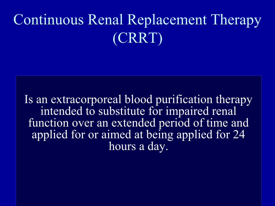
Continuous Renal Replacement Therapy (CRRT)
Is an extracorporeal blood purification therapy intended to substitute for impaired renal
function over an extended period of time and applied for or aimed at being applied for 24
hours a day.
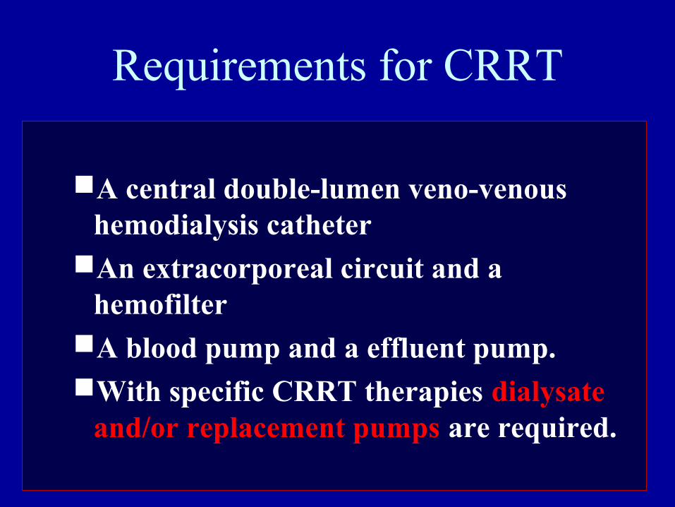
Requirements for CRRT
A central double-lumen veno-venous hemodialysis catheter
An extracorporeal circuit and a hemofilter
A blood pump and a effluent pump. With specific CRRT therapies dialysate
and/or replacement pumps are required.
Indications
• CRRT is better tolerated by hemodynamically unstable patients
because fluid volume, electrolytes and pH are adjusted slowly and steadily over a 24
hour period rather than a 3 – 4 hour period.
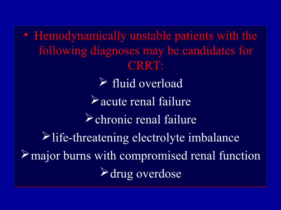
• Hemodynamically unstable patients with the following diagnoses may be candidates for
CRRT: fluid overload
acute renal failurechronic renal failure
life-threatening electrolyte imbalancemajor burns with compromised renal function
drug overdose
Principals of CRRT
• Vascular access.
• Semi-permeable membrane.
• Transport mechanism.
• Dialysate and replacement fluid.
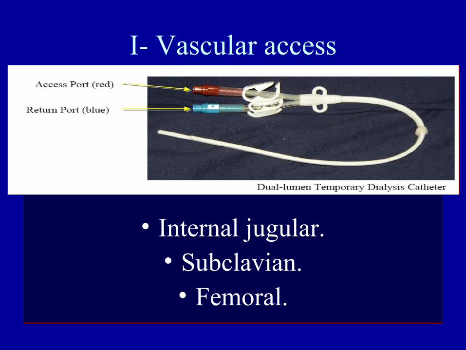
I- Vascular access
• Internal jugular.• Subclavian.
• Femoral.
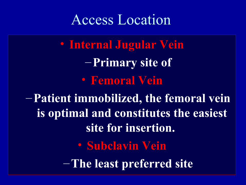
Access Location
• Internal Jugular Vein
–Primary site of
• Femoral Vein
–Patient immobilized, the femoral vein is optimal and constitutes the easiest
site for insertion.
• Subclavin Vein
–The least preferred site
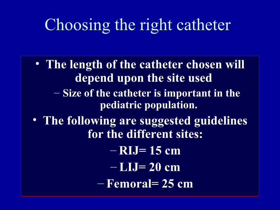
Choosing the right catheter
• The length of the catheter chosen will depend upon the site used
– Size of the catheter is important in the pediatric population.
• The following are suggested guidelines for the different sites:
– RIJ= 15 cm – LIJ= 20 cm
– Femoral= 25 cm
II- Semi-permeable membrane
• The basis of all blood purification therapies.
• Water and some solutes pass through the membrane, while cellular
components and other solutes remain behind.
II- Semi-permeable membrane
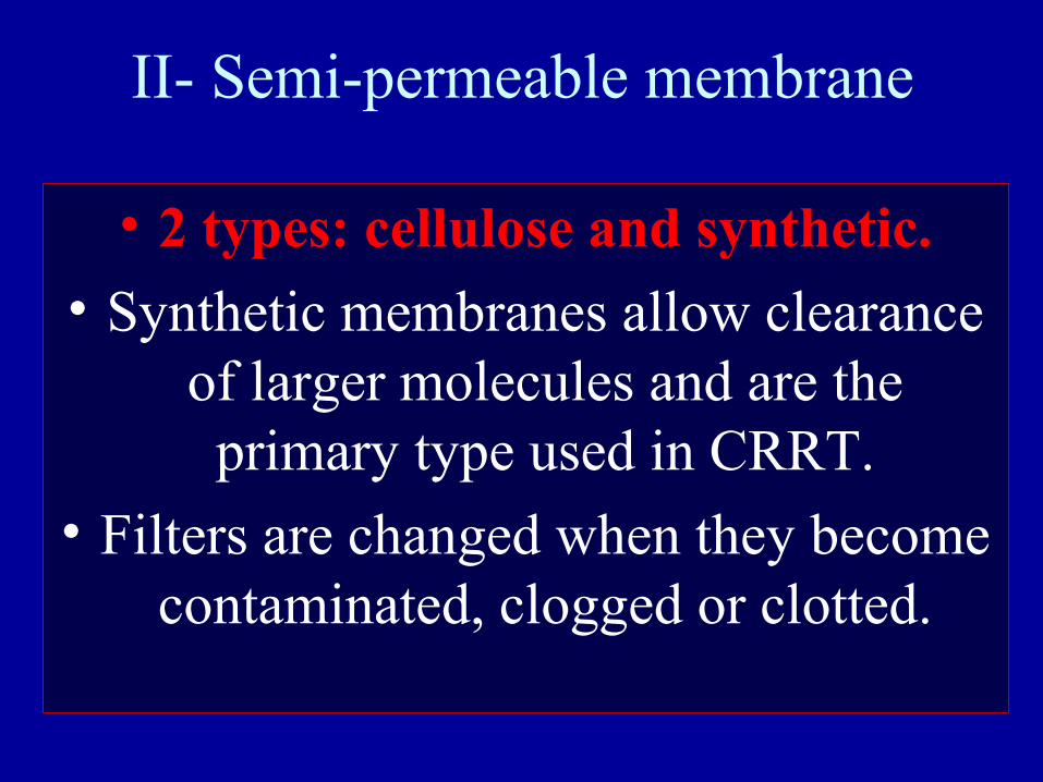
• 2 types: cellulose and synthetic.
• Synthetic membranes allow clearance of larger molecules and are the primary type used in CRRT.
• Filters are changed when they become contaminated, clogged or clotted.
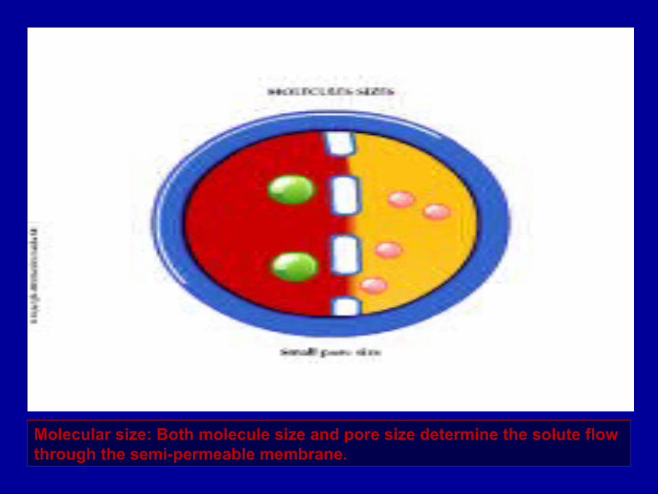
Molecular size: Both molecule size and pore size determine the solute flow through the semi-permeable membrane.
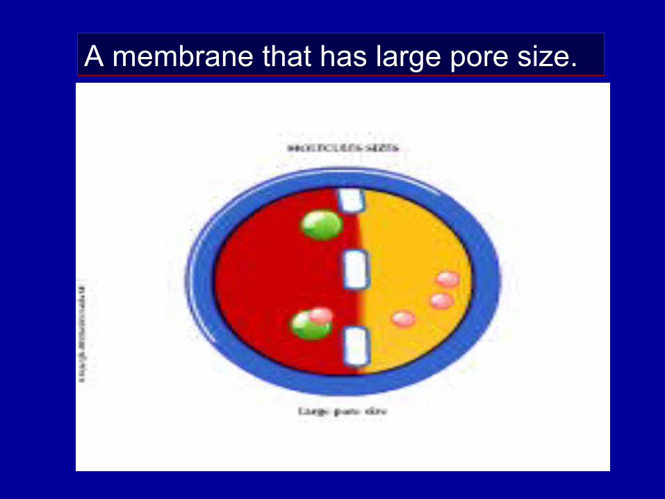
A membrane that has large pore size.
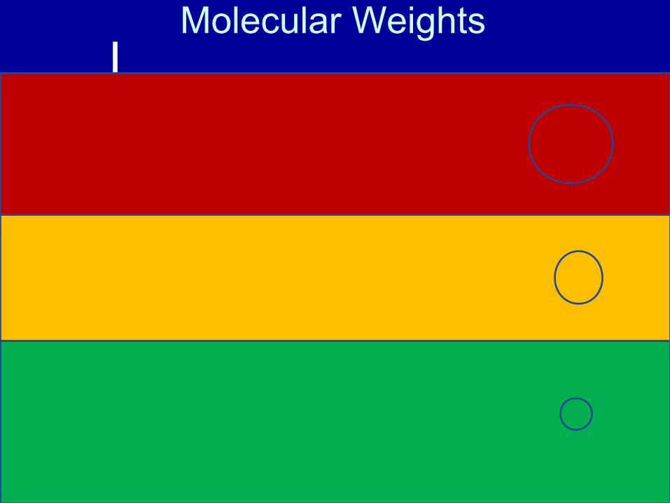
Molecular Weights (Dalton)
100000
50000
10000
5000
1000
500
100
50
10
Albumin (55000 – 60000)
Beta 2 Microglobulin (11800) Inulin (5200)
Vit B12 (1355)
Aluminium/Desforoxamine complex (700)Glucose (180) Uric Acid (168) Creatinine (113) Phosphate (80)
Urea (60) Potassium (35) Phosphorus (31) Sodium (23)
III- Transport mechanisms
a) Ultrafiltration
• The passage of water through a membrane under a pressure gradient.
• Driving pressure can be +ve (push fluid through the filter), or –ve (pull fluid to other side of filter).
• Pressure gradient is created by effluent pump.
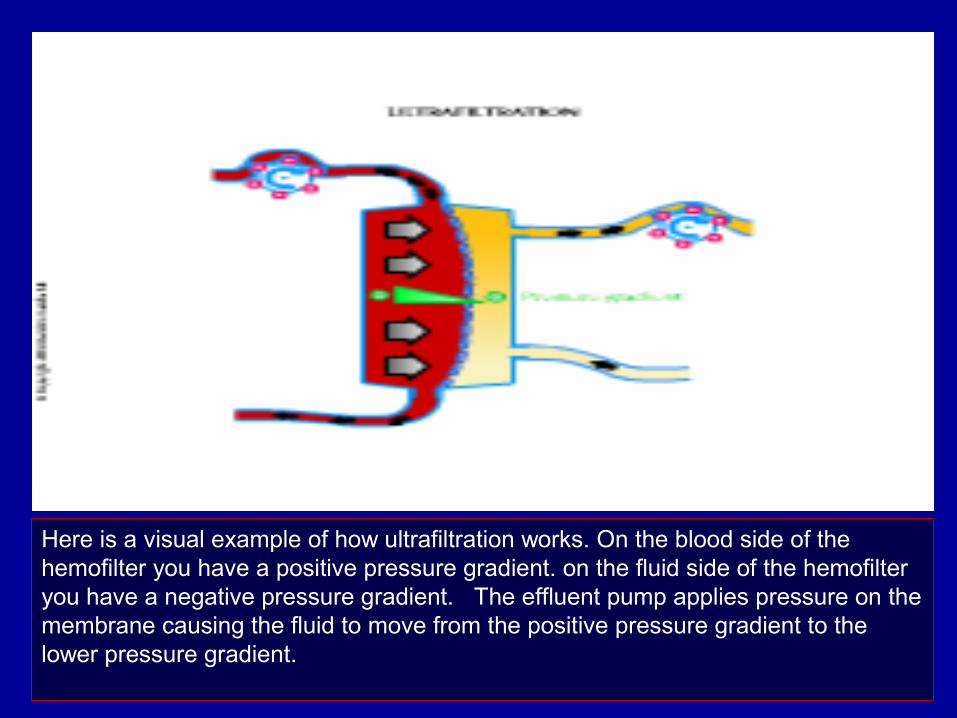
Here is a visual example of how ultrafiltration works. On the blood side of the hemofilter you have a positive pressure gradient. on the fluid side of the hemofilter you have a negative pressure gradient. The effluent pump applies pressure on the membrane causing the fluid to move from the positive pressure gradient to the lower pressure gradient.
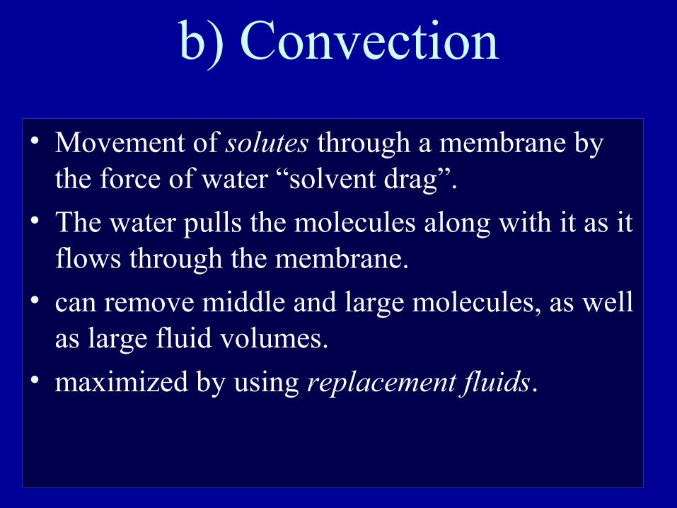
b) Convection
• Movement of solutes through a membrane by the force of water “solvent drag”.
• The water pulls the molecules along with it as it flows through the membrane.
• can remove middle and large molecules, as well as large fluid volumes.
• maximized by using replacement fluids.
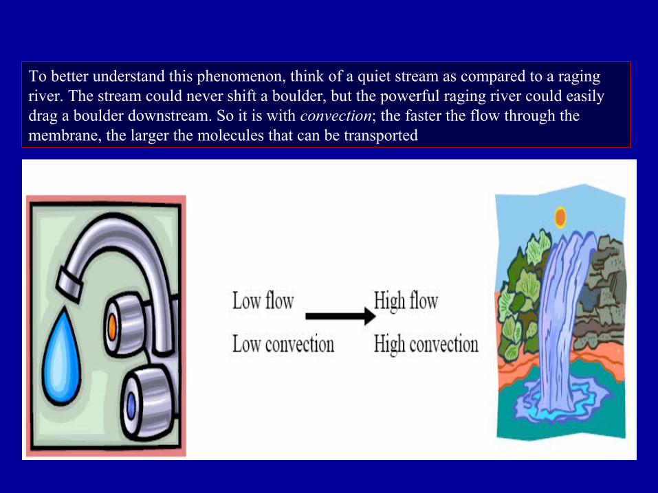
To better understand this phenomenon, think of a quiet stream as compared to a raging river. The stream could never shift a boulder, but the powerful raging river could easily drag a boulder downstream. So it is with convection; the faster the flow through the membrane, the larger the molecules that can be transported
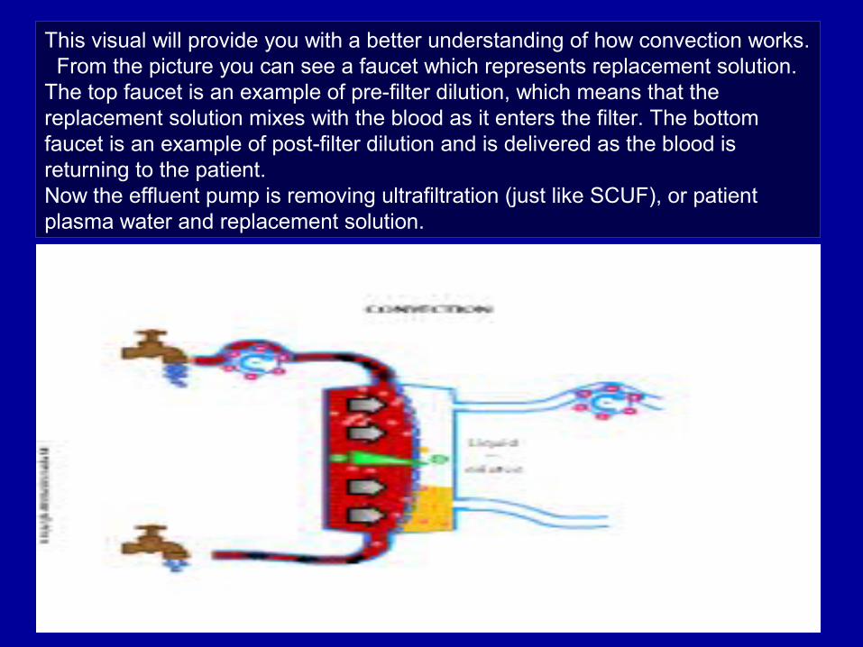
This visual will provide you with a better understanding of how convection works. From the picture you can see a faucet which represents replacement solution. The top faucet is an example of pre-filter dilution, which means that the replacement solution mixes with the blood as it enters the filter. The bottom faucet is an example of post-filter dilution and is delivered as the blood is returning to the patient. Now the effluent pump is removing ultrafiltration (just like SCUF), or patient plasma water and replacement solution.
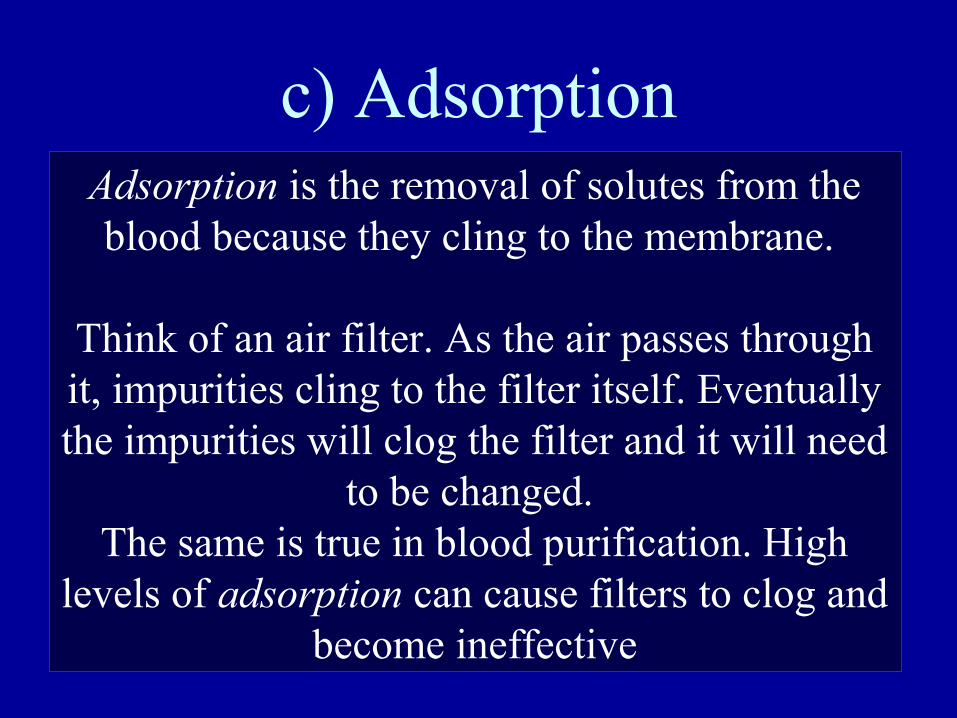
c) AdsorptionAdsorption is the removal of solutes from the blood because they cling to the membrane.
Think of an air filter. As the air passes through it, impurities cling to the filter itself. Eventually the impurities will clog the filter and it will need
to be changed. The same is true in blood purification. High
levels of adsorption can cause filters to clog and become ineffective
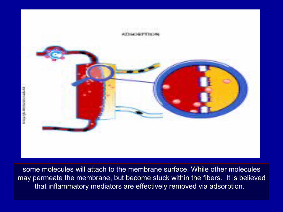
some molecules will attach to the membrane surface. While other molecules may permeate the membrane, but become stuck within the fibers. It is believed
that inflammatory mediators are effectively removed via adsorption.
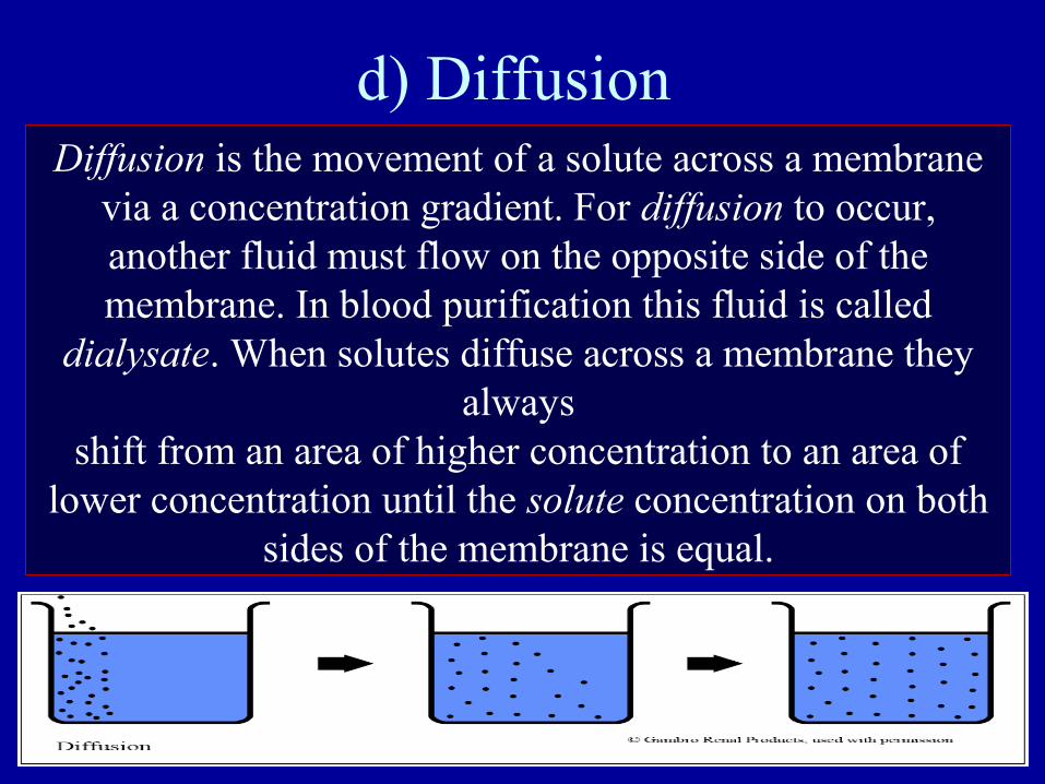
d) Diffusion Diffusion is the movement of a solute across a membrane
via a concentration gradient. For diffusion to occur, another fluid must flow on the opposite side of the membrane. In blood purification this fluid is called
dialysate. When solutes diffuse across a membrane they always
shift from an area of higher concentration to an area of lower concentration until the solute concentration on both
sides of the membrane is equal.
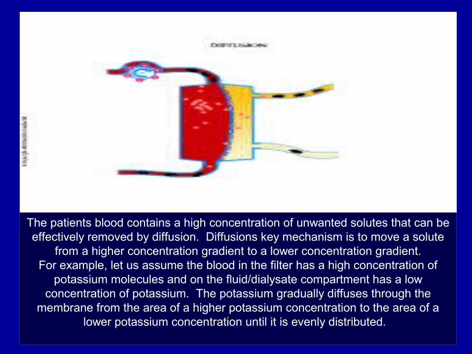
The patients blood contains a high concentration of unwanted solutes that can be effectively removed by diffusion. Diffusions key mechanism is to move a solute
from a higher concentration gradient to a lower concentration gradient.For example, let us assume the blood in the filter has a high concentration of
potassium molecules and on the fluid/dialysate compartment has a low concentration of potassium. The potassium gradually diffuses through the
membrane from the area of a higher potassium concentration to the area of a lower potassium concentration until it is evenly distributed.
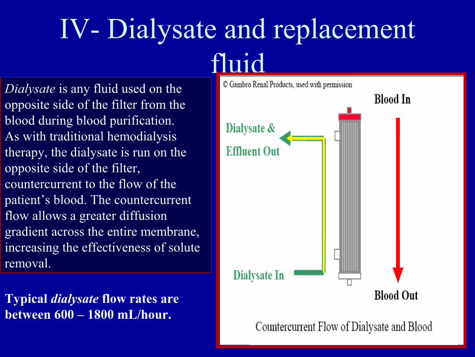
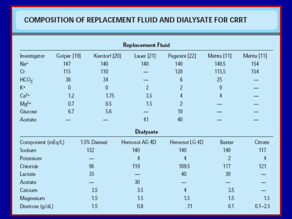
IV- Dialysate and replacement fluid
Dialysate is any fluid used on theopposite side of the filter from theblood during blood purification.As with traditional hemodialysistherapy, the dialysate is run on theopposite side of the filter, countercurrent to the flow of the patient’s blood. The countercurrent flow allows a greater diffusion gradient across the entire membrane,increasing the effectiveness of solute removal.
Typical dialysate flow rates are between 600 – 1800 mL/hour.
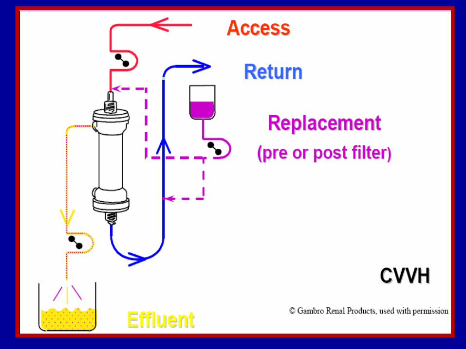
Replacement fluid• Used to increase the amount of convective
solute removal in CRRT.
• Replacement fluids do not replace anything.
• Fluid removal rates are calculated independently of replacement fluid rates.
• The most common replacement fluid is 0.9% Normal Saline.
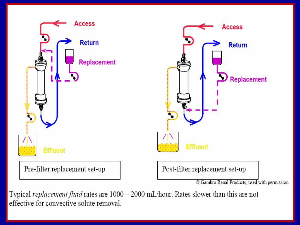
• Can be pre or post filter.
The decision to infuse replacement fluids before or after the filter is made by the
physician. Replacement fluids administered pre-filter reduce filter clotting and can be
administered at faster rates (driving higher convection) than fluids administered post-
filter.
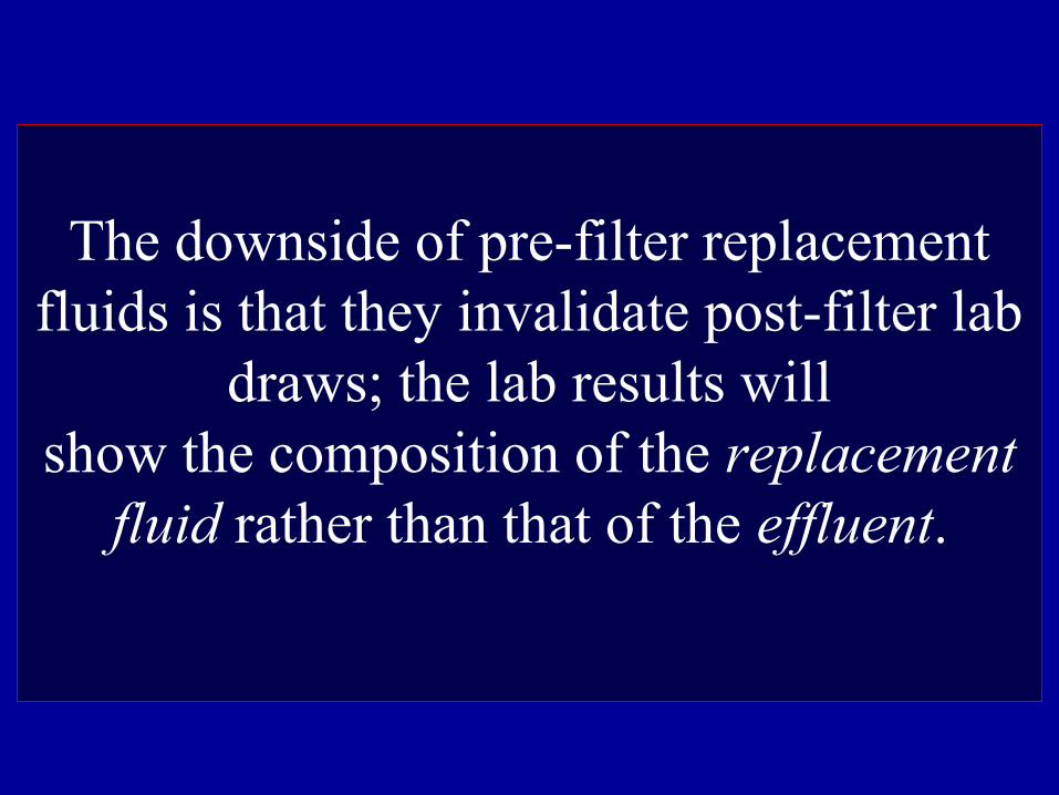
The downside of pre-filter replacement fluids is that they invalidate post-filter lab
draws; the lab results willshow the composition of the replacement
fluid rather than that of the effluent.
Modalities of CRRT
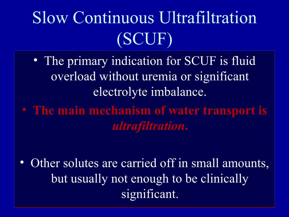
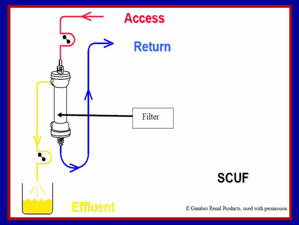
Slow Continuous Ultrafiltration (SCUF)
• The primary indication for SCUF is fluid overload without uremia or significant
electrolyte imbalance.
• The main mechanism of water transport is ultrafiltration.
• Other solutes are carried off in small amounts, but usually not enough to be clinically
significant.
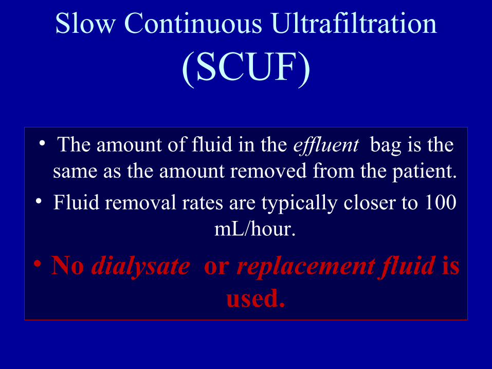
Slow Continuous Ultrafiltration
(SCUF)
• The amount of fluid in the effluent bag is the same as the amount removed from the patient.
• Fluid removal rates are typically closer to 100 mL/hour.
• No dialysate or replacement fluid is used.
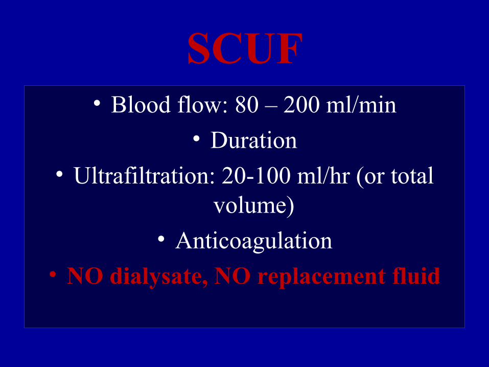
SCUF• Blood flow: 80 – 200 ml/min
• Duration
• Ultrafiltration: 20-100 ml/hr (or total volume)
• Anticoagulation• NO dialysate, NO replacement fluid
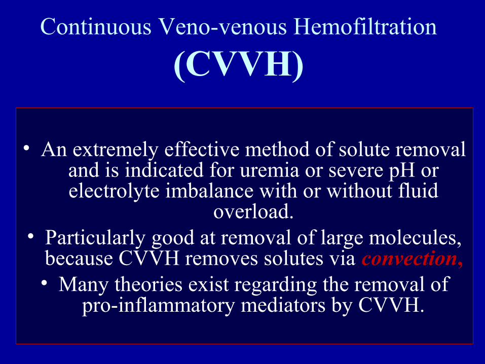
Continuous Veno-venous Hemofiltration
(CVVH)
• An extremely effective method of solute removal and is indicated for uremia or severe pH or electrolyte imbalance with or without fluid
overload.• Particularly good at removal of large molecules,
because CVVH removes solutes via convection,• Many theories exist regarding the removal of
pro-inflammatory mediators by CVVH.
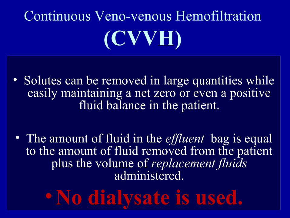
Continuous Veno-venous Hemofiltration
(CVVH)
• Solutes can be removed in large quantities while easily maintaining a net zero or even a positive
fluid balance in the patient.
• The amount of fluid in the effluent bag is equal to the amount of fluid removed from the patient
plus the volume of replacement fluids administered.
• No dialysate is used.
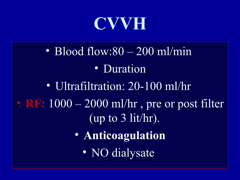
CVVH
• Blood flow:80 – 200 ml/min
• Duration• Ultrafiltration: 20-100 ml/hr
• RF: 1000 – 2000 ml/hr , pre or post filter (up to 3 lit/hr).
• Anticoagulation
• NO dialysate
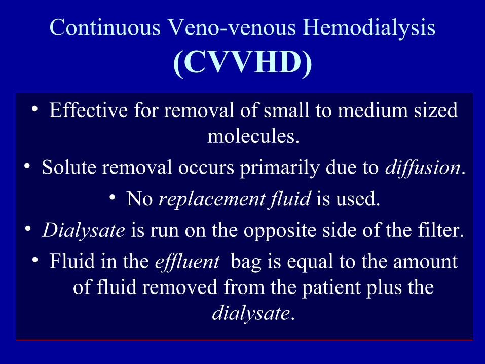
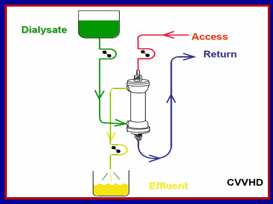
Continuous Veno-venous Hemodialysis
(CVVHD)• Effective for removal of small to medium sized
molecules.
• Solute removal occurs primarily due to diffusion.
• No replacement fluid is used.
• Dialysate is run on the opposite side of the filter.
• Fluid in the effluent bag is equal to the amount of fluid removed from the patient plus the
dialysate.
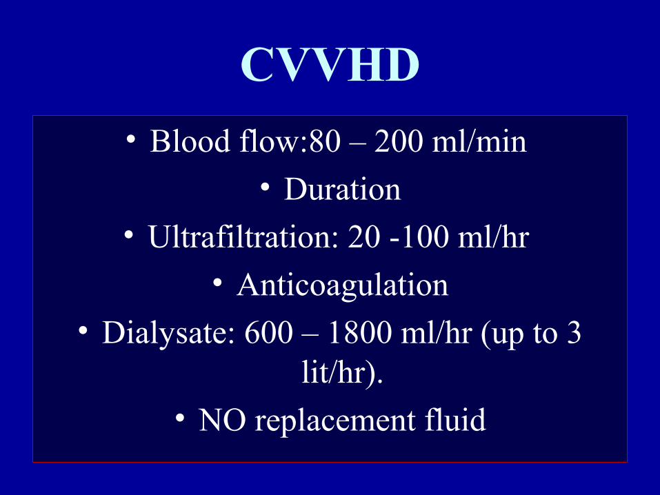
CVVHD
• Blood flow:80 – 200 ml/min
• Duration• Ultrafiltration: 20 -100 ml/hr
• Anticoagulation
• Dialysate: 600 – 1800 ml/hr (up to 3 lit/hr).
• NO replacement fluid
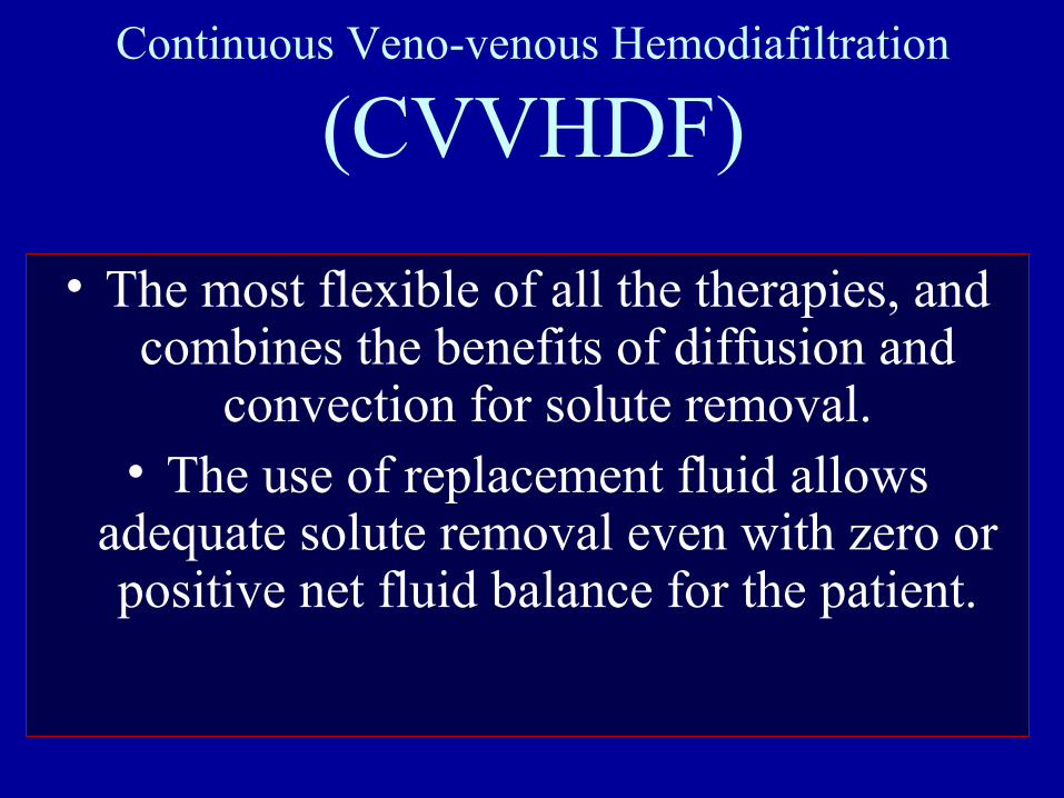
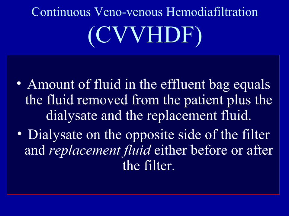
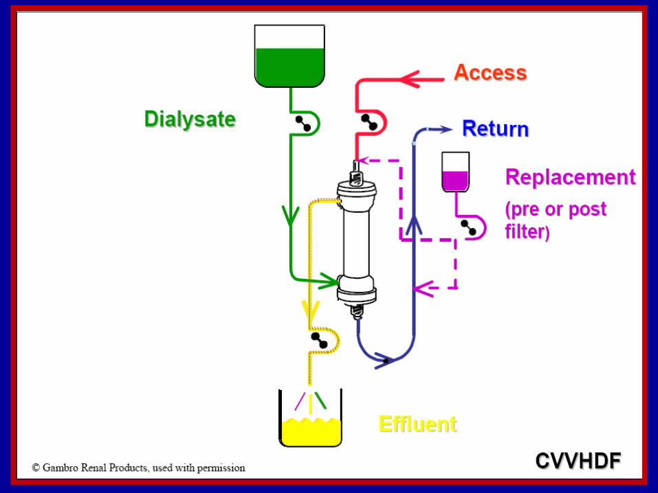
Continuous Veno-venous Hemodiafiltration
(CVVHDF)
• The most flexible of all the therapies, and combines the benefits of diffusion and
convection for solute removal.• The use of replacement fluid allows
adequate solute removal even with zero or positive net fluid balance for the patient.
Continuous Veno-venous Hemodiafiltration
(CVVHDF)
• Amount of fluid in the effluent bag equals the fluid removed from the patient plus the
dialysate and the replacement fluid.• Dialysate on the opposite side of the filter
and replacement fluid either before or after the filter.
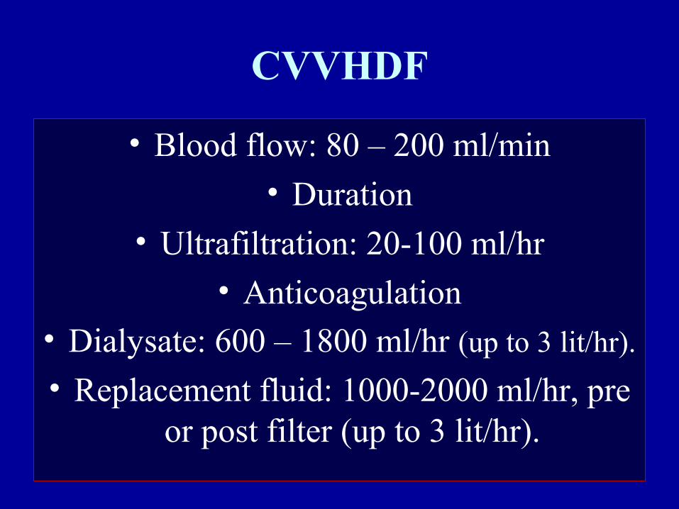
CVVHDF
• Blood flow: 80 – 200 ml/min
• Duration• Ultrafiltration: 20-100 ml/hr
• Anticoagulation• Dialysate: 600 – 1800 ml/hr (up to 3 lit/hr).
• Replacement fluid: 1000-2000 ml/hr, pre or post filter (up to 3 lit/hr).
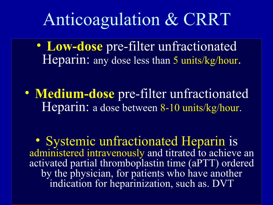
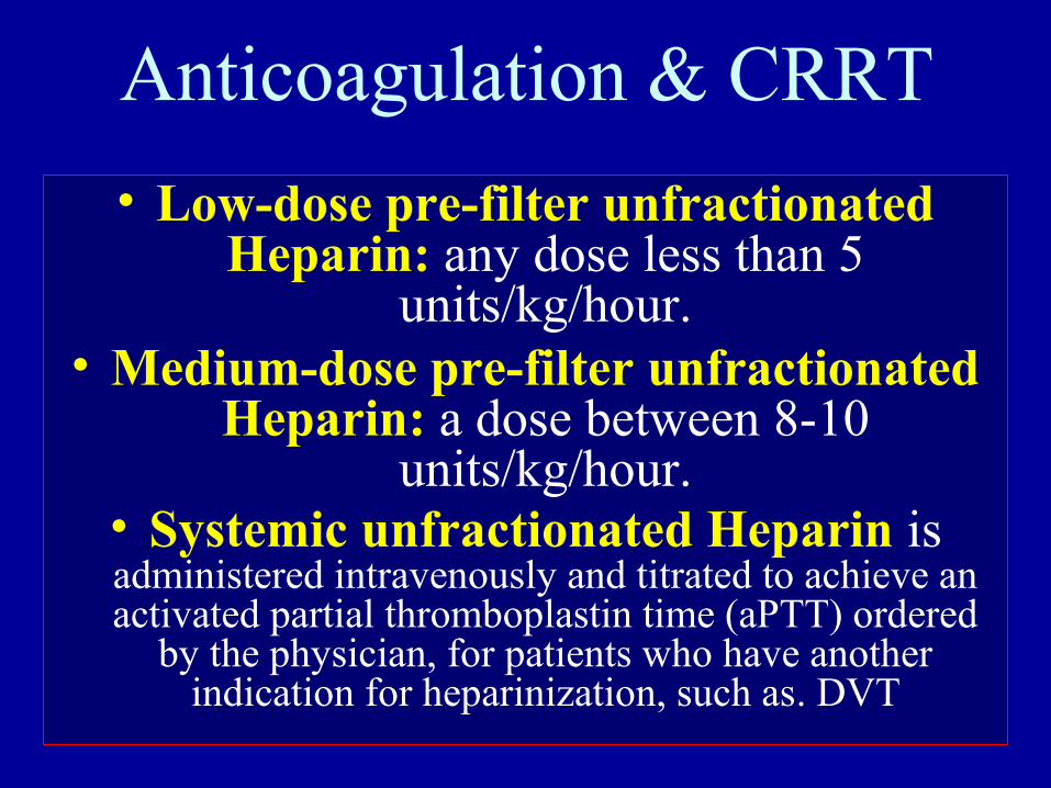
Anticoagulation & CRRT• Low-dose pre-filter unfractionated Heparin: any dose less than 5 units/kg/hour.
• Medium-dose pre-filter unfractionated Heparin: a dose between 8-10 units/kg/hour.
• Systemic unfractionated Heparin is administered intravenously and titrated to achieve an activated partial thromboplastin time (aPTT) ordered
by the physician, for patients who have another indication for heparinization, such as. DVT
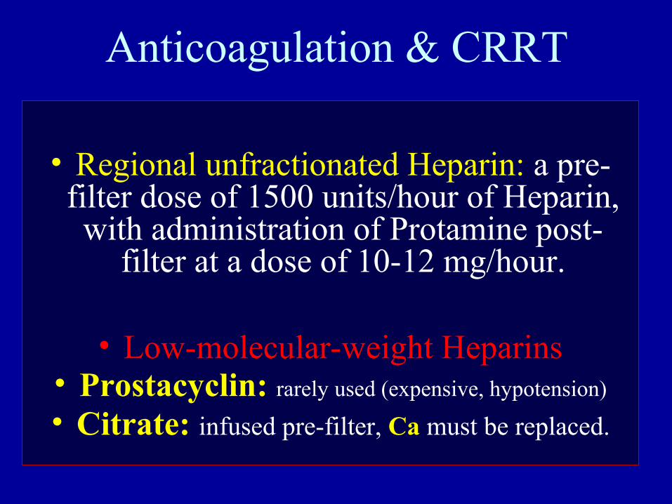
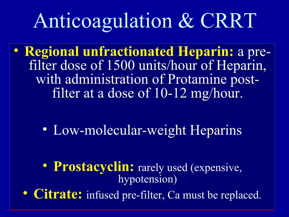
Anticoagulation & CRRT
• Regional unfractionated Heparin: a pre-filter dose of 1500 units/hour of Heparin, with administration of Protamine post-
filter at a dose of 10-12 mg/hour.
• Low-molecular-weight Heparins• Prostacyclin: rarely used (expensive, hypotension)
• Citrate: infused pre-filter, Ca must be replaced.
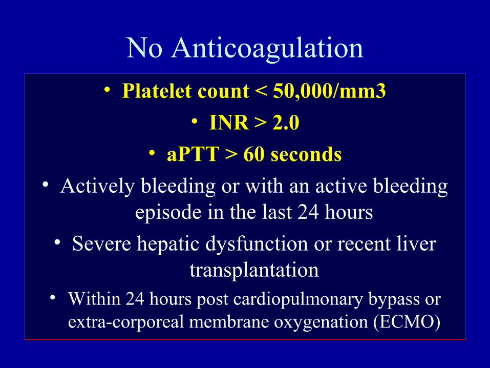
No Anticoagulation• Platelet count < 50,000/mm3
• INR > 2.0
• aPTT > 60 seconds
• Actively bleeding or with an active bleeding episode in the last 24 hours
• Severe hepatic dysfunction or recent liver transplantation
• Within 24 hours post cardiopulmonary bypass or extra-corporeal membrane oxygenation (ECMO)
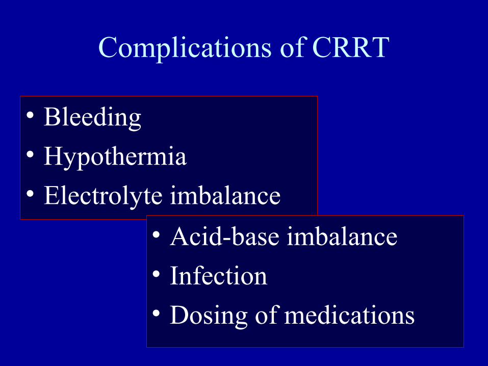
Complications of CRRT
• Bleeding
• Hypothermia
• Electrolyte imbalance
• Acid-base imbalance
• Infection
• Dosing of medications
Continuous Renal Replacement Therapy
CRRT
WHATIs CRRT
HOWTo use CRRT
HOWTo use CRRT
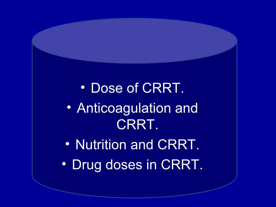
• Dose of CRRT.• Anticoagulation and
CRRT.
• Nutrition and CRRT.
• Drug doses in CRRT.
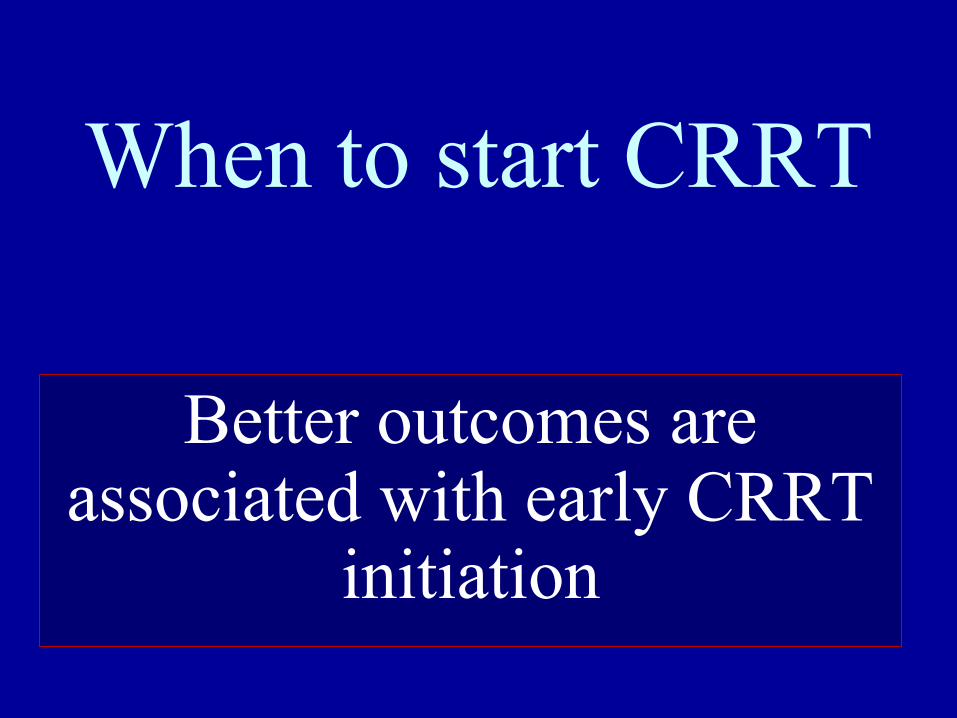
When to start CRRT
Better outcomes are associated with early CRRT
initiation
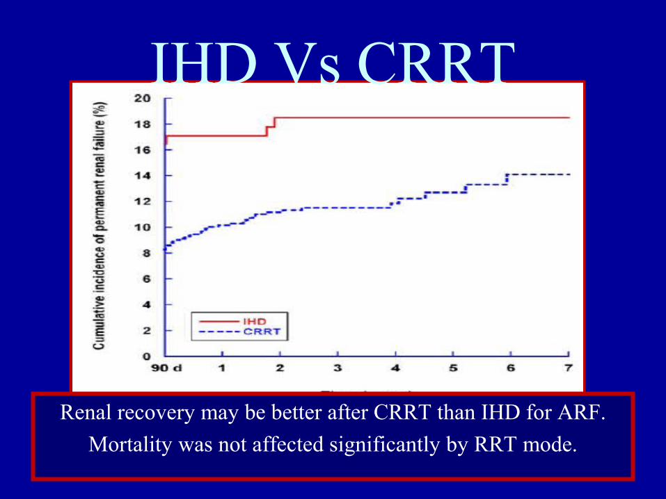
Renal recovery may be better after CRRT than IHD for ARF.
Mortality was not affected significantly by RRT mode.
IHD Vs CRRT
DOSE of CRRT
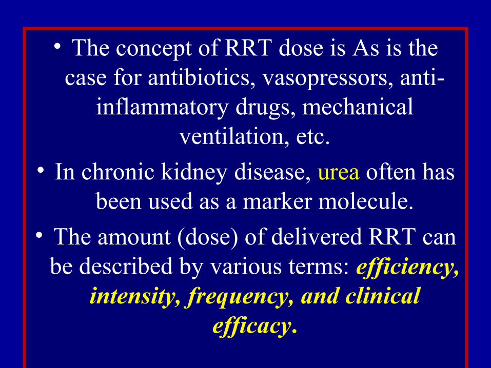
• The concept of RRT dose is As is the case for antibiotics, vasopressors, anti-
inflammatory drugs, mechanical ventilation, etc.
• In chronic kidney disease, urea often has been used as a marker molecule.
• The amount (dose) of delivered RRT can be described by various terms: efficiency,
intensity, frequency, and clinical efficacy.

Efficiency (K)
• The volume of blood cleared of a given solute over a given time.
(mL/min, mL/hr, L/hr, L/24 hrs, etc.)
• During RRT, K depends on solute molecular size and diffusivity, transport modality (convection or diffusion), and circuit
operational characteristics such as blood flow rate, ultrafiltration rate, dialysate flow rate, and membrane and hemodialyzer type and
size.
Intensity (Kt)
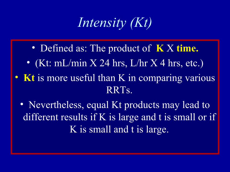
• Defined as: The product of K X time.
• (Kt: mL/min X 24 hrs, L/hr X 4 hrs, etc.)
• Kt is more useful than K in comparing various RRTs.
• Nevertheless, equal Kt products may lead to different results if K is large and t is small or if
K is small and t is large.
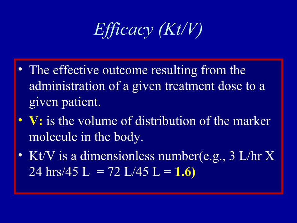
Efficacy (Kt/V)
• The effective outcome resulting from the administration of a given treatment dose to a given patient.
• V: is the volume of distribution of the marker molecule in the body.
• Kt/V is a dimensionless number(e.g., 3 L/hr X 24 hrs/45 L = 72 L/45 L = 1.6)
Limitations• The marker solute cannot and does not
represent all of the solutes that accumulate in renal failure.
• Its kinetics and volume of distribution are also different from other solutes.
• Finally, its removal during RRT is not representative of the removal of other solutes.
• This is true for both end-stage renal failure and acute renal failure.
Anticoagulationand CRRT
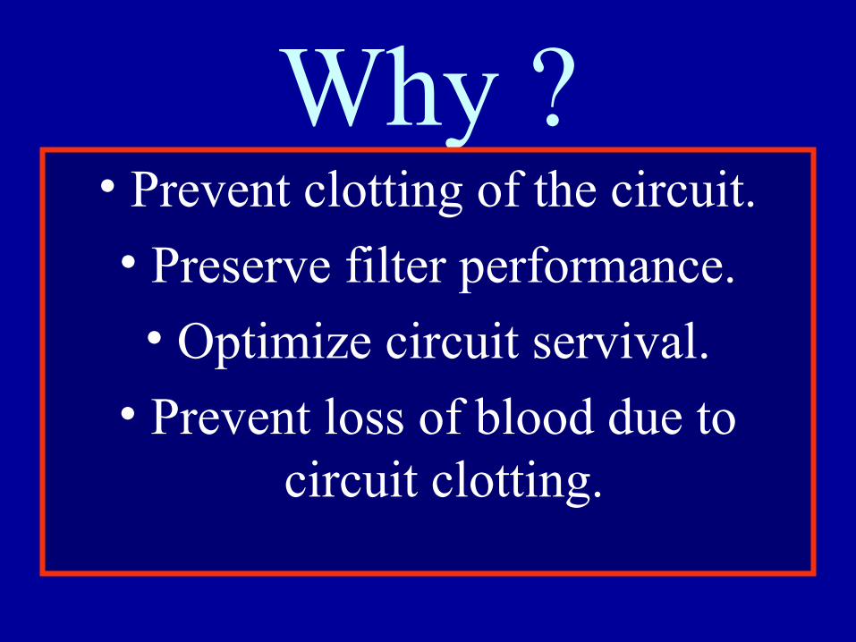
Why ?• Prevent clotting of the circuit.
• Preserve filter performance.
• Optimize circuit servival.
• Prevent loss of blood due to circuit clotting.
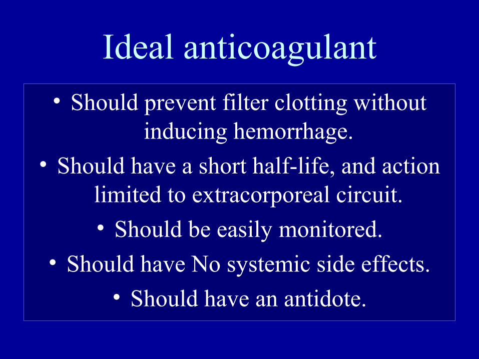
Ideal anticoagulant
• Should prevent filter clotting without inducing hemorrhage.
• Should have a short half-life, and action limited to extracorporeal circuit.
• Should be easily monitored.
• Should have No systemic side effects.• Should have an antidote.
Anticoagulation & CRRT
• Low-dose pre-filter unfractionated Heparin: any dose less than 5
units/kg/hour.• Medium-dose pre-filter unfractionated
Heparin: a dose between 8-10 units/kg/hour.
• Systemic unfractionated Heparin is administered intravenously and titrated to achieve an activated partial thromboplastin time (aPTT) ordered
by the physician, for patients who have another indication for heparinization, such as. DVT
Anticoagulation & CRRT• Regional unfractionated Heparin: a pre-
filter dose of 1500 units/hour of Heparin, with administration of Protamine post-
filter at a dose of 10-12 mg/hour.
• Low-molecular-weight Heparins
• Prostacyclin: rarely used (expensive, hypotension)
• Citrate: infused pre-filter, Ca must be replaced.
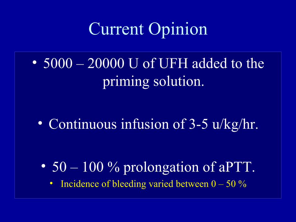
Current Opinion
• 5000 – 20000 U of UFH added to the priming solution.
• Continuous infusion of 3-5 u/kg/hr.
• 50 – 100 % prolongation of aPTT.• Incidence of bleeding varied between 0 – 50 %
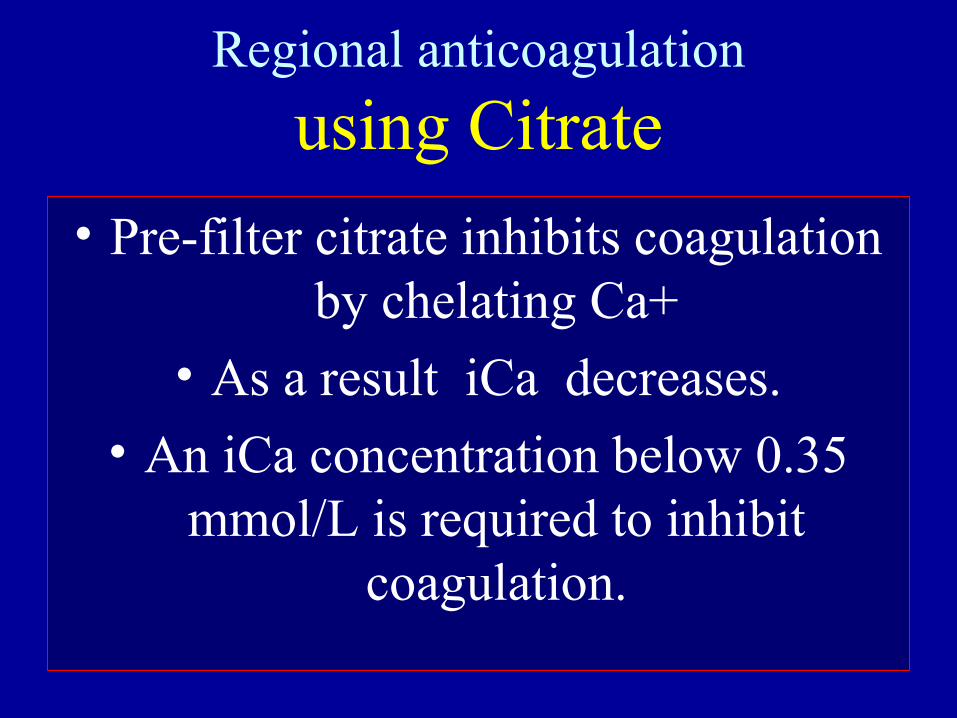
Regional anticoagulation
using Citrate
• Pre-filter citrate inhibits coagulation by chelating Ca+
• As a result iCa decreases.
• An iCa concentration below 0.35 mmol/L is required to inhibit
coagulation.
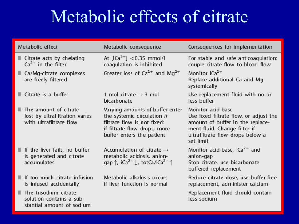
Metabolic effects of citrate
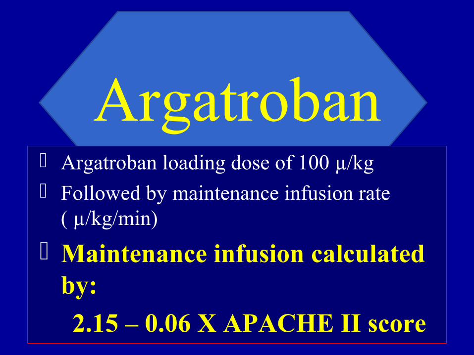
Argatroban
Argatroban loading dose of 100 µ/kg Followed by maintenance infusion rate
( µ/kg/min)
Maintenance infusion calculated by:
2.15 – 0.06 X APACHE II score

Argatroban loading dose of 100 µ/kg Followed by maintenance infusion rate ( µ/kg/min) Maintenance infusion calculated by: 2.15 – 0.06 X APACHE II score
In critically ill patients with HIT and necessity for CRRT , APACHE II can help to predict the required argatroban maintenance dose for anticoagulation.
This predictor identifies decreased argatroban dosing requirements.
Resulting in effective and safe CRRT.
Argatroban
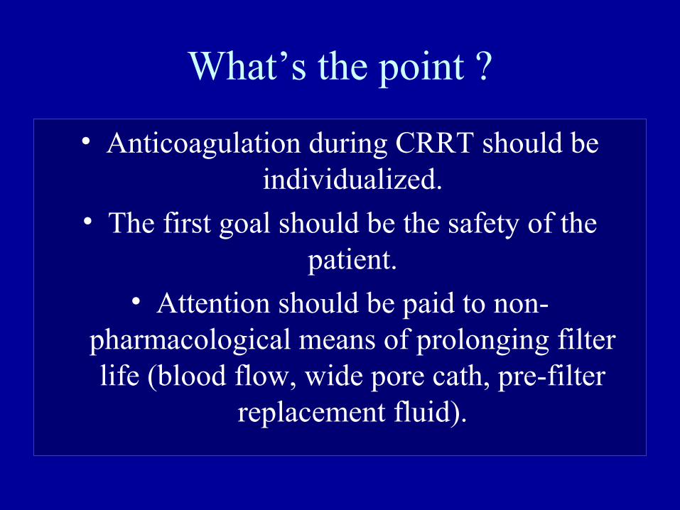
What’s the point ?
• Anticoagulation during CRRT should be individualized.
• The first goal should be the safety of the patient.
• Attention should be paid to non-pharmacological means of prolonging filter life (blood flow, wide pore cath, pre-filter
replacement fluid).
Nutritionand CRRT
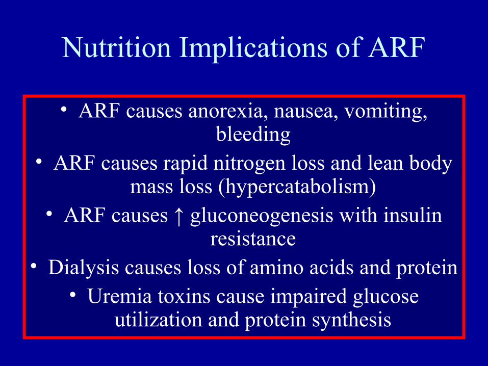
Nutrition Implications of ARF
• ARF causes anorexia, nausea, vomiting, bleeding
• ARF causes rapid nitrogen loss and lean body mass loss (hypercatabolism)
• ARF causes ↑ gluconeogenesis with insulin resistance
• Dialysis causes loss of amino acids and protein• Uremia toxins cause impaired glucose
utilization and protein synthesis
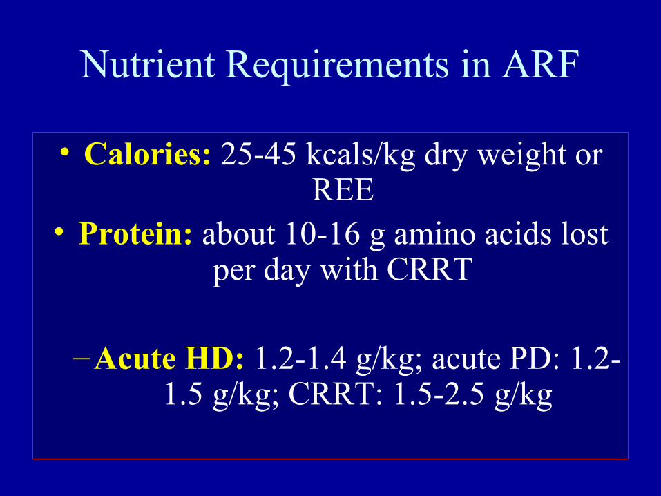
Nutrient Requirements in ARF
• Calories: 25-45 kcals/kg dry weight or REE
• Protein: about 10-16 g amino acids lost per day with CRRT
–Acute HD: 1.2-1.4 g/kg; acute PD: 1.2-1.5 g/kg; CRRT: 1.5-2.5 g/kg
Nutrient Requirements in ARF
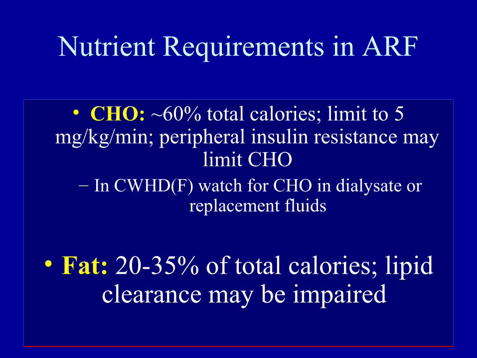
• CHO: ~60% total calories; limit to 5 mg/kg/min; peripheral insulin resistance may
limit CHO– In CWHD(F) watch for CHO in dialysate or
replacement fluids
• Fat: 20-35% of total calories; lipid clearance may be impaired
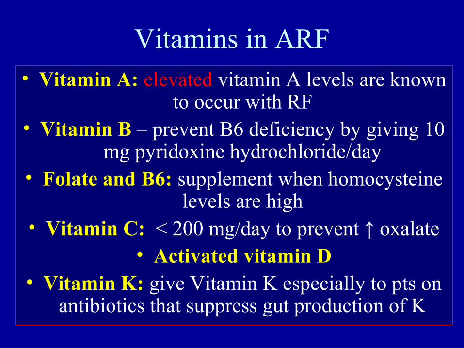
Vitamins in ARF• Vitamin A: elevated vitamin A levels are known
to occur with RF• Vitamin B – prevent B6 deficiency by giving 10
mg pyridoxine hydrochloride/day• Folate and B6: supplement when homocysteine
levels are high• Vitamin C: < 200 mg/day to prevent ↑ oxalate
• Activated vitamin D• Vitamin K: give Vitamin K especially to pts on
antibiotics that suppress gut production of K
Minerals in ARF
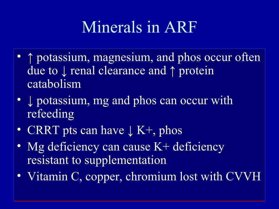
• ↑ potassium, magnesium, and phos occur often due to ↓ renal clearance and ↑ protein catabolism
• ↓ potassium, mg and phos can occur with refeeding
• CRRT pts can have ↓ K+, phos• Mg deficiency can cause K+ deficiency
resistant to supplementation• Vitamin C, copper, chromium lost with CVVH
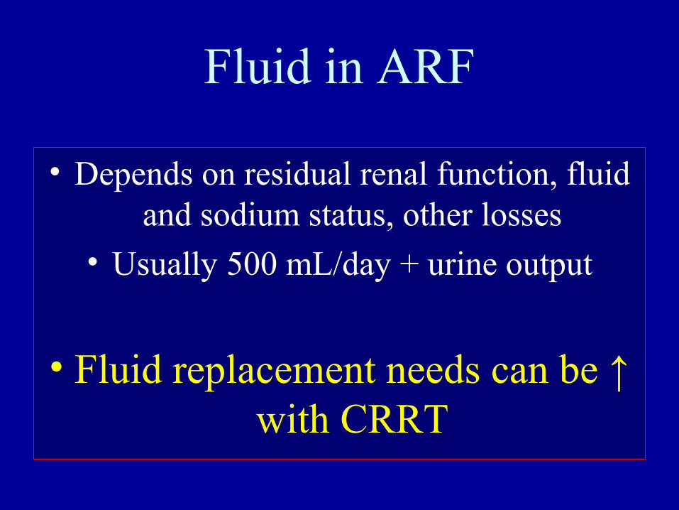
Fluid in ARF
• Depends on residual renal function, fluid and sodium status, other losses
• Usually 500 mL/day + urine output
• Fluid replacement needs can be ↑ with CRRT
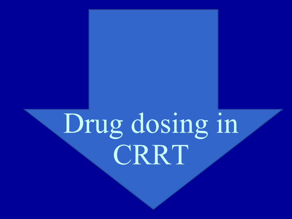
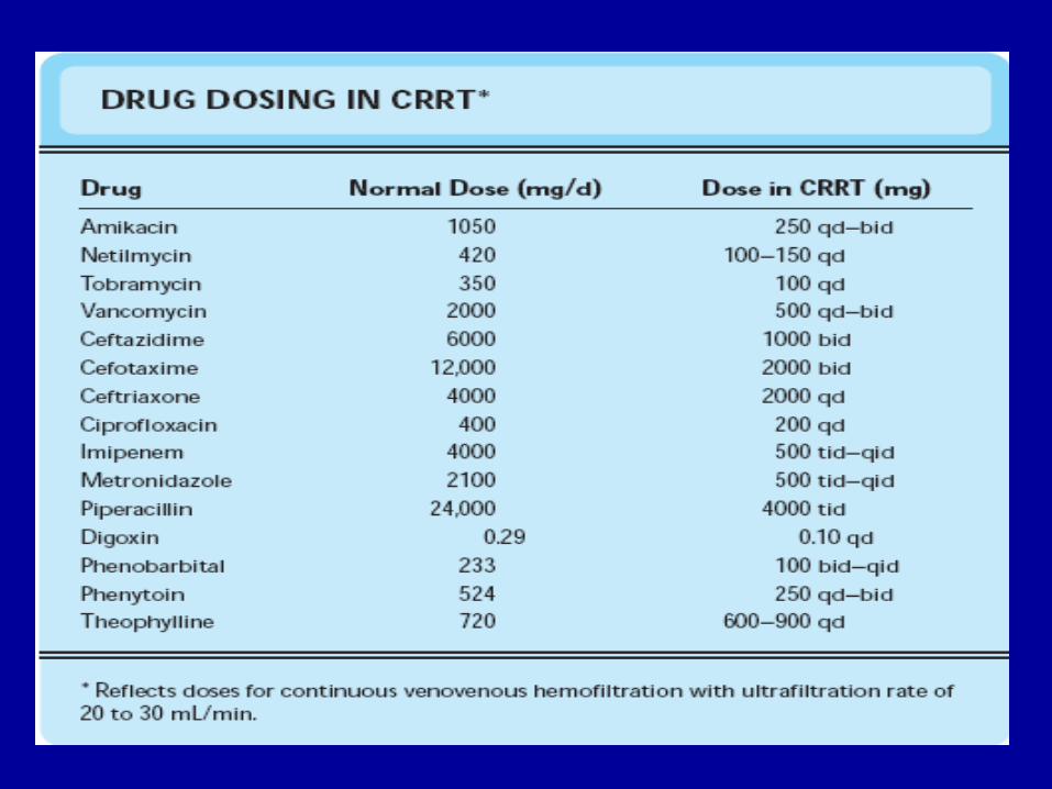
Drug dosing in CRRT
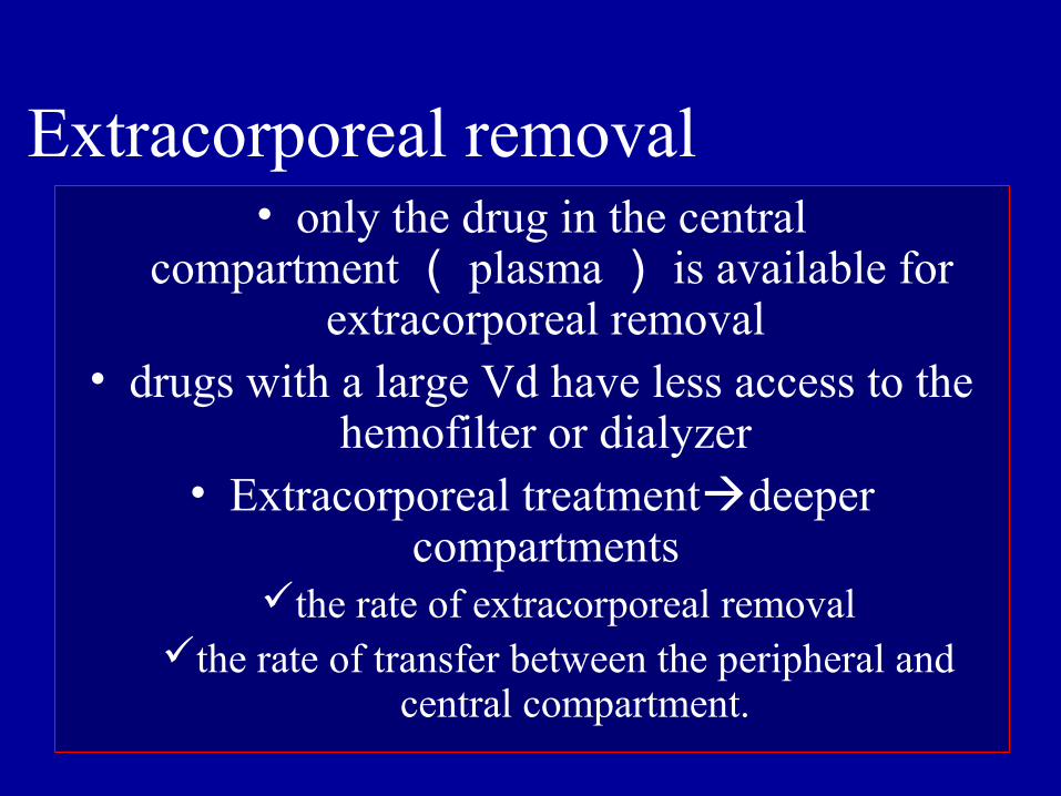
• only the drug in the central compartment( plasma) is available for
extracorporeal removal • drugs with a large Vd have less access to the
hemofilter or dialyzer • Extracorporeal treatmentdeeper
compartments the rate of extracorporeal removal
the rate of transfer between the peripheral and central compartment.
Extracorporeal removal
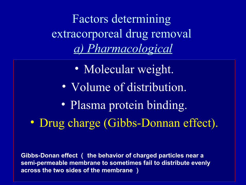
Factors determining extracorporeal drug removal
a) Pharmacological
• Molecular weight.
• Volume of distribution.
• Plasma protein binding.
• Drug charge (Gibbs-Donnan effect).
Gibbs-Donan effect( the behavior of charged particles near a semi-permeable membrane to sometimes fail to distribute evenly across the two sides of the membrane)
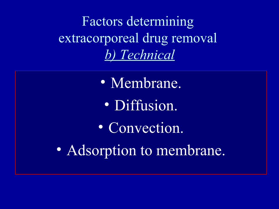
Factors determining extracorporeal drug removal
b) Technical
• Membrane.
• Diffusion.
• Convection.
• Adsorption to membrane.


![THAILAND RENAL REPLACEMENT THERAPY YEAR 2011 · 2020. 8. 27. · THAILAND RENAL REPLACEMENT THERAPY YEAR 2011 ] Page 3 ACKNOWLEDGEMENTS The Thai Renal Replacement Therapy (TRT) committee](https://img.pdfslide.net/doc/110x75/6096115f208ae70f0b464852/thailand-renal-replacement-therapy-year-2011-2020-8-27-thailand-renal-replacement.jpg)


![Renal Replacement Therapy Auto Saved]](https://img.pdfslide.net/doc/110x75/577d27651a28ab4e1ea3d215/renal-replacement-therapy-auto-saved.jpg)