Embed Size (px)
Citation preview

This page intentionally left blank
Cranial nerves are involved in head and neck function, and
processes such as eating, speech and facial expression. This clinically
oriented survey of cranial nerve anatomy and function was written
for students of medicine, dentistry and speech therapy, but will also
be useful for postgraduate physicians and general practitioners, and
specialists in head and neck healthcare (surgeons, dentists, speech
therapists, etc.). After an introductory section surveying cranial
nerve organization and tricky basics such as ganglia, nuclei and brain
stem pathways, the nerves are considered in functional groups: (1)
for chewing and facial sensation; (2) for pharynx and larynx, swal-
lowing and phonation; (3) autonomic components, taste and smell;
(4) vision and eye movements; and (5) hearing and balance. In each
chapter, the main anatomical features of each nerve are followed by
clinical aspects and details of clinical testing. Simple line diagrams
accompany the text. Detailed anatomy is not given.
Stanley Monkhouse is Anatomist at the University of Nottingham
at Derby (Graduate Entry Medicine). He has been an examiner
at the Royal Colleges of Surgeons of England and Ireland; at the
Universities of Nottingham, Leeds, Newcastle-upon-Tyne, London,
Belfast, Dublin (Trinity College), National University of Ireland, King
AbdulAziz University (Jeddah, Saudi Arabia), Amman (Jordan) and
King Faisal University (Dammam, Saudi Arabia).
C R A N I A L N E RV E SFunctional Anatomy

C R A N I A L N E RV E SFunctional Anatomy
S TA N L E Y M O N K H O U S EMA, MB, BChir, PhD
University of Nottingham Medical School at Derby
Sometime Professor of Anatomy at the Royal College of
Surgeons in Ireland; Lecturer in Human Morphology at the
University of Nottingham; and Clinical Assistant in Ear Nose
and Throat, Queen’s Medical Centre, Nottingham
cambridge university pressCambridge, New York, Melbourne, Madrid, Cape Town, Singapore, São Paulo
Cambridge University PressThe Edinburgh Building, Cambridge cb2 2ru, UK
First published in print format
isbn-13 978-0-521-61537-2
isbn-13 978-0-511-13272-8
© Cambridge University Press,2006
2005
Information on this title: www.cambridge.org/9780521615372
This publication is in copyright. Subject to statutory exception and to the provision ofrelevant collective licensing agreements, no reproduction of any part may take placewithout the written permission of Cambridge University Press.
isbn-10 0-511-13272-7
isbn-10 0-521-61537-2
Cambridge University Press has no responsibility for the persistence or accuracy of urlsfor external or third-party internet websites referred to in this publication, and does notguarantee that any content on such websites is, or will remain, accurate or appropriate.
Published in the United States of America by Cambridge University Press, New York
www.cambridge.org
paperback
eBook (NetLibrary)
eBook (NetLibrary)
paperback
C O N T E N T S
List of Figures page vii
List of Tables ix
Acknowledgements xi
A note to the reader xiii
Part I Organization of the cranial nerves 1
1 General considerations 3
2 Cranial nerve motor fibres and nuclei 17
3 Cranial nerve motor pathways: upper and
lower motor neurons 24
4 Cranial nerve sensory fibres, brain stem sensory
nuclei and tracts 31
Parts II–V Individual cranial nerves and functional
considerations 39
5 Survey of cranial nerves and introduction to Parts II–V 41
Part II Trigeminal, facial and hypoglossal nerves 45
6 Cutaneous sensation and chewing 47
7 The trigeminal nerve (V) 50
8 The ophthalmic nerve (Va) 52
9 The maxillary nerve (Vb) 56
10 The mandibular nerve (Vc) 60
11 The facial nerve (VII) 66
12 The hypoglossal nerve (XII) 74
Part III Glossopharyngeal, vagus and accessory nerves 77
13 Swallowing and speaking, bulbar palsy,
pseudobulbar palsy, Broca’s area 79
14 The glossopharyngeal nerve (IX) 83
15 The vagus nerve (X) 86
16 The accessory nerve (XI) 92
Part IV Autonomic components of cranial nerves,
taste and smell 95
17 Parasympathetic components and taste sensation 97
18 Smell: The olfactory nerve (I) 106
19 The sympathetic nervous system in the head 109
Part V Vision, eye movements, hearing and balance:
optic, oculomotor, trochlear, abducens and
vestibulocochlear nerves 113
20 The optic nerve (II) 115
21 The oculomotor (III), trochlear (IV) and
abducens (VI) nerves 121
22 Visual reflexes: the control of eye movements;
clinical testing of II, III, IV and VI 128
23 The vestibulocochlear nerve (VIII) and auditory
and vestibular pathways 133
Further reading 140
Index 143
vi Contents
F I G U R E S
1.1 Attachments of cranial nerves, anterior view page 8
1.2 Attachments of cranial nerves, lateral view 9
1.3 Ganglia and nuclei 12
2.1 Cranial nerve motor nuclei 23
3.1 Corticonuclear pathways 26
4.1 Trigeminal sensory system 34
7.1 Trigeminal nerve 51
8.1 Ophthalmic nerve 53
9.1 Maxillary nerve 57
10.1 Mandibular nerve 61
11.1 Facial nerve (intracranial) 67
11.2 Facial nerve (extracranial) 68
12.1 Hypoglossal nerve 75
14.1 Glossopharyngeal nerve 84
15.1 Vagus nerve 87
16.1 Accessory nerve 93
17.1 Head and neck parasympathetics 100
17.2 Taste pathways 102
18.1 Olfactory pathways 107
20.1 Visual pathways 116
21.1 Oculomotor nerve 122
21.2 Trochlear nerve 123
21.3 Abducens nerve 124
22.1 Pupillary light reflex 128
23.1 Auditory pathways 134
23.2 Vestibular pathways 136
viii Figures
TA B L E S
1.1 Synopsis of cranial nerves page 4
1.2 Attachments and foramina of cranial nerves 7
1.3 Head and neck ganglia 14
2.1 Branchial arches, muscles and nerves 19
2.2 Cranial nerve motor nuclei 21
3.1 Voluntary (somatic and branchiomotor) motor
components of cranial nerves 28
4.1 Cranial nerve sensation, ganglia and nuclei 35
17.1 Parasympathetic components of cranial nerves 98
22.1 Pathways of light and accommodation reflexes 129


AC K N OW L E D G E M E N T S
This book grew from notes first written in 1992 for medical and
surgical students at the Royal College of Surgeons in Ireland.
Comments from students over the years helped me to modify the
text, and I am therefore greatly indebted to those whom I have
taught. The notes were condensed for inclusion in my textbook
Clinical Anatomy (first published by Churchill Livingstone, 2001),
and I acknowledge with thanks the cooperation of staff at Elsevier
in allowing the use of the original notes here.
There are several people who deserve my special thanks. The first
is Eric Clarke who goaded me into action in 1992 and who has been
a constant source of encouragement and practical help. The second
is Dr Gordon Wright MA, MD, Fellow of Clare College, Cambridge,
who in 1970–1971 taught me neuroanatomy with great wit and style,
and who responded to my request for constructive criticism of an
earlier version of the text. Of course, I bear sole responsibility for
errors. I look forward to receiving constructive criticism from others.
And finally, I thank Pauline Graham and her colleagues at
Cambridge University Press.
I would like to think that this book would have met with the
approval of Maxwell Marsden Bull MA, MD, sometime Fellow and
Senior Tutor of Queens’ College, Cambridge. He had a great gift for
expository and analytical teaching, and he showed me that educare
and delectare can be synonymous.
Stanley Monkhouse
Derby 2005

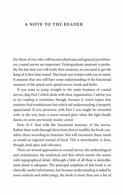
A N OT E TO T H E R E A D E R
For those of you who will become physicians and general practition-
ers, cranial nerves are important. Undergraduate anatomy is proba-
bly the last time you will study their anatomy, so you need to get the
hang of it first time round. This book was written with you in mind.
It assumes that you will have some understanding of the functional
anatomy of the spinal cord, spinal nerves, trunk and limbs.
If you want to jump straight to the main business of cranial
nerves, skip Part I which deals with their organization. I advise you
to try reading it sometime, though, because it covers topics that
students find troublesome but which aid understanding if properly
appreciated. If you persevere with Part I you might be rewarded
with, at the very least, a warm inward glow when the light finally
dawns on some previously murky corner.
Parts II–V deal with the functional anatomy of the nerves.
Rather than work through them from first to twelfth, the book con-
siders them according to function. You will encounter them much
as would an ingested morsel of food. This is unorthodox: it does,
though, lend spice and relevance.
There are several approaches to cranial nerves: the embryological
and evolutionary, the analytical, and that which numbs the senses
with topographical detail. Although a little of all these is desirable,
none alone is adequate. The principal emphasis of this book is on
clinically useful information, but because understanding is aided by
some analysis and embryology, the book is more than just a list of
xiv A note to the reader
points for cramming. I hope that the inclusion of some explanatory
material will stimulate you whilst not obscuring the basics. It is by
no means the last word on the subject, and I expect that research
neuroanatomists will throw up their hands in horror at some of the
generalizations it contains. It is unavoidable that some material
appears more than once, but I hope that this repetition will reinforce
rather than bore.
PART I
O RG A N I Z AT I O N O F T H E
C R A N I A L N E RV E S

Chapter 1
G E N E R A L C O N S I D E R AT I O N S
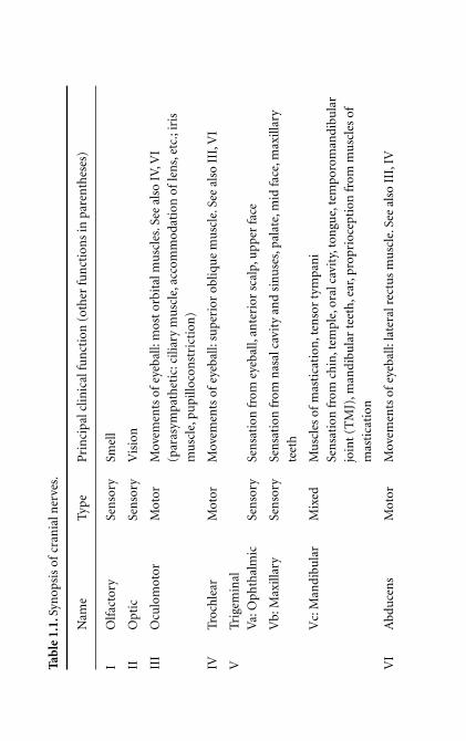
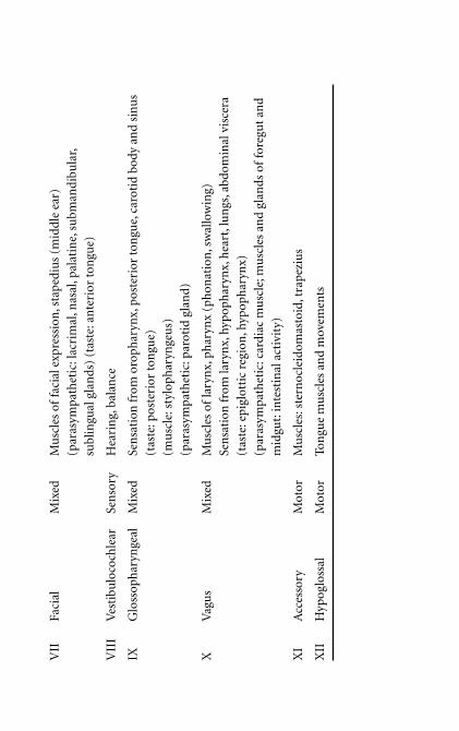
1.1 Cranial nerves and their functions (Table 1.1)
Cranial nerves arise from the brain as twelve pairs. They
pass through or into the cranial bones (thus cranial nerves) and are
numbered I to XII roughly in order from top (rostral) to bottom
(caudal). Their functions are those of the head: some are concerned
with awareness of, and communication with, the environment; and
some are concerned with sustenance, the gut tube and movements
associated with it.
1.2 Cranial nerves and spinal nerves are differently
constituted
Cranial nerves are not equivalent to spinal nerves. All spinal
nerves have similar functions and carry similar types of nerve fibre
(motor, sensory, autonomic, etc.). This is not so for cranial nerves:
• Some cranial nerves contain only sensory fibres, some contain
only motor fibres, and some contain both.
• Some cranial nerves convey parasympathetic fibres, some convey
taste fibres, some convey both, and some neither.
Cranial nerves exhibit great variety and functional special-
ization. This is evident in Table 1.1 which summarizes their
numbers, names and main functions. Learn this table without
further ado, and make sure that you can use names and numbers
Tab
le 1
.1.S
ynop
sis
ofcr
ania
l ner
ves.
Nam
eTy
peP
rin
cipa
l clin
ical
fun
ctio
n (
oth
er fu
nct
ion
s in
par
enth
eses
)
IO
lfac
tory
Sen
sory
Smel
l
IIO
ptic
Sen
sory
Vis
ion
III
Ocu
lom
otor
Mot
orM
ovem
ents
of
eyeb
all:
mos
t or
bita
l mu
scle
s.Se
e al
so I
V,V
I
(par
asym
path
etic
: cili
ary
mu
scle
, acc
omm
odat
ion
of
len
s,et
c.; i
ris
mu
scle
,pu
pillo
con
stri
ctio
n)
IVTr
och
lear
Mot
orM
ovem
ents
of
eyeb
all:
supe
rior
obl
iqu
e m
usc
le.S
ee a
lso
III,
VI
VTr
igem
inal
Va:
Oph
thal
mic
Sen
sory
Sen
sati
on fr
om e
yeba
ll,an
teri
or s
calp
,upp
er fa
ce
Vb:
Max
illar
ySe
nso
rySe
nsa
tion
from
nas
al c
avit
y an
d si
nu
ses,
pala
te,m
id fa
ce,m
axill
ary
teet
h
Vc:
Man
dibu
lar
Mix
edM
usc
les
ofm
asti
cati
on,t
enso
r ty
mpa
ni
Sen
sati
on fr
om c
hin
,tem
ple,
oral
cav
ity,
ton
gue,
tem
poro
man
dibu
lar
join
t (T
MJ)
,man
dibu
lar
teet
h,e
ar,p
ropr
ioce
ptio
n fr
om m
usc
les
of
mas
tica
tion
VI
Abd
uce
ns
Mot
orM
ovem
ents
of
eyeb
all:
late
ral r
ectu
s m
usc
le.S
ee a
lso
III,
IV
VII
Faci
alM
ixed
Mu
scle
s of
faci
al e
xpre
ssio
n,s
tap
ediu
s (m
idd
le e
ar)
(par
asym
path
etic
: lac
rim
al,n
asal
,pal
atin
e,su
bman
dibu
lar,
subl
ingu
al g
lan
ds)
(tas
te: a
nte
rior
ton
gue)
VII
IV
esti
bulo
coch
lear
Sen
sory
Hea
rin
g,ba
lan
ce
IXG
loss
oph
aryn
geal
Mix
edSe
nsa
tion
from
oro
phar
ynx,
post
erio
r to
ngu
e,ca
roti
d bo
dy a
nd
sin
us
(tas
te: p
oste
rior
ton
gue)
(mu
scle
: sty
loph
aryn
geu
s)
(par
asym
path
etic
:par
otid
gla
nd)
XV
agu
sM
ixed
Mu
scle
s of
lary
nx,
phar
ynx
(ph
onat
ion
,sw
allo
win
g)
Sen
sati
on fr
om la
ryn
x,hy
poph
aryn
x,h
eart
,lu
ngs
,abd
omin
al v
isce
ra
(tas
te:e
pigl
otti
c re
gion
,hyp
oph
aryn
x)
(par
asym
path
etic
:car
diac
mu
scle
;mu
scle
s an
d gl
ands
of
fore
gut
and
mid
gut:
inte
stin
al a
ctiv
ity)
XI
Acc
esso
ryM
otor
Mu
scle
s:st
ern
ocle
idom
asto
id,t
rape
ziu
s
XII
Hyp
oglo
ssal
Mot
orTo
ngu
e m
usc
les
and
mov
emen
ts
interchangeably: in the clinical situation the nerves are often
referred to by number only.
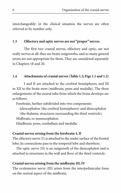
1.3 Olfactory and optic nerves are not “proper” nerves
The first two cranial nerves, olfactory and optic, are not
really nerves at all: they are brain outgrowths, and so many general
terms are not appropriate for them. They are considered separately
in Chapters 18 and 20.
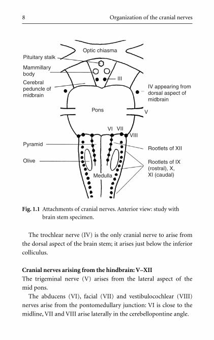
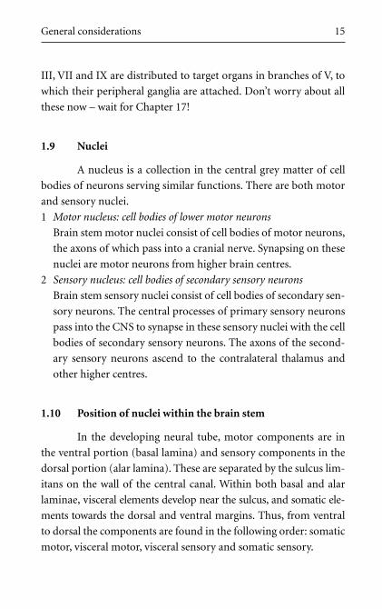
1.4 Attachments of cranial nerves (Table 1.2; Figs 1.1 and 1.2)
I and II are attached to the cerebral hemispheres, and III
to XII to the brain stem (midbrain, pons and medulla). The three
enlargements of the neural tube from which the brain develops are
as follows:
Forebrain, further subdivided into two components:
telencephalon (the cerebral hemispheres) and diencephalon
(the thalamic structures surrounding the third ventricle).
Midbrain, or mesencephalon.
Hindbrain: pons, cerebellum and medulla.
Cranial nerves arising from the forebrain: I, II
The olfactory nerve (I) is attached to the under surface of the frontal
lobe; its connections pass to the temporal lobe and elsewhere.
The optic nerve (II) is an outgrowth of the diencephalon and is
attached to structures in the wall and floor of the third ventricle.
Cranial nerves arising from the midbrain: III, IV
The oculomotor nerve (III) arises from the interpeduncular fossa
on the ventral aspect of the midbrain.
6 Organization of the cranial nerves
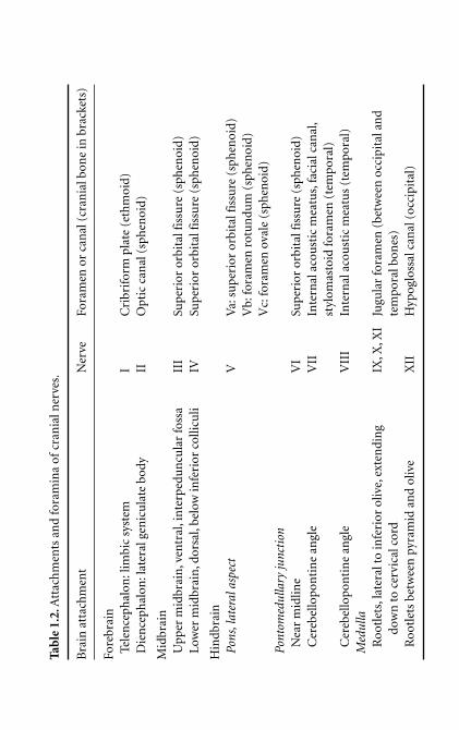
Tab
le 1
.2.A
ttac
hm
ents
an
d fo
ram
ina
ofcr
ania
l ner
ves.
Bra
in a
ttac
hm
ent
Ner
veFo
ram
en o
r ca
nal
(cr
ania
l bon
e in
bra
cket
s)
Fore
brai
nTe
len
ceph
alon
: lim
bic
syst
emI
Cri
brif
orm
pla
te (
eth
moi
d)D
ien
ceph
alon
: lat
eral
gen
icu
late
bod
yII
Opt
ic c
anal
(sp
hen
oid)
Mid
brai
nU
pper
mid
brai
n, v
entr
al, i
nte
rped
un
cula
r fo
ssa
III
Sup
erio
r or
bita
l fis
sure
(sp
hen
oid)
Low
er m
idbr
ain
,dor
sal,
belo
w in
feri
or c
ollic
uli
IVSu
per
ior
orbi
tal f
issu
re (
sph
enoi
d)
Hin
dbra
inPo
ns,l
ater
al a
spec
tV
Va:
supe
rior
orb
ital
fiss
ure
(sp
hen
oid)
Vb:
fora
men
rot
un
dum
(sp
hen
oid)
Vc:
fora
men
ova
le (
sph
enoi
d)Po
ntom
edul
lary
junc
tion
Nea
r m
idlin
eV
ISu
peri
or o
rbit
al fi
ssu
re (
sph
enoi
d)C
ereb
ello
pon
tin
e an
gle
VII
Inte
rnal
aco
ust
ic m
eatu
s,fa
cial
can
al,
styl
omas
toid
fora
men
(te
mpo
ral)
Cer
ebel
lopo
nti
ne
angl
eV
III
Inte
rnal
aco
ust
ic m
eatu
s (t
empo
ral)
Med
ulla
Roo
tlet
s,la
tera
l to
infe
rior
oliv
e,ex
ten
din
g IX
,X,X
IJu
gula
r fo
ram
en (
betw
een
occ
ipit
al a
nd
dow
n to
cer
vica
l cor
dte
mpo
ral b
ones
)R
ootl
ets
betw
een
pyr
amid
an
d ol
ive
XII
Hyp
oglo
ssal
can
al (
occi
pita
l)
The trochlear nerve (IV) is the only cranial nerve to arise from
the dorsal aspect of the brain stem; it arises just below the inferior
colliculus.
Cranial nerves arising from the hindbrain: V–XII
The trigeminal nerve (V) arises from the lateral aspect of the
mid pons.
The abducens (VI), facial (VII) and vestibulocochlear (VIII)
nerves arise from the pontomedullary junction: VI is close to the
midline, VII and VIII arise laterally in the cerebellopontine angle.
8 Organization of the cranial nerves
Optic chiasmaPituitary stalk
Mammillarybody
Cerebralpeduncle ofmidbrain
Pyramid
Olive
Medulla
Rootlets of IX(rostral), X,XI (caudal)
Rootlets of XII
VIII
V
VI VII
Pons
III
IV appearing fromdorsal aspect ofmidbrain
�
Fig. 1.1 Attachments of cranial nerves. Anterior view: study with
brain stem specimen.
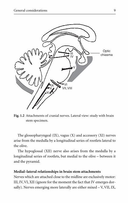
The glossopharyngeal (IX), vagus (X) and accessory (XI) nerves
arise from the medulla by a longitudinal series of rootlets lateral to
the olive.
The hypoglossal (XII) nerve also arises from the medulla by a
longitudinal series of rootlets, but medial to the olive – between it
and the pyramid.
Medial–lateral relationships in brain stem attachments
Nerves which are attached close to the midline are exclusively motor:
III, IV, VI, XII (ignore for the moment the fact that IV emerges dor-
sally). Nerves emerging more laterally are either mixed – V, VII, IX,
General considerations 9
OpticchiasmaIII
IV
V
VIIX
XIX VII,VIII
Fig. 1.2 Attachments of cranial nerves. Lateral view: study with brain
stem specimen.
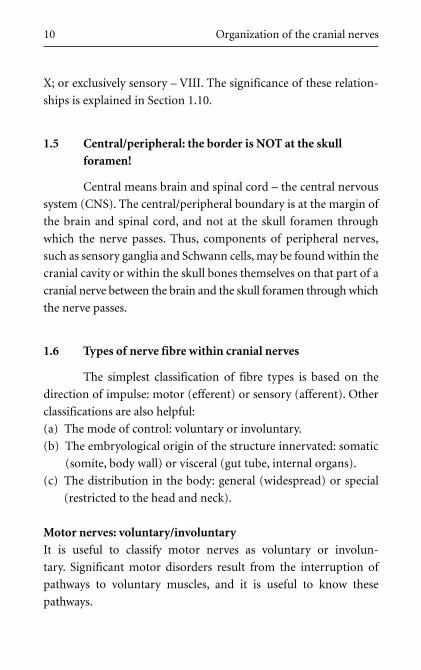
X; or exclusively sensory – VIII. The significance of these relation-
ships is explained in Section 1.10.
1.5 Central/peripheral: the border is NOT at the skull
foramen!
Central means brain and spinal cord – the central nervous
system (CNS). The central/peripheral boundary is at the margin of
the brain and spinal cord, and not at the skull foramen through
which the nerve passes. Thus, components of peripheral nerves,
such as sensory ganglia and Schwann cells, may be found within the
cranial cavity or within the skull bones themselves on that part of a
cranial nerve between the brain and the skull foramen through which
the nerve passes.
1.6 Types of nerve fibre within cranial nerves
The simplest classification of fibre types is based on the
direction of impulse: motor (efferent) or sensory (afferent). Other
classifications are also helpful:
(a) The mode of control: voluntary or involuntary.
(b) The embryological origin of the structure innervated: somatic
(somite, body wall) or visceral (gut tube, internal organs).
(c) The distribution in the body: general (widespread) or special
(restricted to the head and neck).
Motor nerves: voluntary/involuntary
It is useful to classify motor nerves as voluntary or involun-
tary. Significant motor disorders result from the interruption of
pathways to voluntary muscles, and it is useful to know these
pathways.
10 Organization of the cranial nerves
Motor nerves: somatic/visceral
An embryological distinction is also useful: somatic motor, supply-
ing body wall muscles mainly derived from somites, and visceral
motor, supplying muscle associated with yolk sac derivatives and
internal organs. Even though research casts doubt on the validity of
this distinction, it is helpful conceptually and it enables us to predict
with some accuracy the position of nerve roots and motor nuclei
within the brain stem (Sections 1.10 and 2.7).
Sensory nerves: somatic/visceral is not a particularly useful
distinction
Somatic sensation is sensation from body wall structures (soma:
body): in cranial nerves, it includes that from the skin and oral cav-
ity (except taste). Visceral sensation includes that from the alimentary
canal (except the mouth) and taste. The somatic/visceral distinc-
tion is based not upon the nature of the peripheral nerve or neu-
ron, but upon how the information is handled once inside the CNS:
brain stem connections of somatic sensation are different from
those of visceral sensation (Section 4.2).
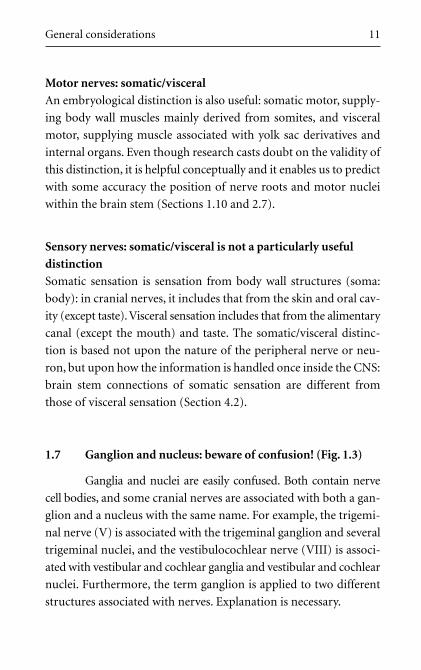
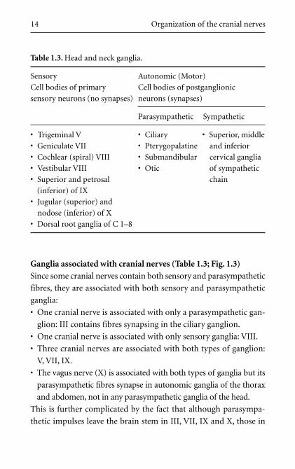
1.7 Ganglion and nucleus: beware of confusion! (Fig. 1.3)
Ganglia and nuclei are easily confused. Both contain nerve
cell bodies, and some cranial nerves are associated with both a gan-
glion and a nucleus with the same name. For example, the trigemi-
nal nerve (V) is associated with the trigeminal ganglion and several
trigeminal nuclei, and the vestibulocochlear nerve (VIII) is associ-
ated with vestibular and cochlear ganglia and vestibular and cochlear
nuclei. Furthermore, the term ganglion is applied to two different
structures associated with nerves. Explanation is necessary.
General considerations 11
A ganglion is simply a swelling. Thus, in a nerve, ganglion means
a swelling on the nerve. It is used to mean the swelling caused by a
collection of nerve cell bodies on a peripheral nerve: cell bodies take
up more space than fibres, so a collection of cell bodies will cause a
swelling.
12 Organization of the cranial nerves
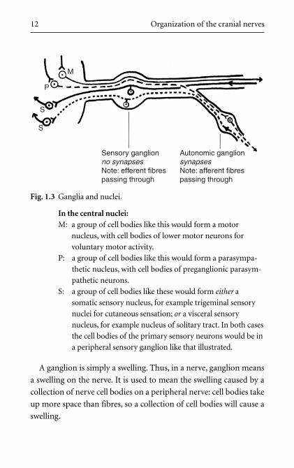
Sensory ganglionno synapsesNote: efferent fibrespassing through
M
P
S
S
Autonomic ganglionsynapsesNote: afferent fibrespassing through
Fig. 1.3 Ganglia and nuclei.
In the central nuclei:M: a group of cell bodies like this would form a motor
nucleus, with cell bodies of lower motor neurons for
voluntary motor activity.
P: a group of cell bodies like this would form a parasympa-
thetic nucleus, with cell bodies of preganglionic parasym-
pathetic neurons.
S: a group of cell bodies like these would form either a
somatic sensory nucleus, for example trigeminal sensory
nuclei for cutaneous sensation; or a visceral sensory
nucleus, for example nucleus of solitary tract. In both cases
the cell bodies of the primary sensory neurons would be in
a peripheral sensory ganglion like that illustrated.

A nucleus is an aggregation of cell bodies in the CNS (exception:
basal ganglia of the brain, the term being of historical significance;
basal nuclei is better).
Ganglia are peripheral; nuclei are central.
1.8 Ganglia (Fig. 1.3)
A ganglion is a collection of nerve cell bodies associated
with a peripheral nerve. There are two types of ganglia: those with
synapses and those without.
1 Ganglia with synapses: autonomic ganglia
These are found on autonomic (visceral motor) pathways, and are
thus autonomic ganglia. Autonomic neurons in cranial nerves are
parasympathetic. Preganglionic neurons convey impulses from
brain stem nuclei and synapse with postganglionic neurons, the
cell bodies of which constitute the ganglia. (For sympathetic gan-
glia, see Chapter 19.)
2 Ganglia without synapses: sensory ganglia
Nearly all primary sensory neurons have their cell bodies in
peripheral ganglia – sensory ganglia. Primary sensory neurons
are usually pseudounipolar: that is to say, the single axon which
arises from the cell body bifurcates into a peripheral process
which passes towards the receptor, and a central process which
passes towards the brain. There are no synapses in sensory gan-
glia. An example of this type of ganglion is found on every nerve
containing sensory fibres; on spinal nerves they are dorsal root
ganglia.
Note that each ganglion is either a sensory ganglion or an auto-
nomic ganglion; there is no such thing as a mixed autonomic and
sensory ganglion.
General considerations 13
Ganglia associated with cranial nerves (Table 1.3; Fig. 1.3)
Since some cranial nerves contain both sensory and parasympathetic
fibres, they are associated with both sensory and parasympathetic
ganglia:
• One cranial nerve is associated with only a parasympathetic gan-
glion: III contains fibres synapsing in the ciliary ganglion.
• One cranial nerve is associated with only sensory ganglia: VIII.
• Three cranial nerves are associated with both types of ganglion:
V, VII, IX.
• The vagus nerve (X) is associated with both types of ganglia but its
parasympathetic fibres synapse in autonomic ganglia of the thorax
and abdomen, not in any parasympathetic ganglia of the head.
This is further complicated by the fact that although parasympa-
thetic impulses leave the brain stem in III, VII, IX and X, those in
14 Organization of the cranial nerves
Table 1.3. Head and neck ganglia.
Sensory Autonomic (Motor)
Cell bodies of primary Cell bodies of postganglionic
sensory neurons (no synapses) neurons (synapses)
Parasympathetic Sympathetic
• Trigeminal V • Ciliary • Superior, middle
• Geniculate VII • Pterygopalatine and inferior
• Cochlear (spiral) VIII • Submandibular cervical ganglia
• Vestibular VIII • Otic of sympathetic
• Superior and petrosal chain
(inferior) of IX
• Jugular (superior) and
nodose (inferior) of X
• Dorsal root ganglia of C 1–8
III, VII and IX are distributed to target organs in branches of V, to
which their peripheral ganglia are attached. Don’t worry about all
these now – wait for Chapter 17!
1.9 Nuclei
A nucleus is a collection in the central grey matter of cell
bodies of neurons serving similar functions. There are both motor
and sensory nuclei.
1 Motor nucleus: cell bodies of lower motor neurons
Brain stem motor nuclei consist of cell bodies of motor neurons,
the axons of which pass into a cranial nerve. Synapsing on these
nuclei are motor neurons from higher brain centres.
2 Sensory nucleus: cell bodies of secondary sensory neurons
Brain stem sensory nuclei consist of cell bodies of secondary sen-
sory neurons. The central processes of primary sensory neurons
pass into the CNS to synapse in these sensory nuclei with the cell
bodies of secondary sensory neurons. The axons of the second-
ary sensory neurons ascend to the contralateral thalamus and
other higher centres.
1.10 Position of nuclei within the brain stem
In the developing neural tube, motor components are in
the ventral portion (basal lamina) and sensory components in the
dorsal portion (alar lamina). These are separated by the sulcus lim-
itans on the wall of the central canal. Within both basal and alar
laminae, visceral elements develop near the sulcus, and somatic ele-
ments towards the dorsal and ventral margins. Thus, from ventral
to dorsal the components are found in the following order: somatic
motor, visceral motor, visceral sensory and somatic sensory.
General considerations 15
In the brain stem, this is preserved in a modified fashion. During
development, it is as if the dorsal aspects of the brain stem were
forcibly parted, each side being pushed laterally, by the enlarging
central canal which becomes the fourth ventricle. The sequence
somatic motor, visceral motor, visceral sensory, somatic sensory in
the brain stem is therefore not so much ventral to dorsal as medial
to lateral. Thus somatic motor nerves (e.g. III, XII) arise near the
midline, nerves with visceral components (e.g. V, VII, IX, X) arise
further laterally, and the entirely sensory VIII most lateral of all.
Refer again to Section 1.4.
16 Organization of the cranial nerves

Chapter 2
C R A N I A L N E RV E M OTO R F I B R E SA N D N U C L E I
2.1 Motor fibres
Motor fibres are present in all cranial nerves except I, II
and VIII.
2.2 Classification of motor components in cranial nerves
In spinal nerves, it is useful to distinguish between somatic
and visceral motor fibres. This is based on the embryological origin
of the muscle innervated.
Somatic motor (voluntary) fibres innervate muscles which
develop from somites: striated muscle. Cell bodies are the ventral
horn cells of the spinal cord grey matter. These muscles are under
voluntary control.
Visceral motor (autonomic, involuntary) fibres innervate muscles
which develop in association with the gut tube and its derivatives
(e.g. bronchial tree), in glands, hair follicles and the heart. Except for
cardiac muscle, it is smooth or non-striated. It is involuntary.
Thus, in the trunk and limbs voluntary may be loosely equated
with striated and somatic, and involuntary with smooth and visceral.
2.3 Additional component in cranial nerves: for
branchial arches
In the head and neck there is an additional group of muscles
which are striated and are under voluntary control, but are classed

18 Organization of the cranial nerves
as visceral because they develop in association with the cranial end
of the gut tube. These are derivatives of the branchial or pharyngeal
arches. Branchial arch muscles are concerned only with the cephalic
end of the gut tube and have no equivalents below the neck; they are
innervated by branchiomotor fibres, found only in cranial nerves,
which originate from branchiomotor nuclei in the brain stem.
2.4 Types of motor nerve fibres
There are thus three types of motor nerve fibres in cranial
nerves:
1 Voluntary – somatic.
2 Voluntary – visceral – branchiomotor (special visceral; special
because confined to the head and neck).
3 Involuntary – visceral – parasympathetic (general visceral; general
because distributed more widely).
Remember: In cranial nerves visceral cannot be equated exclusively
with autonomic or involuntary.
Motor fibres supplying voluntary muscles are found in all cranial
nerves except I, II and VIII. Cranial nerve motor fibres are either
somatic or visceral (somatic and visceral fibres are never found in
the same nerve).
2.5 Motor fibres in cranial nerves
• Somatic motor: III, IV, VI, XII:
– Extrinsic ocular muscles which move the eyeball and upper eye-
lid: oculomotor (III), trochlear (IV) and abducens (VI) nerves.
– Tongue muscles: hypoglossal nerve (XII).
• Branchiomotor: V, VII, IX, X (XI) (Table 2.1).
Cranial nerve motor fibres and nuclei 19
– The five branchial arches consist of ridges of mesoderm pass-
ing ventral–dorsal on either side of the foregut at the head end
of the embryo. For reasons which need not concern us, these
are numbered, cranial–caudal, as I, II, III, IV and VI. Each
branchial arch gives rise to skeletal structures, muscles, nerves
and arteries, the muscles of an arch being innervated by the
nerve of that arch.
Axons and cell bodies of voluntary motor nerves
For both somatic and branchiomotor voluntary fibres, axons in
peripheral nerves pass without interruption from cell bodies in the
brain stem motor nuclei to the muscles of destination. These neu-
rons are called lower motor neurons. Note that their cell bodies are
in the central nervous system.
2.6 Parasympathetic components of cranial nerves
Parasympathetic fibres emerge from the brain in only
four cranial nerves: III, VII, IX and X, and are delivered to their
destinations in branches of V. They innervate the ciliary and iris
Table 2.1. Branchial arches, muscles and nerves.
Branchial arch Muscles Nerves
First Muscles of Mandibular Vc
mastication, etc.
Second Muscles of facial Facial VII
expression, etc.
Third Stylopharyngeus Glossopharyngeal IX
Fourth Pharyngeal muscles Pharyngeal branches of X
Sixth Laryngeal muscles Recurrent laryngeal of X

20 Organization of the cranial nerves
muscles of the eyeball, and the salivary, lacrimal, nasal and palatal
glands. They are arranged with two peripheral neurons: pre- and
postganglionic. Cell bodies of preganglionic neurons are in
brain stem parasympathetic nuclei, and their axons synapse on
postganglionic neurons in peripheral parasympathetic ganglia. See
Chapter 17.
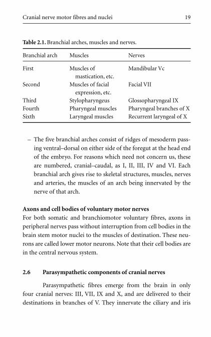
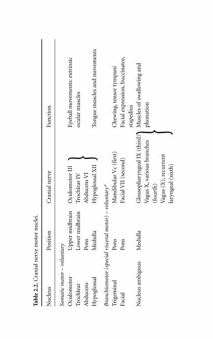
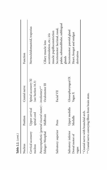
2.7 Brain stem motor nuclei (Table 2.2; Fig. 2.1)
Axons of cranial nerve motor neurons originate from brain
stem nuclei of three types corresponding to the embryological ori-
gin of the muscle groups:
1 Somatic nuclei: These are close to the midline, equivalent to
spinal cord ventral horn cells. Somatic nuclei are oculomotor,
trochlear, abducens and hypoglossal nuclei.
2 Branchiomotor nuclei: These develop lateral to somatic nuclei,
between them and parasympathetic nuclei. Branchiomotor nuclei
are trigeminal motor, facial motor and the nucleus ambiguus
(and probably its cervical extension for the spinal accessory
nerve, see Section 16.3).
3 Parasympathetic nuclei: These are the most laterally placed of the
brain stem motor nuclei, equivalent to lateral horn cells of the
spinal cord. They include Edinger–Westphal, superior and infe-
rior salivatory, and the dorsal motor nucleus of the vagus.
Brain stem motor nuclei thus make up three interrupted columns:
somatic motor, branchiomotor (special visceral motor) and parasym-
pathetic (general visceral motor). This pattern is a useful basis for
further study.
Tab
le 2
.2.C
ran
ial n
erve
mot
or n
ucl
ei.
Nu
cleu
sPo
siti
onC
ran
ial n
erve
Fun
ctio
n
Som
atic
mot
or –
vol
unta
ry
Ocu
lom
otor
Upp
er m
idbr
ain
Ocu
lom
otor
III
Eye
ball
mov
emen
ts: e
xtri
nsi
c
Troc
hle
arL
ower
mid
brai
nTr
och
lear
IV
ocu
lar
mu
scle
s
Abd
uce
ns
Pon
sA
bdu
cen
s V
I
Hyp
oglo
ssal
Med
ulla
Hyp
oglo
ssal
XII
Ton
gue
mu
scle
s an
d m
ovem
ents
Bra
nchi
omot
or (
spec
ial v
isce
ral m
otor
) –
volu
ntar
y*
Trig
emin
al
Pon
sM
andi
bula
r V
c (f
irst
)C
hew
ing,
ten
sor
tym
pan
i
Faci
alPo
ns
Faci
al V
II (
seco
nd)
Faci
al e
xpre
ssio
n,b
ucc
inat
or,
stap
ediu
s
Nu
cleu
s am
bigu
us
Med
ulla
Glo
ssop
har
ynge
al I
X (
thir
d)M
usc
les
ofsw
allo
win
g an
d
Vag
us
X,v
ario
us
bran
ches
phon
atio
n
(fou
rth
)
Vag
us
(X),
recu
rren
t
lary
nge
al (
sixt
h)
}
}
Tab
le 2
.2.(
cont
.)
Nu
cleu
sPo
siti
onC
ran
ial n
erve
Fun
ctio
n
Cer
vica
l acc
esso
ry
Upp
er c
ervi
cal
Spin
al a
cces
sory
XI
Ster
noc
leid
omas
toid
, tra
pez
ius
nu
cleu
ssp
inal
cor
d(s
ee S
ecti
on 1
6.3)
Para
sym
path
etic
(ge
nera
l vis
cera
l mot
or)
– in
volu
ntar
y**
Edi
nge
r–W
estp
hal
Mid
brai
nO
culo
mot
or I
IIC
iliar
y m
usc
le:l
ens
acco
mm
odat
ion
,etc
.;ir
is
mu
scle
:pu
pillo
con
stri
ctio
n
Saliv
ator
y:su
peri
orPo
ns
Faci
al V
IISe
cret
omot
or:l
acri
mal
,nas
al,
pala
te,s
ubm
andi
bula
r,su
blin
gual
glan
ds
Saliv
ator
y:in
feri
orU
pper
med
ulla
Glo
ssop
har
ynge
al I
XSe
cret
omot
or:p
arot
id g
lan
d
Dor
sal m
otor
of
Med
ulla
Vag
us
XH
eart
,for
egu
t an
d m
idgu
t
vagu
sde
riva
tive
s
*C
ran
ial n
erve
wit
h b
ran
chia
l arc
h.
**C
ran
ial n
erve
con
veyi
ng
fibr
es fr
om b
rain
ste
m.
Cranial nerve motor fibres and nuclei 23
Oc
EW
Tr
TMF
Ab
SSNISN
DMNX
HypNA
AccXII
XI
X
IX
VIIVI
V
IV
III
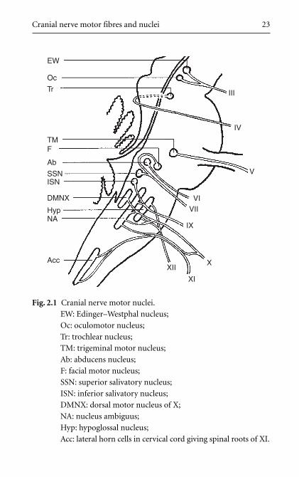
Fig. 2.1 Cranial nerve motor nuclei.
EW: Edinger–Westphal nucleus;
Oc: oculomotor nucleus;
Tr: trochlear nucleus;
TM: trigeminal motor nucleus;
Ab: abducens nucleus;
F: facial motor nucleus;
SSN: superior salivatory nucleus;
ISN: inferior salivatory nucleus;
DMNX: dorsal motor nucleus of X;
NA: nucleus ambiguus;
Hyp: hypoglossal nucleus;
Acc: lateral horn cells in cervical cord giving spinal roots of XI.

Chapter 3
C R A N I A L N E RV E M OTO R PAT H WAYS : U P P E R A N D LOW E RM OTO R N E U RO N S
3.1 Upper and lower motor neurons
Both somatic motor and branchiomotor nerves supply
voluntary muscles. Pathways between motor cortex and muscles may
be thought of as being arranged in two neuronal groups: upper
motor neurons and lower motor neurons. Axons of upper motor
neurons decussate before synapsing with lower motor neurons, so
the right motor cortex controls the left side of the body, and vice
versa – contralateral control.
Upper motor neurons: cortex to nucleus
For cranial nerves, cell bodies of upper motor neurons are in the
head and neck area of the motor cortex. Axons descend, decussating
just before synapsing with cell bodies of lower motor neurons
which make up the motor nucleus of that cranial nerve. The term
upper motor neurons is also used clinically to include fibres from
other brain centres (e.g. parietal lobe, basal ganglia, cerebellum,
reticular formation, midbrain, etc.) that connect with the lower
motor neurons in the cranial nerve nucleus, thus influencing their
activity.
Lower motor neurons: nucleus to muscle
Cell bodies of lower motor neurons form the brain stem nucleus.
Axons leave the brain stem and pass in the cranial nerve to the

Cranial nerve motor pathways 25
destination. Thus, although most of the axon of the lower motor
neuron is part of the peripheral nervous system, the cell body and
first part of the axon is in the central nervous system.
3.2 Corticonuclear and corticobulbar
These terms describe the upper motor neuron pathways
described above. They are often used interchangeably even though,
since bulb means medulla, corticobulbar should be reserved for fibres
passing to nuclei in the medulla.
3.3 Corticonuclear pathways (Fig. 3.1)
Frontal motor cortex
The head and neck area of the frontal motor cortex is found in the
most lateral part of the precentral gyrus of the frontal lobe, imme-
diately anterior to the central sulcus above the lateral fissure. It is
supplied by branches of the middle cerebral artery. Its approximate
surface marking is the pterion.
Corona radiata, internal capsule
Axons of upper motor neurons descend through the corona radiata
and on to the genu of the internal capsule. The arterial supply of
the internal capsule is from the medial and lateral striate branches
of the middle cerebral artery.
Brain stem course
Axons of upper motor neurons descend through the central por-
tions of the cerebral peduncles (crura) of the midbrain ventral to
the substantia nigra and proceed as far as necessary, decussating
just before synapsing on lower motor neuron cell bodies in the
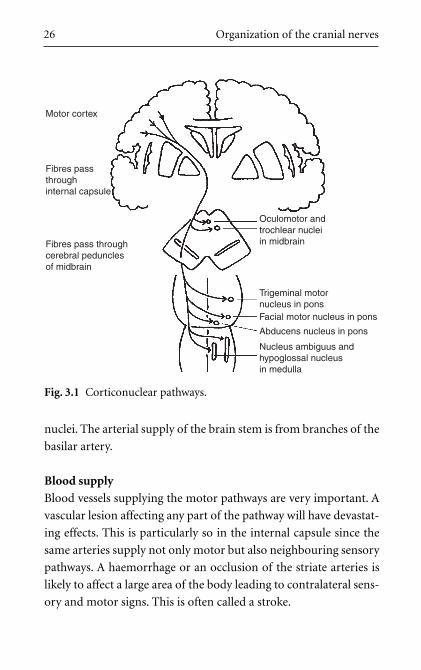
26 Organization of the cranial nerves
nuclei. The arterial supply of the brain stem is from branches of the
basilar artery.
Blood supply
Blood vessels supplying the motor pathways are very important. A
vascular lesion affecting any part of the pathway will have devastat-
ing effects. This is particularly so in the internal capsule since the
same arteries supply not only motor but also neighbouring sensory
pathways. A haemorrhage or an occlusion of the striate arteries is
likely to affect a large area of the body leading to contralateral sens-
ory and motor signs. This is often called a stroke.
Motor cortex
Oculomotor andtrochlear nucleiin midbrain
Trigeminal motornucleus in ponsFacial motor nucleus in pons
Abducens nucleus in pons
Nucleus ambiguus andhypoglossal nucleusin medulla
Fibres passthroughinternal capsule
Fibres pass throughcerebral pedunclesof midbrain
Fig. 3.1 Corticonuclear pathways.
Cranial nerve motor pathways 27
3.4 Bilateral upper motor neuron control of III, IV, VIand part of VII
The pattern in the head and neck, as in the rest of the body, is
that the motor cortex innervates contralateral motor nuclei. However,
muscles which move the eyes, and the eyelids and forehead in asso-
ciation with eye movements, receive bilateral cortical innervation.
The nuclei concerned are the oculomotor (III), trochlear (IV) and
abducens (VI), and that portion of facial (VII) motor nucleus which
innervates orbicularis oculi and frontalis. This must have evolved
in association with, and for the protection of, the sense of sight by
which means we seek sustenance and mates, and avoid danger.
There is limited bilateral control of the other voluntary motor
nuclei as is evidenced by partial recovery of function in patients after
a stroke.
3.5 Upper and lower motor neuron lesions
Lower motor neuron lesion: flaccidity, hyporeflexia, wasting,
ipsilateral
If all lower motor neurons passing to a muscle are severed, the mus-
cle will be completely paralyzed. It will be flaccid (atonic, hypo-
tonic), it will not respond to reflexes (arreflexic, hyporeflexic) since
no impulses reach it, and it will fairly quickly atrophy as a result of
denervation. The injury and the paralysis are on the same side; they
are ipsilateral with respect to each other.
Upper motor neuron lesion: spasticity, hyperreflexia,
contralateral
If upper motor neurons to a muscle are severed, the ability to con-
trol and initiate movement in the muscle may be lost. However,
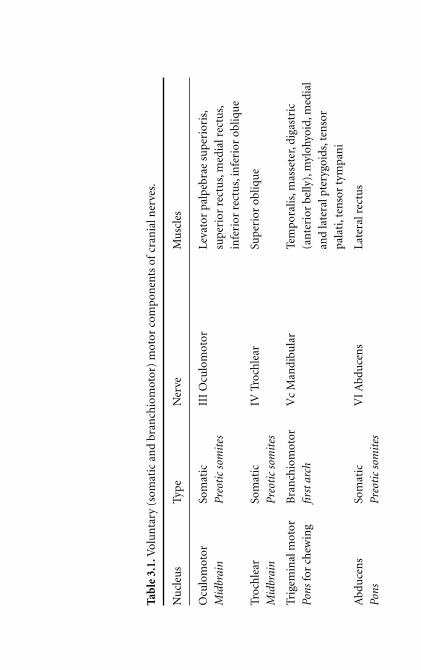
Tab
le 3
.1.V
olu
nta
ry (
som
atic
an
d br
anch
iom
otor
) m
otor
com
pon
ents
of
cran
ial n
erve
s.
Nu
cleu
sTy
pe
Ner
veM
usc
les
Ocu
lom
otor
Som
atic
III
Ocu
lom
otor
Leva
tor
palp
ebra
e su
peri
oris
,
Mid
brai
nP
reot
ic s
omit
essu
peri
or r
ectu
s,m
edia
l rec
tus,
infe
rior
rec
tus,
infe
rior
obl
iqu
e
Troc
hle
arSo
mat
icIV
Tro
chle
arSu
peri
or o
bliq
ue
Mid
brai
nP
reot
ic s
omit
es
Trig
emin
al m
otor
Bra
nch
iom
otor
Vc
Man
dibu
lar
Tem
pora
lis,m
asse
ter,
diga
stri
c
Pons
for
chew
ing
firs
t arc
h(a
nte
rior
bel
ly),
myl
ohyo
id,m
edia
l
and
late
ral p
tery
goid
s,te
nso
r
pala
ti,t
enso
r ty
mpa
ni
Abd
uce
ns
Som
atic
VI
Abd
uce
ns
Late
ral r
ectu
s
Pons
Pre
otic
som
ites
Faci
al m
otor
Bra
nch
iom
otor
VII
Fac
ial
Mu
scle
s of
faci
al e
xpre
ssio
n,
Pons
seco
nd a
rch
bucc
inat
or,s
tap
ediu
s,
occi
pito
fron
talis
, sty
lohy
oid,
diga
stri
c (p
oste
rior
bel
ly),
plat
ysm
a
Nu
cleu
s B
ran
chio
mot
or
ambi
guu
s(s
ee S
ecti
on 1
6.3)
Med
ulla
thir
d ar
chIX
Glo
ssop
har
ynge
alSt
ylop
har
ynge
us
for
swal
low
ing,
four
th a
rch
X V
agu
s,ph
aryn
geal
bra
nch
esM
usc
les
ofph
aryn
x
phon
atio
nsi
xth
arch
X V
agu
s,re
curr
ent
lary
nge
alM
usc
les
ofla
ryn
x
XI
Spin
al a
cces
sory
Ster
noc
leid
omas
toid
,tra
pezi
us
Hyp
oglo
ssal
Som
atic
XII
Hyp
oglo
ssal
Intr
insi
c to
ngu
e m
uscl
es,h
yogl
ossu
s,
Med
ulla
Occ
ipit
al so
mit
esge
nio
glos
sus,
styl
oglo
ssus
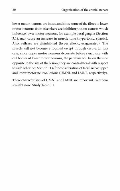
30 Organization of the cranial nerves
lower motor neurons are intact, and since some of the fibres to lower
motor neurons from elsewhere are inhibitory, other centres which
influence lower motor neurons, for example basal ganglia (Section
3.1), may cause an increase in muscle tone (hypertonic, spastic).
Also, reflexes are disinhibited (hyperreflexic, exaggerated). The
muscle will not become atrophied except through disuse. In this
case, since upper motor neurons decussate before synapsing with
cell bodies of lower motor neurons, the paralysis will be on the side
opposite to the site of the lesion; they are contralateral with respect
to each other. See Section 11.6 for consideration of facial nerve upper
and lower motor neuron lesions (UMNL and LMNL, respectively).
These characteristics of UMNL and LMNL are important. Get them
straight now! Study Table 3.1.
Chapter 4
C R A N I A L N E RV E S E N S O RY F I B R E S , B R A I N S T E M S E N S O RYN U C L E I A N D T R AC T S
Note: Sensory fibres carried by the olfactory, optic and vestibulo-
cochlear nerves are not dealt with in this chapter. Consult Chapters
18, 20 and 23.
4.1 The basic plan of sensory systems
The basic sensory system consists of three neuronal groups:
• primary sensory neurons from receptor to central nucleus, with
its cell body in a peripheral sensory ganglion;
• secondary sensory neurons from nucleus to diencephalon (usually
the thalamus);
• tertiary sensory neurons from thalamus to cortex.
There are no synapses outside the brain and spinal cord: the first
synapse is in the central nervous system (CNS) between primary and
secondary sensory neurons.
Primary sensory neuron: receptor to sensory nucleus
This extends from peripheral receptor to CNS. The cell body is situ-
ated in a peripheral ganglion (dorsal root ganglion for spinal nerves)
and the neuron is usually pseudounipolar, that is to say, it gives rise
to a single axon which bifurcates into a peripheral process passing
to the receptor, and a central process passing into the CNS. The cell
body is thus both structurally and electrically out on a limb. The
central process of the primary sensory neuron terminates by synapsing
32 Organization of the cranial nerves
in a central nucleus which consists of cell bodies of the neuron next
in the pathway …
Secondary sensory neurons: sensory nucleus to thalamus
The axons of these neurons ascend from the nucleus, which contains
their cell bodies, to the contralateral thalamus (in the diencephalon),
decussating soon after leaving the nucleus. In the thalamus, they
synapse with the cell bodies of tertiary sensory neurons. (There are
many other destinations of impulses from the nucleus, for example
reticular nuclei and cerebellum, for the dissemination of informa-
tion and its integration with other functions and systems.)
Tertiary sensory neurons: thalamus, internal capsule, cortex
Axons of tertiary sensory neurons extend from the thalamus to
the appropriate area of sensory cortex and elsewhere, the neurons
being known as thalamocortical neurons. These pass through the
internal capsule. The principal sensory cortex for the head, to which
somatic sensation is relayed by the thalamocortical neurons, is found
on the lateral aspect of the parietal lobe behind the central sulcus and
immediately above the lateral fissure. It is adjacent to the head and
neck area of the motor cortex in the frontal lobe.
4.2 Sensory fibres in cranial nerves: somatic and visceral
Cranial nerves which transmit sensory fibres (other than
I, II, VIII) are the trigeminal (V), facial (VII), glossopharyngeal (IX)
and vagus (X). As described earlier (Section 1.6), sensory informa-
tion may be classified as either somatic or visceral.
1 Somatic sensory (somatosensory):
Somatosensory fibres in cranial nerves convey pain, temperature,
tactile and proprioceptive impulses from skin of the scalp, face,

cheek and temple, oral cavity, teeth and gums, nasal cavity and
sinuses, and temporomandibular joint and muscles. The trigem-
inal nerve is the principal somatosensory cranial nerve. All cranial
nerve somatosensory fibres pass to the sensory nuclei of the
trigeminal nerve, irrespective of the cranial nerve through which
the fibres enter the brain stem.
2 Visceral sensory:
Visceral sensory fibres include taste fibres, fibres from the
alimentary canal except the oral cavity, teeth and gums, and
fibres from chemoreceptors and thoracoabdominal viscera. All
cranial nerve visceral sensory fibres pass to the nucleus of the
solitary tract, irrespective of the cranial nerve through which the
fibres enter the brain stem.
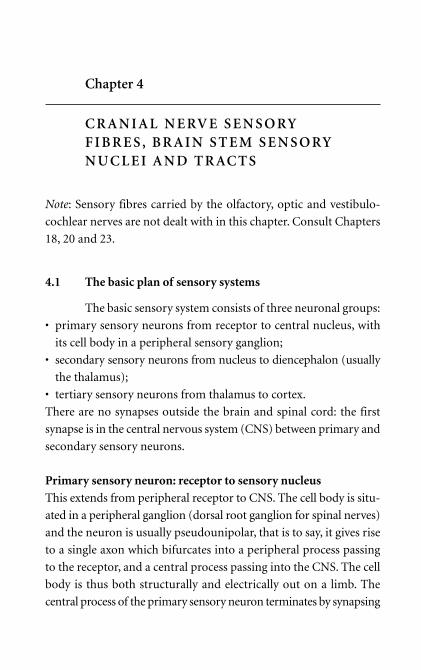
4.3 Somatic sensation (Fig. 4.1)
All but a few somatosensory fibres from structures in the
head are carried in the trigeminal (V) nerve. There are some
somatosensory fibres in the vagus (X) nerve, and a few in the facial
(VII) and glossopharyngeal (IX) nerves from the external ear. Cell
bodies of primary sensory neurons are situated in the peripheral
sensory ganglion (no synapses, remember) of the nerve through
which they enter the brain stem.
Sensory ganglia for somatosensory fibres
Most somatic sensory fibres are carried in the trigeminal nerve:
their cell bodies are in the trigeminal ganglion. The small number
of somatosensory fibres in the vagus nerve (X) have cell bodies
in the jugular (superior) vagal ganglion; those in the facial nerve
(VII) have cell bodies in the geniculate ganglion; and those in the
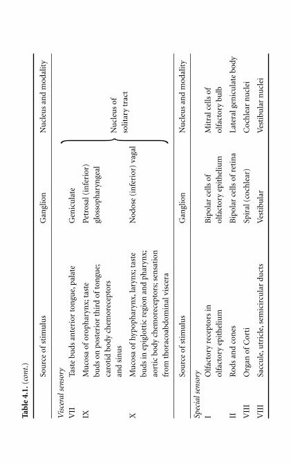
Cranial nerve sensory fibres, brain stem sensory nuclei and tracts 33
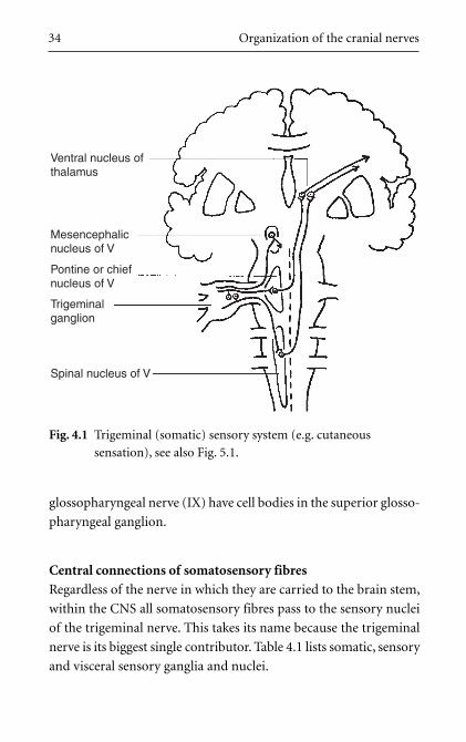
34 Organization of the cranial nerves
glossopharyngeal nerve (IX) have cell bodies in the superior glosso-
pharyngeal ganglion.
Central connections of somatosensory fibres
Regardless of the nerve in which they are carried to the brain stem,
within the CNS all somatosensory fibres pass to the sensory nuclei
of the trigeminal nerve. This takes its name because the trigeminal
nerve is its biggest single contributor. Table 4.1 lists somatic, sensory
and visceral sensory ganglia and nuclei.
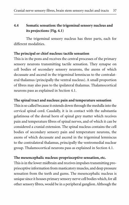
Ventral nucleus ofthalamus
Mesencephalicnucleus of V
Pontine or chiefnucleus of V
Trigeminalganglion
Spinal nucleus of V
Fig. 4.1 Trigeminal (somatic) sensory system (e.g. cutaneous
sensation), see also Fig. 5.1.
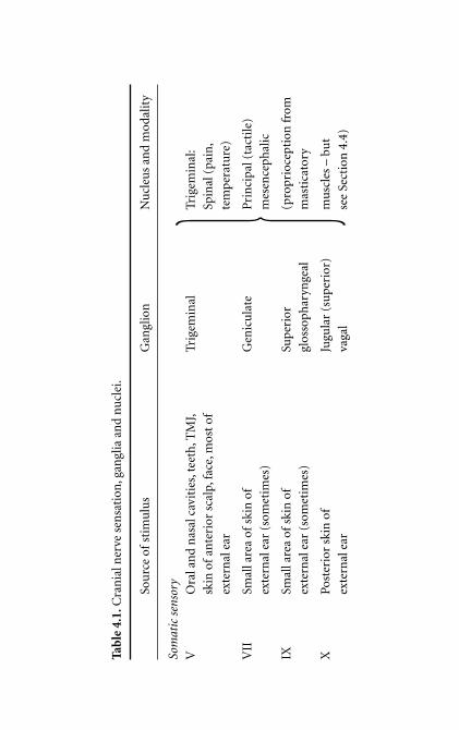
Tab
le 4
.1.C
ran
ial n
erve
sen
sati
on, g
angl
ia a
nd
nu
clei
.
Sou
rce
ofst
imu
lus
Gan
glio
nN
ucl
eus
and
mod
alit
y
Som
atic
sen
sory
VO
ral a
nd
nas
al c
avit
ies,
teet
h,T
MJ,
Trig
emin
alTr
igem
inal
:
skin
of
ante
rior
sca
lp,f
ace,
mos
t of
Spin
al (
pain
,
exte
rnal
ear
tem
pera
ture
)
VII
Smal
l are
a of
skin
of
Gen
icu
late
Pri
nci
pal (
tact
ile)
exte
rnal
ear
(so
met
imes
)m
esen
ceph
alic
IXSm
all a
rea
ofsk
in o
fSu
peri
or
(pro
prio
cept
ion
from
exte
rnal
ear
(so
met
imes
) gl
osso
phar
ynge
alm
asti
cato
ry
XPo
ster
ior
skin
of
Jugu
lar
(su
peri
or)
mu
scle
s –
but
exte
rnal
ear
vaga
lse
e Se
ctio
n 4
.4)�
Tab
le 4
.1.(
cont
.)
Sou
rce
ofst
imu
lus
Gan
glio
nN
ucl
eus
and
mod
alit
y
Vis
cera
l sen
sory
VII
Tast
e bu
ds a
nte
rior
ton
gue,
pala
teG
enic
ula
te
IXM
uco
sa o
for
oph
aryn
x;ta
ste
Petr
osal
(in
feri
or)
buds
on
pos
teri
or t
hir
d of
ton
gue;
glos
soph
aryn
geal
caro
tid
body
ch
emor
ecep
tors
and
sin
us
XM
uco
sa o
fhy
poph
aryn
x,la
ryn
x;ta
ste
Nod
ose
(in
feri
or)
vaga
l
buds
in e
pigl
otti
c re
gion
an
d ph
aryn
x;
aort
ic b
ody
chem
orec
epto
rs;s
ensa
tion
from
th
orac
oabd
omin
al v
isce
ra
Sou
rce
ofst
imu
lus
Gan
glio
nN
ucl
eus
and
mod
alit
y
Spec
ial s
enso
ry
IO
lfac
tory
rec
epto
rs in
B
ipol
ar c
ells
of
Mit
ral c
ells
of
olfa
ctor
y ep
ith
eliu
mol
fact
ory
epit
hel
ium
olfa
ctor
y bu
lb
IIR
ods
and
con
esB
ipol
ar c
ells
of
reti
na
Late
ral g
enic
ula
te b
ody
VII
IO
rgan
of
Cor
tiSp
iral
(co
chle
ar)
Coc
hle
ar n
ucl
ei
VII
ISa
ccu
le,u
tric
le,s
emic
ircu
lar
duct
sV
esti
bula
rV
esti
bula
r n
ucl
ei
�Nu
cleu
s of
solit
ary
trac
t
Cranial nerve sensory fibres, brain stem sensory nuclei and tracts 37
4.4 Somatic sensation: the trigeminal sensory nucleus and
its projections (Fig. 4.1)
The trigeminal sensory nucleus has three parts, each for
different modalities.
The principal or chief nucleus: tactile sensation
This is in the pons and receives the central processes of the primary
sensory neurons transmitting tactile sensation. They synapse on
cell bodies of secondary sensory neurons, the axons of which
decussate and ascend in the trigeminal lemniscus to the contralat-
eral thalamus (principally the ventral nucleus). A small proportion
of fibres may also pass to the ipsilateral thalamus. Thalamocortical
neurons pass as explained in Section 4.1.
The spinal tract and nucleus: pain and temperature sensation
This is so called because it extends down through the medulla into the
cervical spinal cord. Caudally, it is in contact with the substantia
gelatinosa of the dorsal horn of spinal grey matter which receives
pain and temperature fibres of spinal nerves, and of which it can be
considered a cranial extension. The spinal nucleus contains the cell
bodies of secondary sensory pain and temperature neurons, the
axons of which decussate and ascend in the trigeminal lemniscus
to the contralateral thalamus, principally the ventromedial nuclear
group. Thalamocortical neurons pass as explained in Section 4.1.
The mesencephalic nucleus: proprioceptive sensation, etc.
This is in the lower midbrain and receives impulses transmitting pro-
prioceptive information from masticatory muscles, and deep pressure
sensation from the teeth and gums. The mesencephalic nucleus is
unique since it houses primary sensory nerve cell bodies which, for all
other sensory fibres, would be in a peripheral ganglion. Although the

38 Organization of the cranial nerves
details of its connections are not entirely clear, this arrangement allows
other processes of the proprioceptive neurons to make connections
with, for example, the motor nucleus of V, the salivatory nuclei, and
the nucleus ambiguus – for chewing and swallowing (Section 13.1).
4.5 Visceral sensation: nucleus of the solitary tract
Clinically, this is less important than somatic sensation.
Visceral sensory fibres enter the brain stem in the facial (VII), glos-
sopharyngeal (IX) and vagus (X) nerves. Cell bodies of the primary
sensory neurons are in the peripheral sensory ganglion (no synapses)
of the nerve through which they enter the brain stem. Branches of the
trigeminal (V) nerve are involved peripherally in the complex course
of visceral sensory fibres, and visceral sensory fibres are often found
in nerves which carry parasympathetic fibres in the opposite direc-
tion; they are described later in Chapter 17.
Sensory ganglia for visceral sensory fibres
Visceral sensory fibres entering the brain stem in the facial nerve
(VII) have cell bodies in the geniculate ganglion; those in the glosso-
pharyngeal nerve (IX) have cell bodies in the petrosal (inferior)
glossopharyngeal ganglion; and those in the vagus nerve (X) have
cell bodies in the nodose (inferior) vagal ganglion.
Central connections of visceral sensory fibres
Regardless of the nerve in which they are carried to the brain stem,
within the CNS all visceral sensory fibres pass to the solitary tract and
nucleus (nucleus tractus solitarius or NTS) in the medulla. Axons
from the nucleus of the solitary tract pass rostrally by multisynaptic
pathways, possibly bilateral, to the thalamus (ventral posteromedial
nucleus), and thence probably to the insula and the uncus for con-
nections with olfactory centres (Chapter 18).
PARTS II–V
I N D I V I D UA L C R A N I A L
N E RV E S A N D F U N C T I O NA L
C O N S I D E R AT I O N S

Chapter 5
S U RV E Y O F C R A N I A L N E RV E S A N DI N T RO D U C T I O N TO PA RT S I I – V
In the following chapters we consider cranial nerves in groups con-
cerned with their functions. These are, in no particular order, inges-
tion and chewing, cutaneous sensation, swallowing and speaking,
autonomic function, taste and smell, and sight, hearing and balance.
The ingestion of food is dependent on opening the mouth. This
is a function of the mandibular division of the trigeminal (Vc) and
facial (VII) nerves: the mandibular opens the jaw and the facial parts
the lips. The facial, mandibular and hypoglossal (XII) nerves are
involved in taking the food into the mouth and closing the lips.
Chewing is served by the same three nerves: in simple language, the
facial nerve keeps the lips closed, the mandibular nerve moves the
mandible for its mastication, and both facial and hypoglossal nerves
maintain the food between the teeth. Also, the trigeminal senses its
position and consistency, and regulates the force of contraction of
the muscles, and both trigeminal and facial nerves are responsible
for taste perception from the mouth. The trigeminal nerve also has
another important function: the cutaneous sensation of the face and
anterior scalp. It is, except for a small area of skin in the external ear,
the only nerve concerned with this. So, the trigeminal, facial and hypo-
glossal nerves will be considered first.
After we eat and chew, we swallow. The motor components of swal-
lowing are mainly the responsibility of the vagus (X) and glossophar-
yngeal (IX) nerves, with the hypoglossal (XII) nerve also initially
involved. The vagus both innervates the muscles of swallowing, and
senses, albeit unconsciously after the initial stages, its progress. It is
also involved in phonation and speech which are related to swal-
lowing in that many of the muscles and nerves are the same. These
processes are aided by the glossopharyngeal nerve which, with the
vagus, carries sensory information to the brain and participates
in the perception of taste and the control of salivary secretions. The
accessory nerve (XI) is an accessory to the vagus and so it too should
be included in this group. After this, the loose ends of taste sensa-
tion and autonomic function may conveniently be tied up.
There is an embryological basis for studying the nerves in this
order. The cranial end of the developing embryo is dominated by
five pairs of structures which arise on either side of the primitive
pharynx: these are the branchial (or pharyngeal) arches. Mandibular
and facial movements and sensations are the functions of the first
and second arches, of which the nerves are, respectively, the trigem-
inal and facial. Pharyngeal movements and sensations involved in
swallowing are the concern of the third, fourth and sixth arches, and
the nerves of these are the glossopharyngeal (third arch) and the
vagus (fourth and sixth arches) (see Table 3.1 for more details).
This leaves the other main function of the head: the awareness
of our surroundings. Our sense of smell is to a large extent
linked with taste and basic physiological and psychological drives:
it is therefore studied in connection with taste. Finally, vision, eye
movements, balance and hearing are all interrelated and are con-
sidered together.
Thus, the cranial nerves are considered in the following order:
1 the trigeminal, facial and hypoglossal nerves (V, VII, XII);
2 the vagus, glossopharyngeal and accessory nerves (X, IX, XI);
3 autonomic function, taste sensation and olfaction (I);
4 vision and eye movements (II, III, IV, VI), and vestibular func-
tion and hearing (VIII).
42 Individual cranial nerves and functional considerations
Survey of cranial nerves and introduction to Parts II–V 43
Note
In Parts II–V the anatomical course of each nerve is usually described
from its brain stem attachment outwards. However, each functional
group of fibres is described according to the direction taken by the
nerve impulse, motor fibres being described from central to periph-
eral, and sensory fibres from peripheral to central.
Some sections of the text are in note form. Within the text, import-
ant points are in bold print.


PART II
T R I G E M I NA L , FAC I A L A N D
H Y P O G LO S S A L N E RV E S


Chapter 6
C U TA N E O U S S E N S AT I O N A N D C H E W I N G
6.1 Think about it
If you were designing from scratch the cutaneous innervation
of the head and neck, it might strike you as logical to get down on
all fours like a quadruped and tilt your head back so that your face
was the most anterior part of you. In this position it makes sense
that the dorsal aspect of the neck and head should be supplied by
dorsal rami of spinal nerves, and the ventral aspect of the neck
and head (under the chin) by ventral rami. This leaves the entire
anterior aspect of the face, which, in a quadruped, goes first into
new environments, with a cutaneous nerve all to itself – the trigem-
inal. This is exactly how it is. All you have to do is remember that
because we are upright bipeds, the relative positions of the head
and trunk have changed as compared with the quadruped. Think
about it.
Sensory information from the face and scalp is carried back to the
trigeminal sensory nuclei (Section 4.4) in neurons with cell bodies
in the trigeminal ganglion (except for proprioceptive neurons), and
it is relayed to various centres within the brain. Examples of these
central connections can be illustrated by what happens when we wash
our face in the morning. Connections from the trigeminal nuclei
include those to:
1 the sensory cortex and other cortical centres for perception: we know
what we are doing;
2 the limbic system: a habit like this pleases us because our
mothers conditioned us to do it when we were children (quite
wrongly as it happens since soap is bad for the skin);
3 the reticular formation: it wakes us up;
4 the hypothalamus: vasoconstriction or vasodilatation, according
to the temperature of the water.
The second and third divisions of the trigeminal innervate the
roof and floor of the mouth, so it will not surprise you to learn that
they are involved not only with cutaneous sensation but also with
sensation in the oral cavity and with movements of the mandible.
6.2 Motor aspects of ingestion and chewing
The motor aspects of ingestion and chewing are:
• Depression of mandible: lateral pterygoid, mylohyoid, anterior
digastric (mandibular nerve (Vc)).
• Parting of lips: inhibition of orbicularis oris (facial nerve (VII)).
• Removal of food from fork.
• Closing lips: orbicularis oris (facial nerve (VII)).
• Elevation of mandible (occlusion): masseter, temporalis, medial
pterygoid (mandibular nerve (Vc)).
• Tongue movements (hypoglossal nerve (XII)).
• Mandibular movements: temporalis, masseter, pterygoids, etc.
(mandibular nerve (Vc)).
• Maintenance of bolus between teeth (in occlusal plane):
– Buccinator (facial nerve (VII)).
– Tongue (hypoglossal nerve (XII)).
In a baby before weaning, the buccinator (VII) and the tongue (XII)
are the principal muscles of sustenance producing the necessary
sucking forces. Damage to VII in infants, for example birth injuries,
will impair feeding (see Facial nerve injury in babies in Section 11.7).
48 Trigeminal, facial and hypoglossal nerves

6.3 Sensory aspects of ingestion and chewing
Sensory functions of the mandibular nerve are important
in sensing how hard the masticatory muscles must contract in order
to chew effectively without damaging the teeth and gums. These
muscles, masseter in particular, exert great force. This proprioceptive
information is carried to the mesencephalic nucleus of the trigem-
inal nerve (Section 4.4) and thence to other brain stem nuclei.
The consistency of the food is sensed by branches of the mandibu-
lar nerve and when this is judged satisfactory, the bolus is propelled
backwards on to the posterior (glossopharyngeal) portion of the
tongue and swallowing begins. Once the bolus has passed the pos-
terior portion of the tongue, the process is irreversible or, at any rate,
reversible only with a great deal of coughing and spluttering.
6.4 Salivation and taste
Parasympathetic fibres in cranial nerves are secretomotor:
they are concerned with the stimulation of secretions from the sub-
mandibular, sublingual, parotid and minor palatal salivary glands.
These impulses originate in the superior and inferior salivatory
nuclei and pass to the glands through branches of the facial and
glossopharyngeal nerves, and, peripherally, the trigeminal. They are
considered in more detail in Part IV. Impulses from the sensory
nuclei of the trigeminal nerve pass to the salivatory nuclei to influ-
ence salivary production.
Branches of the trigeminal and facial nerves also transmit taste
sensation fibres from the anterior portion of the tongue and the
oral mucosa to the solitary tract and nucleus. Taste is considered
separately in Part IV.
Cutaneous sensation and chewing 49
Chapter 7
T H E T R I G E M I NA L N E RV E ( V )
7.1 Functions
The trigeminal nerve transmits sensation from the skin of
the anterior part of the head, the oral and nasal cavities, the teeth
and the meninges. It has three divisions (ophthalmic, maxillary and
mandibular) subsequently treated as separate nerves. Its mandibu-
lar division also carries motor fibres to muscles used in chewing.
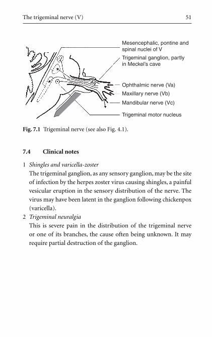
7.2 Attachment, course, divisions (Fig. 7.1)
• Attached to lateral aspect of pons, near middle cerebellar
peduncle.
• Passes below tentorium cerebelli, to middle cranial fossa.
• Trigeminal (sensory) ganglion in depression on temporal bone.
• Splits into ophthalmic (Va), maxillary (Vb) and mandibular (Vc).
7.3 Trigeminal ganglion
• It contains cell bodies of primary sensory neurons in all three
divisions of trigeminal nerve, except those of proprioceptive
neurons (see Chapter 4).
• It is partially surrounded by cerebrospinal fluid in recess of sub-
arachnoid space: trigeminal, or Meckel’s, cave.
The trigeminal nerve (V) 51
7.4 Clinical notes
1 Shingles and varicella-zoster
The trigeminal ganglion, as any sensory ganglion, may be the site
of infection by the herpes zoster virus causing shingles, a painful
vesicular eruption in the sensory distribution of the nerve. The
virus may have been latent in the ganglion following chickenpox
(varicella).
2 Trigeminal neuralgia
This is severe pain in the distribution of the trigeminal nerve
or one of its branches, the cause often being unknown. It may
require partial destruction of the ganglion.
Mesencephalic, pontine andspinal nuclei of V
Trigeminal ganglion, partlyin Meckel’s cave
Ophthalmic nerve (Va)
Maxillary nerve (Vb)
Mandibular nerve (Vc)
Trigeminal motor nucleus
Fig. 7.1 Trigeminal nerve (see also Fig. 4.1).
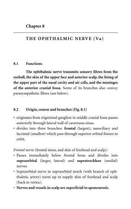
Chapter 8
T H E O P H T H A L M I C N E RV E ( V a )
8.1 Functions
The ophthalmic nerve transmits sensory fibres from the
eyeball, the skin of the upper face and anterior scalp, the lining of
the upper part of the nasal cavity and air cells, and the meninges
of the anterior cranial fossa. Some of its branches also convey
parasympathetic fibres (see below).
8.2 Origin, course and branches (Fig. 8.1)
• originates from trigeminal ganglion in middle cranial fossa passes
anteriorly through lateral wall of cavernous sinus.
• divides into three branches: frontal (largest), nasociliary and
lacrimal (smallest) which pass through superior orbital fissure to
orbit.
Frontal nerve (frontal sinus, and skin of forehead and scalp):
• Passes immediately below frontal bone and divides into
supraorbital (larger, lateral) and supratrochlear (medial)
nerves.
• Supraorbital nerve in supraorbital notch (with branch of oph-
thalmic artery) turns up to supply skin of forehead and scalp
(back to vertex).
• Nerves and vessels in scalp are superficial to aponeurosis.
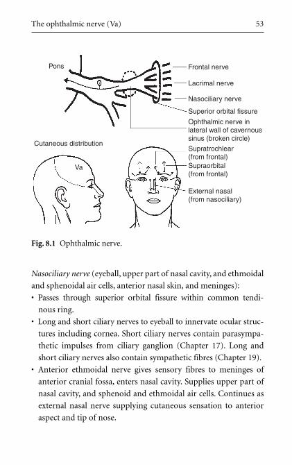
The ophthalmic nerve (Va) 53
Nasociliary nerve (eyeball, upper part of nasal cavity, and ethmoidal
and sphenoidal air cells, anterior nasal skin, and meninges):
• Passes through superior orbital fissure within common tendi-
nous ring.
• Long and short ciliary nerves to eyeball to innervate ocular struc-
tures including cornea. Short ciliary nerves contain parasympa-
thetic impulses from ciliary ganglion (Chapter 17). Long and
short ciliary nerves also contain sympathetic fibres (Chapter 19).
• Anterior ethmoidal nerve gives sensory fibres to meninges of
anterior cranial fossa, enters nasal cavity. Supplies upper part of
nasal cavity, and sphenoid and ethmoidal air cells. Continues as
external nasal nerve supplying cutaneous sensation to anterior
aspect and tip of nose.
External nasal(from nasociliary)
Frontal nervePons
Cutaneous distribution
Va
Lacrimal nerve
Nasociliary nerve
Superior orbital fissure
Ophthalmic nerve inlateral wall of cavernoussinus (broken circle)
Supratrochlear(from frontal)Supraorbital(from frontal)
Fig. 8.1 Ophthalmic nerve.

Lacrimal nerve (lacrimal gland, and small area of adjacent skin and
conjunctiva: not important):
• Superior orbital fissure: lateral to (outside) common tendinous ring.
• Joined by postganglionic parasympathetic fibres from ptery-
gopalatine ganglion. These are distributed to lacrimal gland (see
Section 17.3).
8.3 Nerve fibres: central connections
Somatic sensory fibres: sensory nuclei of trigeminal nerve.
Present in all branches. Axons pass centrally with cell bodies in
trigeminal ganglion. Central axonal processes pass to pons and
trigeminal sensory nuclei (Section 4.4).
Parasympathetic pathways: not important (see Chapter 17).
8.4 Clinical notes
1 Corneal reflex
When the cornea is touched, usually with a wisp of cotton wool,
the subject blinks. This tests V and VII. The nerve impulses pass
thus: cornea, nasociliary nerve, Va, principal sensory nucleus of V,
brain stem interneurons, facial motor nucleus,VII, orbicularis oculi
muscle. Note that the reflex does not test vision or eye movements.
2 Supraorbital injuries
Trauma to the supraorbital margin may damage the supraorbital
and/or supratrochlear nerves causing sensory loss in the scalp.
3 Ethmoid tumours
Malignant tumours of the mucous lining of the ethmoid air cells
may expand into the orbits, damaging branches of Va, particu-
larly the ethmoidal nerves. This may lead to displacement of the
54 Trigeminal, facial and hypoglossal nerves
The ophthalmic nerve (Va) 55
orbital contents causing proptosis and squint, and sensory loss
over the anterior nasal skin.
4 Nasal fractures
Trauma to the nose may damage the external nasal nerve as it
becomes superficial. Sensory loss of the skin down to the tip of
the nose may result.
5 Bilateral cleft lip and palate
In this condition, the central part of the upper lip together with
that part of the palate posterior to it which bears the upper inci-
sors are isolated from surrounding areas. Branches of the maxil-
lary nerve are thus denied access. The isolated area of palate and
lip in these cases are supplied by Va through its external nasal
branch which enters from above. In a unilateral cleft, Vb of one
side is able to innervate the area in an asymmetric fashion.
8.5 Clinical testing
1 Test corneal reflex (see above).
2 Test skin sensation of forehead and anterior scalp.

Chapter 9
T H E M A X I L L A RY N E RV E ( V b )
9.1 Functions
The maxillary nerve transmits sensory fibres from
the skin of the face between the palpebral fissure and the
mouth, from the nasal cavity and sinuses, and from the maxillary
teeth.
At its origin it contains only sensory fibres. Some of its branches
transmit postganglionic parasympathetic fibres from the pterygo-
palatine ganglion which pass to the lacrimal, nasal and palatine
glands (see Section 17.3), and others convey taste (visceral sensory)
fibres from the palate to the nucleus of the solitary tract (see
Section 17.4).
9.2 Origin, course and branches (Fig. 9.1)
• originates from trigeminal ganglion in middle cranial fossa;
• passes in lower part of lateral wall of cavernous sinus;
• meningeal branch (middle cranial fossa – sensory);
• foramen rotundum, to;
• pterygopalatine fossa which divides into main branches, infra-
orbital and zygomatic, and gives other branches to nose, palate and
upper teeth.
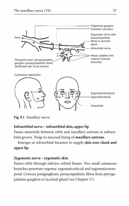
The maxillary nerve (Vb) 57
Infraorbital nerve – infraorbital skin, upper lip
Passes anteriorly between orbit and maxillary antrum in infraor-
bital groove. Twigs to mucosal lining of maxillary antrum.
Emerges at infraorbital foramen to supply skin over cheek and
upper lip.
Zygomatic nerve – zygomatic skin
Enters orbit through inferior orbital fissure. Two small cutaneous
branches penetrate zygoma: zygomaticofacial and zygomaticotem-
poral. Conveys postganglionic parasympathetic fibres from pterygo-
palatine ganglion to lacrimal gland (see Chapter 17).
Pons
Cutaneous distribution
Vb
Zygomaticotemporal
Nasal, palatine andsuperior alveolarbranches
Infraorbital nerve
Trigeminal ganglionForamen rotundum
Zygomatic nerve withparasympatheticfibres to lacrimalgland
Zygomaticofacial
Infraorbital
Pterygoid canal, pterygopalatineganglion (parasympathetic fibresdistributed with Vb as shown)
Fig. 9.1 Maxillary nerve.
58 Trigeminal, facial and hypoglossal nerves
Nasal branches
Pass medially from pterygopalatine fossa through sphenopalatine
foramen into nasal cavity. Supply nasal cavity and sinuses. Branches
also convey postganglionic parasympathetic fibres from pterygopala-
tine ganglion to nasal glands (see Chapter 17).
Superior alveolar (dental) nerves
Branches of infraorbital and palatine nerves pass directly through
maxilla to maxillary teeth, gums and sinus.
Greater and lesser palatine nerves
Hard and soft palate sensation. Branches also convey postganglionic
parasympathetic fibres from pterygopalatine ganglion to minor saliv-
ary glands in the palatal mucosa (see Chapter 17).
Pharyngeal branch
Passes posteriorly to contribute to sensory supply of nasopharynx.
9.3 Nerve fibres: central connections
Somatic sensory fibres: sensory nuclei of trigeminal nerve
Present in all branches of maxillary nerve. Axons pass centrally with
cell bodies in trigeminal ganglion. Central processes pass to pons and
trigeminal sensory nuclei (Section 4.4). Note: sensory fibres travers-
ing pterygopalatine ganglion, for example those from palate and nose, do
not synapse there.
Taste (visceral sensory) fibres: nucleus of solitary tract
From scattered taste buds on palate. Axons ascend in palatine nerves,
through pterygopalatine ganglion (no synapse), pterygoid canal,
greater petrosal and facial nerve (cell bodies in geniculate ganglion).
Central processes enter brain stem through nervus intermedius, pass-
ing to nucleus of solitary tract (see Chapter 17).
The maxillary nerve (Vb) 59
Parasympathetic fibres: superior salivatory nucleus
Superior salivatory nucleus, nervus intermedius, VII, lacrimal, nasal
and palatal glands (see Chapter 17).
9.4 Clinical notes
1 Infraorbital injuries: malar fractures
Trauma to infraorbital margin may cause sensory loss of infra-
orbital skin.
2 Maxillary sinus infections
Infections of the maxillary sinus may cause infraorbital pain or
may cause referred pain to other structures supplied by Vb (e.g.
upper teeth).
3 Maxillary antrum tumours
Malignant tumours of the mucous lining of the maxillary antrum
may expand into the orbit, damaging branches of Vb, particu-
larly the infraorbital. This may lead to anaesthesia over the facial
skin. The orbital contents may also be displaced causing pro-
ptosis and/or a squint.
4 Maxillary teeth abscesses
The roots of the maxillary teeth (especially the second molars)
are intimately related to the maxillary sinus. Root abscesses are
painful.
5 Hay fever
This is usually allergic, but the symptoms could be produced
by involvement of parasympathetic “fellow travellers” with the
maxillary nerve (see Section 17.3).
9.5 Clinical testing
Test skin sensation of lower eyelid, cheek and upper lip.
Chapter 10
T H E M A N D I B U L A R N E RV E ( Vc )
10.1 Functions
The mandibular nerve is a mixed sensory and motor
nerve. It transmits sensory fibres from the skin over the mandible,
side of the cheek and temple, the oral cavity and contents, the
external ear, the tympanic membrane and temporomandibular
joint (TMJ). It also supplies the meninges of the cranial vault.
It is motor to the eight muscles derived from the first branchial arch:
• temporalis, masseter
• medial, lateral pterygoids
• mylohyoid, anterior belly of digastric
• tensor tympani, tensor palati
As an aid to memory, note the four groups of two: tensors, ptery-
goids, big muscles and the last two in the floor of the mouth.
Some of its distal branches also convey parasympathetic secreto-
motor fibres to the salivary glands, and taste fibres from the anter-
ior portion of the tongue.
10.2 Origin, course and branches (Fig. 10.1)
• From trigeminal ganglion in middle cranial fossa.
• Foramen ovale.
• Infratemporal fossa with four main branches: inferior alveolar,
lingual, auriculotemporal, buccal.
• Motor branches to muscles listed above.
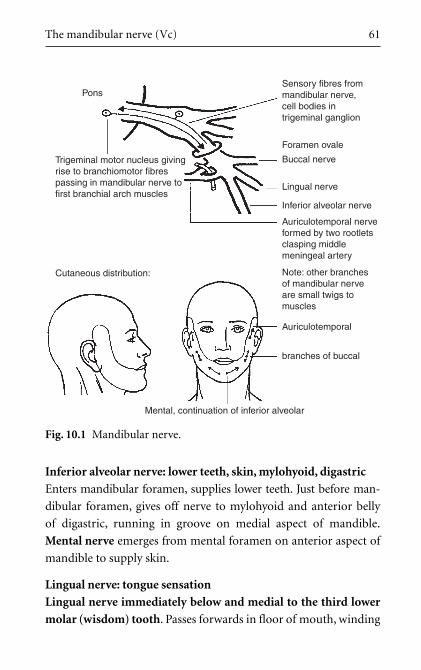
The mandibular nerve (Vc) 61
Inferior alveolar nerve: lower teeth, skin, mylohyoid, digastric
Enters mandibular foramen, supplies lower teeth. Just before man-
dibular foramen, gives off nerve to mylohyoid and anterior belly
of digastric, running in groove on medial aspect of mandible.
Mental nerve emerges from mental foramen on anterior aspect of
mandible to supply skin.
Lingual nerve: tongue sensation
Lingual nerve immediately below and medial to the third lower
molar (wisdom) tooth. Passes forwards in floor of mouth, winding
Sensory fibres frommandibular nerve,cell bodies intrigeminal ganglion
Pons
Trigeminal motor nucleus givingrise to branchiomotor fibrespassing in mandibular nerve tofirst branchial arch muscles
Cutaneous distribution:
Foramen ovale
Buccal nerve
Lingual nerve
Inferior alveolar nerve
Auriculotemporal nerveformed by two rootletsclasping middlemeningeal artery
Note: other branchesof mandibular nerveare small twigs tomuscles
Auriculotemporal
branches of buccal
Mental, continuation of inferior alveolar
Fig. 10.1 Mandibular nerve.
62 Trigeminal, facial and hypoglossal nerves
around submandibular duct. Supplies anterior tongue, gums. Conveys
parasympathetic fibres from superior salivatory nucleus, and taste
fibres to VII; these pass between lingual nerve and VII in chorda
tympani (see Chapter 17).
Auriculotemporal nerve: skin of temple, TMJ, external ear
Arises beneath foramen ovale by two rootlets on either side of mid-
dle meningeal artery. Passes above parotid gland, between TMJ and
external auditory meatus to emerge on side of head. Ascends close
to superficial temporal artery. Supplies TMJ, parotid fascia, skin of
temple, most of skin of external auditory meatus and tympanic
membrane. For short distance between foramen ovale and parotid
gland it conveys parasympathetic fibres for innervation of parotid
gland (see Chapter 17).
Buccal nerve: skin and mucosa of cheek
Supplies sensory fibres to skin and mucosa of cheek (it does NOT
supply buccinator (see Section 11.3)).
Muscular branches: temporal nerves to temporalis, and other
muscular twigs.
Meningeal branches: Small twigs re-enter middle cranial fossa
through foramen ovale, and other foramina, to supply meninges.
10.3 The first branchial arch
The mandibular nerve is the main nerve of the first branchial
arch (the maxillary nerve also contributes). The first branchial arch
gives rise to: a precursor of the mandible (Meckel’s cartilage), the
spine of the sphenoid, the sphenomandibular ligament, the malleus
and incus, and the eight muscles listed above (see Section 10.1).
Arterial components of the first arch degenerate.

The mandibular nerve (Vc) 63
10.4 Nerve fibres: central connections
Somatic sensory fibres: sensory nuclei of the trigeminal nerve
Axons pass centrally with cell bodies in trigeminal ganglion.
Central processes to pons and trigeminal sensory nuclei (see
Section 4.4).
Branchiomotor fibres: trigeminal motor nucleus
Trigeminal motor nucleus in pons, axons to mandibular nerve and
eight muscles of first branchial arch (see above).
Visceral sensory (taste) fibres: nucleus of the solitary tract
Taste fibres from anterior portion of tongue pass in lingual nerve,
chorda tympani and facial nerve. Cell bodies in geniculate ganglion.
Central processes to nucleus of solitary tract (see Section 4.5).
Parasympathetic pathways: salivatory nuclei
Branches of mandibular nerve convey parasympathetic impulses to
submandibular, sublingual and parotid glands (see Chapter 17).
10.5 Clinical notes
1 Lingual nerve
Careless extractions of the third lower molar (wisdom) tooth,
abscesses of its root, etc., or fractures of the angle of the
mandible may all damage the lingual nerve. This may result not
only in loss of somatic sensation from the anterior portion of the
tongue, but also loss of taste sensation and parasympathetic
function.
2 Inferior alveolar nerve and inferior alveolar nerve block
Trauma to the mandible may damage or tear the inferior alveo-
lar nerve in the mandibular canal leading to sensory loss distal to
the lesion. Local anaesthesia of the inferior alveolar nerve is com-
monly performed for dental procedures. Injection of local anaes-
thetics into the oral mucosa on the medial side of the mandible
can also involve the nearby lingual nerve, thus affecting the
tongue and inside of the mouth. In wisdom teeth extractions, the
buccal nerve may also be anaesthetized leading to numbness of
the cheek.
3 Mumps
Mumps is inflammation of the parotid gland causing tension in
the parotid capsule which is innervated by the auriculotemporal
nerve. It gives both local tenderness and referred earache. It is
very uncomfortable.
4 Submandibular duct
The intimate relationship between the submandibular duct and
the lingual nerve is significant in duct infections and surgery. Sub-
mandibular stones are not uncommon because of the mucous
secretions. If the lingual nerve were damaged there would be
sensory loss, both somatic and taste, in the anterior portion of
the tongue.
5 Referred pain to the ear
Disease of the TMJ or swelling of the parotid gland may cause
earache because of referred pain. Also, pain from the lower teeth,
oral cavity and tongue may be referred to the ear. Beware of
patients with cotton wool in the external auditory meatus –
check in the mouth for the disease!
6 Superficial temporal artery biopsy
The auriculotemporal nerve accompanies the superficial tempo-
ral artery on the temple. In cases of temporal arteritis, the nerve
is anaesthetized so that the overlying skin can be incised to
obtain a biopsy of the artery.
64 Trigeminal, facial and hypoglossal nerves
10.6 Clinical testing
1 Sensory: Test skin sensation of chin and lower lip.
2 Motor: feel contractions of masseter, temporalis. Open jaw
against resistance (pterygoids, mylohyoid, anterior digastric).
The mandibular nerve (Vc) 65
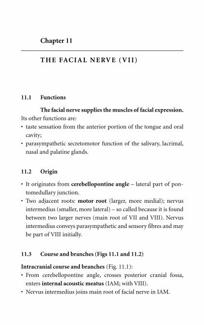
Chapter 11
T H E FAC I A L N E RV E ( V I I )
11.1 Functions
The facial nerve supplies the muscles of facial expression.
Its other functions are:
• taste sensation from the anterior portion of the tongue and oral
cavity;
• parasympathetic secretomotor function of the salivary, lacrimal,
nasal and palatine glands.
11.2 Origin
• It originates from cerebellopontine angle – lateral part of pon-
tomedullary junction.
• Two adjacent roots: motor root (larger, more medial); nervus
intermedius (smaller, more lateral) – so called because it is found
between two larger nerves (main root of VII and VIII). Nervus
intermedius conveys parasympathetic and sensory fibres and may
be part of VIII initially.
11.3 Course and branches (Figs 11.1 and 11.2)
Intracranial course and branches (Fig. 11.1):
• From cerebellopontine angle, crosses posterior cranial fossa,
enters internal acoustic meatus (IAM; with VIII).
• Nervus intermedius joins main root of facial nerve in IAM.

The facial nerve (VII) 67
• Geniculate ganglion is deep in IAM: this houses cell bodies of
sensory fibres (no synapses) in VII. Nerve turns posteriorly into:
• Facial canal running posteriorly along medial wall of tympanic
(middle ear) cavity, and gives branch to stapedius (attached to
stapes);
Pons
Mastoidcavity
VIII
Eustachiantube
Chordatympani
VII atstylomastoid
foramen
Nervusintermedius
Facial canal on medialwall of tympanic cavity
Maxillary nervein foramenrotundum
Pterygopalatineganglion
Canalfor tensortympani
Lingual nerve,submandibular
ganglion
Fig. 11.1 Facial nerve (intracranial). : branchiomotor fibres
from facial motor nucleus to muscles of facial expression,
stapedius, etc.; : parasympathetic preganglionic
fibres from superior salivatory nucleus; : nucleus of
solitary tract receiving visceral sensory fibres, cell bodies in
geniculate (sensory) ganglion; * to stapedius; stippled area
represents bone.
68 Trigeminal, facial and hypoglossal nerves
• Chorda tympani given off just before VII emerges at stylomastoid
foramen; this passes anteriorly across tympanic membrane into
infratemporal fossa where it joins lingual nerve;
• Emerges at stylomastoid foramen.
Extracranial course and branches (Fig. 11.2)
• Outside stylomastoid foramen, small branches of VII supply
occipital belly of occipitofrontalis, stylohyoid and posterior belly
of digastric, and a variable amount of cutaneous sensation from
skin of external auditory meatus.
• Nerve enters parotid gland where it forms intricate plexus.
Branches of VII are superficial in the gland.
• Five groups of branches emerge superficially from anterior border
of parotid gland: temporal, zygomatic, buccal, mandibular and
Branch to occipitalis
Branch to stylohyoid
Position of parotidgland (oval)Cervical branches
Marginal mandibular(vulnerable here)
Temporal
Zygomatic
Buccal
Fig. 11.2 Facial nerve (extracranial).
The facial nerve (VII) 69
cervical. These supply muscles of facial expression including
orbicularis oculi, orbicularis oris, buccinator and platysma.
The most important thing about the intracranial course of VII is
its relationship to the middle ear. The most important thing about
the extracranial course is its relationship to the parotid gland.
11.4 The second branchial arch, otic vesicle and facial nerve
The facial nerve is the nerve of the second branchial arch
which gives rise to part of the hyoid bone, the styloid process, the
stylohyoid ligament, the stapes and the muscles listed in Section
11.3: the muscles of facial expression (which migrate into superfi-
cial layers), stylohyoid, posterior belly of digastric, occipitofrontalis
and stapedius. Arterial components of the second arch degenerate.
The facial nerve develops in association with the otic vesicle, and
with the first branchial pouch (endodermal) and the first branchial
cleft (ectodermal) which lie between the first and second arches.
This accounts for the intimate relationship of the facial nerve
with the derivatives of these structures: the inner, middle and exter-
nal ears.
11.5 Nerve fibres: central connections
Branchiomotor fibres: facial motor nucleus
Facial motor nucleus in pons, axons pass dorsally, then loop ven-
trally around abducens (VI) nucleus, causing elevation (facial col-
liculus) on floor of fourth ventricle. (It has been suggested that this
peculiar course may be a result of the developmental phenomenon
of neurobiotaxis in which a cell body migrates towards the greatest
density of stimuli.)
70 Trigeminal, facial and hypoglossal nerves
Preganglionic parasympathetic fibres: superior salivatory
nucleus
Superior salivatory nucleus in lower pons, preganglionic axons into
nervus intermedius and VII. Some pass to pterygopalatine ganglion
(synapse) for lacrimal, nasal and palatine glands. Some enter chorda
tympani and pass to submandibular ganglion (synapse) for sub-
mandibular and sublingual glands (see Chapter 17). Parasympathetic
fibres not present in extracranial portion.
Visceral sensory (taste) fibres: solitary tract and nucleus
Taste fibres from anterior portion of tongue, through lingual nerve
to chorda tympani, VII. Cell bodies in geniculate ganglion. Central
processes pass in nervus intermedius to solitary tract and nucleus.
Taste fibres from palate pass through pterygoid canal and greater
petrosal nerve to facial nerve, then as above. Taste fibres not present
in extracranial portion of VII.
Somatic sensory fibres: sensory nuclei of the trigeminal nerve
From small and variable area of skin in region of ear, and external
aspect of tympanic membrane, fibres join facial nerve just outside
stylomastoid foramen. Cell bodies in geniculate ganglion. Central
processes pass to sensory nuclei of trigeminal nerve.
11.6 Upper and lower motor neuron lesions of
the facial nerve
In upper motor neuron (UMN) lesions of the facial nerve,
the forehead and orbicularis oculi muscles are largely spared. This
is because there is bilateral cortical control of the upper facial
muscles, and so if corticonuclear fibres on one side of the brain are
interrupted (e.g. in the internal capsule) those of the other side are
unaffected. For the lower facial muscles this is not so: the normal
pattern prevails with only contralateral control.
UMN lesion of the facial nerve
The facial motor nucleus is divided into two parts:
1 that for upper facial muscles (orbicularis oculi and frontalis);
2 that for lower facial muscles.
The lower motor neuron (LMN) cell bodies in (1) receive UMNs
from both cerebral motor cortices, ipsilateral as well as the usual
contralateral. LMNs in (2) do not: they receive only the customary
contralateral innervation. Thus, a unilateral UMN lesion of the
fibres supplying (1) will not result in complete functional loss since
UMN input is also present from the other side. However, for the
lower part of the face, there would be no sparing since the UMN
input comes only from the contralateral cortex. This is usually
obvious since when facial muscles lose their tone, facial wrinkles
also disappear.
The significance of this has been pointed out in Section 3.4. It is
associated with eye movements and eye protection.
LMN lesion of the facial nerve – facial palsy
An LMN lesion, whether of cell bodies in the facial motor nucleus,
or of any part of the peripheral course of the facial nerve, intracra-
nial or extracranial, would result in a complete ipsilateral LMN
lesion of the facial nerve, irrespective of which part of the facial
nucleus was involved. The bilateral UMN innervation to the upper
part of the face would be of no significance since the lesion affects
the more distal LMN. An LMN lesion of the facial nerve is called a
facial palsy.
The facial nerve (VII) 71
11.7 Other clinical notes
1 Parotid disease
Parotid tumours, trauma or surgery may damage branches of
the facial nerve. This would result in an ipsilateral facial palsy
with wasting and functional loss. It would be unlikely to recover.
2 Stapedius: hyperacusis
Dysfunction of the smallest muscle supplied by the facial nerve
can cause a distressing symptom. Stapedius dampens the move-
ments of the ossicular chain and if it is inactive, sounds will be
distorted and echoing. This is hyperacusis. See also Stapedius
paralysis: hyperacusis in Section 23.5.
3 Bell’s palsy
This is a facial palsy, usually of unknown aetiology. It has been
suggested that vascular spasm of the arteries in the facial canal
supplying the nerve might be responsible, or inflammation and
swelling of the nerve within the bony canal.
4 Corneal reflex
A test of the ophthalmic and facial nerves (see Corneal reflex in
Section 8.4).
5 The marginal mandibular branch of the facial nerve
This branch passes on or just below the lower margin of the
mandible. It is superficial even to the palpable facial arterial
pulse and is thus liable to injury. Section of this nerve would
result in paralysis of the muscles of the corner of the mouth:
drooling would occur.
6 Facial nerve injury in babies
As the mastoid process is rudimentary at birth, the facial
nerve is more easily damaged in babies. Birth injuries, or other
trauma, can therefore cause an ipsilateral facial palsy. This is
serious since buccinator, supplied by VII, is necessary for sucking
(feeding).
72 Trigeminal, facial and hypoglossal nerves
The facial nerve (VII) 73
7 Cerebellopontine angle tumours
Tumours in this region would cause signs and symptoms of dam-
age to the facial and vestibulocochlear nerves and cerebellar signs.
These include facial palsy, deafness, vertigo and poor coordination.
8 Acoustic neuroma
This is a tumour of Schwann cells on the vestibular nerve in the
IAM. Since the tumour grows within a bony canal it may com-
press the facial and vestibulocochlear nerves causing a particular
type of deafness (nerve deafness) and an ipsilateral facial palsy.
9 Brain stem lesions
The relationship between the nucleus of the abducens nerve
and the axons of the facial nerve means that a brain stem lesion
may cause a paralysis of the facial nerve in association with a
paralysis of the ipsilateral lateral rectus muscle of the eye.
10 Geniculate herpes, Ramsay Hunt syndrome
The herpes zoster virus may lie dormant in the geniculate gan-
glion (after an attack of chickenpox) and at some later stage cause
a vesicular eruption in the skin around the external auditory mea-
tus supplied by the facial nerve. There may also be signs on the
anterior portion of the tongue as a result of taste fibres with cell
bodies in the geniculate ganglion. The inflammation may spread
to involve the motor fibres in the facial nerve. A LMN lesion aris-
ing from this cause is known as the Ramsay Hunt syndrome.
11.8 Clinical testing
1 Observe the face. Normal facial movements (lips, eyelids, emo-
tions) and the presence of normal facial skin creases indicate an
intact nerve.
2 Test strength by trying to force apart tightly closed eyelids. This
should be difficult.
3 Test corneal reflex (see Corneal reflex in Section 8.4).

Chapter 12
T H E H Y P O G L O S S A L N E RV E ( X I I )
12.1 Function
The hypoglossal nerve supplies the muscles of the tongue.
Movements of the tongue are important in chewing, in the initial
stages of swallowing and in speech. It also conveys fibres from C1
which innervate the strap muscles.
12.2 Origin, course and branches (Fig. 12.1)
The hypoglossal nerve (XII):
• Originates from medulla by vertical series of rootlets between
pyramid and olive (see Section 1.4). Hypoglossal (condylar) canal
in occipital bone.
• Receives motor fibres from C1 and descends to submandibular
region.
• Turns forwards, lateral to external carotid artery, hooking
beneath origin of occipital artery. Passes lateral to hyoglossus
and enters tongue from below.
• Gives descendens hypoglossi to ansa cervicalis carrying fibres from
C1 to strap muscles; other C1 fibres remain with XII to supply
geniohyoid.
• Supplies intrinsic muscles of tongue, hyoglossus, genioglossus and
styloglossus.
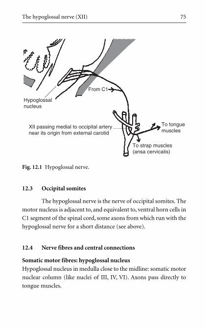
The hypoglossal nerve (XII) 75
12.3 Occipital somites
The hypoglossal nerve is the nerve of occipital somites. The
motor nucleus is adjacent to, and equivalent to, ventral horn cells in
C1 segment of the spinal cord, some axons from which run with the
hypoglossal nerve for a short distance (see above).
12.4 Nerve fibres and central connections
Somatic motor fibres: hypoglossal nucleus
Hypoglossal nucleus in medulla close to the midline: somatic motor
nuclear column (like nuclei of III, IV, VI). Axons pass directly to
tongue muscles.
To tonguemuscles
To strap muscles(ansa cervicalis)
From C1
Hypoglossalnucleus
XII passing medial to occipital arterynear its origin from external carotid
Fig. 12.1 Hypoglossal nerve.
Sensory fibres: uncertain
A few sensory fibres from sternocleidomastoid and trapezius. Cell
bodies (unusually) scattered along length of nerve.
12.5 Clinical notes
1 Hypoglossal nerve lesions
Damage to the hypoglossal nerve in the neck would result in
an ipsilateral lower motor neuron lesion. This would cause
the protruded tongue to deviate to the side of the lesion (see
Section 12.6).
2 Carotid artery surgery, block dissection of neck
The hypoglossal nerve is vulnerable in surgery (e.g. carotid
endarterectomy, block dissection of the neck for malignant dis-
ease) where it passes under the origin of the occipital artery.
3 Bulbar and pseudobulbar palsy
For details refer to Section 13.3.
12.6 Clinical testing
1 Ask the patient to protrude tongue. If it deviates to one side, then
the nerve of that side is damaged – the tongue is pushed to the
paralysed side by muscles of the functioning side.
2 Ask patient to push tongue into cheek, then palpate cheek to feel
tone and strength of tongue muscles.
76 Trigeminal, facial and hypoglossal nerves

PART III
G LO S S O P H A RY N G E A L , VAG U S
A N D AC C E S S O RY N E RV E S

Chapter 13
S WA L LOW I N G A N D S P E A K I N G ,B U L BA R PA L S Y, P S E U D O B U L BA RPA L S Y, B RO C A’ S A R E A
13.1 Swallowing
When food or drink passes on to the posterior part of the
tongue, the muscles supplied by the nucleus ambiguus through the
vagus (X) and glossopharyngeal (IX) nerves propel it backwards and
downwards into the hypopharynx, thence through the cricopha-
ryngeal sphincter to the oesophagus. The nasopharynx is closed by
the palatal muscles (X, Vc), and the Eustachian tube opens (X). The
laryngeal orifice is reduced in size largely as a result of elevation of
the entire laryngeal skeleton by all the muscles attaching to it from
above, and the cricopharyngeal sphincter opens (X). The tongue
muscles (XII) also have an important role in these actions. Most
of the muscles of the pharynx are supplied in one way or another
by the nucleus ambiguus through the vagus (it is unnecessary to
bother with individual pharyngeal muscles).
Sensation from this region is conveyed through the pharyngeal
plexus, the glossopharyngeal (posterior tongue, oropharynx) and
vagus (hypopharynx) nerves, to the nucleus of the solitary tract.
13.2 Speaking
Noise production: phonation
The vocal cords create the narrow slit through which air is directed
to make a sound, much as in an oboe, recorder or organ pipe.
Muscles which move the cords are supplied by the recurrent laryngeal
nerve (X).
Making the noise intelligible: articulation
The pharyngeal muscles (X), the tongue (XII), the muscles of
facial expression (VII), mandibular movements (Vc) and the palate
(X, V) all modify the crude noise produced by the larynx to create
speech.
Pitch
Pitch is modulated principally by tensing (cricothyroid) and relax-
ing (vocalis) the vocal cords. All movements of the vocal cords are
controlled by the nucleus ambiguus through the superior and recur-
rent laryngeal nerves (X). It is unnecessary to learn individual laryn-
geal muscles or their attachments; it is enough to know that they are
all innervated by the nucleus ambiguus. Lesions affecting the nucleus
ambiguus lead to profound swallowing and speech disorders: bulbar
and pseudobulbar palsies.
13.3 Bulbar palsy: ipsilateral lower motor neuron lesion
This is caused by a lesion of the medulla (e.g. vascular, mul-
tiple sclerosis, motor neuron disease) which involves the nucleus
ambiguus and the hypoglossal nucleus. There would be an ipsilateral
lower motor neuron lesion of the muscles of the tongue and pharynx.
Chewing, swallowing and speaking would be affected. Because the
nuclei concerned are in the medulla or bulb, this is called a bulbar
palsy.
80 Glossopharyngeal, vagus and accessory nerves

13.4 Medullary syndromes
Lateral medullary syndrome (Wallenberg’s or posterior inferior
cerebellar artery syndrome)
This is caused by thrombosis of the posterior inferior cerebellar artery
(PICA). Since this supplies the upper lateral medulla, some of the
symptoms are explicable on the basis of the involvement of the spinal
part of the trigeminal sensory nucleus and the nucleus ambiguus: dis-
turbances of facial temperature sensation and bulbar palsy affecting
speech and swallowing. There may also be cerebellar involvement
and balance disorders (possible involvement of cochlear and vestibu-
lar nuclei). The lateral spinothalamic tract conveying pain and tem-
perature sensation from the contralateral trunk and limbs may also
be involved.
Medial medullary syndrome
This involves the hypoglossal nucleus, corticospinal tract and medial
lemniscus. The medial medulla is supplied by the anterior spinal artery.
The lateral medullary syndrome is more likely to cause sensory
symptoms, and the medial medullary syndrome more likely to
cause motor syndromes: look again at Section 1.10 for the reasons
for this.
13.5 Pseudobulbar palsy: contralateral upper
motor neuron lesion
Interruption of the upper motor neuron pathways any-
where between the cortex and the medulla (e.g. internal capsule,
cerebral peduncles) will affect contralateral muscles of the tongue
Swallowing and speaking, palsies, Broca’s area 81

and pharynx. Since this presents as a disorder of the muscles of
speech and swallowing, it is at first sight similar to a bulbar palsy,
but being an upper motor neuron lesion, it is caused by a lesion on
the opposite side. For these reasons it is known as a pseudobulbar
palsy.
13.6 Broca’s motor speech area
This is the area of motor cortex which controls the muscles
of speech. It is immediately above the lateral fissure, deep to the
pterion, usually in the dominant hemisphere (normally the left).
Damage (e.g. occlusion of a branch of the middle cerebral artery)
leads to motor speech aphasia (aphasia – wordless).
82 Glossopharyngeal, vagus and accessory nerves
Chapter 14
T H E G L O S S O P H A RY N G E A L N E RV E ( I X )
14.1 Functions
From a clinical point of view, the glossopharyngeal nerve
is unimportant except for its role in the gag reflex. The main
function of the glossopharyngeal nerve is the sensory supply of
the oropharynx and posterior part of the tongue.
Its other functions are the motor supply to stylopharyngeus; con-
veying parasympathetic fibres part of the way to parotid gland and
sensory supply from the carotid sinus, carotid body, and (sometimes)
skin of the external acoustic meatus and tympanic membrane.
14.2 Origin, course and branches (Fig. 14.1)
• From medulla by a vertical series of rootlets lateral to olive, above
and in series with those of X and XI.
• Passes through jugular foramen (middle portion). Two sensory
ganglia: superior and petrosal (inferior).
• Parasympathetic axons from inferior salivatory nucleus to otic
ganglion (for parotid gland) enter tympanic branch (see Chapter
17). May also convey sensory fibres from ear.
• Nerve descends in neck, supplying stylopharyngeus and carotid
body.
• Passes between internal and external carotid arteries to enter
pharynx. Sensory fibres to pharyngeal plexus supplying mucosa
of pharynx and posterior tongue.
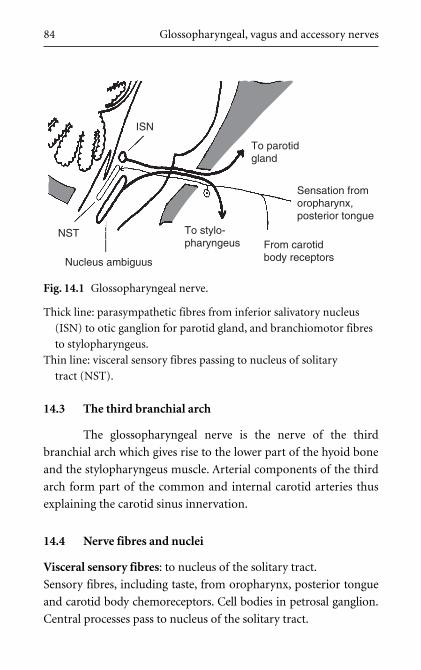
84 Glossopharyngeal, vagus and accessory nerves
14.3 The third branchial arch
The glossopharyngeal nerve is the nerve of the third
branchial arch which gives rise to the lower part of the hyoid bone
and the stylopharyngeus muscle. Arterial components of the third
arch form part of the common and internal carotid arteries thus
explaining the carotid sinus innervation.
14.4 Nerve fibres and nuclei
Visceral sensory fibres: to nucleus of the solitary tract.
Sensory fibres, including taste, from oropharynx, posterior tongue
and carotid body chemoreceptors. Cell bodies in petrosal ganglion.
Central processes pass to nucleus of the solitary tract.
To parotidgland
From carotidbody receptors
To stylo-pharyngeus
Nucleus ambiguus
NST
ISN
Sensation fromoropharynx,posterior tongue
Fig. 14.1 Glossopharyngeal nerve.
Thick line: parasympathetic fibres from inferior salivatory nucleus
(ISN) to otic ganglion for parotid gland, and branchiomotor fibres
to stylopharyngeus.
Thin line: visceral sensory fibres passing to nucleus of solitary
tract (NST).
Branchiomotor fibres: from nucleus ambiguus to stylopharyngeus.
Parasympathetic fibres: from inferior salivatory nucleus.
Preganglionic axons from inferior salivatory nucleus to auricu-
lotemporal nerve and otic ganglion. Postganglionic fibres enter
parotid gland. See Chapter 17.
Somatic sensory fibres: to nuclei of the trigeminal nerve.
From variable portion of skin in external ear, axons pass in tympanic
branch to main trunk of IX. Cell bodies in superior ganglion of IX.
Central processes pass to the sensory trigeminal nuclei.
14.5 Clinical notes and clinical testing
Gag reflex
Sensation supplied by the glossopharyngeal nerve is different in
quality to that supplied by the trigeminal. Place a finger on the
anterior part of the tongue (V) and then the posterior part (IX) to
demonstrate this. The gag reflex is mediated by the glossopharyn-
geal (afferent limb) and the vagus (efferent limb). It is a functional
test of both nerves.
The glossopharyngeal nerve (IX) 85
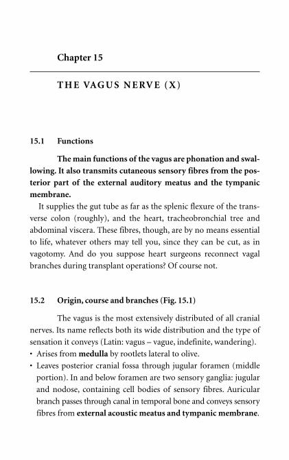
Chapter 15
T H E VAG U S N E RV E ( X )
15.1 Functions
The main functions of the vagus are phonation and swal-
lowing. It also transmits cutaneous sensory fibres from the pos-
terior part of the external auditory meatus and the tympanic
membrane.
It supplies the gut tube as far as the splenic flexure of the trans-
verse colon (roughly), and the heart, tracheobronchial tree and
abdominal viscera. These fibres, though, are by no means essential
to life, whatever others may tell you, since they can be cut, as in
vagotomy. And do you suppose heart surgeons reconnect vagal
branches during transplant operations? Of course not.
15.2 Origin, course and branches (Fig. 15.1)
The vagus is the most extensively distributed of all cranial
nerves. Its name reflects both its wide distribution and the type of
sensation it conveys (Latin: vagus – vague, indefinite, wandering).
• Arises from medulla by rootlets lateral to olive.
• Leaves posterior cranial fossa through jugular foramen (middle
portion). In and below foramen are two sensory ganglia: jugular
and nodose, containing cell bodies of sensory fibres. Auricular
branch passes through canal in temporal bone and conveys sensory
fibres from external acoustic meatus and tympanic membrane.
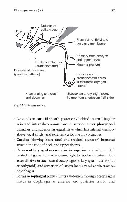
The vagus nerve (X) 87
• Descends in carotid sheath posteriorly behind internal jugular
vein and internal/common carotid arteries. Gives pharyngeal
branches, and superior laryngeal nerve which has internal (sensory
above vocal cords) and external (cricothyroid) branches.
• Cardiac (slowing heart rate) and tracheal (sensory) branches
arise in the root of neck and upper thorax.
• Recurrent laryngeal nerves arise in superior mediastinum: left
related to ligamentum arteriosum, right to subclavian artery. Both
ascend between trachea and oesophagus to laryngeal muscles (not
cricothyroid) and sensation of larynx below vocal cords, trachea,
oesophagus.
• Forms oesophageal plexus. Enters abdomen through oesophageal
hiatus in diaphragm as anterior and posterior trunks and
From skin of EAM andtympanic membrane
Sensory from pharynxand upper larynx
Motor to pharynx
Sensory andbranchiomotor fibresin recurrent laryngealnerves
Subclavian artery (right side),ligamentum arteriosum (left side)
X continuing to thoraxand abdomen
Dorsal motor nucleus(parasympathetic)
Nucleus ambiguus(branchiomotor)
Nucleus ofsolitary tract
Fig. 15.1 Vagus nerve.
88 Glossopharyngeal, vagus and accessory nerves
contributes fibres to abdominal viscera and to coeliac, superior
mesenteric and myenteric plexuses. Branches pass in lesser omen-
tum alongside lesser curvature of stomach to innervate pyloric
antrum (nerves of Latarjet), and to give hepatic branches.
15.3 The fourth and sixth branchial arches: embryological
considerations
The vagus is the nerve of the fourth and sixth branchial
arches. Structures derived from these include the pharyngeal and
laryngeal cartilages and muscles. The sixth arch artery on the left
gives rise to the ductus arteriosus (ligamentum after birth) around
which the left sixth arch nerve, the recurrent laryngeal, is caught
when the artery descends. The sixth arch artery on the right degen-
erates, so the right recurrent laryngeal nerve is related to the most
caudal persisting branchial arch artery, the fourth, which becomes
the right subclavian. The motor function of the vagus in the neck
is branchiomotor (special visceral motor): motor function in the
thorax and abdomen is parasympathetic (general visceral motor).
15.4 Nerve fibres and central connections
Branchiomotor fibres: from nucleus ambiguus
Nucleus ambiguus in medulla: branchiomotor nucleus of the third,
fourth and sixth branchial arches. Axons pass to muscles of pharynx
and larynx.
Parasympathetic fibres: from dorsal motor nucleus of vagus.
Dorsal motor nucleus of vagus (DMNX) in medulla gives pregan-
glionic axons to innervate heart and thoracoabdominal viscera
(foregut and midgut). Cell bodies of postganglionic neurons are
generally in wall of destination organ, for example cardiac, myen-
teric plexuses.
The vagus nerve (X) 89
Somatic sensory fibres: to sensory nuclei of the trigeminal nerve
From posterior wall of external auditory meatus and posterior por-
tion of external surface of tympanic membrane, fibres pass in
auricular branch of X to main trunk in jugular foramen. Cell bod-
ies in jugular (superior) ganglion. Central axonal processes pass to
trigeminal sensory nuclei.
Visceral sensory fibres: to nucleus of the solitary tract.
Taste fibres from epiglottic area, visceral sensory fibres from
hypopharynx, larynx, oesophagus, trachea, thoracoabdominal vis-
cera and aortic baro- and chemo-receptors. Cell bodies in nodose
(inferior) ganglion. Central axonal processes pass to nucleus of
solitary tract.
15.5 An alternative view of the vagus
The vagus controls the entry into the gut tube (swallow-
ing), and mediates sensation of most of the gut tube. This includes
the bronchial tree (a gut tube derivative). What about the heart?
This develops from heart tubes formed by angiogenetic cells initially
found in the wall of the yolk sac, from which the gut tube develops.
The yolk sac seems to be a common theme here. Perhaps we have
been too eager to over-analyze the vagus into parasympathetic,
branchiomotor, and so on. Perhaps the “big picture” is that the
vagus is the yolk sac nerve; the nerve of sustenance. Perhaps it is as
simple as that.
15.6 Clinical notes
1 Gag reflex
See Section 14.5.
90 Glossopharyngeal, vagus and accessory nerves
2 Palatal elevation
Observing the elevation of the palate when a subject says “ah”
tests the motor function in the glossopharyngeal and vagus
nerves.
3 Vagal reflexes: coughing, vomiting, fainting
Irritation of the skin on the posterior wall of the external audi-
tory meatus (supplied by the vagus) can cause coughing (X:
bronchial tree sensation), vomiting (X: alimentary canal sensa-
tion) or syncope (reflex bradycardia).
4 Referred pain
Pain from the pharynx and/or larynx may be referred to the ear.
This is a characteristic presentation of hypopharyngeal tumours.
5 Vocal cords
Movements of the vocal cords are effected by the vagus.
Laryngeal speech indicates that the vagus is intact at least to the
level of the upper thorax. The mediastinal course of the left
recurrent laryngeal nerve means that left mediastinal tumours
may present as voice changes.
6 Thyroid arteries
The arteries of the thyroid gland are closely related to the laryn-
geal branches of the vagus. The superior laryngeal artery is
related to the external laryngeal nerve near the origin of the
artery, and the recurrent laryngeal nerve is related to the inferior
thyroid artery close to the gland. This is relevant to thyroid sur-
gery. Damage to the recurrent laryngeal nerves at this point
nearly always affects fibres innervating the vocal cord abductors
before those affecting adductors. This is serious, since if abduction
is lost, the cords will be adducted and breathing will be difficult.
7 Vagotomy
Vagotomy was performed in patients with gastric ulcers to reduce
gastric acid secretion by the stomach, and to decrease stomach
The vagus nerve (X) 91
emptying by preventing terminal antral contraction. Sometimes
only the nerves of Latarjet were sectioned.
8 Manipulation at surgery
This may cause a reflex bradycardia.
15.7 Clinical testing
If speech is normal, the vagus nerves are fine. Tradition and
convention, however, often demand the charade of testing them.
1 Listen to speech.
2 Gag reflex (see Section 14.5).
3 Testing palatal, pharyngeal movements, and listening to speech
are tests of motor components of IX, X and cranial XI (see later).
They are thus tests of the nucleus ambiguus.

Chapter 16
T H E AC C E S S O RY N E RV E ( X I )
16.1 Parts and functions
The accessory nerve has two parts: cranial and spinal. Oddly
enough, when clinicians refer to the eleventh cranial nerve, or acces-
sory nerve, they almost always mean spinal accessory, which is not
really a cranial nerve at all!
Cranial accessory
This arises from a caudal extension of the nucleus ambiguus by
rootlets below and in series with those of IX and X. It joins the vagus,
from which it is functionally indistinguishable (its name: accessory
vagus). Some people hold that the muscles of the larynx and pharynx
are innervated by the cranial accessory, leaving the vagus ‘proper’
with parasympathetic fibres only, but this is not certain. Clinically,
such distinctions are unnecessary in any case, since when something
goes wrong, it tends to affect a large area of the brain stem such that X
and XI are likely to be affected along with other nerves. This book
considers the cranial accessory no more.
Spinal accessory
Note: This is the one to remember.
This is motor to the muscles bounding the posterior triangle of the
neck: sternocleidomastoid and trapezius.

16.2 Origin and course of spinal accessory (Fig. 16.1)
• Rootlets from upper four or five segments of spinal cord con-
tinue series of rootlets of IX, X and cranial XI.
• Emerge between ventral and dorsal spinal nerve roots, just behind
denticulate ligament.
• Ascends through foramen magnum to enter posterior cranial
fossa.
• Briefly runs with cranial XI before emerging through jugular
foramen (middle compartment).
• Passes deep to sternocleidomastoid which it supplies.
• Enters roof of posterior triangle of neck. Surface marking in poste-
rior triangle: one third of way down posterior border of sternoclei-
domastoid to one third of way up anterior border of trapezius.
The accessory nerve (XI) 93
Cranial roots arisefrom nucleusambiguus and joinvagus – forgetabout them
Spinal roots arise from cells in lateralpart of ventral grey column of cervicalcord. Nerve ascends through foramenmagnum, then through jugular foramento sternocleidomastoid and trapezius
Fig. 16.1 Accessory nerve.
94 Glossopharyngeal, vagus and accessory nerves
16.3 Nerve fibres and nuclei: what fibre types are present?
Cell bodies of spinal accessory motor neurons are in the lat-
eral part of the ventral grey horn of the cervical cord in, apparently, a
caudal extension of the nucleus ambiguus. Other muscles innervated
by the nucleus ambiguus are classed as branchiomotor, yet not every-
one is comfortable with this categorization for trapezius and stern-
ocleidomastoid since it is not clear from which (if any) branchial
arch they arise. It is intriguing to note, though, that branchial arch
muscles are concerned with the cranial end of the gut tube and with
nutrition, and the spinal accessory innervates muscles that move the
head and neck when you are searching for food (e.g. consider a
giraffe).
16.4 Clinical notes
The accessory nerve is vulnerable in the posterior triangle as
it crosses the roof. Such injuries result in paralysis of trapezius (but
not sternocleidomastoid which it has already supplied) and thus
shoulder abduction beyond 90° involving scapular rotation is
impaired (hair grooming, etc.). The accessory nerve may be dam-
aged in dissection of the neck for malignant disease, in biopsy of
enlarged lymph nodes in and around the posterior triangle, or in
penetrating injuries to this region.
16.5 Clinical testing of spinal accessory
1 Ask the patient to shrug the shoulders (trapezius) against resistance.
2 Ask the patient to put hand on head (trapezius: shoulder abduction
beyond 90°).
3 Ask the patient to move the chin towards one shoulder against
resistance (contralateral sternocleidomastoid).
PART IV
AU TO N O M I C C O M P O N E N T S O F
C R A N I A L N E RV E S , TA S T E
A N D S M E L L


Chapter 17
PA R A S Y M PAT H E T I C C O M P O N E N T SA N D TA S T E S E N S AT I O N
17.1 Introduction
Except for the parasympathetic pathway to the eye, from
a clinical point of view, these pathways are unimportant. However,
they are intriguing, and understanding them might bring you satis-
faction. But those to the eye are important (see Edinger–Westphal
nucleus under Section 17.3).
Parasympathetic and taste pathways are considered together in
this book because they share some peripheral pathways, particularly
those that pass between branches of two different cranial nerves (e.g.
chorda tympani, petrosal nerves). Also, on a more pedantic level,
they are regarded as visceral functions – general visceral efferent
(parasympathetic) and special visceral afferent (taste). Although the
pathways are of great embryological interest, they matter very little
clinically: as an example of this, deliberate section of the chorda
tympani may have to be performed in ear surgery, and although
food and drink may lose some savour, and the patient may subse-
quently suffer from a dry mouth, life continues much as before. This
is a nuisance and may be inconvenient, but unless you are a bon
vivant (to which we all may aspire) it is unlikely to be devastating.
17.2 Parasympathetic components (Table 17.1)
These innervate:
• the ciliary muscle and constrictor pupillae muscles;
• lacrimal and salivary glands;
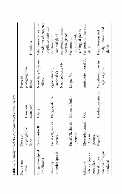
Tab
le 1
7.1.
Para
sym
path
etic
com
pon
ents
of
cran
ial n
erve
s.
Bra
in s
tem
N
erve
of
Ner
ve o
f
nu
cleu
spr
egan
glio
nic
Gan
glio
npo
st-g
angl
ion
ic
fibr
es(s
ynap
ses)
fibr
esFu
nct
ion
s
Edi
nge
r–W
estp
hal
Ocu
lom
otor
III
Cili
ary
Nas
ocili
ary
Va,
shor
tC
iliar
y m
usc
le: a
ccom
-
(mid
brai
n)
cilia
rym
odat
ion
of
len
s,et
c.;
pupi
lloco
nst
rict
ion
Saliv
ator
y:Fa
cial
VII
,gre
ater
Pte
rygo
pala
tin
eZ
ygom
atic
Vb,
Secr
etom
otor
:
supe
rior
(po
ns)
petr
osal
lacr
imal
Va
lacr
imal
gla
nd
Nas
al,p
alat
ine
Vb
Secr
etom
otor
:nas
al,
pala
tin
e gl
ands
Faci
al V
II,c
hor
daSu
bman
dibu
lar
Lin
gual
Vc
Secr
etom
otor
:
tym
pan
isu
bman
dibu
lar,
subl
ingu
al g
lan
ds
Saliv
ator
y:G
loss
oph
aryn
geal
Oti
cA
uri
culo
tem
pora
l Vc
Secr
etom
otor
:par
otid
infe
rior
(u
pper
IX,l
esse
r gl
and
med
ulla
)pe
tros
al
Dor
sal m
otor
Vag
us
XC
ardi
ac,m
yen
teri
cV
ery
shor
t,on
or
inH
eart
;for
egut
an
d
nu
cleu
s of
vagu
sta
rget
org
ans
mid
gut
mu
scle
an
d
(med
ulla
)gl
ands
Parasympathetic components and taste sensation 99
• the sinuatrial node of the heart; and
• glands and muscles of the alimentary canal as far distally as the
junction between midgut and hindgut (approximately the splenic
flexure of the colon) (see Section 15.2).
We have already noted that parasympathetic fibres pass from
branches of one cranial nerve to those of another and, for struc-
tures in the head, all postganglionic parasympathetic fibres, irre-
spective of their origin, attain their destinations in branches of the
trigeminal nerve. These pathways are described below. Table 17.1
summarizes parasympathetic connections and ganglia.
17.3 Parasympathetic pathways in the head (Fig. 17.1)
1 Edinger–Westphal nucleus (III), ciliary ganglion, iris, ciliary
body. This is important (see Chapter 22):
• Edinger–Westphal nucleus on rostral aspect of oculomotor
nucleus in midbrain.
• Axons pass in oculomotor (III) nerve, inferior division, to cil-
iary ganglion (synapse).
• Postganglionic axons to iris and ciliary muscles in nasociliary
and short ciliary nerves.
• Pupillary constriction (miosis), ciliary muscle contraction.
2 Superior salivatory nucleus (VII), greater petrosal nerve,
pterygopalatine ganglion, lacrimal, nasal, palatine glands:
• Superior salivatory nucleus, cerebellopontine angle, internal
auditory meatus (IAM), VII, greater petrosal nerve, carotid
canal/foramen lacerum, greater petrosal nerve, pterygoid
canal (with sympathetic fibres, deep petrosal nerve (see
Section 19.2)).
• Pterygopalatine fossa, pterygopalatine ganglion (synapse).
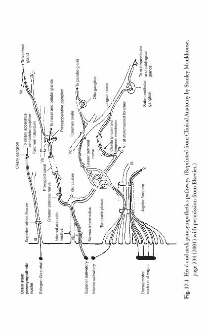
Bra
in s
tem
par
asym
pat
het
icn
ucl
ei
Edi
nger
–Wes
tpha
l
Sup
erio
r sa
livat
ory
Infe
rior
saliv
ator
y
Sup
erio
r or
bita
l fis
sure
III VII
Inte
rnal
aco
ustic
mea
tus
Gre
ater
pet
rosa
l ner
ve
Gen
icul
um
Pte
rygo
id c
analCili
ary
gang
lion
To c
iliar
y ap
para
tus
cons
tric
tor
pupi
llae
Va
To la
crim
algl
and
For
amen
rot
undu
m
Vb
To n
asal
and
pal
atal
gla
nds
Pte
rygo
pala
tine
gang
lion
For
amen
ova
le
Less
er p
etro
sal
nerv
e
Tym
pani
c pl
exus
Jugu
lar
fora
men
Dor
sal m
otor
nucl
eus
of v
agus
IX
X
Ner
vus
inte
rmed
ius
Cho
rda
tym
pani
and
tym
pani
c m
embr
ane
VII
at s
tylo
mas
toid
fora
men
To p
arot
id g
land
Otic
gan
glio
n
Sub
man
dibu
lar
gang
lion
To s
ubm
andi
bula
ran
d su
blin
guia
lgl
ands
Ling
ual n
erve
Vc
Fig.
17.1
Hea
d an
d n
eck
para
sym
path
etic
s pa
thw
ays.
(Rep
rin
ted
from
Clin
ical
An
atom
y by
Sta
nle
y M
onkh
ouse
,
page
234
(20
01)
wit
h p
erm
issi
on fr
om E
lsev
ier.
)
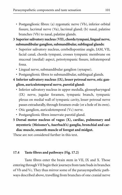
• Postganglionic fibres: (a) zygomatic nerve (Vb), inferior orbital
fissure, lacrimal nerve (Va), lacrimal gland; (b) nasal, palatine
branches (Vb) to nasal, palatine glands.
3 Superior salivatory nucleus (VII), chorda tympani, lingual nerve,
submandibular ganglion, submandibular, sublingual glands:
• Superior salivatory nucleus, cerebellopontine angle, IAM, VII,
facial canal, chorda tympani, crosses tympanic membrane on
mucosal (medial) aspect, petrotympanic fissure, infratemporal
fossa.
• Lingual nerve, submandibular ganglion (synapse).
• Postganglionic fibres to submandibular, sublingual glands.
4 Inferior salivatory nucleus (IX), lesser petrosal nerve, otic gan-
glion, auriculotemporal nerve, parotid gland:
• Inferior salivatory nucleus in upper medulla, glossopharyngeal
(IX) nerve, jugular foramen, tympanic branch, tympanic
plexus on medial wall of tympanic cavity, lesser petrosal nerve
passes extradurally, through foramen ovale (or a hole of its own).
• Otic ganglion, auriculotemporal (Vc) nerve.
• Postganglionic fibres innervate parotid gland.
5 Dorsal motor nucleus of vagus (X), cardiac, pulmonary and
myenteric (Meissner’s, Auerbach’s) ganglia, bronchial and car-
diac muscle, smooth muscle of foregut and midgut.
These are not considered further in this text.
17.4 Taste fibres and pathways (Fig. 17.2)
Taste fibres enter the brain stem in VII, IX and X. Those
entering through VII begin their journeys from taste buds in branches
of Vb and Vc. They thus mirror some of the parasympathetic path-
ways described above, travelling from branches of one cranial nerve
Parasympathetic components and taste sensation 101

to another in nerves which also conduct parasympathetic fibres in
the opposite direction (e.g. chorda tympani, greater petrosal nerve).
Neurons conducting taste sensation centrally have cell bodies
in the sensory ganglia of the nerves through which they enter the
brain stem. Within the brain stem, as with all visceral sensation,
axons pass to the nucleus of the solitary tract (NTS).
1 In facial nerve (VII):
• From anterior portion of tongue and neighbouring mucosa:
– Taste buds in anterior portion of tongue and oral cavity.
– Lingual nerve (Vc), chorda tympani, across tympanic mem-
brane,VII in temporal bone, cell bodies in geniculate ganglion.
102 Autonomic components of cranial nerves, taste and smell
Internal acoustic meatus
Greater petrosalVb
VII
To n
ucle
us o
f sol
itary
trac
tTo
nuc
leus
of s
olita
ry tr
act
From taste receptors inpalate and nasopharynx
Lingual nerve
From taste receptors in anteriortongue and oral cavity
Jugular foramen
IX
From taste receptors in posterior tongueand oropharynxX
From taste receptors in pharynx
Pterygopalatine ganglion –sensory fibres pass through
Pterygoid canal
Geniculate ganglion
Chorda tympani
VII Petrotympanic fissure
Vc
Fig. 17.2 Taste pathways. (Reprinted from Clinical Anatomy by
Stanley Monkhouse, page 235 (2001) with permission from
Elsevier.)
– Central processes through nervus intermedius, brain stem
at cerebellopontine angle to NTS.
Compare this with parasympathetic fibres to submandibular ganglion
(see Superior salivatory nucleus (VII) chorda tympani under Section
17.3).
• From the palate and nasopharynx:
– Taste buds in palate, palatine branches of Vb.
– Taste buds in nasopharynx, pharyngeal branches of Vb.
– Fibres pass into pterygopalatine fossa, without interruption
through pterygopalatine ganglion into pterygoid canal and
greater petrosal nerve, VII in temporal bone, cell bodies in
geniculate ganglion.
– Central processes through nervus intermedius, brain stem
at cerebellopontine angle to NTS.
Compare this course with that of parasympathetic fibres to pterygo-
palatine ganglion (see Superior salivatory nucleus (VII) greater petrosal
nerve under Section 17.3).
2 In glossopharyngeal nerve (IX): from posterior tongue,
oropharynx
• Taste buds in posterior part of tongue and oropharynx.
• IX, cell bodies in sensory ganglia of IX.
• Central processes to NTS.
3 In vagus nerve (X): from epiglottic region, hypopharyngeal wall
• Taste buds in epiglottic region and hypopharyngeal wall.
• X, cell bodies in sensory ganglia of X.
• Central processes to NTS.
17.5 Chorda tympani, branchial arches and petrosal
nerves
The chorda tympani connects the facial and mandibular
nerves, respectively the nerves of the second and first branchial
Parasympathetic components and taste sensation 103

arches. During embryonic development, each of the nerves of
branchial arches 2, 3, 4 and 6 gives off a branch which pass into
the territory of the preceding (cranial) branchial arch (second
arch to first arch, third arch to second arch etc). Since, in order
to take such a course, these branches pass from the caudal to the
cranial sides of the slit between two adjacent branchial arches
(Latin: trema means slit), these nerves are known as the pretrematic
branches.
The chorda tympani is generally accepted as the pretrematic
branch of the second branchial arch nerve. It passes from second
arch tissue to first arch tissue through the tympanic membrane
which is itself in the trema between the first and second arches. It
contains two types of visceral fibre: afferent (taste) and efferent
(parasympathetic). The embryological origin of the petrosal nerves
is less certain.
17.6 Clinical notes
1 Frey’s syndrome
After parotidectomy, cut ends of postganglionic fibres begin to
grow. Should these sprouting fibres find their way into Schwann
cells sheaths occupied before surgery by sympathetic fibres,
stimuli normally producing salivation will instead induce sweat-
ing over the site of the parotid. This is Frey’s syndrome (gusta-
tory sweating) (see Section 14.4).
2 Runny eyes, streaming nose
Runny eyes, runny and blocked up nose might be produced by
overactivity of the pterygopalatine ganglion. This is why the gan-
glion is sometimes called the hay fever ganglion although these
symptoms are usually allergic.
104 Autonomic components of cranial nerves, taste and smell
17.7 Clinical testing of visceral components
1 Salivary glands
Ask the patient to suck something bitter (such as a lemon) to
provoke salivary secretion. This is sometimes done to try to
locate the position of a calculus in the duct of a salivary gland,
usually the submandibular, but is not done to test the neural
pathways since, as we have said, who cares?
2 Taste
Testing taste is possible but hardly worth the trouble.
Parasympathetic components and taste sensation 105
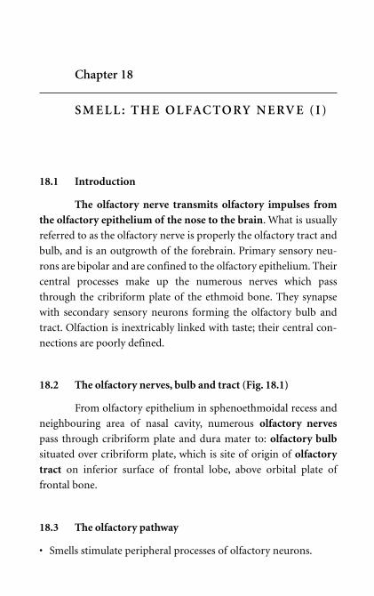
Chapter 18
S M E L L : T H E O L FAC TO RY N E RV E ( I )
18.1 Introduction
The olfactory nerve transmits olfactory impulses from
the olfactory epithelium of the nose to the brain. What is usually
referred to as the olfactory nerve is properly the olfactory tract and
bulb, and is an outgrowth of the forebrain. Primary sensory neu-
rons are bipolar and are confined to the olfactory epithelium. Their
central processes make up the numerous nerves which pass
through the cribriform plate of the ethmoid bone. They synapse
with secondary sensory neurons forming the olfactory bulb and
tract. Olfaction is inextricably linked with taste; their central con-
nections are poorly defined.
18.2 The olfactory nerves, bulb and tract (Fig. 18.1)
From olfactory epithelium in sphenoethmoidal recess and
neighbouring area of nasal cavity, numerous olfactory nerves
pass through cribriform plate and dura mater to: olfactory bulb
situated over cribriform plate, which is site of origin of olfactory
tract on inferior surface of frontal lobe, above orbital plate of
frontal bone.
18.3 The olfactory pathway
• Smells stimulate peripheral processes of olfactory neurons.
Smell: the olfactory nerve (I) 107
• Olfactory neurons are bipolar with cell bodies in olfactory
epithelium.
• Central processes pass through cribriform plate and dura to …
• … synapse in olfactory bulb with secondary sensory neurons
(mitral cells).
The olfactory bulb, tract, striae and connections
Axons pass posteriorly in olfactory tract, through olfactory striae to
limbic system of brain, particularly the uncus and amygdala of the
temporal lobe, thus providing connections with memory circuitry
and much else.
18.4 Olfaction and taste
The pleasures of eating and drinking lie as much with smell as
with taste: it is a common experience that an upper respiratory tract
infection which interferes with the sense of smell impairs the enjoy-
ment of food. Olfaction and taste are clearly closely linked and it is
thought that from the nucleus of the solitary tract, to which taste fibres
pass, axons project to the uncus to connect with olfactory centres.
Olfactory bulb, where bipolar neurons synapse on mitral cells
Axons of mitral cells pass toolfactory areas of forebrainCentral processes
pass throughcribriform plate
Cell bodies ofbipolar neuronsin olfactoryepithelium
Fig. 18.1 Olfactory pathways.
108 Autonomic components of cranial nerves, taste and smell
18.5 Clinical notes
1 Smells and the responses they can provoke
Evidence of olfactory connections to the limbic system are: (1)
smells can trigger memories; (2) smells can provoke emotional
responses; (3) smells have a role in sexual arousal.
2 Anosmia
Head injuries which fracture the cribriform plate may tear olfac-
tory nerves resulting in post-traumatic anosmia. Anosmia can
also be caused by blockage of the nasal cavities, for example a
nasal polyp or malignancy.
3 Cerebrospinal fluid rhinorrhoea
Head injuries may tear the dura mater, leading to cerebrospinal
fluid (CSF) leaking into the nasal cavity and dripping from the
anterior nasal aperture. This should be considered if clear fluid
issues from the nose after a head injury.
4 Temporal lobe epilepsy
Diseases such as epilepsy in the areas to which the olfactory
impulses project (e.g. the temporal lobe) may cause olfactory
hallucinations. The smells which are experienced are usually
unpleasant and are often accompanied by pseudo-purposeful
movements associated with tasting such as licking the lips.
18.6 Clinical testing
Too much trouble – don’t bother. You might just as well rely
on the subjective opinion of the patient which is, after all, what
matters most.
Chapter 19
T H E S Y M PAT H E T I C N E RVO U SS YS T E M I N T H E H E A D
Sympathetic fibres are not conveyed from the brain or brain stem in
cranial nerves, but are found in distal branches of some cranial nerves.
They are not usually considered components of cranial nerves, but they
appear here for the sake of completeness.
19.1 Functions of the sympathetic system in the head
These are similar to those in the rest of the body: secretomo-
tor to sweat glands, vasomotor (especially important for cerebral
vessels), muscles of the hair follicles and so on. In addition, various
structures in the eye receive a sympathetic innervation, particularly
dilator pupillae and part of levator palpebrae superioris muscle.
19.2 Sympathetic pathways to cranial structures
Sympathetic nerve impulses leave the central nervous system
only in the thoracolumbar region of the spinal cord. This means that
if their destination is the head, they leave the spinal cord in upper thor-
acic spinal nerves and thence pass back up to the head. The sympa-
thetic chain is the redistribution system by which means they ascend.
Preganglionic axons: T1, neck of first rib, cervical chain,
synapse in superior cervical ganglion
• Preganglionic axons arise in lateral grey horn of T1 and/or T2
segments of spinal cord, and possibly also C8.
• Ventral roots of appropriate spinal nerves, spinal nerve, anterior
primary ramus, white ramus communicans.
• Sympathetic chain at T1 ganglion near neck of first rib.
• Preganglionic axons for cranial structures do not synapse immed-
iately, but ascend in sympathetic chain, posterior to carotid sheath,
on surface or in substance of prevertebral muscles.
• Synapse in superior cervical ganglion (SCG) at rostral end of
chain immediately below the base of skull.
Postganglionic axons
• From SCG, postganglionic fibres pass to wall of adjacent internal
and external carotid arteries forming plexus around them.
• Plexus continues in walls of internal carotid, through carotid
canal, into cranium and onto the walls of internal carotid artery
and its branches.
• Postganglionic fibres delivered in walls of arteries to skin, eye,
orbit, all cerebral arteries and other structures.
Cavernous sinus, orbital sympathetics, deep petrosal, on
vertebral arteries
• As internal carotid artery passes through cavernous sinus, post-
ganglionic fibres on its wall pass in fibrous strands which con-
nect artery to lateral wall of sinus. Postganglionic fibres thus gain
access to III, IV, Va, VI. This provides another route to orbit and
eye and, through branches of ophthalmic nerve, to scalp.
• Orbital postganglionic fibres pass to levator palpebrae superioris
muscle and to eyeball along III, Va, long and short ciliary nerves.
Fibres traversing ciliary (parasympathetic) ganglion do so with-
out synapsing.
• Deep petrosal nerve leaves carotid plexus in carotid canal.
Joins greater petrosal nerve to form nerve of pterygoid canal to
pterygopalatine fossa. Sympathetic fibres distributed in Vb to
nose, pharynx, palate and face.
110 Autonomic components of cranial nerves, taste and smell
• Some fibres pass through cervical vertebral foramina transver-
saria with vertebral arteries to vessels of vertebrobasilar system.
19.3 Clinical notes
1 Apical lung tumour: Horner’s syndrome
Any interruption of the sympathetic pathway to the eye would
result in Horner’s syndrome. Levator palpebrae superioris is
partly supplied by the sympathetic system, and so would be
weakened leading to drooping of the upper eyelid (ptosis).
Sympathetic denervation of the iris would lead to unopposed
pupillary constriction (miosis), and sympathetic denervation of
sweat glands would result in an absence of sweating (anhidrosis).
A tumour at the apex of the lung invading the neck of the first rib
and interrupting the sympathetic chain would result in such
signs. It may also damage T1 root of the brachial plexus causing
weakness or paralysis of the small muscles of the hand with con-
sequent impairment of grip.
2 Cerebral vasculature and the sympathetic nervous system
The regulation of cerebral blood flow is of great importance.
Pathways described above regulate the calibre of all cerebral vas-
culature in response to physiological and other metabolic needs.
This is performed by the sympathetic supply to arterial wall
smooth muscle.
3 Cervical sympathectomy
This is interruption of the sympathetic pathways to the upper
limb for peripheral vascular disease. The sympathetic chain is
sectioned below T1 ganglion but the procedure is called cervical
because it was often performed through a cervical incision.
Providing that the chain is sectioned below T1 ganglion, which
receives preganglionic impulses from the spinal cord, there will
remain an adequate sympathetic supply to the head.
The sympathetic nervous system in the head 111


PART V
V I S I O N , E Y E M OV E M E N T S ,
H E A R I N G A N D BA L A N C E :
O P T I C , O C U LO M OTO R ,
T RO C H L E A R , A B D U C E N S A N D
V E S T I B U LO C O C H L E A R N E RV E S

Chapter 20
T H E O P T I C N E RV E ( I I )
20.1 Sight
Sight is dependent not only on a substantial portion of
the cerebral cortex, but also upon six cranial nerves (II–VII).
Perception is the function of the retina, optic nerve, tract, radia-
tion and cortex. The oculomotor, trochlear and abducens nerves
move the eye. Eyeball sensations such as pain, touch and pressure
are mediated by the ophthalmic nerve, and the facial nerve inner-
vates orbicularis oculi muscle. This Chapter deals with the optic
pathway: eye movements and their control come later.
20.2 The optic nerve
The optic pathway transmits visual impulses from the
retina to the brain. The optic nerve is the name given to the path-
way between the eyeball and the optic chiasma. It is an artificial
subdivision of the optic pathway. Like the olfactory nerve (Chapter
18), the optic nerve is not really a nerve. It is an outgrowth of the
diencephalon (the thalamic structures). As in the olfactory system,
the primary sensory neurons are bipolar and are confined to the
sensitive epithelium (retina), the axons of secondary sensory neu-
rons forming the optic nerve, chiasma and tract.
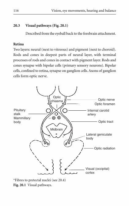
20.3 Visual pathways (Fig. 20.1)
Described from the eyeball back to the forebrain attachment.
Retina
Two layers: neural (next to vitreous) and pigment (next to choroid).
Rods and cones in deepest parts of neural layer, with terminal
processes of rods and cones in contact with pigment layer. Rods and
cones synapse with bipolar cells (primary sensory neurons). Bipolar
cells, confined to retina, synapse on ganglion cells. Axons of ganglion
cells form optic nerve.
116 Vision, eye movements, hearing and balance
Optic nerveOptic foramen
Internal carotidartery
Optic tract
Lateral geniculatebody
Optic radiation
Midbrain
Visual (occipital)cortex
Mammillarybody
Pituitarystalk
Opticchiasma
*Fibres to pretectal nuclei (see 20.4)
Fig. 20.1 Visual pathways.
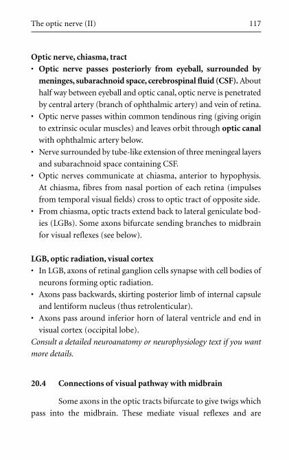
Optic nerve, chiasma, tract
• Optic nerve passes posteriorly from eyeball, surrounded by
meninges, subarachnoid space, cerebrospinal fluid (CSF). About
half way between eyeball and optic canal, optic nerve is penetrated
by central artery (branch of ophthalmic artery) and vein of retina.
• Optic nerve passes within common tendinous ring (giving origin
to extrinsic ocular muscles) and leaves orbit through optic canal
with ophthalmic artery below.
• Nerve surrounded by tube-like extension of three meningeal layers
and subarachnoid space containing CSF.
• Optic nerves communicate at chiasma, anterior to hypophysis.
At chiasma, fibres from nasal portion of each retina (impulses
from temporal visual fields) cross to optic tract of opposite side.
• From chiasma, optic tracts extend back to lateral geniculate bod-
ies (LGBs). Some axons bifurcate sending branches to midbrain
for visual reflexes (see below).
LGB, optic radiation, visual cortex
• In LGB, axons of retinal ganglion cells synapse with cell bodies of
neurons forming optic radiation.
• Axons pass backwards, skirting posterior limb of internal capsule
and lentiform nucleus (thus retrolenticular).
• Axons pass around inferior horn of lateral ventricle and end in
visual cortex (occipital lobe).
Consult a detailed neuroanatomy or neurophysiology text if you want
more details.
20.4 Connections of visual pathway with midbrain
Some axons in the optic tracts bifurcate to give twigs which
pass into the midbrain. These mediate visual reflexes and are
The optic nerve (II) 117
connected to the pretectal nuclei (for the pupillary light reflex) and
the superior colliculus and medial longitudinal fasciculus (for lens
accommodation, eye movements, etc.). These reflexes and control
mechanisms depend upon many other structures and are considered
in Chapter 22.
20.5 Lesions of optic pathway
1 Optic nerve
Section of one optic nerve causes blindness in one eye.
2 Crossing fibres in chiasma
Destruction of crossing fibres in chiasma (e.g. pituitary tumour)
causes blindness in the nasal retina of both eyes. This gives a
bitemporal hemianopia (field loss).
3 Pressure on lateral aspect of chiasma
Pressure on the lateral aspect of the chiasma (e.g. internal carotid
aneurysm) affects fibres from the temporal retina of the ipsilat-
eral eye, giving an ipsilateral nasal hemianopia. This is uncom-
mon. Bilateral internal carotid artery aneurysms would cause a
binasal hemianopia – even more uncommon.
4 Optic tract or geniculate body
Destruction of the right optic tract or LGB would interrupt
pathways from the temporal retina of the right eye and the nasal
retina of the left eye. This would cause blindness in the left side
of both visual fields. This is a homonymous hemianopia. Thus,
destruction of the right optic tract would cause a left homony-
mous hemianopia.
5 Optic radiation and visual cortex
Lesions of the optic radiation and visual cortex are more com-
plex. Consult a detailed neuroanatomy or ophthalmology text if
you want more information.
118 Vision, eye movements, hearing and balance
20.6 Other clinical notes
1 Retinal signs of systemic disease
The retina is the only part of the forebrain that can be viewed
directly by an observer, so retinoscopy is an essential part of the
neurological examination of a patient. Exudates, haemorrhages
and abnormalities of blood vessels may be seen on retinoscopy
and may be signs of generalized disease processes (e.g. diabetes,
rheumatoid arthritis, etc.).
2 Papilloedema and raised intracranial pressure
Because the optic nerve in the orbit is surrounded by subarach-
noid space containing CSF, raised intracranial pressure will com-
press the nerve. This will occlude the central vein before the
central artery (venous blood is at a lower pressure). The retina
will be engorged with blood and the optic disc will bulge into the
vitreous. This is papilloedema, visible on retinoscopy – a reliable
sign of raised intracranial pressure.
3 Blood supply of visual cortex
Although most of the visual cortex is supplied by the posterior
cerebral artery, the cortical area which receives projections from
the macula of the retina is generally supplied by both middle and
posterior cerebral arteries. This is one of the explanations given
for the phenomenon of macular sparing in which vision at the
macula may be preserved even though the surrounding areas of
the visual cortex are no longer functional.
4 Retinal detachment
The optic nerves and retina develop as a diencephalic outgrowth,
the optic vesicle, which contains an extension of the diencephalic
cavity, the third ventricle. As the two layers of the retina grow,
they approach each other and the cavity is obliterated as the two
layers become contiguous. The two layers give rise to the inner
The optic nerve (II) 119
neural layer and the outer pigment layer. The potential space
between them can open up in certain conditions, for example
poor vascular perfusion. This is called retinal detachment; it
causes blindness.
5 Demyelinating diseases
In the optic nerve, a brain outgrowth, myelin is produced by
oligodendrocytes. The nerve may be affected in demyelinating
diseases such as multiple sclerosis. This is not so for other cranial
nerves in which myelin is manufactured by Schwann cells.
20.7 Clinical testing
Accurate assessments require the facilities of ophthalmol-
ogy units, but crude assessment may be done by confrontation.
This involves the examiner facing the patient, and both observing
an object (usually the examiner’s finger) held equidistant between
patient and examiner. The object is at first held out of sight, and as
it is brought towards the centre of the visual field from one extrem-
ity (right, left, top or bottom), the point at which it is first seen by
both patient and examiner is noted. This method is based on com-
paring the patient’s visual fields with those of the examiner, and
thus assumes that the examiner is normal, at least in respect of his
or her visual fields.
All crude tests of vision, of course, also depend on a normally func-
tioning cornea, aqueous humour, iris, lens, vitreous humour, etc.
120 Vision, eye movements, hearing and balance
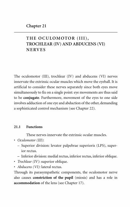
Chapter 21
T H E O C U LO M OTO R ( I I I ) ,TROCHLEAR (IV) AND ABDUCENS (VI)N E RV E S
The oculomotor (III), trochlear (IV) and abducens (VI) nerves
innervate the extrinsic ocular muscles which move the eyeball. It is
artificial to consider these nerves separately since both eyes move
simultaneously to fix on a single point: eye movements are thus said
to be conjugate. Furthermore, movement of the eyes to one side
involves adduction of one eye and abduction of the other, demanding
a sophisticated control mechanism (see Chapter 22).
21.1 Functions
These nerves innervate the extrinsic ocular muscles.
• Oculomotor (III):
– Superior division: levator palpebrae superioris (LPS), super-
ior rectus.
– Inferior division: medial rectus, inferior rectus, inferior oblique.
• Trochlear (IV): superior oblique.
• Abducens (VI): lateral rectus.
Through its parasympathetic components, the oculomotor nerve
also causes constriction of the pupil (miosis) and has a role in
accommodation of the lens (see Chapter 17).
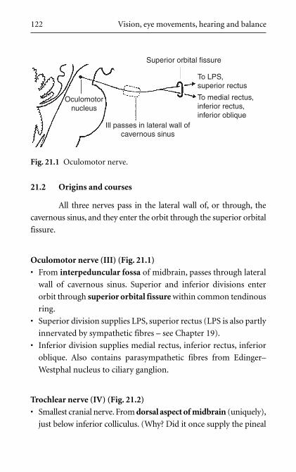
21.2 Origins and courses
All three nerves pass in the lateral wall of, or through, the
cavernous sinus, and they enter the orbit through the superior orbital
fissure.
Oculomotor nerve (III) (Fig. 21.1)
• From interpeduncular fossa of midbrain, passes through lateral
wall of cavernous sinus. Superior and inferior divisions enter
orbit through superior orbital fissure within common tendinous
ring.
• Superior division supplies LPS, superior rectus (LPS is also partly
innervated by sympathetic fibres – see Chapter 19).
• Inferior division supplies medial rectus, inferior rectus, inferior
oblique. Also contains parasympathetic fibres from Edinger–
Westphal nucleus to ciliary ganglion.
Trochlear nerve (IV) (Fig. 21.2)
• Smallest cranial nerve. From dorsal aspect of midbrain (uniquely),
just below inferior colliculus. (Why? Did it once supply the pineal
122 Vision, eye movements, hearing and balance
Superior orbital fissure
To LPS, superior rectus
To medial rectus,inferior rectus,inferior oblique
Ill passes in lateral wall ofcavernous sinus
Oculomotornucleus
Fig. 21.1 Oculomotor nerve.
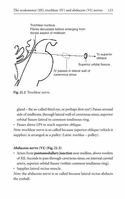
gland – the so-called third eye, or perhaps first eye?) Passes around
side of midbrain, through lateral wall of cavernous sinus, superior
orbital fissure lateral to common tendinous ring.
• Passes above LPS to reach superior oblique.
Note: trochlear nerve is so called because superior oblique (which it
supplies) is arranged as a pulley (Latin: trochlea – pulley).
Abducens nerve (VI) (Fig. 21.3)
• Arises from pontomedullary junction near midline, above rootlets
of XII. Ascends to pass through cavernous sinus, on internal carotid
artery, superior orbital fissure (within common tendinous ring).
• Supplies lateral rectus muscle.
Note: the abducens nerve is so called because lateral rectus abducts
the eyeball.
The oculomotor (III), trochlear (IV) and abducens (VI) nerves 123
Trochlear nucleusFibres decussate before emerging fromdorsal aspect of midbrain
To superioroblique
Superior orbital fissure
IV passes in lateral wall ofcavernous sinus
Fig. 21.2 Trochlear nerve.
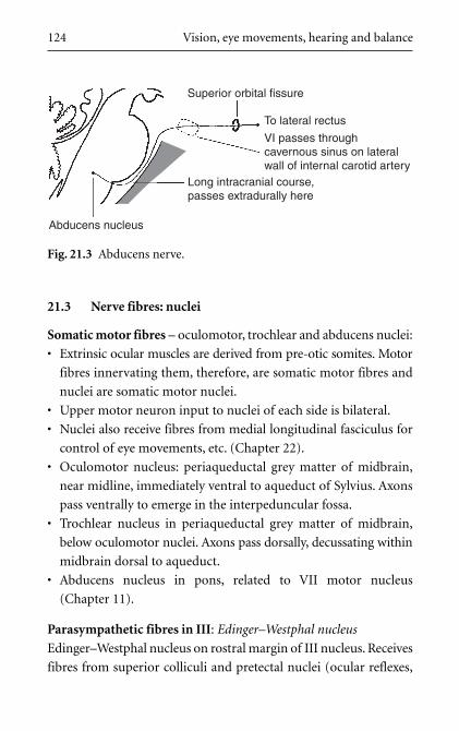
21.3 Nerve fibres: nuclei
Somatic motor fibres – oculomotor, trochlear and abducens nuclei:
• Extrinsic ocular muscles are derived from pre-otic somites. Motor
fibres innervating them, therefore, are somatic motor fibres and
nuclei are somatic motor nuclei.
• Upper motor neuron input to nuclei of each side is bilateral.
• Nuclei also receive fibres from medial longitudinal fasciculus for
control of eye movements, etc. (Chapter 22).
• Oculomotor nucleus: periaqueductal grey matter of midbrain,
near midline, immediately ventral to aqueduct of Sylvius. Axons
pass ventrally to emerge in the interpeduncular fossa.
• Trochlear nucleus in periaqueductal grey matter of midbrain,
below oculomotor nuclei. Axons pass dorsally, decussating within
midbrain dorsal to aqueduct.
• Abducens nucleus in pons, related to VII motor nucleus
(Chapter 11).
Parasympathetic fibres in III: Edinger–Westphal nucleus
Edinger–Westphal nucleus on rostral margin of III nucleus. Receives
fibres from superior colliculi and pretectal nuclei (ocular reflexes,
124 Vision, eye movements, hearing and balance
Superior orbital fissure
To lateral rectus
Long intracranial course,passes extradurally here
Abducens nucleus
VI passes throughcavernous sinus on lateralwall of internal carotid artery
Fig. 21.3 Abducens nerve.
Chapter 22). Preganglionic axons pass in III to ciliary ganglion
(synapse). Postganglionic axons in short ciliary nerves to constrictor
pupillae and ciliary muscles.
21.4 Clinical notes
1 Visual reflexes and clinical testing (see Chapter 22).
2 Midbrain lesions: oculomotor nerve
Vascular or other lesions of the midbrain can affect the oculo-
motor nerve. They may also affect the substantia nigra causing
Parkinsonian symptoms (e.g. resting tremor), the red nucleus
(also causing extrapyramidal symptoms), and the descending
corticospinal fibres in the cerebral peduncles leading to a con-
tralateral upper motor neuron lesions (UMNL). Benedikt’s syn-
drome involves the nerve as it passes through the red nucleus:
oculomotor paralysis with contralateral extrapyramidal dyskine-
sia. In Weber’s syndrome the lesion is more ventral, also involving
motor fibres in the cerebral peduncles: oculomotor paralysis is
associated with contralateral UMNLs.
3 Oculomotor nerve injury
The oculomotor nerve is liable to be stretched as it crosses the
tentorial notch in cases of raised intracranial pressure. Complete
section of the oculomotor nerve would lead to ptosis (partial
paralysis of LPS), lateral squint (unopposed action of superior
oblique and lateral rectus), pupillary dilatation (unopposed
sympathetic activity), loss of accommodation and light reflexes.
Irritation of the nerve may cause spasm of the muscles supplied
by it (e.g. spasm of medial rectus leading to a medial squint).
4 Oculomotor nerve injury: diabetes
It is not uncommon for diabetics to suffer from an acute vasculitis
of the oculomotor nerve. This causes medial squint (somatic
fibres) and ptosis (sympathetic fibres to LPS).
The oculomotor (III), trochlear (IV) and abducens (VI) nerves 125
5 Aneurysms of posterior cerebral artery: oculomotor nerve
Just after the nerve leaves the midbrain it is intimately related to
the posterior cerebral artery, aneurysms of which may compress
the nerve leading to symptoms as described above.
6 Trauma: trochlear nerve
The trochlear nerve is the thinnest and most fragile nerve. It is
vulnerable to trauma. Section of the nerve would result in the
affected eye being turned medially.
7 Intracranial disease: diagnostic usefulness of abducens nerve
The abducens nerve, with a relatively low origin compared to its
destination, has the longest intracranial course of any cranial
nerve. It may be involved in fractures of the base of the skull or
in intracranial disease. Section of the nerve would result in con-
vergent squint (the eye abductor being paralyzed). See also the
effects of raised intracranial pressure: abducens nerve below.
Because of this long intracranial course it is often the first cra-
nial nerve to be affected by intracranial disease. So, if you could
only test one cranial nerve as part of a neurological investiga-
tion, this would be the one!
8 Abducens and facial motor nuclei
Diseases of the brain stem affecting the abducens nucleus may also
involve fibres from the facial motor nucleus which loop around it.
9 Gradenigo’s syndrome: abducens nerve
Since the abducens nerve passes over the apex of the petrous tem-
poral bone, it may be affected by infections of the petrous tem-
poral (petrositis), thus causing weakness of lateral rectus with
consequent medial deviation of the ipsilateral eye (Gradenigo’s
syndrome: rare but interesting).
10 The effects of raised intracranial pressure: oculomotor nerve
When an expanding lesion above the tentorium causes raised
intracranial pressure, the uncus of the temporal lobe may be
126 Vision, eye movements, hearing and balance
squashed into the tentorial notch (herniation of the uncus).
This compresses the midbrain which passes through the tentorial
notch and the nearby oculomotor nerve. The result is pupillary
dilatation (unopposed sympathetic action as the parasympa-
thetic fibres in III are affected), at first unilateral and then bilat-
eral. By this stage, the patient will already be unconscious.
11 The effects of raised intracranial pressure: abducens nerve
As intracranial pressure rises, the cerebrum may be forced back-
wards and downwards, thus stretching the nerve with its long
intracranial course. A lateral rectus palsy (medial squint) would
result. Because this may cause an erroneous diagnosis to be made,
it is known as a false localizing sign.
12 Cavernous sinus thrombosis: all three nerves
Cavernous sinus thrombosis may occur as a result of an infec-
tion of any part of the head that drains through veins to the cav-
ernous sinus (e.g. face, ear, etc.). It affects all the nerves that pass
through or in the wall of the sinus (III, IV, Va, VI). The abducens
nerve is usually affected first because it passes through the sinus,
causing a paralysis of lateral rectus and a resultant medial
squint. Involvement of the ophthalmic nerve may cause severe
pain, and the condition may result ultimately in papilloedema
and visual loss. Since the advent of antibiotic therapy, this con-
dition is much less often encountered than formerly.
The oculomotor (III), trochlear (IV) and abducens (VI) nerves 127
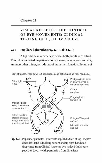
Chapter 22
V I S UA L R E F L E X E S : T H E C O N T RO LO F E Y E M OV E M E N T S ; C L I N I C A LT E S T I N G O F I I , I I I , I V A N D V I
22.1 Pupillary light reflex (Fig. 22.1, Table 22.1)
A light shone into either eye causes both pupils to constrict.
This reflex is elicited on patients, conscious or unconscious, and it is,
amongst other things, a crude test of brain stem function. Because of
Postganglionic fibresin ciliary nerves toconstrictor pupillae
Ciliaryganglion
Preganglionicfibres in III
Edinger–Westphalnucleus
Midbrain pretectalnucleus
Shine lightin eye
Start at top left. Pass down left hand side, along bottom and up right hand side
Impulses passalong optic nerve,chiasma, tract
Before reachinglateral geniculatebody, some fibresbranch to midbrain
Fig. 22.1 Pupillary light reflex (study with Fig. 21.1). Start at top left, pass
down left-hand side, along bottom and up right-hand side.
(Reprinted from Clinical Anatomy by Stanley Monkhouse,
page 269 (2001) with permission from Elsevier.)
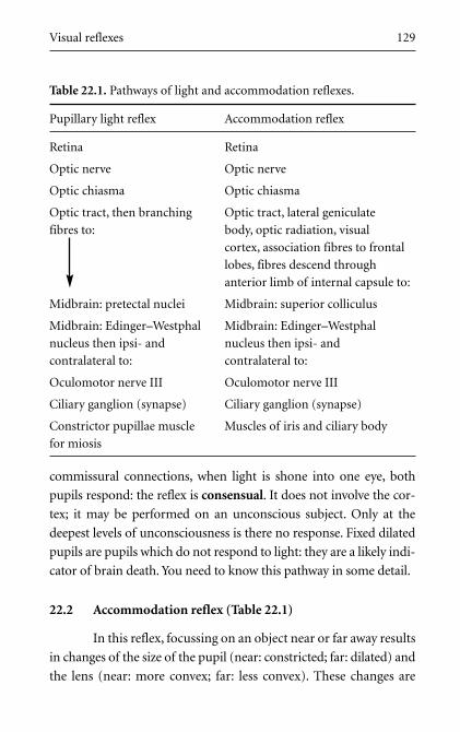
Visual reflexes 129
commissural connections, when light is shone into one eye, both
pupils respond: the reflex is consensual. It does not involve the cor-
tex; it may be performed on an unconscious subject. Only at the
deepest levels of unconsciousness is there no response. Fixed dilated
pupils are pupils which do not respond to light: they are a likely indi-
cator of brain death. You need to know this pathway in some detail.
22.2 Accommodation reflex (Table 22.1)
In this reflex, focussing on an object near or far away results
in changes of the size of the pupil (near: constricted; far: dilated) and
the lens (near: more convex; far: less convex). These changes are
Table 22.1. Pathways of light and accommodation reflexes.
Pupillary light reflex Accommodation reflex
Retina Retina
Optic nerve Optic nerve
Optic chiasma Optic chiasma
Optic tract, then branching Optic tract, lateral geniculate
fibres to: body, optic radiation, visual
cortex, association fibres to frontal
lobes, fibres descend through
anterior limb of internal capsule to:
Midbrain: pretectal nuclei Midbrain: superior colliculus
Midbrain: Edinger–Westphal Midbrain: Edinger–Westphal
nucleus then ipsi- and nucleus then ipsi- and
contralateral to: contralateral to:
Oculomotor nerve III Oculomotor nerve III
Ciliary ganglion (synapse) Ciliary ganglion (synapse)
Constrictor pupillae muscle Muscles of iris and ciliary body
for miosis
130 Vision, eye movements, hearing and balance
equivalent to those made by photographers in stop adjustment and
lens extension on a camera. You will realize that in the accommoda-
tion reflex perception is involved, unlike the pupillary light reflex, and
thus the cortex is involved. There is also a degree of voluntary control
since you can decide to focus on an object. The precise pathways are
not fully understood, but those given in Table 22.1 are probable.
22.3 Argyll Robertson pupil
The Argyll Robertson pupil is one that accommodates but
does not react to light. A comparison of the pathways for the accom-
modation reflex, which functions normally, and the pupillary light
reflex, which does not, indicates that the lesion could be in: (a) the
fibres that pass from the optic tract to the midbrain, (b) the pretec-
tal nuclei or (c) that part of the Edinger–Westphal nucleus which
deals with fibres from the pretectal nuclei.
22.4 Conjugate eye movements and their control
Eye movements involve nuclei of III, IV and VI integrated
by mechanisms which include the frontal eye fields, the superior
colliculus, the pontine gaze centre, the cerebellum, and the medial
longitudinal fasciculus (MLF).
The frontal eye fields mediate voluntary eye movements and are
responsible for saccadic movements by which means we search the
visual fields for an object on which to fix. Saccades are so rapid that
individual visual images are imperceptible until fixation has ensued.
Frontal eye field stimulation causes conjugate movement of the eyes
to the opposite side.
The superior colliculi on the dorsal aspect of the midbrain are
involved in visual reflexes. This part of the brain stem is known as
Visual reflexes 131
the tectum (Latin: roof): tectospinal and spinotectal tracts pass to
and from the spinal cord.
The MLF extends from midbrain to cervical spinal cord. It inte-
grates the nuclei of III, IV and VI with:
• ventral horn cells (motor) of the cervical spinal cord for the con-
trol of head and neck movements involved in visual fixation
movements;
• vestibular nuclei (see Chapter 23);
• auditory nuclei (see Chapter 23);
• the cerebellum.
Consider what happens when you watch a fixed object from a mov-
ing vehicle. As the head and neck turn sideways, the vestibular–
ocular reflex keeps your eyes fixed on the object. Impulses from the
vestibular apparatus, and from the neck muscles by way of the
spinal cord, pass to the MLF and the nuclei of III, IV and VI caus-
ing the extrinsic ocular muscles to bring about a series of saccades
which, although imperceptible to you, continually reset the eyes on
target. These connections are also brought into play in other cir-
cumstances: disorders of the vestibular apparatus (e.g. Ménière’s
disease – see Section 23.5), a loud noise, or pain mediated by a spinal
nerve, can all result in reflex eye movements.
Nystagmus
The MLF is also connected to the cerebellum, and so can be affected
in cerebellar disease producing abnormal eye movements. Disorders of
any part of this system may lead to jerky eye movements – nystagmus.
This can be regarded as ataxia of the eye muscles.
22.5 Internuclear ophthalmoplegia
This occurs when the MLF in the brain stem is damaged (e.g.
multiple sclerosis). The nuclei of III, IV and VI become disconnected,
132 Vision, eye movements, hearing and balance
and uncoordinated movements of the eyes result in strabismus
(squints).
22.6 Clinical testing of eyes and eye movements
1 Pupillary light reflex. Shine a light into one eye and observe both
pupils. This tests II, midbrain, III.
2 Corneal reflex tests Va, brain stem and VII.
3 Observe pupillary size: both should be equal. If not, there may be
a lesion of III or midbrain.
4 Look for nystagmus. Nystagmus present on straight forward gaze
is definitely abnormal. Nystagmus evident at the extremes of eye
movements is only possibly abnormal.
5 Look for a squint. A lesion of the main trunk of the oculomotor,
trochlear or abducens nerves will be obvious.
6 With the head stationary, the patient should be asked to follow
with both eyes together an object moving not-too-quickly (e.g.
the examiner’s finger or a pen) as it describes a large square with
both diagonals. Should any abnormality be observed, each eye
may be tested more carefully. Or, if you want the easy way out,
send the patient to an optician or ophthalmologist.
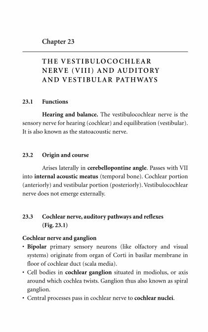
Chapter 23
T H E V E S T I B U LO C O C H L E A R N E RV E ( V I I I ) A N D AU D I TO RY A N D V E S T I B U L A R PAT H WAYS
23.1 Functions
Hearing and balance. The vestibulocochlear nerve is the
sensory nerve for hearing (cochlear) and equilibration (vestibular).
It is also known as the statoacoustic nerve.
23.2 Origin and course
Arises laterally in cerebellopontine angle. Passes with VII
into internal acoustic meatus (temporal bone). Cochlear portion
(anteriorly) and vestibular portion (posteriorly). Vestibulocochlear
nerve does not emerge externally.
23.3 Cochlear nerve, auditory pathways and reflexes
(Fig. 23.1)
Cochlear nerve and ganglion
• Bipolar primary sensory neurons (like olfactory and visual
systems) originate from organ of Corti in basilar membrane in
floor of cochlear duct (scala media).
• Cell bodies in cochlear ganglion situated in modiolus, or axis
around which cochlea twists. Ganglion thus also known as spiral
ganglion.
• Central processes pass in cochlear nerve to cochlear nuclei.
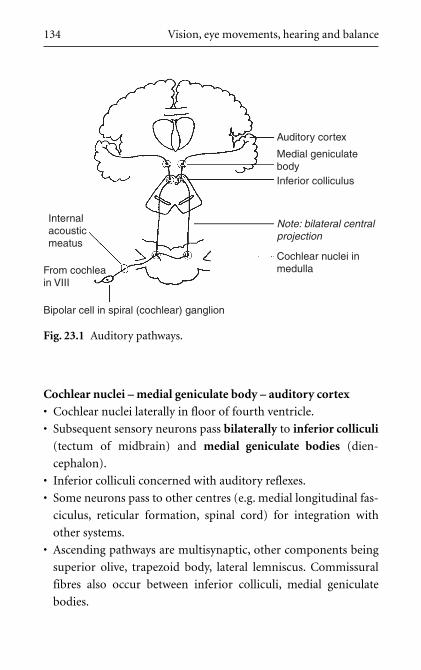
Cochlear nuclei – medial geniculate body – auditory cortex
• Cochlear nuclei laterally in floor of fourth ventricle.
• Subsequent sensory neurons pass bilaterally to inferior colliculi
(tectum of midbrain) and medial geniculate bodies (dien-
cephalon).
• Inferior colliculi concerned with auditory reflexes.
• Some neurons pass to other centres (e.g. medial longitudinal fas-
ciculus, reticular formation, spinal cord) for integration with
other systems.
• Ascending pathways are multisynaptic, other components being
superior olive, trapezoid body, lateral lemniscus. Commissural
fibres also occur between inferior colliculi, medial geniculate
bodies.
134 Vision, eye movements, hearing and balance
Auditory cortex
Medial geniculatebodyInferior colliculus
Cochlear nuclei inmedulla
Bipolar cell in spiral (cochlear) ganglion
From cochleain VIII
Internalacousticmeatus
Note: bilateral centralprojection
Fig. 23.1 Auditory pathways.
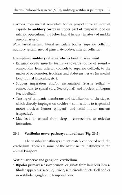
• Axons from medial geniculate bodies project through internal
capsule to auditory cortex in upper part of temporal lobe on
inferior operculum, just below lateral fissure (territory of middle
cerebral artery).
Note: visual system: lateral geniculate bodies, superior colliculi;
auditory system: medial geniculate bodies, inferior colliculi.
Examples of auditory reflexes: when a loud noise is heard
• Extrinsic ocular muscles turn eyes towards source of sound –
connections from inferior colliculi to superior colliculi, to the
nuclei of oculomotor, trochlear and abducens nerves (in medial
longitudinal fasciculus, etc.).
• Sudden inspiration and/or exclamation (startle reflex) –
connections to spinal cord (tectospinal) and nucleus ambiguus
(tectobulbar).
• Tensing of tympanic membrane and stabilization of the stapes,
which directly impinges on cochlea – connections to trigeminal
motor nucleus (tensor tympani) and facial motor nucleus
(stapedius).
• May lead to arousal from sleep – connections to reticular
formation.
23.4 Vestibular nerve, pathways and reflexes (Fig. 23.2)
The vestibular pathways are intimately connected with the
cerebellum. These are some of the oldest neural pathways in the
animal kingdom.
Vestibular nerve and ganglion: cerebellum
• Bipolar primary sensory neurons originate from hair cells in ves-
tibular apparatus: saccule, utricle, semicircular ducts. Cell bodies
in vestibular ganglion in temporal bone.
The vestibulocochlear nerve (VIII), auditory, vestibular pathways 135
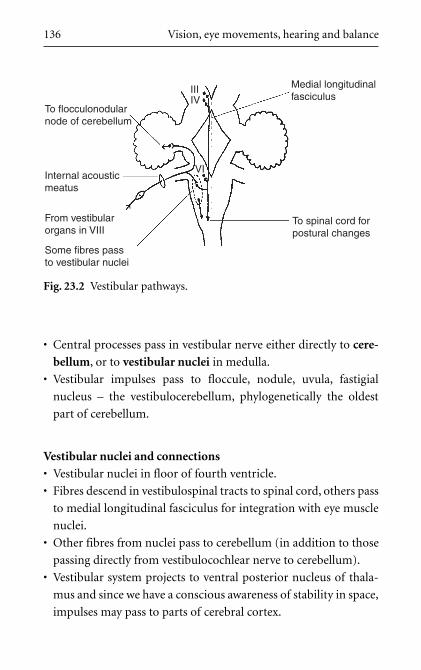
• Central processes pass in vestibular nerve either directly to cere-
bellum, or to vestibular nuclei in medulla.
• Vestibular impulses pass to floccule, nodule, uvula, fastigial
nucleus – the vestibulocerebellum, phylogenetically the oldest
part of cerebellum.
Vestibular nuclei and connections
• Vestibular nuclei in floor of fourth ventricle.
• Fibres descend in vestibulospinal tracts to spinal cord, others pass
to medial longitudinal fasciculus for integration with eye muscle
nuclei.
• Other fibres from nuclei pass to cerebellum (in addition to those
passing directly from vestibulocochlear nerve to cerebellum).
• Vestibular system projects to ventral posterior nucleus of thala-
mus and since we have a conscious awareness of stability in space,
impulses may pass to parts of cerebral cortex.
136 Vision, eye movements, hearing and balance
Medial longitudinalfasciculus
IIIIV
VI
To spinal cord forpostural changes
Some fibres passto vestibular nuclei
From vestibularorgans in VIII
Internal acousticmeatus
To flocculonodularnode of cerebellum
Fig. 23.2 Vestibular pathways.
Examples of vestibular reflexes
• Muscles act to counter unwanted movement – vestibulospinal
connections.
• As we move progressively in one direction, slow eye movements
in opposite direction are followed by rapid movements in same
direction (for eyes to catch up). This is nystagmus. Connections
to the nuclei of oculomotor, trochlear and abducens nerves (in
medial longitudinal fasciculus).
• Connections also from vestibular pathways and cerebellum to
reticular formation (arousal if necessary) including vomiting
centre in medulla.
23.5 Clinical notes
Remember that within the inner ear, the cochlear duct is
continuous with the saccule, utricle and semicircular ducts: they form
an enclosed endolymph containing system derived from the otocyst.
Cochlear disorders, therefore, may affect the vestibular system, and
vice versa as follows:
1 Conductive deafness
This is deafness resulting from defective transmission of sound
waves to the endolymph. It may be caused by blockage of the
external acoustic meatus, a large perforation in the tympanic
membrane (reducing its ability to pick up sound waves), impair-
ment of ossicular movement (e.g. fluid in the tympanic cavity,
ossicular joint disease, dislocation of the ossicular chain) or fix-
ation of footplate of the stapes.
2 Sensorineural deafness
This is deafness resulting from disease of the cochlea, the
vestibulocochlear nerve or the auditory pathway. It is clinically
distinguishable from conductive deafness. It includes the deaf-
ness of old age (presbyacusis).
The vestibulocochlear nerve (VIII), auditory, vestibular pathways 137
3 Nerve deafness
This is sensorineural deafness resulting specifically from disease
of the vestibulocochlear nerve or the auditory pathways. If the
auditory pathways are involved, the deafness is rarely profound
because of the bilateral connections of the cochlear nuclei.
4 Acoustic neuroma
This is a tumour of Schwann cells on the vestibular nerve in the
cerebellopontine angle. If the tumour grows in the internal
acoustic meatus, it will compress both vestibulocochlear and
facial nerves causing a nerve deafness and, eventually, an ipsi-
lateral facial palsy.
5 Stapedius paralysis: hyperacusis
This leads to a peculiar echoing sensation even though sounds
may not be particularly loud (hyperacusis) and it arises in facial
nerve lesions; it may be the first symptom of disease (see
Stapedius: hyperacusis under Section 11.7).
6 Inability to localize sounds in space
A unilateral lesion of the auditory cortex, though not resulting
in profound deafness (because of bilateral pathways), may lead
to difficulty in localizing the source of a sound. This is socially
awkward and can be dangerous.
7 Labyrinthine artery
This is a branch of the vertebrobasilar system and enters the
internal acoustic meatus to supply VII, VIII and the inner ear.
Lesions of this artery, or of the vertebrobasilar system, can cause
vertigo and unsteadiness. Narrowing of the vertebral arteries by
either atherosclerosis or by cervical vertebral osteophytes may
lead to these symptoms with neck movements.
8 Nystagmus
Disorders of the vestibular system, the cerebellum, and/or the
medial longitudinal fasciculus in the brain stem, may lead
138 Vision, eye movements, hearing and balance

to pathological nystagmus with slow eye movements in one
direction followed by quick movements in the other. It requires
investigation.
9 Travel sickness
This common condition illustrates the connections from the
vestibular pathways and cerebellum to the vomiting centre in
the medulla.
10 Ménière’s disease
Prosper Ménière described a condition consisting of attacks of
deafness, vertigo and tinnitus (noises in the head). It arises from
an endolymph disorder and its symptoms reflect the endolym-
phatic continuity between cochlea, saccule, utricle and semi-
circular ducts.
23.6 Clinical testing
1 Simple tuning fork tests and audiometry distinguish between
external and middle ear deafness (conductive) and inner ear and
nerve deafness (sensorineural). The former usually is treatable,
the latter usually is not.
2 Vestibular function can be tested by:
(a) electrical neurophysiological testing.
(b) a long established and still performed test, the caloric test,
which involves irrigating the external auditory meatus with
warm and cold water. Convection currents affect the lateral
semicircular duct which provokes nystagmus. The duration
of this can be measured and compared with results from a
normal subject.
The best and easiest way to test the function of the vestibulocochlear
nerve is to send the patient to the ENT clinic with, of course, a
polite request.
The vestibulocochlear nerve (VIII), auditory, vestibular pathways 139

F U RT H E R R E A D I N G
You may wish to consult reference books for details on topics that
interest you. There are so many books available that this list is only
a lucky dip, others would choose differently.
For topographical anatomy: any large textbook, for example
Standring S et al. Gray’s Anatomy. Churchill Livingstone, 2004.
ISBN 0443071683.
For anatomical anomalies:
Hollinshead WH. Anatomy for Surgeons. Vol. 1: The Head and
Neck, 3rd edition. Harper and Row, 1982.
Schäfer EA, Symington J, Bryce TH. Quain’s Elements of
Anatomy, Vol. III, 11th edition. Longmans, Green and Co, 1909.
For developmental anatomy:
Gilbert SF. Developmental Biology. Sinauer Associates, 2003.
ISBN 0878932585.
Hamilton WJ, Boyd JD, Mossman HW. Human Embryology. The
Macmillan Press Ltd, 1975.
For detailed neuroanatomy:
Kandel ER, Schwartz JH, Jessell TM. Principles of Neural
Science. McGraw-Hill/Appleton & Lange, 2000. ISBN
0838577016.
Ranson SW, Clark SL. Anatomy of the Nervous System, 10th
edition. WB Saunders and Company, 1959.
Butler AB, Hodos W. Comparative Vertebrate Neuroanatomy:
Evolution and Adaptation. Wiley-Liss, 1996. ISBN 0471888893.
For clinical neurology:
Donaghy M (ed.). Brain’s Diseases of the Nervous System.
Oxford University Press, 2001. ISBN 0192626183.
Further reading 141


I N D E X
Entries are listed by terms in common use, thus trigeminal ganglion, notganglion, trigeminal.
abducens 8, 18, 27, 73, 123, 132, 135accessory 5, 9, 42, 77, 92accommodation 118, 121, 125,
129–130acoustic neuroma 73, 138Argyll Robertson pupil 130auditory 62, 86, 131, 133auriculotemporal nerve 62, 64,
85, 101autonomic ganglia 13, 14
Bell’s palsy 72Benedikt’s syndrome 125branchial arch 17, 18, 60, 62–63,
69, 84, 88, 103–104branchiomotor 18, 20, 24, 62,
88, 94Broca’s motor speech area 182buccinator 48, 62, 69, 72buccal nerve 62, 64bulbar palsy 76, 80, 81–82
caloric test 139carotid body 83, 84
cavernous sinus 52, 56, 110, 122,123, 127
cerebellopontine 8, 66, 73, 99,133, 138
cerebellum 6, 30, 130, 131,135–137, 138
cervical sympathectomy 111chewing 38, 41, 48, 49, 50, 74, 80chorda tympani 63, 68, 70, 97,
101, 102, 103–104ciliary body 99ciliary ganglion 14, 53, 99, 122, 125ciliary muscle 97, 99, 125ciliary nerves 53, 99, 110, 125clinical testing of cranial nerves:
abducens 126, 132accessory 94facial 54, 73glossopharyngeal 85hypoglossal 76mandibular 65maxillary 59oculomotor 132olfactory 108
144 Index
clinical testing of cranial nerves:(cont.)
ophthalmic 55optic 120statoacoustic 139trochlear 132vagus 91vestibulocochlear 139visual cortex 117, 118, 119visual reflexes 132
cochlear 11, 81, 133, 134, 135,137–139
corneal reflex 54, 72, 132corticobulbar(nuclear) pathway
25–26cribriform plate of ethmoid
106CSF rhinorrhoea 108
deafness 73, 137–139diencephalon 6, 31, 32, 115, 134dorsal motor nucleus of vagus 88,
101dura mater, innervation of 106
ear, referred pain to 64, 90Edinger–Westphal nucleus 20, 97,
99, 122, 124, 130ethmoid air cells 54external acoustic meatus 83, 86, 137external laryngeal nerve 90external nasal nerve 53, 55eye tests, movements 132
facial canal 67, 72, 101facial expression muscles 66,
68–69, 80
facial motor nucleus 54, 69, 71,126, 135
facial nerve 30, 48, 66, 102, 115,138
fibre type classification 10fixed dilated pupils 129foramen magnum, and XI 93foramen ovale 60, 62, 101foramen rotundum 56forebrain, nerves arising from 6Frey’s syndrome 104frontal eye fields 130frontal motor cortex 25frontal nerve 52
gag reflex 83, 85ganglion, general 11, 12, 13, 14geniculate ganglion 33, 38, 58, 63,
67, 70glossopharyngeal 9, 32, 38, 40, 79,
83, 101, 103Gradenigo’s syndrome 126
herpes zoster 51, 73hindbrain, nerves arising from 8hemianopia 118Horner’s syndrome 111hyperacusis 72, 138hyperreflexia, hypertonia 27, 30hypoglossal 9, 18, 41, 74hypopharynx 79, 89hyporeflexia, hypotonia 27
inferior alveolar 61, 64inferior colliculus 8, 122
inferior salivatory nucleus 83,85, 101
infraorbital 56, 57, 58, 59internal acoustic meatus 66, 133,
138internal capsule 25, 32, 70–71,
81, 117, 135internal carotid artery 110, 118,
123internal jugular vein 87internuclear ophthalmoplegia 131interpeduncular fossa 6, 122, 124
jugular foramen 83, 86, 89, 93, 101
labyrinthine artery 138lacrimal gland 20, 54, 56, 66, 97,
99, 101laryngeal nerve 80larynx 80, 89, 90, 92Latarjet, nerves of 88, 91lateral geniculate body 117, 135lateral ventricle and optic radiation
117lingual 62, 63–65, 70, 101, 102lower motor neurons, lesion 27,
30, 70, 71, 73, 80
mandibular foramen 61mandibular 41, 48, 49, 50, 60, 72,
80, 103mastication 41maxillary 50, 55, 56, 62Meckel’s cartilage 63Meckel’s cave 50medial geniculate body 134–135
medial longitudinal fasciculus 118,124, 130, 134, 136, 137, 138
medulla 6, 37, 74, 80, 81, 101, 136,139
Ménière’s disease 131, 139mental 61–62midbrain 6, 25, 99, 117–118, 125,
132middle meningeal artery 62miosis 99, 111, 121motor pathways 24mumps 64myenteric plexus 88mylohyoid 48, 60, 61–62, 65
nasociliary nerve 52, 53, 54, 99nasopharynx 58, 79, 103nervus intermedius 58, 59, 66,
70, 103neural tube, development of 6nucleus ambiguus 20, 38, 79, 80,
81, 88, 91, 95nystagmus 131, 132, 137, 138–139
occipital artery 74, 76occipital somites 75occipitofrontalis 68, 69oculomotor 6, 18, 27, 121, 132,
135, 137oesophageal plexus 87olfactory 6, 31, 38, 106, 115, 133ophthalmic 50, 52, 72, 110, 115,
121, 127ophthalmoplegia 131optic 6, 115, 130organ of Corti 133
Index 145
oropharynx 79, 83, 84, 103otic ganglion 83, 84, 85, 101
palate 55, 56, 58, 80, 90, 103, 110papilloedema 119, 127parasympathetic ganglia 14, 20
see also ganglion parasympathetic fibres 3, 19, 49,
59, 70, 85, 88, 124parotid gland 62, 63, 64, 68, 83,
101petrosal nerves 70, 97, 99, 101,
103, 110pharyngeal plexus 79, 83phonation 40, 79, 86pons, nerves arising from 8post traumatic anosmia 108posterior inferior cerebellar artery
81pretectal nuclei 117–118, 124, 130pseudobulbar palsy 80, 81–82pterion 25, 82pterygopalatine ganglion 54, 56,
57, 70, 99, 103ptosis 54–55, 59, 111, 125pupil 121, 128–129, 130, 132pupillary light reflex 118, 128,
130, 132pyramid 9, 74, 125
raised intracranial pressure 119,125, 126, 127
Ramsay Hunt syndrome 73recurrent laryngeal nerve 80, 87,
88, 90retina 115, 116–120
saccade 130, 131salivation 49, 104semicircular ducts 135, 137, 139sensory cortex 30, 47sensory ganglia 10, 13, 33, 38, 83,
86, 102see also ganglion
smell 41, 42, 106solitary tract 12, 33, 38, 58, 70, 84,
89somatic motor nuclei 124somatic sensory system 32–35speech 42, 74, 80, 81–83, 90, 91sphenoid air cells 53spiral (cochlear) ganglion 133stapedius 67, 69, 72, 135, 138startle reflex 135statoacoustic nerve 133sternocleidomastoid 76, 92, 93, 94striate arteries 26stroke 26, 27stylomastoid foramen 68, 70sublingual gland 70, 101submandibular 49, 62, 63, 64, 70,
101, 103sucking, in babies 48, 72superficial temporal artery 62, 64superior alveolar nerves 58superior colliculus 118, 130superior laryngeal nerve 87superior orbital fissure 52, 53, 54,
122, 123superior salivatory nucleus 59, 62,
70, 99, 101supraorbital, supratrochlear nerves
52, 54
146 Index
swallowing 38, 41–42, 74, 79, 86, 89sympathetic nervous system in
head 109
taste 49, 105, 107tectum 130–131, 134telencephalon, nerves arising from 6temporal bone 50, 86, 102, 103,
126, 133, 135temporomandibular joint 33, 60tensor tympani 60, 135tongue muscles 18, 75, 76, 79trapezius 76, 92, 93, 94trigeminal 8, 11, 32–34, 50, 58, 70
see also mandibular, maxillary,ophthalmic
trigeminal neuralgia 51trochlear 8, 18, 52, 121, 132, 135,
137tympanic membrane 60, 70, 83,
86, 101, 104, 135tympanic plexus 101
upper motor neuron, lesion 27,30, 70, 71, 81–82, 125
vagotomy 86, 90vagus 9, 14, 33, 38, 42, 86,
101, 103varicella–zoster 51vestibular 11, 42, 73, 81, 131, 133vestibular–ocular reflex 131vestibulocerebellum 136vestibulocochlear 8, 11, 31, 73, 133visceral motor system 13, 15, 16visceral sensory system 12, 15, 131visual reflexes 117, 125, 128visual system 133, 135
Wallenberg’s syndrome 81Weber’s syndrome 125
zoster 51, 73zygomatic nerve 57, 101
Index 147