Embed Size (px)
Citation preview
Emergency Studies in Nuclear Medicine
Dr.Mostafa Sayed Mostafa Prof. of Nuclear Medicine
Assiut University- Egypt.
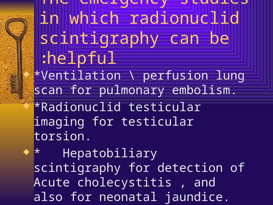
The emergency studies in which radionuclid scintigraphy can be
helpful: ¨ *Ventilation \ perfusion lung scan for
pulmonary embolism. ¨ *Radionuclid testicular imaging for
testicular torsion. ¨ * Hepatobiliary scintigraphy for detection
of Acute cholecystitis , and also for neonatal jaundice.
Cont.
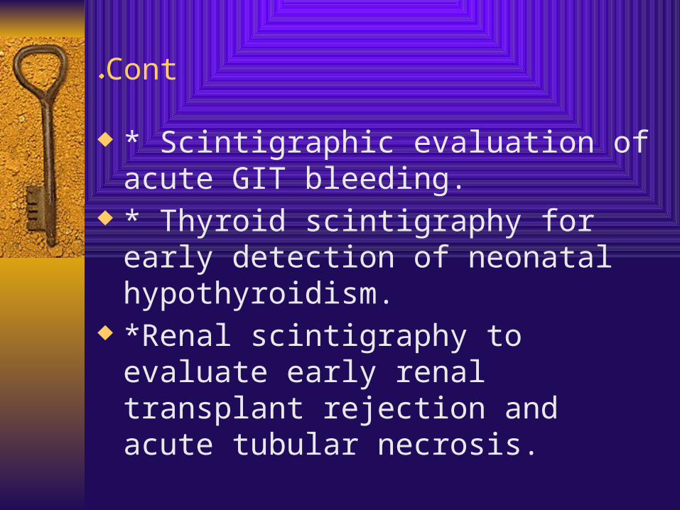
¨ * Scintigraphic evaluation of acute GIT bleeding.
¨ * Thyroid scintigraphy for early detection of neonatal hypothyroidism.
¨ *Renal scintigraphy to evaluate early renal transplant rejection and acute tubular necrosis.
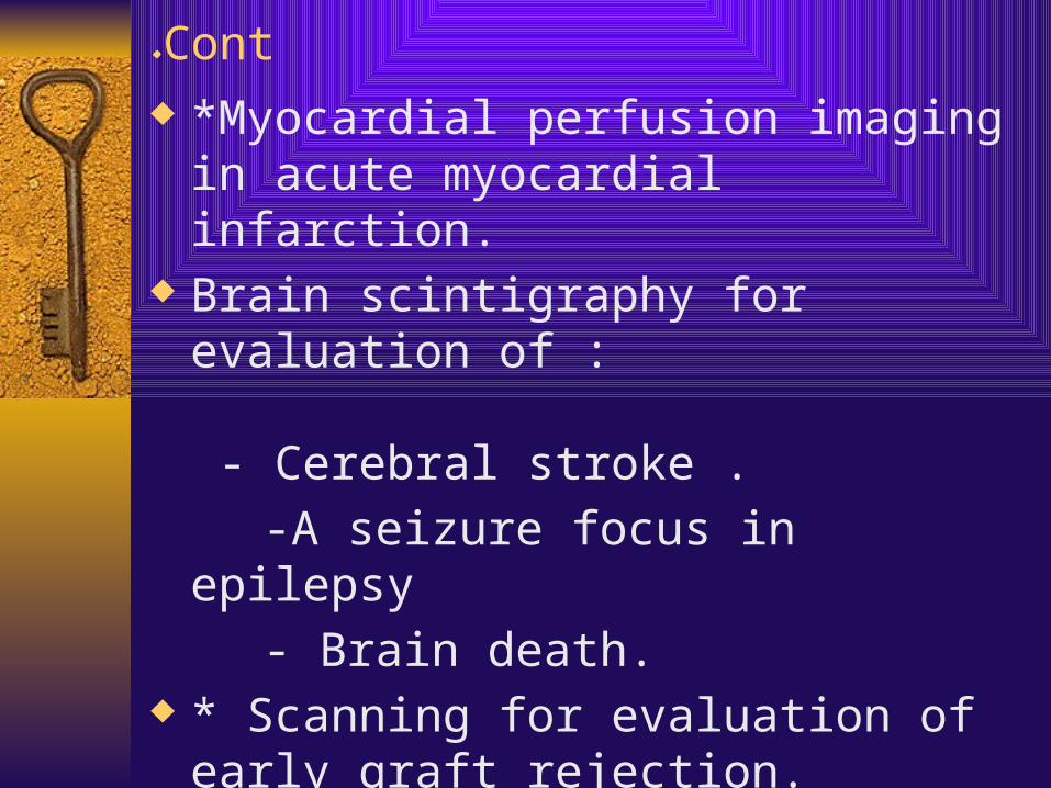
Cont.¨ *Myocardial perfusion imaging in acute
myocardial infarction. ¨ Brain scintigraphy for evaluation of :
- Cerebral stroke . -A seizure focus in epilepsy - Brain death.¨ * Scanning for evaluation of early graft
rejection. ¨ In this presentation I well cover some of
these cases and in next presentation I well talk about the rest.
Ventilation perfusion lung scan for pulmonary embolism:
¨ Idea of the scan:- For successful gas exchange to occur, each broncho-pulmonary segment must has a matched air supply via the bronchial tree and blood supply via the pulmonary artery.
¨ Consequently, if either is deficient in any broncho-pulmonary segment effective gas exchange cannot take place
¨ This is termed mismatch of perfusion ventilation.
¨ In perfusion scan we use Tc-99m MAA after its I.V.injection , it is trapped in the capillary bed of the pulmonary arterial circulation .
¨ In patient with PE the perfusion scan demonstrates an area of decreased radioactivity (cold area) corresponding to the area of decreased blood flow .
¨ To be sure this cold area due to PE we do anther lung scan which is ventilation lung scan using Tc-99m DTPA aerosol .
¨ If ventilation scan is normal in presence of abnormal perfusion scan (mismatched scan) this indicates probability of PE. While if the perfusion scan is normal from the start it exclude PE.
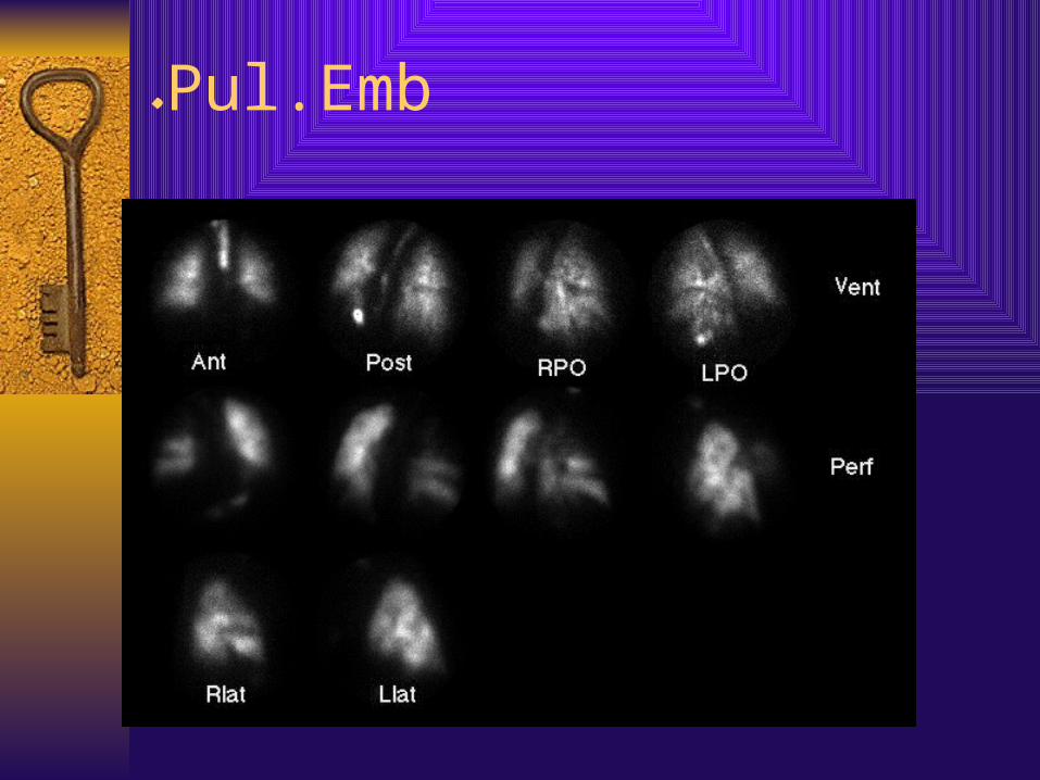
Pul.Emb.
Testicular imaging for testicular torsion:
¨ Spermatic cord torsion is a medical emergency and prompt surgical management is necessary to salvage the involved testis.
¨ Radio nuclide scrotal scintigraphy using Tc-99m pertechnetate is inexpensive diagnostic approach to distinguish testicular torsion from non surgical causes of scrotal pain , such as epididymitis or epididymo-orchitis.
How the study is performed: ¨ -No patient preparation.¨ -The patient is injected I.V. by the Tc-99m
pertechnetate as a bolus injection and radio nuclide angiogram of testicular perfusion is obtained by serial images for 30 min. with lead shielding for lower abdomen, thighs and penis.
¨ -An area of decreased accumulation corresponding to the interrupted blood supply indicates that torsion is possible.
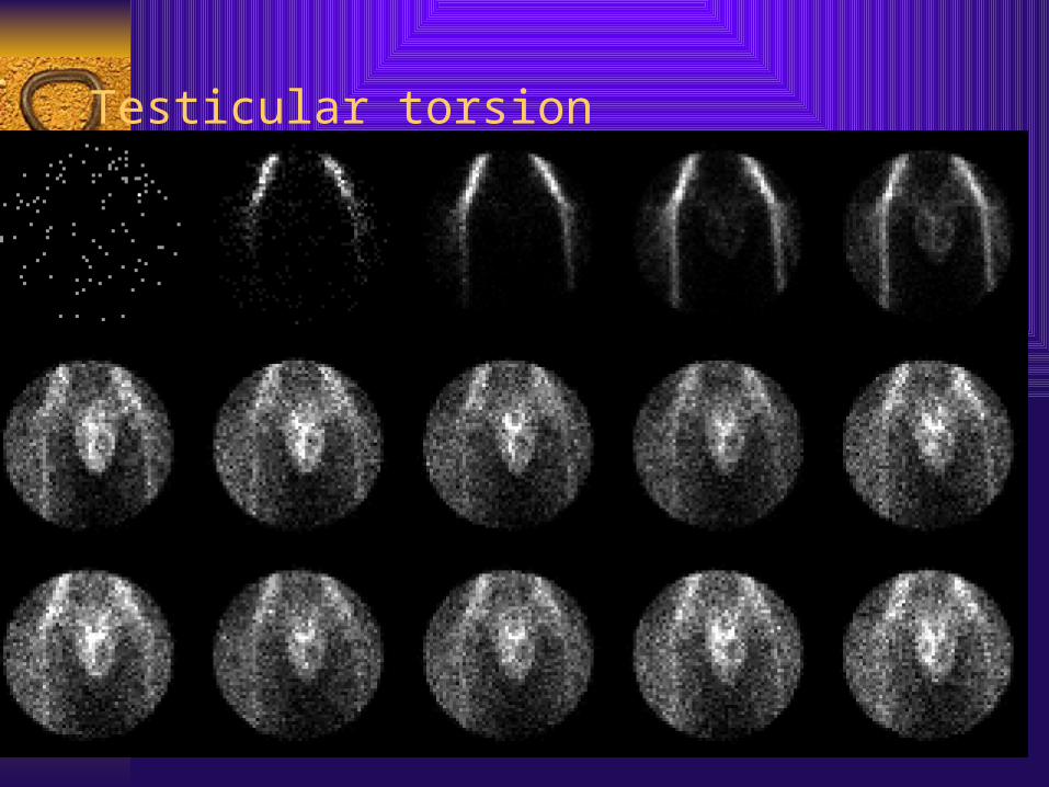
Testicular torsion
Hepatobiliary scan using Tc-99m IDA
¨ Idea of the scan: - Following I.V. injection of the Tc-99m HIDA, rapid clearance from the blood occurs and the radiotracer is handled by the hepatobiliary system in a manner similar to bilirubin. It undergoes rapid polygonal cell uptake , remains unconjugated and is excreted into the biliary ducts . Sufficient activity is noted in the liver and biliary tract that permit imaging after 5 min. of injection.
¨ In normal study the bile duct ,gall bladder and duodenum usually well visualized within 30-40 min. post injection
¨ In pediatrics: Tc-99m HIDA scan is useful in the differential diagnosis of causes of neonatal jaundice which are principally biliary atresia or neonatal hepatitis ,as surgery is required in biliary atresia while laparotomy is unwise in neonatal hepatitis.
¨ In case of biliary atresia there is no radiotracer passage to the intestine.
¨ Also, Tc-99m HIDA scan can confirm the choledcal cyst which is a congenital cystic dilation of the common bile duct and show its connection with biliary tree.
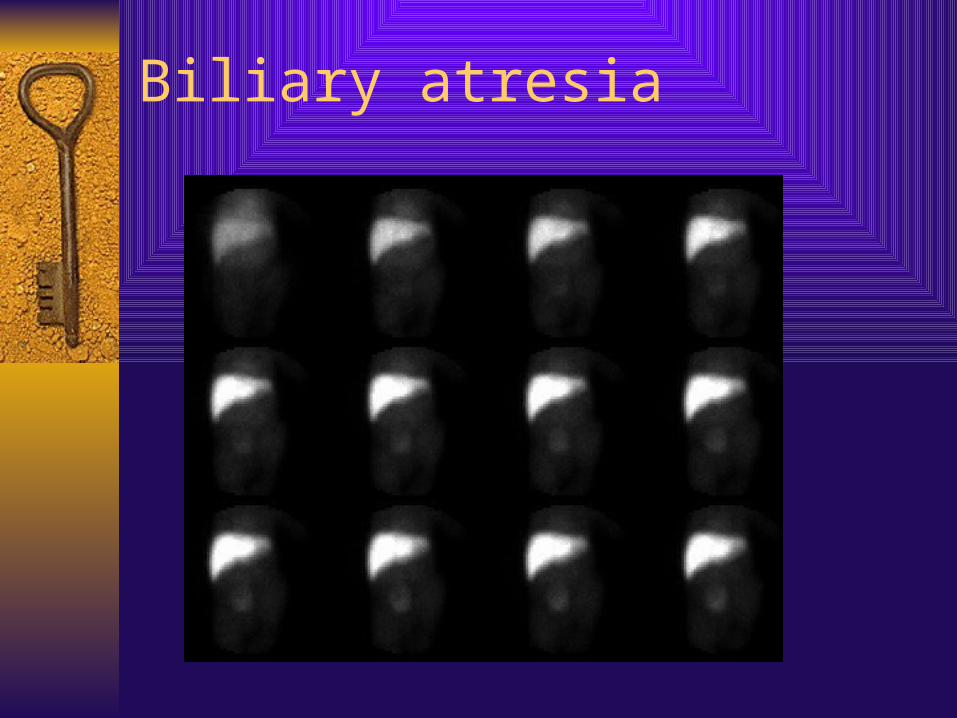
Biliary atresia
¨ * Acute cholecystitis : Since acute cholecystitis is initiated and characterized by cystic duct obstruction , the simple non invasive test that detects cystic duct obstruction is Tc-99m HIDA.
¨ The finding in the scan is non visualization of gall bladder , while the radiotracer reach to the intestine.
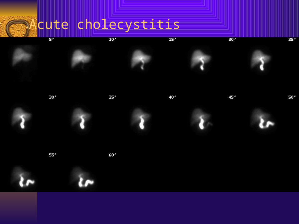
Acute cholecystitis.
GIT bleeding scan.¨ GIT bleeding scan can be used to visualize
suspected bleeding distal to the ligament of Treitz( lower GI tract). The GI bleeding scan can be performed using Tc-99m – RBC or TC-99m sulfur colloid.
¨ Because the radiotracer emits gamma rays that can be detected by a gamma camera, blood extravasated into the GI tract, can be imaged. The series of images can be viewed as a movie (cinemode) increasing the sensitivity of the test
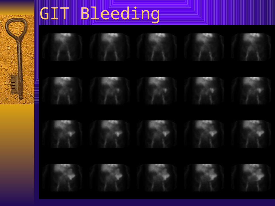
GIT Bleeding
Neonatal Hypothyroidism:
¨ *Rapid diagnosis is essential so that replacement therapy can be initiated before sever damage to central nervous system development occurs.
¨ *Diagnosis depends upon rapid screening of TSH measurements.
¨ *The nuclear medicine studies can rule out athyreosis or absence of the thyroid gland , or demonstrate an ectopic gland which may not provide efficient thyroid hormone , resulting in an elevated level of thyroid-stimulating hormone.
¨ * detection of presence or absence of the thyroid gland is easy with thyroid scan using Tc99m pertechnetate.
THANK YOU