Embed Size (px)
DESCRIPTION
STROKE.
Citation preview
Scott and Eelco F.M. WijdicksPatrick D. Lyden, Lewis B. Morgenstern, Adnan I. Qureshi, Robert H. Rosenwasser, Phillip A.Anthony Furlan, Robert L. Grubb, Randall T. Higashida, Edward C. Jauch, Chelsea Kidwell, Harold P. Adams, Jr, Gregory del Zoppo, Mark J. Alberts, Deepak L. Bhatt, Lawrence Brass,
value of this guideline as an educational tool for neurologistsInterdisciplinary Working Groups: The American Academy of Neurology affirms the
Atherosclerotic Peripheral Vascular Disease and Quality of Care Outcomes in Research Cardiology Council, Cardiovascular Radiology and Intervention Council, and the
the American Heart Association/ American Stroke Association Stroke Council, Clinical Guidelines for the Early Management of Adults With Ischemic Stroke : A Guideline From
Print ISSN: 0039-2499. Online ISSN: 1524-4628 Copyright © 2007 American Heart Association, Inc. All rights reserved.
is published by the American Heart Association, 7272 Greenville Avenue, Dallas, TX 75231Stroke doi: 10.1161/STROKEAHA.107.181486
2007;38:1655-1711; originally published online April 12, 2007;Stroke.
http://stroke.ahajournals.org/content/38/5/1655World Wide Web at:
The online version of this article, along with updated information and services, is located on the
http://stroke.ahajournals.org/content/38/9/e96.full.pdf http://stroke.ahajournals.org/content/38/6/e38.full.pdf
An erratum has been published regarding this article. Please see the attached page for:
http://stroke.ahajournals.org//subscriptions/
is online at: Stroke Information about subscribing to Subscriptions:
http://www.lww.com/reprints Information about reprints can be found online at: Reprints:
document. Permissions and Rights Question and Answer process is available in the
Request Permissions in the middle column of the Web page under Services. Further information about thisOnce the online version of the published article for which permission is being requested is located, click
can be obtained via RightsLink, a service of the Copyright Clearance Center, not the Editorial Office.Strokein Requests for permissions to reproduce figures, tables, or portions of articles originally publishedPermissions:
by guest on November 2, 2012http://stroke.ahajournals.org/Downloaded from

Guidelines for the Early Management of AdultsWith Ischemic Stroke
A Guideline From the American Heart Association/American Stroke Association Stroke Council, Clinical Cardiology
Council, Cardiovascular Radiology and Intervention Council, and theAtherosclerotic Peripheral Vascular Disease and Quality of Care
Outcomes in Research Interdisciplinary Working Groups
The American Academy of Neurology affirms the value of this guidelineas an educational tool for neurologists.
Harold P. Adams, Jr, MD, FAHA, Chair; Gregory del Zoppo, MD, FAHA, Vice Chair;Mark J. Alberts, MD, FAHA; Deepak L. Bhatt, MD;
Lawrence Brass, MD, FAHA†; Anthony Furlan, MD, FAHA; Robert L. Grubb, MD, FAHA;Randall T. Higashida, MD, FAHA; Edward C. Jauch, MD, FAHA; Chelsea Kidwell, MD, FAHA;
Patrick D. Lyden, MD; Lewis B. Morgenstern, MD, FAHA; Adnan I. Qureshi, MD, FAHA;Robert H. Rosenwasser, MD, FAHA; Phillip A. Scott, MD, FAHA; Eelco F.M. Wijdicks, MD, FAHA
Purpose—Our goal is to provide an overview of the current evidence about components of the evaluation and treatment of adultswith acute ischemic stroke. The intended audience is physicians and other emergency healthcare providers who treat patientswithin the first 48 hours after stroke. In addition, information for healthcare policy makers is included.
Methods—Members of the panel were appointed by the American Heart Association Stroke Council’s Scientific StatementOversight Committee and represented different areas of expertise. The panel reviewed the relevant literature with anemphasis on reports published since 2003 and used the American Heart Association Stroke Council’s Levels ofEvidence grading algorithm to rate the evidence and to make recommendations. After approval of the statement by thepanel, it underwent peer review and approval by the American Heart Association Science Advisory and CoordinatingCommittee. It is intended that this guideline be fully updated in 3 years.
Results—Management of patients with acute ischemic stroke remains multifaceted and includes several aspects of care thathave not been tested in clinical trials. This statement includes recommendations for management from the first contactby emergency medical services personnel through initial admission to the hospital. Intravenous administration ofrecombinant tissue plasminogen activator remains the most beneficial proven intervention for emergency treatment ofstroke. Several interventions, including intra-arterial administration of thrombolytic agents and mechanical interven-tions, show promise. Because many of the recommendations are based on limited data, additional research on treatmentof acute ischemic stroke is needed. (Stroke. 2007;38:1655-1711.)
Key Words: AHA Scientific Statements � emergency medical services � stroke � acute cerebral infarction� tissue plasminogen activator
†Deceased.The American Heart Association makes every effort to avoid any actual or potential conflicts of interest that may arise as a result of an outside
relationship or a personal, professional, or business interest of a member of the writing panel. Specifically, all members of the writing group are requiredto complete and submit a Disclosure Questionnaire showing all such relationships that might be perceived as real or potential conflicts of interest.
This guideline was approved by the American Heart Association Science Advisory and Coordinating Committee on January 6, 2007. A single reprintis available by calling 800-242-8721 (US only) or writing the American Heart Association, Public Information, 7272 Greenville Ave, Dallas, TX75231-4596. Ask for reprint No. 71-0398. To purchase additional reprints, call 843-216-2533 or e-mail [email protected].
This guideline has been copublished in Circulation.Expert peer review of AHA Scientific Statements is conducted at the AHA National Center. For more on AHA statements and guidelines development,
visit http://www.americanheart.org/presenter.jhtml?identifier�3023366.Permissions: Multiple copies, modification, alteration, enhancement, and/or distribution of this document are not permitted without the express
permission of the American Heart Association. Instructions for obtaining permission are located at http://www.americanheart.org/presenter.jhtml?identifier�4431. A link to the “Permission Request Form” appears on the right side of the page.
© 2007 American Heart Association, Inc.
Stroke is available at http://www.strokeaha.org DOI: 10.1161/STROKEAHA.107.181486
1655
AHA/ASA Guideline
by guest on November 2, 2012http://stroke.ahajournals.org/Downloaded from
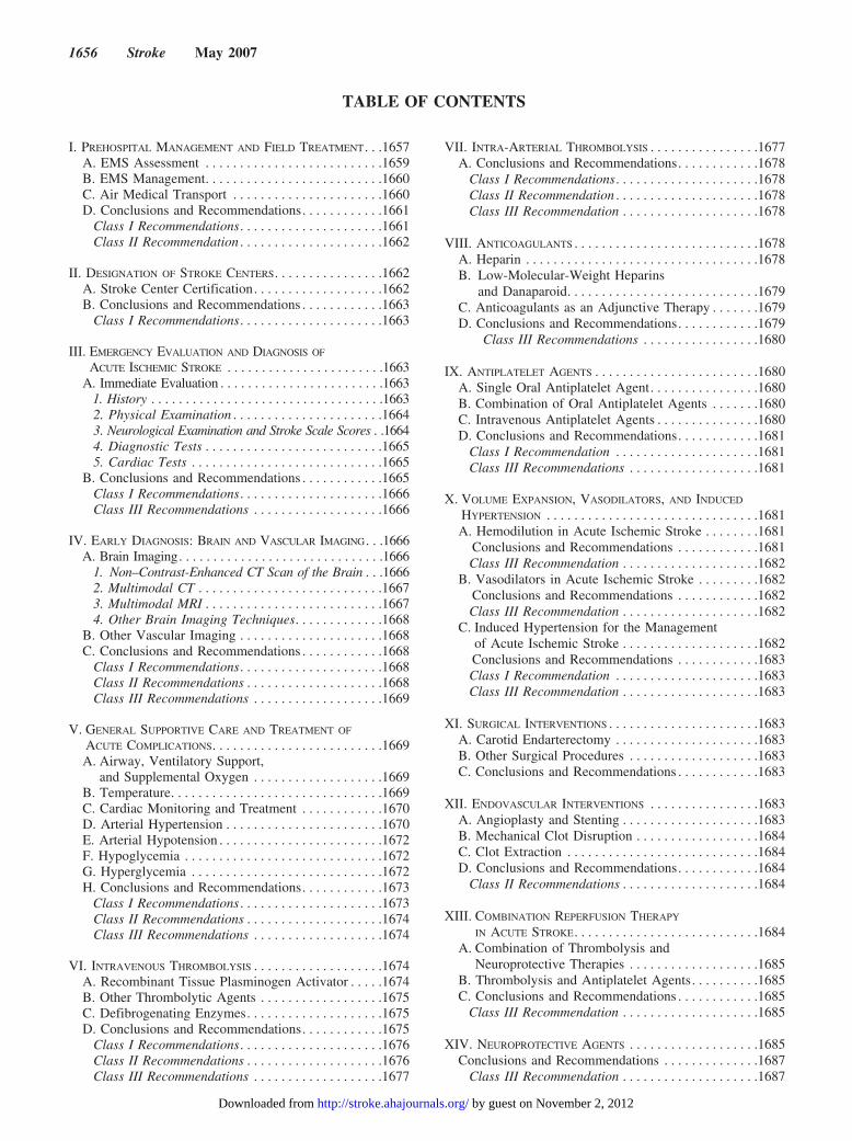
TABLE OF CONTENTS
I. PREHOSPITAL MANAGEMENT AND FIELD TREATMENT. . .1657A. EMS Assessment . . . . . . . . . . . . . . . . . . . . . . . . . .1659B. EMS Management. . . . . . . . . . . . . . . . . . . . . . . . . .1660C. Air Medical Transport . . . . . . . . . . . . . . . . . . . . . .1660D. Conclusions and Recommendations. . . . . . . . . . . .1661
Class I Recommendations. . . . . . . . . . . . . . . . . . . . .1661Class II Recommendation . . . . . . . . . . . . . . . . . . . . .1662
II. DESIGNATION OF STROKE CENTERS. . . . . . . . . . . . . . . .1662A. Stroke Center Certification. . . . . . . . . . . . . . . . . . .1662B. Conclusions and Recommendations . . . . . . . . . . . .1663
Class I Recommendations. . . . . . . . . . . . . . . . . . . . .1663
III. EMERGENCY EVALUATION AND DIAGNOSIS OF
ACUTE ISCHEMIC STROKE . . . . . . . . . . . . . . . . . . . . . . .1663A. Immediate Evaluation . . . . . . . . . . . . . . . . . . . . . . . .1663
1. History . . . . . . . . . . . . . . . . . . . . . . . . . . . . . . . . . .16632. Physical Examination . . . . . . . . . . . . . . . . . . . . . .16643. Neurological Examination and Stroke Scale Scores . .16644. Diagnostic Tests . . . . . . . . . . . . . . . . . . . . . . . . . .16655. Cardiac Tests . . . . . . . . . . . . . . . . . . . . . . . . . . . .1665
B. Conclusions and Recommendations . . . . . . . . . . . .1665Class I Recommendations. . . . . . . . . . . . . . . . . . . . .1666Class III Recommendations . . . . . . . . . . . . . . . . . . .1666
IV. EARLY DIAGNOSIS: BRAIN AND VASCULAR IMAGING. . .1666A. Brain Imaging. . . . . . . . . . . . . . . . . . . . . . . . . . . . . .1666
1. Non–Contrast-Enhanced CT Scan of the Brain . . .16662. Multimodal CT . . . . . . . . . . . . . . . . . . . . . . . . . . .16673. Multimodal MRI . . . . . . . . . . . . . . . . . . . . . . . . . .16674. Other Brain Imaging Techniques. . . . . . . . . . . . .1668
B. Other Vascular Imaging . . . . . . . . . . . . . . . . . . . . .1668C. Conclusions and Recommendations . . . . . . . . . . . .1668
Class I Recommendations. . . . . . . . . . . . . . . . . . . . .1668Class II Recommendations . . . . . . . . . . . . . . . . . . . .1668Class III Recommendations . . . . . . . . . . . . . . . . . . .1669
V. GENERAL SUPPORTIVE CARE AND TREATMENT OF
ACUTE COMPLICATIONS. . . . . . . . . . . . . . . . . . . . . . . . .1669A. Airway, Ventilatory Support,
and Supplemental Oxygen . . . . . . . . . . . . . . . . . . .1669B. Temperature. . . . . . . . . . . . . . . . . . . . . . . . . . . . . . .1669C. Cardiac Monitoring and Treatment . . . . . . . . . . . .1670D. Arterial Hypertension . . . . . . . . . . . . . . . . . . . . . . .1670E. Arterial Hypotension . . . . . . . . . . . . . . . . . . . . . . . .1672F. Hypoglycemia . . . . . . . . . . . . . . . . . . . . . . . . . . . . .1672G. Hyperglycemia . . . . . . . . . . . . . . . . . . . . . . . . . . . .1672H. Conclusions and Recommendations. . . . . . . . . . . .1673
Class I Recommendations . . . . . . . . . . . . . . . . . . . . .1673Class II Recommendations . . . . . . . . . . . . . . . . . . . .1674Class III Recommendations . . . . . . . . . . . . . . . . . . .1674
VI. INTRAVENOUS THROMBOLYSIS . . . . . . . . . . . . . . . . . . .1674A. Recombinant Tissue Plasminogen Activator . . . . .1674B. Other Thrombolytic Agents . . . . . . . . . . . . . . . . . .1675C. Defibrogenating Enzymes. . . . . . . . . . . . . . . . . . . .1675D. Conclusions and Recommendations. . . . . . . . . . . .1675
Class I Recommendations. . . . . . . . . . . . . . . . . . . . .1676Class II Recommendations . . . . . . . . . . . . . . . . . . . .1676Class III Recommendations . . . . . . . . . . . . . . . . . . .1677
VII. INTRA-ARTERIAL THROMBOLYSIS . . . . . . . . . . . . . . . .1677A. Conclusions and Recommendations. . . . . . . . . . . .1678
Class I Recommendations. . . . . . . . . . . . . . . . . . . . .1678Class II Recommendation . . . . . . . . . . . . . . . . . . . . .1678Class III Recommendation . . . . . . . . . . . . . . . . . . . .1678
VIII. ANTICOAGULANTS . . . . . . . . . . . . . . . . . . . . . . . . . . .1678A. Heparin . . . . . . . . . . . . . . . . . . . . . . . . . . . . . . . . . .1678B. Low-Molecular-Weight Heparins
and Danaparoid. . . . . . . . . . . . . . . . . . . . . . . . . . . .1679C. Anticoagulants as an Adjunctive Therapy . . . . . . .1679D. Conclusions and Recommendations. . . . . . . . . . . .1679
Class III Recommendations . . . . . . . . . . . . . . . . .1680
IX. ANTIPLATELET AGENTS . . . . . . . . . . . . . . . . . . . . . . . .1680A. Single Oral Antiplatelet Agent. . . . . . . . . . . . . . . .1680B. Combination of Oral Antiplatelet Agents . . . . . . .1680C. Intravenous Antiplatelet Agents . . . . . . . . . . . . . . .1680D. Conclusions and Recommendations. . . . . . . . . . . .1681
Class I Recommendation . . . . . . . . . . . . . . . . . . . . .1681Class III Recommendations . . . . . . . . . . . . . . . . . . .1681
X. VOLUME EXPANSION, VASODILATORS, AND INDUCED
HYPERTENSION . . . . . . . . . . . . . . . . . . . . . . . . . . . . . . .1681A. Hemodilution in Acute Ischemic Stroke . . . . . . . .1681
Conclusions and Recommendations . . . . . . . . . . . .1681Class III Recommendation . . . . . . . . . . . . . . . . . . . .1682
B. Vasodilators in Acute Ischemic Stroke . . . . . . . . .1682Conclusions and Recommendations . . . . . . . . . . . .1682Class III Recommendation . . . . . . . . . . . . . . . . . . . .1682
C. Induced Hypertension for the Managementof Acute Ischemic Stroke . . . . . . . . . . . . . . . . . . . .1682Conclusions and Recommendations . . . . . . . . . . . .1683Class I Recommendation . . . . . . . . . . . . . . . . . . . . .1683Class III Recommendation . . . . . . . . . . . . . . . . . . . .1683
XI. SURGICAL INTERVENTIONS . . . . . . . . . . . . . . . . . . . . . .1683A. Carotid Endarterectomy . . . . . . . . . . . . . . . . . . . . .1683B. Other Surgical Procedures . . . . . . . . . . . . . . . . . . .1683C. Conclusions and Recommendations . . . . . . . . . . . .1683
XII. ENDOVASCULAR INTERVENTIONS . . . . . . . . . . . . . . . .1683A. Angioplasty and Stenting . . . . . . . . . . . . . . . . . . . .1683B. Mechanical Clot Disruption . . . . . . . . . . . . . . . . . .1684C. Clot Extraction . . . . . . . . . . . . . . . . . . . . . . . . . . . .1684D. Conclusions and Recommendations. . . . . . . . . . . .1684
Class II Recommendations . . . . . . . . . . . . . . . . . . . .1684
XIII. COMBINATION REPERFUSION THERAPY
IN ACUTE STROKE. . . . . . . . . . . . . . . . . . . . . . . . . . .1684A. Combination of Thrombolysis and
Neuroprotective Therapies . . . . . . . . . . . . . . . . . . .1685B. Thrombolysis and Antiplatelet Agents. . . . . . . . . .1685C. Conclusions and Recommendations . . . . . . . . . . . .1685
Class III Recommendation . . . . . . . . . . . . . . . . . . . .1685
XIV. NEUROPROTECTIVE AGENTS . . . . . . . . . . . . . . . . . . .1685Conclusions and Recommendations . . . . . . . . . . . . . .1687
Class III Recommendation . . . . . . . . . . . . . . . . . . . .1687
1656 Stroke May 2007
by guest on November 2, 2012http://stroke.ahajournals.org/Downloaded from
XV. ADMISSION TO THE HOSPITAL AND GENERAL
ACUTE TREATMENT (AFTER HOSPITALIZATION) . . . . .1687A. Admission to the Hospital . . . . . . . . . . . . . . . . . . .1687B. Specialized Stroke Care Units . . . . . . . . . . . . . . . .1687
1. General Care . . . . . . . . . . . . . . . . . . . . . . . . . . . .16882. Nutrition and Hydration . . . . . . . . . . . . . . . . . . . .16883. Infections . . . . . . . . . . . . . . . . . . . . . . . . . . . . . . . .1688
C. Deep Vein Thrombosis and PulmonaryEmbolism . . . . . . . . . . . . . . . . . . . . . . . . . . . . . . . .1689
1. Other Care . . . . . . . . . . . . . . . . . . . . . . . . . . . . . .1689D. Conclusions and Recommendations . . . . . . . . . . .1689
Class I Recommendations. . . . . . . . . . . . . . . . . . . . .1689Class II Recommendations . . . . . . . . . . . . . . . . . . . .1690Class III Recommendations . . . . . . . . . . . . . . . . . . .1690
XVI. TREATMENT OF ACUTE NEUROLOGICAL
COMPLICATIONS . . . . . . . . . . . . . . . . . . . . . . . . . . . . . . .1690A. Ischemic Brain Swelling . . . . . . . . . . . . . . . . . . . .1690B. Hemorrhagic Transformation . . . . . . . . . . . . . . . . .1691C. Seizures. . . . . . . . . . . . . . . . . . . . . . . . . . . . . . . . . .1691D. Conclusions and Recommendations . . . . . . . . . . .1691
Class I Recommendations. . . . . . . . . . . . . . . . . . . . .1691Class II Recommendations . . . . . . . . . . . . . . . . . . . .1692Class III Recommendations . . . . . . . . . . . . . . . . . . .1692
E. Palliative Care. . . . . . . . . . . . . . . . . . . . . . . . . . . . .1692DISCLOSURES . . . . . . . . . . . . . . . . . . . . . . . . . . . . . . . . . . .1693REFERENCES. . . . . . . . . . . . . . . . . . . . . . . . . . . . . . . . . . . .1694
The present document is a comprehensive guideline state-ment on the management of patients with acute ischemic
stroke that supercedes the prior statement and interim up-dates.1–3 These guidelines have been developed by a panel ofphysicians with a broad range of expertise, including vascularneurology, neurocritical care, emergency medicine, neurosur-gery, and interventional neuroradiology/endovascular neuro-surgery. The intended audience for these guidelines includesphysicians, emergency medical services (EMS) personnel,and other medical personnel who deal with the emergencydiagnosis and treatment of patients with suspected ischemicstroke. In addition, components of these guidelines are veryrelevant to health policy decision makers and administrators.The goal of these guidelines is to provide updated recom-mendations that may be used by physicians who provideacute stroke care within the first hours to time of initialdiagnosis, treatment, and initial hospitalization. In addition,the guideline also includes information that should be usefulfor nonphysician EMS personnel and for hospitals. Theemphasis of these guidelines is the diagnosis and emergencytreatment of patients with acute ischemic stroke. Informationabout the management of acute and subacute neurologicaland medical complications is also included. The panel recog-nizes that measures to prevent early recurrent stroke are alsoa component of acute management. In general, the medical orsurgical interventions administered to prevent recurrentstroke are similar to those prescribed to patients with recenttransient ischemic attacks or to other high-risk persons. Thereader is referred to another recent statement that addressesthe management of risk factors, the prescription of antithrom-botic medications, and the use of surgical or endovascularinterventions to prevent recurrent stroke.4
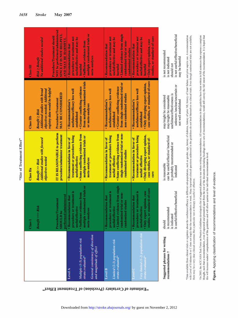
In writing these guidelines, the panel applied the rules ofevidence and the formulation of strength of recommendationsused by other panels of the American Heart Association(AHA)4 (see the Figure and Table 1). The data were collectedthrough a systematic review of the literature. Because of thewide scope of the guidelines, the members of the panel wereassigned primary reviews for individual sections. Then thepanel assessed the complete guidelines. If the panel con-cluded that data supported or did not support the use of aspecific intervention, appropriate recommendations weremade. In some cases in which definitive data were notavailable, no specific recommendation was made. Italicsindicate recommendations that have been changed or addedsince the publication of the previous guideline. In otherinstances, supporting evidence based on clinical trial researchwas not available for a specific intervention, but the panel hasmade a specific recommendation on the basis of pathophys-iological reasoning and expert practice experience. For manyof these interventions, it is unlikely that randomized trials willever be performed. An example is the recommendation toperform endotracheal intubation to protect the airway in acomatose patient.
I. Prehospital Management andField Treatment
Recent data indicate that 29% to 65% of patients with signsor symptoms of acute stroke access their initial medical carevia local EMS (Table 2), which confirms the role of EMS inthe chain of survival.5–13 Notably, an estimated 19% to 60%of stroke patients present within 3 hours of stroke and 14% to32% of those arrive within 2 hours of symptom onset.Although just over half of all stroke patients use EMS accessto health care, those who do utilize EMS comprise themajority of patients presenting within the 3-hourwindow.13–16
EMS activation appears to be a function primarily ofindividuals other than the patient, with one report indicatingthat a family member, paid caregiver, coworker, or otherbystander accounted for 62% to 95% of 9-1-1 activationcalls.6,9 In addition to bystander recognition of a problem,other reported predictors of EMS use by stroke patientsinclude stroke severity,17 presence of intracranial hemor-rhage,9,18 age,9,18 sense of urgency,9 unemployment,6 and race(black).18
The benefits of EMS activation by patients with strokesymptoms appear to occur in both the prehospital andin-hospital settings. Hospital arrival is faster for patients whouse EMS/9-1-1 as their initial medical contact than for thosewho contact their primary physician or hospital directly18 ora primary care site.19 Not surprisingly, EMS use is stronglyassociated with shorter time periods from symptom onset tohospital arrival, although this may reflect a greater sense ofurgency on the patient’s or bystander’s part rather thanreduced transport time.8,9,12 Similarly, EMS use is stronglyassociated with decreased time to initial physician examina-tion,9,10,13,20 initial computed tomography (CT) imaging,9,10,12
and neurological evaluation.9
Adams et al Early Management of Adults With Ischemic Stroke 1657
by guest on November 2, 2012http://stroke.ahajournals.org/Downloaded from
Fig
ure.
Ap
ply
ing
clas
sific
atio
nof
reco
mm
end
atio
nsan
dle
velo
fev
iden
ce.
1658 Stroke May 2007
by guest on November 2, 2012http://stroke.ahajournals.org/Downloaded from
On the basis of the aforementioned information, commu-nities should encourage 9-1-1 activation and use for patientswith symptoms of acute stroke.
Data from the TLL Temple Foundation Stroke Projectcontrolled trial indicate that educational interventions onstroke identification and management targeting patients,EMS, hospitals, and community physicians increasedthrombolytic use in patients with ischemic stroke from 2.21%to 8.65% as compared with communities that did not havesuch programs, which saw only a 0.06% increase. Forpatients with ischemic stroke who were eligible forthrombolytic therapy, rates of tissue-type plasminogen acti-vator (tPA) use increased from 14% to 52% in interventioncommunities. The benefit from this aggressive interventionprogram was sustained at 6 months after intervention.21,22
A. EMS AssessmentEMS assessment begins with the initial 9-1-1 contact (Table2). The role of the dispatch system is to ensure immediatetriage and dispatch of appropriate EMS providers when acutestroke is suspected by either the caller or the dispatcher.23
Data from 2 systems indicate that dispatchers correctlysuspected or identified 52% of patients ultimately proven tohave had a stroke on initial telephone evaluation.7,24 Thesedata imply that educational programs should be aimed atdispatchers to increase their awareness of stroke symptoms.
Stroke should be given a priority dispatch similar to that foracute myocardial infarction or trauma.25
After ambulance arrival on the scene, EMS providersshould obtain a focused history and patient assessment,provide necessary stabilization and treatment, and transportimmediately to the closest, most appropriate facility (Table3). The word appropriate is key because it means that anambulance may bypass a hospital that does not have theresources or institutional commitment to treat patients withstroke if a more appropriate hospital is available within areasonable transport interval. Advance notice to the receivingemergency department (ED) of the impending arrival of apotential stroke patient, along with information on comorbidconditions and estimated time of symptom onset, will speedthe subsequent ED assessment.
Critical elements of the patient’s history must includeinformation on time of symptom onset (Table 4). This mayrequire obtaining information from bystanders or, preferably,transporting witnesses with the patient. Similarly, next of kin,if available, may be needed for information or consent andshould travel to the receiving hospital concurrently. Tele-phone numbers, including cellular telephone numbers, ofwitnesses or relatives may help the ED to clarify the historyor seek consent for treatment. A list of the patient’s medica-tions, or the medication containers themselves, should besought, with particular attention paid to identifying anticoag-ulant (both oral and injectable), antiplatelet, and antihyper-tensive drug use.
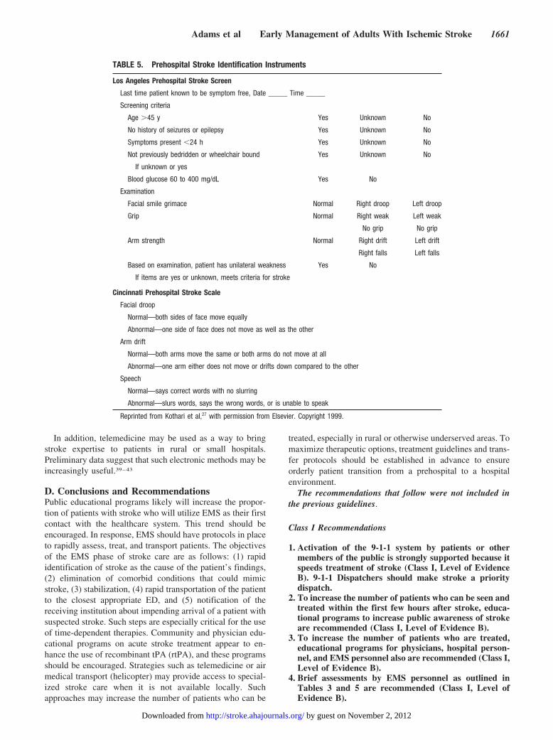
After the patient’s airway, breathing, and circulation(ABCs) are assessed and stabilized, common presenting signsof stroke should be sought and a focused examinationcompleted. Prehospital stroke assessment tools have provedeffective in identifying stroke patients in the field. The LosAngeles Prehospital Stroke Screen uses patient history, phys-ical findings, and finger stick glucose determination toidentify stroke patients.26 The Cincinnati Prehospital Stroke
TABLE 2. Stroke Chain of Survival
Detection Recognition of stroke signs and symptoms
Dispatch Call 9-1-1 and priority EMS dispatch
Delivery Prompt transport and prehospital notification to hospital
Door Immediate ED triage
Data ED evaluation, prompt laboratory studies, and CT imaging
Decision Diagnosis and decision about appropriate therapy
Drug Administration of appropriate drugs or other interventions
TABLE 1. Definition of Classes and Levels of Evidence Used in AHA Recommendations
Classification
Class I Conditions for which there is evidence for and/or general agreement that the procedure or treatment is useful and effective
Class II Conditions for which there is conflicting evidence and/or a divergence of opinion about the usefulness/efficacy of a procedureor treatment
Class IIa The weight of evidence or opinion is in favor of the procedure or treatment.
Class IIb Usefulness/efficacy is less well established by evidence or opinion.
Class III Conditions for which there is evidence and/or general agreement that the procedure or treatment is not useful/effective and insome cases may be harmful
Level of evidence
A Data derived from multiple randomized clinical trials
B Data derived from a single randomized trial or nonrandomized studies
C Consensus opinion of experts
Level of evidence fordiagnostic recommendation
A Data derived from multiple prospective cohort studies that used a reference standard applied by a masked evaluator
B Data derived from a single grade A study or one or more case–control studies or studies that used a reference standardapplied by an unmasked evaluator
C Consensus opinion of experts
Adams et al Early Management of Adults With Ischemic Stroke 1659
by guest on November 2, 2012http://stroke.ahajournals.org/Downloaded from
Scale is an alternative instrument with fewer data elements(Table 5), requiring only 30 to 60 seconds to complete.27
Other prehospital stroke evaluation tools exist, although dataon their validity are limited.
B. EMS ManagementGuidelines for EMS management are presented in Table3.25,28 After initial stabilization, it is recommended thatpatient transport commence as soon as possible, with cardiacmonitoring and intravenous access established during trans-port, if possible. Isotonic crystalloids (most commonly nor-mal saline solution) are recommended for resuscitation, ifneeded. Dextrose-containing fluids should be avoided unlesshypoglycemia is present or strongly suspected because ex-cessive glucose may be injurious to stroke patients. Norecommendations can be offered on the prehospital manage-ment of hypertension in patients with suspected stroke, andintervention is best accomplished after hospital arrival.
It is well recognized that hypoglycemic patients may havesymptoms that mimic an acute stroke, manifesting focalsymptoms, altered speech, and/or cognitive changes, andtherefore EMS assessment of blood glucose has been aroutine practice for many years. A single report suggests thata more selective approach may be possible, with bloodglucose measurement advocated only in the presence of ahistory suspicious for hypoglycemia or inability to obtain
adequate patient information.29 That study is limited, how-ever, by its retrospective methodology and an upper confi-dence interval of 2.4% for the likelihood of failing to identifya hypoglycemic patient. At present, checking blood glucoseconcentrations in most patients with stroke is a prudent step,even among patients without a history of diabetes mellitus oruse of insulin.
The availability of resources to care for patients with acutestroke varies widely both among and within communities.The National Institutes of Health (NIH) Task Force report,“Improving the Chain of Recovery for Acute Stroke in YourCommunity,” recommends identifying hospitals capable ofproviding acute stroke care and creating a transport system tothese centers based on patient location. Such systems requireadvanced planning and frequent updating and should incor-porate EMS representatives, community leaders, hospitals,and physicians to ensure clear guidance for EMS providerswith regard to patient destination.
Identification of an effective neuroprotective therapy mayfurther expand the role of EMS in the treatment of acutestroke. The feasibility of initiation of hypothermia has alsobeen demonstrated in the prehospital setting.30 Of importancefor future research is the fact that it appears possible toincorporate EMS into the research process, with EMS per-sonnel having demonstrated success in facilitating physiciancell phone elicitation of consent from patients and in deliv-ering experimental stroke therapy.31,32
C. Air Medical TransportAir medical (helicopter) transport for patients with acutestroke appears beneficial, although the data are limited.Helicopters may extend the range of thrombolytic therapy torural areas.33 They could deliver teams to administer tPA andsubsequently transfer treated patients,34 expand enrollmentfor acute stroke studies,35 and facilitate early definitivediagnosis and operative intervention in nontraumatic intracra-nial hemorrhage.36 It is important to note that helicoptertransfer of stroke patients for potential thrombolysis is cost-effective for a wide range of system variables.37
Protocols for the use of air medical transfer fromfacilities unable to provide acute stroke care should bedeveloped in advance. Air medical transfer should beconsidered for patients who cannot receive treatmentlocally and who could reach a treating facility within theavailable time window.33,38
TABLE 3. Guidelines for EMS Management of Patients With Suspected Stroke
Recommended Not Recommended
Manage ABCs Dextrose-containing fluids in nonhypoglycemic patients
Cardiac monitoring Hypotension/excessive blood pressure reduction
Intravenous access Excessive intravenous fluids
Oxygen (as required O2 saturation �92%)
Assess for hypoglycemia
Nil per os (NPO)
Alert receiving ED
Rapid transport to closest appropriatefacility capable of treating acute stroke
TABLE 4. Key Components of History
Onset of symptoms
Recent events
Stroke
Myocardial infarction
Trauma
Surgery
Bleeding
Comorbid diseases
Hypertension
Diabetes mellitus
Use of medications
Anticoagulants
Insulin
Antihypertensives
1660 Stroke May 2007
by guest on November 2, 2012http://stroke.ahajournals.org/Downloaded from
In addition, telemedicine may be used as a way to bringstroke expertise to patients in rural or small hospitals.Preliminary data suggest that such electronic methods may beincreasingly useful.39–43
D. Conclusions and RecommendationsPublic educational programs likely will increase the propor-tion of patients with stroke who will utilize EMS as their firstcontact with the healthcare system. This trend should beencouraged. In response, EMS should have protocols in placeto rapidly assess, treat, and transport patients. The objectivesof the EMS phase of stroke care are as follows: (1) rapididentification of stroke as the cause of the patient’s findings,(2) elimination of comorbid conditions that could mimicstroke, (3) stabilization, (4) rapid transportation of the patientto the closest appropriate ED, and (5) notification of thereceiving institution about impending arrival of a patient withsuspected stroke. Such steps are especially critical for the useof time-dependent therapies. Community and physician edu-cational programs on acute stroke treatment appear to en-hance the use of recombinant tPA (rtPA), and these programsshould be encouraged. Strategies such as telemedicine or airmedical transport (helicopter) may provide access to special-ized stroke care when it is not available locally. Suchapproaches may increase the number of patients who can be
treated, especially in rural or otherwise underserved areas. Tomaximize therapeutic options, treatment guidelines and trans-fer protocols should be established in advance to ensureorderly patient transition from a prehospital to a hospitalenvironment.
The recommendations that follow were not included inthe previous guidelines.
Class I Recommendations
1. Activation of the 9-1-1 system by patients or othermembers of the public is strongly supported because itspeeds treatment of stroke (Class I, Level of EvidenceB). 9-1-1 Dispatchers should make stroke a prioritydispatch.
2. To increase the number of patients who can be seen andtreated within the first few hours after stroke, educa-tional programs to increase public awareness of strokeare recommended (Class I, Level of Evidence B).
3. To increase the number of patients who are treated,educational programs for physicians, hospital person-nel, and EMS personnel also are recommended (Class I,Level of Evidence B).
4. Brief assessments by EMS personnel as outlined inTables 3 and 5 are recommended (Class I, Level ofEvidence B).
TABLE 5. Prehospital Stroke Identification Instruments
Los Angeles Prehospital Stroke Screen
Last time patient known to be symptom free, Date _____ Time _____
Screening criteria
Age �45 y Yes Unknown No
No history of seizures or epilepsy Yes Unknown No
Symptoms present �24 h Yes Unknown No
Not previously bedridden or wheelchair bound Yes Unknown No
If unknown or yes
Blood glucose 60 to 400 mg/dL Yes No
Examination
Facial smile grimace Normal Right droop Left droop
Grip Normal Right weak Left weak
No grip No grip
Arm strength Normal Right drift Left drift
Right falls Left falls
Based on examination, patient has unilateral weakness Yes No
If items are yes or unknown, meets criteria for stroke
Cincinnati Prehospital Stroke Scale
Facial droop
Normal—both sides of face move equally
Abnormal—one side of face does not move as well as the other
Arm drift
Normal—both arms move the same or both arms do not move at all
Abnormal—one arm either does not move or drifts down compared to the other
Speech
Normal—says correct words with no slurring
Abnormal—slurs words, says the wrong words, or is unable to speak
Reprinted from Kothari et al,27 with permission from Elsevier. Copyright 1999.
Adams et al Early Management of Adults With Ischemic Stroke 1661
by guest on November 2, 2012http://stroke.ahajournals.org/Downloaded from
5. The use of a stroke identification algorithm such as theLos Angeles or Cincinnati screens is encouraged (ClassI, Level of Evidence B).
6. The panel recommends that EMS personnel begin theinitial management of stroke in the field, as outlined inTable 3 (Class I, Level of Evidence B). The developmentof stroke protocols to be used by EMS personnel isstrongly encouraged.
7. Patients should be transported rapidly for evaluationand treatment to the closest institution that providesemergency stroke care as described in the statement(Class I, Level of Evidence B). In some instances, thismay involve air evacuation. EMS personnel shouldnotify the receiving ED so that the appropriate re-sources may be mobilized.
Class II Recommendation
1. Telemedicine can be an effective method to provideexpert stroke care to patients located in rural areas(Class IIa, Level of Evidence B). Additional researchand experience on the usefulness of telemedicine areencouraged.
II. Designation of Stroke CentersIn an attempt to improve the organization and delivery of careto stroke patients, the Brain Attack Coalition published 2 setsof recommendations, one for primary stroke centers (PSCs)and, more recently, one for comprehensive stroke centers(CSCs).44,45 A PSC has the personnel, programs, expertise,and infrastructure to care for many patients with uncompli-cated strokes, uses many acute therapies (such as intravenousrtPA), and admits such patients into a stroke unit. The CSC isdesigned to care for patients with complicated types ofstrokes, patients with intracerebral hemorrhage or subarach-noid hemorrhage, and those requiring specific interventions(eg, surgery or endovascular procedures) or an intensive careunit type of setting.
The specific elements of a PSC and a CSC will not bereviewed in the present document because they are wellcovered in the articles cited above. Many of the elements ina PSC or a CSC, including stroke units, written care proto-cols, availability of physicians with neurological expertise,and neurosurgical volumes, are associated with improvedoutcomes among patients treated for stroke.44,45 Since thepublication of the PSC article in 2000,44 numerous publishedstudies have demonstrated the utility and effectiveness ofsuch centers.22,46–51 One study found that a PSC increased theuse of intravenous rtPA from 1.5% to 10.2% in 2 years.46
Another study found that 7 of the 11 elements of a PSC wereassociated with increased use of intravenous tPA.47 Addi-tional areas of disease performance that may be added includeperformance of a lipid profile, dysphagia screening, and thepresence of a rehabilitation plan.
The utility of a CSC is beginning to emerge. Studies showthat a CSC increases the use of lytic agents and that a CSCmay improve overall care and outcomes.52,53 In-hospital deathrates were reduced by almost 50% in hospitals with a vascularneurologist and were reduced by 24% in those with a stroketeam.54 Such centers have acted as a regional resource forstroke care with good results and will be pivotal for further
advancements in acute stroke care, stroke prevention, andrehabilitation.38,55,56
A. Stroke Center CertificationThe certification or designation of some hospitals as PSCs orCSCs is progressing rapidly. The American Stroke Associa-tion convened an expert panel to study this issue for PSCs,with the conclusion that a variety of certification processesmight be developed and lead to improved care and out-comes.57 Another panel is currently meeting to evaluatevarious options for CSC certification. One study showed thatself-certification was likely to lead to a significant overesti-mation of a hospital’s compliance with published recommen-dations for a PSC.58 Thus, these data and anecdotal experi-ence suggest that outside independent evaluations of hospitalsas stroke centers should lead to more accurate assessment ofa facility’s true capabilities.
The Joint Commission on the Accreditation of HealthcareOrganizations (JCAHO) began a formal process for thecertification of PSCs in February 2004 (Table 6). As ofFebruary 2006, �200 hospitals in the United States had beencertified as PSCs by the JCAHO. The JCAHO certificationprocess includes a detailed evaluation of a hospital’s staffing,education, disease management programs, outcomes, andinfrastructure (see www.JCAHO.org for details). Severalstates have developed or are exploring a state-based certifi-cation process for PSCs, primarily using the state healthdepartment or a related government agency as the certifier. Atthis time the American Stroke Association and JCAHO havetaken preliminary steps that may lead to a formal certificationprocess for CSCs.
The preferential routing of acute stroke patients to a PSChas been demonstrated to increase the proportion of patientscared for at stroke-capable centers and to increase theproportion of patients treated with thrombolytic therapy to�10%.48 Direct routing of stroke patients whose symptomsstarted �3 hours ago to a PSC or a CSC has been imple-mented or is in the process of implementation in 7 states,covering �25% of the US population. The states of Florida,New Jersey, Maryland, Massachusetts, New Mexico, NewYork, and Texas have laws or policies mandating that acutestroke patients be taken to the nearest stroke center. In otherstates, the limited number of such centers makes preferentialrouting logistically infeasible. Stroke centers in rural areas
TABLE 6. Standardized Measures for Stroke: JCAHO PrimaryStroke Centers
tPA considered
Screen for dysphagia
Deep vein thrombosis prophylaxis
Lipid profile during hospitalization
Smoking cessation
Education about stroke
Plan for rehabilitation considered
Antithrombotic medications started within 48 hours
Antithrombotic medications prescribed at discharge
Anticoagulants prescribed to patients with atrial fibrillation
1662 Stroke May 2007
by guest on November 2, 2012http://stroke.ahajournals.org/Downloaded from
often use helicopter transportation or telemedicine technolo-gies to provide rapid transportation and expertise to expeditetreatment at outlying hospitals.33,53 However, this is clearly anarea that will evolve as the number of stroke centers in-creases, their geographic distribution expands, and the con-cept is embraced by the medical community.
Stroke centers should not be viewed in isolation. Rather,they should be part of a larger support network sometimesreferred to as a stroke system of care. Such a system wouldencompass issues such as prevention, education, acute care,rehabilitation, and quality improvement.59 In addition, as thenumber of stroke centers increases, such facilities may forma network of hospitals that would be useful for testing newtherapies for acute stroke.
B. Conclusions and RecommendationsRobust data demonstrate the efficacy of specialized strokeservices in improving outcomes of patients with stroke. Thus,there is a strong impetus to develop such specialized strokeservices across the United States. Both primary (PSC) andcomprehensive (CSC) centers are needed. At present, theprocess of identification of PSCs is ahead of that used todevelop CSCs. The details of the organization of suchservices may vary among institutions or in different parts ofthe country to reflect demographic or geographic variables.Statewide or regional programs are being developed. Amethod to designate stroke centers, such as the JCAHOprogram, is being used to ensure that centers have theexpertise and resources to provide modern stroke care. Plansfor EMS to bypass institutions that do not have the capabilityto provide modern stroke care need to be developed.
The following recommendations were not included in theprior stroke guidelines.
Class I Recommendations
1. The creation of PSCs is strongly recommended (Class I,Level of Evidence B). The organization of such re-sources will depend on local variables. The design ofseveral community-based PSCs that provide emergencycare and that are closely associated with a CSC, whichprovides more extensive care, has considerable appeal.
2. The development of CSC is recommended (Class I,Level of Evidence C).
3. Certification of stroke centers by an external body,such as JCAHO, is encouraged (Class I, Level ofEvidence B). The panel encourages additional medicalcenters to seek such certification.
4. For patients with suspected stroke, EMS should bypasshospitals that do not have resources to treat stroke andgo to the closest facility capable of treating acute stroke(Class I, Level of Evidence B).
III. Emergency Evaluation and Diagnosis ofAcute Ischemic Stroke
Given the narrow therapeutic windows for treatment of acuteischemic stroke, timely evaluation and diagnosis of ischemicstroke are paramount.60 Hospitals that maintain an ED mustcreate efficient pathways and processes to rapidly identifyand evaluate potential stroke patients. The physician’s eval-uation, diagnostic testing, including neuroimaging, and con-tact with a physician with stroke expertise should be per-formed concurrently. A consensus panel convened by theNational Institute of Neurological Disorders and Stroke(NINDS) established goals for time frames in these steps inthe evaluation of stroke patients in the ED.25,61 At this samesymposium, the “Stroke Chain of Survival” was promoted asa template for identifying critical events in the ED identifi-cation, evaluation, and treatment of stroke patients (Table2).61 By using this template and the time goals, hospitals andEDs can create effective systems for optimizing stroke patientcare.62
All patients with suspected acute stroke should be triagedwith the same priority as patients with acute myocardialinfarction or serious trauma, regardless of the severity of thedeficits. Roughly half of all acute stroke patients access theED through 9-1-1 and EMS. Prehospital notification of thearrival of a patient with a potential stroke expedites evalua-tion and diagnosis, and therefore hospitals should requestnotification from local EMS providers.63,64 For the remaining50% of stroke patients, the ED staff should maintain a highlevel of suspicion for stroke in patients presenting through theED lobby to minimize delays in triage. Early implementationof stroke pathways and stroke team notification should occurin parallel with the ED evaluation and management.
A. Immediate EvaluationThe initial evaluation of a potential stroke patient is similar tothat of other critically ill patients: stabilization of the ABCs.This is quickly followed by a secondary assessment ofneurological deficits and possible comorbidities. The overallgoal is not only to identify patients with possible stroke butalso to exclude stroke mimics (conditions with stroke-likesymptoms), identify other conditions requiring immediateintervention, and determine potential causes of the stroke forearly secondary prevention (Table 7).
1. HistoryThe single most important piece of historical information isthe time of symptom onset. The current definition of the timeof stroke onset is when patients were at their previousbaseline or symptom-free state. For patients unable to providethis information or who awaken with stroke symptoms, thetime of onset is defined as when the patient was last awake
TABLE 7. Stroke Mimics and Clinical Features
Conversion disorder Lack of cranial nerve findings, neurological findings in a nonvascular distribution, inconsistent examination
Hypertensive encephalopathy Headache, delirium, significant hypertension, cerebral edema
Hypoglycemia History of diabetes, serum glucose low, decreased level of consciousness
Complicated migraine History of similar events, preceding aura, headache
Seizures History of seizures, witnessed seizure activity, postictal period
Adams et al Early Management of Adults With Ischemic Stroke 1663
by guest on November 2, 2012http://stroke.ahajournals.org/Downloaded from
and symptom free or known to be “normal.” Often a patient’scurrent symptoms were preceded by similar symptoms thatsubsequently resolved. Currently, for patients who had neu-rological symptoms that completely resolved, the therapeuticclock is reset, and the time of symptom onset begins anew. Itis important to note, however, that the longer the transientneurological deficits last, the greater is the chance of detect-ing neuroanatomically relevant focal abnormalities ondiffusion-weighted and apparent diffusion coefficient imag-ing.65 Whether this represents an increased risk of hemor-rhage with thrombolysis remains to be determined.
Additional historical items include circumstances aroundthe development of the neurological symptoms and featuresthat may point to other potential causes of the symptoms.Although not absolutely accurate, some early historical dataand clinical findings may direct the physician toward adiagnosis of another cause for the patient’s symptoms (Table7). It is important to ask about risk factors for arteriosclerosisand cardiac disease in all patients, as well as any history ofdrug abuse, migraine, seizure, infection, trauma, or preg-nancy. Historical data related to eligibility for therapeuticinterventions in acute ischemic stroke are equally important.66
Bystanders or family witnesses should be asked for informa-tion about onset time and historical issues, and therefore EMSpersonnel should be encouraged to identify witnesses andbring them in the ambulance when patients are unable tospeak or provide history. Validated tools for identification ofstroke patients within an ED are available.67
2. Physical ExaminationThe general physical examination continues from the originalassessment of the ABCs and should include pulse oximetryand body temperature. Examination of the head and neck mayreveal signs of trauma or seizure activity (eg, contusions,tongue lacerations), carotid disease (bruits), or congestiveheart failure (jugular venous distention). The cardiac exami-nation focuses on identifying concurrent myocardial ische-mia, valvular conditions, irregular rhythm, and, in rare cases,aortic dissection, which could precipitate a cardioembolicevent. Similarly, the respiratory and abdominal examinationsseek to identify other comorbidities. Examination of the skinand extremities may also provide insight into importantsystemic conditions such as hepatic dysfunction, coagulopa-thies, or platelet disorders (eg, jaundice, purpura, petechia).
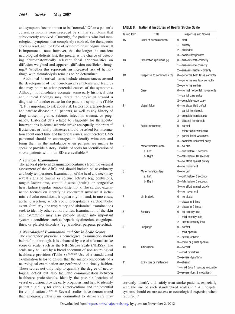
3. Neurological Examination and Stroke Scale ScoresThe emergency physician’s neurological examination shouldbe brief but thorough. It is enhanced by use of a formal strokescore or scale, such as the NIH Stroke Scale (NIHSS). Thescale may be used by a broad spectrum of non-neurologicalhealthcare providers (Table 8).24,68,69 Use of a standardizedexamination helps to ensure that the major components of aneurological examination are performed in a timely fashion.These scores not only help to quantify the degree of neuro-logical deficit but also facilitate communication betweenhealthcare professionals, identify the possible location ofvessel occlusion, provide early prognosis, and help to identifypatient eligibility for various interventions and the potentialfor complications.42,70–72 Several studies have demonstratedthat emergency physicians committed to stroke care may
correctly identify and safely treat stroke patients, especiallywith the use of such standardized scales.73,74 All hospitalsystems should ensure access to neurological expertise whenrequired.75
TABLE 8. National Institutes of Health Stroke Scale
Tested Item Title Responses and Scores
1A Level of consciousness 0—alert
1—drowsy
2—obtunded
3—coma/unresponsive
1B Orientation questions (2) 0—answers both correctly
1—answers one correctly
2—answers neither correctly
1C Response to commands (2) 0—performs both tasks correctly
1—performs one task correctly
2—performs neither
2 Gaze 0—normal horizontal movements
1—partial gaze palsy
2—complete gaze palsy
3 Visual fields 0—no visual field defect
1—partial hemianopia
2—complete hemianopia
3—bilateral hemianopia
4 Facial movement 0—normal
1—minor facial weakness
2—partial facial weakness
3—complete unilateral palsy
5 Motor function (arm) 0—no drift
a. Left 1—drift before 5 seconds
b. Right 2—falls before 10 seconds
3—no effort against gravity
4—no movement
6 Motor function (leg) 0—no drift
a. Left 1—drift before 5 seconds
b. Right 2—falls before 5 seconds
3—no effort against gravity
4—no movement
7 Limb ataxia 0—no ataxia
1—ataxia in 1 limb
2—ataxia in 2 limbs
8 Sensory 0—no sensory loss
1—mild sensory loss
2—severe sensory loss
9 Language 0—normal
1—mild aphasia
2—severe aphasia
3—mute or global aphasia
10 Articulation 0—normal
1—mild dysarthria
2—severe dysarthria
11 Extinction or inattention 0—absent
1—mild (loss 1 sensory modality)
2—severe (loss 2 modalities)
1664 Stroke May 2007
by guest on November 2, 2012http://stroke.ahajournals.org/Downloaded from
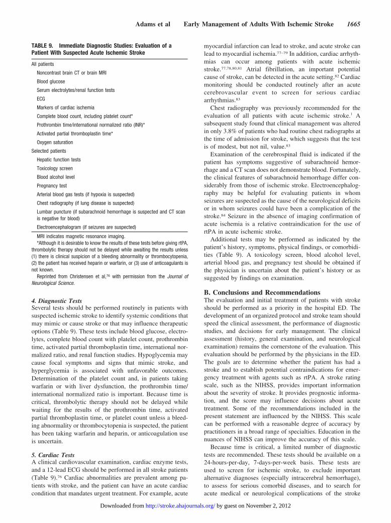
4. Diagnostic TestsSeveral tests should be performed routinely in patients withsuspected ischemic stroke to identify systemic conditions thatmay mimic or cause stroke or that may influence therapeuticoptions (Table 9). These tests include blood glucose, electro-lytes, complete blood count with platelet count, prothrombintime, activated partial thromboplastin time, international nor-malized ratio, and renal function studies. Hypoglycemia maycause focal symptoms and signs that mimic stroke, andhyperglycemia is associated with unfavorable outcomes.Determination of the platelet count and, in patients takingwarfarin or with liver dysfunction, the prothrombin time/international normalized ratio is important. Because time iscritical, thrombolytic therapy should not be delayed whilewaiting for the results of the prothrombin time, activatedpartial thromboplastin time, or platelet count unless a bleed-ing abnormality or thrombocytopenia is suspected, the patienthas been taking warfarin and heparin, or anticoagulation useis uncertain.
5. Cardiac TestsA clinical cardiovascular examination, cardiac enzyme tests,and a 12-lead ECG should be performed in all stroke patients(Table 9).76 Cardiac abnormalities are prevalent among pa-tients with stroke, and the patient can have an acute cardiaccondition that mandates urgent treatment. For example, acute
myocardial infarction can lead to stroke, and acute stroke canlead to myocardial ischemia.77–79 In addition, cardiac arrhyth-mias can occur among patients with acute ischemicstroke.77,78,80,81 Atrial fibrillation, an important potentialcause of stroke, can be detected in the acute setting.82 Cardiacmonitoring should be conducted routinely after an acutecerebrovascular event to screen for serious cardiacarrhythmias.83
Chest radiography was previously recommended for theevaluation of all patients with acute ischemic stroke.1 Asubsequent study found that clinical management was alteredin only 3.8% of patients who had routine chest radiographs atthe time of admission for stroke, which suggests that the testis of modest, but not nil, value.83
Examination of the cerebrospinal fluid is indicated if thepatient has symptoms suggestive of subarachnoid hemor-rhage and a CT scan does not demonstrate blood. Fortunately,the clinical features of subarachnoid hemorrhage differ con-siderably from those of ischemic stroke. Electroencephalog-raphy may be helpful for evaluating patients in whomseizures are suspected as the cause of the neurological deficitsor in whom seizures could have been a complication of thestroke.84 Seizure in the absence of imaging confirmation ofacute ischemia is a relative contraindication for the use ofrtPA in acute ischemic stroke.
Additional tests may be performed as indicated by thepatient’s history, symptoms, physical findings, or comorbidi-ties (Table 9). A toxicology screen, blood alcohol level,arterial blood gas, and pregnancy test should be obtained ifthe physician is uncertain about the patient’s history or assuggested by findings on examination.
B. Conclusions and RecommendationsThe evaluation and initial treatment of patients with strokeshould be performed as a priority in the hospital ED. Thedevelopment of an organized protocol and stroke team shouldspeed the clinical assessment, the performance of diagnosticstudies, and decisions for early management. The clinicalassessment (history, general examination, and neurologicalexamination) remains the cornerstone of the evaluation. Thisevaluation should be performed by the physicians in the ED.The goals are to determine whether the patient has had astroke and to establish potential contraindications for emer-gency treatment with agents such as rtPA. A stroke ratingscale, such as the NIHSS, provides important informationabout the severity of stroke. It provides prognostic informa-tion, and the score may influence decisions about acutetreatment. Some of the recommendations included in thepresent statement are influenced by the NIHSS. This scalecan be performed with a reasonable degree of accuracy bypractitioners in a broad range of specialties. Education in thenuances of NIHSS can improve the accuracy of this scale.
Because time is critical, a limited number of diagnostictests are recommended. These tests should be available on a24-hours-per-day, 7-days-per-week basis. These tests areused to screen for ischemic stroke, to exclude importantalternative diagnoses (especially intracerebral hemorrhage),to assess for serious comorbid diseases, and to search foracute medical or neurological complications of the stroke
TABLE 9. Immediate Diagnostic Studies: Evaluation of aPatient With Suspected Acute Ischemic Stroke
All patients
Noncontrast brain CT or brain MRI
Blood glucose
Serum electrolytes/renal function tests
ECG
Markers of cardiac ischemia
Complete blood count, including platelet count*
Prothrombin time/international normalized ratio (INR)*
Activated partial thromboplastin time*
Oxygen saturation
Selected patients
Hepatic function tests
Toxicology screen
Blood alcohol level
Pregnancy test
Arterial blood gas tests (if hypoxia is suspected)
Chest radiography (if lung disease is suspected)
Lumbar puncture (if subarachnoid hemorrhage is suspected and CT scanis negative for blood)
Electroencephalogram (if seizures are suspected)
MRI indicates magnetic resonance imaging.*Although it is desirable to know the results of these tests before giving rtPA,
thrombolytic therapy should not be delayed while awaiting the results unless(1) there is clinical suspicion of a bleeding abnormality or thrombocytopenia,(2) the patient has received heparin or warfarin, or (3) use of anticoagulants isnot known.
Reprinted from Christensen et al,76 with permission from the Journal ofNeurological Science.
Adams et al Early Management of Adults With Ischemic Stroke 1665
by guest on November 2, 2012http://stroke.ahajournals.org/Downloaded from
(Table 9). Examination of the cerebrospinal fluid has alimited role in the evaluation of patients with suspectedstroke. Additional diagnostic studies, including cardiac andvascular imaging, often are time consuming and may delayemergency treatment. Thus, most of these tests are not doneuntil after the acute treatment or after the patient is admittedto the hospital.
The recommendations that follow are similar to thoseincluded in previous statements except recommendation 1under Class III.
Class I Recommendations
1. An organized protocol for the emergency evaluation ofpatients with suspected stroke is recommended (Class I,Level of Evidence B). The goal is to complete anevaluation and to decide treatment within 60 minutes ofthe patient’s arrival in an ED. Designation of an acutestroke team that includes physicians, nurses, and labo-ratory/radiology personnel is encouraged. Patients withstroke should have a careful clinical assessment, includ-ing neurological examination.
2. The use of a stroke rating scale, preferably the NIHSS,is recommended (Class I, Level of Evidence B). Hospi-tals (ie, administration) must provide the necessaryresources to use such a scale.
3. A limited number of hematologic, coagulation, andbiochemistry tests are recommended during the initialemergency evaluation (Table 9) (Class I, Level ofEvidence B).
4. Patients with clinical or other evidence of acute cardiacor pulmonary disease may warrant chest x-ray (Class I,Level of Evidence B).
5. An ECG is recommended because of the high incidenceof heart disease in patients with stroke (Class I, Level ofEvidence B).
Class III Recommendations
1. Most patients with stroke do not need a chest x-ray aspart of their initial evaluation (Class III, Level ofEvidence B). This is a change from the previousguideline.
2. Most patients with stroke do not need an examinationof the cerebrospinal fluid (Class III, Level of EvidenceB). The yield of brain imaging is very high for detectionof intracranial hemorrhage. The clinical course ofsubarachnoid hemorrhage or acute central nervoussystem infections usually is distinct from that of ische-mic stroke. Examination of the cerebrospinal fluid maybe indicated for evaluation of a patient with a strokethat may be secondary to an infectious illness.
IV. Early Diagnosis: Brain and Vascular ImagingA. Brain ImagingAs therapeutic options evolve, brain imaging strategies areplaying an increasingly important role in the initial evaluationof patients with acute stroke (Table 9). Brain imagingfindings, including the size, location, and vascular distribu-tion of the infarction, as well as the presence of bleeding,affect both short-term and long-term treatment decisions. Inaddition, information about the possible degree of reversibil-ity of ischemic injury, intracranial vessel status, and cerebral
hemodynamic status may be obtained by modern imagingstudies.85 Neuroimaging tests might improve selection ofpatients who could be treated with reperfusion therapies byidentifying those with regions of salvageable brain tissue, alow risk for hemorrhagic transformation, or occlusions oflarge arteries that might or might not be amenable to therapy.CT and magnetic resonance imaging (MRI) are being used asinitial imaging options. The most commonly obtained brainimaging test is noncontrast CT, but individual centers able toobtain MRI with efficiency equal to that of CT are using anMRI strategy in patients without MR contraindications.86–90
Additional research is required.91,92 As a result, it is generallyagreed that the performance of these tests should not delaytreatment with intravenous rtPA.86,91–93
1. Non–Contrast-Enhanced CT Scan of the BrainIt is agreed that emergency, non–contrast-enhanced CTscanning of the brain accurately identifies most cases ofintracranial hemorrhage and helps discriminate nonvascularcauses of neurological symptoms (eg, brain tumor). The priorguidelines recommended that CT be the primary diagnosticbrain imaging study for evaluation of patients with suspectedstroke.94 Although CT is the “criterion standard” with whichother brain imaging studies are compared, it is relativelyinsensitive in detecting acute and small cortical or subcorticalinfarctions, especially in the posterior fossa.95 In most cases,the use of a contrast infusion does not provide additionalinformation and is not necessary unless it is required for CTangiography (and, more recently, CT perfusion) or concernexists about a brain tumor or infectious process.
With the advent of rtPA treatment, interest has grown inusing CT to identify subtle, early signs of ischemic braininjury (early infarct signs) or arterial occlusion (hyperdensevessel sign) that might affect decisions about treatment. Inaddition, the loss of the gray-white differentiation in thecortical ribbon (particularly at the lateral margins of theinsula) or the lentiform nucleus and sulcal effacement canoften be detected within 6 hours in up to 82% of patients withlarge-vessel anterior circulation occlusions.96,97 These signsare associated with poorer outcomes.98,99
In addition, widespread signs of early infarction are corre-lated with a higher risk of hemorrhagic transformation aftertreatment with thrombolytic agents. In combined data from 2trials of intravenous rtPA administered within 3 hours ofsymptom onset, CT evidence of early edema or mass effectwas accompanied by an 8-fold increase in the risk ofsymptomatic hemorrhage.66 In a second analysis, early infarctsigns involving more than one third of the territory of themiddle cerebral artery (MCA) were not independently asso-ciated with increased risk of adverse outcome after rtPAtreatment, and as a group these patients still benefited fromtherapy.100 In a European trial in which thrombolytic therapywas administered within 6 hours of symptom onset, patientsestimated to have involvement of more than one third of theterritory of the MCA had an increased risk of intracerebralhemorrhage, whereas those with less involvement benefitedthe most from thrombolytic treatment.99,101 However, physi-cians’ ability to reliably and reproducibly recognize the earlyCT changes is variable.102–106 Use of scoring systems for
1666 Stroke May 2007
by guest on November 2, 2012http://stroke.ahajournals.org/Downloaded from
early CT changes may improve identification of cerebralischemia and may provide valuable prognostic informationbut is not validated for outcome or patient selection for acutetreatments.107,108 Further studies are needed to determine thesignificance of early infarct signs and their role in treatmentdecision making.109
For patients who are candidates for treatment with rtPA,the goal is to complete the CT examination within 25 minutesof arrival at the ED, with the study interpreted within anadditional 20 minutes (door-to-interpretation time of 45minutes).61 A subsequent CT scan often is obtained if thepatient worsens neurologically and may be especially helpfulin identifying hemorrhagic transformation after administra-tion of rtPA.66
2. Multimodal CTRecent technological advances have led to increased interestin more sophisticated multimodal approaches to acute strokeimaging. The multimodal CT approach may include noncon-trast CT, perfusion CT, and CT angiography studies. Twotypes of perfusion techniques are currently available. Whole-brain perfusion CT provides a map of cerebral blood volume,and it is postulated that regions of hypoattenuation on thesecerebral blood volume maps represent the ischemic core.110
Although this technique has the advantage of providingwhole-brain coverage, it is limited by its inability to providemeasures of cerebral blood flow or mean transit time.Alternatively, the second technique, dynamic perfusion CT,has the potential to provide absolute measures of cerebralblood flow, mean transit time, and cerebral blood volume.Dynamic perfusion CT is currently limited to 2 to 4 brainslices and provides incomplete visualization of all pertinentvascular territories.
Recent reports demonstrate a high degree of sensitivity andspecificity for detecting cerebral ischemia with both of theseperfusion CT techniques.111–113 In addition, several studieshave suggested that perfusion CT may be able to differentiatethresholds of reversible and irreversible ischemia and thusidentify the ischemic penumbra.114,115
Helical CT angiography provides a means to rapidly andnoninvasively evaluate the vasculature, both intracraniallyand extracranially, in acute, subacute, and chronic strokesettings and thus to provide potentially important informationabout the presence of vessel occlusions or stenoses.116,117 Thefeasibility of this technique has been demonstrated in theacute stroke setting, with preliminary data suggesting highdiagnostic accuracy for evaluation of large-vessel intracranialocclusions as compared with ultrasound and digital subtrac-tion angiography.118–120
These techniques have the advantage of relatively rapiddata acquisition and can be performed with conventional CTequipment. Disadvantages include iodine contrast and addi-tional radiation exposure. The role of perfusion CT and CTangiography in making acute treatment decisions has not yetbeen established.
3. Multimodal MRIThe multimodal MRI approach for acute stroke evaluationincludes diffusion-weighted imaging (DWI), perfusion-weighted imaging (PWI), MR angiography, gradient echo,
and often fluid-attenuated inversion recovery or T2-weightedsequences. Standard MRI sequences (T1 weighted, T2weighted, and proton density) are relatively insensitive to thechanges of acute ischemia.121 DWI allows visualization ofischemic regions within minutes of symptom onset122–131 andearly identification of the lesion size, site, and age. It candetect relatively small cortical or subcortical lesions, includ-ing those in the brain stem or cerebellum, areas often poorlyvisualized with standard CT scan techniques. DWI alsoprovides information about the involved vascular territoryand has a high sensitivity (88% to 100%) and specificity(95% to 100%) for detecting ischemic lesions, even at veryearly time points.
PWI, usually performed with the rapid administration of anintravenous paramagnetic contrast agent, provides relativemeasures of cerebral hemodynamic status. Investigations ofthe best PWI analytical method focus on identifying thehighest correlation of ischemic volume with acute clinicaldeficits (symptomatic hypoperfusion) or with volume ofchronic infarct (tissue at risk).
Studies have demonstrated that the initial volumes of thelesions seen on DWI and PWI correlate well with the finalsize of the stroke found on follow-up brain imaging.129,132,133
In addition, these lesion volumes correlate well with severityof stroke as rated by both clinical scales and outcomes. Thesefindings suggest that DWI might provide helpful early prog-nostic information.125,132
The ischemic penumbra is roughly approximated on MRIas regions of perfusion change without a correspondingdiffusion abnormality (diffusion–perfusion mismatch). How-ever, several studies indicate that, at least in some circum-stances, the initial diffusion abnormality is reversible and thevisually thresholded perfusion volumes overestimate the pen-umbra.134,135 Sequential MRI studies performed in patientsbeing treated with thrombolytic therapy have shown that thetechnique may visualize salvage of mismatch-defined pen-umbral tissue with smaller volumes of infarction amongpatients who have successful recanalization.134,136
Efforts are under way to develop multiparametric MRIcriteria that could identify regions of irreversible infarctionfrom potentially reversible ischemia or portend a high risk ofhemorrhagic complications after thrombolytic therapy.137–139
A recent phase II trial of intravenous administration of thethrombolytic agent desmoteplase showed a signal of potentialtherapeutic benefit when MRI was used to select patients withdiffusion–perfusion mismatch for treatment 3 to 9 hours fromonset.140 However, insufficient evidence currently exists torecommend this approach for selecting patients for acutetherapies in routine practice.
Two prospective studies recently demonstrated that MRI isas accurate as CT in detecting hyperacute intraparenchymalhemorrhage in patients presenting with stroke symptomswithin 6 hours of onset when gradient echo MRI sequenceswere used.88,141 These findings suggest that MRI may be usedas the sole imaging modality to evaluate acute stroke patients,including candidates for thrombolytic treatment. However, inpatients presenting with symptoms suggestive of subarach-noid hemorrhage, a CT scan should be performed.
Adams et al Early Management of Adults With Ischemic Stroke 1667
by guest on November 2, 2012http://stroke.ahajournals.org/Downloaded from
Gradient echo sequences also have the ability to detectclinically silent prior microbleeds not visualized on CT. Somedata suggest that microbleeds represent markers of bleeding-prone angiopathy and increased risk of hemorrhagic transfor-mation after antithrombotic and thrombolytic therapy.142–144
However, other studies have not found an increased risk inpatients with small numbers of microbleeds.145 The impor-tance of the presence of large numbers of microbleeds onMRI in thrombolytic decision making remains uncertain.
MR angiography is increasingly used for noninvasivescreening of the extracranial and intracranial circulation.When compared with digital subtraction angiography fordetection of cervical and intracranial stenoses, sensitivity andspecificity have ranged from 70% to 100%.146,147 In theintracranial vasculature, MR angiography is useful in identi-fying acute proximal large-vessel occlusions but cannotreliably identify distal or branch occlusions.
A potential diagnostic advantage of MRI over CT innon-tPA situations in suspected stroke has been demon-strated. MRI is better at distinguishing acute, small cortical,small deep, and posterior fossa infarcts; at distinguishingacute from chronic ischemia; and at identifying subclinicalsatellite ischemic lesions that provide information on strokemechanism.95,124,129,148–167 Limitations of MRI in the acutesetting include cost, relatively limited availability of the test,and patient contraindications such as claustrophobia, cardiacpacemakers, or metal implants. Advantages include theavoidance of exposure to ionizing radiation and iodinatedcontrast and greater spatial resolution.
4. Other Brain Imaging TechniquesOxygen-15 positron-emission tomography may quantify re-gional brain perfusion and oxygen consumption.168–172 How-ever, logistical and pragmatic considerations limit the appli-cation of positron-emission tomography in the setting ofacute stroke. Xenon-enhanced CT provides a quantitativemeasurement of cerebral blood flow by using inhaled xenonbut is not currently widely available.173 Single-photon emis-sion CT, which is minimally invasive and measures relativecerebral blood flow, might be able to identify thresholds forreversible ischemia and could be helpful in predicting out-comes or monitoring responses to treatment.174–176 Limita-tions include lack of availability, expense, and difficultyassociated with tracer preparation.
B. Other Vascular ImagingIn addition to the aforementioned CT and MR angiography,transcranial Doppler ultrasonography, carotid duplex sonog-raphy, and catheter angiography have been used to detectintracranial or extracranial vessel abnormalities. TranscranialDoppler ultrasonography and angiography have been used tomonitor the effects of thrombolytic therapy over time and canhelp to determine prognosis.177–179
In patients whose symptoms started �8 hours ago, thesetests may be helpful in selecting candidates for intervention.A variety of ancillary tests are available to help cliniciansreach accurate pathophysiological and etiologic stroke diag-noses and provide information that can be critical for effec-tive prevention of recurrent stroke.180,181 Vascular imaging is
a key component of the evaluation. The selection of testsneeds to be tailored to the individual patient and clinicalsetting.
C. Conclusions and RecommendationsBrain imaging remains a required component of the emer-gency assessment of patients with suspected stroke. Both CTand MRI are options for imaging the brain, but for most casesand at most institutions, CT remains the most practical initialbrain imaging test. A physician skilled in assessing CT orMRI studies should be available to examine the initial scan.In particular, the scan should be evaluated for evidence ofearly signs of infarction. Baseline CT findings, including thepresence of ischemic changes involving more than one thirdof a hemisphere, have not been predictors of responses totreatment with rtPA when the agent is administered within the3-hour treatment window. Information about multimodal CTand MRI of the brain suggests that these diagnostic studiesmay help in the diagnosis and treatment of patients with acutestroke. Imaging of the intracranial or extracranial vasculaturein the emergency assessment of patients with suspectedstroke is useful at institutions providing endovascular recan-alization therapies. The usefulness of vascular imaging forpredicting responses to treatment before intravenous admin-istration of thrombolytic agents has not been demonstrated.
Class I Recommendations
1. Imaging of the brain is recommended before initiatingany specific therapy to treat acute ischemic stroke(Class I, Level of Evidence A). This recommendation hasnot changed from the previous guideline.
2. In most instances, CT will provide the information tomake decisions about emergency management (Class I,Level of Evidence A). This recommendation has notchanged from the previous guideline.
3. The brain imaging study should be interpreted by aphysician with expertise in reading CT or MRI studiesof the brain (Class I, Level of Evidence C). Thisrecommendation has been added since the previousguideline.
4. Some findings on CT, including the presence of a denseartery sign, are associated with poor outcomes afterstroke (Class I, Level of Evidence A). This recommen-dation has not changed from the previous guideline.
5. Multimodal CT and MRI may provide additional in-formation that will improve diagnosis of ischemicstroke (Class I, Level of Evidence A). This recommen-dation has been added since the previous guideline.
Class II Recommendations
1. Nevertheless, data are insufficient to state that, with theexception of hemorrhage, any specific CT finding (in-cluding evidence of ischemia affecting more than onethird of a cerebral hemisphere) should preclude treat-ment with rtPA within 3 hours of onset of stroke (ClassIIb, Level of Evidence A). This recommendation has notchanged from the previous guideline.
2. Vascular imaging is necessary as a preliminary step forintra-arterial administration of pharmacologicalagents, surgical procedures, or endovascular interven-
1668 Stroke May 2007
by guest on November 2, 2012http://stroke.ahajournals.org/Downloaded from
tions (Class IIa, Level of Evidence B). This recommen-dation has not changed from the previous guideline.
Class III Recommendations
1. Emergency treatment of stroke should not be delayedin order to obtain multimodal imaging studies (ClassIII, Level of Evidence C). This recommendation hasbeen added since the previous guideline.
2. Vascular imaging should not delay treatment of pa-tients whose symptoms started <3 hours ago and whohave acute ischemic stroke (Class III, Level of EvidenceB). This recommendation has been added since theprevious guideline.
V. General Supportive Care and Treatment ofAcute Complications
A. Airway, Ventilatory Support, andSupplemental OxygenMaintaining adequate tissue oxygenation is important in thesetting of acute cerebral ischemia. The goals are to preventhypoxia and potential worsening of the brain injury. The mostcommon causes of hypoxia are partial airway obstruction,hypoventilation, aspiration pneumonia, and atelectasis. Pa-tients with decreased consciousness or signs of brain stemdysfunction have the greatest risk of airway compromisebecause of impaired oropharyngeal mobility and loss ofprotective reflexes.182–184 The prognosis of patients whorequire endotracheal intubation generally is poor; �50% ofthese patients are dead within 30 days after stroke.185,186
Pneumonia is among the leading complications of stroke andis an important cause of death after the cerebrovascularevent.187 It is most likely to develop among patients who areseriously ill, and prevention of early aspiration and protectionof the airway may be a way to lessen this complication.
Elective intubation also may help in the management ofpatients who have severely increased levels of intracranialpressure or have malignant brain edema after stroke.185,188 Noclinical trial has tested the utility of endotracheal intubation inthe management of critically ill patients with stroke, and weanticipate that none will be done. It is generally agreed that anendotracheal tube should be placed if the airway isthreatened.182,188
After ischemic stroke, some patients develop Cheyne-Stokes pattern of respiration, with decreases in oxygensaturation that can be readily reversed with oxygen supple-mentation.189 A small pilot study found that high-flow oxy-gen may be associated with a transient improvement inneurological impairments.190 The results of a controlled studydo not support the use of supplemental oxygen at 3 L/min formost patients with acute ischemic stroke.191 However, pa-tients with acute stroke should be monitored with pulseoximetry with a target oxygen saturation level �92%.25,192
Most patients with acute stroke do not need supplementaloxygen; however, if pulse oximetry or blood gas determina-tion indicates hypoxia, oxygen should be administered.
Hyperbaric oxygen may be used to treat patients withischemic neurological symptoms secondary to air embolismor caisson disease.193 Studies that have tested the use ofhyperbaric oxygen in stroke have been inconclusive or have
shown that the intervention does not improve outcomes.194–196
Data from a small trial suggest that hyperbaric oxygentherapy may be harmful.196 A systematic review found noevidence that hyperbaric oxygen improved outcomes afterstroke or brain injury.197 At present, data do not support theroutine use of hyperbaric oxygen in the treatment of patientswith acute ischemic stroke.
B. TemperatureIncreased body temperature (fever) in the setting of acuteischemic stroke is associated with poor neurological outcome(increased risk of morbidity and mortality), possibly second-ary to increased metabolic demands, enhanced release ofneurotransmitters, and increased free radical production.198–205
The source of any fever should be ascertained. The fever maybe secondary to a cause of stroke, such as infective endocar-ditis, or may represent a complication, such as pneumonia.
Because of the negative effects of fever, lowering anacutely elevated body temperature might improve the prog-nosis of patients with stroke.206 Measures include antipyreticmedications and cooling devices. Small clinical trials havetested the utility of aspirin, ibuprofen, or acetaminophen inlowering body temperatures and in improving outcomes afterstroke. Sulter et al207 found that either aspirin or acetamino-phen was modestly successful in achieving normothermia butthat patients with a temperature �38°C were relativelyunresponsive to treatment. In a small, randomized trial,Kasner et al208 administered 3900 mg of acetaminophen dailyto afebrile patients with stroke. They concluded that themedication might prevent hyperthermia or modestly promotehypothermia but that the effects were not likely to have arobust clinical impact. Dippel et al209 tested 2 different dosesof acetaminophen in a small clinical trial. They concludedthat a daily dosage of 6000 mg might have a potentialbeneficial effect in lowering body temperature. A smallplacebo-controlled trial found that acetaminophen mightlower body temperature by a mean of 0.26°C within 4 hoursof starting treatment.210 In another clinical trial, Dippel etal210 compared the effects of placebo, ibuprofen, or acetamin-ophen on body temperature in 75 patients with recent stroke.Although treating fever after stroke makes intuitive sense, nodata demonstrate that the use of medications to lower bodytemperature among either febrile or afebrile patients im-proves neurological outcomes after stroke. Seeking andtreating the source of fever are reasonable.
Hypothermia has been shown to be neuroprotective inexperimental and focal hypoxic brain injury models. Hypo-thermia may delay depletion of energy reserves, lessenintracellular acidosis, slow influx of calcium into ischemiccells, suppress production of oxygen free radicals, and lessenthe impact of excitatory amino acids.211 Deep hypothermiaoften is administered to protect the brain in major operativeprocedures. Mild to moderate hypothermia is associated withimproved neurological outcomes among patients with cardiacarrest.212–214 Conversely, a multicenter clinical trial found thatmild hypothermia administered during surgery for treatmentof a ruptured intracranial aneurysm did not improve outcomesafter subarachnoid hemorrhage.215 Several small clinicalstudies have evaluated the feasibility of inducing modest
Adams et al Early Management of Adults With Ischemic Stroke 1669
by guest on November 2, 2012http://stroke.ahajournals.org/Downloaded from
hypothermia for treatment of patients with acute ischemicstroke.216–223 Two small studies evaluated the utility ofhypothermia in treating patients with malignant cerebralinfarctions; results were mixed.224,225 Potential side effects oftherapeutic hypothermia include hypotension, cardiac ar-rhythmias, and pneumonia.226 In a systematic review ofavailable data, Correia et al227 could find no evidence thatphysical cooling would improve outcomes after stroke. Al-though strong experimental and clinical evidence indicatesthat induced hypothermia can protect the brain in the pres-ence of hypoxia or ischemia, including after cardiac arrest,data about the utility of induced hypothermia for treatment ofpatients with stroke are not yet available. Hypothermia isdiscussed in more detail in the neuroprotection section of thisstatement.
C. Cardiac Monitoring and TreatmentAlthough patients with heart disease are at high risk forischemic stroke, both myocardial ischemia and cardiac ar-rhythmias are potential complications of acute cerebrovascu-lar diseases.228,229 Patients with infarctions of the right hemi-sphere, particularly those involving the insula, may have anincreased risk of cardiac complications, presumably second-ary to disturbances in autonomic nervous system func-tion.78,230–234 ECG changes secondary to stroke include ST-segment depression, QT dispersion, inverted T waves, andprominent U waves.235–237 Elevations in blood levels ofenzymes attributable to injury to myocardial muscle also maybe found.79 The most common arrhythmia detected in thesetting of stroke is atrial fibrillation, which either may berelated to the cause of stroke or may be a complication. Otherpotentially life-threatening cardiac arrhythmias are relativelyuncommon, but sudden death may occur.77,238 No clinicaltrials have tested the utility of cardiac monitoring for mostpatients with ischemic stroke or the use of cardiac protectiveagents or medications to prevent serious cardiac arrhythmias.Still, general consensus exists that patients with acute ische-mic stroke should have cardiac monitoring for at least the first24 hours and that any serious cardiac arrhythmia should betreated. The utility of prophylactic administration of medica-tions to prevent cardiac arrhythmias among patients withstroke is not known.
D. Arterial HypertensionAn elevated blood pressure is often detected in the first hoursafter stroke. Elevations in blood pressure �160 mm Hg aredetected in �60% of patients with acute stroke.239 Bothelevated and low blood pressures are associated with pooroutcome after stroke.240 For every 10-mm Hg increase�180 mm Hg, the risk of neurological deterioration increasedby 40% and the risk of poor outcome increased by 23%. Theelevation in blood pressure may be secondary to the stress ofthe cerebrovascular event, a full bladder, nausea, pain, pre-existing hypertension, a physiological response to hypoxia, ora response to increased intracranial pressure.241,242 In a studythat correlated acute blood pressure values with other find-ings in the setting of acute stroke, Vemmos et al243 found thatamong patients with most subtypes of ischemic stroke,elevated blood pressure was correlated with a past history of
hypertension or severity of neurological impairments. Thesame investigators found a U-shaped relationship betweendeath and admission blood pressure; both elevated and lowadmission levels were associated with high rates of early andlate death.243 They also correlated death due to brain injuryand brain edema with high initial blood pressure levels.Castillo et al240 have reported similar findings. Using the datafrom the Glycine Antagonist in Neuroprotection (GAIN)study, Aslanyan et al244 found that an elevated baseline meanarterial blood pressure was not independently associated withan unfavorable outcome after stroke. However, elevations inmean blood pressure during the first days after stroke had anunfavorable effect on outcomes. The same investigatorsshowed that an elevated pulse pressure (the difference be-tween systolic and diastolic blood pressure values) wasindependently associated with poor outcomes 3 months afterstroke.245
Theoretical reasons for lowering blood pressure includereducing the formation of brain edema, lessening the risk ofhemorrhagic transformation of the infarction, preventingfurther vascular damage, and forestalling early recurrentstroke. In addition, urgent antihypertensive therapy may beneeded to treat patients with stroke who also have hyperten-sive encephalopathy, aortic dissection, acute renal failure,acute pulmonary edema, or acute myocardial infarction.242,246
Conversely, aggressive treatment of blood pressure may leadto neurological worsening by reducing perfusion pressure toischemic areas of the brain.242,247,248
In a majority of patients, a decline in blood pressure occurswithin the first hours after stroke even without any specificmedical treatment.241 The blood pressure often falls sponta-neously when the patient is moved to a quiet room, the patientis allowed to rest, the bladder is emptied, or the pain iscontrolled. In addition, treatment of increased intracranialpressure may result in a decline in arterial blood pressure.
There are several questions about the management of arterialhypertension in the setting of acute stroke.249–251 Should patientspreviously taking antihypertensive medications continue takingthem during the first hours after stroke? Are some of thesemedications contraindicated or indicated? Should new antihy-pertensive agents be started? What level of blood pressure wouldmandate initiation of new antihypertensive treatment? Whichmedication should be administered in this situation? Unfortu-nately, definite answers to these questions are not available.Since the publication of the last guidelines, several clinicalstudies have provided additional information.
A randomized trial testing nimodipine found that unfavorableoutcomes among patients treated with the medication wereassociated with lowering of the blood pressure.252,253 Reanalyz-ing some of the data from the studies of nimodipine, Fogelholmet al254 noted that favorable outcomes were associated withhigher levels of blood pressure among patients with mild tomoderate strokes receiving nimodipine. The converse was trueamong patients with severe strokes. In a study that involved 115patients admitted within 24 hours of stroke, Oliveira-Filho etal255 noted that systolic blood pressure dropped by �28% duringthe first day whether or not medications were prescribed. Theynoted an adverse effect on outcomes with lowering of the bloodpressure, with an association found with degree in decline. Each
1670 Stroke May 2007
by guest on November 2, 2012http://stroke.ahajournals.org/Downloaded from
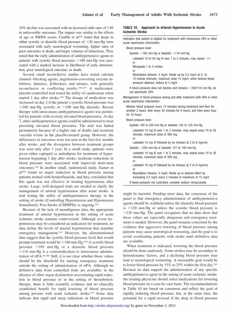
10% decline was associated with an increased odds ratio of 1.89in unfavorable outcomes. The impact was similar to the effectsof age or NIHSS scores. Castillo et al240 noted that drops ineither systolic or diastolic blood pressure of �20 mm Hg wereassociated with early neurological worsening, higher rates ofpoor outcomes or death, and larger volumes of infarctions. Theynoted that the early administration of antihypertensive agents topatients with systolic blood pressures �180 mm Hg was asso-ciated with a marked increase in likelihood of early deteriora-tion, poor neurological outcome, or death.
Several small inconclusive studies have tested calciumchannel–blocking agents, angiotensin-converting enzyme in-hibitors, diuretics, �-blockers, and nitrates, with generallyinconclusive or conflicting results.256,257 A multicenter,placebo-controlled trial tested the utility of candesartan whenstarted 1 day after stroke.258 The dosage of medication wasincreased on day 2 if the patient’s systolic blood pressure was�160 mm Hg systolic or �100 mm Hg diastolic. Rescuetherapy with intravenous antihypertensive agents was permit-ted for patients with severely elevated blood pressures. At day7, other antihypertensive agents could be administered to treatpersisting elevated blood pressures. The trial was haltedprematurely because of a higher rate of deaths and recurrentvascular events in the placebo-treated group. However, thedifferences in outcomes were not seen in the first few monthsafter stroke, and the divergence between treatment groupswas seen only after 1 year. In a small study, patients weregiven either captopril or amlodipine for treatment of hyper-tension beginning 1 day after stroke; moderate reductions inblood pressure were associated with improved short-termoutcomes.259 In another small, randomized study, Eames etal260 found no major reduction in blood pressure amongpatients treated with bendrofluazide, and they concluded thatthis agent was not effective in treating hypertension afterstroke. Large, well-designed trials are needed to clarify themanagement of arterial hypertension after acute stroke. Atrial testing the utility of antihypertensive therapy in thesetting of stroke (Controlling Hypertension and HypotensionImmediately Post-Stroke [CHHIPS]) is ongoing.261
Because of the lack of unambiguous data, the appropriatetreatment of arterial hypertension in the setting of acuteischemic stroke remains controversial. Although severe hy-pertension may be considered an indication for treatment, nodata define the levels of arterial hypertension that mandateemergency management.247 However, the aforementioneddata suggest that the systolic blood pressure level that wouldprompt treatment would be �180 mm Hg.240 A systolic bloodpressure �185 mm Hg or a diastolic blood pressure�110 mm Hg is a contraindication to intravenous adminis-tration of rtPA.66,262 Still, it is not clear whether those valuesshould be the threshold for starting emergency treatmentoutside the setting of administration of rtPA. Although nodefinitive data from controlled trials are available, in theabsence of other organ dysfunction necessitating rapid reduc-tion in blood pressure or in the setting of thrombolytictherapy, there is little scientific evidence and no clinicallyestablished benefit for rapid lowering of blood pressureamong persons with acute ischemic stroke.247 Some dataindicate that rapid and steep reductions in blood pressure
might be harmful. Pending more data, the consensus of thepanel is that emergency administration of antihypertensiveagents should be withheld unless the diastolic blood pressureis �120 mm Hg or unless the systolic blood pressure is�220 mm Hg. The panel recognizes that no data show thatthese values are especially dangerous and emergency treat-ment is needed. However, the panel remains concerned by theevidence that aggressive lowering of blood pressure amongpatients may cause neurological worsening, and the goal is toavoid overtreating patients with stroke until definitive dataare available.
When treatment is indicated, lowering the blood pressureshould be done cautiously. Some strokes may be secondary tohemodynamic factors, and a declining blood pressure maylead to neurological worsening. A reasonable goal would beto lower blood pressure by 15% to 25% within the first day.263
Because no data support the administration of any specificantihypertensive agent in the setting of acute ischemic stroke,the treating physician should select medications for loweringblood pressure on a case-by-case basis. The recommendationsin Table 10 are based on consensus and reflect the goal ofrapidly reducing blood pressure, but, at the same time, thepotential for a rapid reversal if the drop in blood pressure
TABLE 10. Approach to Arterial Hypertension in AcuteIschemic Stroke
Indication that patient is eligible for treatment with intravenous rtPA or otheracute reperfusion intervention
Blood pressure level
Systolic �185 mm Hg or diastolic �110 mm Hg
Labetalol 10 to 20 mg IV over 1 to 2 minutes, may repeat �1;orNitropaste 1 to 2 inches;orNicardipine infusion, 5 mg/h, titrate up by 2.5 mg/h at 5- to15-minute intervals, maximum dose 15 mg/h; when desired bloodpressure attained, reduce to 3 mg/h
If blood pressure does not decline and remains �185/110 mm Hg, donot administer rtPA
Management of blood pressure during and after treatment with rtPA or otheracute reperfusion intervention
Monitor blood pressure every 15 minutes during treatment and then foranother 2 hours, then every 30 minutes for 6 hours, and then every hourfor 16 hours
Blood pressure level
Systolic 180 to 230 mm Hg or diastolic 105 to 120 mm Hg
Labetalol 10 mg IV over 1 to 2 minutes, may repeat every 10 to 20minutes, maximum dose of 300 mg;orLabetalol 10 mg IV followed by an infusion at 2 to 8 mg/min
Systolic �230 mm Hg or diastolic 121 to 140 mm Hg
Labetalol 10 mg IV over 1 to 2 minutes, may repeat every 10 to 20minutes, maximum dose of 300 mg;orLabetalol 10 mg IV followed by an infusion at 2 to 8 mg/min;orNicardipine infusion, 5 mg/h, titrate up to desired effect byincreasing 2.5 mg/h every 5 minutes to maximum of 15 mg/h
If blood pressure not controlled, consider sodium nitroprusside
Adams et al Early Management of Adults With Ischemic Stroke 1671
by guest on November 2, 2012http://stroke.ahajournals.org/Downloaded from
leads to neurological worsening. The selection of an agentmay be influenced by other medical conditions; for example,the presence of asthma would contraindicate the administra-tion of a �-blocker. Because of a prolonged effect and thepotential for a precipitous decline in blood pressure associ-ated with the sublingual administration of nifedipine, thisagent with this route of administration is notrecommended.264
Among patients who are candidates for treatment withintravenous rtPA, attention to management of blood pressureis critical before, during, and after the administration of themedication.263 Excessively high blood pressure is associatedwith an increased risk of symptomatic hemorrhagic transfor-mation.66,262,265,266 Failure to meet the blood pressure param-eters of previous guidelines may be one of the explanationsfor an increased risk of hemorrhagic complications afteradministration of rtPA.267–269 Presumably, similar risks ofbleeding will be associated with elevations of blood pressureamong patients receiving other acute pharmacological ormechanical interventions to improve the perfusion to thebrain. The suggestion to withhold such therapies amongpatients with markedly elevated blood pressure is based onpotential safety risks. Conversely, excessively high bloodpressures could be lowered successfully with medications topermit treatment with intravenous rtPA.
Arterial hypertension is a recognized risk factor for strokeand recurrent stroke. Many patients were taking medicationsbefore their stroke or are found to have sustained hyperten-sion after their stroke. These patients will need long-termantihypertensive treatment; the primary question is the timingof the institution of such therapy. Limited data are availableto guide these decisions. On the basis of the trial of cande-sartan, it appears that medications can be administered with areasonable degree of safety when started �1 day afterstroke.258 The timing of the reinstitution of treatment and theselection of medications will depend on the patient’s neuro-logical status, the underlying stroke mechanism, the patient’sability to swallow medications, and the presence of concom-itant diseases. Presumably, most patients with mild to mod-erate strokes who are not at high risk for increased intracra-nial pressure may have their prestroke antihypertensivemedications restarted 24 hours after their vascular event. Thepanel strongly endorses clinical research that will provideinformation about the safety and efficacy of restarting anti-hypertensive therapy among patients with stroke.
E. Arterial HypotensionPersistent arterial hypotension is rare among patients withacute ischemic stroke, but it is associated with an increasedlikelihood of an unfavorable outcome.270 Castillo et al240
noted that the rates of neurological worsening, poor neuro-logical outcomes, or death increased when the baselinesystolic blood pressure was �100 mm Hg or the diastolicblood pressure was �70 mm Hg. The cause of hypotensionshould be sought; among the potential causes are aorticdissection, volume depletion, blood loss, and decreasedcardiac output secondary to myocardial ischemia or cardiacarrhythmias. Patients with stroke may have depleted bloodvolume. Correction of hypovolemia and optimization of
cardiac output are important priorities during the first hoursafter stroke. Treatment includes volume replacement withnormal saline and correction of cardiac arrhythmias, such asslowing a ventricular response to rapid atrial fibrillation. Ifthese measures are ineffective, vasopressor agents such asdopamine may be used. Trials have tested the utility ofvolume expansion and drug-induced hypertension for treat-ment of acute ischemic stroke. These measures are describedlater in the present guideline.
F. HypoglycemiaBecause hypoglycemia may produce neurological signs thatmimic ischemic stroke and because hypoglycemia itself maylead to brain injury, prompt measurement of the serumglucose concentration and rapid correction of a low serumglucose level are important.
G. HyperglycemiaHyperglycemia will be detected on admission in approxi-mately one third of patients with stroke.271–273 Most patientshave moderate elevations of glucose levels.274 Clinical stud-ies demonstrate that the presence of hyperglycemia is asso-ciated with poor outcomes after ischemic stroke, includingamong patients treated with thrombolytic agents.275–279 Ahistory of diabetes mellitus also is associated with a pooreroutcome after stroke.280,281
The detrimental effects of hyperglycemia are not clearlyunderstood but include increasing tissue acidosis secondaryto anaerobic glycolysis, lactic acidosis, and free radicalproduction.281,282 Hyperglycemia also may affect the blood–brain barrier and the development of brain edema281 and maybe associated with an increased risk of hemorrhagic transfor-mation of the infarction.283 Unfortunately, the contribution ofhyperglycemia to poor outcomes may be affected by otherfactors.284 Elevations of blood glucose concentration may besecondary to the stress of the acute cerebrovascular event.285
In particular, hyperglycemia after stroke in nondiabetic pa-tients may be a stress response.285 Candelise et al286 foundthat hyperglycemia is a marker of a more severe stroke. Thus,the poor outcomes among patients with hyperglycemia mayin part reflect the seriousness of the vascular event itself.Recent clinical and imaging studies have highlighted theimportance of hyperglycemia as a negative prognostic fac-tor.271,278–280,282 Baird et al280 found that persistent hypergly-cemia (blood glucose level �200 mg/dL) during the first 24hours after stroke independently predicted expansion of thevolume of ischemic stroke and poor neurological outcomes.These reports provide reasonable evidence that persistentelevations of blood glucose levels are associated with neuro-logical worsening.281,287 These data suggest that managementof hyperglycemia is an important part of acute managementof patients with ischemic stroke. The outstanding issues relateto the effectiveness of management of stroke-associatedhyperglycemia in improving outcomes after stroke and thelevel of glucose concentration that should be sought duringthe first 24 hours. On the basis of the aforementioned data, thepanel concluded that the level of hyperglycemia that previ-ously mandated emergency treatment in the setting of strokewas too high. The exact level of blood glucose that should
1672 Stroke May 2007
by guest on November 2, 2012http://stroke.ahajournals.org/Downloaded from
prompt interventions is not known. A reasonable approachwould be to initiate treatment among patients with a bloodglucose level �200 mg/dL. The management of hyperglyce-mia among patients with stroke likely will be influenced bythe approach to treating elevated blood glucose levels amongpatients with other critical illnesses, including patients whohave had cardiac arrest. The changes are dramatic. Severalstudies have looked at the utility of intensive insulin protocolsin treating critically ill patients with a broad range ofillnesses.288–293 In general, the desired level of blood glucosehas been in the range of 80 to 140 mg/dL. Frequent moni-toring of blood glucose levels and adjustments of insulin arerequired. These studies have demonstrated a reduction indeath and important complications, including infections andrenal failure, with aggressive management of hyperglycemia.The frequency of hypoglycemic episodes appears to be low.Physicians treating patients with severe stroke should beaware of the new regimens for treating hyperglycemia. Grayet al274 found that plasma glucose levels spontaneouslydecline in many patients. A small clinical trial tested thesafety of short-term administration of glucose, insulin, andpotassium to patients with moderate hyperglycemia.294 Theintervention could be given safely, but plasma glucose levelswere not significantly lower than in the control group. Asubsequent report about a larger cohort treated by the sameinvestigators found that infusions of glucose, insulin, andpotassium significantly reduced blood pressure, but no dif-ferences in outcomes were noted 7 days after stroke.295 Brunoet al296 treated 24 patients with markedly elevated serumglucose levels (mean 14.7 mmol/L) within 12 hours of onsetof stroke. With insulin infusions that required frequentadjustments in response to serum levels, they were able toachieve desired glucose control within 5 hours. Symptomatichypoglycemia occurred in 5 patients. It is unclear whether theintervention affected outcomes. A British trial (United King-dom Glucose Insulin in Stroke Trial) is testing the utility of astrategy of achieving euglycemia after stroke.274 Despite thelack of data to guide decisions about management, consensusexists that hyperglycemia should be controlled after stroke.297
A reasonable goal would be to treat those patients’ elevatedglucose concentrations (140 to 180 mg/dL). The approachwould be similar to that prescribed for other acutely illpatients with concomitant hyperglycemia.
H. Conclusions and RecommendationsMost of the recommendations about general acute manage-ment are based on limited data. Some of the aspects of acutemanagement may never be tested in clinical trials, whereasother aspects of treatment, such as the best strategy fortreatment of hyperglycemia or arterial hypertension, likelywill be clarified by ongoing or future clinical research.Pending such trials, many of the suggestions that follow arebased on consensus and thus are Grade C recommendations.
Class I Recommendations
1. Airway support and ventilatory assistance are recom-mended for the treatment of patients with acute strokewho have decreased consciousness or who have bulbardysfunction causing compromise of the airway (Class
I, Level of Evidence C). This recommendation has notchanged from previous statements.
2. Hypoxic patients with stroke should receive supplemen-tal oxygen (Class I, Level of Evidence C). This recom-mendation has not changed since the previous guideline.
3. It is generally agreed that sources of fever should betreated and antipyretic medications should be admin-istered to lower temperature in febrile patients withstroke (Class I, Level of Evidence C). This recommen-dation has not changed from previous statements. Med-ications such as acetaminophen can lower body tem-perature modestly, but the effectiveness of treatingeither febrile or nonfebrile patients to improve neuro-logical outcomes is not established. Additional re-search on utility of emergency administration of anti-pyretic medications is under way.
4. General agreement supports the use of cardiac moni-toring to screen for atrial fibrillation and other poten-tially serious cardiac arrhythmias that would necessi-tate emergency cardiac interventions. It is generallyagreed that cardiac monitoring should be performedduring the first 24 hours after onset of ischemic stroke(Class I, Level of Evidence B). This recommendationhas not changed from previous statements.
5. The management of arterial hypertension remainscontroversial. Data to guide recommendations fortreatment are inconclusive or conflicting. Many pa-tients have spontaneous declines in blood pressureduring the first 24 hours after onset of stroke. Untilmore definitive data are available, it is generallyagreed that a cautious approach to the treatment ofarterial hypertension should be recommended (ClassI, Level of Evidence C). Patients who have othermedical indications for aggressive treatment of bloodpressure should be treated. This recommendation hasnot changed from previous statements.
6. Patients who have elevated blood pressure and areotherwise eligible for treatment of rtPA may havetheir blood pressure lowered so that their systolicblood pressure is <185 mm Hg and their diastolicblood pressure is <110 mm Hg (Class I, Level ofEvidence B) before lytic therapy is started. Thisrecommendation has not changed from previous state-ments. If medications are given to lower blood pres-sure, the clinician should be sure that the bloodpressure is stabilized at the lower level before treatingwith rtPA and maintained below 180/105 mm Hg forat least the first 24 hours after intravenous rtPAtreatment. Because the maximum interval from strokeonset until treatment with rtPA is short, many patientswith sustained hypertension above recommended lev-els cannot be treated with intravenous rtPA.
7. Until other data become available, consensus existsthat the previously described blood pressure recom-mendations should be followed in patients undergoingother acute interventions to recanalize occluded ves-sels, including intra-arterial thrombolysis (Class I,Level of Evidence C). This recommendation has beenadded since the previous guideline.
8. It is generally agreed that patients with markedlyelevated blood pressure may have their blood pressurelowered. A reasonable goal would be to lower bloodpressure by �15% during the first 24 hours afteronset of stroke. The level of blood pressure that would
Adams et al Early Management of Adults With Ischemic Stroke 1673
by guest on November 2, 2012http://stroke.ahajournals.org/Downloaded from
mandate such treatment is not known, but consensusexists that medications should be withheld unless thesystolic blood pressure is >220 mm Hg or the diastolicblood pressure is >120 mm Hg (Class I, Level ofEvidence C). This recommendation has changed fromprevious statements in that a potential goal for loweringblood pressure is now included. Research testing theeffects of early treatment of arterial hypertension onoutcomes after stroke is under way. The panel looksforward to any data that will clarify this managementdecision.
9. It is generally agreed that the cause of arterial hypo-tension in the setting of acute stroke should be sought.Hypovolemia should be corrected with normal saline,and cardiac arrhythmias that might be reducing car-diac output should be corrected (Class I, Level ofEvidence C). This recommendation was not included inprevious statements. The utility of volume expansionand the use of medications to increase blood pressureto treat ischemic stroke are discussed elsewhere in thepresent guideline.
10. It is generally agreed that hypoglycemia should betreated in patients with acute ischemic stroke (Class I,Level of Evidence C). The goal is to achieve normo-glycemia. Marked elevation of blood glucose levelsshould be avoided. This recommendation was includedin previous statements.
Class II Recommendations
1. No data are available to guide selection of medicationsfor the lowering of blood pressure in the setting of acuteischemic stroke. The recommended medications anddoses included in Table 10 are based on general con-sensus (Class IIa, Level of Evidence C). The recommen-dations in Table 10 have changed from the previousstatements.
2. Evidence from one clinical trial indicates that initiationof antihypertensive therapy within 24 hours of stroke isrelatively safe. Thus, it is generally agreed that antihy-pertensive medications should be restarted at �24hours for patients who have preexisting hypertensionand are neurologically stable unless a specific contra-indication to restarting treatment is known (Class IIa,Level of Evidence B). This recommendation was notincluded in previous statements.
3. Evidence indicates that persistent hyperglycemia (>140mg/dL) during the first 24 hours after stroke is associ-ated with poor outcomes, and thus it is generally agreedthat hyperglycemia should be treated in patients withacute ischemic stroke. The minimum threshold de-scribed in previous statements likely was too high, andlower serum glucose concentrations (possibly >140 to185 mg/dL) probably should trigger administration ofinsulin, similar to the procedure in other acute situa-tions accompanied by hyperglycemia (Class IIa, Levelof Evidence C). This is a change from previous state-ments. Close monitoring of glucose concentrations withadjustment of insulin doses to avoid hypoglycemia isrecommended. Simultaneous administration of glucoseand potassium also may be appropriate. The results ofongoing research should clarify the management ofhyperglycemia after stroke.
Class III Recommendations
1. Nonhypoxic patients with acute ischemic stroke do notneed supplemental oxygen therapy (Class III, Level ofEvidence B). This recommendation has not changedfrom previous statements.
2. Data on the utility of hyperbaric oxygen are inconclu-sive, and some data imply that the intervention may beharmful. Thus, with the exception of stroke secondaryto air embolization, this intervention is not recom-mended for treatment of patients with acute ischemicstroke (Class III, Level of Evidence B). This recommen-dation has changed from previous statements.
3. Although data demonstrate the efficacy of hypothermiafor improving neurological outcomes after cardiac ar-rest, the utility of induced hypothermia for the treat-ment of patients with ischemic stroke is not established.At the present time, insufficient evidence exists torecommend hypothermia for treatment of patients withacute stroke (Class III, Level of Evidence B). Thisrecommendation has not changed from previous state-ments. Additional research on the safety and efficacy ofinduced hypothermia for treatment of patients withstroke is under way.
VI. Intravenous ThrombolysisA. Recombinant Tissue Plasminogen ActivatorIntravenous thrombolytic therapy for acute stroke is nowgenerally accepted.298 The US Food and Drug Administration(FDA) approved the use of intravenous rtPA in 1996, partlyon the basis of the results of the NINDS rtPA Stroke Study,in which 624 patients with ischemic stroke were treated withplacebo or rtPA (0.9 mg/kg IV, maximum 90 mg) within 3hours of symptom onset, with approximately one half treatedwithin 90 minutes.66 The study was conducted in 2 parts. Inpart I, the primary end point was neurological improvement at24 hours, as indicated by complete neurological recovery oran improvement of �4 points on the NIHSS. In part II, thepivotal efficacy trial, the primary end point was a global oddsratio for a favorable outcome, defined as complete or nearlycomplete neurological recovery 3 months after stroke. Favor-able outcomes were achieved in 31% to 50% of patientstreated with rtPA, as compared with 20% to 38% of patientsgiven placebo. The benefit was similar 1 year after stroke.299
The major risk of treatment was symptomatic brain hemor-rhage, which occurred in 6.4% of patients treated with rtPAand 0.6% of patients given placebo. However, the death ratein the 2 treatment groups was similar at 3 months (17% versus20%) and 1 year (24% versus 28%).66,299 Although thepresence of edema or mass effect on baseline CT scan wasassociated with higher risk of symptomatic intracranial hem-orrhage, follow-up study demonstrated that the presence ofearly ischemic changes on CT scan was not associated withadverse outcome.100,266 Indeed, in the Australian trial ofstreptokinase, early CT evidence of edema was not associatedwith an increased risk of hemorrhagic conversion.301 Thelikelihood of favorable outcome also was affected by theseverity of deficits and the patient’s age. Patients with mild tomoderate strokes (NIHSS score �20) and persons youngerthan 75 years of age had the greatest potential for a favorableresponse to treatment.302 The chances of a complete or nearly
1674 Stroke May 2007
by guest on November 2, 2012http://stroke.ahajournals.org/Downloaded from
complete recovery among patients with severe stroke (NIHSSscore of �20) improved with treatment, but overall success inthis group of critically ill patients was low.302 In 2 large trials,the European Cooperative Acute Stroke Study (ECASS) andECASS-II, intravenous rtPA was not more effective thanplacebo in improving neurological outcomes 3 months afterstroke.303,304 The dosage of rtPA used in ECASS was mar-ginally higher than that used in the NINDS trials, and patientswere treated up to 6 hours after stroke. Patients with CTevidence of low attenuation (edema and/or ischemia) involv-ing more than one third of the territory of the MCA were lesslikely to have a good outcome after treatment with rtPA thanwere those who received placebo.98,305 However, the numberswere small, and the difference did not reach statisticalsignificance. A post hoc analysis concluded that the patientstreated within 3 hours appeared to benefit from rtPA.306 In theECASS-II trial, 800 patients were assigned randomly totreatment with either rtPA (0.9 mg/kg IV) or placebo. Morethan one third of the patients in each group made an excellentrecovery, and no significant benefit was noted from treat-ment. A post hoc analysis of ECASS-II showed that thelikelihood of either death or dependency was lower amongthe patients treated with rtPA.303 The trial included vigorousmethodology to avoid recruitment of patients with CTchanges consistent with multilobar infarctions.98 As a result,the strokes among the patients admitted in ECASS-II wereless severe than in the other studies, and the generally morefavorable prognosis among patients may have reduced thelikelihood of detecting a therapeutic effect. Still, the rate ofsymptomatic intracranial hemorrhage was increased withrtPA treatment (8.8% versus 3.4%). An American trial testedrtPA up to 5 hours after stroke.307 Results were similar in thatapproximately one third of the patients in both treatmentgroups made an excellent recovery. The rate of symptomatichemorrhage was higher in the treatment group (7% versus1.1%). Subsequent to the approval of rtPA for treatment ofpatients with acute ischemic stroke, several groups reportedon the utility of the treatment in a community setting.269,308–313
Some groups reported rates of intracranial hemorrhage andfavorable outcomes that are similar to those found in theNINDS trials, but others have not. It is now clear that the riskof hemorrhage is proportional to the degree to which theNINDS protocol is not followed.269,314,315 Besides a risk ofintracranial hemorrhage, other potential adverse experiencesinclude systemic bleeding, myocardial rupture if the agent isgiven within a few days of acute myocardial infarction, andreactions such as anaphylaxis or angioedema, although theseevents are rare.298 Debate about time of initiation of rtPAtreatment merits attention. The NINDS investigators reporteda time-to-treatment interaction in a subgroup analysis of theNINDS rtPA Trial.60 Treatment with rtPA initiated within 90minutes of symptom onset was associated with an odds ratioof 2.11 (95% confidence interval, 1.33 to 3.55) for favorableoutcome at 3 months as compared with placebo. In compar-ison, the odds ratio for good outcome at 3 months fortreatment with rtPA initiated within 90 to 180 minutes was1.69 (95% confidence interval, 1.09 to 2.62). The investiga-tors concluded that the earlier that treatment is initiated, thebetter the result. A subsequent pooled analysis of all large,
multicenter, placebo-controlled trials of rtPA for acute strokeconfirmed a time effect, but the upper limit of the treatmentwindow may be as late as 5 to 6 hours.316 Investigation of theearly time epoch in the NINDS trial revealed a potentialconfounder in the original data: 19% of the patients treatedwith rtPA between 91 and 180 minutes after stroke onset hadan NIHSS score of �5 compared with 4% of the placebopatients. On the basis of this observation, it has beensuggested that the relative preponderance of mild strokes witha likely good outcome in the rtPA treatment group mayexplain the entire benefit reported for patients treated be-tween 91 and 180 minutes. Subsequent reanalysis showedthat the imbalance in patients with minor stroke did notexplain the difference between treatment and placebo.317 Theadjusted odds ratio for 3-month favorable outcome (oddsratios for treatment compared with placebo) for the subgroupof patients from the NINDS rtPA Stroke Trial with NIHSSscore of �5 at baseline and time from stroke onset totreatment of 91 to 180 minutes was statistically significant infavor of treatment. Indeed, when all possible subgroups wereexamined separately, no effect of the severity imbalancecould be shown to influence the overall result that rtPAtherapy positively influenced outcome. In a separate analysisby an independent group, an identical finding was reached:Baseline imbalances in the numbers of patients with mildstroke do not explain the overall study result.318
B. Other Thrombolytic AgentsClinical trials of streptokinase were halted prematurely be-cause of unacceptably high rates of hemorrhage, and thisagent should not be used.319–322 Other intravenously admin-istered thrombolytic agents, including reteplase, urokinase,anistreplase, and staphylokinase, might have been consideredfor treatment of patients with acute ischemic stroke. None ofthese agents has been tested extensively. Tenecteplase ap-pears promising as an effective thrombolytic with fewerbleeding complications than wild-type rtPA, but pivotalstudies are under way.323 Desmoteplase has been tested in apilot study; results appear promising.189,324
C. Defibrogenating EnzymesAncrod, an enzyme derived from snake venom that degradesfibrinogen, was tested in a series of clinical studies. Apreliminary trial found that ancrod treatment improved out-comes after stroke, with patients with blood fibrinogen levels�100 mg/dL having the best responses.325,326 A subsequentstudy found a favorable benefit–risk profile for patients.Further studies of ancrod continue, given the potentiallyfavorable combination of antithrombotic activity and mildthrombolytic effect.327,328
D. Conclusions and RecommendationsIntravenous administration of rtPA is the only FDA-approvedmedical therapy for treatment of patients with acute ischemicstroke.3 Its use is associated with improved outcomes for abroad spectrum of patients who can be treated within 3 hoursof stroke onset. Earlier treatment (ie, within 90 minutes) maybe more likely to result in a favorable outcome. Latertreatment, at 90 to 180 minutes, also is beneficial. Patients
Adams et al Early Management of Adults With Ischemic Stroke 1675
by guest on November 2, 2012http://stroke.ahajournals.org/Downloaded from
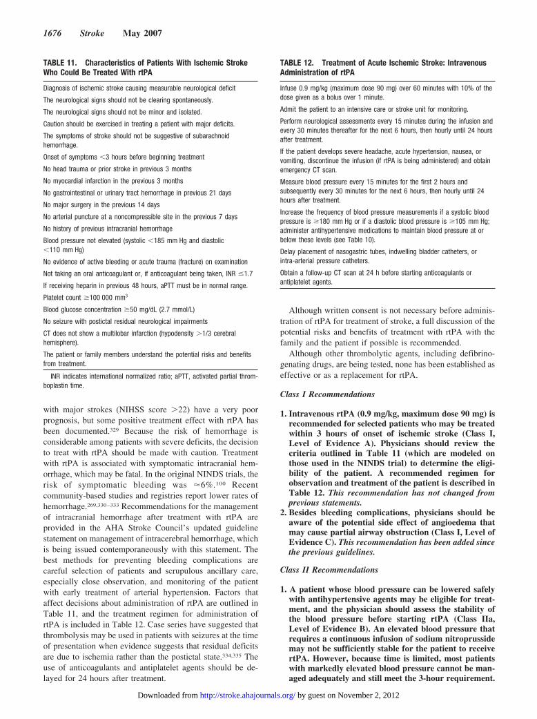
with major strokes (NIHSS score �22) have a very poorprognosis, but some positive treatment effect with rtPA hasbeen documented.329 Because the risk of hemorrhage isconsiderable among patients with severe deficits, the decisionto treat with rtPA should be made with caution. Treatmentwith rtPA is associated with symptomatic intracranial hem-orrhage, which may be fatal. In the original NINDS trials, therisk of symptomatic bleeding was �6%.100 Recentcommunity-based studies and registries report lower rates ofhemorrhage.269,330–333 Recommendations for the managementof intracranial hemorrhage after treatment with rtPA areprovided in the AHA Stroke Council’s updated guidelinestatement on management of intracerebral hemorrhage, whichis being issued contemporaneously with this statement. Thebest methods for preventing bleeding complications arecareful selection of patients and scrupulous ancillary care,especially close observation, and monitoring of the patientwith early treatment of arterial hypertension. Factors thataffect decisions about administration of rtPA are outlined inTable 11, and the treatment regimen for administration ofrtPA is included in Table 12. Case series have suggested thatthrombolysis may be used in patients with seizures at the timeof presentation when evidence suggests that residual deficitsare due to ischemia rather than the postictal state.334,335 Theuse of anticoagulants and antiplatelet agents should be de-layed for 24 hours after treatment.
Although written consent is not necessary before adminis-tration of rtPA for treatment of stroke, a full discussion of thepotential risks and benefits of treatment with rtPA with thefamily and the patient if possible is recommended.
Although other thrombolytic agents, including defibrino-genating drugs, are being tested, none has been established aseffective or as a replacement for rtPA.
Class I Recommendations
1. Intravenous rtPA (0.9 mg/kg, maximum dose 90 mg) isrecommended for selected patients who may be treatedwithin 3 hours of onset of ischemic stroke (Class I,Level of Evidence A). Physicians should review thecriteria outlined in Table 11 (which are modeled onthose used in the NINDS trial) to determine the eligi-bility of the patient. A recommended regimen forobservation and treatment of the patient is described inTable 12. This recommendation has not changed fromprevious statements.
2. Besides bleeding complications, physicians should beaware of the potential side effect of angioedema thatmay cause partial airway obstruction (Class I, Level ofEvidence C). This recommendation has been added sincethe previous guidelines.
Class II Recommendations
1. A patient whose blood pressure can be lowered safelywith antihypertensive agents may be eligible for treat-ment, and the physician should assess the stability ofthe blood pressure before starting rtPA (Class IIa,Level of Evidence B). An elevated blood pressure thatrequires a continuous infusion of sodium nitroprussidemay not be sufficiently stable for the patient to receivertPA. However, because time is limited, most patientswith markedly elevated blood pressure cannot be man-aged adequately and still meet the 3-hour requirement.
TABLE 11. Characteristics of Patients With Ischemic StrokeWho Could Be Treated With rtPA
Diagnosis of ischemic stroke causing measurable neurological deficit
The neurological signs should not be clearing spontaneously.
The neurological signs should not be minor and isolated.
Caution should be exercised in treating a patient with major deficits.
The symptoms of stroke should not be suggestive of subarachnoidhemorrhage.
Onset of symptoms �3 hours before beginning treatment
No head trauma or prior stroke in previous 3 months
No myocardial infarction in the previous 3 months
No gastrointestinal or urinary tract hemorrhage in previous 21 days
No major surgery in the previous 14 days
No arterial puncture at a noncompressible site in the previous 7 days
No history of previous intracranial hemorrhage
Blood pressure not elevated (systolic �185 mm Hg and diastolic�110 mm Hg)
No evidence of active bleeding or acute trauma (fracture) on examination
Not taking an oral anticoagulant or, if anticoagulant being taken, INR �1.7
If receiving heparin in previous 48 hours, aPTT must be in normal range.
Platelet count �100 000 mm3
Blood glucose concentration �50 mg/dL (2.7 mmol/L)
No seizure with postictal residual neurological impairments
CT does not show a multilobar infarction (hypodensity �1/3 cerebralhemisphere).
The patient or family members understand the potential risks and benefitsfrom treatment.
INR indicates international normalized ratio; aPTT, activated partial throm-boplastin time.
TABLE 12. Treatment of Acute Ischemic Stroke: IntravenousAdministration of rtPA
Infuse 0.9 mg/kg (maximum dose 90 mg) over 60 minutes with 10% of thedose given as a bolus over 1 minute.
Admit the patient to an intensive care or stroke unit for monitoring.
Perform neurological assessments every 15 minutes during the infusion andevery 30 minutes thereafter for the next 6 hours, then hourly until 24 hoursafter treatment.
If the patient develops severe headache, acute hypertension, nausea, orvomiting, discontinue the infusion (if rtPA is being administered) and obtainemergency CT scan.
Measure blood pressure every 15 minutes for the first 2 hours andsubsequently every 30 minutes for the next 6 hours, then hourly until 24hours after treatment.
Increase the frequency of blood pressure measurements if a systolic bloodpressure is �180 mm Hg or if a diastolic blood pressure is �105 mm Hg;administer antihypertensive medications to maintain blood pressure at orbelow these levels (see Table 10).
Delay placement of nasogastric tubes, indwelling bladder catheters, orintra-arterial pressure catheters.
Obtain a follow-up CT scan at 24 h before starting anticoagulants orantiplatelet agents.
1676 Stroke May 2007
by guest on November 2, 2012http://stroke.ahajournals.org/Downloaded from
This recommendation has not changed from previousguidelines.
2. A patient with a seizure at the time of onset of strokemay be eligible for treatment as long as the physician isconvinced that residual impairments are secondary tostroke and not a postictal phenomenon (Class IIa, Levelof Evidence C). This recommendation differs from theprevious statements and represents a broadening of eli-gibility for treatment with rtPA.
Class III Recommendations
1. The intravenous administration of streptokinase fortreatment of stroke is not recommended (Class III,Level of Evidence A). This recommendation has notchanged from previous guidelines.
2. The intravenous administration of ancrod, tenect-eplase, reteplase, desmoteplase, urokinase, or otherthrombolytic agents outside the setting of a clinical trialis not recommended (Class III, Level of Evidence C).This recommendation is new.
VII. Intra-Arterial ThrombolysisThe 2003 guidelines concluded that intra-arterial administra-tion of at least one specific thrombolytic agent, recombinantprourokinase, appears to be of some benefit in treatment ofcarefully selected patients with acute ischemic stroke second-ary to occlusion of the MCA.2 This conclusion was based onthe results of a prospective, randomized, placebo-controlledphase III study testing the effectiveness of intra-arterialthrombolysis with prourokinase among patients with strokeof �6 hours’ duration secondary to occlusion of the MCA.336
In the primary intent-to-treat analysis, 40% of the 121patients treated with recombinant prourokinase and 25% ofthe 59 control patients had a modified Rankin Scale score of0 to 2 at 90 days (P�0.04). Recanalization of the MCA wasachieved in 66% of the patients treated with recombinantprourokinase and 18% of the patients in the control group(P�0.001). Intracranial hemorrhage with neurological dete-rioration within 24 hours of treatment occurred in 10% ofpatients treated with recombinant prourokinase and in 2% ofthe control group (P�0.06). No difference in overall deathrate was seen between the 2 groups. The FDA has notapproved the drug, and recombinant prourokinase is notcurrently available for clinical use. Extrapolation to rtPA, thewidely used intravenous cerebral thrombolytic drug, and tourokinase, which is chemically similar to the prourokinaseprodrug, is based on consensus and case series data. Extrap-olation to other lytic agents is more speculative.
The intra-arterial approach has been promoted because ahigh concentration of thrombolytic agents may be deliveredinto the thrombus.337 Despite the uncontrolled observationthat recanalization rates may be higher with intra-arterialthrombolysis than with intravenous thrombolysis,337 clinicalbenefit may be counterbalanced by delays to initiating treat-ment with the intra-arterial approach. On the basis of 1999–2001 National Hospital Discharge Survey data,338 there were1 796 513 admissions for ischemic stroke between 1999 and2001. Of these admissions, 1314 (0.07%) underwent intra-arterial thrombolysis, and 11 283 (0.6%) underwent intrave-nous thrombolysis. A second estimate is derived from the
Greater Buffalo and Erie County stroke study that suggestedthat intravenous or intra-arterial thrombolysis was used in1.4% and 0.3% of 1590 patients admitted in 11 hospitals.329
Intra-arterial administration of rtPA to patients with ischemicstroke who are expected to have limited response to intrave-nous therapy has gained popularity. Potential reasons includesevere neurological deficits (NIHSS score �10), presentationbetween 3 and 6 hours after symptom onset, recent history ofmajor surgical procedures, and occlusion of major cervicaland/or intracranial vessels. However, only limited data sup-port the usefulness of intra-arterial therapy in these situations.
New evidence since the publication of the last guidelines issummarized in subsequent sections. More emphasis has beenplaced on deriving information from the initial angiogram,with emphasis on the site of occlusion and identification ofcollateral supply to the affected region. New data suggest thatthis information may be incorporated into a scheme to stratifypatients into the expected rate of recanalization and short-term outcome after intra-arterial thrombolysis.337,339,340
A small randomized, multicenter trial compared intrave-nous urokinase with intra-arterial urokinase within the first 6hours of acute ischemic stroke.341 Patients fulfilling theselection criteria were randomly assigned to receive uroki-nase 900 000 U via intravenous (n�14) or intra-arterial(n�13) approach. The study was terminated prematurelybecause 7 patients (26%) died: 4 in the intravenous group and3 in the intra-arterial group. Although patients treated withintra-arterial therapy showed greater and earlier improve-ment, no significant difference was seen in primary andsecondary outcomes. Recently, a cohort study was reportedfrom Japan’s Multicenter Stroke Investigator’s Collaboration(J-MUSIC).342 The modified Rankin Scale score at dischargewas lower in the urokinase group than in the control group(mean, 2.8 in the urokinase group versus 3.3 in the controlgroup; P�0.03). A favorable outcome (modified RankinScale score of 0 to 2) was more frequently observed in theurokinase group (51%) than in the control group (34%;P�0.01). A third study343 randomized 16 patients withangiographic evidence of posterior circulation vascular oc-clusion who presented within 24 hours of symptom onset toeither intra-arterial prourokinase or conservative manage-ment. Some imbalance between groups existed, with greaterseverity of deficit at baseline observed in the treatment arm.Good outcomes were observed in 4 of 8 patients whoreceived intra-arterial urokinase and in 1 of 8 patients in thecontrol group.
It has been proposed that patients be selected for intra-arterial thrombolysis on the basis of radiological criteria. Anonrandomized study344 compared outcomes of 83 patientswith or without a hyperdense artery sign on initial CT scantreated with intravenous or intra-arterial rtPA. An increasedlikelihood of favorable outcomes, indicated by a significantimprovement in the discharge NIHSS score, was noted withintra-arterial rtPA treatment, irrespective of the presence orabsence of hyperdense artery sign. A less favorable outcomein discharge NIHSS score was noted with intravenous rtPA inpatients with a hyperdense artery sign than in those withoutthe hyperdense artery sign. This suggests that differentialresponse to intravenous rtPA in patients with hyperdense
Adams et al Early Management of Adults With Ischemic Stroke 1677
by guest on November 2, 2012http://stroke.ahajournals.org/Downloaded from
artery sign344 on initial CT scan is not observed withintra-arterial thrombolysis.
A. Conclusions and RecommendationsSince the publication of the last guidelines, no new Class Ievidence has been published. Intra-arterial administration ofat least one specific thrombolytic agent appears to be ofbenefit in the treatment of carefully selected patients withacute ischemic stroke secondary to occlusion of the MCA.New evidence about the use of intra-arterial urokinase inpatients with vertebral or basilar artery occlusion treatedwithin 24 hours of symptom onset and patients with embolicstroke involving the anterior circulation within 4.5 hours ofsymptom onset suggests that intra-arterial therapy may beused. Patients who are evaluated within 6 hours of symptomsbut who are ineligible to receive intravenous thrombolysisbecause of recent surgery or other procedures may becandidates for intra-arterial thrombolysis.345,346
New criteria have been established to determine the qual-ifications of physicians who can perform intra-arterialthrombolysis on the basis of recent statements from profes-sional organizations and clinical trials.
Class I Recommendations
1. Intra-arterial thrombolysis is an option for treatmentof selected patients who have major stroke of <6 hours’duration due to occlusions of the MCA and who are nototherwise candidates for intravenous rtPA (Class I,Level of Evidence B). This recommendation has notchanged since previous guidelines.
2. Treatment requires the patient to be at an experiencedstroke center with immediate access to cerebral angiog-raphy and qualified interventionalists. Facilities areencouraged to define criteria to credential individualswho can perform intra-arterial thrombolysis (Class I,Level of Evidence C). This recommendation has beenadded since previous guidelines.
Class II Recommendation
1. Intra-arterial thrombolysis is reasonable in patients whohave contraindications to use of intravenous thrombolysis,such as recent surgery (Class IIa, Level of Evidence C).This recommendation was not included in the previousguideline.
Class III Recommendation
1. The availability of intra-arterial thrombolysis shouldgenerally not preclude the intravenous administrationof rtPA in otherwise eligible patients (Class III, Level ofEvidence C). This recommendation has not changedfrom previous guidelines.
VIII. AnticoagulantsPhysicians have used anticoagulants to treat patients withacute ischemic stroke for �50 years. These medicationscontinue to be prescribed commonly.347 Despite their wide-spread use, the usefulness of emergency anticoagulation is thesubject of debate.348–352 Disagreements exist about the bestagent to administer, the route of administration, the use of abolus dose to start treatment, the level of anticoagulation
required, and the duration of treatment. In a small randomizedtrial, Toth353 found no increase in serious bleeding compli-cations with the use of a bolus dose to start heparin antico-agulation. A weight-based nomogram for administration ofheparin after stroke has been developed.354 This approachseems to lessen the necessity for frequent adjustments indose. In the past, panels of the AHA have concluded that thedata about the utility of heparin in the management of strokeare either uncertain or largely negative.1–3,355,356 Besides theuncertainty about efficacy, the safety concern exists thaturgent anticoagulation may lead to symptomatic intracranialhemorrhage. Physicians have been uncertain about the sever-ity of neurological impairments or the initial CT findings thatwould contraindicate the early use of heparin.
Anticoagulants often are prescribed to patients with recentstroke in an effort to prevent early recurrent stroke and toimprove neurological outcomes. The Cerebral EmbolismStudy Group estimated that the risk of early recurrentembolism was �12% among untreated patients with embolicstroke.357,358 A Norwegian trial testing urgent anticoagulationamong patients with recent stroke and atrial fibrillation foundthe risk of recurrent stroke to be �8% in 1 week.359 Othertrials testing anticoagulants in stroke have found the rates ofearly recurrent stroke to be much lower (in the range of0.3%/d to 0.5%/d).360–362 These relatively low rates mean thatdetection of a therapeutic effect in prevention of earlyrecurrent stroke by anticoagulation will be difficult.
A. HeparinThe International Stroke Trial tested 2 doses (5000 U/d or25 000 U/d) of subcutaneously administered heparin whenthe medication was started within 48 hours of stroke.361
Although the trial included randomization in its design,investigators and patients knew the nature of the treatment.Dual randomization meant that approximately one half of thepatients receiving heparin also were receiving aspirin. Neithermonitoring of the level of anticoagulation nor adjustment ofdosages to biological responses was done. Thus, some pa-tients may have received excessive doses of heparin, with anincreased risk of bleeding complications, and others mayhave had inadequate dosages, with a resultant loss of effec-tiveness. In addition, patients enrolled in this very large trialdid not need to have a brain imaging study before treatment.Although heparin was effective in lowering the risk of earlyrecurrent stroke, an increased rate of bleeding complicationsnegated this benefit. A subgroup analysis looking at theeffects of heparin among patients with atrial fibrillation didnot demonstrate a benefit from the agent.363
A Swedish study testing the utility of heparin for treatmentof patients with progressing stroke did not demonstrate abenefit from the anticoagulant.364 Recently, 2 small Europeantrials have tested the utility of heparin in treatment of patientswith recent stroke.365,366 Investigators tested continuouslyintravenously administered heparin starting with a bolus dosewith adjustments in dosage in response to activated partialthromboplastin time in a small clinical trial that enrolledpatients within 12 hours of onset of stroke. The multicentertrial treated 32 patients with heparin and 35 with aspirin(control). No significant differences in outcomes, recurrent
1678 Stroke May 2007
by guest on November 2, 2012http://stroke.ahajournals.org/Downloaded from
ischemic stroke, hemorrhagic worsening, or death were notedbetween the 2 treatment groups. A single-center study in-volved the randomization of patients with acute nonlacunarhemispheric infarctions to treatment with either adjustedintravenous infusions of heparin without an initial bolus doseor saline.365 The trial enrolled 418 patients (208 on heparin)within 3 hours of onset of stroke. Thirteen heparin-treatedpatients had symptomatic hemorrhagic complications (6.2%)(7 were fatal), and symptomatic hemorrhagic events werediagnosed in 3 control patients (1.4%). Favorable outcomes at90 days were noted in 81 of 208 patients treated with heparin(38.9%) and 60 of 210 patients (28.6%) in the control group.A meta-analysis of the studies of heparin found no benefitfrom administration of the medication.367 However, thisanalysis was performed before the 2 recent reports aboutheparin were published. Nevertheless, the results of therelatively small study by Camerlingo et al365 likely would bedwarfed by the data from the International Stroke Trial.
B. Low-Molecular-Weight Heparinsand DanaparoidSeveral trials have tested low-molecular-weight (LMW) he-parins or danaparoid for treatment of patients with acuteischemic strokes. Results generally have been negative. Atrial in Hong Kong tested 2 doses of subcutaneously admin-istered nadroparin given over 10 days after stroke.368 Al-though no benefit from treatment was found at the end of thetreatment period or at 3 months, those who received the largerdose of nadroparin had a significantly lower death rate at 6months than did the control group. Another trial of nadroparinfailed to find any improvement in the rate of favorableoutcomes with treatment, but the rate of serious bleeding wasincreased with the larger of 2 doses of LMW heparin.369
Berge et al359 compared the utility of dalteparin or aspirin forprevention of early recurrent stroke or improvement inneurological outcome among patients with presumed cardio-embolic stroke. Although no significant differences werenoted in outcomes or the rates of recurrent strokes, thepatients taking aspirin had fewer second events. The rate ofbleeding complications also was higher among patients whowere treated with dalteparin than among those given aspirin.A German trial compared 4 different doses of certoparin; nodifferences in rates of favorable outcomes were noted amongthe groups, but the rate of serious bleeding complications washighest among the group that received the largest dose of theLMW heparin.370 An aspirin-controlled trial tested 2 differentdoses of subcutaneously administered tinazaparin in patientswith recent stroke, with no differences in favorable outcomes,rates of recurrent stroke, death, or bleeding complications.371
A randomized, double-blind, placebo-controlled trial testedthe utility of a continuous intravenous infusion of the LMWheparinoid (danaparoid) in improving outcomes after acuteischemic stroke.362 The trial halted recruitment of patientswith moderate to severe stroke (NIHSS scores of �15)because of an increased risk of symptomatic intracranialhemorrhages. The medication did not lessen the risk ofneurological worsening or early recurrent stroke, includingamong patients with cardioembolic events. No improvementin the chance of having a favorable or very favorable outcome
was found at 3 months. The trial included prespecifiedsubgroup analyses among patients with different subtypes ofischemic stroke. The only subgroup that showed benefit fromtreatment included patients with stroke secondary to large-artery atherosclerosis.372 This finding may be supported bythe results of the study by Lovett et al,373 which found that therisk of early recurrent stroke was highest among patients withsevere large-artery atherosclerotic disease. A study examin-ing the usefulness of a LMW heparin in treating patients withstroke secondary to intracranial stenotic disease has beenperformed in Hong Kong and Singapore, but the results havenot yet been published.
Some studies have compared the utility of heparin or LMWheparins in treatment of patients with recent stroke. Woessneret al374 compared the usefulness of subcutaneously adminis-tered enoxaparin or adjusted-dose heparin in a multicentertrial that randomized patients with either high-grade arterialstenoses or a cardioembolic source for stroke. No significantdifferences were noted between the 2 regimens. Another trialinvestigated the efficacy of subcutaneously administeredenoxaparin in comparison to heparin for prevention of throm-boembolic events among patients with lower-limb paralysisafter stroke.375 The 2 medications had equal efficacy.
C. Anticoagulants as an Adjunctive TherapyThe administration of anticoagulants or antiplatelet agents iscurrently contraindicated during the first 24 hours aftertreatment with intravenous rtPA. This restriction is based onthe regimen used in the NINDS trials.66 Arterial reocclusionmay follow successful recanalization with thrombolysis.376
Thus, there is interest in the use of an anticoagulant that maymaintain arterial patency after thrombolytic therapy. Thetrials of intra-arterial administration of prourokinase usedheparin as part of the treatment regimen, and the controlgroup received only heparin.336,377,378 In the first study, boththe success of recanalization and the risk of hemorrhage wereincreased among the patients who received the larger of the 2doses of adjunctive heparin. Two small studies have testedthe use of intravenously administered heparin after treatmentwith rtPA.379,380 No increase in bleeding complications hasbeen reported. Heparin also has been given in combinationwith abciximab with a reasonable degree of safety.381 Theexperience with adjunctive anticoagulation is limited. Neithersafety nor effectiveness has been established, and additionalresearch is needed.
D. Conclusions and RecommendationsThe results of the recent trials show that early administrationof either heparin or a LMW heparin/danaparoid is associatedwith an increased risk of bleeding complications. Thesemedications increase the risk of symptomatic hemorrhagictransformation of ischemic strokes, especially among personswith severe events. These medications are also associatedwith a risk of serious bleeding in other parts of the body.Although the likelihood of bleeding appears to be lower thanthat associated with the administration of thrombolyticagents, it is sufficiently high to require convincing evidenceof efficacy to justify urgent anticoagulation. The risk ofbleeding appears not to be greatly affected by the use of a
Adams et al Early Management of Adults With Ischemic Stroke 1679
by guest on November 2, 2012http://stroke.ahajournals.org/Downloaded from
bolus dose to start treatment or by the route of administration(subcutaneous or intravenous). Monitoring of the level ofanticoagulation and adjustment of the dosages in response tolevels probably increase the safety of treatment.
Present data indicate that early administration of heparin orthe LMW heparins/danaparoid does not lower the risk ofearly recurrent stroke, including among patients with cardio-embolic stroke. Early administration of anticoagulants doesnot lessen the risk of early neurological worsening. Data arenot sufficient to indicate whether anticoagulants might haveefficacy among some potentially high-risk groups, such aspersons with intracardiac or intra-arterial thrombi. The effi-cacy of urgent anticoagulation is not established for treatmentof patients with vertebrobasilar disease or an arterialdissection.
Most trials have not demonstrated the efficacy of antico-agulation in improving outcomes after acute ischemic stroke.One relatively small trial found that intravenous heparin,when administered within 3 hours of onset of stroke topatients with nonlacunar stroke, may improve outcomes. Inlight of the generally negative data, the results of this trialmay need to be replicated. Because the time window forpotentially effective treatment with heparin is the same as forintravenously administered rtPA, a study may be needed totest the relative efficacy of heparin or thrombolysis.
The role of anticoagulants as an adjunctive therapy inaddition to mechanical or pharmacological thrombolysis hasnot been defined.
The following recommendations have not changed fromprevious guidelines.
Class III Recommendations
1. Urgent anticoagulation with the goal of preventingearly recurrent stroke, halting neurological worsening,or improving outcomes after acute ischemic stroke isnot recommended for treatment of patients with acuteischemic stroke (Class III, Level of Evidence A). Thisrecommendation may change if additional data demon-strate the usefulness of very early intravenous admin-istration of anticoagulants for treatment of patientswith infarctions secondary to large-artery thrombosisor cardioembolism. Urgent anticoagulation should notbe used in lieu of intravenous thrombolysis for treat-ment of otherwise eligible patients (Class III, Level ofEvidence A).
2. Urgent anticoagulation is not recommended for pa-tients with moderate to severe strokes because of anincreased risk of serious intracranial hemorrhagic com-plications (Class III, Level of Evidence A).
3. Initiation of anticoagulant therapy within 24 hours oftreatment with intravenously administered rtPA is notrecommended (Class III, Level of Evidence B).
IX. Antiplatelet AgentsThe 2003 guideline2 recommendation that aspirin could beused after stroke was consistent with the recent Joint Guide-line Statement from the AHA and the American Academy ofNeurology.356 The administration of aspirin as an adjunctivetherapy, within 24 hours of the use of thrombolytic agents,was not recommended. Aspirin was not indicated as a
substitute for other acute interventions, especially intravenousadministration of rtPA, for the treatment of acute ischemicstroke.
A. Single Oral Antiplatelet AgentAspirin is the only oral antiplatelet agent that has beenevaluated for the treatment of acute ischemic stroke. Twolarge trials360,361 each showed a nonsignificant trend inreduction in death or disability when treatment with aspirinwas initiated within 48 hours of stroke. A small increase inbleeding complications was also noted. When the data fromthe 2 trials were combined, a modest but statistically signif-icant benefit from aspirin was noted. The primary effectseemed to be in prevention of recurrent events. It is not clearwhether aspirin limited the neurological consequences of theacute ischemic stroke itself.
The use of ticlopidine, clopidogrel, or dipyridamole in thesetting of acute ischemic stroke has not been evaluated.382
Initiation of treatment with clopidogrel in a daily dose of 75mg does not cause maximal platelet inhibition for �5 days.383
This delay poses the issue of an early therapeutic effect fortreatment of patients with acute stroke. A bolus dose ofclopidogrel 300 mg inhibits platelet aggregation rapidly.384 A300-mg loading dose of clopidogrel followed by daily dosesof 75 mg/d has been recommended for treatment of patientswith acute coronary syndrome (ACS) who have aspirinallergies.385 No data are available about the utility of thisstrategy in treating patients with acute ischemic stroke.386,387
B. Combination of Oral Antiplatelet AgentsAlthough the combination of clopidogrel and aspirin isprescribed for patients with ACS, this combination has notbeen studied in the setting of acute ischemic stroke. No dataare available about the use of other combinations of antiplate-let agents in the management of acute ischemic stroke.
C. Intravenous Antiplatelet AgentsThe platelet glycoprotein IIb/IIIa inhibitors have been con-sidered in the treatment of acute ischemic stroke because theymay increase the rate of spontaneous recanalization andimprove microvascular patency.388,389 A small case series390
demonstrated that intravenous abciximab administered be-tween 3 and 24 hours after symptom onset in patients withsupratentorial ischemic stroke may attenuate ischemic lesiongrowth as demonstrated by serial DWI. A double-blind,placebo-controlled, dose-escalation trial randomly allocated74 patients to receive either an escalating dose of abciximab(54 patients) or placebo (20 patients) in a 3:1 ratio within 24hours after ischemic stroke onset.390,391 No symptomaticintracerebral hemorrhages occurred in any group, whichsuggests that intravenous abciximab is relatively safe whenadministered within 24 hours of symptom onset in selectedpatients with ischemic stroke. A second randomized, double-blind, placebo-controlled phase II trial randomized 400 pa-tients within 6 hours of ischemic stroke onset to intravenousabciximab or placebo.392 The rates of symptomatic hemor-rhage during the first 5 days after stroke were not signifi-cantly different between the abciximab and placebo groups(3.6% versus 1%). Treatment with abciximab showed a
1680 Stroke May 2007
by guest on November 2, 2012http://stroke.ahajournals.org/Downloaded from
nonsignificant shift in favorable outcomes defined by modi-fied Rankin Scale scores at 3 months, after adjustment forbaseline severity of stroke, age, and interval fromstroke.391,392 A phase III, multinational, multicenter, random-ized, double-blind, placebo-controlled study evaluating thesafety and efficacy of abciximab in patients with acuteischemic stroke has been halted because of an increased rateof bleeding. Details are not yet available.
D. Conclusions and RecommendationsNo new data are available about the utility of oral antiplateletagents (used singly or in combination) for treatment ofpatients with acute ischemic stroke. Currently available datademonstrate a small but statistically significant decline in riskof mortality and morbidity when aspirin is started within 48hours after onset of stroke. It appears that the primary effectsof the aspirin are due to reduction of early recurrent strokerather than limitation of the neurological consequences of thestroke. No data are available on the utility of other antiplateletagents, including clopidogrel, given as monotherapy or incombination with aspirin for treatment of patients with acuteischemic stroke. The relative indications for the long-termadministration of antiplatelet agents to prevent recurrentstroke are beyond the scope of this statement. However, thepanel recommends the administration of such agents as partof management after acute stroke.
Ongoing research is testing the usefulness of intravenouslyadministered antiplatelet agents (glycoprotein IIb/IIIa recep-tor blockers) when given alone or in combination with otherinterventions. Preliminary evidence suggests that theseagents, when used alone, have an acceptable safety profile,but considerably more research is needed to determinewhether these agents have a role in the management of acutestroke in conjunction with other therapies.
Class I Recommendation
1. The oral administration of aspirin (initial dose is 325mg) within 24 to 48 hours after stroke onset is recom-mended for treatment of most patients (Class I, Level ofEvidence A). This recommendation has changed in thata dose of aspirin is now included.
Class III Recommendations
1. Aspirin should not be considered a substitute for otheracute interventions for treatment of stroke, includingthe intravenous administration of rtPA (Class III, Levelof Evidence B). These recommendations have notchanged from previous statements.
2. The administration of aspirin as an adjunctive therapywithin 24 hours of thrombolytic therapy is not recom-mended (Class III, Level of Evidence A). This recom-mendation has not changed.
3. The administration of clopidogrel alone or in combina-tion with aspirin is not recommended for the treatmentof acute ischemic stroke (Class III, Level of EvidenceC). This recommendation was not included in the previ-ous statement. The panel supports research testing theusefulness of emergency administration of clopidogrelin the treatment of patients with acute stroke.
4. Outside the setting of clinical trials, the intravenousadministration of antiplatelet agents that inhibit theglycoprotein IIb/IIIa receptor is not recommended(Class III, Level of Evidence B). This recommendationhas been added since the last guideline was published.
X. Volume Expansion, Vasodilators, andInduced Hypertension
A. Hemodilution in Acute Ischemic StrokeIt is known that patients experiencing acute ischemic strokemay have a variety of abnormalities that increase whole-blood viscosity, including leukocyte activation, red cell ag-gregation, and reduced red cell deformability.393–395 In addi-tion, elevated fibrinogen levels, which add to plasmaviscosity, have been documented. On the basis of thesefindings, hemodilution, with or without venesection, has beenstudied. The goal is to improve cerebral blood flow tohyperperfuse potentially viable brain tissue supplied by lep-tomeningeal collaterals in an attempt to perfuse the ischemicpenumbra.396–400
Volume expansion and hemodilution have been demon-strated to improve blood flow but may reduce oxygen-carrying capacity at hematocrits �30%.401–408 As the hemat-ocrit is reduced, oxygen delivery increases as much as 28% to30%; however, at levels lower than this, oxygen deliverybegins to decline.409
In patients with cerebral infarction, cerebral autoregulatorymechanisms are impaired, and hemodilution has been notedto increase cerebral blood flow in the infarcted as well as thecontralateral hemisphere. Oxygen-carrying molecules such asperfluorocarbons or diaspirin cross-linked hemoglobin havebeen shown to reduce brain infarct size in animal models buthave not yet been assessed and cannot be recommended inpatients.383,410–417
Investigations of intravascular volume and hemodilution inpatients began in the 1960s. Initial reports, from studies thatwere not controlled and not randomized, were encouragingand suggested clinical benefits of intravascular volume ex-pansion. In a review of recent trials for hemodilution instroke, a combination of plasma volume expansion with orwithout venesection was used.387,416–422 Several trials usedplasma volume expansion alone, either dextran 40 or hy-droxyethyl starch and albumin in one trial.405,423–425 In alltrials, and in the Multicenter Austrian Hemodilution StrokeTrial387 study in particular, hemodilution did not significantlyreduce deaths within the first 4 weeks but did influencedeaths within 3 to 6 months. Hemodilution also had nosignificant influence on death, dependency, orinstitutionalization/long-term care. In several trials, hemodi-lution was associated with a tendency toward reduction indeep venous thrombosis and pulmonary embolism at 3 to 6months. Despite volume expansion and hemodilution, the riskof significant cardiac events did not increase.
Conclusions and RecommendationsThe present data indicate that intentional hemodilution, withor without venesection in clinical practice, does not reducecase fatality or improve functional outcome in survivors. Thedata do not support the use of hypervolemia and isovolumic
Adams et al Early Management of Adults With Ischemic Stroke 1681
by guest on November 2, 2012http://stroke.ahajournals.org/Downloaded from
hemodilution protocols, including dextran, albumin, and hy-droxyethyl starch. The only possible exception for the use ofhemodilution is in stroke patients with severe polycythe-mia.426 Maintenance of a normal circulating blood volumewith regulation of metabolic parameters within physiologicalranges is desirable.
Class III Recommendation
1. Hemodilution with or without venesection and volumeexpansion is not recommended for treatment of pa-tients with acute ischemic stroke (Class III, Level ofEvidence A). This recommendation has not changedsince the previous guidelines were published.
B. Vasodilators in Acute Ischemic StrokeMethylxanthine derivatives are known vasodilators that alsohave other activities such as inhibition of platelet aggregation,reduction of free radical release, and inhibition ofthromboxane A2 synthesis.404,427–429 On the basis of thesecharacteristics, methylxanthine derivatives, specifically pen-toxifylline, propentofylline, and pentifylline, have been eval-uated in the setting of acute ischemic stroke.430,431 In one trial,pentoxifylline and propentofylline were administered viacontinuous intravenous infusion over a 3-day period inpatients experiencing an acute ischemic event.432 Other stud-ies continued infusions for 5 days, as in the trial by Chan andKay in 1993, or 7 days, as in the trial of Huber et al in1993.433,434
Early case fatality, defined as within 4 weeks, was evalu-ated in several trials of pentoxifylline.432–435 The trial con-ducted by Chan and Kay found a large reduction in the oddsof early death; however, this was not borne out in otherstudies. Two trials demonstrated a nonsignificant reduction inearly death or deterioration.433,435 One trial compared thecombination of pentoxifylline and aspirin with aspirin aloneand found a significant decrease in short-term deaths.433
However, this trial studied early death and deterioration, andno significant reduction in these combined events was ob-served. The trial by Huber et al in 1993 included 30 patientsafter propentofylline treatment and found no difference in latecase fatality.434 The trial by Chan and Kay administeredaspirin to both patient groups with the theoretical advantagethat aspirin may potentiate the antiplatelet effects of pentoxi-fylline, which may explain the apparent clinical effectiveness.The trial by Hsu et al demonstrated that patients withprofound neurological deficit derived the greatest benefitfrom pentoxifylline.432
Conclusions and RecommendationsOn the basis of current data, neither pentoxifylline norpentofylline has been shown to improve outcomes afterstroke.
Class III Recommendation
1. The administration of medications such as pentoxifyl-line is not recommended for treatment of patients withacute ischemic stroke (Class III, Level of Evidence A).This recommendation has not changed since the previousguideline was published.
C. Induced Hypertension for the Management ofAcute Ischemic StrokeIn the setting of acute stroke, patients may have an ischemicpenumbra of brain tissue, which may have impaired perfusionbut may not be irreversibly damaged. In this situation, a localdrop in cerebral blood flow occurs, and cerebral arteriolesdilate in an attempt to compensate and maintain flow to thepotentially salvageable ischemic tissue.436 The magnitude ofthe low perfusion and duration of the ischemia are importantvariables.437,438 It is intuitively attractive that reperfusion ofthis area by dilatation of leptomeningeal collaterals may beadvantageous.
The optimal management of blood pressure in patientsexperiencing acute ischemic neurological events remainscontroversial.247,439,440 Markedly elevated blood pressure af-ter an acute ischemic event may increase the risk of conver-sion from an ischemic to a hemorrhagic lesion, with life-threatening implications.441 However, inducing hypertensionto increase cerebral blood flow has been attractive on thebasis of experimental data.437 Preliminary studies suggest thatthere may be a role for induced hypertension.247,442–446 Otherstudies by Olsen demonstrated that the ischemic penumbrahas impaired autoregulation and that induction of elevatedblood pressure partially restores perfusion to the penumbra,as demonstrated by single-photon emission CT imaging.447,448
In a study by Rordorf et al, 30 patients with acute ischemicevents were treated with intravenous phenylephrine, whichwas titrated to improvement of neurological deficit, as dem-onstrated in 33% of the patients.445,446 In this trial, patientswere entered within 12 hours of the onset of symptoms butwere excluded in the presence of recent or known cardiacischemic events, congestive heart failure, intracerebral hem-orrhage, or edema related to a completed infarction. Patientswere maintained hypertensive for a period of 1 to 6 days, andneurological status at discharge was improved in those whohad responded to the therapy. In other reports by Hillis et al,patients were randomized to induced hypertension or conven-tional management.443,444 Serial DWI and PWI studies wereperformed before and during the period of induced hyperten-sion. After 3 days of treatment, patients who were treatedwith induced hypertension showed an improvement in neu-rological examination, no change in DWI lesion volume, andreductions in PWI abnormality.
Wityk et al used induced hypertension for treatment ofpatients who were within 12 hours of onset of symptoms butwho could not be treated with thrombolytic therapy.449,450
They also advocated the use of DWI and PWI as a means todemonstrate improved perfusion to monitor such patients inan intensive care setting and when intervention is undertakento achieve a target systolic blood pressure or mean arterialpressure of 20% to 30% above baseline. Other treatmentoptions include withdrawing previous antihypertensive ther-apy, augmenting intravenous fluids, or starting intravenousphenylephrine. As part of the treatment paradigm, serialneurological examinations are performed to determinewhether neurological improvement occurs; patients whoshow no improvement in a 30- to 60-minute period areconsidered nonresponders, and the intervention is stopped.Patients who have responded to this mode of therapy are
1682 Stroke May 2007
by guest on November 2, 2012http://stroke.ahajournals.org/Downloaded from
maintained at that level and weaned according to neurologicalexamination and functional imaging. Data demonstrating thesafety and efficacy of this strategy are needed.
Conclusions and RecommendationsPreliminary and small clinical studies suggest that drug-induced hypertension could be used in the management ofsome patients with acute ischemic stroke. However, datafrom large clinical trials are not available. Thus, the efficacyof this treatment strategy has not been established. Theadministration of vasopressors may be complicated by sideeffects, including myocardial ischemia, in some patients withstroke. Some patients may not be able to be treated with thistherapy. The safety of drug-induced hypertension for treat-ment of stroke in a broad spectrum of patients has not beenestablished. Further research is needed.
Class I Recommendation
1. In exceptional cases, a physician may prescribe vaso-pressors to improve cerebral blood flow. If drug-induced hypertension is used, close neurological andcardiac monitoring is recommended (Class I, Level ofEvidence C). This recommendation has been added sincethe previous guideline was published.
Class III Recommendation
1. Drug-induced hypertension, outside the setting of clin-ical trials, is not recommended for treatment of mostpatients with acute ischemic stroke (Class III, Level ofEvidence B). This recommendation has been added sincethe previous guideline was published.
XI. Surgical InterventionsA. Carotid EndarterectomyLittle information exists about the effectiveness of surgicaltreatment of patients with acute ischemic stroke. Most casesof immediate operation are performed in the setting of anacute stroke after carotid endarterectomy. Emergency carotidendarterectomy generally is not performed in other settings ofacute ischemic stroke because the risks of the procedure areperceived to be high. The sudden restoration of blood flowmight increase the development of brain edema or lead tohemorrhagic transformation, especially among patients withmajor infarctions. In addition, the time required for detectingthe arterial lesion and mobilizing the operating room limitsthe utility of surgery.
However, some surgeons report encouraging results fromemergency operations for patients with severe stenosis orocclusion of the internal carotid artery existing for �24hours.324,386,451–460 In general, improvement after surgery wasfound among patients with mild to moderate neurologicalimpairments. Still, the data are limited, and the usefulness ofurgent surgery among patients with severe neurologicaldeficits is even less clear.
The indications for immediate carotid endarterectomy in apatient with an acute ipsilateral ischemic stroke and anintraluminal thrombus associated with an atheroscleroticplaque at the carotid bifurcation are controversial. The mor-bidity associated with surgery appears to be high among
patients with an intraluminal thrombus demonstrated bycerebral angiography.461–464 Although some groups reportlow rates of complications and good neurological outcomeswith immediate surgery,461–463 others have reported betterresults when the patients are treated initially with anticoagu-lants followed by delayed operation.464
B. Other Surgical ProceduresImmediate extracranial-intracranial arterial bypass for treat-ment of ischemic stroke failed to improve outcomes and wasassociated with a high risk of intracranial hemorrhage.465
However, some surgeons have reported favorable results withemergency bypass procedures.466,467 In an occasional patientwith an acute neurological deficit secondary to an embolus ofthe MCA, outcome might be improved by an emergencymicrosurgical embolectomy of the MCA.468,469 Experiencewith immediate surgical procedures for treatment of acuteischemic stroke in the vertebrobasilar circulation is extremelylimited.
C. Conclusions and RecommendationsData on the safety and effectiveness of carotid endarterecto-my and other operations for treatment of patients with acuteischemic stroke are not sufficient to permit a recommenda-tion. Surgical procedures may have serious risks and may notfavorably alter the outcome of the patient.
XII. Endovascular InterventionsSeveral endovascular interventions are being evaluated forthe treatment of intracranial or extracranial arterial occlusionsleading to acute ischemic stroke.470–473 Options includeemergency angioplasty and stenting, mechanical disruption ofthe clot, and extraction of the thrombus. In most cases, themechanical intervention has been combined with either intra-venous or intra-arterial thrombolytic therapy.
A. Angioplasty and StentingLimited data are available about the use of angioplasty andstenting in the emergency treatment of intracranial or ex-tracranial lesions in patients with acute ischemic stroke.474–476
Angioplasty and stenting have been used to treat patients withacute stroke secondary to carotid artery dissection.477 In oneseries, emergency angioplasty and stenting of the internalcarotid artery were performed in conjunction with intra-arte-rial thrombolysis in 25 patients who had acute carotid arteryocclusion with secondary artery-to-artery embolism to theMCA.478 Results were compared with another group of 25patients who were treated medically; favorable outcomeswere more frequent (56% versus 26%) among patients withendovascular treatment. Jovin et al479 were successful inachieving recanalization in 23 of 25 patients who hademergency stenting of the extracranial internal carotid artery.Brekenfeld et al480 treated 350 patients with intra-arterialurokinase and noted that recanalization could be increasedwith angioplasty and implantation of stents. Angioplasty withor without stenting has been combined with emergencyadministration of thrombolytic agents in patients with occlu-sions in the vertebrobasilar circulation.481,482
Adams et al Early Management of Adults With Ischemic Stroke 1683
by guest on November 2, 2012http://stroke.ahajournals.org/Downloaded from
B. Mechanical Clot DisruptionNoser et al483 treated 16 patients with occlusion of the MCAand 16 others with occlusion of the internal carotid arterywith aggressive mechanical clot disruption. In most cases, theendovascular treatment was an adjunct to thrombolysis. ASwiss study of 350 patients treated with intra-arterial phar-macological thrombolysis found that mechanical fragmenta-tion improved the success for recanalization.480 Berlis et al484
used an endovascular photoacoustic device to speedrecanalization.
C. Clot ExtractionDevices have been used to extract thrombi from occludedintracranial arteries.485,486 In the Mechanical Embolus Re-moval in Cerebral Embolism (MERCI) trial, vessels wereopened with a device that removed the thrombus from anintracranial artery.487–489 The device was associated withrapid opening of the artery, but the efficacy in recanalizationand safety results achieved with the MERCI retrieval systemwere similar to those achieved with intra-arterial prouroki-nase in the Prolyse in Acute Cerebral Thromboembolism II(PROACT II) trial.336 The rate of recanalization of the MCAin MERCI was 45%, and it was 66% in PROACT II. InMERCI, 17 patients also received thrombolytic medicationswhen the device was unable to achieve recanalization, but theoutcomes of these patients were not reported. Although theFDA has approved the use of the MERCI device for reopen-ing intracranial arteries, its clinical utility has not beenestablished.
D. Conclusions and RecommendationsThe area of endovascular treatment of patients with acuteischemic stroke shows great promise. A number of techniquesand devices are being studied. Already, the FDA has ap-proved one device to extract a thrombus from an occludedintracranial artery. Other devices likely will be approved inthe future. Emergency angioplasty also may achieve a role inmanagement. As with the intra-arterial administration ofthrombolytics, the use of these devices will be limited tothose comprehensive stroke centers that have the resourcesand physician expertise to perform these procedures safely.
Class II Recommendations
1. Although the MERCI device is a reasonable interven-tion for extraction of intra-arterial thrombi in carefullyselected patients, the panel also recognizes that theutility of the device in improving outcomes after strokeis unclear (Class IIb, Level of Evidence B). This recom-mendation has been added since the previous guideline.The panel also recommends that the device be studiedin additional clinical trials that will define its role in theemergency management of stroke. This is the first timethat a panel has made a recommendation about endovas-cular treatment of patients with acute ischemic stroke.
2. The usefulness of other mechanical endovascular treat-ments is not established (Class IIb, Level of EvidenceC). These devices should be used in the setting ofclinical trials. This recommendation has not changedfrom previous guidelines.
XIII. Combination Reperfusion Therapyin Acute Stroke
Initial studies of thrombolytic therapy in acute ischemicstroke involved a single pharmacological agent given eitherintravenously or intra-arterially. Unfortunately, neither intra-venous nor intra-arterial thrombolysis with only a singlepharmacological agent is an efficient way to rapidly recana-lize occluded major brain arteries. Even when it works,intravenous or intra-arterial rtPA takes at least 15 to 30minutes to reopen an occluded major vessel such as the MCA,and no evidence indicates that other available thrombolyticagents are faster. Large-vessel occlusions of the internalcarotid artery or basilar artery often are resistant to intrave-nous or intra-arterial thrombolysis with 1 agent. Recenttranscranial Doppler ultrasonography studies suggest only a30% complete recanalization rate for MCA occlusion afteradministration of intravenous rtPA, a 48% partial recanaliza-tion rate, and a 27% reocclusion rate.376 The MCA completerecanalization rate with intra-arterial recombinant prouroki-nase was only 20% after 2 hours, with a 63% partialrecanalization rate and a 10% reocclusion rate within the firsthour of treatment.336 The low rate of complete recanalizationand the high rate of reocclusion with stroke thrombolysis isnot surprising when it is considered that not even aspirin isallowed for 24 hours after intravenous rtPA.
Faster and more complete recanalization should translateinto better patient outcomes. To achieve this, the trend inACS has been to use multiple pharmacological agents and,increasingly, percutaneous coronary intervention. The impe-tus is the more rapid and complete recanalization of occludedor stenosed coronary arteries. The standard treatment in manyACS patients includes antiplatelet therapy with aspirin, clo-pidogrel, glycoprotein IIb/IIIa blockers, antithrombotic ther-apy with heparin or LMW heparin, and direct percutaneouscoronary intervention.490 In patients with ACS, Thrombolysisin Myocardial Infarction (TIMI) 14487 reported the highestTIMI 3 complete recanalization rates with reduced-doseintravenous rtPA combined with the glycoprotein IIb/IIIaantagonist abciximab. However, the rate of brain hemorrhagewas increased in patients �75 years of age who receivedreduced-dose reteplase and abciximab in the Global Utiliza-tion of Streptokinase and tPA for Occluded Arteries(GUSTO) V trial.488
To improve the efficiency of acute stroke thrombolysis ina way that is similar to treatment of ACS, multimodalcombination therapies will need to be developed. Suchcombination therapy should not only increase the likelihoodof favorable outcomes but should also reduce the likelihoodof intracranial hemorrhage. The PROACT I study demon-strated that the recanalization efficacy and safety of recom-binant prourokinase was affected by the concomitant use ofheparin377; although the large dose of anticoagulant wasassociated with a higher rate of recanalization, it also wascomplicated by more bleeding. A major stimulus to thedevelopment of stroke thrombectomy devices has been thedesire to improve the speed and completeness of recanaliza-tion as compared with an intra-arterially administered drugalone (see the section on endovascular therapy). Externalultrasound has also been shown to enhance intravenous rtPA
1684 Stroke May 2007
by guest on November 2, 2012http://stroke.ahajournals.org/Downloaded from
in the Combined Lysis of Thrombus in Brain ischemia Using2 MHz Transcranial Ultrasound and Systemic tPA (CLOT-BUST) study.491 In CLOTBUST, complete MCA recanaliza-tion with dramatic clinical recovery occurred in 49% ofpatients receiving intravenous rtPA combined with transcra-nial ultrasound versus 30% in patients receiving intravenousrtPA only.
Another approach being evaluated is the initiation ofintravenous thrombolysis in the interim period between initialevaluation and intra-arterial treatment.72,492 The Interven-tional Management of Stroke (IMS) study reported on 80patients with an initial NIHSS score �10 treated withintravenous thrombolysis followed by intra-arterialthrombolysis.492 Patients received intravenous rtPA (0.6 mg/kg, 60 mg maximum over 30 minutes) started within 3 hoursof onset. Additional rtPA was then administered via micro-catheter in 62 patients with persistent occlusion at the site ofthe thrombus up to a total dose of 22 mg over 2 hours ofinfusion or until recanalization occurred. The 80 subjects hada median baseline NIHSS score of 18. The 3-month mortalityrate in treated patients (16%) was numerically lower than butnot statistically different from the mortality rate observed inthe placebo- (24%) and rtPA-treated subjects (21%) in theNINDS rtPA Stroke Trial. The rate of symptomatic intrace-rebral hemorrhage (6.3%) in IMS subjects was similar to thatof rtPA-treated subjects (6.6%) but higher than the rate inplacebo-treated subjects (1.0%; P�0.018) in the NINDS rtPAStroke Trial. IMS subjects had a significantly better outcomeat 3 months than NINDS placebo-treated subjects for alloutcome measures. The new data support the previous phaseI study72 and single-center case series493 supporting the safetyof this approach. On the basis of the results of the phase IIstudy, a phase III study has been planned.
The ongoing IMS II study again uses reduced-dose intra-venous rtPA combined with intra-arterial rtPA but nowdelivered through an ultrasonic EKOS catheter in an attemptto accelerate thrombolysis. Intra-arterial administration ofalteplase after intravenous administration of alteplase (0.6mg/kg) may provide benefit in patients with initial NIHSSscore �10 who present within 3 hours of symptom onset.
A. Combination of Thrombolysis andNeuroprotective TherapiesThere have also been very limited studies of combinationcytoprotective and reperfusion therapy in acute stroke (lube-luzole,494 hypothermia,495 magnesium,31 clomethiazole496).Until randomized clinical efficacy trials can be completed,combination reperfusion therapies for acute stroke will re-main experimental for the foreseeable future.
B. Thrombolysis and Antiplatelet AgentsUse of platelet glycoprotein IIb/IIIa inhibitors has been sug-gested to prevent platelet activation and reocclusion and is beingevaluated in several phase I studies as adjunctive therapy tointra-arterial thrombolysis. Other clinical trials of combinationplasminogen activator and glycoprotein IIb/IIIa inhibitors, somebased on perfusion brain imaging, are under way. Three recentlycompleted studies497–499 have suggested that intra-arterial ad-ministration of thrombolysis in combination with platelet
glycoprotein IIb/IIIa inhibitors may be safe and may poten-tially improve outcome. A multicenter study497 reported theresults of treating patients with acute vertebrobasilar occlu-sion using a combination of an intravenous bolus of abcix-imab (0.25 mg/kg) followed by 12-hour infusion therapy(0.125 �g/kg per minute) and intra-arterial rtPA in 47patients. The results were compared with a retrospectivecohort of 41 patients treated by intra-arterial rtPA mono-therapy (median dosage, 40 mg). The rates of symptomaticintracranial hemorrhages were 13% and 12% for combinationtreatment and thrombolytic treatments, respectively. Thecombination treatment had higher rates of complete recana-lization (45% versus 22%), favorable outcomes (34% versus17%), and survival (62% versus 32%) compared withthrombolysis alone. A second study498 evaluated 26 patientswith acute ischemic stroke (NIHSS score �10). Combineduse of intravenous abciximab and intra-arterial urokinasethrombolysis in 10 patients was compared with intra-arterialurokinase alone in 16 patients. The recanalization rate washigher in the combined urokinase and abciximab group thanin the urokinase group (90% versus 44%) with a trend ofbetter functional outcome (50% versus 80%). No significantdifference was noted in rates of symptomatic intracerebralhemorrhage (25% versus 30%). A prospective, nonrandom-ized, open-label trial500 was conducted to evaluate the safetyof an escalating dose of reteplase in conjunction with intra-venous abciximab in patients with acute ischemic stroke (3 to6 hours after symptom onset) in 20 patients. The safetystopping rule was not activated in any of the tiers. Onesymptomatic intracerebral hemorrhage was observed in 1 ofthe 20 patients (1 U tier). Partial or complete recanalizationwas observed in 13 of the 20 patients. Thirteen patientsdemonstrated early neurological improvement, and favorableoutcome at 1 month was observed in 6 patients.
C. Conclusions and RecommendationsThe potential for combination interventions to restore perfu-sion to the brain given with or without neuroprotectivetherapies has great appeal. However, currently available datado not provide conclusive evidence for either the safety orefficacy of combinations of medications to improve cerebralperfusion. Data are limited with regard to the usefulness ofmechanical devices to augment the effects of pharmacologi-cal thrombolysis to treat acute ischemic stroke. No data areavailable to demonstrate the efficacy of a neuroprotectiveintervention as a complement to thrombolysis or other ther-apies to restore perfusion.
Class III Recommendation
1. At present, combinations of interventions to restoreperfusion cannot be recommended outside the setting ofclinical trials (Class III, Level of Evidence B). Thisrecommendation has been added since the previousguidelines were published.
XIV. Neuroprotective AgentsMedications that limit the cellular effects of acute ischemia orreperfusion may limit neurological injury after stroke. Poten-tial therapeutic strategies include curbing the effects of
Adams et al Early Management of Adults With Ischemic Stroke 1685
by guest on November 2, 2012http://stroke.ahajournals.org/Downloaded from
excitatory amino acids, such as glutamate, transmembranefluxes of calcium, intracellular activation of proteases, apo-ptosis, free radical damage, inflammatory responses, andmembrane repair. Although numerous interventions haveshown promise in experimental studies, most clinical trialstesting these therapies have produced disappointing results. Insome circumstances, treated patients had poorer outcomesthan did controls or the rates of adverse experiences wereunacceptably high.501 Although some of these clinical studieswere small or may not have been well designed, others havebeen sufficiently large and methodologically strong to pro-duce important information.502 New medications and innova-tive clinical trial designs likely will result in future evidencethat neuroprotective strategies could be helpful in treatmentof stroke. These therapies could be administered alone or incombination with other interventions, including treatments torestore perfusion to the brain. One of the potential advantagesof neuroprotective medications is that they could be started inthe field and before completion of brain imaging studies.31
Nimodipine is approved for the prevention of ischemicstroke among persons with recent aneurysmal subarachnoidhemorrhage.503 The medication was tested in a large numberof clinical trials with generally negative results.253,504–506 Ameta-analysis of the trials performed before 1994 found nobenefit from treatment.507 Subsequently, Horn et al508 treatedpatients within 6 hours of onset of stroke and found no benefitfrom nimodipine. In some cases, outcomes were pooreramong patients treated with nimodipine than among con-trols.253,508 Presumably, the higher rates of poor outcomeswere secondary to the antihypertensive effects of nimodi-pine.253 Trials of flunarizine, isradipine, and darodipine alsowere negative.509–511 Although nicardipine is used to treatelevated blood pressure in the setting of stroke, the agent hashad limited testing for treatment of the stroke itself.512 Asystematic review of the calcium channel–blocking agentsfound no evidence that these medications are effective inimproving outcomes after stroke.513
Several N-methyl-D-aspartate (NMDA) antagonists havebeen tested in clinical trials, with largely negative results. Inseveral instances, the rates of serious adverse experienceswere high. Although a pilot study found that selfotel mightimprove outcomes, subsequent clinical trials were haltedprematurely because of an increased rate of unfavorableoutcomes among treated patients.514–516 Other clinical trialstested aptiganel, but it was associated with a high rate of sideeffects, and no improvement in outcomes was found.517,518
Dextrorphan was associated with a high rate of adverseeffects.519 Although remacemide may lower the frequency ofischemic neurological complications associated with cardiacsurgery, its effectiveness in the setting of stroke is notestablished.520,521 The NMDA antagonist AR-R15896AR wastested in a clinical trial, but side effects, including psychiatricdisturbances, meant that the agent could not be tolerated bythe patient.522 A preliminary study of the �-amino-3-hydroxy-5-methyl-4-isoxazolepropionic acid antagonist ZK200775 showed that the medication led to declines in con-sciousness and possible neurological worsening. Studies ofthe glycine antagonist gavestinel found that the medicationwas relatively safe, but the likelihood of favorable outcomes
was not improved.523–525 The agent also was not effective inimproving outcomes among patients with intracerebral hem-orrhage.526 High doses of licostinel, an antagonist of glycineat the NMDA receptor, were associated with numerous sideeffects.527 Another glycine antagonist was associated withhepatotoxicity and likely will not be tested further.528 Thepolyamine glutamate antagonist eliprodil was tested in aclinical trial, but side effects were unacceptably high.529 Asystematic review of the trials testing these medicationsfound no improvements in rates of either death or favorableoutcomes with treatment.530 Neither fosphenytoin nor si-patrigine has demonstrated effectiveness in treatment of acutestroke.531,532 Repinotan, a serotonin agonist, has shown somepromise in preliminary studies.533
Lubeluzole was tested in several clinical trials, includingone that evaluated the combination of the medication andrtPA.534 Although a pilot study suggested that the agent wassafe and might reduce the death rate, subsequent largerclinical trials found no effects in reducing deaths or improv-ing outcomes after stroke.535–537 A subsequent analysis of thetrials concluded that there was no evidence for the effective-ness of lubeluzole.538 Trials tested the efficacy of clomethia-zole, a �-aminobutyric acid agonist, alone or in combinationwith rtPA.496 The medication also was used to treat patientswith hemorrhagic stroke.539,540 Larger clinical trials failed todemonstrate efficacy of clomethiazole in improving out-comes after ischemic stroke.541–545 Diazepam is being tested,but the results of the trials are not available.546 Although adose-escalation study of naloxone found the medication to besafe, no hint of efficacy was noted.547 No benefit has beenreported from treatment with the opioid antagonistnalmefene, as noted in other clinical trials.548,549 The neuro-protective agent NXY-059 (Cerovive) has been tested inclinical trials in acute stroke.550 A recently published clinicaltrial showed a shift in the rate of favorable outcomes, asmeasured by the modified Rankin Scale, among patientstreated with NXY-059.551 The rate of symptomatic bleedingamong patients given rtPA seemed to be reduced by the useof NXY-059. Other outcomes were not improved. The resultsof a second study, which have not been published but whichhave been announced, did not demonstrate efficacy with theuse of NXY-059. A pilot study testing the combination ofcaffeine and alcohol when started within 6 hours of strokefound the intervention to be relatively safe.552 Additionalresearch on this combination is needed.
Magnesium has been tested in a series of clinical studies.Although preliminary studies showed that magnesium waswell tolerated and might improve outcomes, a subsequentlarger clinical trial was negative.553–556 However, the agentwas given up to 12 hours after onset of stroke. Another studyis testing the utility of early administration of magnesium inthe field.31 A trial of tirilazad was halted prematurely when aninterim analysis showed that the medication was not likely tobe effective.557,558 A review of all trials testing tirilazad,including in the treatment of subarachnoid hemorrhage,concluded that the medication did not improve outcomes.559
A dose-escalation study suggested that ebselen might be safeand effective in improving outcomes after stroke, and anotherclinical trial is under way.560 A small clinical trial found that
1686 Stroke May 2007
by guest on November 2, 2012http://stroke.ahajournals.org/Downloaded from
edaravone might improve outcomes.561 Neither of thesemedications is available in the United States.
Citicoline, an agent that appears to stabilize membranes,has been tested in several clinical studies.562–564 The trials didnot demonstrate efficacy from treatment. A subsequent meta-analysis reported that patients with moderate to severe strokemight be helped if the medication was started within 24 hoursof onset of symptoms.565 This finding should not be consid-ered definitive but rather a rationale for further testing of themedication in this subgroup of patients. Several trials ofGM1-ganglioside, which also may stabilize membranes, havenot demonstrated improved outcomes with treatment.566–569
A systematic review of this agent did not demonstrate anybenefit from treatment.570 Piracetam also has been tested inseveral clinical trials, with mixed results.571–574 Reviews ofthe medication also have reached differing conclusions;although the agent may be effective in some patients withischemic stroke, there may be a trend for increased risk ofdeath among patients treated with piracetam.575,576 At present,the data are not sufficiently clear to draw a conclusion aboutthe utility of this medication.
Medications that reduce the inflammatory response toischemia also have been evaluated. A randomized trial ofenlimomab (an intercellular adhesion molecule-1 antagonist)found that the rates of poorer outcomes, including death, wereincreased among patients receiving the agent.577 Another trialtested a neutrophil inhibitory factor; although the medicationwas safe, it did not improve outcomes.578 A small studytesting cerebrolysin, an agent that has potential neurotrophicand neuroprotective actions, found that the agent was safe andmight improve outcomes.579 Preliminary studies of trafermin(basic fibroblast growth factor) show conflicting results. Onestudy found that the agent was well tolerated, but anothershowed a higher death rate among treated patients.502,580
Other potential neuroprotective therapies, which are beingtested, include erythropoietin, interferon-�, adenosine A1
receptor agonists, and nitric oxide synthase inhibitors.Hypothermia is a potent intervention that slows cerebral
metabolism and protects neurons in settings of acute ische-mia. Neurological outcomes after cardiac arrest appear to beimproved by early institution of hypothermia.212,213 Hypo-thermia also has been used to treat patients with severe brainedema.225 This physical intervention could be combined withneuroprotective medications or therapies to restore perfu-sion.581 Cooling may be achieved by the use of externalcooling devices or endovascular approaches.216,219,222,582 Thedesired level of body temperature has not been determined. Inaddition, cooling may be associated with serious adversereactions, including hypotension, cardiac arrhythmias, orinfections.226,583 These complications appear to be associatedwith the degree of induced hypothermia, with the risk of sideeffects being correlated with prolonged hypothermia andlower temperatures. In addition, the time required to achievethe desired body temperature may be considerable, and the“therapeutic effect” of the lower temperature might not beachieved until after most of the acute cellular effects ofischemia have occurred.218 Pilot studies suggest that coolingmay be feasible and safe; it might be a therapeutic alternativefor treatment of acute stroke.218,220 However, a recent clinical
trial found that induced hypothermia with modest lowering oftemperature did not lessen the ischemic consequences ofsurgery for treatment of ruptured aneurysms.215 In addition, asystematic review found no definitive evidence that eitherphysical or chemical cooling interventions improved out-comes after acute ischemic stroke.227 Early induced hypother-mia holds promise, and additional research is under way.211
Conclusions and RecommendationsConsiderable experimental and clinical research is requiredbefore an intervention with identified neuroprotective effectscan be recommended for treatment of patients with acuteischemic stroke. Several steps to improve research have beenrecommended.584 It is hoped that ongoing studies of neuro-protective interventions, including hypothermia, potentiallytested alone or in combination with measures to restoreperfusion, will demonstrate safety and efficacy.
Class III Recommendation
1. At present, no intervention with putative neuroprotec-tive actions has been established as effective in improv-ing outcomes after stroke, and therefore none currentlycan be recommended (Class III, Level of Evidence A).This recommendation has not changed from previousguidelines.
XV. Admission to the Hospital and GeneralAcute Treatment (After Hospitalization)
A. Admission to the HospitalApproximately 25% of patients may have neurological wors-ening during the first 24 to 48 hours after stroke. It is difficultto predict which patients will deteriorate.277,585–588 Besidesprogression of the initial stroke, the potential for preventableneurological or medical complications also means that pa-tients with stroke should be admitted to the hospital in mostcircumstances.589–593 The goals of treatment after admissionto the hospital are to (1) observe for changes in the patient’scondition that might prompt initiation of medical or surgicalinterventions, (2) provide observation and treatment to reducethe likelihood of bleeding complications after the use of rtPA,(3) facilitate medical or surgical measures aimed at improv-ing outcome after stroke, (4) begin measures to preventsubacute complications, (5) plan for long-term therapies toprevent recurrent stroke, and (6) start efforts to restoreneurological function through rehabilitation and good sup-portive care.
B. Specialized Stroke Care UnitsSeveral studies, performed mainly in Europe, demonstrate theutility of comprehensive stroke units in lessening the rates ofmortality and morbidity after stroke.594–606 The positiveeffects can persist for years. The benefits from treatment in astroke unit are comparable to the effects achieved withintravenous administration of rtPA.607 In addition, stroke unitcare can be given to a broad number of patients regardless ofthe interval from stroke or severity of the neurologicalimpairments, including patients who cannot be treated withthrombolytic therapy. In general, a European stroke unitincludes a geographically defined facility staffed by skilled
Adams et al Early Management of Adults With Ischemic Stroke 1687
by guest on November 2, 2012http://stroke.ahajournals.org/Downloaded from
professionals, including physicians, nurses, and rehabilitationpersonnel. The unit may have monitoring capabilities topermit close observation of changes in patients’ neurologicalstatus or medical complications.608,609 European stroke unitsusually do not include intensive care unit–level treatment,including ventilatory assistance. Regular communicationsand coordinated care also are key aspects of the unit.Standardized stroke orders or integrated stroke pathwaysimprove adherence to best practices for treatment of patientswith stroke.610–612
Such specialized care is included in recommendations for aCSC. It should be noted that most stroke units in the UnitedStates have much shorter lengths of stay than do the unitsevaluated in the European studies, and most do not incorpo-rate comprehensive rehabilitation care.
1. General CareMost of the individual components of general medical man-agement after admission to the hospital have not been testedin clinical studies.182,590–592,613 Thus, recommendations arebased on customary care and the finding from multiplerandomized trials that efficient delivery of the combination ofthese treatments in a stroke unit yields better outcomes thandoes less organized delivery of these therapies in generalmedical wards. The patient’s neurological status and vitalsigns should be assessed frequently during the first 24 hoursafter admission. Most patients are first treated with bed rest,but mobilization usually begins as soon as the patient’scondition is considered stable. Some patients may haveneurological worsening on movement to an upright posture.Thus, close observation should be included during the tran-sition to sitting or standing. Early mobilization is favoredbecause it lessens the likelihood of complications such aspneumonia, deep vein thrombosis, pulmonary embolism, andpressure sores.613 In addition, prolonged immobility may leadto contractures, orthopedic complications, or pressure pal-sies.590,614,615 Frequent turning, the use of alternating pressuremattresses, and close surveillance of the skin help to preventpressure sores. Measures to avoid falls are an important partof mobilization.616
2. Nutrition and HydrationSustaining nutrition is important because dehydration ormalnutrition may slow recovery.617,618 Dehydration is a po-tential cause of deep vein thrombosis after stroke. Impair-ments of swallowing are associated with a high risk ofpneumonia.619 Some patients cannot receive food or fluidsbecause of impairments in swallowing or mental status.Patients with infarctions of the brain stem, multiple strokes,major hemispheric lesions, or depressed consciousness are atthe greatest risk for aspiration. Swallowing impairments areassociated with an increased risk of death.620 An abnormalgag reflex, impaired voluntary cough, dysphonia, incompleteoral-labial closure, a high NIHSS score, or cranial nervepalsies should alert the physician to the risk.621–623 A pre-served gag reflex may not indicate safety with swallowing.624
An assessment of the ability to swallow is important beforethe patient is allowed to eat or drink. A water swallow testperformed at the bedside is a useful screening tool, anddocumentation of this assessment is included as a quality-of-
care measure after stroke. A wet voice after swallowing is apredictor of a high risk for aspiration. A videofluoroscopicmodified barium swallow examination may be performed ifindicated.
Most patients are treated initially with intravenous fluids.Intravenous hyperalimentation is rarely necessary. Whennecessary, a nasogastric or nasoduodenal tube may be in-serted to provide feedings and to expedite administration ofmedications.625 Some patients may not tolerate a nasogastrictube, which also is associated with risk for aspiration pneu-monia. Placement of a percutaneous endoscopic gastrostomy(PEG) tube often is used to treat patients who will needprolonged tube feedings.626 Although this device usuallyrequires less care, complications, including involuntary re-moval of the tube or peritonitis, may occur.627 The risk ofaspiration pneumonia is not eliminated by the use of a PEG.Some evidence suggests that use of the PEG tube is superiorto nasogastric tube feedings.628
The Feed or Ordinary Diet (FOOD) trials examined (1) theeffect of administration of nutritional supplements in out-comes of patients with stroke who could swallow, (2) theeffect of initiation of nasogastric feeding started within 7 daysof stroke in comparison to later intervals on outcomes, and (3)the effect of PEG feedings on outcomes in comparison tonasogastric feedings.629,630 The studies that addressed thelatter 2 questions were relatively small and, although nega-tive, do not provide definitive data. The trial showed thatsupplemental nutrition was not necessary.
Bowel management to avoid constipation and fecal impac-tion or diarrhea also is a component of ancillary care.631 Somefeedings administered via a PEG or nasogastric tube maycause osmotic gradients that lead to diarrhea.
3. InfectionsPneumonia, which is most likely to occur in seriouslyaffected, immobile patients and those who are unable tocough, is an important cause of death after stroke.590,619,632–634
Aslanyan et al633 found that the development of pneumoniawas associated with an increased risk of death (hazard ratio2.2) or unfavorable outcome (odds ratio, 3.8).633 The appear-ance of fever after stroke should prompt a search forpneumonia, and appropriate antibiotic therapy should beadministered. Protection of the airway and suctioning mayhelp to lower the risk of aspiration. Measures to treat nauseaand vomiting also may lower the risk of aspiration pneumo-nia. Exercise and encouragement to take deep breaths mayhelp to lessen the development of atelectasis. Prophylacticadministration of levofloxacin was not successful in lesseningthe risk of pneumonia or other infections in the first days afterstroke.635
Urinary tract infections are relatively common amongpatients with stroke. This complication is most likely associ-ated with more seriously affected patients.633 Bacteremia orsepsis is a potential complication. Screening of the urine forevidence of infection should be performed whenever a patientdevelops a fever after stroke. Some patients, especially thosewith major impairments, are at high risk for urinary inconti-nence.636 To ease care and to avoid skin complications, somepatients will need an indwelling bladder catheter to treat
1688 Stroke May 2007
by guest on November 2, 2012http://stroke.ahajournals.org/Downloaded from
incontinence or urinary retention. Whenever possible, pro-longed use of an indwelling catheter should be avoidedbecause of the attendant increased risk of urinary infections.Condom catheters are not satisfactory. Intermittent catheter-ization may be needed. Acidification of the urine also maylessen the risk of infection. Anticholinergic agents may helpin recovery of bladder function. Although prophylactic ad-ministration of antibiotics usually is not done, the medica-tions should be prescribed for patients with evidence ofurinary tract infections.
C. Deep Vein Thrombosis andPulmonary EmbolismPulmonary embolism accounts for �10% of deaths afterstroke, and the complication may be detected in �1% ofpatients who have had a stroke.637 Pulmonary emboli gener-ally arise from venous thrombi that develop in a paralyzedlower extremity or pelvis. Besides being associated with alife-threatening pulmonary event, symptomatic deep veinthrombosis also slows recovery and rehabilitation afterstroke. The risk of deep vein thrombosis is highest amongimmobilized and older patients with severe stroke.638–642
The institution of measures to prevent deep vein thrombo-sis after stroke is a quality indicator for stroke centers in theUnited States.643,644 The options for lowering the risk of deepvein thrombosis include early mobilization, antithromboticagents, and the use of external compression devices. Thebenefit of early mobilization is described above. Anticoagu-lants are given to prevent deep vein thrombosis and pulmo-nary embolism among seriously ill patients. Much of thesupport for the use of anticoagulants comes from clinicalstudies testing these agents in treating bedridden patientsother than those with stroke.645,646 Meta-analysis demon-strated that these medications were effective among patientswith stroke.647 Several clinical trials have demonstrated theutility of heparin and the LMW heparins in preventing deepvein thrombosis. There does not appear to be a significantdifference in efficacy or safety between unfractionated hep-arin and the LMW heparins.648–660 The risk of seriousbleeding complications seems to be relatively low.654 Long-term treatment usually involves the use of oral anticoagulantssuch as warfarin. Ridker et al661 found that low-intensitywarfarin therapy was effective in preventing recurrent venousthromboembolism. Ximelagatran also appears to be effectivein lowering the risk of deep vein thrombosis, but this agent isnot yet available in the United States. Aspirin also may beeffective for patients who have contraindications to antico-agulants, although direct comparisons with anticoagulants arenot available.662,663 Experience with the use of externalcompression of the veins in the lower extremities such asstockings or alternating pressure devices is limited.664–666
External compression may be useful for managing patientswho cannot be treated with antithrombotic agents.654,655
Patients with pulmonary embolism from thrombi in the lowerextremities and a contraindication for antithrombotic treat-ment may need the placement of a device to occlude theinferior vena cava.
1. Other CareAfter stabilization of the patient’s condition, rehabilitation,measures to prevent long-term complications, patient andfamily education, and family support may be started. Somepatients may need treatment for depression. In addition, thepatient should be evaluated to determine the most likely causeof stroke. Medical or surgical measures to prevent recurrentstroke should be initiated. Administration of antithromboticagents (either antiplatelet agents or, in some cases, antico-agulants) before discharge is a quality-of-care indicator forstroke treatment in the United States. Measures to treathyperlipidemia, diabetes mellitus, hypertension, and codevel-oping heart disease also are important. Lifestyle changesinclude cessation of smoking and changes in diet. Changes inactivity will reflect the patient’s neurological impairmentsand overall health.
D. Conclusions and RecommendationsThe management of patients after admission to the hospitalremains a key component of overall treatment, and it is asimportant as the acutely administered therapies. The compo-nents of this aspect of treatment dovetail with the acuteinterventions to restore perfusion. In addition, these compo-nents of management can be given to the large number ofpatients who are not eligible for treatment with the acutelyadministered interventions. These therapies can improveoutcomes by lessening complications and speeding recoveryfrom stroke.
Class I Recommendations
1. The use of comprehensive specialized stroke care(stroke units) incorporating rehabilitation is recom-mended (Class I, Level of Evidence A). This recommen-dation is unchanged from the previous guideline.
2. The use of standardized stroke care order sets isrecommended to improve general management (ClassI, Level of Evidence B). This recommendation was not inprevious guidelines.
3. Early mobilization of less severely affected patients andmeasures to prevent subacute complications of strokeare recommended (Class I, Level of Evidence C). Thisrecommendation is unchanged from the previousguideline.
4. Assessment of swallowing before starting eating ordrinking is recommended (Class I, Level of EvidenceB). This recommendation is new.
5. Patients with suspected pneumonia or urinary tractinfections should be treated with antibiotics (Class I,Level of Evidence B). This recommendation is similar toprevious guidelines.
6. Subcutaneous administration of anticoagulants is rec-ommended for treatment of immobilized patients toprevent deep vein thrombosis (Class I, Level of Evi-dence A). The ideal timing for starting these medica-tions is not known. This recommendation is unchangedfrom the previous guideline.
7. Treatment of concomitant medical diseases is recom-mended (Class I, Level of Evidence C). This recommen-dation is unchanged from the previous guideline.
Adams et al Early Management of Adults With Ischemic Stroke 1689
by guest on November 2, 2012http://stroke.ahajournals.org/Downloaded from
8. Early institution of interventions to prevent recurrentstroke is recommended (Class I, Level of Evidence C).This recommendation is similar to previous guidelines.
Class II Recommendations
1. Patients who cannot take food and fluids orally shouldreceive nasogastric, nasoduodenal, or PEG feedings tomaintain hydration and nutrition while undergoingefforts to restore swallowing (Class IIa, Level of Evi-dence B). The timing of the placement of a PEG isuncertain. This recommendation is new.
2. Aspirin is a potential intervention to prevent deep veinthrombosis but is less effective than anticoagulants(Class IIa, Level of Evidence A). This recommendationhas been strengthened.
3. The use of intermittent external compression devices isrecommended for treatment of patients who cannotreceive anticoagulants (Class IIa, Level of Evidence B).This recommendation is unchanged from the previousguideline.
Class III Recommendations
1. Nutritional supplements are not needed (Class III,Level of Evidence B). This recommendation is new.
2. Prophylactic administration of antibiotics is not recom-mended (Class III, Level of Evidence B). This recom-mendation is new.
3. If possible, the placement of indwelling bladder cathe-ters should be avoided because of the associated risk ofurinary tract infections (Class III, Level of Evidence C).Some patients may need prolonged catheter drainage ofthe bladder, and measures to lower risk of infectionshould be taken. This recommendation is similar toprevious guidelines.
XVI. Treatment of AcuteNeurological Complications
The most important acute neurological complications ofstroke are (1) swelling of the ischemic tissue causing masseffect; (2) hemorrhagic transformation of the infarction withor without mass effect; and, less commonly, (3) seizures. Arecent study (worsening defined as a decreasing NIHSS scoreof �1 point) found that more than one third of patientsdeteriorated because of progressive stroke, approximately onethird because of brain swelling, 11% because of recurrentcerebral ischemia, and 10% because of parenchymalhemorrhage.667
A. Ischemic Brain SwellingBrain swelling, when associated with astrocytic ischemia, isdue to a cytotoxic reaction mediated by multiple factors,including free radicals.668 Progressive clinical deteriorationdue to swelling from MCA occlusion is seen more often inwomen and in patients with additional vascular territorialinfarctions on initial CT.669 Brain swelling typically occurs inpatients who have had an occlusion of the stem of the MCAand appears �4 days after the onset.670–674 Dramatic earlyswelling has been described and attributed to reperfusionedema and possibly effects of rtPA. The term malignant hasbeen affixed to brain swelling to delineate a group of patientswith a large territorial infarct that swells within 24 hours,
causing brain herniation signs.675 The proportion of patientswith this so-called malignant form is unknown, and itsclinical profile is not well defined. Rapid deterioration fromcerebellar infarcts with swelling is also more common andmay be associated with sudden apnea from brain stemcompression and cardiac arrhythmias. The overall risk ofbrain swelling in patients with anterior circulation ischemicstroke is low and is estimated to be 10% to 20%.376,377 Theincidence in posterior circulation stroke is unknown. Animaging study may predict deterioration from swelling. EarlyCT scan hypodensity, defined as �12 hours after onset, of�50% of the MCA territory and the presence of hyperdenseMCA signs were independent predictors of neurologicaldeterioration.671 In addition, patients with MCA infarctionwho developed a mass effect on CT scan as identified bycompression of the frontal horn, shift of the septum pelluci-dum, and, later, shift of the pineal gland are at risk ofworsening clinically and developing clinical signs of brainherniation.671 Perfusion CT scan performed within 6 hours ofsymptom onset could determine which patient would deteri-orate from swelling. Large hypoattenuation (defined asgreater than two thirds of the MCA territory) on enhanced CTand large hypoperfusion on CT perfusion maps predicteddevelopment of “malignant MCA infarct” with high sensitiv-ity of 91% and specificity of 94%.676
Very few clinical signs predict clinical deterioration. Pa-tients with bilateral ptosis and involvement of the nondomi-nant hemisphere may be at a higher risk. Multivariableanalysis found that a history of hypertension, history of heartfailure, presence of elevated white blood cell count, presenceof �50% MCA hypodensity, and involvement of additionalvascular territory increased the development of fatal brainedema.677 The need for early mechanical ventilation increasesthe risk of death.678
In patients with MCA infarctions who develop brainedema, increased intracranial pressure probably occurs late inthe course, if at all.679 Therefore, aggressive management ofintracranial pressure in patients with early developing cere-bral edema is not an established goal. One of the principles inthe management of brain edema after MCA infarction is toprevent further deterioration from tissue displacement andbrain stem shift.
Similarly, management of cerebellar swelling should in-clude a decompressive suboccipital craniotomy to remove thenecrotic tissue. Initial management of brain swelling shouldinclude restriction of free water to avoid hypo-osmolar fluidthat may worsen edema. Factors that could exacerbate swell-ing such as hypoxemia, hypercarbia, and hyperthermia shouldbe corrected. The head of the bed can be elevated at 20° to30° in an attempt to help venous drainage. Antihypertensiveagents, particularly those that include cerebral vasodilatation,should be avoided.
The treatment of patients with raised intracranial pressure,present at a late stage, is directed toward standard measuresthat include hyperventilation, osmotic diuretics, drainage ofcerebral fluid, or decompressive surgery.680 No clinical trialsaddress the efficacy of any of these aggressive managementstrategies after stroke. No evidence indicates that hyperven-tilation, corticosteroids in conventional or large doses, furo-
1690 Stroke May 2007
by guest on November 2, 2012http://stroke.ahajournals.org/Downloaded from
semide, mannitol, or glycerol or other measures that reduceintracranial pressure improve outcome in patients with ische-mic brain swelling.681–690 Mannitol is typically used at 0.25 to0.5 g/kg IV administered over 20 minutes, lowers intracranialpressure, and can be given every 6 hours.691 The usualmaximal dose is 2 g/kg. The effect of mannitol in patientswith ischemic brain swelling is unknown, but it is often usedas a temporizing measure before patients undergo decompres-sive craniectomy. Despite intensive medical management, thedeath rate is estimated to be as high as 50% to 70%.465,466
Decompressive surgery, including hemicraniectomy anddurotomy with temporal lobe resection, remains the mostattractive option for ischemic brain swelling.692–701 Timing ofsurgery is poorly defined, with some opting for early (within24 hours) intervention.702 Recent studies have found thatdecompressive craniectomy may have a less favorable out-come than suggested.703 More disability may be expected inolder patients (�55 years of age) and in patients withdominant infarctions.704 Additional surgery, “strokectomy,”of parts of the frontal or temporal lobe may be needed inyounger patients who fail to improve.705 The timing ofdecompressive hemicraniectomy and indications remain un-clear. Several randomized, controlled clinical trials are underway in Europe.706 An alternative option to control brainswelling is moderate hypothermia, defined as 33°C to 34°C,with the use of noninvasive or invasive cooling devices; thisstrategy is unproven, but feasibility trials are under way.
Patients with cerebellar infarct often develop acute hydro-cephalus. If hydrocephalus is present, drainage of cerebralfluid through an intraventricular catheter can rapidly lowerintracranial pressure; however, a concern about upwardherniation of cerebellar tissue remains.707–710 Suboccipitalcraniotomy is the treatment to relieve both hydrocephalus andbrain stem compression caused by large cerebellarinfarctions.703,698,710–716
B. Hemorrhagic TransformationConsiderable information exists about the natural rate of earlyhemorrhagic transformation of ischemic stroke.717–723 Somestudies suggest that almost all infarctions have some elementof petechial hemorrhage. With the use of CT, one prospectivestudy estimated that �5% of infarctions spontaneously de-veloped symptomatic hemorrhagic transformations fromfrank hematomas.724 The location, size, and cause of strokecan influence the development of this complication. Furtherinformation about the influence of hemorrhagic transforma-tion outcome in stroke is needed. Small asymptomatic pete-chiae are much less important than hematomas, which can beassociated with neurological decline. The use of all anti-thrombotics, but especially anticoagulants and thrombolyticagents, increases the likelihood of serious hemorrhagic trans-formation.279,725–727 The early use of aspirin is also associatedwith a small increase in the risk of clinical detectablehemorrhage.
Management of patients with hemorrhagic infarction de-pends on the amount of bleeding and its symptoms and mayinclude clot evacuation in deteriorating patients. A recentstudy found that hemorrhagic transformation in stroke pa-tients was detected in one third of patients admitted to acute
rehabilitation, but functional outcome was not significantlydifferent from that of patients who did not have hemorrhagictransformation on the first CT scan.728 Hemorrhagic conver-sion in patients with cerebellar infarct significantly increasedthe risk of deterioration.729 Recently, it has been suggestedthat gadolinium enhancement on T1-weighted MR images ispredictive of hemorrhagic transformation, but this finding isunconfirmed.730
C. SeizuresThe reported frequency of seizures during the first days ofstroke ranges from 2% to 23% depending on study de-signs.704,731–734 The true risk of seizures appears to be towardthe lower end of the estimate. Seizures are more likely tooccur within 24 hours of stroke and are usually partial, withor without secondary generalization. Recurrent seizures de-velop in �20% to 80% of patients; however, recent estimateshave found the rate of early postischemic stroke seizures torange from 2% to 33%. Late seizures vary from 3% to67%.735,736 The risk of late seizures is higher in patients withpreexisting dementia.737 Status epilepticus is uncommon.738
No data are available on the utility of prophylactic adminis-tration of anticonvulsants after stroke. Few data are availableon the efficacy of anticonvulsants in the treatment of strokepatients who have experienced seizures. Thus, recommenda-tions are based on the established management of seizuresthat may complicate any neurological illness.
D. Conclusions and RecommendationsConsiderable research is needed on the prevention andtreatment of neurological complications of acute ischemicstroke, including seizures, hemorrhagic transformation of theinfarction, and brain edema. The latter, which is a leadingcause of death after a major ischemic stroke, is a pressingissue. At present, neither medical nor surgical interventionshave been established as effective in controlling brain edema,preventing the neurological consequences of increased intra-cranial pressure or herniation, or improving outcomes afterstroke. Although several medical interventions are usedtraditionally to control edema and although surgical proce-dures may be a life-saving measure, it appears that earlierinterventions may be associated with better clinical outcomesthan waiting for the patient to have signs of profoundneurological dysfunction, such as herniation. Until additionaldata are available, the recommendations that follow are basedon general consensus or limited information.
Class I Recommendations
1. Patients with major infarctions affecting the cerebralhemisphere or cerebellum are at high risk for compli-cating brain edema and increased intracranial pres-sure. Measures to lessen the risk of edema and closemonitoring of the patient for signs of neurologicalworsening during the first days after stroke are recom-mended (Class I, Level of Evidence B). This recommen-dation has not changed since the previous guidelines.Because many hospitals may not have neurosurgicalexpertise, transfer of patients at risk for malignant
Adams et al Early Management of Adults With Ischemic Stroke 1691
by guest on November 2, 2012http://stroke.ahajournals.org/Downloaded from
brain edema to an institution that has such expertiseshould be considered. This recommendation is new.
2. Patients with acute hydrocephalus secondary to anischemic stroke most commonly affecting the cerebel-lum can be treated with placement of a ventriculardrain (Class I, Level of Evidence B). This recommenda-tion has not changed since the previous guidelines.
3. Decompressive surgical evacuation of a space-occupying cerebellar infarction is a potentially life-saving measure, and clinical recovery may be very good(Class I, Level of Evidence B). Although data fromclinical trials are not available, it is recommended forpatients with major cerebellar infarction. This recom-mendation has not changed since the previous guidelines.
4. Recurrent seizures after stroke should be treated in amanner similar to other acute neurological conditions(Class I, Level of Evidence B). This recommendation hasnot changed since the previous guidelines.
Class II Recommendations
1. Although aggressive medical measures, including osmo-therapy, have been recommended for treatment ofdeteriorating patients with malignant brain edema af-ter large cerebral infarction, these measures are un-proven (Class IIa, Level of Evidence C). Hyperventila-tion is a short-lived intervention. Medical measuresmay delay decompressive surgery. This recommenda-tion has not changed since the previous guidelines.
2. Decompressive surgery for malignant edema of thecerebral hemisphere may be life-saving, but the impactof morbidity is unknown. Both the age of the patientand the side of the infarction (dominant versus non-dominant hemisphere) may affect decisions about sur-gery. Although the surgery may be recommended fortreatment of seriously affected patients, the physicianshould advise the patient’s family about the potentialoutcomes, including survival with severe disability(Class IIa, Level of Evidence B). This recommendationhas been modified.
3. No specific recommendation is made for treatment of pa-tients with asymptomatic hemorrhagic transformation afterischemic stroke (Class IIb, Level of Evidence C). This recom-mendation is new. Treatment of symptomatic hemorrhagictransformation is addressed in the intracerebral hemorrhagemanagement guideline being issued contemporaneously withthis statement. Measures to lessen the likelihood of hemor-rhagic complications of thrombolytic agents or other inter-
ventions to restore or improve perfusion such as carefulcontrol of arterial blood pressure are recommended.
Class III Recommendations
1. Because of lack of evidence of efficacy and the potentialto increase the risk of infectious complications, cortico-steroids (in conventional or large doses) are not recom-mended for treatment of cerebral edema and increasedintracranial pressure complicating ischemic stroke(Class III, Level of Evidence A). This recommendationhas not changed since the previous guidelines.
2. Prophylactic administration of anticonvulsants topatients with stroke but who have not had seizures isnot recommended (Class III, Level of Evidence C).This recommendation has not changed since the pre-vious guidelines.
E. Palliative CareUnfortunately, some patients with stroke have a fatal braininjury. These critically ill persons have profound neuro-logical impairments such as a persistent vegetative state orevidence of unstable vital signs. Other patients with strokehave serious preexisting medical or neurological illnesses,such as dementia, that have caused severe impairments,and the new cerebrovascular event may add more disabil-ity. Despite the interventions that are described in thisoutline, the prognosis of such patients often is very poor.Many people would not want to survive if a devastatingstroke would lead to a persistent vegetative state or othercondition of devastating incapacity.
An increasing number of patients have advanced direc-tive statements that provide instructions about emergencytreatment in a situation such as a massive stroke. Physi-cians should honor those directives. In other circum-stances, such directives may not be available, and thepatient’s neurological status usually precludes decisionmaking. Occasionally, a guardian with medical power ofattorney can make the decision. Otherwise, the physicianshould involve family members. The physician shouldprovide clear information about the nature of the stroke,the prognosis, and the treatment options. The familyshould be given the opportunity to select or withholdmedical interventions. In such situation, the medical caremay emphasize measures to keep the patient comfortableand to support the family during the terminal aspects of thestroke.
1692 Stroke May 2007
by guest on November 2, 2012http://stroke.ahajournals.org/Downloaded from
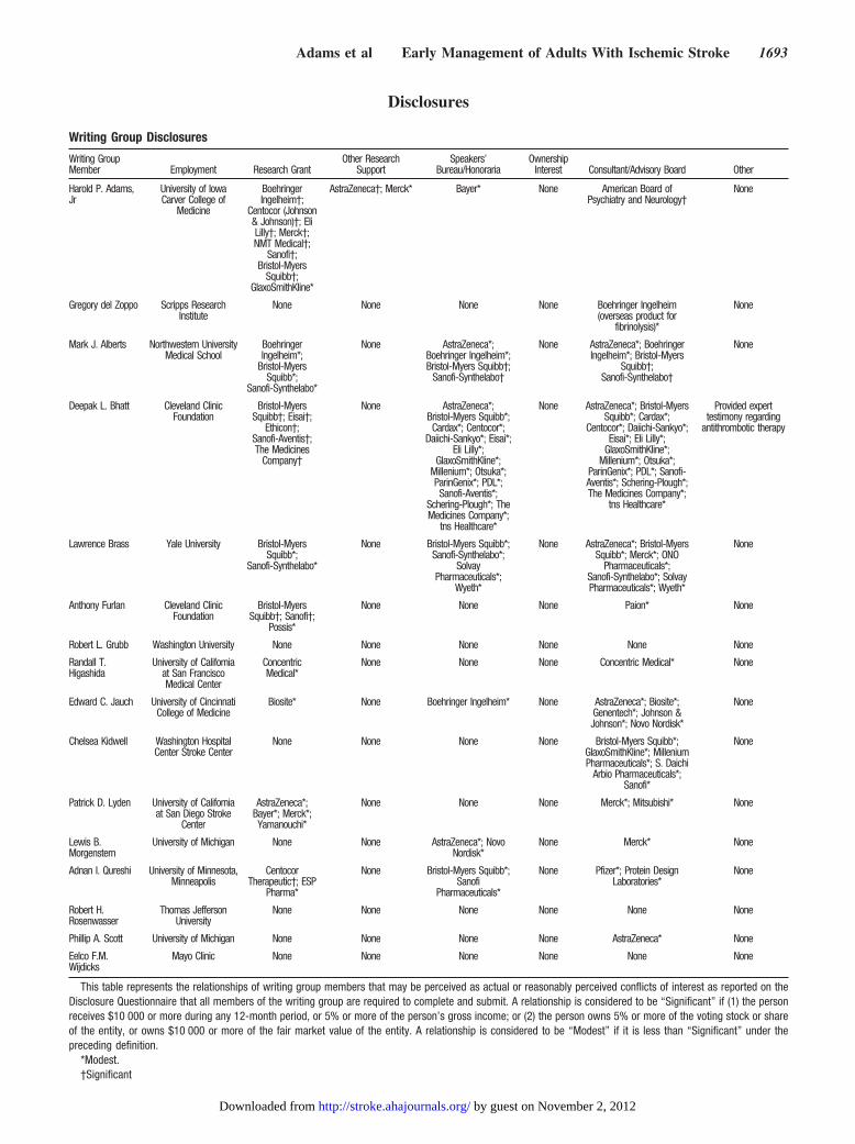
Disclosures
Writing Group Disclosures
Writing GroupMember Employment Research Grant
Other ResearchSupport
Speakers’Bureau/Honoraria
OwnershipInterest Consultant/Advisory Board Other
Harold P. Adams,Jr
University of IowaCarver College of
Medicine
BoehringerIngelheim†;
Centocor (Johnson& Johnson)†; EliLilly†; Merck†;NMT Medical†;
Sanofi†;Bristol-Myers
Squibb†;GlaxoSmithKline*
AstraZeneca†; Merck* Bayer* None American Board ofPsychiatry and Neurology†
None
Gregory del Zoppo Scripps ResearchInstitute
None None None None Boehringer Ingelheim(overseas product for
fibrinolysis)*
None
Mark J. Alberts Northwestern UniversityMedical School
BoehringerIngelheim*;
Bristol-MyersSquibb*;
Sanofi-Synthelabo*
None AstraZeneca*;Boehringer Ingelheim*;Bristol-Myers Squibb†;
Sanofi-Synthelabo†
None AstraZeneca*; BoehringerIngelheim*; Bristol-Myers
Squibb†;Sanofi-Synthelabo†
None
Deepak L. Bhatt Cleveland ClinicFoundation
Bristol-MyersSquibb†; Eisai†;
Ethicon†;Sanofi-Aventis†;The Medicines
Company†
None AstraZeneca*;Bristol-Myers Squibb*;Cardax*; Centocor*;
Daiichi-Sankyo*; Eisai*;Eli Lilly*;
GlaxoSmithKline*;Millenium*; Otsuka*;ParinGenix*; PDL*;Sanofi-Aventis*;
Schering-Plough*; TheMedicines Company*;
tns Healthcare*
None AstraZeneca*; Bristol-MyersSquibb*; Cardax*;
Centocor*; Daiichi-Sankyo*;Eisai*; Eli Lilly*;
GlaxoSmithKline*;Millenium*; Otsuka*;
ParinGenix*; PDL*; Sanofi-Aventis*; Schering-Plough*;The Medicines Company*;
tns Healthcare*
Provided experttestimony regarding
antithrombotic therapy
Lawrence Brass Yale University Bristol-MyersSquibb*;
Sanofi-Synthelabo*
None Bristol-Myers Squibb*;Sanofi-Synthelabo*;
SolvayPharmaceuticals*;
Wyeth*
None AstraZeneca*; Bristol-MyersSquibb*; Merck*; ONO
Pharmaceuticals*;Sanofi-Synthelabo*; SolvayPharmaceuticals*; Wyeth*
None
Anthony Furlan Cleveland ClinicFoundation
Bristol-MyersSquibb†; Sanofi†;
Possis*
None None None Paion* None
Robert L. Grubb Washington University None None None None None None
Randall T.Higashida
University of Californiaat San FranciscoMedical Center
ConcentricMedical*
None None None Concentric Medical* None
Edward C. Jauch University of CincinnatiCollege of Medicine
Biosite* None Boehringer Ingelheim* None AstraZeneca*; Biosite*;Genentech*; Johnson &Johnson*; Novo Nordisk*
None
Chelsea Kidwell Washington HospitalCenter Stroke Center
None None None None Bristol-Myers Squibb*;GlaxoSmithKline*; MilleniumPharmaceuticals*; S. Daichi
Arbio Pharmaceuticals*;Sanofi*
None
Patrick D. Lyden University of Californiaat San Diego Stroke
Center
AstraZeneca*;Bayer*; Merck*;Yamanouchi*
None None None Merck*; Mitsubishi* None
Lewis B.Morgenstern
University of Michigan None None AstraZeneca*; NovoNordisk*
None Merck* None
Adnan I. Qureshi University of Minnesota,Minneapolis
CentocorTherapeutic†; ESP
Pharma*
None Bristol-Myers Squibb*;Sanofi
Pharmaceuticals*
None Pfizer*; Protein DesignLaboratories*
None
Robert H.Rosenwasser
Thomas JeffersonUniversity
None None None None None None
Phillip A. Scott University of Michigan None None None None AstraZeneca* None
Eelco F.M.Wijdicks
Mayo Clinic None None None None None None
This table represents the relationships of writing group members that may be perceived as actual or reasonably perceived conflicts of interest as reported on theDisclosure Questionnaire that all members of the writing group are required to complete and submit. A relationship is considered to be “Significant” if (1) the personreceives $10 000 or more during any 12-month period, or 5% or more of the person’s gross income; or (2) the person owns 5% or more of the voting stock or shareof the entity, or owns $10 000 or more of the fair market value of the entity. A relationship is considered to be “Modest” if it is less than “Significant” under thepreceding definition.
*Modest.†Significant
Adams et al Early Management of Adults With Ischemic Stroke 1693
by guest on November 2, 2012http://stroke.ahajournals.org/Downloaded from
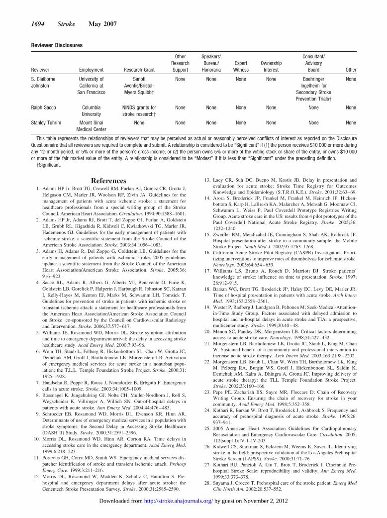
References1. Adams HP Jr, Brott TG, Crowell RM, Furlan AJ, Gomez CR, Grotta J,
Helgason CM, Marler JR, Woolson RF, Zivin JA. Guidelines for themanagement of patients with acute ischemic stroke: a statement forhealthcare professionals from a special writing group of the StrokeCouncil, American Heart Association. Circulation. 1994;90:1588–1601.
2. Adams HP Jr, Adams RJ, Brott T, del Zoppo GJ, Furlan A, GoldsteinLB, Grubb RL, Higashida R, Kidwell C, Kwiatkowski TG, Marler JR,Hademenos GJ. Guidelines for the early management of patients withischemic stroke: a scientific statement from the Stroke Council of theAmerican Stroke Association. Stroke. 2003;34:1056–1083.
3. Adams H, Adams R, Del Zoppo G, Goldstein LB. Guidelines for theearly management of patients with ischemic stroke: 2005 guidelinesupdate: a scientific statement from the Stroke Council of the AmericanHeart Association/American Stroke Association. Stroke. 2005;36:916–923.
4. Sacco RL, Adams R, Albers G, Alberts MJ, Benavente O, Furie K,Goldstein LB, Gorelick P, Halperin J, Harbaugh R, Johnston SC, KatzanI, Kelly-Hayes M, Kenton EJ, Marks M, Schwamm LH, Tomsick T.Guidelines for prevention of stroke in patients with ischemic stroke ortransient ischemic attack: a statement for healthcare professionals fromthe American Heart Association/American Stroke Association Councilon Stroke: co-sponsored by the Council on Cardiovascular Radiologyand Intervention. Stroke. 2006;37:577–617.
5. Williams JE, Rosamond WD, Morris DL. Stroke symptom attributionand time to emergency department arrival: the delay in accessing strokehealthcare study. Acad Emerg Med. 2000;7:93–96.
6. Wein TH, Staub L, Felberg R, Hickenbottom SL, Chan W, Grotta JC,Demchuk AM, Groff J, Bartholomew LK, Morgenstern LB. Activationof emergency medical services for acute stroke in a nonurban popu-lation: the T.L.L. Temple Foundation Stroke Project. Stroke. 2000;31:1925–1928.
7. Handschu R, Poppe R, Rauss J, Neundorfer B, Erbguth F. Emergencycalls in acute stroke. Stroke. 2003;34:1005–1009.
8. Rossnagel K, Jungehulsing GJ, Nolte CH, Muller-Nordhorn J, Roll S,Wegscheider K, Villringer A, Willich SN. Out-of-hospital delays inpatients with acute stroke. Ann Emerg Med. 2004;44:476–483.
9. Schroeder EB, Rosamond WD, Morris DL, Evenson KR, Hinn AR.Determinants of use of emergency medical services in a population withstroke symptoms: the Second Delay in Accessing Stroke Healthcare(DASH II) Study. Stroke. 2000;31:2591–2596.
10. Morris DL, Rosamond WD, Hinn AR, Gorton RA. Time delays inaccessing stroke care in the emergency department. Acad Emerg Med.1999;6:218–223.
11. Porteous GH, Corry MD, Smith WS. Emergency medical services dis-patcher identification of stroke and transient ischemic attack. PrehospEmerg Care. 1999;3:211–216.
12. Morris DL, Rosamond W, Madden K, Schultz C, Hamilton S. Pre-hospital and emergency department delays after acute stroke: theGenentech Stroke Presentation Survey. Stroke. 2000;31:2585–2590.
13. Lacy CR, Suh DC, Bueno M, Kostis JB. Delay in presentation andevaluation for acute stroke: Stroke Time Registry for OutcomesKnowledge and Epidemiology (S.T.R.O.K.E.). Stroke. 2001;32:63–69.
14. Arora S, Broderick JP, Frankel M, Frankel M, Heinrich JP, Hicken-bottom S, Karp H, LaBresh KA, Malarcher A, Mensah G, Moomaw CJ,Schwamm L, Weiss P; Paul Coverdell Prototype Registries WritingGroup. Acute stroke care in the US: results from 4 pilot prototypes of thePaul Coverdell National Acute Stroke Registry. Stroke. 2005;36:1232–1240.
15. Zweifler RM, Mendizabal JE, Cunningham S, Shah AK, Rothrock JF.Hospital presentation after stroke in a community sample: the MobileStroke Project. South Med J. 2002;95:1263–1268.
16. California Acute Stroke Pilot Registry (CASPR) Investigators. Priori-tizing interventions to improve rates of thrombolysis for ischemic stroke.Neurology. 2005;64:654–659.
17. Williams LS, Bruno A, Rouch D, Marriott DJ. Stroke patients’knowledge of stroke: influence on time to presentation. Stroke. 1997;28:912–915.
18. Barsan WG, Brott TG, Broderick JP, Haley EC, Levy DE, Marler JR.Time of hospital presentation in patients with acute stroke. Arch InternMed. 1993;153:2558–2561.
19. Wester P, Radberg J, Lundgren B, Peltonen M; Seek-Medical-Attention-in-Time Study Group. Factors associated with delayed admission tohospital and in-hospital delays in acute stroke and TIA: a prospective,multicenter study. Stroke. 1999;30:40–48.
20. Menon SC, Pandey DK, Morgenstern LB. Critical factors determiningaccess to acute stroke care. Neurology. 1998;51:427–432.
21. Morgenstern LB, Bartholomew LK, Grotta JC, Staub L, King M, ChanW. Sustained benefit of a community and professional intervention toincrease acute stroke therapy. Arch Intern Med. 2003;163:2198–2202.
22. Morgenstern LB, Staub L, Chan W, Wein TH, Bartholomew LK, KingM, Felberg RA, Burgin WS, Groff J, Hickenbottom SL, Saldin K,Demchuk AM, Kalra A, Dhingra A, Grotta JC. Improving delivery ofacute stroke therapy: the TLL Temple Foundation Stroke Project.Stroke. 2002;33:160–166.
23. Pepe PE, Zachariah BS, Sayre MR, Floccare D; Chain of RecoveryWriting Group. Ensuring the chain of recovery for stroke in yourcommunity. Acad Emerg Med. 1998;5:352–358.
24. Kothari R, Barsan W, Brott T, Broderick J, Ashbrock S. Frequency andaccuracy of prehospital diagnosis of acute stroke. Stroke. 1995;26:937–941.
25. 2005 American Heart Association Guidelines for CardiopulmonaryResuscitation and Emergency Cardiovascular Care. Circulation. 2005;112(suppl I):IV-1–IV-203.
26. Kidwell CS, Starkman S, Eckstein M, Weems K, Saver JL. Identifyingstroke in the field: prospective validation of the Los Angeles PrehospitalStroke Screen (LAPSS). Stroke. 2000;31:71–76.
27. Kothari RU, Pancioli A, Liu T, Brott T, Broderick J. Cincinnati Pre-hospital Stroke Scale: reproducibility and validity. Ann Emerg Med.1999;33:373–378.
28. Suyama J, Crocco T. Prehospital care of the stroke patient. Emerg MedClin North Am. 2002;20:537–552.
Reviewer Disclosures
Reviewer Employment Research Grant
OtherResearchSupport
Speakers’Bureau/
HonorariaExpert
WitnessOwnership
Interest
Consultant/AdvisoryBoard Other
S. ClaiborneJohnston
University ofCalifornia at
San Francisco
SanofiAventis/Bristol-Myers Squibb†
None None None None BoehringerIngelheim for
Secondary StrokePrevention Trials†
None
Ralph Sacco ColumbiaUniversity
NINDS grants forstroke research†
None None None None None None
Stanley Tuhrim Mount SinaiMedical Center
None None None None None None None
This table represents the relationships of reviewers that may be perceived as actual or reasonably perceived conflicts of interest as reported on the DisclosureQuestionnaire that all reviewers are required to complete and submit. A relationship is considered to be “Significant” if (1) the person receives $10 000 or more duringany 12-month period, or 5% or more of the person’s gross income; or (2) the person owns 5% or more of the voting stock or share of the entity, or owns $10 000or more of the fair market value of the entity. A relationship is considered to be “Modest” if it is less than “Significant” under the preceding definition.
†Significant.
1694 Stroke May 2007
by guest on November 2, 2012http://stroke.ahajournals.org/Downloaded from
29. Abarbanell NR. Is prehospital blood glucose measurement necessary insuspected cerebrovascular accident patients? Am J Emerg Med. 2005;23:823–827.
30. Virkkunen I, Yli-Hankala A, Silfvast T. Induction of therapeutic hypo-thermia after cardiac arrest in prehospital patients using ice-coldRinger’s solution: a pilot study. Resuscitation. 2004;62:299–302.
31. Saver JL, Kidwell C, Eckstein M, Starkman S; FAST-MAG Pilot TrialInvestigators. Prehospital neuroprotective therapy for acute stroke:results of the Field Administration of Stroke Therapy-Magnesium(FAST-MAG) pilot trial. Stroke. 2004;35:e106–e108.
32. Crocco T, Gullett T, Davis SM, Flores N, Sauerbeck L, Jauch E,Threlkeld B, Pio B, Ottaway M, Pancioli A, Chenier T. Feasibility ofneuroprotective agent administration by prehospital personnel in anurban setting. Stroke. 2003;34:1918–1922.
33. Silliman SL, Quinn B, Huggett V, Merino JG. Use of a field-to-strokecenter helicopter transport program to extend thrombolytic therapy torural residents. Stroke. 2003;34:729–733.
34. Chalela JA, Kasner SE, Jauch EC, Pancioli AM. Safety of air medicaltransportation after tissue plasminogen activator administration in acuteischemic stroke. Stroke. 1999;30:2366–2368.
35. Conroy MB, Rodriguez SU, Kimmel SE, Kasner SE. Helicopter transferoffers a potential benefit to patients with acute stroke. Stroke. 1999;30:2580–2584.
36. Silbergleit R, Burney RE, Draper J, Nelson K. Outcome of patients afterair medical transport for management of nontraumatic acute intracranialbleeding. Prehospital Disaster Med. 1994;9:252–256.
37. Silbergleit R, Scott PA, Lowell MJ, Silbergleit R. Cost-effectiveness ofhelicopter transport of stroke patients for thrombolysis. Acad EmergMed. 2003;10:966–972.
38. Rymer MM, Thrutchley DE; Stroke Team at the Mid America Brain andStroke Institute. Organizing regional networks to increase acute strokeintervention. Neurol Res. 2005;27(suppl 1):S9–S16.
39. Hess DC, Wang S, Hamilton W, Lee S, Pardue C, Waller JL, Gross H,Nichols F, Hall C, Adams RJ. REACH: clinical feasibility of a ruraltelestroke network. Stroke. 2005;36:2018–2020.
40. Audebert HJ, Kukla C, Clarmann von Claranau S, Kuhn J, Vatankhah B,Schenkel J, Ickenstein GW, Haberl RL, Horn M; TEMPiS Group.Telemedicine for safe and extended use of thrombolysis in stroke: theTelemedic Pilot Project for Integrative Stroke Care (TEMPiS) inBavaria. Stroke. 2005;36:287–291.
41. Fisher M. Developing and implementing future stroke therapies: thepotential of telemedicine. Ann Neurol. 2005;58:666–671.
42. Shafqat S, Kvedar JC, Guanci MM, Chang Y, Schwamm LH. Role fortelemedicine in acute stroke: feasibility and reliability of remote admin-istration of the NIH Stroke Scale. Stroke. 1999;30:2141–2145.
43. Handschu R, Littmann R, Reulbach U, Gaul C, Heckmann JG, Neun-dorfer B, Scibor M. Telemedicine in emergency evaluation of acutestroke: interrater agreement in remote video examination with a novelmultimedia system. Stroke. 2003;34:2842–2846.
44. Alberts MJ, Hademenos G, Latchaw RE, Jagoda A, Marler JR, MaybergMR, Starke RD, Todd HW, Viste KM, Girgus M, Shephard T, Emr M,Shwayder P, Walker MD; Brain Attack Coalition. Recommendations forthe establishment of primary stroke centers. JAMA. 2000;283:3102–3109.
45. Alberts MJ, Latchaw RE, Selman WR, Shephard T, Hadley MN, BrassLM, Koroshetz W, Marler JR, Booss J, Zorowitz RD, Croft JB, MagnisE, Mulligan D, Jagoda A, O’Connor R, Cawley CM, Connors JJ,Rose-DeRenzy JA, Emr M, Warren M, Walker MD; Brain AttackCoalition. Recommendations for comprehensive stroke centers: a con-sensus statement from the Brain Attack Coalition. Stroke. 2005;36:1597–1616.
46. Lattimore SU, Chalela J, Davis L, DeGraba T, Ezzeddine M, HaymoreJ, Nyquist P, Baird AE, Hallenbeck J, Warach S; NINDS SuburbanHospital Stroke Center. Impact of establishing a primary stroke center ata community hospital on the use of thrombolytic therapy: the NINDSSuburban Hospital Stroke Center experience. Stroke. 2003;34:e55–e57.
47. Douglas VC, Tong DC, Gillum LA, Zhao S, Brass LM, Dostal J,Johnston SC. Do the Brain Attack Coalition’s criteria for stroke centersimprove care for ischemic stroke? Neurology. 2005;64:422–427.
48. Wojner-Alexandrov AW, Alexandrov AV, Rodriguez D, Persse D,Grotta JC. Houston Paramedic and Emergency Stroke Treatment andOutcomes Study (HoPSTO). Stroke. 2005;36:1512–1518.
49. Gropen TI, Gagliano PJ, Blake CA, Blake CA, Sacco RL, KwiatkowskiT, Richmond NJ, Leifer D, Libman R, Azhar S, Daley MB; NYSDOHStroke Center Designation Project Workgroup. Quality improvement in
acute stroke: the New York State Stroke Center Designation Project.Neurology. 2006;67:88–93.
50. Audebert HJ, Schenkel J, Heuschmann PU, Bogdahn U, Haberl RL;Telemedic Pilot Project for Integrative Stroke Care Group. Effects of theimplementation of a telemedical stroke network: the Telemedic PilotProject for Integrative Stroke Care (TEMPiS) in Bavaria, Germany.Lancet Neurol. 2006;5:742–748.
51. Birbeck GL, Zingmond DS, Cui X, Vickrey BG. Multispecialty strokeservices in California hospitals are associated with reduced mortality.Neurology. 2006;66:1527–1532.
52. Rymer MM, Thurtchley D, Summers D; America Brain and StrokeInstitute Stroke Team. Expanded modes of tissue plasminogen activatordelivery in a comprehensive stroke center increases regional acute strokeinterventions. Stroke. 2003;34:e58–e60.
53. Frey JL, Jahnke HK, Goslar PW, Partovi S, Flaster MS. tPA by tele-phone: extending the benefits of a comprehensive stroke center. Neu-rology. 2005;64:154–156.
54. Gillum LA, Johnston SC. Characteristics of academic medical centersand ischemic stroke outcomes. Stroke. 2001;32:2137–2142.
55. Dion JE. Management of ischemic stroke in the next decade: strokecenters of excellence. J Vasc Interv Radiol. 2004;5(pt 2):S133–S141.
56. Silverman IE, Beland DK, Bohannon RW, Ohki SK, Spiegel GR.Expanding the range of therapies for acute ischemic stroke: the earlyexperience of the Regional Stroke Center at Hartford Hospital. ConnMed. 2004;68:419–429.
57. Adams R, Acker J, Alberts M, Andrews L, Atkinson R, Fenelon K,Furlan A, Girgus M, Horton K, Hughes R, Koroshetz W, Latchaw R,Magnis E, Mayberg M, Pancioli A, Robertson RM, Shephard T, SmithR, Smith SC Jr, Smith S, Stranne SK, Kenton EJ 3rd, Bashe G, ChavezA, Goldstein L, Hodosh R, Keitel C, Kelly-Hayes M, Leonard A,Morgenstern L, Wood JO; Advisory Working Group on Stroke CenterIdentification Options of the American Stroke Association. Recommen-dations for improving the quality of care through stroke centers andsystems: an examination of stroke center identification options: multi-disciplinary consensus recommendations from the Advisory WorkingGroup on Stroke Center Identification Options of the American StrokeAssociation. Stroke. 2002;33:e1–e7.
58. Kidwell CS, Shephard T, Tonn S, Lawyer B, Murdock M, Koroshetz W,Alberts M, Hademenos GJ, Saver JL. Establishment of primary strokecenters: a survey of physician attitudes and hospital resources. Neu-rology. 2003;60:1452–1456.
59. Schwamm LH, Pancioli A, Acker JE 3rd, Goldstein LB, Zorowitz RD,Shephard TJ, Moyer P, Gorman M, Johnston SC, Duncan PW, GorelickP, Frank J, Stranne SK, Smith R, Federspiel W, Horton KB, Magnis E,Adams RJ; American Stroke Association’s Task Force on the Devel-opment of Stroke Systems. Recommendations for the establishment ofstroke systems of care: recommendations from the American StrokeAssociation’s Task Force on the Development of Stroke Systems.Stroke. 2005;36:690–703.
60. Marler JR, Tilley BC, Lu M, Brott TG, Lyden PC, Grotta JC, BroderickJP, Levine SR, Frankel MP, Horowitz SH, Haley EC Jr, LewandowskiCA, Kwiatkowski TP. Early stroke treatment associated with betteroutcome: the NINDS rt-PA stroke study. Neurology. 2000;55:1649–1655.
61. Marler JR, Jones PW, Emr M. Proceedings of a national symposium onrapid identification and treatment of acute stroke. Washington, DC;December 12–13, 1996.
62. Asimos AW, Norton HJ, Price MF, Cheek WM. Therapeutic yield andoutcomes of a community teaching hospital code stroke protocol. AcadEmerg Med. 2004;11:361–370.
63. Belvis R, Cocho D, Marti-Fabregas J, Pagonabarraga J, Aleu A,Garcia-Bargo MD, Pons J, Coma E, Garcia-Alfranca F, Jimenez-Fabrega X, Marti-Vilalta JL. Benefits of a prehospital stroke codesystem: feasibility and efficacy in the first year of clinical practice inBarcelona, Spain. Cerebrovasc Dis. 2005;19:96–101.
64. Bray JE, Martin J, Cooper G, Barger B, Bernard S, Bladin C. Aninterventional study to improve paramedic diagnosis of stroke. PrehospEmerg Care. 2005;9:297–302.
65. Kidwell CS, Alger JR, Di Salle F, Starkman S, Villablanca P, BentsonJ, Saver JL. Diffusion MRI in patients with transient ischemic attacks.Stroke. 1999;30:1174–1180.
66. The National Institute of Neurological Disorders and Stroke rt-PAStroke Study Group. Tissue plasminogen activator for acute ischemicstroke. N Engl J Med. 1995;333:1581–1587.
Adams et al Early Management of Adults With Ischemic Stroke 1695
by guest on November 2, 2012http://stroke.ahajournals.org/Downloaded from
67. Nor AM, Davis J, Sen B, Shipsey D, Louw SJ, Dyker AG, Davis M,Ford GA. The Recognition of Stroke in the Emergency Room (ROSIER)scale: development and validation of a stroke recognition instrument.Lancet Neurol. 2005;4:727–734.
68. Goldstein LB, Samsa GP. Reliability of the National Institutes of HealthStroke Scale: extension to non-neurologists in the context of a clinicaltrial. Stroke. 1997;28:307–310.
69. Cote R, Hachinski VC, Shurvell BL, Norris JW, Wolfson C. TheCanadian Neurological Scale: a preliminary study in acute stroke.Stroke. 1986;17:731–737.
70. Krieger DW. Should a hospital without a neurologist use t-PA to treatstroke? Cleve Clin J Med. 1999;66:585–586.
71. Frankel MR, Morgenstern LB, Kwiatkowski T, Lu M, Tilley BC, Bro-derick JP, Libman R, Levine SR, Brott T. Predicting prognosis afterstroke: a placebo group analysis from the National Institute of Neuro-logical Disorders and Stroke rt-PA Stroke Trial. Neurology. 2000;55:952–959.
72. Lewandowski CA, Frankel M, Tomsick TA, Broderick J, Frey J, ClarkW, Starkman S, Grotta J, Spilker J, Khoury J, Brott T. Combinedintravenous and intra-arterial r-TPA versus intra-arterial therapy of acuteischemic stroke: Emergency Management of Stroke (EMS) BridgingTrial. Stroke. 1999;30:2598–2605.
73. Kothari RU, Brott T, Broderick JP, Hamilton CA. Emergency phy-sicians: accuracy in the diagnosis of stroke. Stroke. 1995;26:2238–2241.
74. Morgenstern LB, Lisabeth LD, Mecozzi AC, Smith MA, Longwell PJ,McFarling DA, Risser JM. A population-based study of acute stroke andTIA diagnosis. Neurology. 2004;62:895–900.
75. Goldstein LB, Simel DL. Is this patient having a stroke? JAMA. 2005;293:2391–2402.
76. Christensen H, Fogh Christensen A, Boysen G. Abnormalities on ECGand telemetry predict stroke outcome at 3 months. J Neurol Sci. 2005;234:99–103.
77. Oppenheimer SM, Hachinski VC. The cardiac consequences of stroke.Neurol Clin. 1992;10:167–176.
78. Oppenheimer SM. Neurogenic cardiac effects of cerebrovasculardisease. Curr Opin Neurol. 1994;7:20–24.
79. Dimant J, Grob D. Electrocardiographic changes and myocardialdamage in patients with acute cerebrovascular accidents. Stroke. 1977;8:448–455.
80. Norris JW, Froggatt GM, Hachinski VC. Cardiac arrhythmias in acutestroke. Stroke. 1978;9:392–396.
81. Mikolich JR, Jacobs WC, Fletcher GF. Cardiac arrhythmias in patientswith acute cerebrovascular accidents. JAMA. 1981;246:1314–1317.
82. Vingerhoets F, Bogousslavsky J, Regli F, Van Melle G. Atrial fibril-lation after acute stroke. Stroke. 1993;24:26–30.
83. Sagar G, Riley P, Vohrah A. Is admission chest radiography of anyclinical value in acute stroke patients? Clin Radiol. 1996;51:499–502.
84. Bladin CF, Alexandrov AV, Bellavance A, Bornstein N, Chambers B,Cote R, Lebrun L, Pirisi A, Norris JW. Seizures after stroke: a pro-spective multicenter study. Arch Neurol. 2000;57:1617–1622.
85. Kidwell CS, Villablanca JP, Saver JL. Advances in neuroimaging ofacute stroke. Curr Atheroscler Rep. 2000;2:126–135.
86. Kang DW, Chalela JA, Dunn W, Warach S; NIH-Suburban StrokeCenter Investigators. MRI screening before standard tissue plasminogenactivator therapy is feasible and safe. Stroke. 2005;36:1939–1943.
87. Hjort N, Butcher K, Davis SM, Kidwell CS, Koroshetz WJ, Rother J,Schellinger PD, Warach S, Ostergaard L; UCLA Thrombolysis Inves-tigators. Magnetic resonance imaging criteria for thrombolysis in acutecerebral infarct. Stroke. 2005;36:388–397.
88. Kidwell CS, Chalela JA, Saver JL, Starkman S, Hill MD, Demchuk AM,Butman JA, Patronas N, Alger JR, Latour LL, Luby ML, Baird AE,Leary MC, Tremwel M, Ovbiagele B, Fredieu A, Suzuki S, VillablancaJP, Davis S, Dunn B, Todd JW, Ezzeddine MA, Haymore J, Lynch JK,Davis L, Warach S. Comparison of MRI and CT for detection of acuteintracerebral hemorrhage. JAMA. 2004;292:1823–1830.
89. Rother J, Schellinger PD, Gass A, Siebler M, Villringer A, Fiebach JB,Fiehler J, Jansen O, Kucinski T, Schoder V, Szabo K, Junge-Hulsing GJ,Hennerici M, Zeumer H, Sartor K, Weiller C, Hacke W; Kompeten-znetzwerk Schlaganfall Study Group. Effect of intravenous thrombolysison MRI parameters and functional outcome in acute stroke �6 hours.Stroke. 2002;33:2438–2445.
90. Schellinger PD, Jansen O, Fiebach JB, Pohlers O, Ryssel H, Heiland S,Steiner T, Hacke W, Sartor K. Feasibility and practicality of MRimaging of stroke in the management of hyperacute cerebral ischemia.AJNR Am J Neuroradiol. 2000;21:1184–1189.
91. Keir SL, Wardlaw JM. Systematic review of diffusion and perfusionimaging in acute ischemic stroke. Stroke. 2000;31:2723–2731.
92. Zivin JA. Perfusion-weighted imaging/diffusion-weighted imagingmismatch on MRI can now be used to select patients for recombinanttissue plasminogen activator beyond 3 hours: con. Stroke. 2005;36:1105–1106.
93. Powers WJ. Testing a test: a report card for DWI in acute stroke.Neurology. 2000;54:1549–1551.
94. Adams HP Jr, Brott TG, Furlan AJ, Gomez CR, Grotta J, Helgason CM,Kwiatkowski T, Lyden PD, Marler JR, Torner J, Feinberg W, MaybergM, Thies W. Guidelines for thrombolytic therapy for acute stroke: asupplement to the guidelines for the management of patients with acuteischemic stroke: a statement for healthcare professionals from a SpecialWriting Group of the Stroke Council, American Heart Association.Circulation. 1996;94:1167–1174.
95. Mullins ME, Schaefer PW, Sorensen AG, Halpern EF, Ay H, He J,Koroshetz WJ, Gonzalez RG. CT and conventional and diffusion-weighted MR imaging in acute stroke: study in 691 patients at presen-tation to the emergency department. Radiology. 2002;224:353–360.
96. Moulin T, Cattin F, Crepin-Leblond T, Halpern EF, Ay H, He J,Koroshetz WJ, Gonzalez RG. Early CT signs in acute middle cerebralartery infarction: predictive value for subsequent infarct locations andoutcome. Neurology. 1996;47:366–375.
97. von Kummer R, Nolte PN, Schnittger H, Halpern EF, Ay H, He J,Koroshetz WJ, Gonzalez RG. Detectability of cerebral hemisphereischaemic infarcts by CT within 6 h of stroke. Neuroradiology. 1996;38:31–33.
98. von Kummer R, Allen KL, Holle R, Bozzao L, Bastianello S, ManelfeC, Bluhmki E, Ringleb P, Meier DH, Hacke W. Acute stroke: usefulnessof early CT findings before thrombolytic therapy. Radiology. 1997;205:327–333.
99. Hacke W, Kaste M, Fieschi C, Toni D, Lesaffre E, von Kummer R,Boysen G, Bluhmki E, Hoxter G, Mahagne MH, et al. Intravenousthrombolysis with recombinant tissue plasminogen activator for acutehemispheric stroke: the European Cooperative Acute Stroke Study(ECASS). JAMA. 1995;274:1017–1025.
100. Patel SC, Levine SR, Tilley BC, Grotta JC, Lu M, Frankel M, Haley ECJr, Brott TG, Broderick JP, Horowitz S, Lyden PD, Lewandowski CA,Marler JR, Welch KM; National Institute of Neurological Disorders andStroke rt-PA Stroke Study Group. Lack of clinical significance of earlyischemic changes on computed tomography in acute stroke. JAMA.2001;286:2830–2838.
101. Larrue V, von Kummer R, del Zoppo G, Bluhmki E. Hemorrhagictransformation in acute ischemic stroke: potential contributing factors inthe European Cooperative Acute Stroke Study. Stroke. 1997;28:957–960.
102. Schriger DL, Kalafut M, Starkman S, Krueger M, Saver JL. Cranialcomputed tomography interpretation in acute stroke: physician accuracyin determining eligibility for thrombolytic therapy. JAMA. 1998;279:1293–1297.
103. Marks MP, Holmgren EB, Fox AJ, Patel S, von Kummer R, FroehlichJ. Evaluation of early computed tomographic findings in acute ischemicstroke. Stroke. 1999;30:389–392.
104. Wardlaw JM, Dorman PJ, Lewis SC, Patel S, von Kummer R, FroehlichJ. Can stroke physicians and neuroradiologists identify signs of earlycerebral infarction on CT? J Neurol Neurosurg Psychiatry. 1999;67:651–653.
105. Grotta JC, Chiu D, Lu M, Patel S, Levine SR, Tilley BC, Brott TG,Haley EC Jr, Lyden PD, Kothari R, Frankel M, Lewandowski CA,Libman R, Kwiatkowski T, Broderick JP, Marler JR, Corrigan J, Huff S,Mitsias P, Talati S, Tanne D. Agreement and variability in the interpre-tation of early CT changes in stroke patients qualifying for intravenousrtPA therapy. Stroke. 1999;30:1528–1533.
106. Kalafut MA, Schriger DL, Saver JL, Starkman S. Detection of early CTsigns of �1/3 middle cerebral artery infarctions: interrater reliability andsensitivity of CT interpretation by physicians involved in acute strokecare. Stroke. 2000;31:1667–1671.
107. Barber PA, Demchuk AM, Zhang J, Buchan AM; ASPECTS StudyGroup. Validity and reliability of a quantitative computed tomographyscore in predicting outcome of hyperacute stroke before thrombolytictherapy: Alberta Stroke Programme Early CT Score. Lancet. 2000;355:1670–1674.
108. Demchuk AM, Hill MD, Barber PA, Silver B, Patel SC, Levine SR;NINDS rtPA Stroke Study Group, NIH. Importance of early ischemic
1696 Stroke May 2007
by guest on November 2, 2012http://stroke.ahajournals.org/Downloaded from
computed tomography changes using ASPECTS in NINDS rtPA StrokeStudy. Stroke. 2005;36:2110–2115.
109. Wardlaw JM, Mielke O. Early signs of brain infarction at CT: observerreliability and outcome after thrombolytic treatment—systematicreview. Radiology. 2005;235:444–453.
110. Ezzeddine MA, Lev MH, McDonald CT, Rordorf G, Oliveira-Filho J,Aksoy FG, Farkas J, Segal AZ, Schwamm LH, Gonzalez RG, KoroshetzWJ. CT angiography with whole brain perfused blood volume imaging:added clinical value in the assessment of acute stroke. Stroke. 2002;33:959–966.
111. Kloska SP, Nabavi DG, Gaus C, Nam EM, Klotz E, Ringelstein EB,Heindel W. Acute stroke assessment with CT: do we need multimodalevaluation? Radiology. 2004;233:79–86.
112. Wintermark M, Fischbein NJ, Smith WS, Ko NU, Quist M, Dillon WP.Accuracy of dynamic perfusion CT with deconvolution in detectingacute hemispheric stroke. AJNR Am J Neuroradiol. 2005;26:104–112.
113. Schramm P, Schellinger PD, Klotz E, Kallenberg K, Fiebach JB,Kulkens S, Heiland S, Knauth M, Sartor K. Comparison of perfusioncomputed tomography and computed tomography angiography sourceimages with perfusion-weighted imaging and diffusion-weightedimaging in patients with acute stroke of less than 6 hours’ duration.Stroke. 2004;35:1652–1658.
114. Klotz E, Konig M. Perfusion measurements of the brain: using dynamicCT for the quantitative assessment of cerebral ischemia in acute stroke.Eur J Radiol. 1999;30:170–184.
115. Wintermark M, Reichhart M, Thiran JP, Maeder P, Chalaron M,Schnyder P, Bogousslavsky J, Meuli R. Prognostic accuracy of cerebralblood flow measurement by perfusion computed tomography, at the timeof emergency room admission, in acute stroke patients. Ann Neurol.2002;51:417–432.
116. Lev MH, Farkas J, Rodriguez VR, Schwamm LH, Hunter GJ, PutmanCM, Rordorf GA, Buonanno FS, Budzik R, Koroshetz WJ, GonzalezRG. CT angiography in the rapid triage of patients with hyperacutestroke to intraarterial thrombolysis: accuracy in the detection of largevessel thrombus. J Comput Assist Tomogr. 2001;25:520–528.
117. Esteban JM, Cervera V. Perfusion CT and angio CT in the assessmentof acute stroke. Neuroradiology. 2004;46:705–715.
118. Wildermuth S, Knauth M, Brandt T, Winter R, Sartor K, Hacke W. Roleof CT angiography in patient selection for thrombolytic therapy in acutehemispheric stroke. Stroke. 1998;29:935–938.
119. Shrier DA, Tanaka H, Numaguchi Y, Konno S, Patel U, Shibata D. CTangiography in the evaluation of acute stroke. AJNR Am J Neuroradiol.1997;18:1011–1020.
120. Verro P, Tanenbaum LN, Borden NM, Sen S, Eshkar N. CTangiography in acute ischemic stroke: preliminary results. Stroke. 2002;33:276–278.
121. Mohr JP, Biller J, Hilal SK, Yuh WT, Tatemichi TK, Hedges S, Tali E,Nguyen H, Mun I, Adams HP Jr, et al. Magnetic resonance versuscomputed tomographic imaging in acute stroke. Stroke. 1995;26:807–812.
122. Warach S, Gaa J, Siewert B, Wielopolski P, Edelman RR. Acute humanstroke studied by whole brain echo planar diffusion-weighted magneticresonance imaging. Ann Neurol. 1995;37:231–241.
123. Warach S, Chien D, Li W, Ronthal M, Edelman RR. Fast magneticresonance diffusion-weighted imaging of acute human stroke [publishedcorrection appears in Neurology. 1992;1742:2192]. Neurology. 1992;42:1717–1723.
124. Lutsep HL, Albers GW, DeCrespigny A, Kamat GN, Marks MP,Moseley ME. Clinical utility of diffusion-weighted magnetic resonanceimaging in the assessment of ischemic stroke. Ann Neurol. 1997;41:574–580.
125. Barber PA, Darby DG, Desmond PM, Yang Q, Gerraty RP, Jolley D,Donnan GA, Tress BM, Davis SM. Prediction of stroke outcome withechoplanar perfusion- and diffusion-weighted MRI. Neurology. 1998;51:418–426.
126. Lee LJ, Kidwell CS, Alger J, Starkman S, Saver JL. Impact on strokesubtype diagnosis of early diffusion-weighted magnetic resonanceimaging and magnetic resonance angiography. Stroke. 2000;31:1081–1089.
127. Lovblad KO, Laubach HJ, Baird AE, Curtin F, Schlaug G, Edelman RR,Warach S. Clinical experience with diffusion-weighted MR in patientswith acute stroke. AJNR Am J Neuroradiol. 1998;19:1061–1066.
128. Ay H, Buonanno FS, Rordorf G, Schaefer PW, Schwamm LH, Wu O,Gonzalez RG, Yamada K, Sorensen GA, Koroshetz WJ. Normal
diffusion-weighted MRI during stroke-like deficits. Neurology. 1999;52:1784–1792.
129. Barber PA, Darby DG, Desmond PM, Gerraty RP, Yang Q, Li T, JolleyD, Donnan GA, Tress BM, Davis SM. Identification of major ischemicchange: diffusion-weighted imaging versus computed tomography.Stroke. 1999;30:2059–2065.
130. van Everdingen KJ, van der Grond J, Kappelle LJ, Ramos LM, MaliWP. Diffusion-weighted magnetic resonance imaging in acute stroke.Stroke. 1998;29:1783–1790.
131. Gonzalez RG, Schaefer PW, Buonanno FS, Schwamm LH, Budzik RF,Rordorf G, Wang B, Sorensen AG, Koroshetz WJ. Diffusion-weightedMR imaging: diagnostic accuracy in patients imaged within 6 hours ofstroke symptom onset. Radiology. 1999;210:155–162.
132. Lovblad KO, Baird AE, Schlaug G, Benfield A, Siewert B, Voetsch B,Connor A, Burzynski C, Edelman RR, Warach S. Ischemic lesionvolumes in acute stroke by diffusion-weighted magnetic resonanceimaging correlate with clinical outcome. Ann Neurol. 1997;42:164–170.
133. Tong DC, Yenari MA, Albers GW, O’Brien M, Marks MP, MoseleyME. Correlation of perfusion- and diffusion-weighted MRI with NIHSSscore in acute (�6.5 hour) ischemic stroke. Neurology. 1998;50:864–870.
134. Kidwell CS, Alger JR, Saver JL. Beyond mismatch: evolving paradigmsin imaging the ischemic penumbra with multimodal magnetic resonanceimaging. Stroke. 2003;34:2729–2735.
135. Sobesky J, Zaro Weber O, Lehnhardt FG, Hesselmann V, Neveling M,Jacobs A, Heiss WD. Does the mismatch match the penumbra?Magnetic resonance imaging and positron emission tomography in earlyischemic stroke. Stroke. 2005;36:980–985.
136. Jansen O, Schellinger P, Fiebach J, Hacke W, Sartor K. Early recanal-isation in acute ischaemic stroke saves tissue at risk defined by MRI.Lancet. 1999;353:2036–2037.
137. Wu O, Koroshetz WJ, Ostergaard L, Buonanno FS, Copen WA,Gonzalez RG, Rordorf G, Rosen BR, Schwamm LH, Weisskoff RM,Sorensen AG. Predicting tissue outcome in acute human cerebral ische-mia using combined diffusion- and perfusion-weighted MR imaging.Stroke. 2001;32:933–942.
138. Jacobs MA, Mitsias P, Soltanian-Zadeh H, Santhakumar S, Ghanei A,Hammond R, Peck DJ, Chopp M, Patel S. Multiparametric MRI tissuecharacterization in clinical stroke with correlation to clinical outcome:part 2. Stroke. 2001;32:950–957.
139. Kidwell CS, Alger JR, Saver JL, et al. MR signatures of infarction vs.salvageable penumbra in acute human stroke: a preliminary model[abstract]. Stroke. 2000;31:285.
140. Hacke W, Albers G, Al-Rawi Y, Bogousslavsky J, Davalos A, EliasziwM, Fischer M, Furlan A, Kaste M, Lees KR, Soehngen M, Warach S;DIAS Study Group. The Desmoteplase in Acute Ischemic Stroke Trial(DIAS): a phase II MRI-based 9-hour window acute stroke thrombolysistrial with intravenous desmoteplase. Stroke. 2005;36:66–73.
141. Fiebach JB, Schellinger PD, Gass A, Kucinski T, Siebler M, VillringerA, Olkers P, Hirsch JG, Heiland S, Wilde P, Jansen O, Rother J, HackeW, Sartor K; Kompetenznetzwerk Schlaganfall B5. Stroke magneticresonance imaging is accurate in hyperacute intracerebral hemorrhage: amulticenter study on the validity of stroke imaging. Stroke. 2004;35:502–506.
142. Kidwell CS, Saver JL, Villablanca JP, Duckwiler G, Fredieu A, GoughK, Leary MC, Starkman S, Gobin YP, Jahan R, Vespa P, Liebeskind DS,Alger JR, Vinuela F. Magnetic resonance imaging detection ofmicrobleeds before thrombolysis: an emerging application. Stroke.2002;33:95–98.
143. Wong KS, Chan YL, Liu JY, Gao S, Lam WW. Asymptomaticmicrobleeds as a risk factor for aspirin-associated intracerebral hemor-rhages. Neurology. 2003;60:511–513.
144. Chalela JA, Kang DW, Warach S. Multiple cerebral microbleeds: MRImarker of a diffuse hemorrhage-prone state. J Neuroimaging. 2004;14:54–57.
145. Kakuda W, Thijs VN, Lansberg MG, Bammer R, Wechsler L, Kemp S,Moseley ME, Marks MP, Albers GW; DEFUSE Investigators. Clinicalimportance of microbleeds in patients receiving IV thrombolysis. Neu-rology. 2005;65:1175–1178.
146. Qureshi AI, Isa A, Cinnamon J, Fountain J, Ottenlips JR, Braimah J,Frankel MR. Magnetic resonance angiography in patients with braininfarction. J Neuroimaging. 1998;8:65–70.
147. Scarabino T, Carriero A, Giannatempo GM, Marano R, De Matthaeis P,Bonomo L, Salvolini U. Contrast-enhanced MR angiography (CE MRA)
Adams et al Early Management of Adults With Ischemic Stroke 1697
by guest on November 2, 2012http://stroke.ahajournals.org/Downloaded from
in the study of the carotid stenosis: comparison with digital subtractionangiography (DSA). J Neuroradiol. 1999;26:87–91.
148. Fiebach JB, Schellinger PD, Jansen O, Meyer M, Wilde P, Bender J,Schramm P, Juttler E, Oehler J, Hartmann M, Hahnel S, Knauth M,Hacke W, Sartor K. CT and diffusion-weighted MR imaging in ran-domized order: diffusion-weighted imaging results in higher accuracyand lower interrater variability in the diagnosis of hyperacute ischemicstroke. Stroke. 2002;33:2206–2210.
149. Urbach H, Flacke S, Keller E, Textor J, Berlis A, Hartmann A, Reul J,Solymosi L, Schild HH. Detectability and detection rate of acutecerebral hemisphere infarcts on CT and diffusion-weighted MRI. Neu-roradiology. 2000;42:722–727.
150. Lansberg MG, Albers GW, Beaulieu C, Marks MP. Comparison ofdiffusion-weighted MRI and CT in acute stroke. Neurology. 2000;54:1557–1561.
151. Baird AE, Lovblad KO, Schlaug G, Edelman RR, Warach S. Multipleacute stroke syndrome: marker of embolic disease? Neurology. 2000;54:674–678.
152. Seifert T, Enzinger C, Storch MK, Pichler G, Niederkorn K, Fazekas F.Acute small subcortical infarctions on diffusion weighted MRI: clinicalpresentation and aetiology. J Neurol Neurosurg Psychiatry. 2005;76:1520–1524.
153. Caso V, Budak K, Georgiadis D, Schuknecht B, Baumgartner RW.Clinical significance of detection of multiple acute brain infarcts ondiffusion weighted magnetic resonance imaging. J Neurol NeurosurgPsychiatry. 2005;76:514–518.
154. Wessels T, Rottger C, Jauss M, Kaps M, Traupe H, Stolz E. Identifi-cation of embolic stroke patterns by diffusion-weighted MRI in clin-ically defined lacunar stroke syndromes. Stroke. 2005;36:757–761.
155. Smajlovic D, Sinanovic O. Sensitivity of the neuroimaging techniques inischemic stroke. Med Arh. 2004;58:282–284.
156. Arauz A, Murillo L, Cantu C, Barinagarrementeria F, Higuera J. Pro-spective study of single and multiple lacunar infarcts using magneticresonance imaging: risk factors, recurrence, and outcome in 175 con-secutive cases. Stroke. 2003;34:2453–2458.
157. Gerraty RP, Parsons MW, Barber PA, Darby DG, Desmond PM, TressBM, Davis SM. Examining the lacunar hypothesis with diffusion andperfusion magnetic resonance imaging. Stroke. 2002;33:2019–2024.
158. Takahashi K, Kobayashi S, Matui R, Yamaguchi S, Yamashita K. Thedifferences of clinical parameters between small multiple ischemiclesions and single lesion detected by diffusion-weighted MRI. ActaNeurol Scand. 2002;106:24–29.
159. Wityk RJ, Goldsborough MA, Hillis A, Beauchamp N, Barker PB,Borowicz LM Jr, McKhann GM. Diffusion- and perfusion-weightedbrain magnetic resonance imaging in patients with neurologic compli-cations after cardiac surgery. Arch Neurol. 2001;58:571–576.
160. Stapf C, Hofmeister C, Hartmann A, Marx P, Mast H. Predictive valueof clinical lacunar syndromes for lacunar infarcts on magnetic resonancebrain imaging. Acta Neurol Scand. 2000;101:13–18.
161. Ay H, Oliveira-Filho J, Buonanno FS, Ezzeddine M, Schaefer PW,Rordorf G, Schwamm LH, Gonzalez RG, Koroshetz WJ. Diffusion-weighted imaging identifies a subset of lacunar infarction associatedwith embolic source. Stroke. 1999;30:2644–2650.
162. Etgen T, Grafin von Einsiedel H, Rottinger M, Winbeck K, Conrad B,Sander D. Detection of acute brainstem infarction by using DWI/MRI.Eur Neurol. 2004;52:145–150.
163. Saur D, Kucinski T, Grzyska U, Eckert B, Eggers C, Niesen W, SchoderV, Zeumer H, Weiller C, Rother J. Sensitivity and interrater agreementof CT and diffusion-weighted MR imaging in hyperacute stroke. AJNRAm J Neuroradiol. 2003;24:878–885.
164. Keir SL, Wardlaw JM, Bastin ME, Dennis MS. In which patients isdiffusion-weighted magnetic resonance imaging most useful in routinestroke care? J Neuroimaging. 2004;14:118–122.
165. Wen HM, Lam WW, Rainer T, Fan YH, Leung TW, Chan YL, WongKS. Multiple acute cerebral infarcts on diffusion-weighted imaging andrisk of recurrent stroke. Neurology. 2004;63:1317–1319.
166. Oliveira-Filho J, Ay H, Schaefer PW, Buonanno FS, Chang Y, GonzalezRG, Koroshetz WJ. Diffusion-weighted magnetic resonance imagingidentifies the “clinically relevant” small-penetrator infarcts. ArchNeurol. 2000;57:1009–1014.
167. Marks MP, de Crespigny A, Lentz D, Enzmann DR, Albers GW,Moseley ME. Acute and chronic stroke: navigated spin-echo diffusion-weighted MR imaging. Radiology. 1996;199:403–408.
168. Baron JC, Bousser MG, Rey A, Guillard A, Comar D, Castaigne P.Reversal of focal “misery-perfusion syndrome” by extra-intracranial
arterial bypass in hemodynamic cerebral ischemia: a case study with15O positron emission tomography. Stroke. 1981;12:454–459.
169. Furlan M, Marchal G, Viader F, Derlon JM, Baron JC. Spontaneousneurological recovery after stroke and the fate of the ischemicpenumbra. Ann Neurol. 1996;40:216–226.
170. Heiss WD, Huber M, Fink GR, Herholz K, Pietrzyk U, Wagner R,Wienhard K. Progressive derangement of periinfarct viable tissue inischemic stroke. J Cereb Blood Flow Metab. 1992;12:193–203.
171. Heiss WD, Grond M, Thiel A, Ghaemi M, Sobesky J, Rudolf J, BauerB, Wienhard K. Permanent cortical damage detected by flumazenilpositron emission tomography in acute stroke. Stroke. 1998;29:454–461.
172. Read SJ, Hirano T, Abbott DF, Sachinidis JI, Tochon-Danguy HJ, ChanJG, Egan GF, Scott AM, Bladin CF, McKay WJ, Donnan GA. Iden-tifying hypoxic tissue after acute ischemic stroke using PET and 18F-fluoromisonidazole. Neurology. 1998;51:1617–1621.
173. Yonas H, Gur D, Claassen D, Wolfson SK Jr, Moossy J. Stable xenon-enhanced CT measurement of cerebral blood flow in reversible focalischemia in baboons. J Neurosurg. 1990;73:266–273.
174. Ueda T, Sakaki S, Yuh WT, Nochide I, Ohta S. Outcome in acute strokewith successful intra-arterial thrombolysis and predictive value of initialsingle-photon emission-computed tomography. J Cereb Blood FlowMetab. 1999;19:99–108.
175. Grotta JC, Alexandrov AV. tPA-associated reperfusion after acutestroke demonstrated by SPECT. Stroke. 1998;29:429–432.
176. Berrouschot J, Barthel H, von Kummer R, Nochide I, Ohta S. 99mtechnetium-ethyl-cysteinate-dimer single-photon emission CT canpredict fatal ischemic brain edema. Stroke. 1998;29:2556–2562.
177. Christou I, Alexandrov AV, Burgin WS, Wojner AW, Felberg RA,Malkoff M, Grotta JC. Timing of recanalization after tissue plasminogenactivator therapy determined by transcranial Doppler correlates withclinical recovery from ischemic stroke. Stroke. 2000;31:1812–1816.
178. Demchuk AM, Burgin WS, Christou I, Felberg RA, Barber PA, HillMD, Alexandrov AV. Thrombolysis in brain ischemia (TIBI) transcra-nial Doppler flow grades predict clinical severity, early recovery, andmortality in patients treated with intravenous tissue plasminogen acti-vator. Stroke. 2001;32:89–93.
179. Alexandrov AV, Wojner AW, Grotta JC; CLOTBUST Investigators.CLOTBUST: design of a randomized trial of ultrasound-enhancedthrombolysis for acute ischemic stroke. J Neuroimaging. 2004;14:108–112.
180. Albers GW, Hart RG, Lutsep HL, Newell DW, Sacco RL. Addendum tothe supplement to the guidelines for the management of transient ische-mic attacks. Stroke. 2000;31:1001.
181. Wolf PA, Clagett GP, Easton JD, Goldstein LB, Gorelick PB,Kelly-Hayes M, Sacco RL, Whisnant JP. Preventing ischemic stroke inpatients with prior stroke and transient ischemic attack: a statement forhealthcare professionals from the Stroke Council of the American HeartAssociation. Stroke. 1999;30:1991–1994.
182. Hacke W, Krieger D, Hirschberg M. General principles in the treatmentof acute ischemic stroke. Cerebrovasc Dis. 1991;1(suppl 1):93–99.
183. Krieger D, Hacke W. The intensive care of the stroke patient. In: BarnettHJM, Mohr JP, Stein BM, Yatsu FM, eds. Stroke: Pathophysiology,Diagnosis and Management. 3rd ed. New York, NY: Churchill Liv-ingstone; 1998.
184. Milhaud D, Popp J, Thouvenot E, Heroum C, Bonafe A. Mechanicalventilation in ischemic stroke. J Stroke Cerebrovasc Dis. 2004;13:183–188.
185. Grotta J, Pasteur W, Khwaja G, Hamel T, Fisher M, Ramirez A. Electiveintubation for neurologic deterioration after stroke. Neurology. 1995;45:640–644.
186. Bushnell CD, Phillips-Bute BG, Laskowitz DT, Lynch JR, Chilukuri V,Borel CO. Survival and outcome after endotracheal intubation for acutestroke. Neurology. 1999;52:1374–1381.
187. Katzan IL, Cebul RD, Husak SH, Lynch JR, Chilukuri V, Borel CO. Theeffect of pneumonia on mortality among patients hospitalized for acutestroke. Neurology. 2003;60:620–625.
188. Adams HP Jr. Management of patients with acute ischaemic stroke.Drugs. 1997;54(suppl 3):60–69; discussion 69–70.
189. Nachtmann A, Siebler M, Rose G, Lynch JR, Chilukuri V, Borel CO.Cheyne-Stokes respiration in ischemic stroke. Neurology. 1995;45:820–821.
190. Singhal AB, Benner T, Roccatagliata L, Koroshetz WJ, Schaefer PW,Lo EH, Buonanno FS, Gonzalez RG, Sorensen AG. A pilot study of
1698 Stroke May 2007
by guest on November 2, 2012http://stroke.ahajournals.org/Downloaded from
normobaric oxygen therapy in acute ischemic stroke. Stroke. 2005;36:797–802.
191. Ronning OM, Guldvog B. Should stroke victims routinely receive sup-plemental oxygen? A quasi-randomized controlled trial. Stroke. 1999;30:2033–2037.
192. Treib J, Grauer MT, Woessner R, Morgenthaler M. Treatment of strokeon an intensive stroke unit: a novel concept. Intensive Care Med.2000;26:1598–1611.
193. Bitterman H, Melamed Y. Delayed hyperbaric treatment of cerebral airembolism. Isr J Med Sci. 1993;29:22–26.
194. Nighoghossian N, Trouillas P. Hyperbaric oxygen in the treatment ofacute ischemic stroke: an unsettled issue. J Neurol Sci. 1997;150:27–31.
195. Nighoghossian N, Trouillas P, Adeleine P, Salord F. Hyperbaric oxygenin the treatment of acute ischemic stroke: a double-blind pilot study.Stroke. 1995;26:1369–1372.
196. Rusyniak DE, Kirk MA, May JD, Kao LW, Brizendine EJ, Welch JL,Cordell WH, Alonso RJ; Hyperbaric Oxygen in Acute Ischemic StrokeTrial Pilot Study. Hyperbaric oxygen therapy in acute ischemic stroke:results of the Hyperbaric Oxygen in Acute Ischemic Stroke Trial PilotStudy. Stroke. 2003;34:571–574.
197. Alternative Therapy Evaluation Committee for the Insurance Corpor-ation of British Columbia. A review of the scientific evidence on thetreatment of traumatic brain injuries and strokes with hyperbaric oxygen.Brain Inj. 2003;17:225–236.
198. Azzimondi G, Bassein L, Nonino F, Fiorani L, Vignatelli L, Re G,D’Alessandro R. Fever in acute stroke worsens prognosis: a prospectivestudy. Stroke. 1995;26:2040–2043.
199. Reith J, Jorgensen HS, Pedersen PM, Nakayama H, Raaschou HO,Jeppesen LL, Olsen TS. Body temperature in acute stroke: relation tostroke severity, infarct size, mortality, and outcome. Lancet. 1996;347:422–425.
200. Castillo J, Davalos A, Marrugat J, Noya M. Timing for fever-relatedbrain damage in acute ischemic stroke. Stroke. 1998;29:2455–2460.
201. Ginsberg MD, Busto R. Combating hyperthermia in acute stroke: asignificant clinical concern. Stroke. 1998;29:529–534.
202. Hajat C, Hajat S, Sharma P. Effects of poststroke pyrexia on strokeoutcome: a meta-analysis of studies in patients. Stroke. 2000;31:410–414.
203. Wang Y, Lim LL, Levi C, Heller RF, Fisher J. Influence of admissionbody temperature on stroke mortality. Stroke. 2000;31:404–409.
204. Kammersgaard LP, Jorgensen HS, Rungby JA, Reith J, Nakayama H,Weber UJ, Houth J, Olsen TS. Admission body temperature predictslong-term mortality after acute stroke: the Copenhagen Stroke Study.Stroke. 2002;33:1759–1762.
205. Zaremba J. Hyperthermia in ischemic stroke. Med Sci Monit. 2004;10:RA148–RA153.
206. Jorgensen HS, Reith J, Nakayama H, Kammersgaard LP, Raaschou HO,Olsen TS. What determines good recovery in patients with the mostsevere strokes? The Copenhagen Stroke Study. Stroke. 1999;30:2008–2012.
207. Sulter G, Elting JW, Maurits N, Luyckx GJ, De Keyser J. Acetylsalicylicacid and acetaminophen to combat elevated body temperature in acuteischemic stroke. Cerebrovasc Dis. 2004;17:118–122.
208. Kasner SE, Wein T, Piriyawat P, Villar-Cordova CE, Chalela JA,Krieger DW, Morgenstern LB, Kimmel SE, Grotta JC. Acetaminophenfor altering body temperature in acute stroke: a randomized clinical trial.Stroke. 2002;33:130–134.
209. Dippel DW, van Breda EJ, van Gemert HM, van der Worp HB, MeijerRJ, Kappelle LJ, Koudstaal PJ. Effect of paracetamol (acetaminophen)on body temperature in acute ischemic stroke: a double-blind, ran-domized phase II clinical trial. Stroke. 2001;32:1607–1612.
210. Dippel DW, van Breda EJ, van der Worp HB, van Gemert HM, MeijerRJ, Kappelle LJ, Koudstaal PJ; PISA Investigators. Effect of parac-etamol (acetaminophen) and ibuprofen on body temperature in acuteischemic stroke: PISA, a phase II double-blind, randomized, placebo-controlled trial [ISRCTN98608690]. BMC Cardiovasc Disord.2003;3:2.
211. Hammer MD, Krieger DW. Hypothermia for acute ischemic stroke: notjust another neuroprotectant. Neurologist. 2003;9:280–289.
212. Bernard SA, Gray TW, Buist MD, Jones BM, Silvester W, Gutteridge G,Smith K. Treatment of comatose survivors of out-of-hospital cardiacarrest with induced hypothermia. N Engl J Med. 2002;346:557–563.
213. Bernard SA, Buist M. Induced hypothermia in critical care medicine: areview. Crit Care Med. 2003;31:2041–2051.
214. The Hypothermia After Cardiac Arrest Study Group. Mild hypothermiato improve the neurologic outcome after cardiac arrest. N Engl J Med.2002;346:2041–2051.
215. Todd MM, Hindman BJ, Clarke WR, Torner JC; Intraoperative Hypo-thermia for Aneurysm Surgery Trial (IHAST) Investigators. Mild intra-operative hypothermia during surgery for intracranial aneurysm. N EnglJ Med. 2005;352:135–145.
216. Schwab S, Schwarz S, Aschoff A, Keller E, Hacke W. Moderate hypo-thermia and brain temperature in patients with severe middle cerebralartery infarction. Acta Neurochir Suppl. 1998;71:131–134.
217. Schwab S, Schwarz S, Spranger M, Keller E, Bertram M, Hacke W.Moderate hypothermia in the treatment of patients with severe middlecerebral artery infarction. Stroke. 1998;29:2461–2466.
218. Kammersgaard LP, Rasmussen BH, Jorgensen HS, Reith J, Weber U,Olsen TS. Feasibility and safety of inducing modest hypothermia inawake patients with acute stroke through surface cooling: a case-controlstudy: the Copenhagen Stroke Study. Stroke. 2000;31:2251–2256.
219. Georgiadis D, Schwarz S, Kollmar R, Schwab S. Endovascular coolingfor moderate hypothermia in patients with acute stroke: first results of anovel approach. Stroke. 2001;32:2550–2553.
220. Krieger DW, De Georgia MA, Abou-Chebl A, Andrefsky JC, Sila CA,Katzan IL, Mayberg MR, Furlan AJ. Cooling for Acute Ischemic BrainDamage (COOL AID): an open pilot study of induced hypothermia inacute ischemic stroke. Stroke. 2001;32:1847–1854.
221. Wang H, Olivero W, Lanzino G, Elkins W, Rose J, Honings D, RoddeM, Burnham J, Wang D. Rapid and selective cerebral hypothermiaachieved using a cooling helmet. J Neurosurg. 2004;100:272–277.
222. Slotboom J, Kiefer C, Brekenfeld C, Ozdoba C, Remonda L, NedeltchevK, Arnold M, Mattle H, Schroth G. Locally induced hypothermia fortreatment of acute ischaemic stroke: a physical feasibility study. Neu-roradiology. 2004;46:923–934.
223. Marion DW. Controlled normothermia in neurologic intensive care. CritCare Med. 2004;32(suppl):S43–S45.
224. Milhaud D, Thouvenot E, Heroum C, Escuret E. Prolonged moderatehypothermia in massive hemispheric infarction: clinical experience.J Neurosurg Anesthesiol. 2005;17:49–53.
225. Georgiadis D, Schwarz S, Aschoff A, Schwab S. Hemicraniectomy andmoderate hypothermia in patients with severe ischemic stroke. Stroke.2002;33:1584–1588.
226. Olsen TS, Weber UJ, Kammersgaard LP. Therapeutic hypothermia foracute stroke. Lancet Neurol. 2003;2:410–416.
227. Correia M, Silva M, Veloso M. Cooling therapy for acute stroke.Cochrane Database Syst Rev. 2000;(2):CD001247.
228. Kocan MJ. Cardiovascular effects of acute stroke. Prog CardiovascNurs. 1999;14:61–67.
229. Kolin A, Norris JW. Myocardial damage from acute cerebral lesions.Stroke. 1984;15:990–993.
230. Korpelainen JT, Sotaniemi KA, Makikallio A, Huikuri HV, Myllyla VV.Dynamic behavior of heart rate in ischemic stroke. Stroke. 1999;30:1008–1013.
231. Korpelainen JT, Sotaniemi KA, Huikuri HV, Myllya VV. Abnormalheart rate variability as a manifestation of autonomic dysfunction inhemispheric brain infarction. Stroke. 1996;27:2059–2063.
232. Lane RD, Wallace JD, Petrosky PP, Schwartz GE, Gradman AH.Supraventricular tachycardia in patients with right hemisphere strokes.Stroke. 1992;23:362–366.
233. Tokgozoglu SL, Batur MK, Topuoglu MA, Saribas O, Kes S, Oto A.Effects of stroke localization on cardiac autonomic balance and suddendeath. Stroke. 1999;30:1307–1311.
234. Orlandi G, Fanucchi S, Strata G, Pataleo L, Landucci Pellegrini L,Prontera C, Martini A, Murri L. Transient autonomic nervous systemdysfunction during hyperacute stroke. Acta Neurol Scand. 2000;102:317–321.
235. Afsar N, Fak AS, Metzger JT, Van Melle G, Kappenberger L, Bogous-slavsky J. Acute stroke increases QT dispersion in patients withoutknown cardiac diseases. Arch Neurol. 2003;60:346–350.
236. McDermott MM, Lefevre F, Arron M, Martin GJ, Biller J. ST segmentdepression detected by continuous electrocardiography in patients withacute ischemic stroke or transient ischemic attack. Stroke. 1994;25:1820–1824.
237. Chua HC, Sen S, Cosgriff RF, Gerstenblith G, Beauchamp NJ Jr,Oppenheimer SM. Neurogenic ST depression in stroke. Clin NeurolNeurosurg. 1999;101:44–48.
Adams et al Early Management of Adults With Ischemic Stroke 1699
by guest on November 2, 2012http://stroke.ahajournals.org/Downloaded from
238. Britton M, de Faire U, Helmers C, Miah K, Ryding C, Wester PO.Arrhythmias in patients with acute cerebrovascular disease. Acta MedScand. 1979;205:425–428.
239. Robinson T, Waddington A, Ward-Close S, Taub N, Potter J. Thepredictive role of 24-hour compared to causal blood pressure levels onoutcome following acute stroke. Cerebrovasc Dis. 1997;7:264–272.
240. Castillo J, Leira R, Garcia MM, Serena J, Blanco M, Davalos A. Bloodpressure decrease during the acute phase of ischemic stroke is associatedwith brain injury and poor stroke outcome. Stroke. 2004;35:520–526.
241. Phillips SJ. Pathophysiology and management of hypertension in acuteischemic stroke. Hypertension. 1994;23:131–136.
242. Johnston KC, Mayer SA. Blood pressure reduction in ischemic stroke: atwo-edged sword? Neurology. 2003;61:1030–1031.
243. Vemmos KN, Spengos K, Tsivgoulis G, Zakopoulos N, Manios E,Kotsis V, Daffertshofer M, Vassilopoulos D. Factors influencing acuteblood pressure values in stroke subtypes. J Hum Hypertens. 2004;18:253–259.
244. Aslanyan S, Fazekas F, Weir CJ, Horner S, Lees KR; GAIN Interna-tional Steering Committee and Investigators. Effect of blood pressureduring the acute period of ischemic stroke on stroke outcome: a tertiaryanalysis of the GAIN International Trial. Stroke. 2003;34:2420–2425.
245. Aslanyan S, Weir CJ, Lees KR; GAIN International Steering Committeeand Investigators. Elevated pulse pressure during the acute period ofischemic stroke is associated with poor stroke outcome. Stroke. 2004;35:e153–e155.
246. Kaplan NM. Management of hypertensive emergencies. Lancet. 1994;344:1335–1338.
247. Powers WJ. Acute hypertension after stroke: the scientific basis fortreatment decisions. Neurology. 1993;43(pt 1):461–467.
248. Goldstein LB. Blood pressure management in patients with acute ische-mic stroke. Hypertension. 2004;43:137–141.
249. Verstappen A, Thijs V. What do we (not) know about the managementof blood pressure in acute stroke? Curr Neurol Neurosci Rep. 2004;4:505–509.
250. Chalmers J. The management of blood pressure in acute stroke. LancetNeurol. 2003;2:593.
251. Hillis AE. Systemic blood pressure and stroke outcome and recurrence.Curr Atheroscler Rep. 2004;6:274–280.
252. Ahmed N, Wahlgren NG. Effects of blood pressure lowering in the acutephase of total anterior circulation infarcts and other stroke subtypes.Cerebrovasc Dis. 2003;15:235–243.
253. Wahlgren NG, MacMahon DG, DeKeyser J, Indredavik B, Ryman T.Intravenous Nimodipine West European Stroke Trial (INWEST) ofnimodipine in the treatment of acute ischaemic stroke. Cerebrovasc Dis.1994;4:204–210.
254. Fogelholm R, Palomaki H, Erila T, Rissanen A, Kaste M. Bloodpressure, nimodipine, and outcome of ischemic stroke. Acta NeurolScand. 2004;109:200–204.
255. Oliveira-Filho J, Silva SC, Trabuco CC, Pedreira BB, Sousa EU,Bacellar A. Detrimental effect of blood pressure reduction in the first 24hours of acute stroke onset. Neurology. 2003;61:1047–1051.
256. Bath P. High blood pressure as risk factor and prognostic predictor inacute ischaemic stroke: when and how to treat it? Cerebrovasc Dis.2004;17(suppl 1):51–57.
257. Chalmers J. Trials on blood pressure-lowering and secondary strokeprevention. Am J Cardiol. 2003;91:3G–8G.
258. Schrader J, Luders S, Kulschewski A, Berger J, Zidek W, Treib J,Einhaupl K, Diener HC, Dominiak P; Acute Candesartan CilexetilTherapy in Stroke Survivors Study Group. The ACCESS Study: eval-uation of Acute Candesartan Cilexetil Therapy in Stroke Survivors.Stroke. 2003;34:1699–1703.
259. Rodriguez-Garcia JL, Botia E, de La Sierra A, Villanueva MA,Gonzalez-Spinola J. Significance of elevated blood pressure and itsmanagement on the short-term outcome of patients with acute ischemicstroke. Am J Hypertens. 2005;18:379–384.
260. Eames PJ, Robinson TG, Panerai RB, Potter JF. Bendrofluazide fails toreduce elevated blood pressure levels in the immediate post-strokeperiod. Cerebrovasc Dis. 2005;19:253–259.
261. Potter J, Robinson T, Ford G, James M, Jenkins D, Mistri A, Bulpitt C,Drummond A, Jagger C, Knight J, Markus H, Beevers G, Dewey M,Lees K, Moore A, Paul S; The CHHIPS Trial Group. CHHIPS (Con-trolling Hypertension and Hypotension Immediately Post-Stroke) PilotTrial: rationale and design. J Hypertens. 2005;23:649–655.
262. Brott T, Lu M, Kothari R, Fagan SC, Frankel M, Grotta JC, BroderickJ, Kwiatkowski T, Lewandowski C, Haley EC, Marler JR, Tilley BC.
Hypertension and its treatment in the NINDS rt-PA Stroke Trial. Stroke.1998;29:1504–1509.
263. Grossman E, Ironi AN, Messerli FH. Comparative tolerability profile ofhypertensive crisis treatments. Drug Saf. 1998;19:99–122.
264. Grossman E, Messerli FH, Grodzicki T, Kowey P. Should a moratoriumbe placed on sublingual nifedipine capsules given for hypertensiveemergencies and pseudoemergencies? JAMA. 1996;276:1328–1331.
265. Patel SC, Mody A. Cerebral hemorrhagic complications of thrombolytictherapy. Prog Cardiovasc Dis. 1999;42:217–233.
266. The NINDS t-PA Stroke Study Group. Intracerebral hemorrhage afterintravenous t-PA therapy for ischemic stroke. Stroke. 1997;28:2109–2118.
267. Lopez-Yunez AM, Bruno A, Williams LS, Yilmaz E, Zurru C, Biller J.Protocol violations in community-based rTPA stroke treatment are asso-ciated with symptomatic intracerebral hemorrhage. Stroke. 2001;32:12–16.
268. Katzan IL, Furlan AJ, Lloyd LE, Frank JI, Harper DL, Hinchey JA,Hammel JP, Qu A, Sila CA. Use of tissue-type plasminogen activator foracute ischemic stroke: the Cleveland area experience. JAMA. 2000;283:1151–1158.
269. Katzan IL, Hammer MD, Furlan AJ, Hixson ED, Nadzam DM;Cleveland Clinic Health System Stroke Quality Improvement Team.Quality improvement and tissue-type plasminogen activator for acuteischemic stroke: a Cleveland update. Stroke. 2003;34:799–800.
270. Leonardi-Bee J, Bath PM, Phillips SJ, Sandercock PA; IST Collabo-rative Group. Blood pressure and clinical outcomes in the InternationalStroke Trial. Stroke. 2002;33:1315–1320.
271. Williams LS, Rotich J, Qi R, Fineberg N, Espay A, Bruno A, FinebergSE, Tierney WR. Effects of admission hyperglycemia on mortality andcosts in acute ischemic stroke. Neurology. 2002;59:67–71.
272. Scott JF, Robinson GM, French JM, O’Connell JE, Alberti KG, GrayCS. Prevalence of admission hyperglycaemia across clinical subtypes ofacute stroke. Lancet. 1999;353:376–377.
273. Baird TA, Parsons MW, Barber PA, Butcher KS, Desmond PM, TressBM, Colman PG, Jerums G, Chambers BR, Davis SM. The influence ofdiabetes mellitus and hyperglycaemia on stroke incidence and outcome.J Clin Neurosci. 2002;9:618–626.
274. Gray CS, Hildreth AJ, Alberti GK, O’Connell JE; GIST Collaboration.Poststroke hyperglycemia: natural history and immediate management.Stroke. 2004;35:122–126.
275. Bruno A, Biller J, Adams HP Jr, Clarke WR, Woolson RF, Williams LS,Hansen MD; Trial of ORG 10172 in Acute Stroke Treatment (TOAST)Investigators. Acute blood glucose level and outcome from ischemicstroke. Neurology. 1999;52:280–284.
276. Bruno A, Levine SR, Frankel MR, Brott TG, Lin Y, Tilley BC, LydenPD, Broderick JP, Kwiatkowski TG, Fineberg SE; NINDS rt-PA StrokeStudy Group. Admission glucose level and clinical outcomes in theNINDS rt-PA Stroke Trial. Neurology. 2002;59:669–674.
277. Davalos A, Castillo J. Potential mechanisms of worsening. CerebrovascDis. 1997;7(suppl 5):19–24.
278. Alvarez-Sabin J, Molina CA, Ribo M, Arenillas JF, Montaner J, HuertasR, Santamarina E, Rubiera M. Impact of admission hyperglycemia onstroke outcome after thrombolysis: risk stratification in relation to timeto reperfusion. Stroke. 2004;35:2493–2498.
279. Leigh R, Zaidat OO, Suri MF, Lynch G, Sundararajan S, Sunshine JL,Tarr R, Selman W, Landis DM, Suarez JI. Predictors of hyperacuteclinical worsening in ischemic stroke patients receiving thrombolytictherapy. Stroke. 2004;35:1903–1907.
280. Baird TA, Parsons MW, Phanh T, Butcher KS, Desmond PM, TressBM, Colman PG, Chambers BR, Davis SM. Persistent poststroke hyper-glycemia is independently associated with infarct expansion and worseclinical outcome. Stroke. 2003;34:2208–2214.
281. Lindsberg PJ, Roine RO. Hyperglycemia in acute stroke. Stroke. 2004;35:363–364.
282. Parsons MW, Barber PA, Desmond PM, Baird TA, Darby DG, ByrnesG, Tress BM, Davis SM. Acute hyperglycemia adversely affects strokeoutcome: a magnetic resonance imaging and spectroscopy study. AnnNeurol. 2002;52:20–28.
283. Kase CS, Furlan AJ, Wechsler LR, Higashida RT, Rowley HA, HartRG, Molinari GF, Frederick LS, Roberts HC, Gebel JM, Sila CA, SchulzGA, Roberts RS, Gent M. Cerebral hemorrhage after intra-arterialthrombolysis for ischemic stroke: the PROACT II trial. Neurology.2001;57:1603–1610.
1700 Stroke May 2007
by guest on November 2, 2012http://stroke.ahajournals.org/Downloaded from
284. Weir CJ, Murray GD, Dyker AG, Lees KR. Is hyperglycaemia anindependent predictor of poor outcome after acute stroke? Results of along-term follow up study. BMJ. 1997;314:1303–1306.
285. Capes SE, Hunt D, Malmberg K, Pathak P, Gerstein HC. Stress hyper-glycemia and prognosis of stroke in nondiabetic and diabetic patients: asystematic overview. Stroke. 2001;32:2426–2432.
286. Candelise L, Landi G, Orazio EN, Boccardi E. Prognostic significanceof hyperglycemia in acute stroke. Arch Neurol. 1985;42:661–663.
287. Kagansky N, Levy S, Knobler H. The role of hyperglycemia in acutestroke. Arch Neurol. 2001;58:1209–1212.
288. Goldberg PA, Sakharova OV, Barrett PW, Falko LN, Roussel MG, BakL, Blake-Holmes D, Marieb NJ, Inzucchi SE. Improving glycemiccontrol in the cardiothoracic intensive care unit: clinical experience intwo hospital settings. J Cardiothorac Vasc Anesth. 2004;18:690–697.
289. Chant C, Wilson G, Friedrich JO. Validation of an insulin infusionnomogram for intensive glucose control in critically ill patients. Phar-macotherapy. 2005;25:352–359.
290. Kanji S, Singh A, Tierney M, Meggison H, McIntyre L, Hebert PC.Standardization of intravenous insulin therapy improves the efficiencyand safety of blood glucose control in critically ill adults. Intensive CareMed. 2004;30:804–810.
291. Finney SJ, Zekveld C, Elia A, Evans TW. Glucose control and mortalityin critically ill patients. JAMA. 2003;290:2041–2047.
292. Pittas AG, Siegel RD, Lau J. Insulin therapy for critically ill hospitalizedpatients: a meta-analysis of randomized controlled trials. Arch InternMed. 2004;164:2005–2011.
293. Van den Berghe G, Wouters PJ, Bouillon R, Weekers F, Verwaest C,Schetz M, Vlasselaers D, Ferdinande P, Lauwers P. Outcome benefit ofintensive insulin therapy in the critically ill: insulin dose versusglycemic control. Crit Care Med. 2003;31:359–366.
294. Scott JF, Robinson GM, French JM, O’Connell JE, Alberti KG, GrayCS. Glucose potassium insulin infusions in the treatment of acute strokepatients with mild to moderate hyperglycemia: the Glucose Insulin inStroke Trial (GIST). Stroke. 1999;30:793–799.
295. Scott JF, Robinson GM, French JM, O’Connell JE, Alberti KG, GrayCS. Blood pressure response to glucose potassium insulin therapy inpatients with acute stroke with mild to moderate hyperglycaemia.J Neurol Neurosurg Psychiatry. 2001;70:401–404.
296. Bruno A, Saha C, Williams LS, Shankar R. IV insulin during acutecerebral infarction in diabetic patients. Neurology. 2004;62:1441–1442.
297. Levetan CS. Effect of hyperglycemia on stroke outcomes. Endocr Pract.2004;10(suppl 2):34–39.
298. Lyden PD. Thrombolytic Therapy for Acute Stroke. 2nd ed. Totowa, NJ:Humana Press; 2005.
299. Kwiatkowski TG, Libman RB, Frankel M, Tilley BC, Morgenstern LB,Lu M, Broderick JP, Lewandowski CA, Marler JR, Levine SR, Brott T;National Institute of Neurological Disorders and Stroke RecombinantTissue Plasminogen Activator Stroke Study Group. Effects of tissueplasminogen activator for acute ischemic stroke at one year. N EnglJ Med. 1999;340:1781–1787.
300. Deleted in proof.301. Gilligan A, Markus R, Read S, Srikanth V, Hirano T, Fitt G, Arends M,
Chambers BR, Davis SM, Donnan GA; Australian Streptokinase TrialInvestigators. Baseline blood pressure but not early computedtomography changes predicts major hemorrhage after streptokinase inacute ischemic stroke. Stroke. 2002;33:2236–2242.
302. The NINDS t-PA Stroke Study Group. Generalized efficacy of t-PA foracute stroke: subgroup analysis of the NINDS t-PA Stroke Trial. Stroke.1997;28:2119–2125.
303. Hacke W, Kaste M, Fieschi C, von Kummer R, Davalos A, Meier D,Larrue V, Bluhmki E, Davis S, Donnan G, Schneider D, Diez-Tejedor E,Trouillas P; Second European-Australasian Acute Stroke Study Inves-tigators. Randomised double-blind placebo-controlled trial ofthrombolytic therapy with intravenous alteplase in acute ischaemicstroke (ECASS II). Lancet. 1998;352:1245–1251.
304. Kaste M, Hacke W, Fieschi C, et al. Results of the European Coop-erative Acute Stroke Study (ECASS). Cerebrovasc Dis. 1995;5:225.
305. von Kummer R, Hacke W. Safety and efficacy of intravenous tissueplasminogen activator and heparin in acute middle cerebral artery stroke.Stroke. 1992;23:646–652.
306. Steiner T, Bluhmki E, Kaste M, Toni D, Trouillas P, von Kummer R,Hacke W; ECASS Study Group. The ECASS 3-hour cohort: secondaryanalysis of ECASS data by time stratification: European CooperativeAcute Stroke Study. Cerebrovasc Dis. 1998;8:198–203.
307. Clark WM, Wissman S, Albers GW, Jhamandas JH, Madden KP,Hamilton S. Recombinant tissue-type plasminogen activator (alteplase)for ischemic stroke 3 to 5 hours after symptom onset: the ATLANTISStudy: a randomized controlled trial: Alteplase Thrombolysis for AcuteNoninterventional Therapy in Ischemic Stroke. JAMA. 1999;282:2019–2026.
308. Albers GW, Bates VE, Clark WM, Bell R, Verro P, Hamilton SA.Intravenous tissue-type plasminogen activator for treatment of acutestroke: the Standard Treatment with Alteplase to Reverse Stroke(STARS) study. JAMA. 2000;283:1145–1150.
309. Grond M, Stenzel C, Schmulling S, Rudolf J, Neveling M, Lechleuthner A,Schneweis S, Heiss WD. Early intravenous thrombolysis for acute ischemicstroke in a community-based approach. Stroke. 1998;29:1544–1549.
310. Derex L, Hermier M, Adeleine P, Pialat JB, Wiart M, Berthezene Y,Phillippeau F, Honnorat J, Froment JC, Trouillas P, Nighoghossian N.Clinical and imaging predictors of intracerebral haemorrhage in strokepatients treated with intravenous tissue plasminogen activator. J NeurolNeurosurg Psychiatry. 2005;76:70–75.
311. Trouillas P, Nighoghossian N, Getenet JC, Riche G, Neuschwander P,Froment JC, Turjman F, Jin JX, Malicier D, Fournier G, Gabry AL,Ledoux X, Derex L, Berthezene Y, Adeleine P, Xie J, Ffrench P,Dechavanne M. Open trial of intravenous tissue plasminogen activatorin acute carotid territory stroke: correlations of outcome with clinicaland radiological data. Stroke. 1996;27:882–890.
312. Tanne D, Verro P, Mansbach H, et al. Overview and summary of phaseIV data on use of t-PA for acute ischemic stroke. Stroke Interven-tionalist. 1998;1:3.
313. Tanne D, Bates V, Verro P, Kasner SE, Binder JR, Patel SC, MansbachHH, Daley S, Schultz LR, Karanjia PN, Scott P, Dayno JM, Vereczkey-Porter K, Benesch C, Book D, Coplin WM, Dulli D, Levine SR; t-PAStroke Survey Group. Initial clinical experience with IV tissue plasmin-ogen activator for acute ischemic stroke: a multicenter survey. Neu-rology. 1999;53:424–427.
314. Katzan IL, Hammer MD, Hixson ED, Furlan AJ, Abou-Chebl A,Nadzam DM; Cleveland Clinic Health System Stroke QualityImprovement Team. Utilization of intravenous tissue plasminogen acti-vator for acute ischemic stroke. Arch Neurol. 2004;61:346–350.
315. Graham GD. Tissue plasminogen activator for acute ischemic stroke in clinicalpractice: a meta-analysis of safety data. Stroke. 2003;34:2847–2850.
316. Hacke W, Donnan G, Fieschi C, Kaste M, von Kummer R, Broderick JP,Brott T, Frankel M, Grotta JC, Haley EC Jr, Kwiatkowski T, Levine SR,Lewandowski C, Lu M, Lyden P, Marler JR, Patel S, Tilley BC, Albers G,Bluhmki E, Wilhelm M, Hamilton S; ATLANTIS Trials Investigators,ECASS Trials Investigators, NINDS rt-PA Study Group Investigators.Association of outcome with early stroke treatment: pooled analysis ofATLANTIS, ECASS, and NINDS rt-PA stroke trials. Lancet. 2004;363:768–774.
317. Kwiatkowski T, Libman R, Tilley BC, Lewandowski C, Grotta JC,Lyden P, Levine SR, Brott T; National Institute of Neurological Dis-orders and Stroke Recombinant Tissue Plasminogen Activator StrokeStudy Group. The impact of imbalances in baseline stroke severity onoutcome in the National Institute of Neurological Disorders and StrokeRecombinant Tissue Plasminogen Activator Stroke Study. Ann EmergMed. 2005;45:377–384.
318. Wardlaw JM, Lindley RI, Lewis S. Thrombolysis for acute ischemic stroke: stilla treatment for the few by the few. West J Med. 2002;176:198–199.
319. Donnan GA, Hommel M, Davis SM, McNeil JJ, Steering Committees ofthe ASK and MAST-E Trials, Australian Streptokinase Trial. Strep-tokinase in acute ischaemic stroke. Lancet. 1995;346:56.
320. Hommel M, Boissel JP, Cornu C, Boutitie F, Lees KR, Besson G, LeysD, Amarenco P, Bogaert M; MAST Study Group. Termination of trial ofstreptokinase in severe acute ischaemic stroke. Lancet. 1995;345:57.
321. The Multicenter Acute Stroke Trial–Europe Study Group. Thrombolytictherapy with streptokinase in acute ischemic stroke. N Engl J Med.1996;335:145–150.
322. Multicentre Acute Stroke Trial–Italy (MAST-I) Group. Randomisedcontrolled trial of streptokinase, aspirin, and combination of both intreatment of acute ischaemic stroke. Lancet. 1995;346:1509–1514.
323. Haley EC Jr, Lyden PD, Johnston KC, Hemmen TM; TNK in StrokeInvestigators. A pilot dose-escalation safety study of tenecteplase inacute ischemic stroke. Stroke. 2005;36:607–612.
324. Walters BB, Ojemann RG, Heros RC. Emergency carotid endarterecto-my. J Neurosurg. 1987;66:817–823.
325. Sherman DG, Atkinson RP, Chippendale T, Levin KA, Ng K, Futrell N,Hsu CY, Levy DE. Intravenous ancrod for treatment for acute ischemic
Adams et al Early Management of Adults With Ischemic Stroke 1701
by guest on November 2, 2012http://stroke.ahajournals.org/Downloaded from
stroke: the STAT study: a randomized controlled trial: Stroke Treatmentwith Ancrod Trial. JAMA. 2000;283:2395–2403.
326. The Ancrod Stroke Study Investigators. Ancrod for the treatment ofacute ischemic brain infarction. Stroke. 1994;25:1755–1759.
327. Liu M, Counsell C, Zhao XL, Wardlaw J. Fibrinogen depleting agentsfor acute ischaemic stroke. Cochrane Database Syst Rev. 2003;(3):CD000091.
328. Sherman DG. Antithrombotic and hypofibrinogenetic therapy in acuteischemic stroke: what is the next step? Cerebrovasc Dis. 2004;17(suppl1):138–143.
329. Qureshi AI, Kirmani JF, Sayed MA, Safdar A, Ahmed S, Ferguson R,Hershey LA, Qazi KJ; Buffalo Metropolitan Area and Erie CountyStroke Study Group. Time to hospital arrival, use of thrombolytics, andin-hospital outcomes in ischemic stroke. Neurology. 2005;64:2115–2120.
330. Dick AP, Straka J. IV tPA for acute ischemic stroke: results of the first101 patients in a community practice. Neurologist. 2005;11:305–308.
331. Nadeau JO, Shi S, Fang J, Kapral MK, Richards JA, Silver FL, Hill MD;Investigators for the Registry of the Canadian Stroke Network. TPA usefor stroke in the Registry of the Canadian Stroke Network. Can J NeurolSci. 2005;32:433–439.
332. Grotta JC, Burgin WS, El-Mitwalli A, Long M, Campbell M, Mor-genstern LB, Malkoff M, Alexandrov AV. Intravenous tissue-type plas-minogen activator therapy for ischemic stroke: Houston experience 1996to 2000. Arch Neurol. 2001;58:2009–2013.
333. Merino JG, Silver B, Wong E, Foell B, Demaerschalk B, Tamayo A,Poncha F, Hachinski V; Southwestern Ontario Stroke Program.Extending tissue plasminogen activator use to community and ruralstroke patients. Stroke. 2002;33:141–146.
334. Sylaja PN, Dzialowski I, Krol A, Roy J, Federico P, Demchuk AM;Calgary Stroke Program. Role of CT angiography in thrombolysisdecision-making for patients with presumed seizure at stroke onset.Stroke. 2006;37:915–917.
335. Selim M, Kumar S, Fink J, Schlaug G, Caplan LR, Linfante I. Seizureat stroke onset: should it be an absolute contraindication tothrombolysis? Cerebrovasc Dis. 2002;14:54–57.
336. Furlan A, Higashida R, Wechsler L, Gent M, Rowley H, Kase C, PessinM, Ahuja A, Callahan F, Clark WM, Silver F, Rivera F. Intra-arterialprourokinase for acute ischemic stroke: the PROACT II study: a ran-domized controlled trial: Prolyse in Acute Cerebral Thromboembolism.JAMA. 1999;282:2003–2011.
337. Qureshi AI. Endovascular treatment of cerebrovascular diseases andintracranial neoplasms. Lancet. 2004;363:804–813.
338. Qureshi AI, Suri MF, Nasar A, He W, Kirmani JF, Divani AA, Presti-giacomo CJ, Low RB. Thrombolysis for ischemic stroke in the UnitedStates: data from National Hospital Discharge Survey 1999–2001. Neu-rosurgery. 2005;57:647–654.
339. Qureshi AI. New grading system for angiographic evaluation of arterialocclusions and recanalization response to intra-arterial thrombolysis inacute ischemic stroke. Neurosurgery. 2002;50:1405–1414; discussion1414–1415.
340. Mohammad Y, Xavier AR, Christoforidis G, Bourekas E, Slivka A.Qureshi grading scheme for angiographic occlusions strongly correlateswith the initial severity and in-hospital outcome of acute ischemicstroke. J Neuroimaging. 2004;14:235–241.
341. Ducrocq X, Bracard S, Taillandier L, Anxionnat R, Lacour JC,Guillemin F, Debouverie M, Bollaert PE. Comparison of intravenousand intra-arterial urokinase thrombolysis for acute ischaemic stroke.J Neuroradiol. 2005;32:26–32.
342. Inoue T, Kimura K, Minematsu K, Yamaguchi T; Japan MulticenterStroke Investigator’s Collaboration. A case-control analysis of intra-ar-terial urokinase thrombolysis in acute cardioembolic stroke. Cere-brovasc Dis. 2005;19:225–228.
343. Macleod MR, Davis SM, Mitchell PJ, Gerraty RP, Fitt G, Hankey GJ,Stewart-Wynne EG, Rosen D, McNeil JJ, Bladin CF, Chambers BR,Herkes GK, Young D, Donnan GA. Results of a multicentre, ran-domised controlled trial of intra-arterial urokinase in the treatment ofacute posterior circulation ischaemic stroke. Cerebrovasc Dis. 2005;20:12–17.
344. Agarwal P, Kumar S, Hariharan S, Eshkar N, Verro P, Cohen B, Sen S.Hyperdense middle cerebral artery sign: can it be used to select intra-arterial versus intravenous thrombolysis in acute ischemic stroke? Cere-brovasc Dis. 2004;17:182–190.
345. Chalela JA, Katzan I, Liebeskind DS, Rasmussen P, Zaidat O, Suarez JI,Chiu D, Klucznick RP, Jauch E, Cucchiara BL, Saver J, Kasner SE.
Safety of intra-arterial thrombolysis in the postoperative period. Stroke.2001;32:1365–1369.
346. Choi JH, Bateman BT, Mangla S, Marshall RS, Prabhakaran S, ChongJ, Mohr JP, Mast H, Pile-Spellman J. Endovascular recanalizationtherapy in acute ischemic stroke. Stroke. 2006;37:419–424.
347. Al-Sadat A, Sunbulli M, Chaturvedi S. Use of intravenous heparin byNorth American neurologists: do the data matter? Stroke. 2002;33:1574–1577.
348. Adams HP Jr. Emergency use of anticoagulation for treatment ofpatients with ischemic stroke. Stroke. 2002;33:856–861.
349. Caplan LR. Resolved: heparin may be useful in selected patients withbrain ischemia. Stroke. 2003;34:230–231.
350. Donnan GA, Davis SM. Heparin in stroke: not for most, but the con-troversy lingers. Stroke. 2003;34:232–233.
351. Moonis M, Fisher M. Considering the role of heparin and low-molecular-weight heparins in acute ischemic stroke. Stroke. 2002;33:1927–1933.
352. Sandercock P. Full heparin anticoagulation should not be used in acuteischemic stroke. Stroke. 2003;34:231–232.
353. Toth C. The use of a bolus of intravenous heparin while initiatingheparin therapy in anticoagulation following transient ischemic attack orstroke does not lead to increased morbidity or mortality. Blood CoagulFibrinolysis. 2003;14:463–468.
354. Toth C, Voll C. Validation of a weight-based nomogram for the use ofintravenous heparin in transient ischemic attack or stroke. Stroke. 2002;33:670–674.
355. Coull BM, Williams LS, Goldstein LB, Meschia JF, Heitzman D,Chaturvedi S, Johnston KC, Starkman S, Morgenstern LB, WilterdinkJL, Levine SR, Saver JL; American Academy of Neurology; AmericanStroke Association. Anticoagulants and antiplatelet agents in acute is-chemic stroke: report of the Joint Stroke Guideline Development Com-mittee of the American Academy of Neurology and the American StrokeAssociation (a division of the American Heart Association). Neurology.2002;59:13–22.
356. Coull BM, Williams LS, Goldstein LB, Meschia JF, Heitzman D,Chaturvedi S, Johnston KC, Starkman S, Morgenstern LB, WilterdinkJL, Levine SR, Saver JL; Joint Stroke Guideline Development Com-mittee of the American Academy of Neurology; American Stroke Asso-ciation. Anticoagulants and antiplatelet agents in acute ischemic stroke:report of the Joint Stroke Guideline Development Committee of theAmerican Academy of Neurology and the American Stroke Association(a division of the American Heart Association). Stroke. 2002;33:1934–1942.
357. Cerebral Embolism Study Group. Immediate anticoagulation of embolicstroke: brain hemorrhage and management options. Stroke. 1984;15:779–789.
358. Cerebral Embolism Study Group. Cardioembolic stroke, early anticoag-ulation, and brain hemorrhage. Arch Intern Med. 1987;147:636–640.
359. Berge E, Abdelnoor M, Nakstad PH, Sandset PM; HAEST Study Group.Low molecular-weight heparin versus aspirin in patients with acuteischaemic stroke and atrial fibrillation: a double-blind randomised study:Heparin in Acute Embolic Stroke Trial. Lancet. 2000;355:1205–1210.
360. CAST (Chinese Acute Stroke Trial) Collaborative Group. CAST: ran-domised placebo-controlled trial of early aspirin use in 20,000 patientswith acute ischaemic stroke. Lancet. 1997;349:1641–1649.
361. International Stroke Trial Collaborative Group. The International StrokeTrial (IST): a randomised trial of aspirin, subcutaneous heparin, both, orneither among 19435 patients with acute ischaemic stroke. Lancet.1997;349:1569–1581.
362. The Publications Committee for the Trial of ORG 10172 in Acute StrokeTreatment (TOAST) Investigators. Low molecular weight heparinoid,ORG 10172 (danaparoid), and outcome after acute ischemic stroke: arandomized controlled trial. JAMA. 1998;279:1265–1272.
363. Saxena R, Lewis S, Berge E, Sandercock PA, Koudstaal PJ. Risk ofearly death and recurrent stroke and effect of heparin in 3169 patientswith acute ischemic stroke and atrial fibrillation in the InternationalStroke Trial. Stroke. 2001;32:2333–2337.
364. Roden-Jullig A, Britton M. Effectiveness of heparin treatment for pro-gressing ischaemic stroke: before and after study. J Intern Med. 2000;248:287–291.
365. Camerlingo M, Salvi P, Belloni G, Gamba T, Cesana BM, Mamoli A.Intravenous heparin started within the first 3 hours after onset ofsymptoms as a treatment for acute nonlacunar hemispheric cerebralinfarctions. Stroke. 2005;36:2415–2420.
1702 Stroke May 2007
by guest on November 2, 2012http://stroke.ahajournals.org/Downloaded from
366. Chamorro A, Busse O, Obach V, Toni D, Sandercock P, Reverter JC,Cervera A, Torres F, Davalos A; RAPID Investigators. The rapid anti-coagulation prevents ischemic damage study in acute stroke: final resultsfrom the writing committee. Cerebrovasc Dis. 2005;19:402–404.
367. Gubitz G, Counsell C, Sandercock P, Signorini D. Anticoagulants foracute ischaemic stroke. Cochrane Database Syst Rev. 2000;(2):CD000024.
368. Kay R, Wong KS, Yu YL, Chan YW, Tsoi TH, Ahuja AT, Chan FL,Fong KY, Law CB, Wong A. Low-molecular-weight heparin for thetreatment of acute ischemic stroke. N Engl J Med. 1995;333:1588–1593.
369. Chamorro A. Heparin in acute ischemic stroke: the case for a newclinical trial. Cerebrovasc Dis. 1999;9(suppl 3):16–23.
370. Diener HC, Ringelstein EB, von Kummer R, Langohr HD, BewermeyerH, Landgraf H, Hennerici M, Welzel D, Grave M, Brom J, Weidinger G;Therapy of Patients With Acute Stroke (TOPAS) Investigators.Treatment of acute ischemic stroke with the low-molecular-weightheparin certoparin: results of the TOPAS trial. Stroke. 2001;32:22–29.
371. Bath PM, Lindenstrom E, Boysen G, De Deyn P, Friis P, Leys D,Marttila R, Olsson J, O’Neill D, Orgogozo J, Ringelstein B, van derSande J, Turpie AG. Tinzaparin in Acute Ischaemic Stroke (TAIST): arandomised aspirin-controlled trial. Lancet. 2001;358:702–710.
372. Adams HP Jr, Bendixen BH, Leira E, Chang KC, Davis PH, WoolsonRF, Clarke WR, Hansen MD. Antithrombotic treatment of ischemicstroke among patients with occlusion or severe stenosis of the internalcarotid artery: a report of the Trial of Org 10172 in Acute StrokeTreatment (TOAST). Neurology. 1999;53:122–125.
373. Lovett JK, Coull AJ, Rothwell PM. Early risk of recurrence by subtypeof ischemic stroke in population-based incidence studies. Neurology.2004;62:569–573.
374. Woessner R, Grauer M, Bianchi O, Mueller M, Moersdorf S, Berlit P,Goertler M, Grotemeyer KH, Sliwka U, Stoll M, Treib J. Treatment withAnticoagulants in Cerebral Events (TRACE). Thromb Haemost. 2004;91:690–693.
375. Hillbom M, Erila T, Sotaniemi K, Tatlisumak T, Sarna S, Kaste M.Enoxaparin vs heparin for prevention of deep-vein thrombosis in acuteischaemic stroke: a randomized, double-blind study. Acta Neurol Scand.2002;106:84–92.
376. Alexandrov AV, Grotta JC. Arterial reocclusion in stroke patientstreated with intravenous tissue plasminogen activator. Neurology. 2002;59:862–867.
377. del Zoppo GJ, Higashida RT, Furlan AJ, Pessin MS, Rowley HA, GentM; PROACT Investigators. PROACT: a phase II randomized trial ofrecombinant pro-urokinase by direct arterial delivery in acute middlecerebral artery stroke: Prolyse in Acute Cerebral Thromboembolism.Stroke. 1998;29:4–11.
378. Furlan AJ, Kanoti G. When is thrombolysis justified in patients withacute ischemic stroke? A bioethical perspective. Stroke. 1997;28:214–218.
379. Grond M, Rudolf J, Neveling M, Stenzel C, Heiss W-D. Risk ofimmediate heparin after rt-PA therapy in acute ischemic stroke. Cere-brovasc Dis. 1997;7:318–323.
380. Schmulling S, Rudolf J, Strotmann-Tack T, Grond M, Schneweis S,Sobesky J, Thiel A, Heiss WD. Acetylsalicylic acid pretreatment, con-comitant heparin therapy and the risk of early intracranial hemorrhagefollowing systemic thrombolysis for acute ischemic stroke. CerebrovascDis. 2003;16:183–190.
381. Mandava P, Lick SD, Rahman MA, Langsjoen H, Reddy KV, Nelson J,Kent TA. Initial safety experience of abciximab and heparin for acuteischemic stroke. Cerebrovasc Dis. 2005;19:276–278.
382. Albers GW, Amarenco P, Easton JD, Sacco RL, Teal P. Antithromboticand thrombolytic therapy for ischemic stroke: the Seventh ACCP Con-ference on Antithrombotic and Thrombolytic Therapy. Chest. 2004;126(suppl):483S–512S.
383. Qureshi AI, Luft AR, Sharma M, Guterman LR, Hopkins LN. Pre-vention and treatment of thromboembolic and ischemic complicationsassociated with endovascular procedures, part I: pathophysiological andpharmacological features. Neurosurgery. 2000;46:1344–1359.
384. Gurbel PA, Cummings CC, Bell CR, Alford AB, Meister AF, Sere-bruany VL; Plavix Reduction Of New Thrombus Occurrence(PRONTO) trial. Onset and extent of platelet inhibition by clopidogrelloading in patients undergoing elective coronary stenting: the PlavixReduction Of New Thrombus Occurrence (PRONTO) trial. Am Heart J.2003;145:239–247.
385. Harrington RA, Becker RC, Ezekowitz M, Meade TW, O’Connor CM,Vorchheimer DA, Guyatt GH. Antithrombotic therapy for coronary
artery disease: the Seventh ACCP Conference on Antithrombotic andThrombolytic Therapy. Chest. 2004;126(suppl):513S–548S.
386. McCormick PW, Spetzler RF, Bailes JE, Zabramski JM, Frey JL.Thromboendarterectomy of the symptomatic occluded internal carotidartery. J Neurosurg. 1992;76:752–758.
387. Aichner FT, Fazekas F, Brainin M, Polz W, Mamoli B, Zeiler K.Hypervolemic hemodilution in acute ischemic stroke: the MulticenterAustrian Hemodilution Stroke Trial (MAHST). Stroke. 1998;29:743–749.
388. Lapchak PA, Araujo DM. Therapeutic potential of platelet glycoproteinIIb/IIIa receptor antagonists in the management of ischemic stroke.Am J Cardiovasc Drugs. 2003;3:87–94.
389. Janardhan V, Qureshi AI. Mechanisms of ischemic brain injury. CurrCardiol Rep. 2004;6:117–123.
390. Mitsias PD, Lu M, Silver B, Morris D, Ewing JR, Daley S, Lewan-dowski C, Katramados A, Papamitsakis NI, Ebadian HB, Zhao Q,Soltanian-Zadeh H, Hearshen D, Patel SC, Chopp M. MRI-guided, opentrial of abciximab for ischemic stroke within a 3- to 24-hour window.Neurology. 2005;65:612–615.
391. The Abciximab in Ischemic Stroke Investigators. Abciximab in acuteischemic stroke: a randomized, double-blind, placebo-controlled, dose-escalation study. Stroke. 2000;31:601–609.
392. Abciximab Emergent Stroke Treatment Trial Investigators. Emergencyadministration of abciximab for treatment of patients with acute ische-mic stroke: results of a randomized phase 2 trial. Stroke. 2005;36:880–890.
393. Kiyohara Y, Ueda K, Hasuo Y, Fujii I, Yanai T, Wada J, Kawano H,Shikata T, Omae T, Fujishima M. Hematocrit as a risk factor of cerebralinfarction: long-term prospective population survey in a Japanese ruralcommunity. Stroke. 1986;17:687–692.
394. Harrison MJ. Influence of haematocrit in the cerebral circulation. Cere-brovasc Brain Metab Rev. 1989;1:55–67.
395. Harrison MJ. Protection against ischaemia: the basis of acute stroketherapy. Curr Opin Neurol Neurosurg. 1992;5:33–38.
396. Belayev L, Busto R, Zhao W, Clemens JA, Ginsberg MD. Effect ofdelayed albumin hemodilution on infarction volume and brain edemaafter transient middle cerebral artery occlusion in rats. J Neurosurg.1997;87:595–601.
397. Belayev L, Liu Y, Zhao W, Busto R, Ginsberg MD. Human albumintherapy of acute ischemic stroke: marked neuroprotective efficacy atmoderate doses and with a broad therapeutic window. Stroke. 2001;32:553–560.
398. Belayev L, Pinard E, Nallet H, Seylaz J, Liu Y, Riyamongkol P, ZhaoW, Busto R, Ginsberg MD. Albumin therapy of transient focal cerebralischemia: in vivo analysis of dynamic microvascular responses. Stroke.2002;33:1077–1084.
399. Liu Y, Belayev L, Zhao W, Busto R, Belayev A, Ginsberg MD. Neu-roprotective effect of treatment with human albumin in permanent focalcerebral ischemia: histopathology and cortical perfusion studies. EurJ Pharmacol. 2001;428:193–201.
400. Handschu R, Garling A, Heuschmann PU, Kolominsky-Rabas PL,Erbguth F, Neundorfer B. Acute stroke management in the local generalhospital. Stroke. 2001;32:866–870.
401. Vorstrup S, Andersen A, Juhler M, Brun B, Boysen G. Hemodilutionincreases cerebral blood flow in acute ischemic stroke. Stroke. 1989;20:884–889.
402. Hartmann A, Dettmers C, Beyenburg S. Effect of hemodilution onregional cerebral blood flow. Acta Neurol Scand Suppl. 1989;127:36–48.
403. Hartmann A, Dettmers C, Lagreze H, Tsuda Y. Blood flow and clinicalcourse in patients with ischemic stroke without cerebrospecific therapy.Acta Neurochir Suppl (Wien). 1993;57:130–135.
404. Hartmann A, Rommel T, Dettmers C, Tsuda Y, Lagreze H, Broich K.Hemodilution in cerebral infarcts. Arzneimittelforschung. 1991;41:348–351.
405. Hartmann A, Tsuda Y, Lagreze H. Effect of hypervolaemic haemodi-lution of regional cerebral blood flow in patients with acute ischaemicstroke: a controlled study with hydroxyethylstarch. J Neurol. 1987;235:34–38.
406. Wood JH, Polyzoidis KS, Kee DB Jr, Tsuda Y, Lagreze H, Broich K.Augmentation of cerebral blood flow induced by hemodilution in strokepatients after superficial temporal-middle cerebral arterial bypassoperation. Neurosurgery. 1984;15:535–539.
407. Wood JH, Simeone FA, Fink EA, Golden MA. Hypervolemic hemodi-lution in experimental focal cerebral ischemia: elevation of cardiac
Adams et al Early Management of Adults With Ischemic Stroke 1703
by guest on November 2, 2012http://stroke.ahajournals.org/Downloaded from
output, regional cortical blood flow, and ICP after intravascular volumeexpansion with low molecular weight dextran. J Neurosurg. 1983;59:500–509.
408. Wood JH, Simeone FA, Kron RE, Snyder LL. Experimental hyper-volemic hemodilution: physiological correlations of cortical blood flow,cardiac output, and intracranial pressure with fresh blood viscosity andplasma volume. Neurosurgery. 1984;14:709–723.
409. Chien S. Haemorheology in disease: pathophysiological significanceand therapeutic implications. Clin Hemorheol. 1981;419–442.
410. Aronowski J, Strong R, Grotta JC. Combined neuroprotection and reper-fusion therapy for stroke: effect of lubeluzole and diaspirin cross-linkedhemoglobin in experimental focal ischemia. Stroke. 1996;27:1571–1576; discussion 1576–1577.
411. Kline RA, Negendank W, McCoy L, Berguer R. Beneficial effects ofisovolemic hemodilution using a perfluorocarbon emulsion in a strokemodel. Am J Surg. 1991;162:103–106.
412. Kline RA, Negendank W, McCoy LE. Treatment of cerebral ischemiawith Dextran-40 or Fluosol DA 20%. Biomater Artif Cells Immobili-zation Biotechnol. 1992;20:979–983.
413. Standl T. Artificial oxygen carriers as red blood cell substitutes: per-fluorocarbons and cell-free hemoglobin. Infusionsther Transfusionsmed.2000;27:128–137.
414. Standl T. Haemoglobin-based erythrocyte transfusion substitutes. ExpertOpin Biol Ther. 2001;1:831–843.
415. Yamashita K, Yamaguchi S, Kobayashi S. Effect of perfluorochemicalson experimental cerebral ischemia [in Japanese]. No To Shinkei. 1989;41:1205–1210.
416. Scandinavian Stroke Study Group. Multicenter trial of hemodilution inacute ischemic stroke: results of subgroup analyses. Stroke. 1988;19:464–471.
417. Frei A, Cottier C, Wunderlich P, Ludin E. Glycerol and dextrancombined in the therapy of acute stroke: a placebo-controlled,double-blind trial with a planned interim analysis. Stroke. 1987;18:373–379.
418. Scandinavian Stroke Study Group. Multicenter trial of hemodilution inischemic stroke: background and study protocol. Stroke. 1985;16:885–890.
419. Hemodilution in acute stroke. Stroke. 1986;17:332.420. Scandinavian Stroke Study Group. Multicenter trial of hemodilution in
acute ischemic stroke, I: results in the total patient population. Stroke.1987;18:691–699.
421. Italian Acute Stroke Study Group. The Italian hemodilution trial in acutestroke. Stroke. 1987;18:670–676.
422. The Hemodilution in Stroke Study Group. Hypervolemic hemodilutiontreatment of acute stroke: results of a randomized multicenter trial usingpentastarch. Stroke. 1989;20:317–323.
423. Schneider R. Current status of hemodilution therapy [in German]. ActaMed Austriaca. 1991;18(suppl 1):37–40.
424. Schneider R, Hacke W, Kiesewetter H, Jung F. Hemodilution in acuteischemic insults: comparative study on the clinical and hemorheologiceffectiveness of 10% HES 200/0.5 and 10% dextran 40 [in German].Fortschr Med. 1985;103:1031–1034.
425. Schneider R, Kutzim P. Hemodilution [in German]. Fortschr NeurolPsychiatr. 1988;56:361–372.
426. Pearson TC, Wetherley-Mein G. Vascular occlusive episodes andvenous haematocrit in primary proliferative polycythaemia. Lancet.1978;2:1219–1222.
427. Andine P, Rudolphi KA, Fredholm BB, Hagberg H. Effect of propento-fylline (HWA 285) on extracellular purines and excitatory amino acidsin CA1 of rat hippocampus during transient ischaemia. Br J Pharmacol.1990;100:814–818.
428. Miyashita K, Nakajima T, Ishikawa A, Miyatake T. An adenosineuptake blocker, propentofylline, reduces glutamate release in gerbilhippocampus following transient forebrain ischemia. Neurochem Res.1992;17:147–150.
429. Ward A, Clissold SP. Pentoxifylline: a review of its pharmacodynamicand pharmacokinetic properties, and its therapeutic efficacy. Drugs.1987;34:50–97.
430. Bluhm RE, Molnar J, Cohen MM. The effect of pentoxifylline on theenergy metabolism of ischemic gerbil brain. Clin Neuropharmachol.1985;8:280-285.
431. DeLeo J, Schubert P, Kreutzberg GW. Protection against ischemic braindamage using propentofylline in gerbils. Stroke. 1988;19:1535–1539.
432. Hsu CY, Norris JW, Hogan EL, Bladin P, Dinsdale HB, Yatsu FM,Earnest MP, Scheinberg P, Caplan LR, Karp HR. Pentoxifylline in acute
nonhemorrhagic stroke: a randomized, placebo-controlled double-blindtrial. Stroke. 1988;19:716–722.
433. Chan YW, Kay CS. Pentoxifylline in the treatment of acute ischaemicstroke: a reappraisal in Chinese stroke patients. Clin Exp Neurol. 1993;30:110–116.
434. Huber M, Kittner B, Hojer C, Fink GR, Neveling M, Heiss WD. Effectof propentofylline on regional cerebral glucose metabolism in acuteischemic stroke. J Cereb Blood Flow Metab. 1993;13:526–530.
435. Wong WJ, HH, Lo YK, Chu FL. A control trial of pentoxifylline plusglycerine in the treatment of acute ischemic stroke. In: Abstracts of the7th Asian Congress of Neurology; September 1987. 45.
436. Heiss WD, Graf R. The ischemic penumbra. Curr Opin Neurol. 1994;7:11–19.
437. Astrup J, Siesjo BK, Symon L. Thresholds in cerebral ischemia: theischemic penumbra. Stroke. 1981;12:723–725.
438. Heiss WD, Sobesky J, Hesselmann V. Identifying thresholds forpenumbra and irreversible tissue damage. Stroke. 2004;35(suppl1):2671–2674.
439. Bath PM, Bath FJ, Asplund K. Pentoxifylline, propentofylline andpentifylline for acute ischaemic stroke. Cochrane Database Syst Rev.2000;(2):CD000162.
440. Powers WJ, Zazulia AR, Videen TO, Adams RE, Yundt KD, AiyagariV, Grubb RL Jr, Diringer MN. Autoregulation of cerebral blood flowsurrounding acute (6 to 22 hours) intracerebral hemorrhage. Neurology.2001;57:18–24.
441. Caplan LR. Worsening in ischemic stroke patients: is it time for a newstrategy? Stroke. 2002;33:1443–1445.
442. Bath FJ, Bath P. What is the correct management of blood pressure inacute stroke: the Blood Pressure in Acute Stroke Collaboration. Cere-brovasc Dis. 1997;7:205–213.
443. Hillis AE, Barker PB, Beauchamp NJ, Winters BD, Mirski M, Wityk RJ.Restoring blood pressure reperfused Wernicke’s area and improvedlanguage. Neurology. 2001;56:670–672.
444. Hillis AE, Ulatowski JA, Barker PB, Torbey M, Ziai W, Beauchamp NJ,Oh S, Wityk RJ. A pilot randomized trial of induced blood pressureelevation: effects on function and focal perfusion in acute and subacutestroke. Cerebrovasc Dis. 2003;16:236–246.
445. Rordorf G, Cramer SC, Efird JT, Schwamm LH, Buonanno F, KoroshetzWJ. Pharmacological elevation of blood pressure in acute stroke: clinicaleffects and safety. Stroke. 1997;28:2133–2138.
446. Rordorf G, Koroshetz WJ, Ezzeddine MA, Segal AZ, Buonanno FS. Apilot study of drug-induced hypertension for treatment of acute stroke.Neurology. 2001;56:1210–1213.
447. Olsen TS. Regional cerebral blood flow after occlusion of the middlecerebral artery. Acta Neurol Scand. 1986;73:321–337.
448. Olsen TS, Larsen B, Herning M, Skriver EB, Lassen NA. Blood flowand vascular reactivity in collaterally perfused brain tissue: evidence ofan ischemic penumbra in patients with acute stroke. Stroke. 1983;14:332–341.
449. Wityk RJ, Restrepo L. Hypoperfusion and its augmentation in patientswith brain ischemia. Curr Treat Options Cardiovasc Med. 2003;5:193–199.
450. Wityk RJ, Stern BJ. Ischemic stroke: today and tomorrow. Crit CareMed. 1994;22:1278–1293.
451. Meyer FB, Sundt TM Jr, Piepgras DG, Sandok BA, Forbes G.Emergency carotid endarterectomy for patients with acute carotidocclusion and profound neurological deficits. Ann Surg. 1986;203:82–89.
452. Schneider C, Johansen K, Konigstein R, Metzner C, Oettinger W.Emergency carotid thromboendarterectomy: safe and effective. WorldJ Surg. 1999;23:1163–1167.
453. Kasper GC, Wladis AR, Lohr JM, Roedersheimer LR, Reed RL, MillerTJ, Welling RE. Carotid thromboendarterectomy for recent totalocclusion of the internal carotid artery. J Vasc Surg. 2001;33:242–249;discussion 249–250.
454. Gertler JP, Blankensteijn JD, Brewster DC, Moncure AC, CambriaRP, LaMuraglia GM, Darling RC Jr, Abbott WM. Carotid endarter-ectomy for unstable and compelling neurologic conditions: do resultsjustify an aggressive approach? J Vasc Surg. 1994;19:32– 40; dis-cussion 40 – 42.
455. Eckstein HH, Schumacher H, Dorfler A, Forsting M, Jansen O, RinglebP, Allenberg JR. Carotid endarterectomy and intracranial thrombolysis:simultaneous and staged procedures in ischemic stroke. J Vasc Surg.1999;29:459–471.
1704 Stroke May 2007
by guest on November 2, 2012http://stroke.ahajournals.org/Downloaded from
456. Eckstein HH, Schumacher H, Klemm K, Laubach H, Kraus T, RinglebP, Dorfler A, Weigand M, Bardenheuer H, Allenberg JR. Emergencycarotid endarterectomy. Cerebrovasc Dis. 1999;9:270–281.
457. Eckstein HH, Schumacher H, Laubach H, Ringleb P, Forsting M,Dorfler A, Bardenheuer H, Allenberg JR. Early carotid endarterectomyafter non-disabling ischaemic stroke: adequate therapeutical option inselected patients. Eur J Vasc Endovasc Surg. 1998;15:423–428.
458. Gay JL, Curtil A, Buffiere S, Favre JP, Barral X. Urgent carotid arteryrepair: retrospective study of 21 cases. Ann Vasc Surg. 2002;16:401–406.
459. Huber R, Muller BT, Seitz RJ, Siebler M, Modder U, Sandmann W.Carotid surgery in acute symptomatic patients. Eur J Vasc EndovascSurg. 2003;25:60–67.
460. Sbarigia E, Toni D, Speziale F, Falcou A, Sacchetti ML, Panico MA,Fiorelli M, Argentino C, Ducasse E, Fiorani P. Emergency and earlycarotid endarterectomy in patients with acute ischemic stroke selectedwith a predefined protocol: a prospective pilot study. Int Angiol. 2003;22:426–430.
461. Sundt TM, Sandok BA, Whisnant JP. Carotid endarterectomy: compli-cations and preoperative assessment of risk. Mayo Clin Proc. 1975;50:301–306.
462. Biller J, Adams HP Jr, Boarini D, Godersky JC, Smoker WR, KongableG. Intraluminal clot of the carotid artery: a clinical-angiographic corre-lation of nine patients and literature review. Surg Neurol. 1986;25:467–477.
463. Heros RC. Carotid endarterectomy in patients with intraluminalthrombus. Stroke. 1988;19:667–668.
464. Buchan A, Gates P, Pelz D, Barnett HJ. Intraluminal thrombus in thecerebral circulation: implications for surgical management. Stroke.1988;19:681–687.
465. Crowell RM. STA-MCA bypass for acute focal cerebral ischemia. In:Schmiedek P, ed. Microsurgery for Stroke. New York, NY: SpringerVerlag; 1977:244–250.
466. Yoshimoto Y, Kwak S. Superficial temporal artery–middle cerebralartery anastomosis for acute cerebral ischemia: the effect of smallaugmentation of blood flow. Acta Neurochir (Wien). 1995;137:128–137, discussion 137.
467. Kakinuma K, Ezuka I, Takai N, Yamamoto K, Sasaki O. The simpleindicator for revascularization of acute middle cerebral artery occlusionusing angiogram and ultra-early embolectomy. Surg Neurol. 1999;51:332–341.
468. Meyer FB, Piepgras DG, Sundt TM Jr, Yanagihara T. Emergencyembolectomy for acute occlusion of the middle cerebral artery. J Neu-rosurg. 1985;62:639–647.
469. Linskey ME, Sekhar LN, Hecht ST. Emergency embolectomy forembolic occlusion of the middle cerebral artery after internal carotidartery balloon test occlusion: case report. J Neurosurg. 1992;77:134–138.
470. Harrigan MR, Guterman LR. Endovascular treatment of acute stroke.Neurosurg Clin N Am. 2005;16:433–444, xi.
471. Molina CA, Saver JL. Extending reperfusion therapy for acute ischemicstroke: emerging pharmacological, mechanical, and imaging strategies.Stroke. 2005;36:2311–2320.
472. Nesbit GM, Luh G, Tien R, Barnwell SL. New and future endovasculartreatment strategies for acute ischemic stroke. J Vasc Interv Radiol.2004;15(pt 2):S103–S110.
473. Leary MC, Saver JL, Gobin YP, Jahan R, Duckwiler GR, Vinuela F,Kidwell CS, Frazee J, Starkman S. Beyond tissue plasminogen activator:mechanical intervention in acute stroke. Ann Emerg Med. 2003;41:838–846.
474. Hayashi K, Kitagawa N, Takahata H, Morikawa M, Yoshioka T,Shabani HK, Kitange G, Ochi M, Kaminogo M, Shibata S. Endovasculartreatment for cervical carotid artery stenosis presenting with progressingstroke: three case reports. Surg Neurol. 2002;58:148–154; discussion154.
475. Du Mesnil De Rochemont R, Sitzer M, Neumann-Haefelin T, HarmjanzA, Berkefeld J. Endovascular recanalization of acute atherothromboticcarotid artery occlusion holds up progressive stroke. Neuroradiology.2004;46:583–586.
476. Gupta R, Schumacher HC, Mangla S, Meyers PM, Duong H, KhandjiAG, Marshall RS, Mohr JP, Pile-Spellman J. Urgent endovascular re-vascularization for symptomatic intracranial atherosclerotic stenosis.Neurology. 2003;61:1729–1735.
477. Cohen JE, Leker RR, Gotkine M, Gomori M, Ben-Hur T. Emergentstenting to treat patients with carotid artery dissection: clinically and
radiologically directed therapeutic decision making. Stroke. 2003;34:e254–e257.
478. Nedeltchev K, Brekenfeld C, Remonda L, Ozdoba C, Do DD, Arnold M,Mattle HP, Schroth G. Internal carotid artery stent implantation in 25patients with acute stroke: preliminary results. Radiology. 2005;237:1029–1037.
479. Jovin TG, Gupta R, Uchino K, Jungreis CA, Wechsler LR, HammerMD, Tayal A, Horowitz MB. Emergent stenting of extracranial internalcarotid artery occlusion in acute stroke has a high revascularization rate.Stroke. 2005;36:2426–2430.
480. Brekenfeld C, Remonda L, Nedeltchev K, v Bredow F, Ozdoba C, WiestR, Arnold M, Mattle HP, Schroth G. Endovascular neuroradiologicaltreatment of acute ischemic stroke: techniques and results in 350patients. Neurol Res. 2005;27(suppl 1):S29–S35.
481. Kirton A, Wong JH, Mah J, Ross BC, Kennedy J, Bell K, Hill MD.Successful endovascular therapy for acute basilar thrombosis in anadolescent. Pediatrics. 2003;112(pt 1):e248–e251.
482. Lin DD, Gailloud P, Beauchamp NJ, Aldrich EM, Wityk RJ, MurphyKJ. Combined stent placement and thrombolysis in acute vertebrobasilarischemic stroke. AJNR Am J Neuroradiol. 2003;24:1827–1833.
483. Noser EA, Shaltoni HM, Hall CE, Alexandrov AV, Garami Z,Cacayorin ED, Song JK, Grotta JC, Campbell MS 3rd. Aggressivemechanical clot disruption: a safe adjunct to thrombolytic therapy inacute stroke? Stroke. 2005;36:292–296.
484. Berlis A, Lutsep H, Barnwell S, Norbash A, Wechsler L, Jungreis CA,Woolfenden A, Redekop G, Hartmann M, Schumacher M. Mechanicalthrombolysis in acute ischemic stroke with endovascular photoacousticrecanalization. Stroke. 2004;35:1112–1116.
485. Yu W, Binder D, Foster-Barber A, Malek R, Smith WS, Higashida RT.Endovascular embolectomy of acute basilar artery occlusion. Neurology.2003;61:1421–1423.
486. Schumacher HC, Meyers PM, Yavagal DR, Harel NY, Elkind MS, MohrJP, Pile-Spellman J. Endovascular mechanical thrombectomy of anoccluded superior division branch of the left MCA for acute cardio-embolic stroke. Cardiovasc Intervent Radiol. 2003;26:305–308.
487. Antman EM, Giugliano RP, Gibson CM, McCabe CH, Coussement P,Kleiman NS, Vahanian A, Adgey AA, Menown I, Rupprecht HJ, Vander Wieken R, Ducas J, Scherer J, Anderson K, Van de Werf F,Braunwald E; The TIMI 14 Investigators. Abciximab facilitates the rateand extent of thrombolysis: results of the Thrombolysis in MyocardialInfarction (TIMI) 14 trial. Circulation. 1999;99:2720–2732.
488. Lincoff AM, Califf RM, Van de Werf F, Willerson JT, White HD,Armstrong PW, Guetta V, Gibler WB, Hochman JS, Bode C, VahanianA, Steg PG, Ardissino D, Savonitto S, Bar F, Sadowski Z, Betriu A,Booth JE, Wolski K, Waller M, Topol EJ; Global Use of Strategies ToOpen Coronary Arteries Investigators (GUSTO). Mortality at 1 yearwith combination platelet glycoprotein IIb/IIIa inhibition andreduced-dose fibrinolytic therapy vs conventional fibrinolytic therapyfor acute myocardial infarction: GUSTO V randomized trial. JAMA.2002;288:2130–2135.
489. Smith WS, Sung G, Starkman S, Saver JL, Kidwell CS, Gobin YP,Lutsep HL, Nesbit GM, Grobelny T, Rymer MM, Silverman IE,Higashida RT, Budzik RF, Marks MP; MERCI Trial Investigators.Safety and efficacy of mechanical embolectomy in acute ischemicstroke: results of the MERCI trial. Stroke. 2005;36:1432–1438.
490. Antman EM, Anbe DT, Armstrong PW, Bates ER, Green LA, Hand M,Hochman JS, Krumholz HM, Kushner FG, Lamas GA, Mullany CJ,Ornato JP, Pearle DL, Sloan MA, Smith SC Jr, Alpert JS, Anderson JL,Faxon DP, Fuster V, Gibbons RJ, Gregoratos G, Halperin JL, HiratzkaLF, Hunt SA, Jacobs AK; American College of Cardiology/AmericanHeart Association Task Force on Practice Guidelines; Canadian Cardio-vascular Society. ACC/AHA guidelines for the management of patientswith ST-elevation myocardial infarction: a report of the AmericanCollege of Cardiology/American Heart Association Task Force onPractice Guidelines (Committee to Revise the 1999 Guidelines for theManagement of Patients with Acute Myocardial Infarction). Circulation.2004;110:e82–e292.
491. Alexandrov AV, Molina CA, Grotta JC, Garami Z, Ford SR,Alvarez-Sabin J, Montaner J, Saqqur M, Demchuk AM, Moye LA, HillMD, Wojner AW; CLOTBUST Investigators. Ultrasound-enhancedsystemic thrombolysis for acute ischemic stroke. N Engl J Med. 2004;351:2170–2178.
492. The IMS Study Investigators. Combined intravenous and intra-arterialrecanalization for acute ischemic stroke: the Interventional Managementof Stroke Study. Stroke. 2004;35:904–911.
Adams et al Early Management of Adults With Ischemic Stroke 1705
by guest on November 2, 2012http://stroke.ahajournals.org/Downloaded from
493. Ernst R, Pancioli A, Tomsick T, Kissela B, Woo D, Kanter D, Jauch E,Carrozzella J, Spilker J, Broderick J. Combined intravenous and intra-arterial recombinant tissue plasminogen activator in acute ischemicstroke. Stroke. 2000;31:2552–2557.
494. Grotta J. Combination therapy with lubeluzole and t-PA in the treatmentof acute ischemic stroke. Paper presented at: American Heart Associa-tion’s 23rd International Joint Conference on Stroke; Anaheim, Calif;February, 1998.
495. De Georgia MA, Krieger DW, Abou-Chebl A, Devlin TG, Jauss M,Davis SM, Koroshetz WJ, Rordorf G, Warach S. Cooling for AcuteIschemic Brain Damage (COOL AID): a feasibility trial of endovascularcooling. Neurology. 2004;63:312–317.
496. Lyden P, Jacoby M, Schim J, Albers G, Mazzeo P, Ashwood T,Nordlund A, Odergren T. The Clomethiazole Acute Stroke Study intissue-type plasminogen activator-treated stroke (CLASS-T): finalresults. Neurology. 2001;57:1199–1205.
497. Eckert B, Koch C, Thomalla G, Kucinski T, Grzyska U, Roether J, AlfkeK, Jansen O, Zeumer H. Aggressive therapy with intravenous abciximaband intra-arterial rtPA and additional PTA/stenting improves clinicaloutcome in acute vertebrobasilar occlusion: combined local fibrinolysisand intravenous abciximab in acute vertebrobasilar stroke treatment(FAST): results of a multicenter study. Stroke. 2005;36:1160–1165.
498. Lee DH, Jo KD, Kim HG, Choi SJ, Jung SM, Ryu DS, Park MS. Localintraarterial urokinase thrombolysis of acute ischemic stroke with orwithout intravenous abciximab: a pilot study. J Vasc Interv Radiol.2002;13:769–774.
499. Qureshi AI, Harris-Lane P, Kirmani JF, Janjua N, Divani AA,Mohammad YM, Suarez JI, Montgomery MO. Intra-arterial reteplaseand intravenous abciximab in patients with acute ischemic stroke: anopen-label, dose-ranging, phase I study. Neurosurgery. 2006;59:789-796; discussion 796-797.
500. Qureshi AI, Suri MF, Ali Z, Ringer AJ, Boulos AS, Nakada MT,Alberico RA, Martin LB, Guterman LR, Hopkins LN. Intraarterialreteplase and intravenous abciximab for treatment of acute ischemicstroke: a preliminary feasibility and safety study in a non-human primatemodel. Neuroradiology. 2005;47:845–854.
501. Lutsep HL, Clark WM. Neuroprotection in acute ischaemic stroke:current status and future potential. Drugs R D. 1999;1:3–8.
502. Wahlgren NG, Ahmed N. Neuroprotection in cerebral ischaemia: factsand fancies—the need for new approaches. Cerebrovasc Dis. 2004;17(suppl 1):153–166.
503. Mayberg MR, Batjer HH, Dacey R, Diringer M, Haley EC, Heros RC,Sternau LL, Torner J, Adams HP Jr, Feinberg W, et al. Guidelines forthe management of aneurysmal subarachnoid hemorrhage: a statementfor healthcare professionals from a special writing group of the StrokeCouncil, American Heart Association. Stroke. 1994;25:2315–2328.
504. Kaste M, Fogelholm R, Erila T, Palomaki H, Murros K, Rissanen A,Sarna S. A randomized, double-blind, placebo-controlled trial of nimo-dipine in acute ischemic hemispheric stroke. Stroke. 1994;25:1348–1353.
505. Bogousslavsky J, Regli F, Zumstein V, Kobberling W. Double-blindstudy of nimodipine in non-severe stroke. Eur Neurol. 1990;30:23–26.
506. The American Nimodipine Study Group. Clinical trial of nimodipine inacute ischemic stroke [published correction appears in Stroke. 1992;23:615]. Stroke. 1992;23:3–8.
507. Mohr JP, Orgogozo JM, Harrison MJG, Hennerici M, Wahlgren NG,Gelmers JH, Martinez-Vila E, Dycka J, Tettenborn D. Meta-analysis oforal nimodipine trials in acute ischemic stroke. Cerebrovasc Dis. 1994;4:197–203.
508. Horn J, de Haan RJ, Vermeulen M, Limburg M. Very Early NimodipineUse in Stroke (VENUS): a randomized, double-blind, placebo-controlled trial. Stroke. 2001;32:461–465.
509. Franke CL, Palm R, Dalby M, Schoonderwaldt HC, Hantson L, ErikssonB, Lang-Jenssen L, Smakman J. Flunarizine in Stroke Treatment (FIST):a double-blind, placebo-controlled trial in Scandinavia and the Neth-erlands. Acta Neurol Scand. 1996;93:56–60.
510. Azcona A, Lataste X. Isradipine in patients with acute ischaemiccerebral infarction: an overview of the ASCLEPIOS Programme. Drugs.1990;40(suppl 2):52–57.
511. Oczkowski WJ, Hachinski VC, Bogousslavsky J, Barnett HJ, CarruthersSG. A double-blind, randomized trial of PY108-068 in acute ischemiccerebral infarction. Stroke. 1989;20:604–608.
512. Rosenbaum D, Zabramski J, Frey J, Yatsu F, Marler J, Spetzler R,Grotta J. Early treatment of ischemic stroke with a calcium antagonist.Stroke. 1991;22:437–441.
513. Horn J, Limburg M. Calcium antagonists for acute ischemic stroke.Cochrane Database Syst Rev. 2000;(2):CD001928.
514. Grotta J, Clark W, Coull B, Pettigrew LC, Mackay B, Goldstein LB,Meissner I, Murphy D, LaRue L. Safety and tolerability of the glutamateantagonist CGS 19755 (selfotel) in patients with acute ischemic stroke:results of a phase IIa randomized trial. Stroke. 1995;26:602–605.
515. Morris GF, Bullock R, Marshall SB, Marmarou A, Maas A, MarshallLF; the Selfotel Investigators. Failure of the competitive N-methyl-D-aspartate antagonist selfotel (CGS 19755) in the treatment of severehead injury: results of two phase III clinical trials. J Neurosurg. 1999;91:737–743.
516. Davis SM, Lees KR, Albers GW, Diener HC, Markabi S, Karlsson G,Norris J. Selfotel in acute ischemic stroke: possible neurotoxic effects ofan NMDA antagonist. Stroke. 2000;31:347–354.
517. Albers GW, Goldstein LB, Hall D, Lesko LM; Aptiganel Acute StrokeInvestigators. Aptiganel hydrochloride in acute ischemic stroke: a ran-domized controlled trial. JAMA. 2001;286:2673–2682.
518. Dyker AG, Edwards KR, Fayad PB, Hormes JT, Lees KR. Safety andtolerability study of aptiganel hydrochloride in patients with an acuteischemic stroke. Stroke. 1999;30:2038–2042.
519. Albers GW, Atkinson RP, Kelley RE, Rosenbaum DM; DextrorphanStudy Group. Safety, tolerability, and pharmacokinetics of theN-methyl-D-aspartate antagonist dextrorphan in patients with acutestroke. Stroke. 1995;26:254–258.
520. Arrowsmith JE, Harrison MJ, Newman SP, Stygall J, Timberlake N,Pugsley WB. Neuroprotection of the brain during cardiopulmonarybypass: a randomized trial of remacemide during coronary artery bypassin 171 patients. Stroke. 1998;29:2357–2362.
521. Dyker AG, Lees KR. Remacemide hydrochloride: a double-blind,placebo-controlled, safety and tolerability study in patients with acuteischemic stroke. Stroke. 1999;30:1796–1801.
522. Diener HC, AlKhedr A, Busse O, Hacke W, Zingmark PH, Jonsson N,Basun H; Study Group. Treatment of acute ischaemic stroke with thelow-affinity, use-dependent NMDA antagonist AR-R15896AR: a safetyand tolerability study. J Neurol. 2002;249:561–568.
523. Lees KR, Asplund K, Carolei A, Davis SM, Diener HC, Kaste M,Orgogozo JM, Whitehead J; GAIN International Investigators. GlycineAntagonist (gavestinel) in Neuroprotection (GAIN International) inpatients with acute stroke: a randomised controlled trial. Lancet. 2000;355:1949–1954.
524. Lees KR, Lavelle JF, Cunha L, Diener HC, Sanders EA, Tack P, WesterP; GAIN Phase II European Study Group. Glycine antagonist(GV150526) in acute stroke: a multicentre, double-blind placebo-controlled phase II trial. Cerebrovasc Dis. 2001;11:20–29.
525. Sacco RL, DeRosa JT, Haley EC Jr, Levin B, Ordronneau P, Phillips SJ,Rundek T, Snipes RG, Thompson JL; Glycine Antagonist in Neuropro-tection Americas Investigators. Glycine antagonist in neuroprotectionfor patients with acute stroke: GAIN Americas: a randomized controlledtrial. JAMA. 2001;285:1719–1728.
526. Haley EC Jr, Thompson JL, Levin B, Davis S, Lees KR, Pittman JG,DeRosa JT, Ordronneau P, Brown DL, Sacco RL; GAIN Americas andGAIN International Investigators. Gavestinel does not improve outcomeafter acute intracerebral hemorrhage: an analysis from the GAIN Inter-national and GAIN Americas studies. Stroke. 2005;36:1006–1010.
527. Albers GW, Clark WM, Atkinson RP, Madden K, Data JL, WhitehouseMJ. Dose escalation study of the NMDA glycine-site antagonist lico-stinel in acute ischemic stroke. Stroke. 1999;30:508–513.
528. Dyker AG, Lees KR. Safety and tolerability of GV150526 (a glycine siteantagonist at the N-methyl-D-aspartate receptor) in patients with acutestroke. Stroke. 1999;30:986–992.
529. Lees KR. Cerestat and other NMDA antagonists in ischemic stroke.Neurology. 1997;49(suppl 4):S66–S69.
530. Muir KW, Lees KR. Excitatory amino acid antagonists for acute stroke.Cochrane Database Syst Rev. 2003;(3):CD001244.
531. Muir KW, Holzapfel L, Lees KR. Phase II clinical trial of sipatrigine(619C89) by continuous infusion in acute stroke. Cerebrovasc Dis.2000;10:431–436.
532. Yasuda T, Gold HK, Leinbach RC, Saito T, Guerrero JL, Jang IK, HoltR, Fallon JT, Collen D. Lysis of plasminogen activator-resistant plate-let-rich coronary artery thrombus with combined bolus injection ofrecombinant tissue-type plasminogen activator and antiplateletGPIIb/IIIa antibody. J Am Coll Cardiol. 1990;16:1728–1735.
533. Lutsep HL. Repinotan Bayer. Curr Opin Investig Drugs. 2002;3:924–927.
1706 Stroke May 2007
by guest on November 2, 2012http://stroke.ahajournals.org/Downloaded from
534. Grotta J; Combination Therapy Stroke Trial Investigators. CombinationTherapy Stroke Trial: recombinant tissue-type plasminogen activatorwith/without lubeluzole. Cerebrovasc Dis. 2001;12:258–263.
535. Diener HC, Hacke W, Hennerici M, Radberg J, Hantson L, De KeyserJ; Lubeluzole International Study Group. Lubeluzole in acute ischemicstroke: a double-blind, placebo-controlled phase II trial. Stroke. 1996;27:76–81.
536. Grotta J; the US and Canadian Lubeluzole Ischemic Stroke StudyGroup. Lubeluzole treatment of acute ischemic stroke. Stroke. 1997;28:2338–2346.
537. Diener HC, Cortens M, Ford G, Grotta J, Hacke W, Kaste M, KoudstaalPJ, Wessel T. Lubeluzole in acute ischemic stroke treatment: adouble-blind study with an 8-hour inclusion window comparing a 10-mgdaily dose of lubeluzole with placebo. Stroke. 2000;31:2543–2551.
538. Gandolfo C, Sandercock P, Conti M. Lubeluzole for acute ischaemicstroke. Cochrane Database Syst Rev. 2002;(1):CD001924.
539. Wahlgren NG, Bornhov S, Sharma A, Cederin B, Rosolacci T, AshwoodT, Claesson L, for the CLASS Study Group. The Clomethiazole AcuteStroke Study (CLASS): efficacy results in 545 patients classified as totalanterior circulation syndrome (TACS). J Stroke Cerebrovasc Dis. 1999;8:1–10.
540. Wahlgren NG, Diez-Tejedor E, Teitelbaum J, Arboix A, Leys D,Ashwood T, Grossman E. Results in 95 hemorrhagic stroke patientsincluded in CLASS, a controlled trial of clomethiazole versus placebo inacute stroke patients. Stroke. 2000;31:82–85.
541. Wahlgren NG, Ranasinha KW, Rosolacci T, Franke CL, van Erven PM,Ashwood T, Claesson L. Clomethiazole acute stroke study (CLASS):results of a randomized, controlled trial of clomethiazole versus placeboin 1360 acute stroke patients. Stroke. 1999;30:21–28.
542. Deleted in proof.543. Wester P, Strand T, Wahlgren NG, Ashwood T, Osswald G. An open
study of clomethiazole in patients with acute cerebral infarction. Cere-brovasc Dis. 1998;8:188–190.
544. Zingmark PH, Ekblom M, Odergren T, Ashwood T, Lyden P, KarlssonMO, Jonsson EN. Population pharmacokinetics of clomethiazole and itseffect on the natural course of sedation in acute stroke patients. Br J ClinPharmacol. 2003;56:173–183.
545. Lyden P, Shuaib A, Ng K, Levin K, Atkinson RP, Rajput A, WechslerL, Ashwood T, Claesson L, Odergren T, Salazar-Grueso E;CLASS-I/H/T Investigators. Clomethiazole Acute Stroke Study in is-chemic stroke (CLASS-I): final results. Stroke. 2002;33:122–128.
546. Major ongoing stroke trials. Stroke. 2002;33:646–655.547. Olinger CP, Adams HP Jr, Brott TG, Biller J, Barsan WG, Toffol GJ,
Eberle RW, Marler JR. High-dose intravenous naloxone for thetreatment of acute ischemic stroke. Stroke. 1990;21:721–725.
548. Clark WM, Raps EC, Tong DC, Kelly RE; the Cervene Stroke StudyInvestigators. Cervene (nalmefene) in acute ischemic stroke: final resultsof a phase III efficacy study. Stroke. 2000;31:1234–1239.
549. Clarke W, Ertag W, Orecchio E, Reps E. Cervene in acute ischemicstroke: results of a double-blind, placebo-controlled, dose-comparisonstudy. J Stroke Cerebrovasc Dis. 1999;8:224–230.
550. Lees KR, Sharma AK, Barer D, Ford GA, Kostulas V, Cheng YF,Odergren T. Tolerability and pharmacokinetics of the nitrone NXY-059in patients with acute stroke. Stroke. 2001;32:675–680.
551. Lees KR, Zivin JA, Ashwood T, Davalos A, Davis SM, Diener HC,Grotta J, Lyden P, Shuaib A, Hardemark HG, Wasiewski WW;Stroke-Acute Ischemic NXY Treatment (SAINT I) Trial Investi-gators. NXY-059 for acute ischemic stroke. N Engl J Med. 2006;354:588 – 600.
552. Piriyawat P, Labiche LA, Burgin WS, Aronowski JA, Grotta JC. Pilotdose-escalation study of caffeine plus ethanol (caffeinol) in acute ische-mic stroke. Stroke. 2003;34:1242–1245.
553. Muir KW, Lees KR. A randomized, double-blind, placebo-controlledpilot trial of intravenous magnesium sulfate in acute stroke. Stroke.1995;26:1183–1188.
554. Muir KW, Lees KR. Dose optimization of intravenous magnesiumsulfate after acute stroke. Stroke. 1998;29:918–923.
555. Lampl Y, Gilad R, Geva D, Eshel Y, Sadeh M. Intravenous adminis-tration of magnesium sulfate in acute stroke: a randomized double-blindstudy. Clin Neuropharmacol. 2001;24:11–15.
556. Muir KW, Lees KR, Ford I, Davis S; Intravenous Magnesium Efficacyin Stroke (IMAGES) Study Investigators. Magnesium for acute stroke(Intravenous Magnesium Efficacy in Stroke trial): randomised con-trolled trial. Lancet. 2004;363:439–445.
557. The RANTTAS Investigators. A randomized trial of tirilazad mesylatein patients with acute stroke (RANTTAS). Stroke. 1996;27:1453–1458.
558. Haley EC Jr; RANTTAS II Investigators. High-dose tirilazad for acutestroke (RANTTAS II). Stroke. 1998;29:1256–1257.
559. Bath PM, Iddenden R, Bath FJ, Orgogozo JM; Tirilazad InternationalSteering Committee. Tirilazad for acute ischaemic stroke. CochraneDatabase Syst Rev. 2001;(4):CD002087.
560. Yamaguchi T, Sano K, Takakura K, Saito I, Shinohara Y, Asano T,Yasuhara H; Ebselen Study Group. Ebselen in acute ischemic stroke: aplacebo-controlled, double-blind clinical trial. Stroke. 1998;29:12–17.
561. Effect of a novel free radical scavenger, edaravone (MCI-186), on acutebrain infarction: randomized, placebo-controlled, double-blind study atmulticenters. Cerebrovasc Dis. 2003;15:222–229.
562. Clark WM, Warach SJ, Pettigrew LC, Gammans RE, Sabounjian LA;Citicoline Stroke Study Group. A randomized dose-response trial ofciticoline in acute ischemic stroke patients. Neurology. 1997;49:671–678.
563. Clark WM, Williams BJ, Selzer KA, Zweifler RM, Sabounjian LA,Gammans RE. A randomized efficacy trial of citicoline in patients withacute ischemic stroke. Stroke. 1999;30:2592–2597.
564. Clark WM, Wechsler LR, Sabounjian LA, Schwiderski UE; CiticolineStroke Study Group. A phase III randomized efficacy trial of 2000 mgciticoline in acute ischemic stroke patients. Neurology. 2001;57:1595–1602.
565. Davalos A, Castillo J, Alvarez-Sabin J, Secades JJ, Mercadal J, Lopez S,Cobo E, Warach S, Sherman D, Clark WM, Lozano R. Oral citicoline inacute ischemic stroke: an individual patient data pooling analysis ofclinical trials. Stroke. 2002;33:2850–2857.
566. Lenzi GL, Grigoletto F, Gent M, Roberts RS, Walker MD, Easton JD,Carolei A, Dorsey FC, Rocca WA, Bruno R, et al. Early treatment ofstroke with monosialoganglioside GM-1: efficacy and safety results ofthe Early Stroke Trial. Stroke. 1994;25:1552–1558.
567. Ganglioside GM1 in acute ischemic stroke: the SASS Trial. Stroke.1994;25:1141–1148.
568. Bassi S, Albizzati MG, Sbacchi M, Frattola L, Massarotti M.Double-blind evaluation of monosialoganglioside (GM1) therapy instroke. J Neurosci Res. 1984;12:493–498.
569. Argentino C, Sacchetti ML, Toni D, Savoini G, D’Arcangelo E, ErminioF, Federico F, Milone FF, Gallai V, Gambi D, et al. GM1 gangliosidetherapy in acute ischemic stroke: Italian Acute Stroke Study–Hemodi-lution � Drug. Stroke. 1989;20:1143–1149.
570. Candelise L, Ciccone A. Gangliosides for acute ischaemic stroke.Cochrane Database Syst Rev. 2001;(4):CD000094.
571. De Deyn PP, Reuck JD, Deberdt W, Vlietinck R, Orgogozo JM;members of the Piracetam in Acute Stroke Study (PASS) Group.Treatment of acute ischemic stroke with piracetam. Stroke. 1997;28:2347–2352.
572. De Reuck J, Van Vleymen B. The clinical safety of high-dose piracetam:its use in the treatment of acute stroke. Pharmacopsychiatry. 1999;32(suppl 1):33–37.
573. Huber W. The role of piracetam in the treatment of acute and chronicaphasia. Pharmacopsychiatry. 1999;32(suppl 1):38–43.
574. Orgogozo JM. Piracetam in the treatment of acute stroke. Pharmacopsy-chiatry. 1999;32(suppl 1):25–32.
575. Martinez-Vila E, Sieira PI. Current status and perspectives of neuropro-tection in ischemic stroke treatment. Cerebrovasc Dis. 2001;11(suppl1):60–70.
576. Ricci S, Celani MG, Cantisani AT, Righetti E. Piracetam for acuteischaemic stroke. Cochrane Database Syst Rev. 2002;(4):CD000419.
577. Enlimomab Acute Stroke Trial Investigators. Use of anti-ICAM-1therapy in ischemic stroke: results of the Enlimomab Acute Stroke Trial.Neurology. 2001;57:1428–1434.
578. Krams M, Lees KR, Hacke W, Grieve AP, Orgogozo J, Ford GA. AcuteStroke Therapy by Inhibition of Neutrophils (ASTIN): an adaptivedose-response study of UK-279,276 in acute ischemic stroke. Stroke.2003;34:2543–2548.
579. Ladurner G, Kalvach P, Moessler H; Cerebrolysin Study Group. Neu-roprotective treatment with cerebrolysin in patients with acute stroke: arandomised controlled trial. J Neural Transm. 2005;112:415–428.
580. Bogousslavsky J, Victor SJ, Salinas EO, Pallay A, Donnan GA, FieschiC, Kaste M, Orgogozo JM, Chamorro A, Desmet A; European-Australian Fiblast (Trafermin) in Acute Stroke Group. Fiblast (tra-fermin) in acute stroke: results of the European-Australian phase II/IIIsafety and efficacy trial. Cerebrovasc Dis. 2002;14:239–251.
Adams et al Early Management of Adults With Ischemic Stroke 1707
by guest on November 2, 2012http://stroke.ahajournals.org/Downloaded from
581. Schmid-Elsaesser R, Hungerhuber E, Zausinger S, Baethmann A,Reulen HJ. Combination drug therapy and mild hypothermia: apromising treatment strategy for reversible, focal cerebral ischemia.Stroke. 1999;30:1891–1899.
582. Keller E, Imhof HG, Gasser S, Terzic A, Yonekawa Y. Endovascularcooling with heat exchange catheters: a new method to induce andmaintain hypothermia. Intensive Care Med. 2003;29:939–943.
583. Feigin VL, Anderson CS, Rodgers A, Anderson NE, Gunn AJ. Theemerging role of induced hypothermia in the management of acutestroke. J Clin Neurosci. 2002;9:502–507.
584. Fisher M. Ongoing trials and future directions for acute ischemic stroketreatment. Adv Neurol. 2003;92:401–408.
585. Davalos A, Cendra E, Teruel J, Martinez M, Genis D. Deterioratingischemic stroke: risk factors and prognosis. Neurology. 1990;40:1865–1869.
586. Roden-Jullig A. Progressing stroke: epidemiology. Cerebrovasc Dis.1997;7(suppl 5):2–5.
587. Castillo J. Deteriorating stroke: diagnostic criteria, predictors, mech-anisms and treatment. Cerebrovasc Dis. 1999;9(suppl 3):1–8.
588. Yamamoto H, Bogousslavsky J, van Melle G. Different predictors ofneurological worsening in different causes of stroke. Arch Neurol. 1998;55:481–486.
589. Hack W, Kaste M, Bogousslavsky J, Brainin M, Chamorro A, Lees K,Leys D, Kwiecinski H, Toni P, Langhorne P, Diener C, Hennerici M,Ferro J, Sivenius J, Gunnar N, Bath P, Olsen TS, Gugging M; EuropeanStroke Initiative Executive Committee and the EUSI Writing Com-mittee. European Stroke Initiative recommendations for stroke man-agement: update 2003. Cerebrovasc Dis. 2003;16:311–337.
590. van der Worp HB, Kappelle LJ. Complications of acute ischaemicstroke. Cerebrovasc Dis. 1998;8:124–132.
591. Johnston KC, Li JY, Lyden PD, Hanson SK, Feasby TE, Adams RJ,Faught RE Jr, Haley EC Jr; RANTTAS Investigators. Medical andneurological complications of ischemic stroke: experience from theRANTTAS trial. Stroke. 1998;29:447–453.
592. Langhorne P, Stott DJ, Robertson L, MacDonald J, Jones L, McAlpineC, Dick F, Taylor GS, Murray G. Medical complications after stroke: amulticenter study. Stroke. 2000;31:1223–1229.
593. Zorowitz RD, Tietjen GE. Medical complications after stroke. J StrokeCerebrovasc Dis. 1999;8:192–196.
594. Stroke Unit Trialists Collaboration. How do stroke units improve patientoutcomes? A collaborative systematic review of the randomized trials.Stroke. 1997;28:2139–2144.
595. Stroke Unit Trialists’ Collaboration. Collaborative systematic review ofthe randomised trials of organised inpatient (stroke unit) care afterstroke. BMJ. 1997;314:1151–1159.
596. Organised inpatient (stroke unit) care for stroke. Cochrane DatabaseSyst Rev. 2002;(1):CD000197.
597. Ronning OM, Guldvog B. Stroke unit versus general medical wards, II:neurological deficits and activities of daily living: a quasi-randomizedcontrolled trial. Stroke. 1998;29:586–590.
598. Ronning OM, Guldvog B. Stroke units versus general medical wards, I:twelve- and eighteen-month survival: a randomized, controlled trial.Stroke. 1998;29:58–62.
599. Indredavik B, Slordahl SA, Bakke F, Rokseth R, Haheim LL. Stroke unittreatment: long-term effects. Stroke. 1997;28:1861–1866.
600. Indredavik B, Bakke F, Solberg R, Rokseth R, Haaheim LL, Holme I.Benefit of a stroke unit: a randomized controlled trial. Stroke. 1991;22:1026–1031.
601. Indredavik B, Bakke F, Slordahl SA, Rokseth R, Haheim LL. Stroke unittreatment: 10-year follow-up. Stroke. 1999;30:1524–1527.
602. Indredavik B, Bakke F, Slordahl SA, Rokseth R, Haheim LL. Stroke unittreatment improves long-term quality of life: a randomized controlledtrial. Stroke. 1998;29:895–899.
603. Indredavik B. Stroke units: the Norwegian experience. Cerebrovasc Dis.2003;15(suppl 1):19–20.
604. Stegmayr B, Asplund K, Hulter-Asberg K, Norrving B, Peltonen M,Terent A, Wester PO; Riks-Stroke Collaboration. Stroke units in theirnatural habitat: can results of randomized trials be reproduced in routineclinical practice? Stroke. 1999;30:709–714.
605. Rudd AG, Hoffman A, Irwin P, Lowe D, Pearson MG. Stroke unit careand outcome: results from the 2001 National Sentinel Audit of Stroke(England, Wales, and Northern Ireland). Stroke. 2005;36:103–106.
606. Koton S, Tanne D, Bornstein NM, Green MS. Triggering risk factors forischemic stroke: a case-crossover study. Neurology. 2004;63:2006–2010.
607. Gilligan AK, Thrift AG, Sturm JW, Dewey HM, Macdonell RA,Donnan GA. Stroke units, tissue plasminogen activator, aspirin andneuroprotection: which stroke intervention could provide the greatestcommunity benefit? Cerebrovasc Dis. 2005;20:239–244.
608. Davis SM, Donnan GA. Stroke unit design: high tech versus low tech.Stroke. 2004;35:1021.
609. Indredavik B. Intensive monitoring should not be the routine. Stroke.2004;35:1019–1020.
610. Kwan J, Sandercock P. In-hospital care pathways for stroke. CochraneDatabase Syst Rev. 2004;(4):CD002924.
611. California Acute Stroke Pilot Registry Investigators. The impact ofstandardized stroke orders on adherence to best practices. Neurology.2005;65:360–365.
612. Minkman MM, Schouten LM, Huijsman R, van Splunteren PT. Inte-grated care for patients with a stroke in the Netherlands: results andexperiences from a national Breakthrough Collaborative Improvementproject. Int J Integr Care. 2005;5:e14.
613. Langhorne P. Measures to improve recovery in the acute phase of stroke.Cerebrovasc Dis. 1999;9(suppl 5):2–5.
614. Zorowitz RD, Hughes MB, Idank D, Ikai T, Johnston MV. Shoulderpain and subluxation after stroke: correlation or coincidence? Am JOccup Ther. 1996;50:194–201.
615. Linn SL, Granat MH, Lees KR. Prevention of shoulder subluxation afterstroke with electrical stimulation. Stroke. 1999;30:963–968.
616. Tutuarima JA, van der Meulen JH, de Haan RJ, van Straten A, LimburgM. Risk factors for falls of hospitalized stroke patients. Stroke. 1997;28:297–301.
617. Choi-Kwon S, Yang YH, Kim EK, Jeon MY, Kim JS. Nutritional statusin acute stroke: undernutrition versus overnutrition in different strokesubtypes. Acta Neurol Scand. 1998;98:187–192.
618. Gariballa SE, Parker SG, Taub N, Castleden C. Influence of nutritionalstatus on clinical outcome after acute stroke. Am J Clin Nutr. 1998;68:275–281.
619. Martino R, Foley N, Bhogal S, Diamant N, Speechley M, Teasell R.Dysphagia after stroke: incidence, diagnosis, and pulmonary compli-cations. Stroke. 2005;36:2756–2763.
620. Mann G, Hankey GJ, Cameron D. Swallowing function after stroke:prognosis and prognostic factors at 6 months. Stroke. 1999;30:744–748.
621. Daniels SK, Brailey K, Foundas AL. Lingual discoordination and dys-phagia following acute stroke: analyses of lesion localization. Dys-phagia. 1999;14:85–92.
622. Daniels SK, Ballo LA, Mahoney MC, Foundas AL. Clinical predictorsof dysphagia and aspiration risk: outcome measures in acute strokepatients. Arch Phys Med Rehabil. 2000;81:1030–1033.
623. Elmstahl S, Bulow M, Ekberg O, Petersson M, Tegner H. Treatment ofdysphagia improves nutritional conditions in stroke patients. Dysphagia.1999;14:61–66.
624. Addington WR, Stephens RE, Gilliland KA. Assessing the laryngealcough reflex and the risk of developing pneumonia after stroke: aninterhospital comparison. Stroke. 1999;30:1203–1207.
625. O’Mahony D, McIntyre AS. Artificial feeding for elderly patients afterstroke. Age Ageing. 1995;24:533–535.
626. James A, Kapur K, Hawthorne AB. Long-term outcome of percutaneousendoscopic gastrostomy feeding in patients with dysphagic stroke. AgeAgeing. 1998;27:671–676.
627. Wijdicks EF, McMahon MM. Percutaneous endoscopic gastrostomyafter acute stroke: complications and outcome. Cerebrovasc Dis. 1999;9:109–111.
628. Tsao JW, Hemphill JC 3rd, Johnston SC, Smith WS, Bonovich DC.Initial Glasgow Coma Scale score predicts outcome followingthrombolysis for posterior circulation stroke. Arch Neurol. 2005;62:1126–1129.
629. Dennis MS, Lewis SC, Warlow C; FOOD Trial Collaboration. Routineoral nutritional supplementation for stroke patients in hospital (FOOD):a multicentre randomised controlled trial. Lancet. 2005;365:755–763.
630. Dennis MS, Lewis SC, Warlow C; FOOD Trial Collaboration. Effect oftiming and method of enteral tube feeding for dysphagic stroke patients(FOOD): a multicentre randomised controlled trial. Lancet. 2005;365:764–772.
631. Harari D, Norton C, Lockwood L, Swift C. Treatment of constipationand fecal incontinence in stroke patients: randomized controlled trial.Stroke. 2004;35:2549–2555.
632. Nakagawa T, Sekizawa K, Arai H, Kikuchi R, Manabe K, Sasaki H.High incidence of pneumonia in elderly patients with basal gangliainfarction. Arch Intern Med. 1997;157:321–324.
1708 Stroke May 2007
by guest on November 2, 2012http://stroke.ahajournals.org/Downloaded from
633. Aslanyan S, Weir CJ, Diener HC, Kaste M, Lees KR; GAIN Interna-tional Steering Committee and Investigators. Pneumonia and urinarytract infection after acute ischaemic stroke: a tertiary analysis of theGAIN International trial. Eur J Neurol. 2004;11:49–53.
634. Field TS, Green TL, Roy K, Pedersen J, Hill MD. Trends inhospital admission for stroke in Calgary. Can J Neurol Sci. 2004;31:387–393.
635. Chamorro A, Horcajada JP, Obach V, Vargas M, Revilla M, Torres F,Cervera A, Planas AM, Mensa J. The Early Systemic Prophylaxis ofInfection After Stroke study: a randomized clinical trial. Stroke. 2005;36:1495–1500.
636. Ween JE, Alexander MP, D’Esposito M, Roberts M. Incontinence afterstroke in a rehabilitation setting: outcome associations and predictivefactors. Neurology. 1996;47:659–663.
637. Wijdicks EF, Scott JP. Pulmonary embolism associated with acutestroke. Mayo Clin Proc. 1997;72:297–300.
638. Desmukh M, Bisignani M, Landau P, Orchard TJ. Deep vein thrombosisin rehabilitating stroke patients: incidence, risk factors and prophylaxis.Am J Phys Med Rehabil. 1991;70:313–316.
639. Warlow C, Ogston D, Douglas AS. Deep venous thrombosis of the legsafter strokes, part I: incidence and predisposing factors. BMJ. 1976;1:1178–1181.
640. Kelly J, Rudd A, Lewis RR, Coshall C, Moody A, Hunt BJ. Venousthromboembolism after acute ischemic stroke: a prospective study usingmagnetic resonance direct thrombus imaging. Stroke. 2004;35:2320–2325.
641. Kelly J, Rudd A, Lewis R, Hunt BJ. Venous thromboembolism afteracute stroke. Stroke. 2001;32:262–267.
642. Sun KK, Wang C, Guli XT, Luo Q. Risk factors and clinical features ofdeep venous thrombosis: a report of 388 cases [in Chinese]. ZhonghuaJie He He Hu Xi Za Zhi. 2004;27:727–730.
643. Jacobs BS, Baker PL, Roychoudhury C, Mehta RH, Levine SR.Improved quality of stroke care for hospitalized Medicare beneficiariesin Michigan. Stroke. 2005;36:1227–1231.
644. Roychoudhury C, Jacobs BS, Baker PL, Schultz D, Mehta RH, LevineSR. Acute ischemic stroke in hospitalized medicare patients: evaluationand treatment. Stroke. 2004;35:e22–23.
645. Gould MK, Dembitzer AD, Sanders GD, Garber AM. Low-molecular-weight heparins compared with unfractionated heparin for treatment ofacute deep venous thrombosis: a cost-effectiveness analysis. Ann InternMed. 1999;130:789–799.
646. Hyers TM, Agnelli G, Hull RD, Weg JG, Morris TA, Samama M,Tapson V. Antithrombotic therapy for venous thromboembolic disease.Chest. 1998;114(suppl):561S–578S.
647. Sandercock PA, van den Belt AG, Lindley RI, Slattery J. Antithrombotictherapy in acute ischaemic stroke: an overview of the completed ran-domised trials. J Neurol Neurosurg Psychiatry. 1993;56:17–25.
648. Counsell C, Sandercock P. Low-molecular-weight heparins or heparin-oids versus standard unfractionated heparin for acute ischaemic stroke.Cochrane Database Syst Rev. 2001;(4):CD000119.
649. McCarthy ST, Turner JJ, Robertson D, Hawkey CJ, Macey DJ.Low-dose heparin as a prophylaxis against deep-vein thrombosis afteracute stroke. Lancet. 1977;2:800–801.
650. McCarthy ST, Turner J. Low-dose subcutaneous heparin in the pre-vention of deep-vein thrombosis and pulmonary emboli following acutestroke. Age Ageing. 1986;15:84–88.
651. Sandset PM, Dahl T, Stiris M, Rostad B, Scheel B, Abildgaard U. Adouble-blind and randomized placebo-controlled trial of low molecularweight heparin once daily to prevent deep-vein thrombosis in acuteischemic stroke. Semin Thromb Hemost. 1990;16(suppl):25–33.
652. Turpie AG. Prophylaxis of venous thromboembolism in stroke patients.Semin Thromb Hemost. 1997;23:155–157.
653. Turpie AG, Levine MN, Hirsh J, Carter CJ, Jay RM, Powers PJ, AndrewM, Magnani HN, Hull RD, Gent M. Double-blind randomised trial ofOrg 10172 low-molecular-weight heparinoid in prevention of deep-veinthrombosis in thrombotic stroke. Lancet. 1987;1:523–526.
654. Kamphuisen PW, Agnelli G, Sebastianelli M. Prevention of venousthromboembolism after acute ischemic stroke. J Thromb Haemost.2005;3:1187–1194.
655. Davis SM, Donnan GA. Effective prophylaxis for deep venousthrombosis after stroke: both low-dose anticoagulation and stockings formost cases. Stroke. 2004;35:2910.
656. Dennis MS. Effective prophylaxis for deep vein thrombosis after stroke:low-dose anticoagulation rather than stockings alone: against. Stroke.2004;35:2912–2913.
657. Adams HP Jr. Effective prophylaxis for deep vein thrombosis afterstroke: low-dose anticoagulation rather than stockings alone: for. Stroke.2004;35:2911–2912.
658. Harvey RL. Prevention of venous thromboembolism after stroke. TopStroke Rehabil. 2003;10:61–69.
659. Harvey RL, Lovell LL, Belanger N, Roth EJ. The effectiveness ofanticoagulant and antiplatelet agents in preventing venous thromboem-bolism during stroke rehabilitation: a historical cohort study. Arch PhysMed Rehabil. 2004;85:1070–1075.
660. Busch M, Masuhr F. Thromboprophylaxis and early antithrombotictherapy in patients with acute ischemic stroke and cerebral venous andsinus thrombosis. Eur J Med Res. 2004;9:199–206.
661. Ridker PM, Goldhaber SZ, Glynn RJ. Low-intensity versusconventional-intensity warfarin for prevention of recurrent venousthromboembolism. N Engl J Med. 2003;349:2164–2167; author reply2164–2167.
662. Antiplatelet Trialists’ Collaboration. Collaborative overview of ran-domised trials of antiplatelet therapy, III: reduction in venousthrombosis and pulmonary embolism by antiplatelet prophylaxis amongsurgical and medical patients. BMJ. 1994;308:235–246.
663. Prevention of pulmonary embolism and deep vein thrombosis with lowdose aspirin: Pulmonary Embolism Prevention (PEP) trial. Lancet. 2000;355:1295–1302.
664. Black PM, Crowell RM, Abbott WM. External pneumatic calf com-pression reduces deep venous thrombosis in patients with rupturedintracranial aneurysms. Neurosurgery. 1986;18:25–28.
665. Kamran SI, Downey D, Ruff RL. Pneumatic sequential compressionreduces the risk of deep vein thrombosis in stroke patients. Neurology.1998;50:1683–1688.
666. Mazzone C, Chiodo Grandi F, Sandercock P, Miccio M, Salvi R.Physical methods for preventing deep vein thrombosis in stroke.Cochrane Database Syst Rev. 2002;(1):CD001922.
667. Weimar C, Mieck T, Buchthal J, Ehrenfeld CE, Schmid E, Diener HC;German Stroke Study Collaboration. Neurologic worsening during theacute phase of ischemic stroke. Arch Neurol. 2005;62:393–397.
668. Heo JH, Han SW, Lee SK. Free radicals as triggers of brain edemaformation after stroke. Free Radic Biol Med. 2005;39:51–70.
669. Maramattom BV, Bahn MM, Wijdicks EF. Which patient fares worseafter early deterioration due to swelling from hemispheric stroke? Neu-rology. 2004;63:2142–2145.
670. Heinsius T, Bogousslavsky J, Van Melle G. Large infarcts in the middlecerebral artery territory: etiology and outcome patterns. Neurology.1998;50:341–350.
671. Manno EM, Nichols DA, Fulgham JR, Wijdicks EF. Computed tomo-graphic determinants of neurologic deterioration in patients with largemiddle cerebral artery infarctions. Mayo Clin Proc. 2003;78:156–160.
672. Wijdicks EF, Diringer MN. Middle cerebral artery territory infarctionand early brain swelling: progression and effect of age on outcome.Mayo Clin Proc. 1998;73:829–836.
673. Qureshi AI, Suarez JI, Yahia AM, Mohammad Y, Uzun G, Suri MF,Zaidat OO, Ayata C, Ali Z, Wityk RJ. Timing of neurologic deterio-ration in massive middle cerebral artery infarction: a multicenter review.Crit Care Med. 2003;31:272–277.
674. Ropper AH, Shafran B. Brain edema after stroke: clinical syndrome andintracranial pressure. Arch Neurol. 1984;41:26–29.
675. Berrouschot J, Sterker M, Bettin S, Koster J, Schneider D. Mortality ofspace-occupying (‘malignant’) middle cerebral artery infarction underconservative intensive care. Intensive Care Med. 1998;24:620–623.
676. Ryoo JW, Na DG, Kim SS, Lee KH, Lee SJ, Chung CS, Choi DS.Malignant middle cerebral artery infarction in hyperacute ischemicstroke: evaluation with multiphasic perfusion computed tomographymaps. J Comput Assist Tomogr. 2004;28:55–62.
677. Kasner SE, Demchuk AM, Berrouschot J, Schmutzhard E, Harms L,Verro P, Chalela JA, Abbur R, McGrade H, Christou I, Krieger DW.Predictors of fatal brain edema in massive hemispheric ischemic stroke.Stroke. 2001;32:2117–2123.
678. Gujjar AR, Deibert E, Manno EM, Duff S, Diringer MN. Mechanicalventilation for ischemic stroke and intracerebral hemorrhage: indi-cations, timing, and outcome. Neurology. 1998;51:447–451.
679. Frank JI. Large hemispheric infarction, deterioration, and intracranialpressure. Neurology. 1995;45:1286–1290.
680. Schwab S, Aschoff A, Spranger M, Albert F, Hacke W. The value ofintracranial pressure monitoring in acute hemispheric stroke. Neurology.1996;47:393–398.
Adams et al Early Management of Adults With Ischemic Stroke 1709
by guest on November 2, 2012http://stroke.ahajournals.org/Downloaded from
681. Bauer RB, Tellez H. Dexamethasone as treatment in cerebrovasculardisease, II: a controlled study in acute cerebral infarction. Stroke. 1973;4:547–555.
682. Bayer AJ, Pathy MS, Newcombe R. Double-blind randomised trial ofintravenous glycerol in acute stroke. Lancet. 1987;1:405–408.
683. Larive LL, Rhoney DH, Parker D Jr, Coplin WM, Carhuapoma JR.Introducing hypertonic saline for cerebral edema: an academic centerexperience. Neurocrit Care. 2004;1:435–440.
684. Larsson O, Marinovich N, Barber K. Double-blind trial of glyceroltherapy in early stroke. Lancet. 1976;1:832–834.
685. Manno EM, Adams RE, Derdeyn CP, Powers WJ, Diringer MN. Theeffects of mannitol on cerebral edema after large hemispheric cerebralinfarct. Neurology. 1999;52:583–587.
686. Mathew NT, Rivera VM, Meyer JS, Charney JZ, Hartmann A.Double-blind evaluation of glycerol therapy in acute cerebral infarction.Lancet. 1972;2:1327–1329.
687. Mulley G, Wilcox RG, Mitchell JR. Dexamethasone in acute stroke.BMJ. 1978;2:994–996.
688. Norris JW. Steroid therapy in acute cerebral infarction. Arch Neurol.1976;33:69–71.
689. Norris JW, Hachinski VC. High dose steroid treatment in cerebralinfarction. BMJ (Clin Res Ed). 1986;292:21–23.
690. Schwab S, Spranger M, Schwarz S, Hacke W. Barbiturate coma insevere hemispheric stroke: useful or obsolete? Neurology. 1997;48:1608–1613.
691. Marshall LF, Smith RW, Rauscher LA, Shapiro HM. Mannitol doserequirements in brain-injured patients. J Neurosurg. 1978;48:169 –172.
692. Carter BS, Ogilvy CS, Candia GJ, Rosas HD, Buonanno F. One-yearoutcome after decompressive surgery for massive nondominant hemi-spheric infarction. Neurosurgery. 1997;40:1168 –1175; discussion1175–1176.
693. Delashaw JB, Broaddus WC, Kassell NF, Haley EC, Pendleton GA,Vollmer DG, Maggio WW, Grady MS. Treatment of right hemisphericcerebral infarction by hemicraniectomy. Stroke. 1990;21:874–881.
694. Hacke W, Schwab S, Horn M, Spranger M, De Georgia M, von KummerR. ‘Malignant’ middle cerebral artery territory infarction: clinical courseand prognostic signs. Arch Neurol. 1996;53:309–315.
695. Kalia KK, Yonas H. An aggressive approach to massive middle cerebralartery infarction. Arch Neurol. 1993;50:1293–1297.
696. Kondziolka D, Fazl M. Functional recovery after decompressive crani-ectomy for cerebral infarction. Neurosurgery. 1988;23:143–147.
697. Rieke K, Schwab S, Krieger D, von Kummer R, Aschoff A, SchuchardtV, Hacke W. Decompressive surgery in space-occupying hemisphericinfarction: results of an open, prospective trial. Crit Care Med. 1995;23:1576–1587.
698. Rieke K, Krieger D, Aschoff A, et al. Therapeutic strategies in space-occupying cerebellar infarction based on clinical, neuroradiological, andneurophysical logical data. Cerebrovasc Dis. 1993;3:45–55.
699. Robertson SC, Lennarson P, Hasan DM, Traynelis VC. Clinical courseand surgical management of massive cerebral infarction. Neurosurgery.2004;55:55–61; discussion 61–62.
700. Sakai K, Iwahashi K, Terada K, Gohda Y, Sakurai M, Matsumoto Y.Outcome after external decompression for massive cerebral infarction.Neurol Med Chir (Tokyo). 1998;38:131–135; discussion 135–136.
701. Schwab S, Rieke K, Aschoff A. Hemicraniectomy in space-occupyinghemispheric infarction. Cerebrovasc Dis. 1996;6:325–329.
702. Schwab S, Steiner T, Aschoff A, Schwarz S, Steiner HH, Jansen O,Hacke W. Early hemicraniectomy in patients with complete middlecerebral artery infarction. Stroke. 1998;29:1888–1893.
703. Fandino J, Keller E, Barth A, Landolt H, Yonekawa Y, Seiler RW.Decompressive craniotomy after middle cerebral artery infarction: ret-rospective analysis of patients treated in three centres in Switzerland.Swiss Med Wkly. 2004;134:423–429.
704. Kilincer C, Asil T, Utku U, Hamamcioglu MK, Turgut N, Hicdonmez T,Simsek O, Ekuklu G, Cobanoglu S. Factors affecting the outcome ofdecompressive craniectomy for large hemispheric infarctions: a pro-spective cohort study. Acta Neurochir (Wien). 2005;147:587–594; dis-cussion 594.
705. Curry WT Jr, Sethi MK, Ogilvy CS, Carter BS. Factors associatedwith outcome after hemicraniectomy for large middle cerebral arteryterritory infarction. Neurosurgery. 2005;56:681– 692; discussion681– 692.
706. Hofmeijer J, van der Worp HB, Kappelle LJ. Treatment of space-occupying cerebral infarction. Crit Care Med. 2003;31:617– 625.
707. Greenberg J, Skubick D, Shenkin H. Acute hydrocephalus in cerebellarinfarct and hemorrhage. Neurology. 1979;29:409–413.
708. Hornig CR, Rust DS, Busse O, Jauss M, Laun A. Space-occupyingcerebellar infarction: clinical course and prognosis. Stroke. 1994;25:372–374.
709. Horwitz NH, Ludolph C. Acute obstructive hydrocephalus caused bycerebellar infarction: treatment alternatives. Surg Neurol. 1983;20:13–19.
710. Mathew P, Teasdale G, Bannan A, Oluoch-Olunya D. Neurosurgicalmanagement of cerebellar haematoma and infarct. J Neurol NeurosurgPsychiatry. 1995;59:287–292.
711. Chen HJ, Lee TC, Wei CP. Treatment of cerebellar infarction by decom-pressive suboccipital craniectomy. Stroke. 1992;23:957–961.
712. Pranesh MB, Dinesh Nayak S, Mathew V, Prakash B, Natarajan M,Rajmohan V, Murali R, Pehlaj A. Hemicraniectomy for large middlecerebral artery territory infarction: outcome in 19 patients. J NeurolNeurosurg Psychiatry. 2003;74:800–802.
713. Cho DY, Chen TC, Lee HC. Ultra-early decompressive craniectomy formalignant middle cerebral artery infarction. Surg Neurol. 2003;60:227–232; discussion 232–233.
714. Cockroft KM. Hemicraniectomy after massive hemispheric cerebralinfarction: are we ready for a prospective randomised controlled trial?J Neurol Neurosurg Psychiatry. 2004;75:179–180.
715. Uhl E, Kreth FW, Elias B, Goldammer A, Hempelmann RG, Liefner M,Nowak G, Oertel M, Schmieder K, Schneider GH. Outcome and prog-nostic factors of hemicraniectomy for space occupying cerebralinfarction. J Neurol Neurosurg Psychiatry. 2004;75:270–274.
716. Gupta R, Connolly ES, Mayer S, Elkind MSV. Hemicraniectomy formassive middle cerebral artery territory infarction: a systematic review.Stroke. 2004;35:539–543.
717. Beghi E, Bogliun G, Cavaletti G, Sanguineti I, Tagliabue M, AgostoniF, Macchi I. Hemorrhagic infarction: risk factors, clinical and tomo-graphic features, and outcome: a case-control study. Acta Neurol Scand.1989;80:226–231.
718. Jaillard A, Cornu C, Durieux A, Moulin T, Boutitie F, Lees KR,Hommel M; MAST-E Group. Hemorrhagic transformation in acuteischemic stroke: the MAST-E study. Stroke. 1999;30:1326–1332.
719. Lodder J, Krijne-Kubat B, Broekman J. Cerebral hemorrhagic infarctionat autopsy: cardiac embolic cause and the relationship to the cause ofdeath. Stroke. 1986;17:626–629.
720. Motto C, Aritzu E, Boccardi E, De Grandi C, Piana A, Candelise L.Reliability of hemorrhagic transformation diagnosis in acute ischemicstroke. Stroke. 1997;28:302–306.
721. Motto C, Ciccone A, Aritzu E, Boccardi E, De Grandi C, Piana A,Candelise L; MAST-I Collaborative Group. Hemorrhage after an acuteischemic stroke. Stroke. 1999;30:761–764.
722. Okada Y, Yamaguchi T, Minematsu K, Miyashita T, Sawada T,Sadoshima S, Fujishima M, Omae T. Hemorrhagic transformation incerebral embolism. Stroke. 1989;20:598–603.
723. Toni D, Fiorelli M, Bastianello S, Sacchetti ML, Sette G, Argentino C,Montinaro E, Bozzao L. Hemorrhagic transformation of brain infarct:predictability in the first 5 hours from stroke onset and influence onclinical outcome. Neurology. 1996;46:341–345.
724. Hornig CR, Dorndorf W, Agnoli AL. Hemorrhagic cerebral infarction:a prospective study. Stroke. 1986;17:179–185.
725. Bogousslavsky J, Regli F. Anticoagulant-induced intracerebralbleeding in brain ischemia: evaluation in 200 patients with TIAs, embolifrom the heart, and progressing stroke. Acta Neurol Scand. 1985;71:464–471.
726. Derex L, Hermier M, Adeleine P, Pialat JB, Wiart M, Berthezene Y,Philippeau F, Honnorat J, Froment JC, Trouillas P, Nighoghossian N.Clinical and imaging predictors of intracerebral haemorrhage in strokepatients treated with intravenous tissue plasminogen activator. J NeurolNeurosurg Psychiatry. 2005;76:70–75.
727. Warach S, Latour LL. Evidence of reperfusion injury, exacerbated bythrombolytic therapy, in human focal brain ischemia using a novelimaging marker of early blood-brain barrier disruption. Stroke. 2004;35(suppl 1):2659–2661.
728. Bayramoglu M, Karatas M, Leblebici B, Cetin N, Sozay S, Turhan N.Hemorrhagic transformation in stroke patients. Am J Phys Med Rehabil.2003;82:48–52.
1710 Stroke May 2007
by guest on November 2, 2012http://stroke.ahajournals.org/Downloaded from
729. Koh MG, Phan TG, Atkinson JL, Wijdicks EF. Neuroimaging in dete-riorating patients with cerebellar infarcts and mass effect. Stroke. 2000;31:2062–2067.
730. Vo KD, Santiago F, Lin W, Hsu CY, Lee Y, Lee JM. MR imagingenhancement patterns as predictors of hemorrhagic transformationin acute ischemic stroke. AJNR Am J Neuroradiol. 2003;24:674–679.
731. Burn J, Dennis M, Bamford J, Sandercock P, Wade D, Warlow C.Epileptic seizures after a first stroke: the Oxfordshire Community StrokeProject. BMJ. 1997;315:1582–1587.
732. Davalos A, de Cendra E, Molins A, et al. Epileptic seizures at the onsetof stroke. Cerebrovasc Dis. 1992;2:327–331.
733. Kilpatrick CJ, Davis SM, Hopper JL, Rossiter SC. Early seizures afteracute stroke: risk of late seizures. Arch Neurol. 1992;49:509–511.
734. Pohlmann-Eden B, Cochius J, Hoch D, Hennerici MG. Stroke andepilepsy: critical review of the literature. Cerebrovasc Dis. 1997;7:2–9.
735. Awada A, Omojola MF, Obeid T. Late epileptic seizures after cerebralinfarction. Acta Neurol Scand. 1999;99:265–268.
736. Camilo O, Goldstein LB. Seizures and epilepsy after ischemic stroke.Stroke. 2004;35:1769–1775.
737. Cordonnier C, Henon H, Derambure P, Pasquier F, Leys D. Influence ofpre-existing dementia on the risk of post-stroke epileptic seizures.J Neurol Neurosurg Psychiatry. 2005;76:1649–1653.
738. Rumbach L, Sablot D, Berger E, Tatu L, Vuillier F, Moulin T. Statusepilepticus in stroke: report on a hospital-based stroke cohort. Neu-rology. 2000;54:350–354.
Adams et al Early Management of Adults With Ischemic Stroke 1711
by guest on November 2, 2012http://stroke.ahajournals.org/Downloaded from
In the version of the article “Guidelines for the Early Management of Adults With IschemicStroke: A Guideline From the American Heart Association/American Stroke Association StrokeCouncil, Clinical Cardiology Council, Cardiovascular Radiology and Intervention Council, andthe Atherosclerotic Peripheral Vascular Disease and Quality of Care Outcomes in ResearchInterdisciplinary Working Groups” by Adams et al that published ahead of print on April 12,2007, and in the May 2007 issue of Stroke1, several changes were required. The following updateshave been made to the current online version of this article and were incorporated into the versionthat appeared in the May 22, 2007, issue of Circulation2:
1. On page 1669, in the next to last paragraph of the first column, the sentence “However,patients with acute stroke should be monitored with pulse oximetry with a target oxygen saturationlevel �92%” should read “However, patients with acute stroke should be monitored with pulseoximetry with a target oxygen saturation level �92%.”
2. In Table 10, line 9, “Nicardipine infusion, 5 mg/h, titrate up by 0.25 mg/h at 5- to 15-minuteintervals. . . . ” should read “Nicardipine infusion, 5 mg/h, titrate up by 2.5 mg/h at 5- to 15-minuteintervals. . . . ”
3. On page 1674, first column, lines 3 and 4, “. . . systolic blood pressure is �220 mm Hg orthe mean blood pressure is �120 mm Hg. . .” should read “. . .systolic blood pressure is�220 mm Hg or the diastolic blood pressure is �120 mm Hg. . .”
4. In Table 11, the 15th entry, “Not taking an oral anticoagulant or, if anticoagulant being taken,INR �1.5” should read “Not taking an oral anticoagulant or, if anticoagulant being taken, INR�1.7”.
5. On page 1679, in the first column, line 13, the sentence “A meta-analysis of the studies ofheparin found a benefit from administration of the medication.367” should read “A meta-analysisof the studies of heparin found no benefit from administration of the medication.367”
1[Correction for Vol 38, Number 5, May 2007. Pages 1655–1711.]2[Vol 115, Number 20, May 22, 2007. Pages e478–e534.](Stroke. 2007;38:e38.)© 2007 American Heart Association, Inc.
Stroke is available at http://www.strokeaha.org DOI: 10.1161/STROKEAHA.106.110011
e38
Correction
by guest on November 2, 2012http://stroke.ahajournals.org/Downloaded from

In the AHA/ASA Guideline by Adams et al,1 “Guidelines for the Early Management of AdultsWith Ischemic Stroke: A Guideline From the American Heart Association/American StrokeAssociation Stroke Council, Clinical Cardiology Council, Cardiovascular Radiology and Inter-vention Council, and the Atherosclerotic Peripheral Vascular Disease and Quality of CareOutcomes in Research Interdisciplinary Working Groups,” several changes are required:
1. On page 1662, in the last paragraph of the second column, the sentence “The states of Florida,New Jersey, Maryland, Massachusetts, Michigan, New Mexico, and Texas have laws orpolicies mandating that acute stroke patients be taken to the nearest stroke center” should read“The states of Florida, New Jersey, Maryland, Massachusetts, New Mexico, New York, andTexas have laws or policies mandating that acute stroke patients be taken to the nearest strokecenter.”
2. On page 1671, in the first paragraph of the second column, “(Table 10)” should be removedfrom the sentence so that it now reads: “Pending more data, the consensus of the panel is thatemergency administration of antihypertensive agents should be withheld unless the diastolicblood pressure is �120 mm Hg or unless the systolic blood pressure is �220 mm Hg.”
The following updates have been made to the current online version of this article and areincorporated into the version that appeared in Circulation.2
1[Correction for Vol 38, Number 5, May 2007. Pages 1655–1711.]2[Vol 115, Number 20, May 22, 2007. Pages e478–e534.](Stroke. 2007;38:e96).© 2007 American Heart Association, Inc.
Stroke is available at http://stroke.ahajournals.org DOI: 10.1161/STROKEAHA.106.123456e96
Correction
by guest on November 2, 2012http://stroke.ahajournals.org/Downloaded from