Embed Size (px)
Citation preview
DefinitionBirth injury is used to denote:avoidable and unavoidable
mechanical, hypoxic and ischemic injury
affecting the infant during labor and delivery.
• Birth injuries may result from :
1.Inappropriate or deficient medical skill or attention.
2.They may occur, despite skilled and competent obstetric care.
Definition
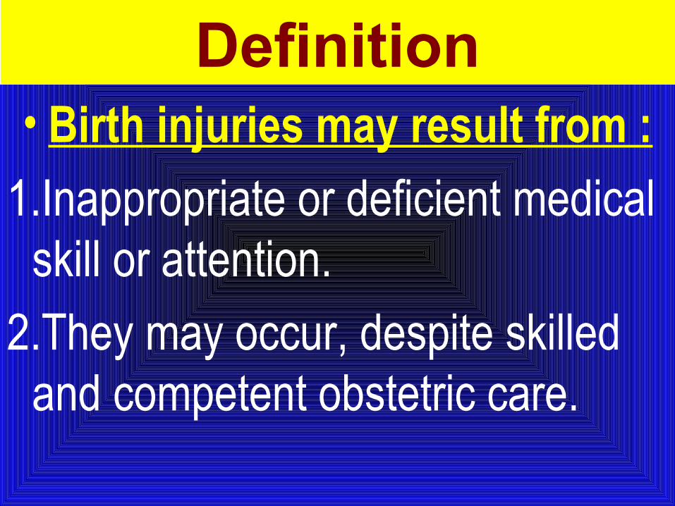
Incidence Has been estimated at 2-7/1,000 live births.
Predisposing factors:1. Macrosomia, 2. Prematurity, 3. Cephalopelvic disproportion,4. Dystocia, 5. Prolonged labor, and 6. Breech presentation.
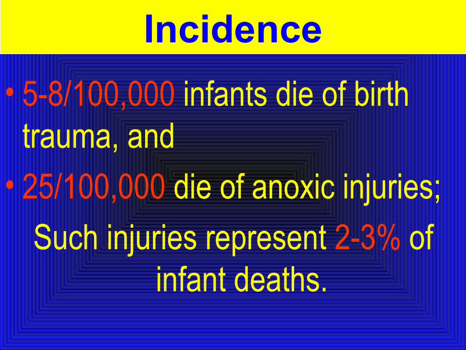
• 5-8/100,000 infants die of birth trauma, and
• 25/100,000 die of anoxic injuries;
Such injuries represent 2-3% of infant deaths.
Incidence
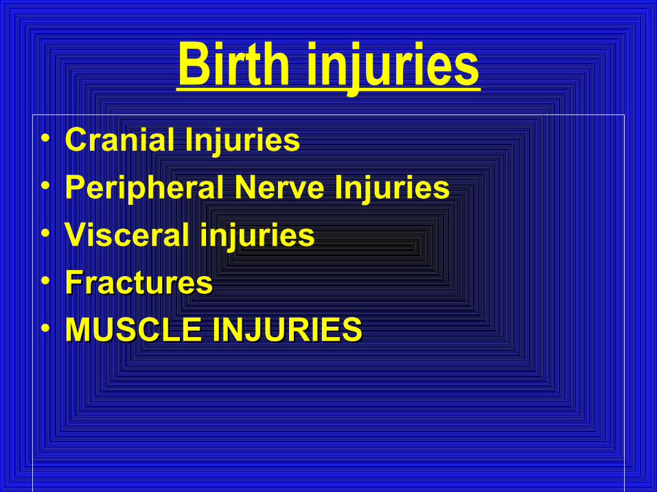
Birth injuries• Cranial Injuries
• Peripheral Nerve Injuries
• Visceral injuries
• FracturesFractures• MUSCLE INJURIESMUSCLE INJURIES
Cranial Injuries
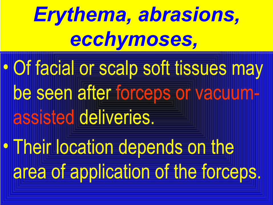
Erythema, abrasions, ecchymoses,
• Of facial or scalp soft tissues may be seen after forceps or vacuum-assisted deliveries.
• Their location depends on the area of application of the forceps.
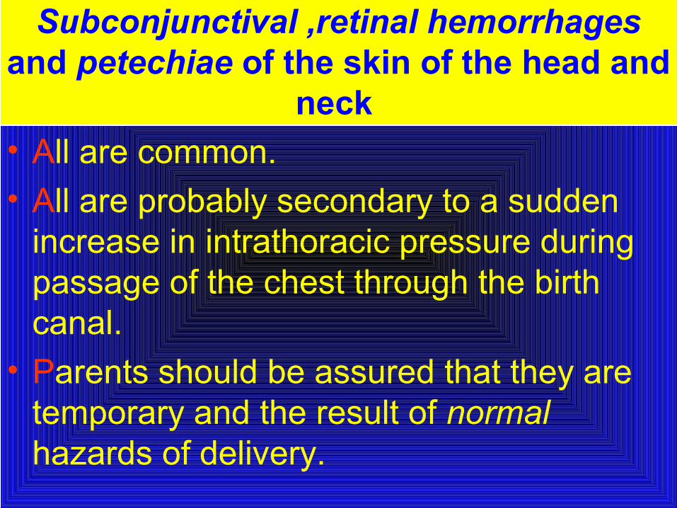
Subconjunctival ,retinal hemorrhages and petechiae of the skin of the head and
neck • All are common. • All are probably secondary to a sudden
increase in intrathoracic pressure during passage of the chest through the birth canal.
• Parents should be assured that they are temporary and the result of normal hazards of delivery.
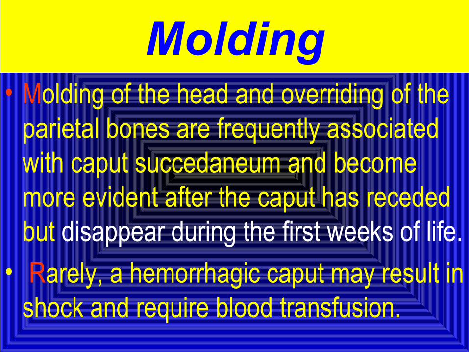
Molding• Molding of the head and overriding of the
parietal bones are frequently associated with caput succedaneum and become more evident after the caput has receded but disappear during the first weeks of life.
• Rarely, a hemorrhagic caput may result in shock and require blood transfusion.
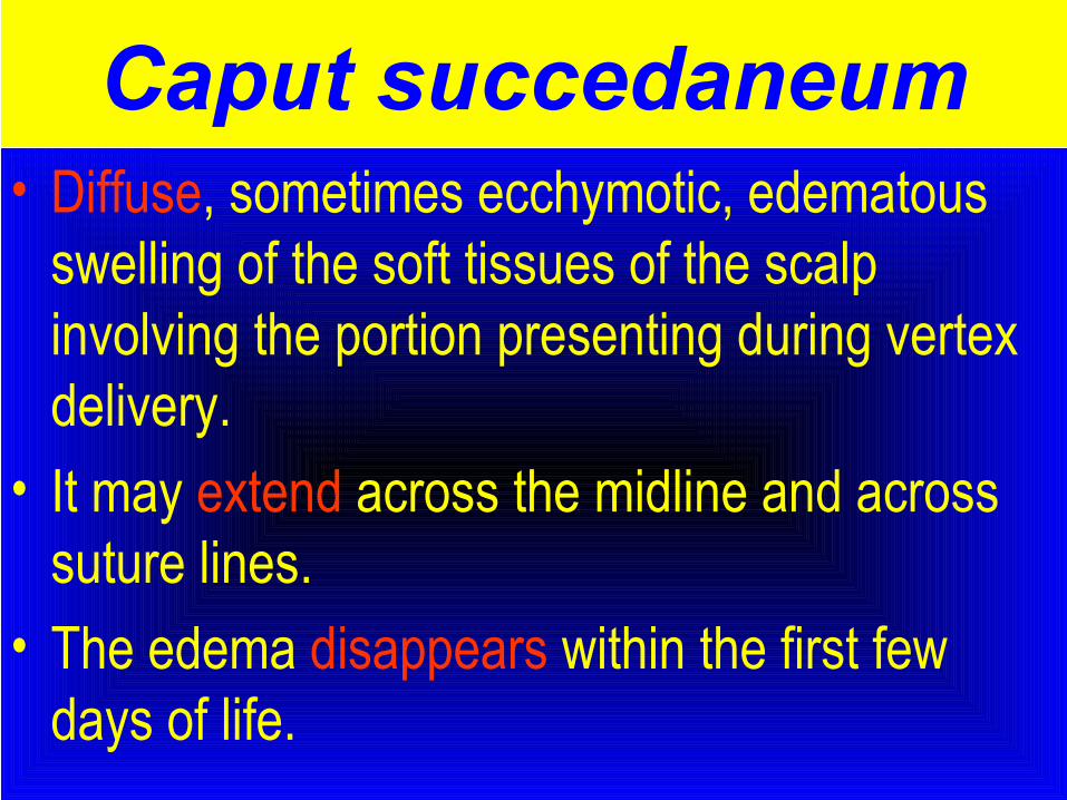
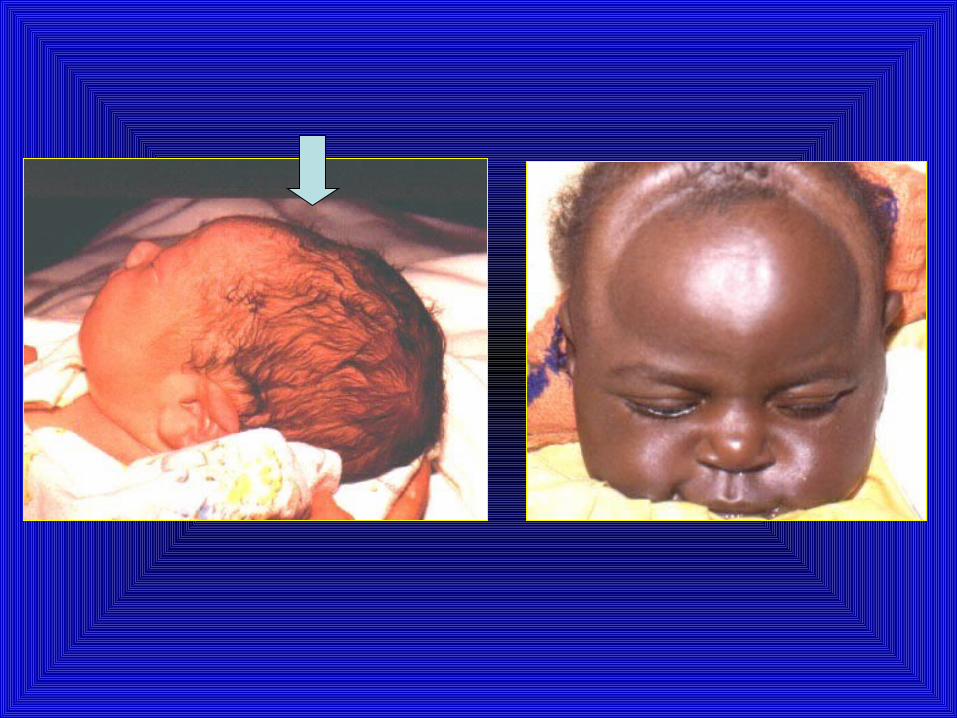
Caput succedaneum• Diffuse, sometimes ecchymotic, edematous
swelling of the soft tissues of the scalp involving the portion presenting during vertex delivery.
• It may extend across the midline and across suture lines.
• The edema disappears within the first few days of life.

• Analogous swelling, discoloration, and distortion of the face are seen in face presentations.
• No specific treatment is needed, but if there are extensive ecchymoses, phototherapy for hyperbilirubinemia may be indicated.
Caput succedaneumCaput succedaneum
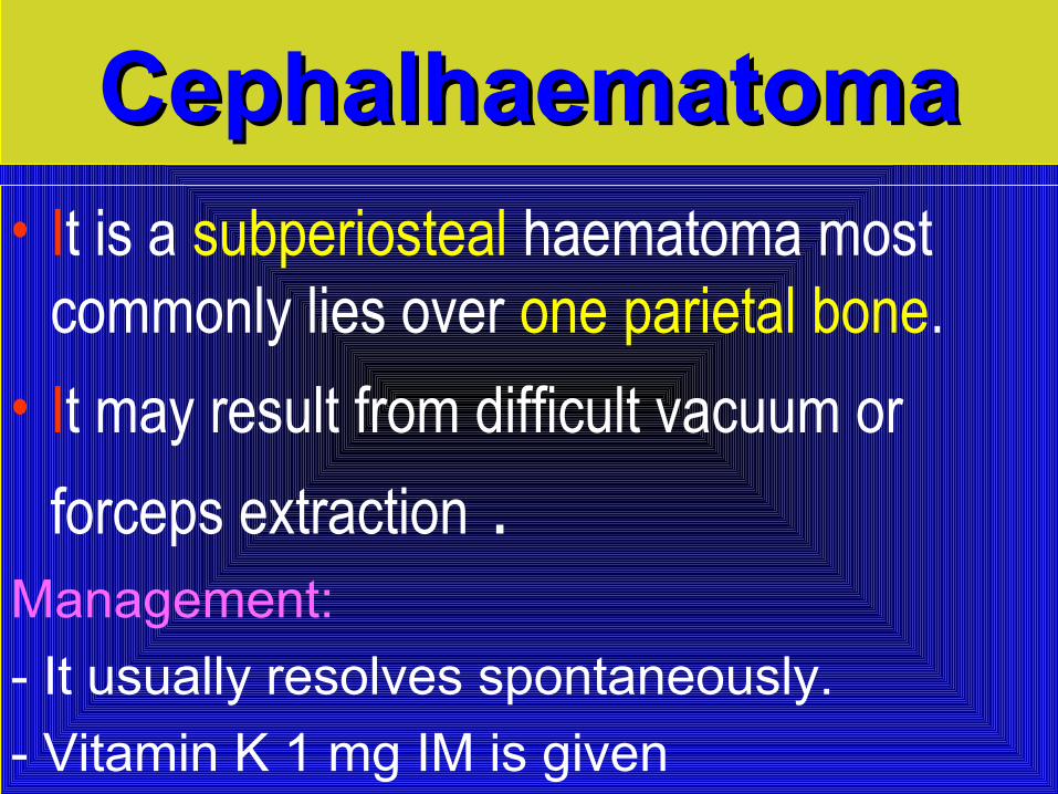
CephalhaematomaCephalhaematoma• It is a subperiosteal haematoma most
commonly lies over one parietal bone.
• It may result from difficult vacuum or
forceps extraction .Management:
- It usually resolves spontaneously.
- Vitamin K 1 mg IM is given
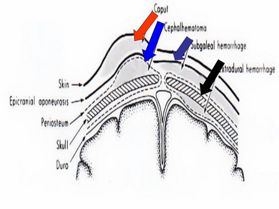
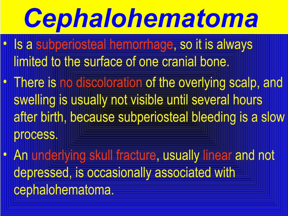
Cephalohematoma • Is a subperiosteal hemorrhage, so it is always
limited to the surface of one cranial bone. • There is no discoloration of the overlying scalp, and
swelling is usually not visible until several hours after birth, because subperiosteal bleeding is a slow process.
• An underlying skull fracture, usually linear and not depressed, is occasionally associated with cephalohematoma.
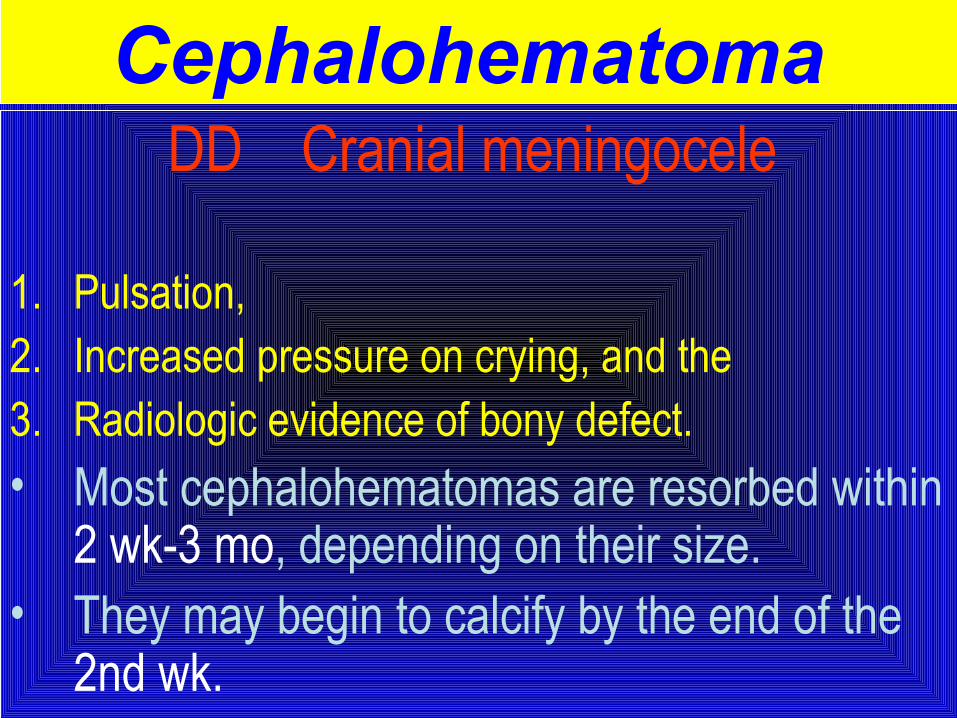
DD Cranial meningocele
1. Pulsation, 2. Increased pressure on crying, and the 3. Radiologic evidence of bony defect. • Most cephalohematomas are resorbed within
2 wk-3 mo, depending on their size. • They may begin to calcify by the end of the
2nd wk.
Cephalohematoma
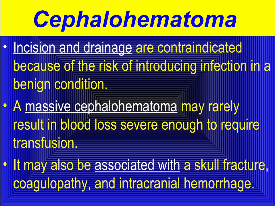
• Incision and drainage are contraindicated because of the risk of introducing infection in a benign condition.
• A massive cephalohematoma may rarely result in blood loss severe enough to require transfusion.
• It may also be associated with a skull fracture, coagulopathy, and intracranial hemorrhage.
Cephalohematoma
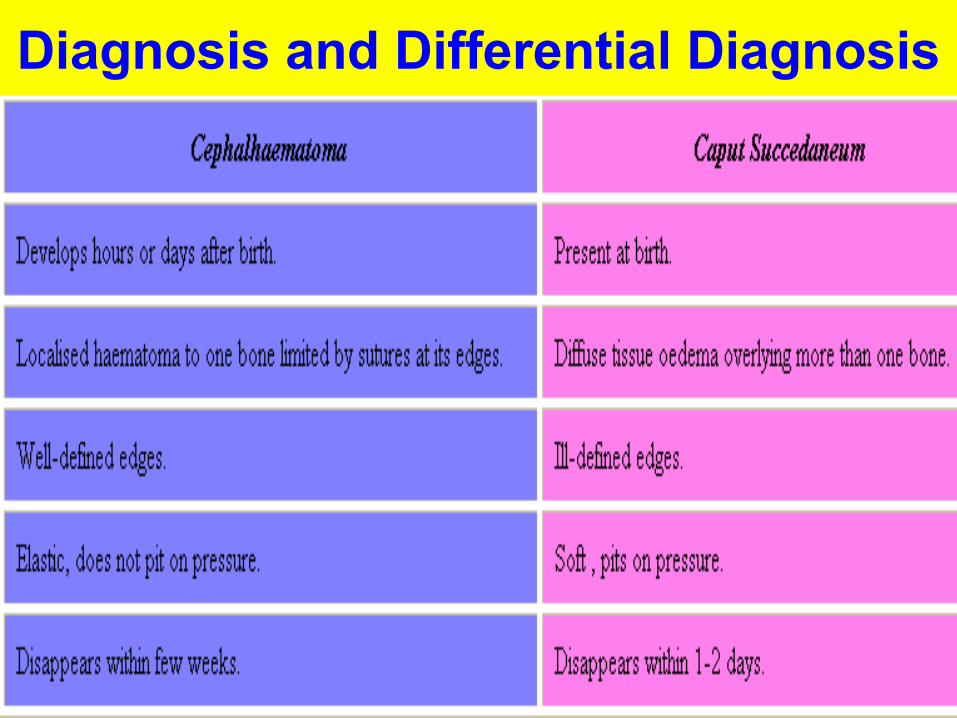
Diagnosis and Differential Diagnosis
Fractures of the skull May occur as a result of pressure from :
1. Forceps or from
2. The maternal symphysis pubis.
3. Sacral promontory, or
4. Ischial spines.
Fracture Skull:Usually occurs due to difficult forceps delivery.
It may be:
(1) Vault fracture:
• Usually affecting the frontal or parietal bone.• It may be linear or depressed fracture. • It needs no treatment unless there is intracranial
haemorrhage.
(2) Fracture base:
• Usually associated with intracranial haemorrhage.
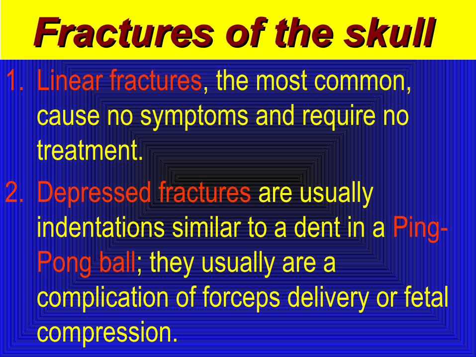
1. Linear fractures, the most common, cause no symptoms and require no treatment.
2. Depressed fractures are usually indentations similar to a dent in a Ping-Pong ball; they usually are a complication of forceps delivery or fetal compression.
Fractures of the skullFractures of the skull
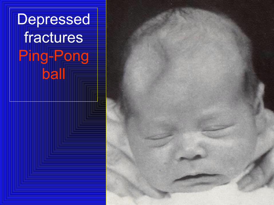
Depressed fractures
Ping-Pong ball
• Affected infants may be asymptomatic unless there is associated intracranial injury.
• It is advisable to elevate severe depressions to prevent cortical injury from sustained pressure.
Fractures of the skull
• Fracture of the Occipital bone almost causes fatal hemorrhage due to disruption of the underlying vascular sinuses.
• It may result during breech deliveries from traction on the hyperextended spine of the infant with the head fixed in the maternal pelvis.
Fractures of the skull

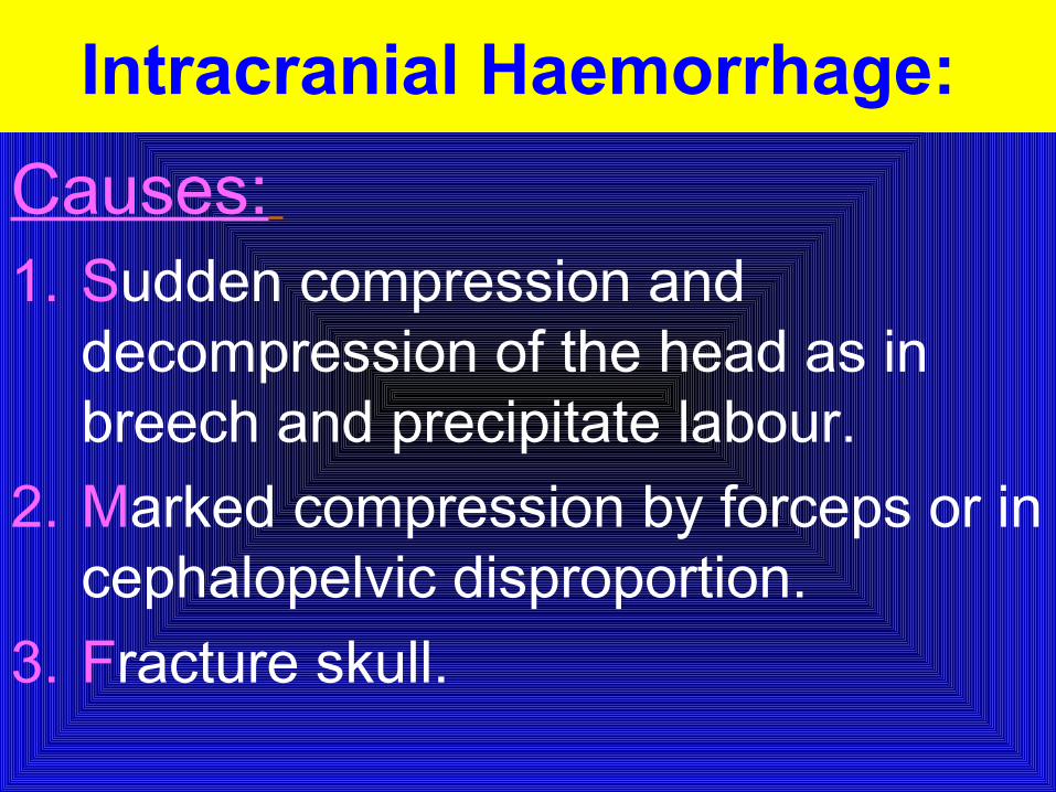
Intracranial Haemorrhage:
Causes: 1. Sudden compression and
decompression of the head as in breech and precipitate labour.
2. Marked compression by forceps or in cephalopelvic disproportion.
3. Fracture skull.
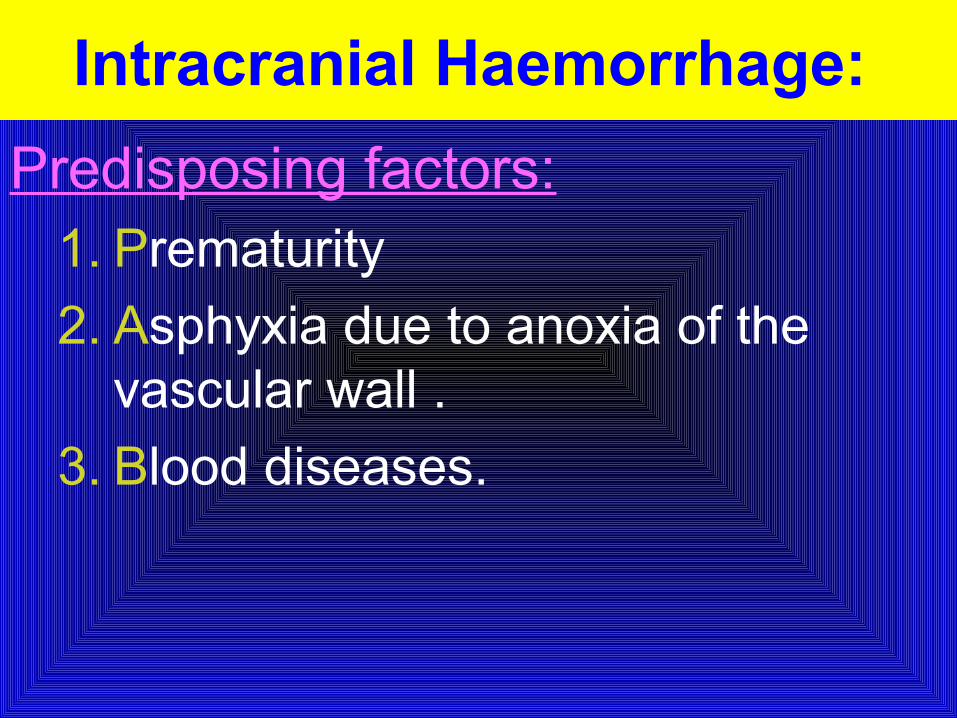
Predisposing factors:1. Prematurity
2. Asphyxia due to anoxia of the vascular wall .
3. Blood diseases.
Intracranial Haemorrhage:
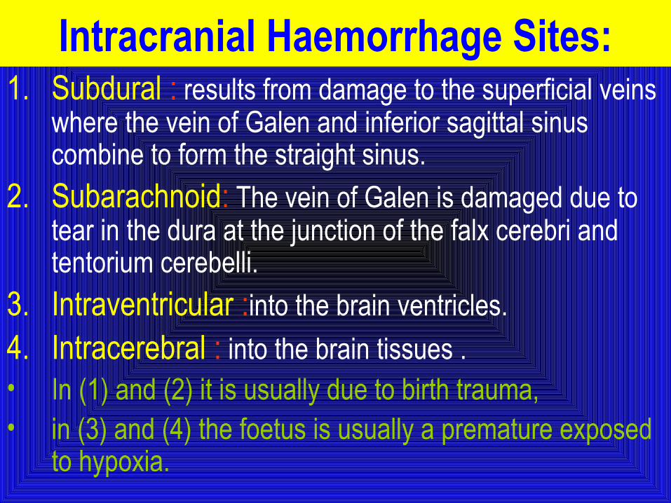
1. Subdural : results from damage to the superficial veins where the vein of Galen and inferior sagittal sinus combine to form the straight sinus.
2. Subarachnoid: The vein of Galen is damaged due to tear in the dura at the junction of the falx cerebri and tentorium cerebelli.
3. Intraventricular :into the brain ventricles.
4. Intracerebral : into the brain tissues .• In (1) and (2) it is usually due to birth trauma, • in (3) and (4) the foetus is usually a premature exposed
to hypoxia.
Intracranial Haemorrhage Sites:
Clinical picture:
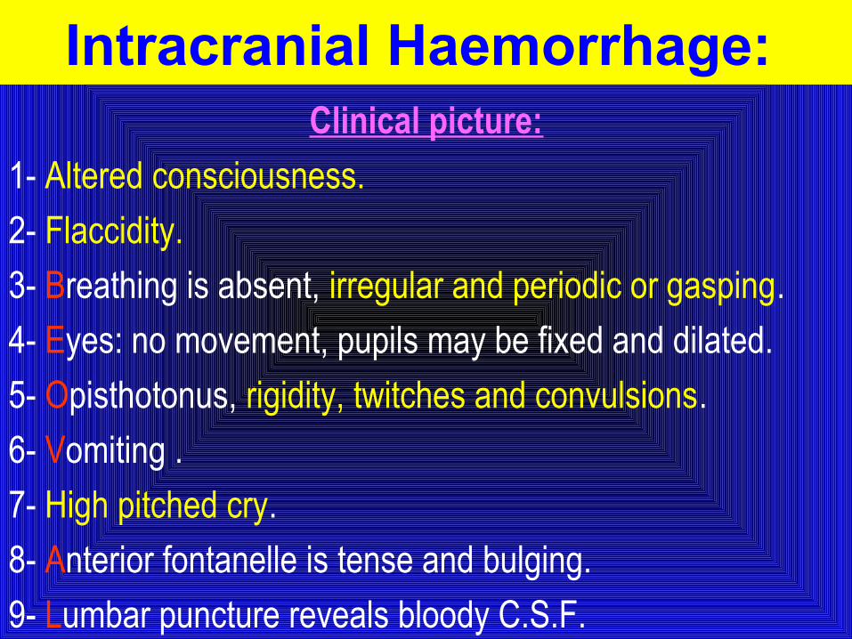
1- Altered consciousness.
2- Flaccidity.
3- Breathing is absent, irregular and periodic or gasping.
4- Eyes: no movement, pupils may be fixed and dilated.
5- Opisthotonus, rigidity, twitches and convulsions.
6- Vomiting .
7- High pitched cry.
8- Anterior fontanelle is tense and bulging.
9- Lumbar puncture reveals bloody C.S.F.
Intracranial Haemorrhage:
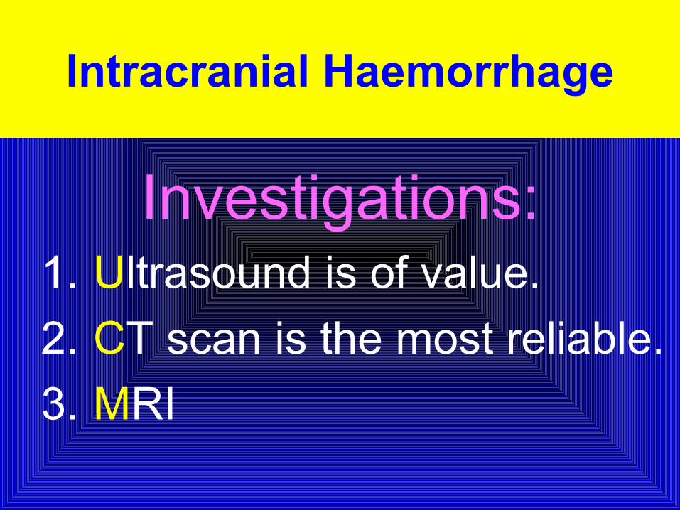
Investigations:1. Ultrasound is of value.
2. CT scan is the most reliable.
3. MRI
Intracranial Haemorrhage
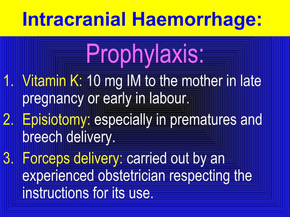
Prophylaxis:1. Vitamin K: 10 mg IM to the mother in late
pregnancy or early in labour.2. Episiotomy: especially in prematures and
breech delivery.3. Forceps delivery: carried out by an
experienced obstetrician respecting the instructions for its use.
Intracranial Haemorrhage:
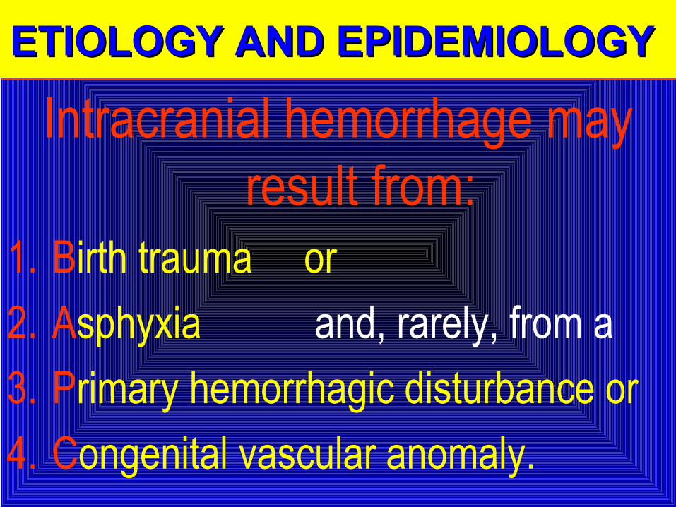
ETIOLOGY AND EPIDEMIOLOGY ETIOLOGY AND EPIDEMIOLOGY ETIOLOGY AND EPIDEMIOLOGY ETIOLOGY AND EPIDEMIOLOGY
Intracranial hemorrhage may result from:
1. Birth trauma or
2. Asphyxia and, rarely, from a
3. Primary hemorrhagic disturbance or
4. Congenital vascular anomaly.
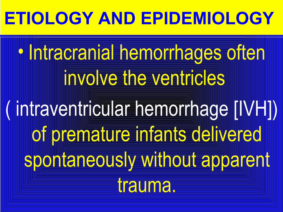
• Intracranial hemorrhages often involve the ventricles
( intraventricular hemorrhage [IVH]) of premature infants delivered
spontaneously without apparent trauma.
ETIOLOGY AND EPIDEMIOLOGY
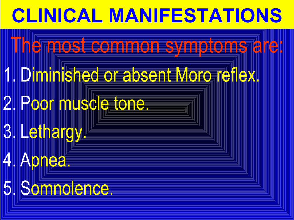
The most common symptoms are:1. Diminished or absent Moro reflex.
2. Poor muscle tone.
3. Lethargy.
4. Apnea.
5. Somnolence.
CLINICAL MANIFESTATIONS
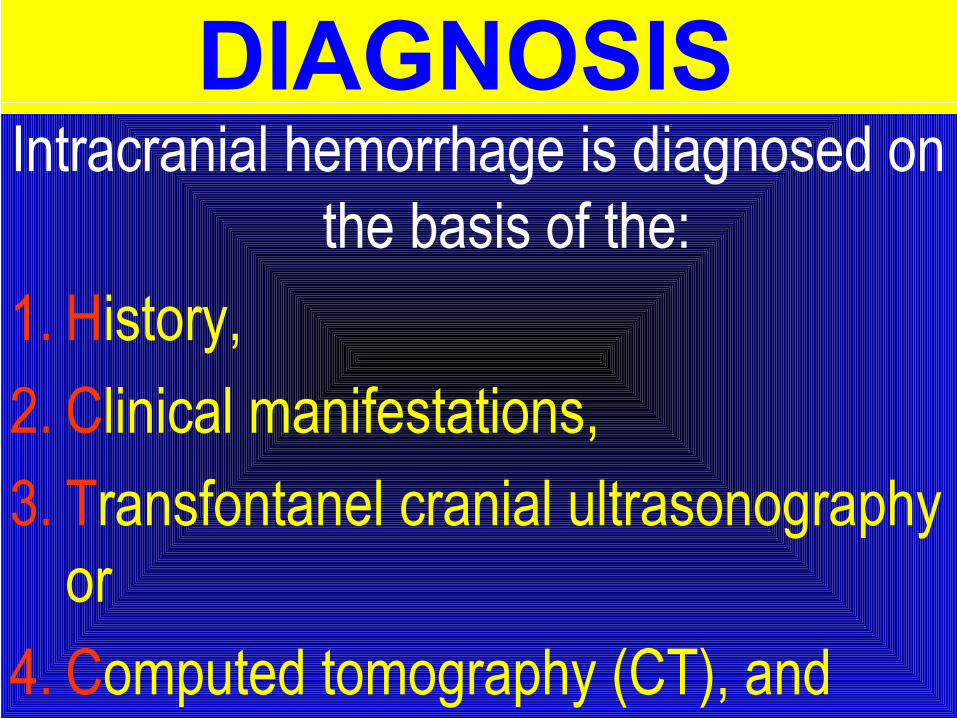
DIAGNOSIS Intracranial hemorrhage is diagnosed on
the basis of the:
1. History,
2. Clinical manifestations,
3. Transfontanel cranial ultrasonography or
4. Computed tomography (CT), and
Lumbar puncture DIAGNOSIS
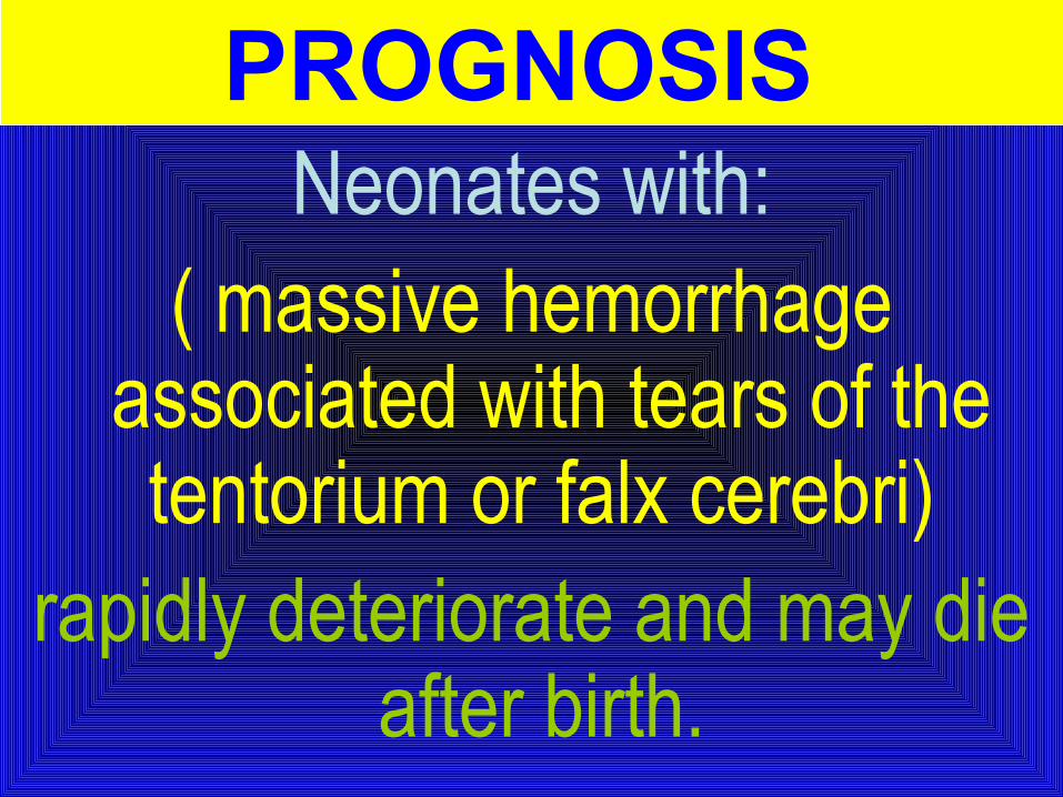
PROGNOSIS Neonates with:
( massive hemorrhage associated with tears of the tentorium or falx cerebri)
rapidly deteriorate and may die after birth.
PREVENTION The incidence of traumatic intracranial
hemorrhage may be reduced by: judicious management of cephalopelvic
disproportion and operative delivery.
Spine and Spinal CordStrong traction exerted:
1. When the spine is hyperextended or 2. When the direction of pull is lateral, or 3. Forceful longitudinal traction on the trunk
while the head is still firmly engaged in the pelvis:
(may produce fracture and separation of the vertebrae).
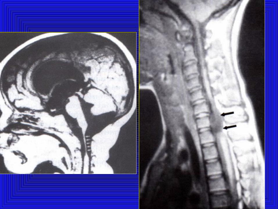
1. Areflexia,2. Loss of sensation, and 3. Complete paralysis of
voluntary motion Occur below the level of injury
Spine and Spinal CordSpine and Spinal Cord
Peripheral Nerve Injuries
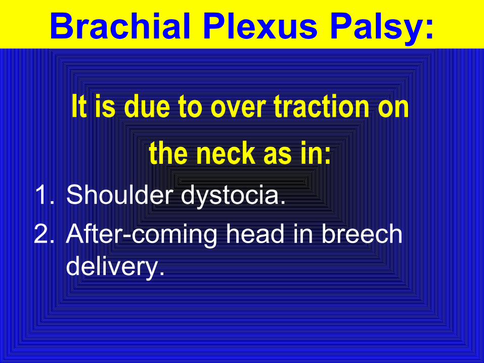
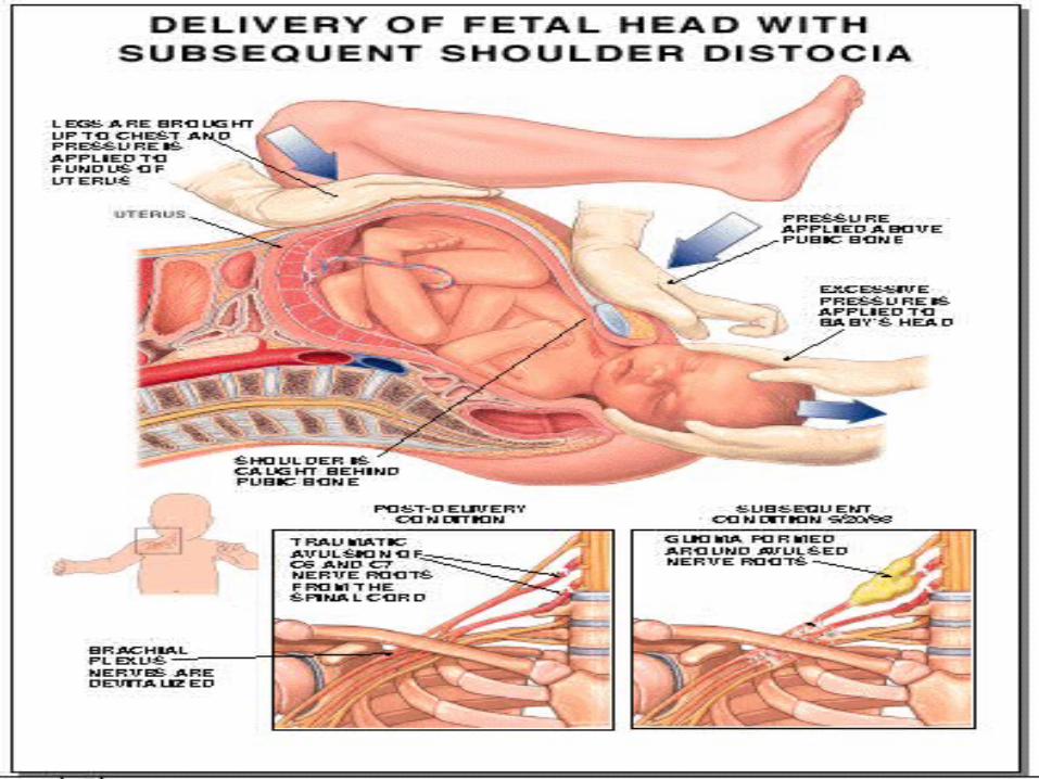
Brachial Plexus Palsy:
It is due to over traction on
the neck as in: 1. Shoulder dystocia.
2. After-coming head in breech delivery.
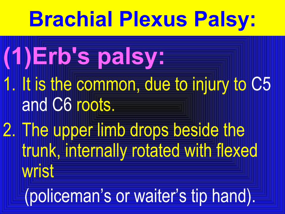
(1)Erb's palsy:1. It is the common, due to injury to C5
and C6 roots.2. The upper limb drops beside the
trunk, internally rotated with flexed wrist (policeman’s or waiter’s tip hand).
Brachial Plexus Palsy:
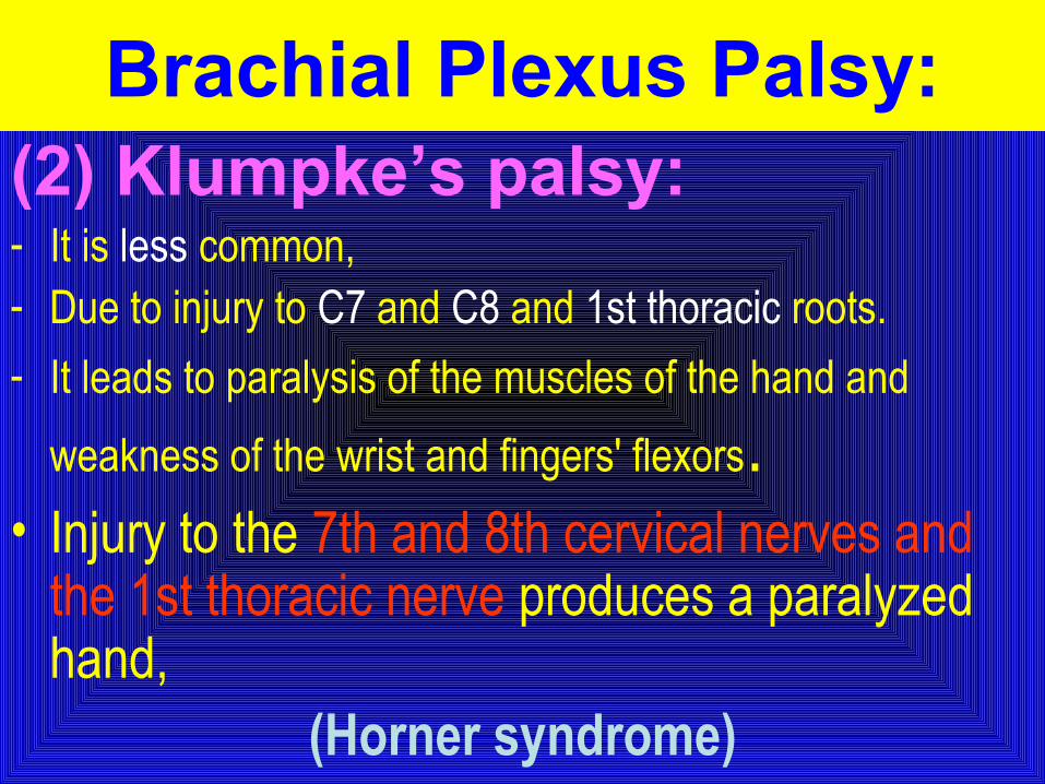
(2) Klumpke’s palsy:- It is less common,- Due to injury to C7 and C8 and 1st thoracic roots.
- It leads to paralysis of the muscles of the hand and
weakness of the wrist and fingers' flexors.• Injury to the 7th and 8th cervical nerves and
the 1st thoracic nerve produces a paralyzed hand,
(Horner syndrome)
Brachial Plexus Palsy:
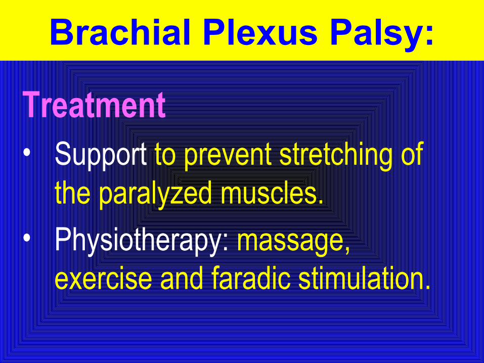
Treatment • Support to prevent stretching of
the paralyzed muscles.• Physiotherapy: massage,
exercise and faradic stimulation.
Brachial Plexus Palsy:
8
7
9
4
5
6
3
2
1
Roots
Trunks
Cords
Nerves
ANATOMY OF THE BRACHIAL PLEXUS
UlnarMedianRadial
7
8
9
5
Lateral PosteriorMedial
4
6
Upper Middle Lower
1
2
3
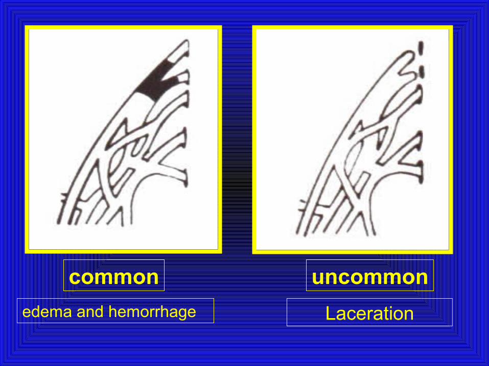
common uncommon
edema and hemorrhage Laceration
The prognosisThe prognosis• Depends on whether the nerve was
merely injured or was lacerated. • If the paralysis was due to edema and
hemorrhage about the nerve fibers, function should return within a few months;
• If due to laceration, permanent damage may result.
TreatmentTreatment• Partial immobilization and appropriate
positioning to prevent development of contractures.
• In upper arm paralysis: the arm should be abducted, with external rotation at the shoulder and with full supination of the forearm and slight extension at the wrist with the palm turned toward the face.
• In lower arm or hand paralysis: the wrist should be splinted in a neutral position and padding placed in the fist.
• Gentle massage and range of motion exercises may be started by 7-10 days of age.
TreatmentTreatment
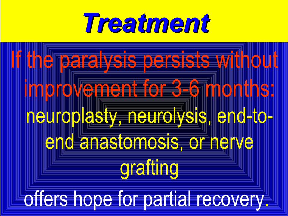
If the paralysis persists without improvement for 3-6 months: neuroplasty, neurolysis, end-to-
end anastomosis, or nerve grafting
offers hope for partial recovery.
TreatmentTreatment
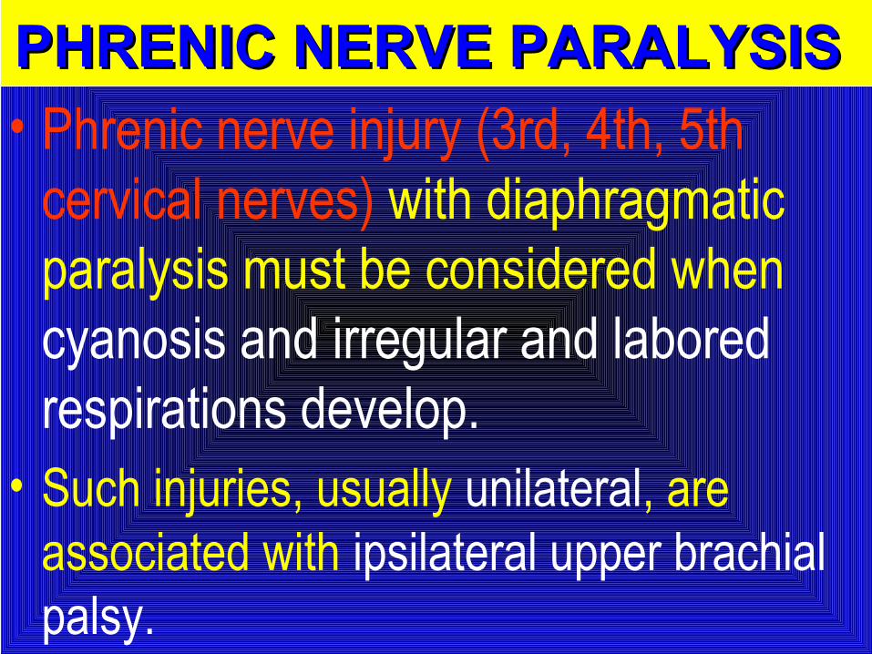
PHRENIC NERVE PARALYSIS PHRENIC NERVE PARALYSIS • Phrenic nerve injury (3rd, 4th, 5th
cervical nerves) with diaphragmatic paralysis must be considered when cyanosis and irregular and labored respirations develop.
• Such injuries, usually unilateral, are associated with ipsilateral upper brachial palsy.
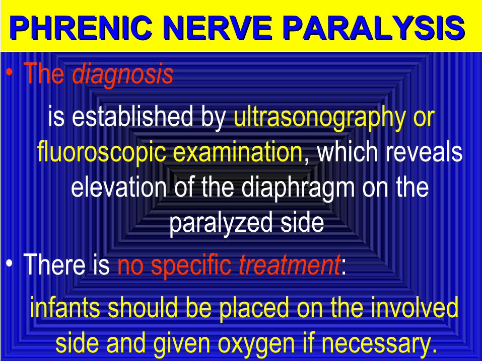
• The diagnosis
is established by ultrasonography or fluoroscopic examination, which reveals
elevation of the diaphragm on the paralyzed side
• There is no specific treatment:
infants should be placed on the involved side and given oxygen if necessary.
PHRENIC NERVE PARALYSISPHRENIC NERVE PARALYSIS
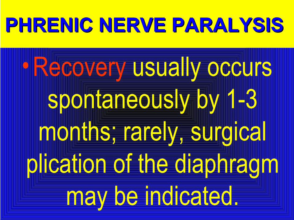
• Recovery usually occurs spontaneously by 1-3
months; rarely, surgical plication of the diaphragm
may be indicated.
PHRENIC NERVE PARALYSISPHRENIC NERVE PARALYSIS
Facial Palsy (Bell’s palsy):Facial Palsy (Bell’s palsy):
- It is usually due to pressure by the forceps blade on the facial nerve at:
1. Its exit from the stylomastoid foramen or
2. In its course over the mandibular ramus.
- It appears within 1-2 days after delivery due to resultant oedema and
haemorrhage around the nerve.
Manifestations:
1. There is paresis of the facial muscles on the affected side with:
2. Partially opened eye and:
3. Flattening of the nasolabial fold.
4. The mouth angle is deviated towards the healthy side.
Spontaneous recovery usually occurs
within 14 days.
Facial Palsy (Bell’s palsy):Facial Palsy (Bell’s palsy):
• When the infant cries, there is movement only on the non paralyzed side of the face, and the mouth is drawn to that side.
• On the affected side the forehead is smooth, the eye cannot be closed, the nasolabial fold is absent, and the corner of the mouth drops.
FACIAL NERVE PALSYFACIAL NERVE PALSY
• The prognosis depends on whether the nerve was injured by pressure or whether the nerve fibers were torn.
• Care of the exposed eye is essential.
FACIAL NERVE PALSYFACIAL NERVE PALSY
• Improvement occurs within few weeks.
• Neuroplasty may be indicated when the paralysis is persistent.
FACIAL NERVE PALSYFACIAL NERVE PALSY

Other peripheral Other peripheral nervesnerves
are seldom injured in utero or at birth except when they are involved in fractures or
hemorrhages.
V) VISCERAL INJURIESV) VISCERAL INJURIES
((Liver, spleen and kidney)Liver, spleen and kidney) may be injured in breech may be injured in breech delivery which should be delivery which should be
avoided by holding the fetus avoided by holding the fetus from its hips.from its hips.
FracturesFractures
BONE INJURIESThese usually occur during difficult
breech delivery.(A) Vertebral Column Injuries:• These are fatal if associated with spinal cord
transection above C4 ,due to diaphragmatic paralysis.
(B) Femur, Humerus and Clavicle:• Managed by splint to the long bone and a sling for
clavicular fracture.
CLAVICLE This bone is fractured during labor and
delivery
more frequently than any other bone; It is particularly vulnerable when there is:
1. Difficulty in delivery of the shoulder in vertex presentations and of
2. The extended arms in breech deliveries.
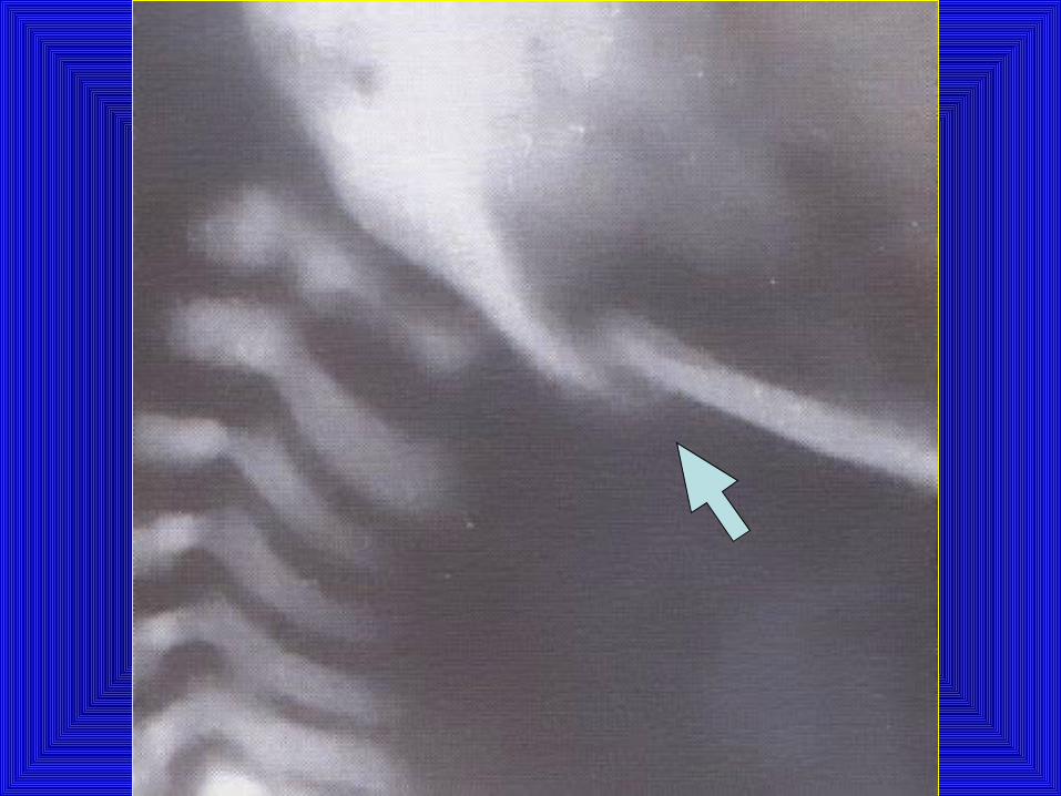
• The infant characteristically does not move the arm freely on the affected side;
• Crepitus and bony irregularity may be palpated, and
• Discoloration is occasionally visible over the fracture site.
CLAVICLE

MUSCLE INJURIESMUSCLE INJURIESStrenomastoid injury
Due to :• Exaggerated lateral flexion of the neck
leading to torticollis and swelling in the muscle.
• It is usually improved within 2 weeks but permanent torticollis may continue.