Embed Size (px)
Citation preview
FOCAL HEPATIC LESIONS
•Yasser Asiri R1
Objective :
1. Identify the most important imaging
features of common benign liver tumors
2. Identify the most important imaging
features of malignant lesions
3. Know the diagnosis of hepatocellular
carcinoma
Introduction • Extensive use of imaging studies has
increased the detection rates of hepatic lesions
• A mass can be found either incidentally or during screening for liver cancer in patients with cirrhosis
• These can be benignant or malignant and thus the right approach for assessing these masses is important
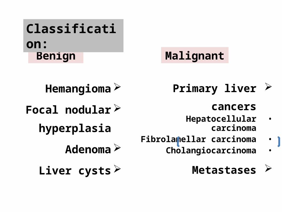
Hemangioma
Focal nodular
hyperplasia
Adenoma
Liver cysts
Primary liver cancers•Hepatocellular carcinoma•Fibrolamellar carcinoma•Cholangiocarcinoma
Metastases
Benign Malignant
Classification:

• Symptomatic or Incidentally detected• History of Hepatitis or extra hepatic
malignant tumor• Liver function tests• Cirrhotic or Non cirrhotic
BENIGN LIVER LESIONS
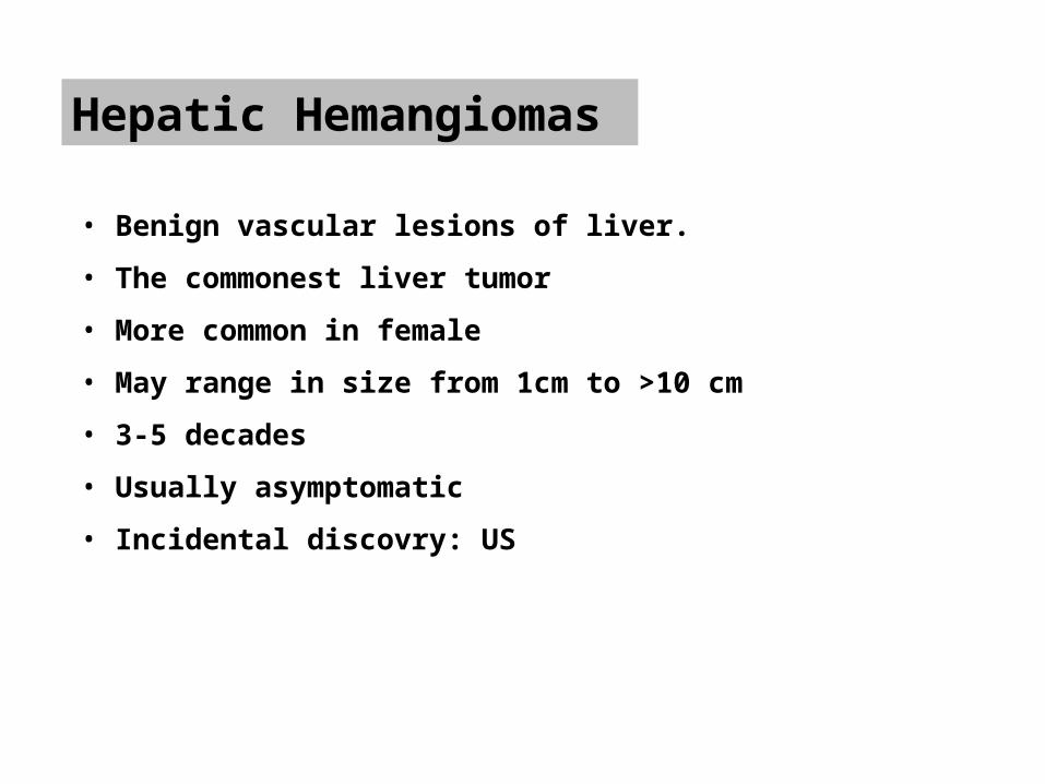
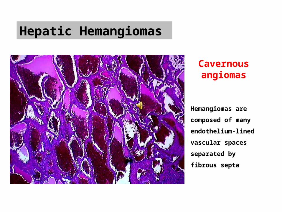
Hepatic Hemangiomas
• Benign vascular lesions of liver. • The commonest liver tumor• More common in female • May range in size from 1cm to >10 cm• 3-5 decades• Usually asymptomatic• Incidental discovry: US
Hepatic Hemangiomas
Hemangiomas are composed of many endothelium-lined vascular spaces separated by fibrous septa
Cavernous angiomas
Hepatic Hemangiomas
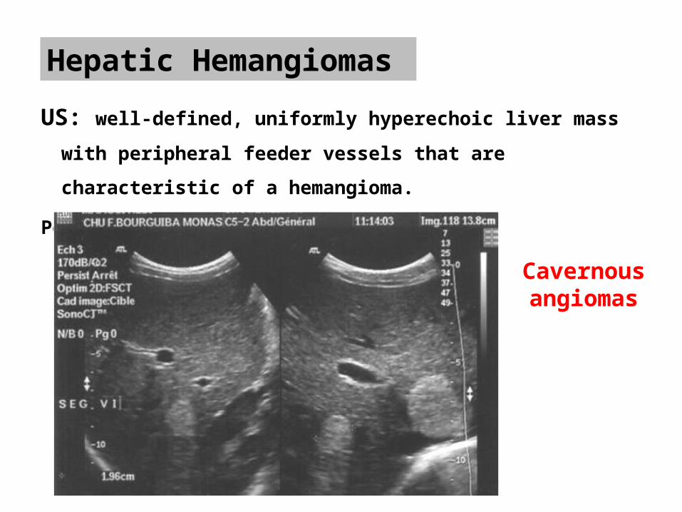
US: well-defined, uniformly hyperechoic liver mass with peripheral feeder
vessels that are characteristic of a hemangioma.
Posterior acoustic enhancment.
Cavernous angiomas

Hepatic Hemangiomas
CT: The pathognomonic features of caverneous hemangioma: peripheral
nodular and discontinuous enhancement and progressive centripetal fill-in
The attenuation of the enhancement is similar to the aorta.
IV-
HAP
PVP
DP
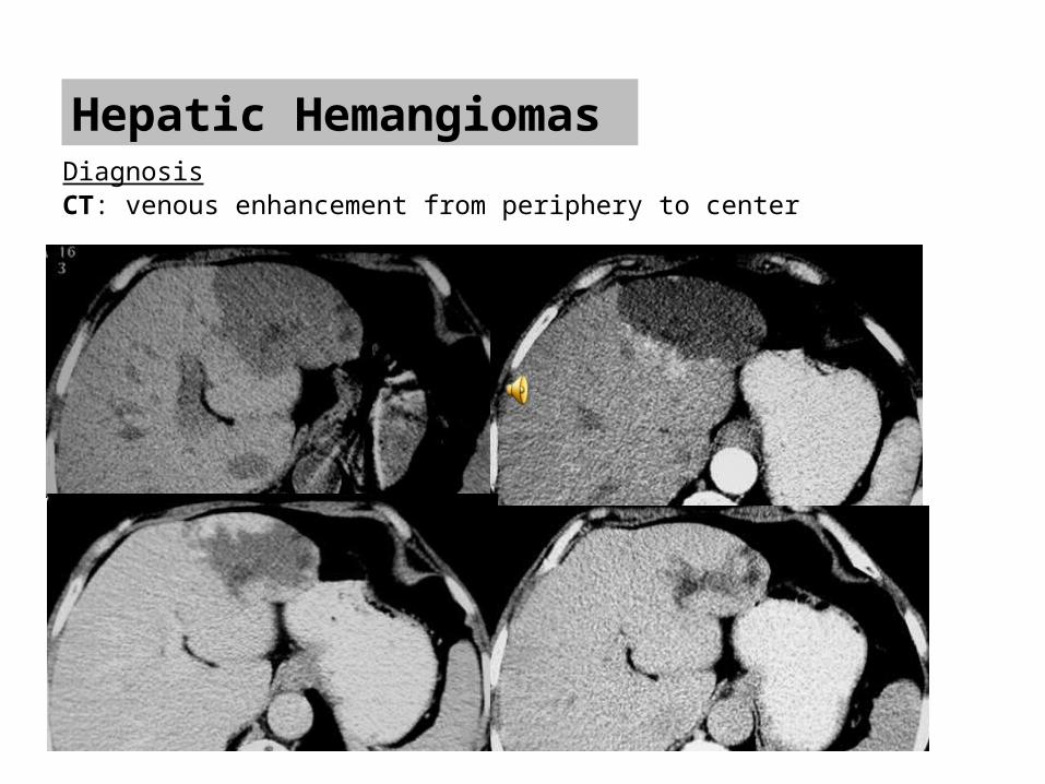
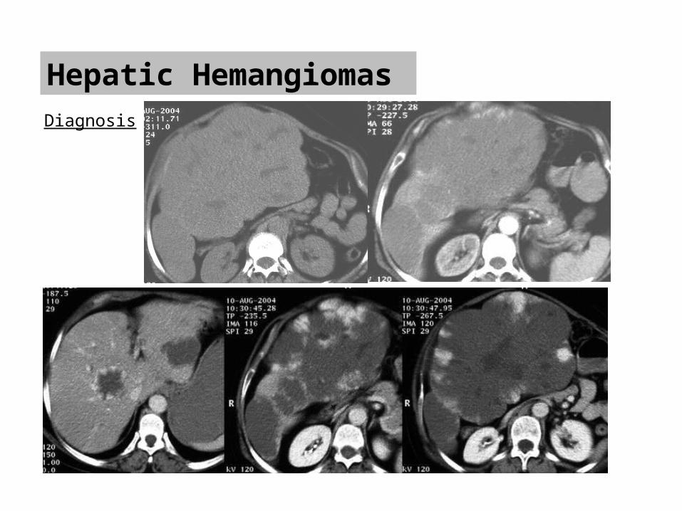
Hepatic Hemangiomas DiagnosisCT: venous enhancement from periphery to center
Hepatic Hemangiomas
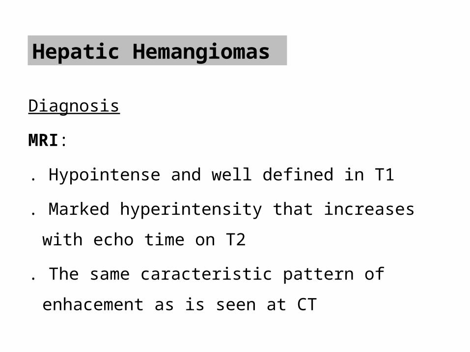
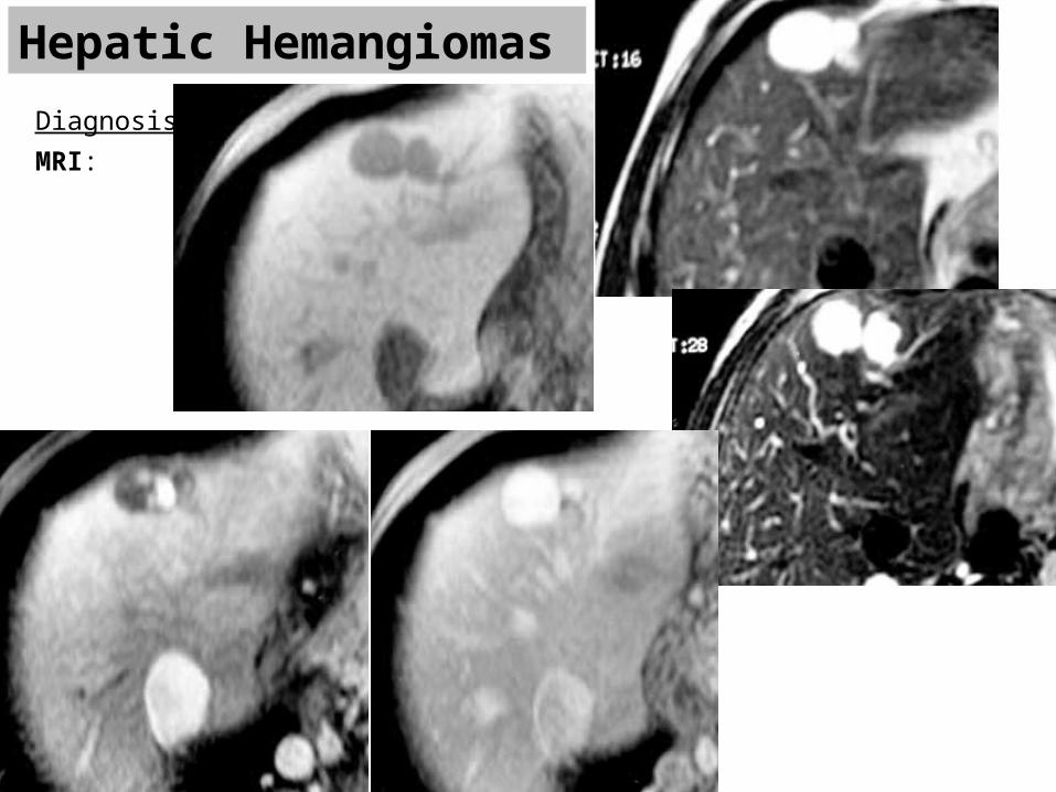
Diagnosis
MRI:
. Hypointense and well defined in T1
. Marked hyperintensity that increases with echo time on
T2
. The same caracteristic pattern of enhacement as is seen at
CT
Hepatic Hemangiomas DiagnosisMRI:
Hepatic Hemangiomas Diagnosis
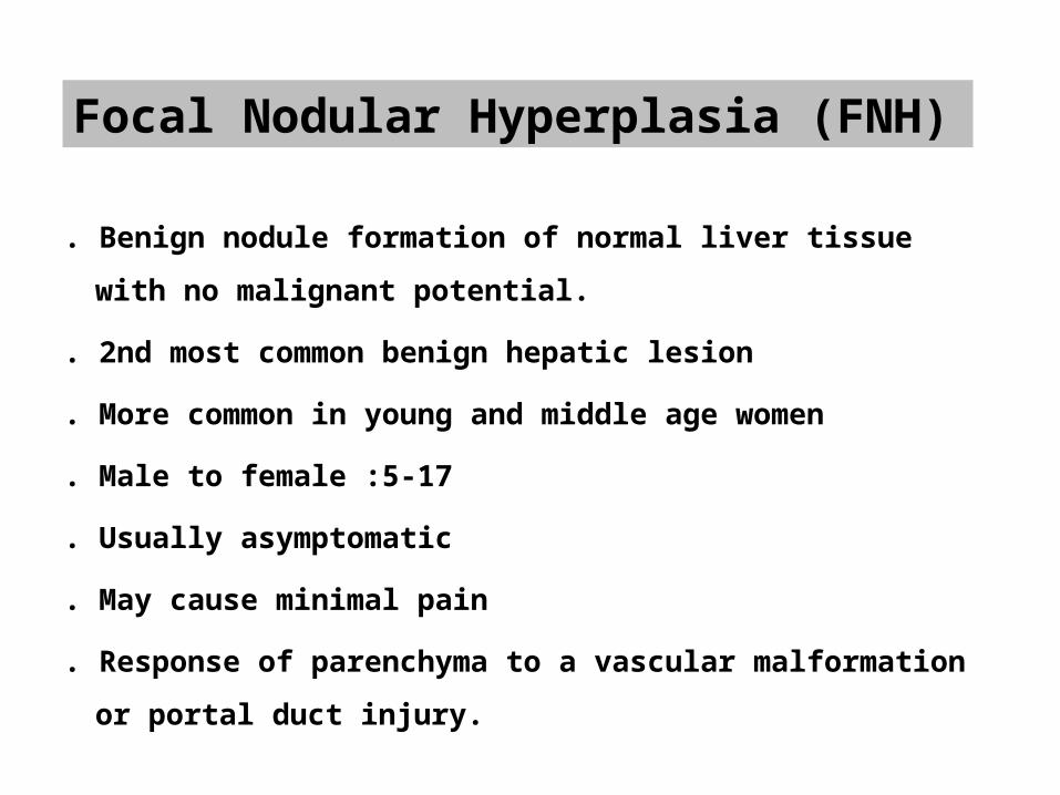
Focal Nodular Hyperplasia (FNH)
. Benign nodule formation of normal liver tissue with no malignant
potential.
. 2nd most common benign hepatic lesion
. More common in young and middle age women
. Male to female :5-17
. Usually asymptomatic
. May cause minimal pain
. Response of parenchyma to a vascular malformation or portal duct injury.
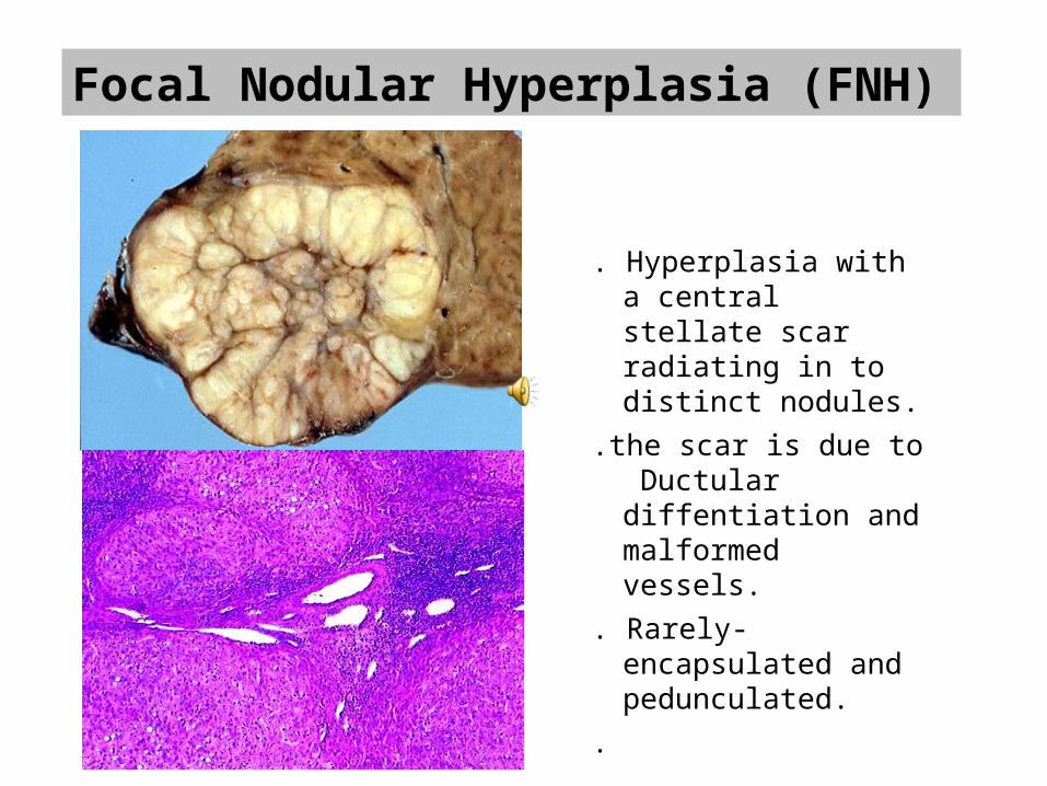
Focal Nodular Hyperplasia (FNH)
. Hyperplasia with a central stellate scar radiating in to distinct nodules.
.the scar is due to Ductular diffentiation and malformed vessels.
. Rarely- encapsulated and pedunculated.
.
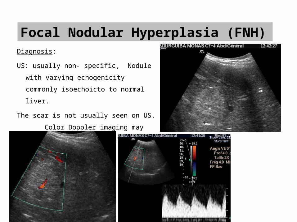
Focal Nodular Hyperplasia (FNH)Diagnosis:
US: usually non- specific, Nodule with varying
echogenicity commonly isoechoicto to normal
liver.
The scar is not usually seen on US.
Color Doppler imaging may show central vessels
with spoke wheel configuration
Focal Nodular Hyperplasia (FNH)Diagnosis: CT
. Central scar formed by a ductules and venules rather than a true fibrous scar.
. Brisk homogeneous enhancement with quick wash out. In the portovenous phase it will only shows unenhanced scar. Usually th e scar shows late enhancment
. Well defined
. Early homogenesation
. Hypodense fibrous bands and septa that arise from the scar
. On delayed phase images the central scar may remain hyperattenuating
. Without capsule
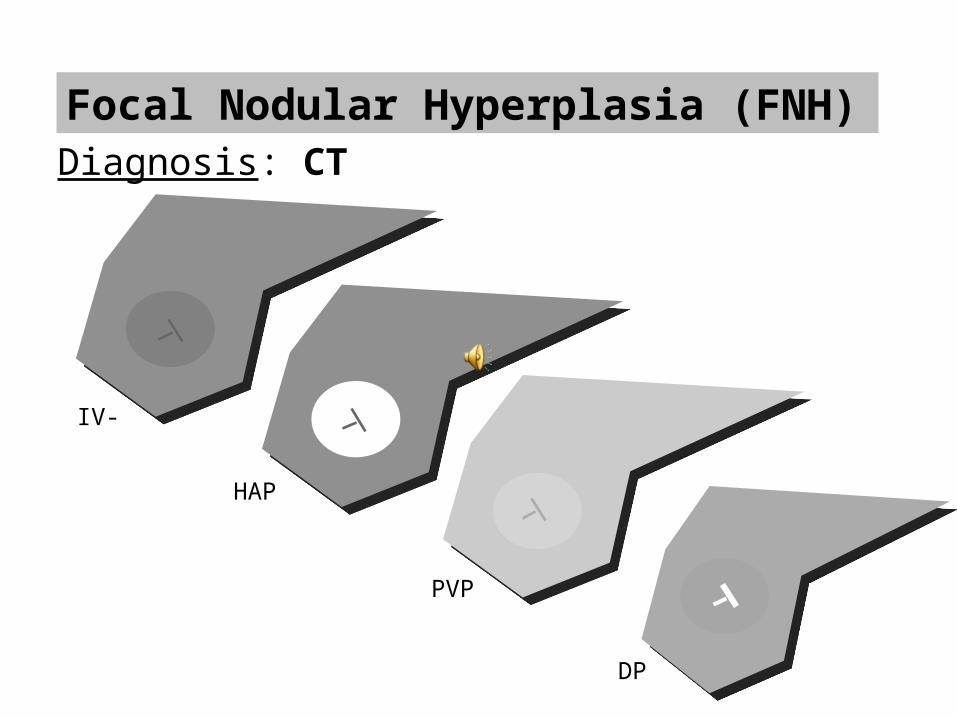
Focal Nodular Hyperplasia (FNH)Diagnosis: CT
HAP
PVP
DP
IV-
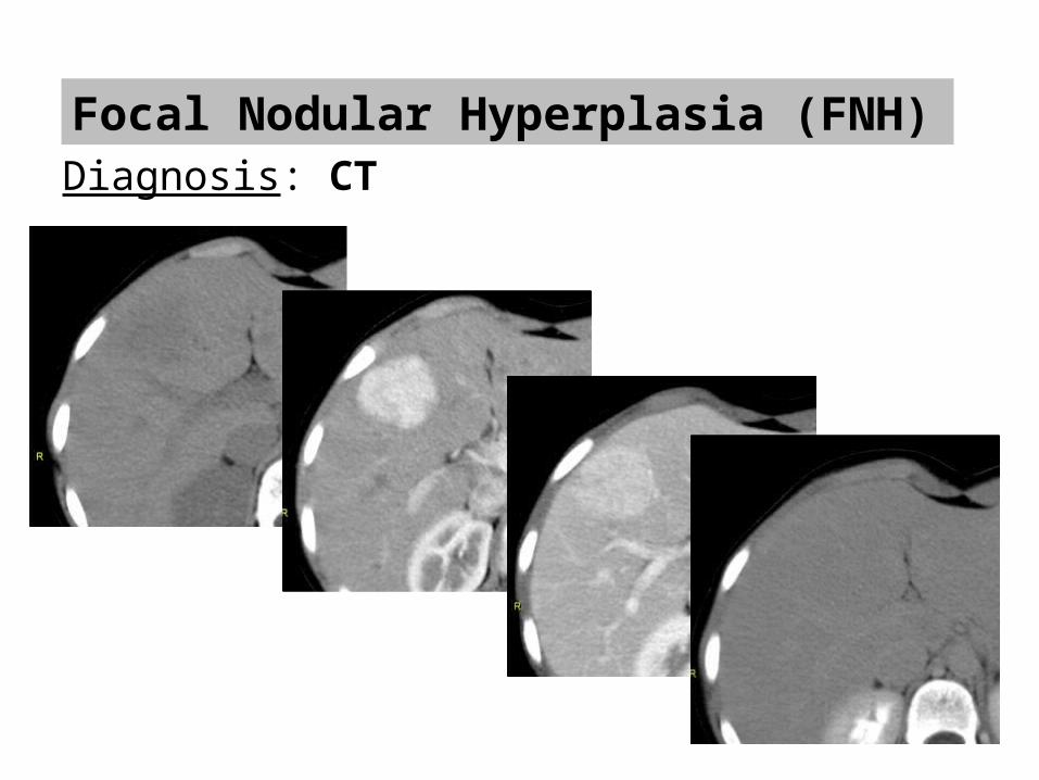
Focal Nodular Hyperplasia (FNH)Diagnosis: CT
Focal Nodular Hyperplasia (FNH)
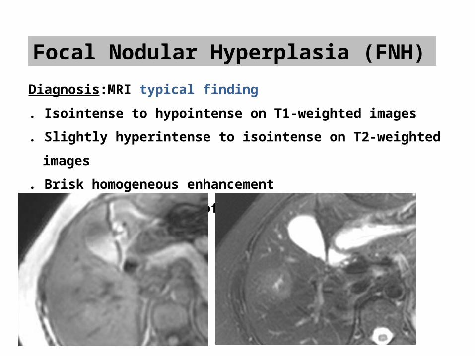
Diagnosis:MRI typical finding
. Isointense to hypointense on T1-weighted images
. Slightly hyperintense to isointense on T2-weighted images
. Brisk homogeneous enhancement
. Delayed enhancement of the central scar
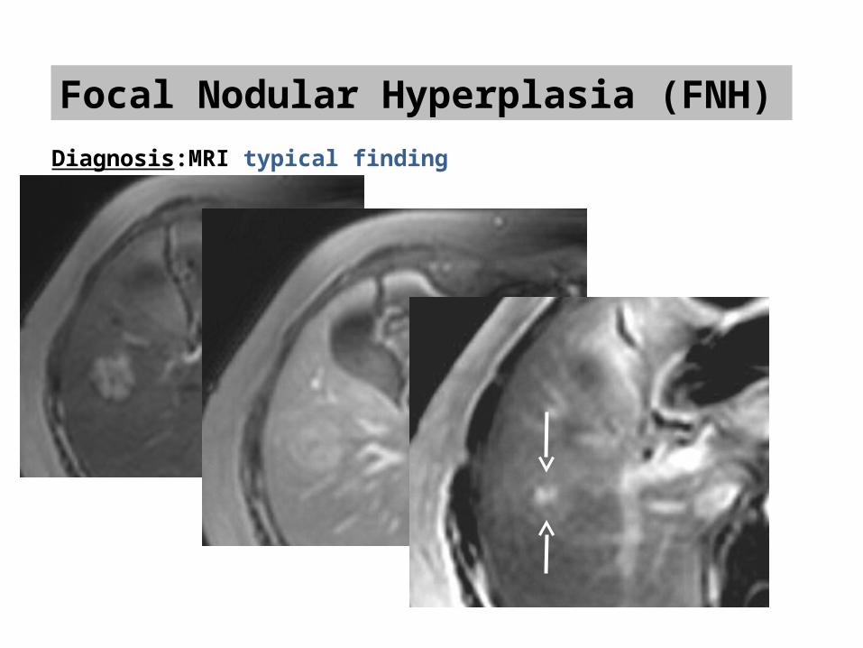
Focal Nodular Hyperplasia (FNH)Diagnosis:MRI typical finding
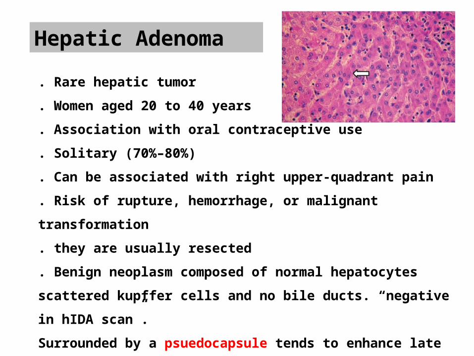
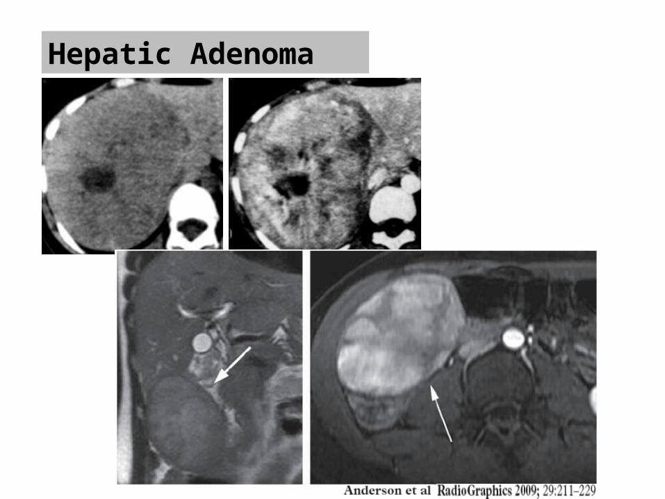
Hepatic Adenoma
. Rare hepatic tumor
. Women aged 20 to 40 years
. Association with oral contraceptive use
. Solitary (70%–80%)
. Can be associated with right upper-quadrant pain
. Risk of rupture, hemorrhage, or malignant transformation. they are usually resected. Benign neoplasm composed of normal hepatocytes scattered kupffer cells and no bile ducts. “negative in hIDA scan”. Surrounded by a psuedocapsule tends to enhance late
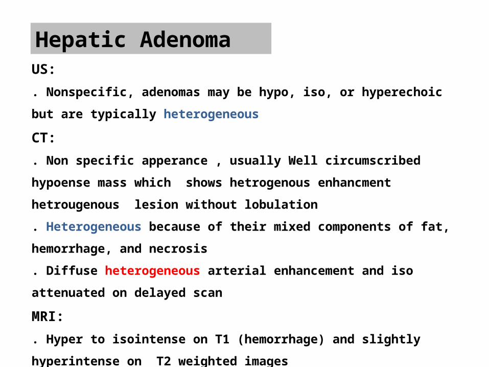
Hepatic AdenomaUS:
. Nonspecific, adenomas may be hypo, iso, or hyperechoic but are typically
heterogeneous
CT:
. Non specific apperance , usually Well circumscribed hypoense mass which
shows hetrogenous enhancment hetrougenous lesion without lobulation
. Heterogeneous because of their mixed components of fat, hemorrhage, and
necrosis
. Diffuse heterogeneous arterial enhancement and iso attenuated on delayed scan
MRI:
. Hyper to isointense on T1 (hemorrhage) and slightly hyperintense on T2 weighted
images
. Same appearance on contrast-enhanced image as CT scan
Hepatic Adenoma
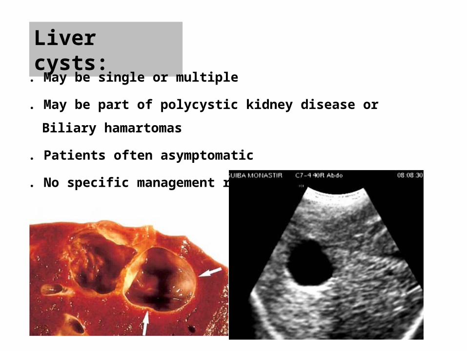
Liver cysts:
. May be single or multiple
. May be part of polycystic kidney disease or Biliary hamartomas
. Patients often asymptomatic
. No specific management required
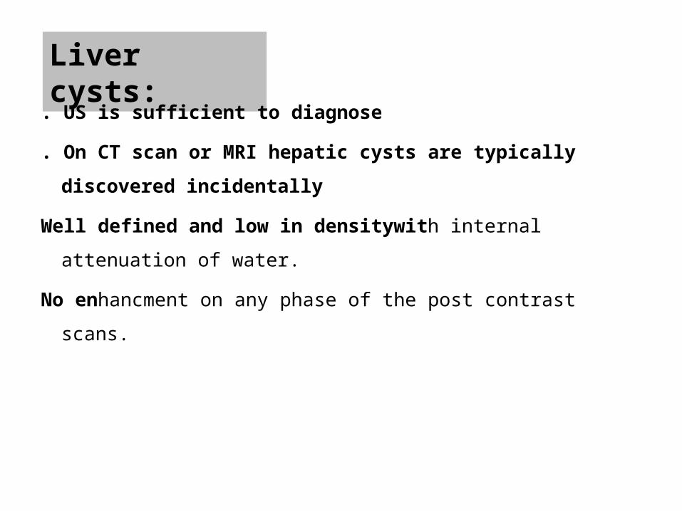
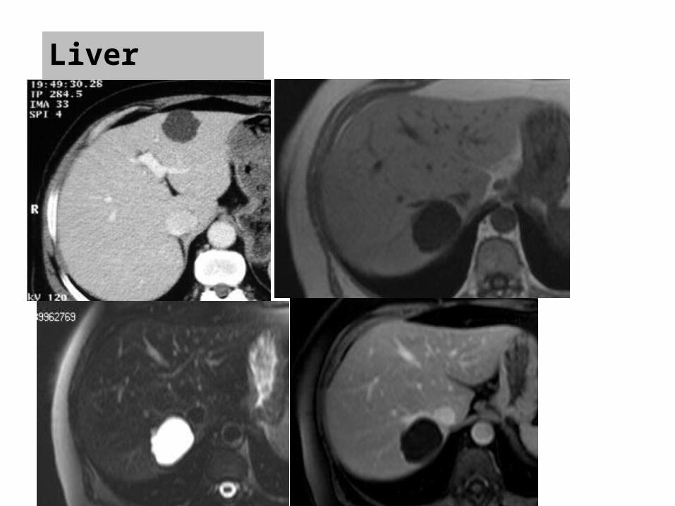
Liver cysts:
. US is sufficient to diagnose
. On CT scan or MRI hepatic cysts are typically discovered incidentally
Well defined and low in densitywith internal attenuation of water.
No enhancment on any phase of the post contrast scans.
Liver cysts:

MALIGNANT LIVER LESIONS
Hepatocellular Carcinoma (HCC)
•The most common primary tumour liver tumor.
•Rarely occurs before age of 40 and peaks at 70 years
•Male to female: 4/1
•Cirrhosis is the strongest predisposing factor for HCC
•HCC is locally invasive and tends to invade the portal vien, IVC and bile
ducts.
•Most HCCs develop by means of a multistep progression: from a low-
grade dysplastic nodule to a high-grade dysplastic nodule, to a
dysplastic nodule with a focus of HCC, and finally to convert carcinoma.
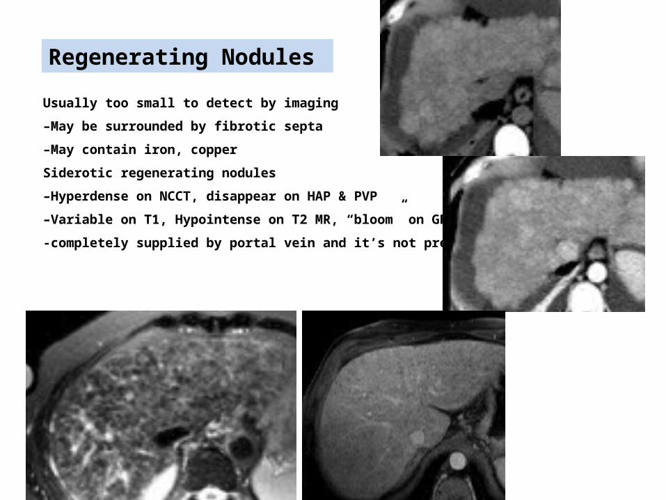
Usually too small to detect by imaging
–May be surrounded by fibrotic septa
–May contain iron, copper
Siderotic regenerating nodules
–Hyperdense on NCCT, disappear on HAP & PVP
–Variable on T1, Hypointense on T2 MR, “bloom” on GRE
-completely supplied by portal vein and it’s not premalignant.
Regenerating Nodules
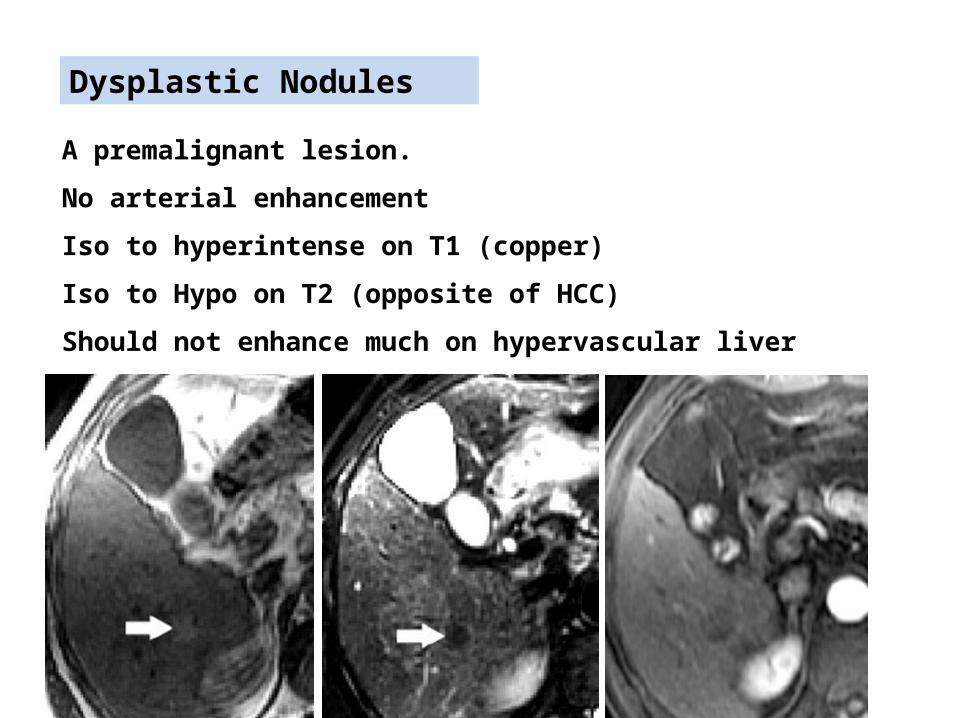
Dysplastic Nodules
A premalignant lesion.
No arterial enhancement
Iso to hyperintense on T1 (copper)
Iso to Hypo on T2 (opposite of HCC)
Should not enhance much on hypervascular liver lesions
Hepatocellular Carcinoma (HCC)
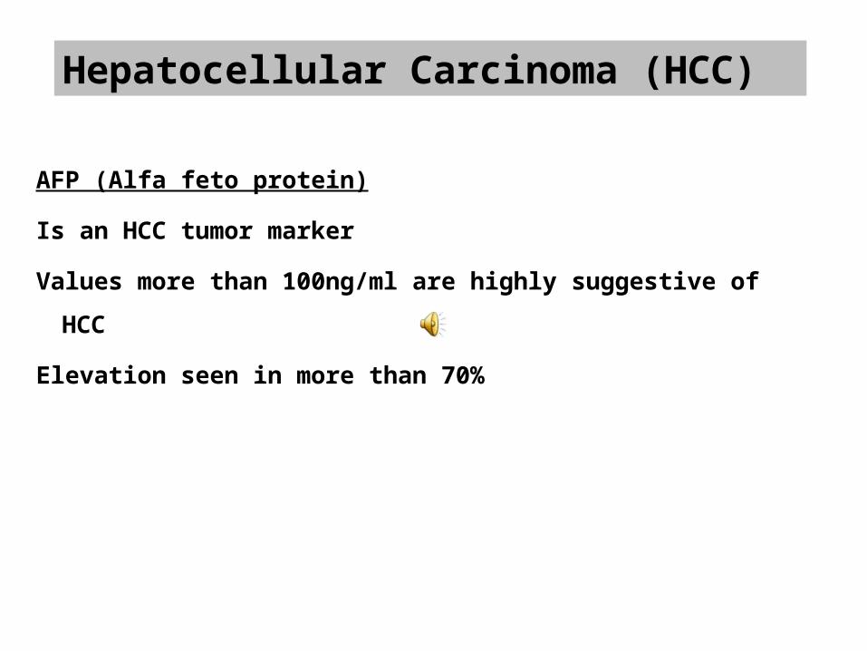
AFP (Alfa feto protein)
Is an HCC tumor marker
Values more than 100ng/ml are highly suggestive of HCC
Elevation seen in more than 70%
Hepatocellular Carcinoma (HCC)
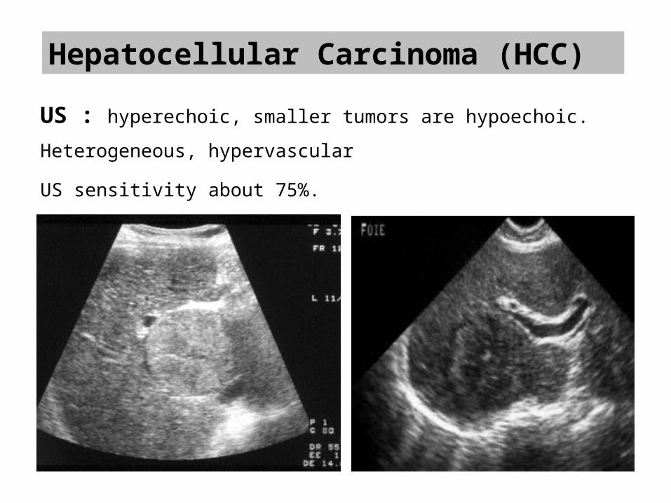
US : hyperechoic, smaller tumors are hypoechoic.
Heterogeneous, hypervascular
US sensitivity about 75%.
Arterial Phase:
liver(30-35 sec)
HCC as supplied by arterial branch/neovascularization
Hepatocellular Carcinoma (HCC)
Venous Phase:
HCC which is enhanced during arterial phase has lost its contrast,
hence no enhancement of the tumor but rest of the liver enhances.
Contrast in brightness of the lesion with respect to surrounding liver.
Enhancement
Wash out phenomenan
CT or MR
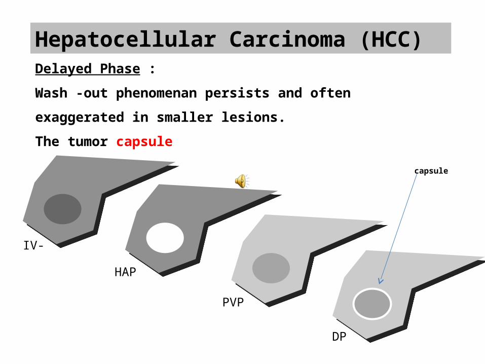
Hepatocellular Carcinoma (HCC)Delayed Phase :
Wash -out phenomenan persists and often exaggerated in smaller
lesions.
The tumor capsule
IV-
HAP
PVP
DP
capsule
Hepatocellular Carcinoma (HCC)
MRI . Variable intensity of HCC on T1
. 35% hyper, 25% iso-, 40 % hypo
. Hyperintense (T1) often well-differentiated, contain fat, copper,
glycogene
. Almost always hyperintense on T2 MR
. The tumor capsule is hypointense on both T1- and T2-weighted
images in most cases
. Other Features: Focal fat
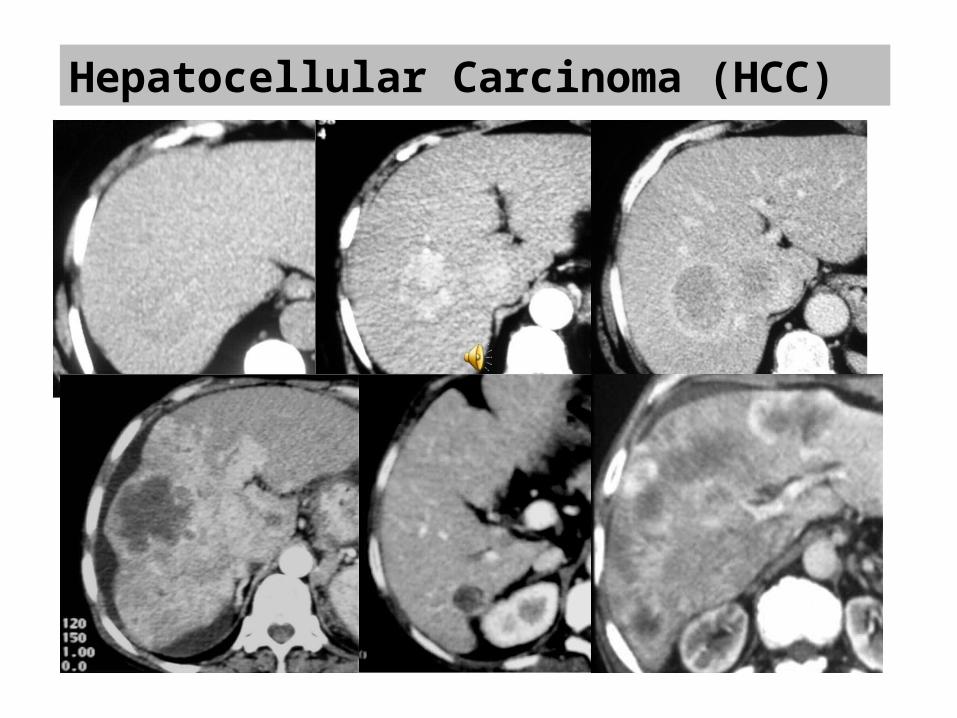
Hepatocellular Carcinoma (HCC)
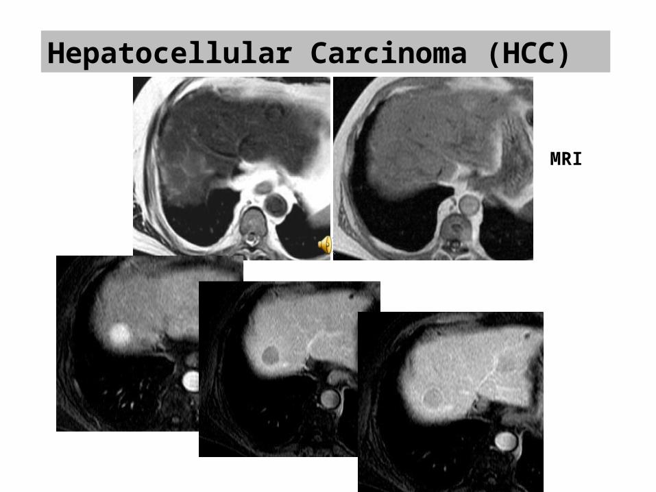
Hepatocellular Carcinoma (HCC)
MRI
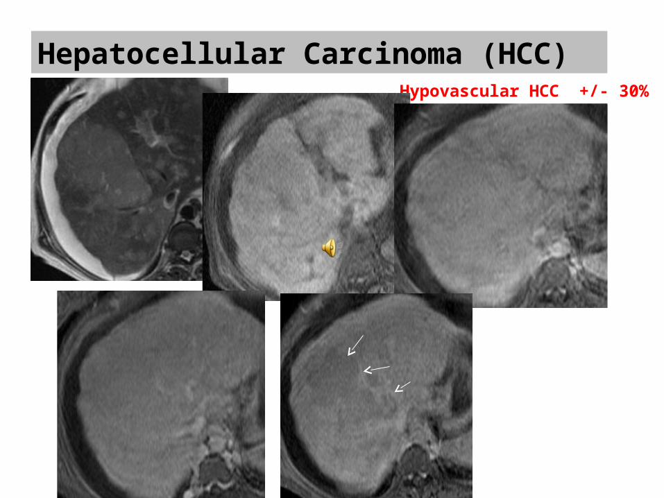
Hepatocellular Carcinoma (HCC)Hypovascular HCC +/- 30%
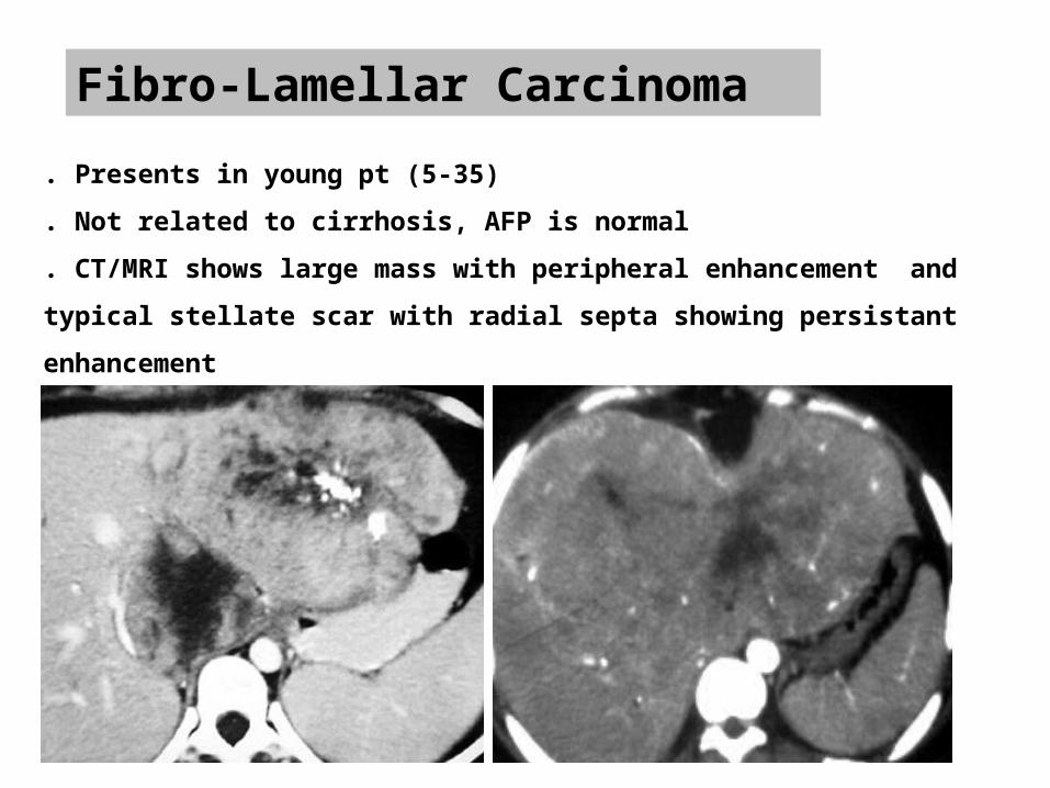
Fibro-Lamellar Carcinoma
. Presents in young pt (5-35)
. Not related to cirrhosis, AFP is normal
. CT/MRI shows large mass with peripheral enhancement and typical stellate scar with
radial septa showing persistant enhancement
. Calcifications

Metastatic disease
. Most common malignant hepatic tumor
. Presence of extrahepatic malignancy should be sought in patients with
characteristic liver lesions per imaging studies. Physical exam and
history is very helpful.
. Common primaries : colon, breast, lung, stomach, pancreases, and
melanoma
. Mild cholestatic picture (ALP, LDH) with preserved liver function
. CT or US guided biopsy provides definitive diagnosis but not always
required.
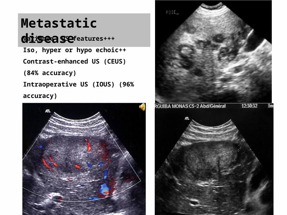
Metastatic diseaseVariable US features+++
Iso, hyper or hypo echoic++
Contrast-enhanced US (CEUS) (84% accuracy)
Intraoperative US (IOUS) (96% accuracy)
Typical feature
Metastatic disease
. Most liver metastases are hypovascular and are best imaged during the
portal venous phase (colon, stomach and pancreas)
. Hypervascular metastases enhancing on the arterial phase (neuroendocrine
tumors, renal cell, breast, melanoma, thyroid)
. Calcification may be present with metastases from mucinous
gastrointestinal tract tumors and from primary ovarian, breast, lung, renal,
and thyroid cancer
. Other features : Hemorrhagic or cystic metastases
Metastatic disease. On MRI, metastases are variable but are usually hypo- to isointense on T WI and
iso- to hyperintense on T2 WI
. Metastatic tumors with liquefactive necrosis or cystic neoplasms show higher signal
intensity on T2 WI
. Metastases may show central hypointensity on T2WI (coagulative necrosis, fibrin,
and mucin)
. High T1 signal intensity can be seen with metastases from melanoma, colonic
adenocarcinoma, ovarian adenocarcinoma, multiple myeloma and pancreatic
mucinous cystic tumor
. Comparing T2-weighted (TE 90) and T2-weighted (TE 160) sequences, metastases
become less intense Characterization
. T1-weighted 3D dynamic contrast-enhanced MRI Detection
Wide spectrum of apperance on CT depends on the primary neoplasm
•Well defined , low density soli mass with peripheral enhancement e.g. Colon cancer
•Hypervascular metastasis show diffused enhancement on arterial phase .
•Can be calcified when the primary neoplasm is mucinous adenocarcinomas, osteocarcinoma,
chondrosarcoma .•Cystic metastasis from mucinous colon
carcinoma, carcinoid or lung.

Metastatic disease
Conclusion :. MDCT and MRI are the most commonly used imaging
modalities for detection and characterization of focal hepatic
lesion
. Imaging modalities can make diagnosis for:
Hepatic cyst
Caverneous hemangioma
Typical FNH
HCC
. For others lesions biopsy will be often necessary


![Erasmus 2012 benign liver [Alleen-lezen] · B. Secondary benign liver lesions 1. Abscess C. Hepatic Pseudolesions 1. Focal Steatosis, Focal spared Area in Fatty Liver 2. ... • Incidental](https://img.pdfslide.net/doc/110x75/5b0382477f8b9a2e228c816d/erasmus-2012-benign-liver-alleen-lezen-secondary-benign-liver-lesions-1-abscess.jpg)