Embed Size (px)
Citation preview
1
Medical Biotechnology of Gene Therapy A Promising
Treatment for Cancer
(Breast Cancer as Example)
Prepared By
MostafaAskarAskarElmetwaly
I mmunology &C a nc e r
NC R R T, E AE A, C a ir o,E gypt
2013
2
Medical Biotechnology of Gene Therapy A Promising Treatment for Cancer
(Breast Cancer as Example)
Abstract
Breast cancer is one of the most common cancers among women around the
world. It accounts for 22.9% of all the cancers and 18% of all female cancers in the
world. One million new cases of breast cancer are diagnosed every year. Pakistan has
more alarming situation with 90,000 new cases and ending up into 40,000 deaths
annually. The risk factor for a female to develop breast cancer as compared with male is
100: 1. The traditional way of treatment is by surgery, chemotherapy or radiotherapy.
Advanced breast cancer is very difficult to treat with any of the traditional treatment
options. A new treatment option in the form of gene therapy can be a promising
treatment for breast cancer. Gene therapy provides treatment option in the form of
targeting mutated gene, expression of cancer markers on the surface of cells, blocking
the metastasis and induction of apoptosis, etc. Gene therapy showed very promising
results for treatment of various cancers. All this is being trialed, experimented and
practiced. Therefore, there is an immense need that this kind of work should be
started in Pakistan. There are many good research institutes as well as well-reputed
hospitals in Pakistan. Presently, there is a need to develop collaboration between
research institutes and hospitals, so that the basic work and clinical trials can be done to
treat breast cancer patients in the country. This collaboration will prove to be very
healthy and will not only strength research institute but also will be very beneficial for
cancer patients.
Keywords: Breast cancer, Metastasis, Mutation, Apoptosis, Research Centers,
Collaboration.
3
Introduction
Breast cancer has a very high ratio of occurrence in United States and probably all
over the world among major cancers. Breast cancer is at third number in the list of
most frequently occurring cancers after stomach and lung cancers. In women, it is the
most common cancer and accounts for 21% of new cancer cases throughout the
world. According to this ratio, 6.2% women in developed countries and 2.2% in
developing countries on average are at life time risk of breast cancer. Breast cancer is the
most frequently diagnosed cancer in women with a ratio of nearly 1 in 5 patients.
Cumulative level of breast cancer is very low, in comparison with other countries. An
average woman would have estimated probability of 1-2% to be treated for breast cancer
by the time she reaches her 65th birthday. Surgery, hormonal therapy, radiotherapy and
chemotherapy are the major treatments of breast cancer. These reduce the risk of breast
cancer up to certain levels. These approaches can only suppress the tumor or cancer
growth but they are unable to cure the disease permanently. As gene therapy is in use to
treat several genetic disorders including cancer, so it can be a very promising
treatment option for breast cancer patients as well. Till date many gene therapy
approaches have been adopted in pre-clinical and clinical trials. Gene therapy can be
broadly divided in six categories: (1) molecular chemotherapy, (2) antiangiogenic gene
therapy, (3) pro-apoptotic gene therapy, (4) mutation compensation, (5) genetic
modulation of resistance/sensitivity, and (6) genetic immune-potentiation.
Genetic Basis of Breast Cancer
Amplifications, deletions and gene mutations come under the somatic changes of
genome ofbreast cancer. Oncogenes present on regions of several chromosomes get
amplified in breast tumors. Tumor suppressor genes mutations arealso cause of breast
carcinomas. TP53 (Tumor Protein P53) which encodes the p53 transcription factor, and
the CDH1 which encodes the cadherin cell adhesionmolecule are the tumor
suppressorgenes. The function of p53 is to suppress the cell proliferation through a
4
multiprotein regulatory pathway and it focuses around the control of apoptosis and
retinoblastoma gene. Cellular-stresses activate p53 and these independent pathways of
p53 depend upon distinct upstream regulatory kinases. These include a pathway
dependent on the alternativeproduct of the INK4 gene, p14ARF, (which is activated by
expression of oncogenes), an ataxia-telangectasia mutated (ATM)/human homologue of
Rad53 (Chk2)-dependent pathway activated by DNA double-strand breaks and a
third ATM, Chk2 and p14ARF independent pathway whose activity is increased by
cytotoxic anti-tumour agents and ultraviolet light. This pathway may be mediated by
other kinases such as the ATM relative ataxia-telangectasia and Rad3-related
protein (ATR). The anti-proliferative action of p53 is exerted by induction of reversible
or irreversible cell cycle arrest or apoptosis. In solid tumors, p53is the most commonly
mutated tumor suppressor gene. Patients with rare hereditary disease, Li-Fraumeni
syndrome also have a mutated p53 gene in their germline cells. Mutated p53 gene is
expected to present in 50% of all cancers. Mutant proteins are a defective target for
binding of sequence specific DNA and in this way also for the transactivation of
genes, upregulated by the wild-type proteins. There are different mechanisms of p53
inactivation in breast cancer. In virus-associated cancers sequestration or enhanced
degradation of p53 occurs through interaction with virally encoded proteins. Binding
of MDM2 with p53 promotes the ubiquitinationof the C-terminus of p53 and, hence,
subsequent degradation. Association of p53 and MDM2 is prevented by interaction
of p14ARF with MDM2 and thus stabilizing p53. Overexpression ofMDM2 or
deletion or epigenetic silencing of p14ARF can start inappropriate degradation of p53.
There is another mechanism of inactivation which involves cytoplasmic sequestration of
p53 protein and preventing nuclear localisationof the protein and thus inhibiting its
activity. The other two very important caretaker genes are wild type BRCA1 and
BRCA2 and their function is to repair the damaged DNA. BRCA1 is present on
chromosome 17. Various studiesindicate thatmutations in BRCA1 are associated with
50 percent to 85 percent life-time risk of developing breast cancer. Carriers of
5
BRCA1 gene mutation often develop breast cancer at a younger age as compared
to the general population. BRCA2 gene is located on chromosome 13. Women carrying
BRCA2 mutations have similarchances of cancer development as are the BRCA1
carriers. Men carrying BRCA2 mutation have an increased risk of developing breast
cancer. Chromosomal instability results due todeletions or defects in BRCA genes as
cell loses the ability to repair the mistakes which occurduring the normal process of
replication, transcription and translation naturally.
Gene Therapy Approaches
The genetic alterations in tumor suppressor genes (TSGs) are the basis for the
breast carcinomas. Strategies have been employed which target correction of these
TSGs to cure breast carcinomas permanently. When introduced in breast cancer cells,
many TSGs have shown induction of apoptosis or cell cycle arrest including p53,
p21, p27, p16,Rb, BRCA1, BRCA2, Testin, Maspin. To date most trials target the
most common altered TSGs in cancer which is p53. Human breast cancer cells
when transduced with viral wild type p53 showed sufficient restoration of the normal
balance of cell proliferation and cell death of the breast cancer cell demonstrated transfer
of p53 into lung cancer by an adenoviral vector initially. Now, we will discuss some
approaches to treat breast cancer by gene therapy.
1. Antisense Technology
Antisense technology involves the use of antisense molecules that can specifically
inhibit the expression of pathogenic genes. Antisense oligodeoxynucleotides are
short ssDNA molecules that modify gene expression by blocking the transfer of
genetic information into protein. Many genes relevant to breast cancer such as αV
integrin, ribosomal protein P2, c-erbB-2, methylenetetrahydrofolatereductase, and
protein kinase C-alpha (PKC-α), have been successfully targeted by antisense
oligonucleotides. LY900003 is currently in clinical development and it presents an
antisense oligonucleotide to specifically block PKC-α. Ribonucleic interference (RNAi)
6
technology is another closely related area. RNAi showed specific downregulation of c-
myc which was sufficient to inhibit the growth of breast cancer MCF-7 cells in vitro and
in vivo. Ribozymes are another approach in ablation of oncogenes. They are RNA
molecules capable of acting as enzymes. Ribozymes were used for the treatment of
human immunodeficiency virus (HIV) initially. Hammerhead ribozyme is the simplest
and is approximately 30 nucleotides long. Possibility to produce trans-acting
ribozymes directed against RNA sequences of interest highlighted the importance
of hammerhead ribozymes. Since then, genetically modified ribozymes have been
designed, produced and given to cells to ‘knock down’ the expression of specific
genes. Keeping this in view, adenovirus-mediated ribozyme targeting of HER-2/neu
showed inhibition of in vivo growth of breast cancer cells in a mouse model.
Another strategy targets disruption of normal cellular localization of growth receptors.
The proto-oncogene erbB-2 is of particular interest. Down regulation of cell surface
erbB-2 levels and induction of apoptosis in erbB-2-overexpressing breast cancer cells
resulted on delivery of a gene encoding an anti-erbB-2 intracellular single-chain
antibody (sFv).
2. Suicide Genes
In late 1980s, it was suggested that there is a possibility that introduction of so-
called suicide genes renders cancer cells more sensitive to chemotherapeutics or
toxins. Suicide genetherapy has two categories : toxin gene therapyand enzyme-
activating pro-drug therapy. Toxin gene therapy includes the transfection of genes that
express toxic molecules. On the other hand, enzyme-activating pro-drug therapy
explains thetransfection of genes able to express enzymes that can selectively activate
specific pro-drugs. The latter one has been variously called gene-directed enzyme pro-
drug therapy (GDEPT), gene pro-drug activation therapy (GPAT), suicide gene
therapy or virally directed enzyme pro-drug therapy (VDEPT). Gene-directed
enzyme pro-drug therapy is a two phase treatment of cancer. The gene for the
7
enzyme is delivered to the breast cancer cell in the first phase while in the second a
nontoxic pro-drug is administered, which is then converted into a toxic metabolite
by the foreign enzyme expressed in the tumor. The enzymes recommended for
GDEPT for breast cancer are divided into two categories. The first one contains
‘foreign’ enzymes which are non mammalian in origin, such as carboxy peptidase
G2 (CPG2), bacterial cytosine deaminaseand viral TK. The second category
contains enzymes which are of human origin, such as cytochrome P450
isophorms. Most probably, the two best examples of this strategy are cytosine
deaminase (CD) and thymidine kinase (TK), which convert 5-fluorocytosine and
ganciclovir(GCV), respectively, into their toxic drug forms. Many experimentalmodels
have shown that combination therapy with cytokines and suicidal genes can be used. An
adenoviral vector carrying the interleukin-2 (IL-2), granulocyte-macrophage colony-
stimulating factor (GM-CSF) or HSV-1 thymidine kinase (HSV-TK) was injected
in breast cancer cells grown as xenografts in BALB/c mice. Both cytokine genes
combined with HSV-TK showed a sufficient reduction in tumor growth in
comparison with HSV-TK alone . Efficacy of GDEPT system is enhanced by a double
transfer of suicide genes. Two distinct types of pro-drugs are activated by transfecting
cancerous cells with two different suicide genes. Results reveal that, the antitumoral
effect exerted by two different suicide gene systems (CD and cytochrome P450 2B1) is
more efficient to each single system alone for murine mammary tumors.
3. Gene Therapy to Induce Apoptosis
Impaired apoptotic signaling plays an important role in tumor initiation and progression
in different types of cancers. Apoptotic resistance of cancerous cells has hazardous
effects because not only it increases the growth of the tumor but also provides
resistance to host defense mechanism and different types of therapeutics.
Identification of genes involved in apoptotic induction offers a good target for
breast cancer gene therapy. Strategies have been employed which deliver those genes
8
that are responsible for apoptotic death and also transmit death signals to the adjacent
tumor cells. These proapoptotic strategies in breast cancer include functional
replacement of TSGs, suicide gene therapy, death receptor and ligand systems and
pathways, BCL-2 family proteins. The first gene which was identified with a function
of inhibiting apoptosis was BCL-2 and it plays an important role in breast cancer.
Experiments on nude mice have showed that BCL-xs gene which is a dominant
negative repressor of Bcl-2 and Bcl-xL induced apoptosis in human mammary tumors
. The down regulation of proapoptotic Bik has been linked to the development of
breast cancers. Significant induction of apoptosis was observed in orthotopictumor
tissues in nude mice and in four breast cancer cell lines in vitro. In breast cancer cells,
there are number of death receptor and ligand systems that function in regulation of
apoptosis. These include tumor necrosis factor-related apoptosis-inducing ligands
(TRAIL), Fas ligand (FasL) and tumor necrosis factor-α. Experimental data showed
that death of breast tumor cells occurred due to the rapid production and expression
of TRAIL protein on introduction of TRAIL gene into subcutaneous human breast
cancer xenografts in nude mice and breast cancer cells using an adenoviral vector.
Several proteins have been identified which help in promoting tumorigenesis by
inhibiting apoptosis. Major stress-inducible Hsp70 (also known as Hsp72 or Hsp70i)
may be categorized as cancer-relevant antiapoptotic protein. Tumor cell lines and
human breast tumors express Hsp70. Classical transfection of antisense Hsp70
cDNA(asHsp70) or inhibition of its synthesis by adenoviral transfer causes
significant breast cancer cells death. Bcl-2 and Bcl-XL could not save death
induction of breast tumor cells by as Hsp70. There is a good possibility to
treattherapies resistant cancers by neutralizing Hsp70. The adenovirus type 5 E1A
protein hasdemonstrated antitumor effects through the induction of apoptosis,
inhibition of cell cycle progression and sensitization to chemotherapeutic agents and
radiation.Regression of tumors in nude mice and induction of apoptosis in breast
9
cancer cells is demonstrated by adenovirus-directed expression of dominant-negative
ER receptor
4. Anti-Angiogenic Gene Therapy
Angiogenesis plays an important role in establishment and spread of tumor. In this
regard, antiangiogenic gene therapy can prove a good approach for treating breast
carcinomas. Plasmids encoding angiostatin and endostatin and complexed with
Liposomes inhibited breast cancer in nude mice. Antiangiogenic therapy alone cannot
control the promulgated breast cancer in humans. Combining antiangiogenic therapy
with other strategies, both conventional and gene transfer-based can produce good
results. Antiangiogenic therapy can be complemented by both chemotherapic and
hormonal approaches due to their different mechanisms of antitumoral action. In a
recent experiment, it was observed that the association of angiostatin with
tamoxifenproduced better results than either approach used alone in a transgenic
mouse model of breast cancer . Further, the potential efficacy of delivery of
endostatingene can be used to treat metastatic breast cancer to brain. Recombinant
proteins that inhibit angiogenesis have shown regression in tumors of mouse
models when administered systemically.
5. Immunotherapy
An immune response can be harnessed and used for eradicating out the cancerous
cells. Immunotherapy can be divided into two functional types: active immunotherapy
and passive immunotherapy. The former approach employs tumor vaccines and
immune stimulatorycytokines stimulates the patient’s immune response to generate
an antitumor immunity; while the later approach explains how to administer pre-
formed elements of the immune system, such as antitumor cytokines, tumor
antibodies, or tumoricidal effector cells to patients, with a plan of directly kill the
cancer cell. An immune response is triggered on alarm signals induced by
pathogens . Activation of a number of immune effector cells can be triggered by
10
direct introduction of immune stimulatory molecules encoding genes into breast
cancer cells. Systemic injection of cytokines such as IL-2 was focus of initial studies.
Biologically active cytokine can be delivered at a tumor site without manipulating with
tumor cells ex vivo. Usage of autologous or allogenic genetically modified normal
cells for local secretion of immune stimulatory molecules was recommended for this
purpose. The survival of mice with intracerebral breast cancer metastasis was prolonged
by intratumoral injection of IL-2-secreting syngeneic/allogeneic fibroblasts
transfected with DNA from breast cancer cells (Lichtor et al., 2005). T-cell subsets
(CD8+ and natural killer (NK)/LAK cells) mediated the antitumor response
predominantly. A variety of other cytokines such as the GM-CSF, IL-4, ILl-12, IL-
18, interferon-alpha and IL-23 have been used.
6. Genetic Vaccines
Genetic vaccines are another approach. In the last three decades, many tumors associated
with antigens have been identified and cloned. These antigens have been used to produce
vaccines for immunotherapy of cancer. Most tumor antigens are utilized as
immunotherapy targets because they are aberrantly expressed on tumor cells. In this
regard, a growth advantage for breast cancer cells has been provided by aberrant
expression of Her-2/neu/erbB-2 overexpression. Currently, under investigation genes
for the vaccine production in breast carcinomas are carcino-embryonic antigen, Fos-
related antigen 1 (Fra-1), tumor cell-associated extracellular matrix metalloproteinase
inducer (EMMPRIN), MAGE-1, MUC-1, hTERT, and B7-H4 . A recent approach is a
combination of different vaccines. By utilizing various cytokines and co-stimulatory
molecules as molecular adjuvants combination vaccine strategies tend to enhance
the effect of genetic vaccines in cancers. In this way, these approaches combine the
‘genetic’ and ‘conventional’ immunotherapeutic approaches. Studies suggest that GM-
CSF enhances antigen processing and presentation by dendritic cells so it has been
used as vaccine adjuvant for breast cancer (Von Mehren et al., 2001). In another
11
study, it was observed that injecting mice with Her-2/neu encoding plasmids and
augmentation with cytokines enhances the efficacy against breast cancer (Chang et
al., 2004).
7. Dendratic Cells (DCs) Applications
DCs are powerful antigen-presenting cells and have central role in generation of
immune responses. They process the antigens and present epitopes to the surface MHC
molecules to have interaction with T cells. Many costimulatory molecules and cytokines
that are required to sustain and direct the immune response are expressed by DCs. Many
studies for producing anticancer vaccines have focused on using DCs ability to
present tumor antigens in a proper way. Gene transfer approaches including DCs rely
on direct modification of DCs by gene modification in vitro and then
administration of modified APCs or DCs as a vaccine. For modification purposes,
genes encoding breast cancer antigens, cytokines and molecules involved in antigen
presentation were used. Suppression of breast cancer in BALB-neuT mice was observed
with a vaccine having modified DCs by adenoviruses encoding nonfunctional
tumor antigens, such as nonsignaling HER-2/neu. CD8+cytotoxic T lymphocytes
induced by DCs transduced with a Tat fusion protein (from the HIV) having the
extracellular domain of Her2/neu killed Her2/neu-expressing breast cancer cells
specifically. For breast cancer therapy both CD4+and CD8+mammoglobin-specific T
cells can be induced by dendritic cells transduced with Tat mammaglobilin.
8. Monoclonal Antibodies
Another approach is transferring genes encoding antibodies to known tumor antigens in
vivo. Genetically modified antibodies molecules contain a constant region of human
antibody molecule and a variable region of monoclonal antibody directed against antigen
of interest. This antibody has long half-life in human body due to the absence of
neutralizing immune response and maintains all the functions of natural antibodies at the
same time. Herceptin is a well-studied example of it. This humanized monoclonal
12
antibody binds directly to Her-2-positive tumor cells and displays efficient inhibition of
tumor growth. Antibody-dependent cellular toxicity is the basis of tumor rejection
strategy. In this case, killing mechanisms are activated directly when Fc surface receptor
on opsonized breast cancer cells binds with natural killer cells.
9. Miscellaneous Gene Therapy Approaches
Evidences suggest that downregulation of Major Histocompatibility Complex
(MHC) molecules, costimulatory molecules and some other ligands can help
escaping breast cancer cells from immune control. CD4+activation takes place due to
presentation of MHC II and this results in secretion of cytokines. These secreted
cytokines along with presented MHC I CD8+cytotoxic Tcells and result in cell
necrosis. Host immune system recognition and antitumor activity can be enhanced by
introducing such deficient receptors and ligands using gene therapy approach.
Conventional therapies fail in many cases of breast cancers due to the resistance to
DNA damaging agents such as chemotherapy and radiotherapy. Different
mechanisms responsible for breast cancer multi drug resistance have been elucidated.
The main mechanisms include modulation in apoptotic function, multidrug
resistance-associated protein 1 (MRP1, ABCC1), deletion or amplification of
topoisomerase II, breast cancer resistance protein (ABCP, BCRP, MXR, ABCG2)
and cellular overproduction of P-Glycoprotein. Down regulation of apoptosis and
over-expression of antiapoptotic genes play a vital role in failure of radiotherapy and
chemotherapy at the present stage of breast cancer treatment. It isstated that p53
plays no part in chemotherapy and radiotherapy resistant tumors so this can be the
basis for TSG combination therapies. Combination with other anticancer treatments
can be beneficial due to the low toxicity of p53 in initial trials and linkage of
p53 with apoptosis. Keeping this in view, adenovirus p53 can demonstrate tumor
regression and sensitize local and metastatic breast cancer by doxorubicin therapy.
After both chemo and radio therapies, the cellular response to DNA damage is mediated
13
by p53. p53 results in cell cycle arrest and finally induce apoptosis. When DNA is
damaged by radiotherapy, there is a cell cycle arrest at the G1 checkpoint as well as p53
dependent transcription activation of p21. The loss of this checkpoint is linked with
decrease in tumor cell apoptosis. ErbB-2 a proto-oncogene is widely studied in breast
cancer. A high correlation is reported between resistance to different therapies and over
expression of epidermal growth factor receptor. Human breast cancer cells were
radiosensitized by overexpression of dominant negative epidermal growth factor
receptor CD533 mediated by adenoviruses. Human telomerase reverse transcriptase
(TERT) is a potential target for active-specific immunotherapy. However, it has been
proved difficult to induce effective specific tumor antigen-specific immunity
consistently by various TERT vaccine formulations. To overcome this difficulty,
new adjuvant strategies such as utilizing chemokines to attract T-cells and antigen-
presenting cells have been employed. In a studies, tested the hypothesis that
thermosensitivity can be enhanced by heat-directed suicide gene therapy in locally
recurrent breast cancer (LRBC). Given the relapse rate in the following
thermoradiotherapy for LRBC and the fact that cells can be intrinsically resistant to
heat. An adenoviral vector (Ad.70b.CDTK) was constructed in which the hsp70b
promoter controls the expression of dual prodrug-activating E.coli cytosine
deaminase/herpes simplex virus thymidine kinase (CDTK) fusion gene. When
expressed in the presence of the prodrugs 5-fluorocytosine (5-FC) and ganciclovir
(GCV), the CDTK fusion protein has been shown previously to be highly cytotoxic in a
variety of tumor cell types both in vitro and in vivo. Importantly, this therapeutic
strategy was shown to act synergistically with heat treatment in a human prostate
cancer model . Here it was shown for the first time that adenovirus-mediated, heat-
directed expression of a CDTK fusion gene can significantly decrease the survival
of thermoresistant human breast cancer cell lines treated with clinically relevant
doses of pro-drugs and heat. In a novel approach, the survivin which is a member of
inhibitor of apoptosis (IAP) gene family has been used for treating breast cancer.
14
Survivin is localized to components of the mitotic apparatus and is expressed in mitosis
in a cell cycle–dependent fashion. Survivin is present in very low amounts in normal
adult tissues but its expression is upregulated in most human cancers cells. Survivin
pathway in cancer was targeted with a replication-deficient adenovirus encoding
asurvivin Thr34→Ala mutant, which abolishes a phosphorylation site for p34cdc2-
cyclin B1. Here, it was shown that initiation of the mitochondrial apoptotic
pathway was caused in various tumor cell types, exhibited no toxicity for normal
human cells, and suppression of tumor growth in three different xenograft breast
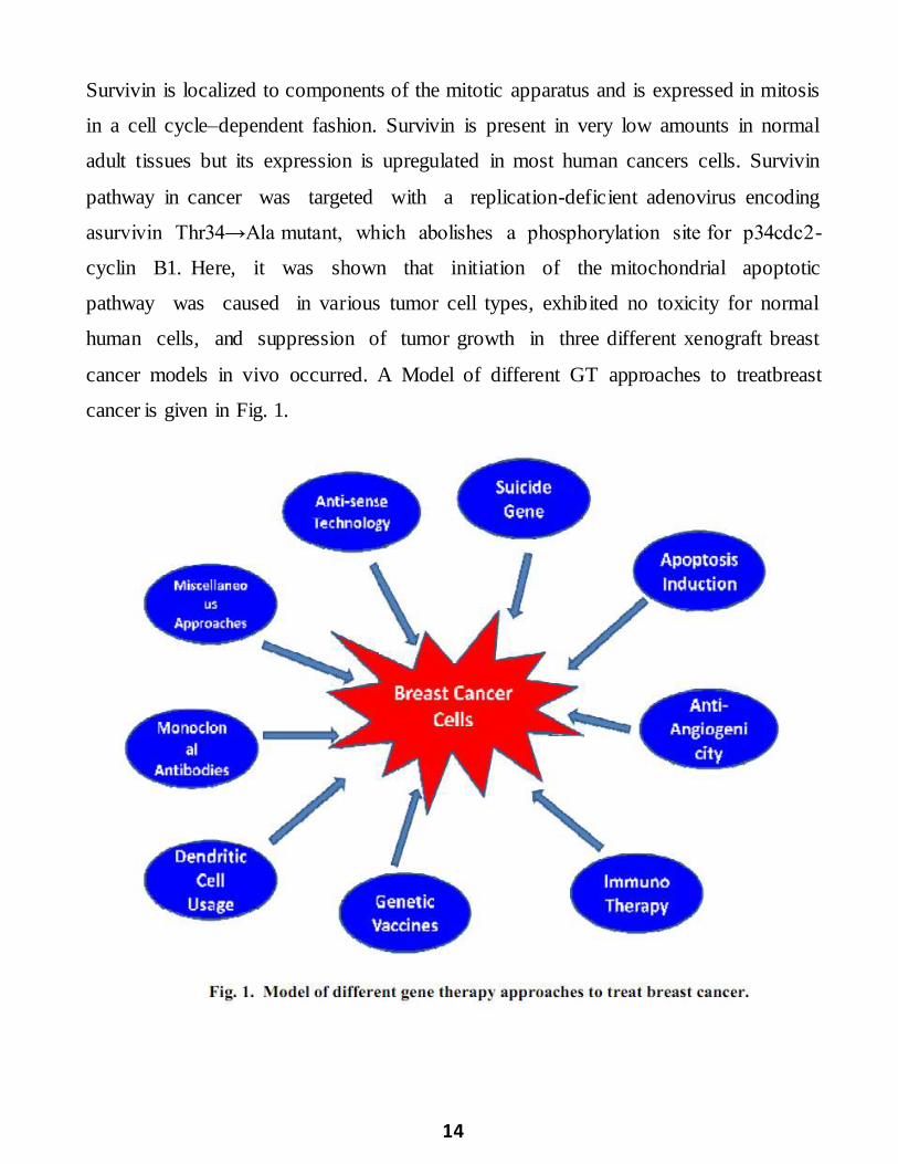
cancer models in vivo occurred. A Model of different GT approaches to treatbreast
cancer is given in Fig. 1.
15
Conclusion
Till-date many strategies have been used for treating breast cancer and majority of them
have focused onp53. For gene therapy intra tumoral route is a best choice of
administration. Although adenoviral vectors present higher transgene expression but a
small minority of patients provide clinical evidence of tumor regression. Antiangiogenic
gene therapy is also a good approach but antiangiogenis therapies cannot alone treat
breast cancer. They need augmentation by other therapies like conventional therapy.
Intramuscular delivery route of antiangiogenic factor gene can be helpful in treating
metastatic breast cancer to the brain. Immunotherapy for breast cancer is another
approach. New genetic vaccines can be developed by better understanding the nature
of antigens which provoke better antitumor response and by identification of novel
tumor antigens. Breast cancer gene therapy is a difficult task to perform. Till-date,
transgene expression has not been achieved up to the desire. For this, there is a need to
device new gene therapy strategies and new transgenes should be searched for. Also
there is a huge need to focus on new gene therapy vectors because presently used
vectors have not showed great success. A better understanding of molecular pathways
which lead to breast cancer is also required. Pakistan has very high ratio of deaths due
to breast cancer so pre clinical and clinical trials should be immediately started
here for better treatment of breast cancer in the country. Gene therapy can only
be made more beneficent if more and more countries put their efforts in it and an
effective linkage could be developed among hospitals and research centers locally.
16
References
Medicine, Oncology And Radiotherapy Institute (NORI) Hospital, Atomic
Energy Commission Nuclear medicine/Cancer Treatment, Institute of nuclear
medicine and oncology (INMOL) (http://www.paec.gov.pk/nori/patient.htm)
Molecular Biology (CEMB) (www.cemb.edu.pk)
SchoolofBiologicalSciences(SBS)(http://www.pu.edu.pk/home/department/59Sch
ool of Biological Sciences)
(ASAB-NUST) (www.asab.edu.nust.pk)
Iqbal, J., K. Bano, A. Saeed, M. Akramand Z Aziz. 2010. Survival of women
with locally advanced breast cancer at a teaching hospital in Lahore. Journal of
Pakistan Medical Association, 60 (9): 721-725.
Davidoff, A.M. and A.C. Nathwani. 2004. Antiangiogenic gene therapy for
cancer treatment. Current Hematological Reports, 3: 267-273.
Brade, A. M., P. Szmitko, D. Ngo, and H.J. Klamut. 2003. Heat-Directed Tumor
Cell Fusion. Human Gene Therapy, 14(5): 447-461.
Ruppert, J.M., M. Wright, M. Rosenfeld, J. Grushcow, G. Bilbao and D.T.
Curiel. 1997. Gene therapy strategies for carcinoma of the breast. Breast Cancer
Research and Treatment, 44: 93-114.