Embed Size (px)
Citation preview

Gingivitis
By: Maryam Arbab
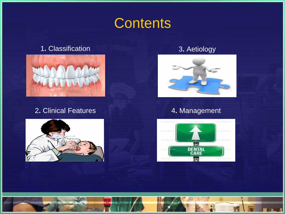
Contents
1. Classification
2. Clinical Features
3. Aetiology
4. Management
CLASSIFICATION OF
GINGIVITIS
Course Based Classification
Distribution Based Classification
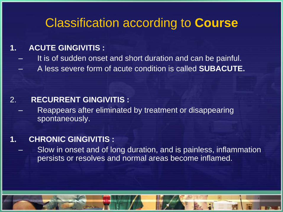
Classification according to Course
1. ACUTE GINGIVITIS :
– It is of sudden onset and short duration and can be painful.
– A less severe form of acute condition is called SUBACUTE.
2. RECURRENT GINGIVITIS :
– Reappears after eliminated by treatment or disappearing spontaneously.
1. CHRONIC GINGIVITIS :
– Slow in onset and of long duration, and is painless, inflammation persists or resolves and normal areas become inflamed.
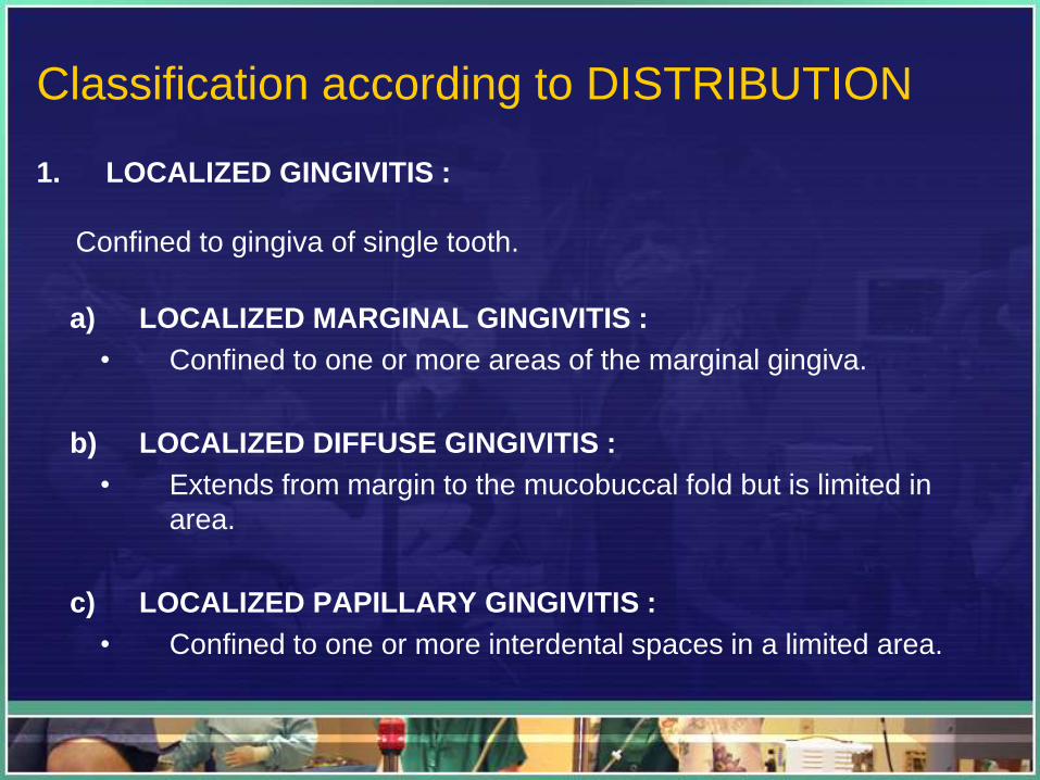
Classification according to DISTRIBUTION
1. LOCALIZED GINGIVITIS :
Confined to gingiva of single tooth.
a) LOCALIZED MARGINAL GINGIVITIS :
• Confined to one or more areas of the marginal gingiva.
b) LOCALIZED DIFFUSE GINGIVITIS :
• Extends from margin to the mucobuccal fold but is limited in
area.
c) LOCALIZED PAPILLARY GINGIVITIS :
• Confined to one or more interdental spaces in a limited area.
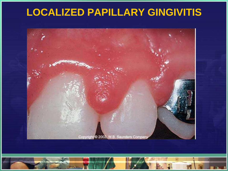
LOCALIZED PAPILLARY GINGIVITIS
Cont..
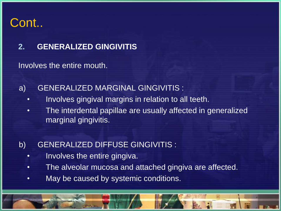
2. GENERALIZED GINGIVITIS
Involves the entire mouth.
a) GENERALIZED MARGINAL GINGIVITIS :
• Involves gingival margins in relation to all teeth.
• The interdental papillae are usually affected in generalized
marginal gingivitis.
b) GENERALIZED DIFFUSE GINGIVITIS :
• Involves the entire gingiva.
• The alveolar mucosa and attached gingiva are affected.
• May be caused by systemic conditions.
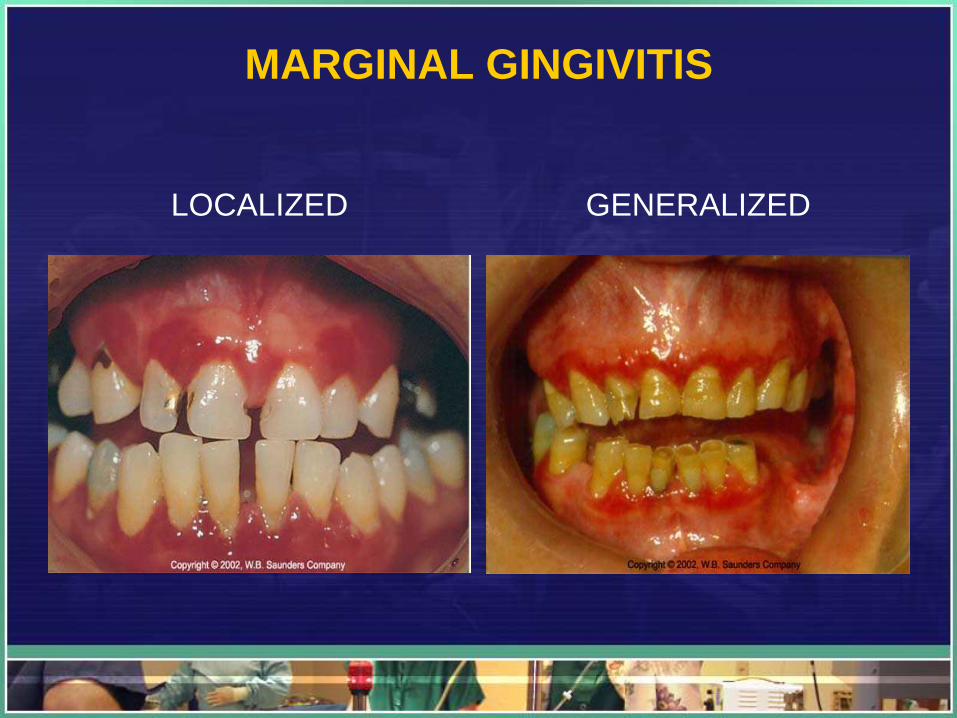
MARGINAL GINGIVITIS
LOCALIZED GENERALIZED
Section 2 : Clinical Features
CLINICAL FINDINGS
A systemic clinical approach requires an orderly
examination of the gingiva for :
Color
Contour
Consistency
Position
Ease and Severity of bleeding
Pain
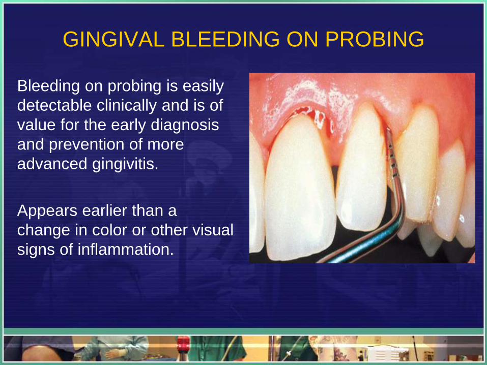
GINGIVAL BLEEDING ON PROBING
Bleeding on probing is easily
detectable clinically and is of
value for the early diagnosis
and prevention of more
advanced gingivitis.
Appears earlier than a
change in color or other visual
signs of inflammation.
COLOR CHANGES IN GINGIVITIS
1. The color changes may be marginal, diffuse, or
patch like depending on the underlying condition.
2. Color changes vary with the intensity of the
inflammation. Initially, it is red erythema.
Cont..
3. In severe inflammation, the red color gradually
becomes dull, whitish gray
4. Many systemic diseases may cause color changes
in the oral mucosa, including the gingiva.
5. The deposition of iron in hemochromatosis may
produce a bluish-gray pigmentation of the oral
mucosa. In jaundice, the oral mucosa acquires a
yellowish color.
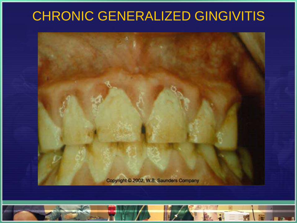
CHRONIC GENERALIZED GINGIVITIS
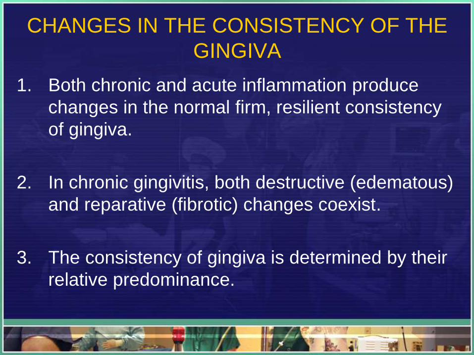
CHANGES IN THE CONSISTENCY OF THE
GINGIVA
1. Both chronic and acute inflammation produce
changes in the normal firm, resilient consistency
of gingiva.
2. In chronic gingivitis, both destructive (edematous)
and reparative (fibrotic) changes coexist.
3. The consistency of gingiva is determined by their
relative predominance.
CHANGES IN THE POSITION OF THE
GINGIVA
RECESSION :
• The exposure of the root surface by an apical shift in the
position of the gingiva.
• Recession refers to the location of the gingiva, not its
condition.
• Recession may be localized to one tooth or a group of
teeth, or it may be generalized through-out the mouth.
A. ACTUAL POSITION OF THE GINGIVA :
• The level of epithelial attachment on the tooth.
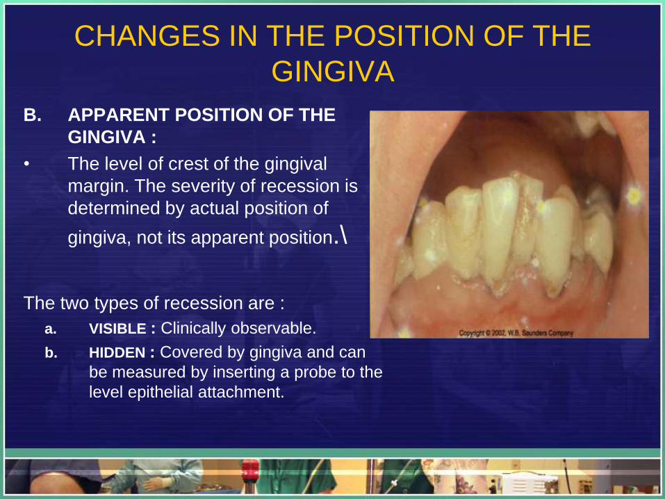
CHANGES IN THE POSITION OF THE
GINGIVA
B. APPARENT POSITION OF THE
GINGIVA :
• The level of crest of the gingival
margin. The severity of recession is
determined by actual position of
gingiva, not its apparent position.\
The two types of recession are :
a. VISIBLE : Clinically observable.
b. HIDDEN : Covered by gingiva and can
be measured by inserting a probe to the
level epithelial attachment.
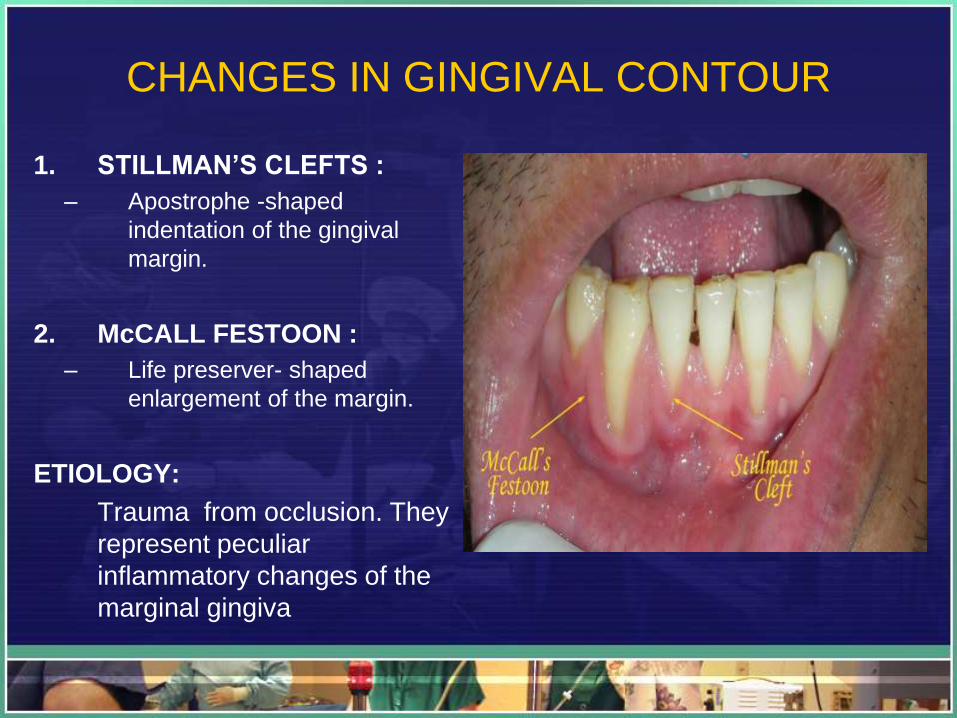
CHANGES IN GINGIVAL CONTOUR
1. STILLMAN’S CLEFTS :
– Apostrophe -shaped
indentation of the gingival
margin.
2. McCALL FESTOON :
– Life preserver- shaped
enlargement of the margin.
ETIOLOGY:
Trauma from occlusion. They
represent peculiar
inflammatory changes of the
marginal gingiva
CHANGES IN SURFACE TEXTURE OF THE
GINGIVA
1. Loss of surface stippling is an early sign of
gingivitis.
2. In chronic inflammation the surface is either
smooth and shiny or nodular, depending on
whether the dominant changes are exudative or
fibrotic.

Aetiology
Causes & Risk Factors
• Some people are more prone to getting gingivitis than others.
• Gingivitis is particularly likely to occur in people with diabetes, AIDS, or
leukemia.
• Other factors linked to an elevated risk of gingivitis include:
– puberty
– pregnancy
– menopause
– smoking
– poor-fitting fillings and crowns (also known as caps)
– mouth breathing
– genetics
– allergic reaction (e.g., cinnamon gum)
– vitamin C deficiency (scurvy)
– niacin (vitamin B3) deficiency (pellagra)
– poorly aligned teeth or poorly fitted mouth appliances (such as retainers or crowns)
– medications (e.g., use of the female contraceptive pill)
Cont..
• Some medications are also associated with
gingivitis, including:
– cyclosporine* (used to treat rheumatoid arthritis and
other autoimmune diseases)
– phenytoin (used to control epilepsy and other seizures)
– calcium channel blockers such as nifedipine (used treat
high blood pressure and other heart conditions)
– Some viruses can also infect the mouth. The one most
likely to attack the gums is the herpes virus.
Management
• Signs and Symptoms
• Investigations/Diagnosis
• Treatment
• Prevention
• Complications
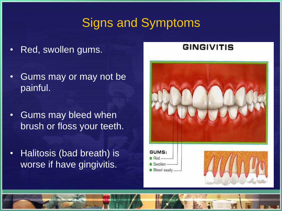
Signs and Symptoms
• Red, swollen gums.
• Gums may or may not be
painful.
• Gums may bleed when
brush or floss your teeth.
• Halitosis (bad breath) is
worse if have gingivitis.
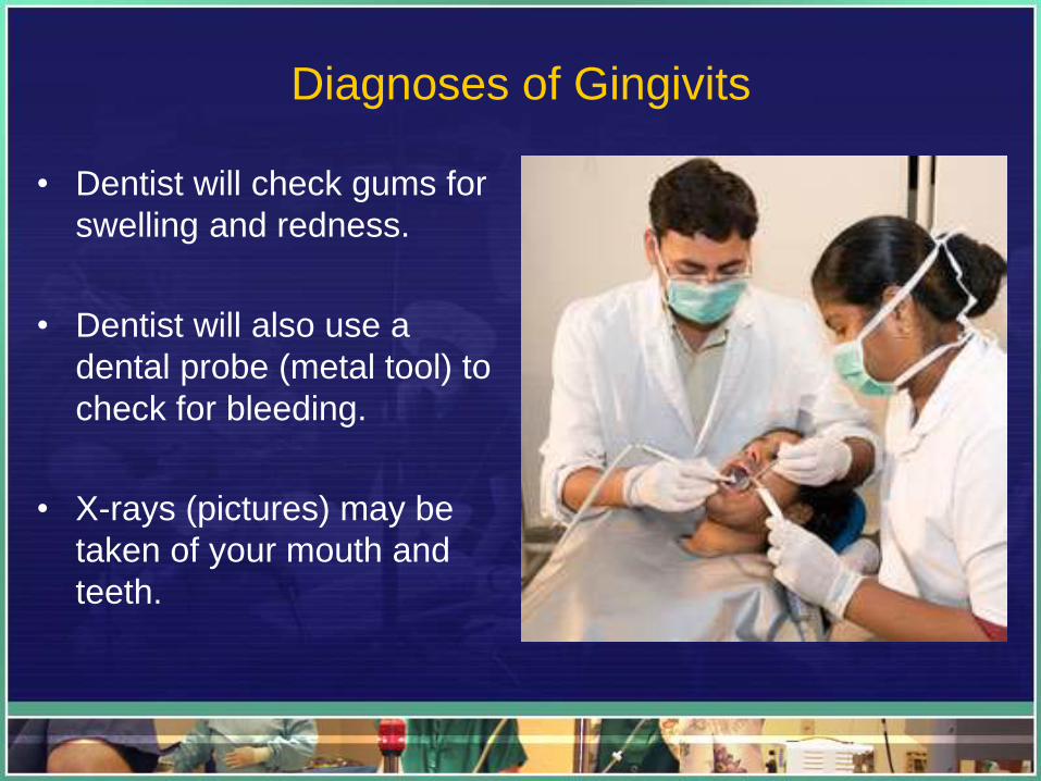
Diagnoses of Gingivits
• Dentist will check gums for
swelling and redness.
• Dentist will also use a
dental probe (metal tool) to
check for bleeding.
• X-rays (pictures) may be
taken of your mouth and
teeth.
Prevention is better than Cure
• Brush teeth two times a day after meals with fluoride toothpaste
• Use dental floss to clean between teeth at least once a day
• Ask dentist if should use a dental rinse, and what kind may work best
• See dentist regularly. Ask dentist how often should see him for dental
cleanings and exams
Cont..
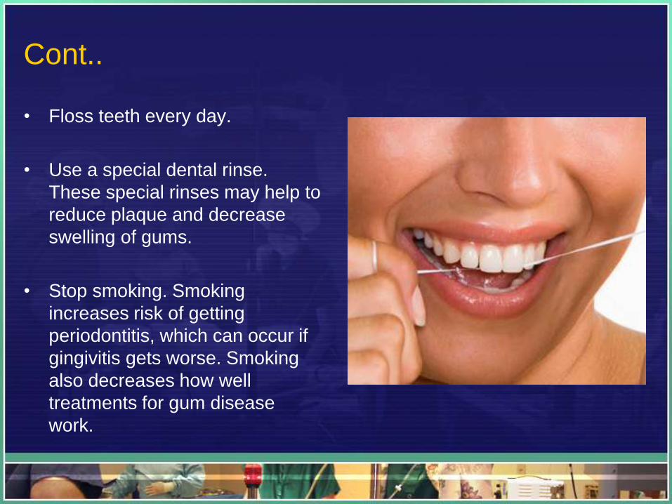
• Floss teeth every day.
• Use a special dental rinse.
These special rinses may help to
reduce plaque and decrease
swelling of gums.
• Stop smoking. Smoking
increases risk of getting
periodontitis, which can occur if
gingivitis gets worse. Smoking
also decreases how well
treatments for gum disease
work.
Conclusion
• The conclusion is very important to maintain the
healthy teeth and beautiful smile
• Prevent plaque from appearing at the gums and
teeth that will cause gingivitis or periodontitis
• Brushing and flushing are very important to prevent
any formation of the hard plaque ,infection and
inflammation for a good and healthy mouth

THANK YOU!!