Embed Size (px)
Citation preview

HAVE CRANIO-VERTEBRAL HAVE CRANIO-VERTEBRAL JUNCTION ANOMALIES BEEN JUNCTION ANOMALIES BEEN OVERLOOKED AS A CAUSE OF OVERLOOKED AS A CAUSE OF
VERTEBRO-BASILAR VERTEBRO-BASILAR INSUFFICIENCY?INSUFFICIENCY?
Deepak Agrawal, Naveen KDeepak Agrawal, Naveen KDepartments of Neurosurgery and *Nuclear medicine,Departments of Neurosurgery and *Nuclear medicine,
All India Institute of Medical Sciences, New Delhi-110029All India Institute of Medical Sciences, New Delhi-110029

BACKGROUNDBACKGROUND association of VBI with CVJ association of VBI with CVJ
anomalies is severely anomalies is severely underestimatedunderestimated
x-rays of the Cx spine are done in only x-rays of the Cx spine are done in only 30% of pts with VBI & 30% of pts with VBI & only 11% pts only 11% pts have proper flexhave proper flexnn/ext/extnn x-rays done x-rays done
Lorenstan KJ, Schrospshire LC, Ahn HS. Congenital odontoid aplasia and Lorenstan KJ, Schrospshire LC, Ahn HS. Congenital odontoid aplasia and
posterior circulation stroke in childhood. posterior circulation stroke in childhood. Ann NeurolAnn Neurol 1988;23-410-413 1988;23-410-413

BACKGROUNDBACKGROUND
posterior circulation ischemia has a higher posterior circulation ischemia has a higher morbidity and mortalitymorbidity and mortality
Fifty percent of these patients who are Fifty percent of these patients who are managed conservatively progress to managed conservatively progress to develop infarction develop infarction

BACKGROUNDBACKGROUND
Diagnosing even a percentage of the Diagnosing even a percentage of the patients with VBI as having CVJ anomalies patients with VBI as having CVJ anomalies may have major therapeutic & prognostic may have major therapeutic & prognostic implications.implications.

Aims and ObjectivesAims and Objectives::
Using 99Tc ECD brain SPECT to document Using 99Tc ECD brain SPECT to document the presence of posterior circulation the presence of posterior circulation cerebral ischemia in patients with CVJ cerebral ischemia in patients with CVJ anomalies and correlate with symptoms of anomalies and correlate with symptoms of VBI. VBI.

PROSPECTIVE STUDY DONE OVER A PROSPECTIVE STUDY DONE OVER A SIX MONTH PERIODSIX MONTH PERIOD

STUDY DESIGNSTUDY DESIGN
19 PATIENTS WITH FIXED AAD
Clinical assessment & Brain SPECT on admission
CONTROL GROUP(7 PTS)
VBI GROUP(12 PTS)
TOO + PF TOO + PF
Rpt SPECT at 4 weeks Rpt SPECT at 4 weeks
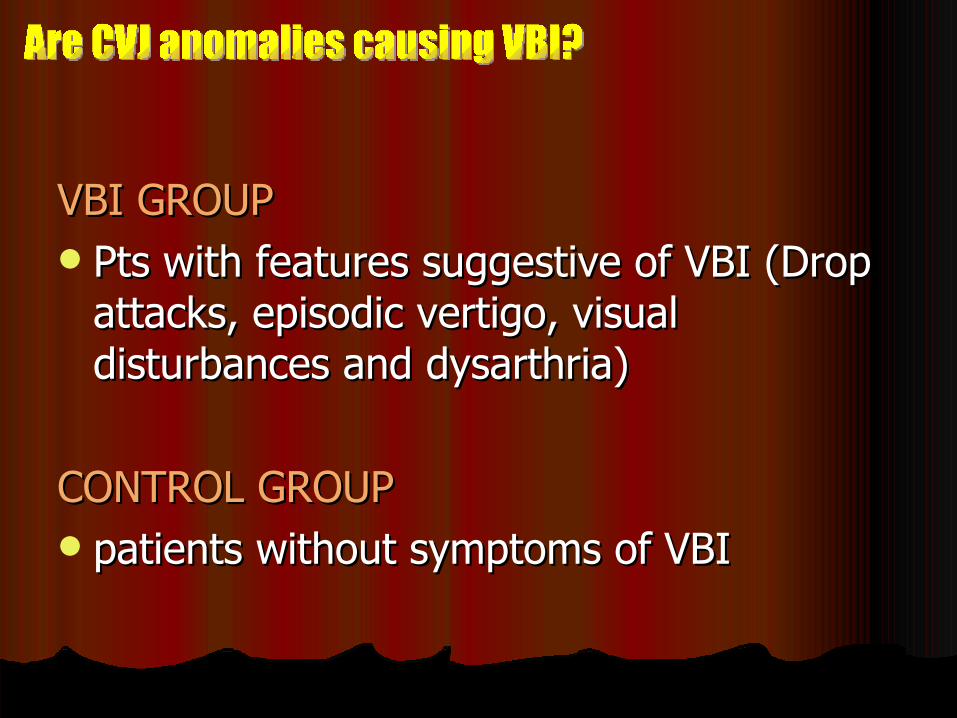
VBI GROUPVBI GROUP Pts with features suggestive of VBI (Drop Pts with features suggestive of VBI (Drop
attacks, episodic vertigo, visual attacks, episodic vertigo, visual disturbances and dysarthria)disturbances and dysarthria)
CONTROL GROUPCONTROL GROUP patients without symptoms of VBI patients without symptoms of VBI

Operative procedureOperative procedure combined TOO and Occipito-cervical combined TOO and Occipito-cervical
fusion from occiput to C3, using contoured fusion from occiput to C3, using contoured loop and sublaminar wiring with bone loop and sublaminar wiring with bone graft placement.graft placement.
Both procedures were carried out Both procedures were carried out consecutively in a single sitting.consecutively in a single sitting.

Patients with reducible AAD, requiring only Patients with reducible AAD, requiring only occipito-cervical fusion were excluded occipito-cervical fusion were excluded from the study to maintain uniformity.from the study to maintain uniformity.
Postoperatively the neck was immobilized Postoperatively the neck was immobilized using a philadelphia collar for a period of using a philadelphia collar for a period of three months.three months.

SPECT scanning was done using 99Tcm-SPECT scanning was done using 99Tcm-ECD on a dual headed GE 'Varicam' ECD on a dual headed GE 'Varicam' scanner.scanner.
The final data was displayed on a 10 The final data was displayed on a 10 grade color scale and semi quantitative grade color scale and semi quantitative analysis performed. analysis performed.
SPECTSPECT

Regional cerebellar perfusion <10% of Regional cerebellar perfusion <10% of contralateral lobe, or in case of bilateral contralateral lobe, or in case of bilateral involvement, less than 20% of basal gangliainvolvement, less than 20% of basal ganglia
ABNORMAL SPECT SCANABNORMAL SPECT SCAN

OBSERVATIONSOBSERVATIONS
RadiologyRadiology AADAAD 1919 BIBI 1515 Occipitalisation of atlasOccipitalisation of atlas 1414 kippel-feil anomalykippel-feil anomaly 99 cerebellar infarctscerebellar infarcts 22
(Both in VBI group)(Both in VBI group)

OBSERVATIONSOBSERVATIONS ( (VBI GroupVBI Group))
Clinical featuresClinical features Vertigo and drop attacksVertigo and drop attacks 1010 IncoordinationIncoordination 88 visual symptomsvisual symptoms 44

RESULTSRESULTS(Preoperative SPECT)(Preoperative SPECT)
Decreased cerebellar perfusion in Decreased cerebellar perfusion in 75% (n=9) of the patients in the 75% (n=9) of the patients in the VBI group compared to 14% (n=1) VBI group compared to 14% (n=1) in the control groupin the control group(p=0.019, fischer exact, 2 tailed).(p=0.019, fischer exact, 2 tailed).
1
9
0
2
4
6
8
10
Control Gp VBI Gp
Number of pts


RESULTSRESULTS Following surgery, five patients (55%) in Following surgery, five patients (55%) in
the symptomatic group and none in the the symptomatic group and none in the control group had improvement in control group had improvement in cerebellar perfusion.cerebellar perfusion.
All five patients showing improvement on All five patients showing improvement on SPECT also had improvement in their SPECT also had improvement in their symptoms of VBI following surgery symptoms of VBI following surgery

Two pts in VBI group developed meningitis in Two pts in VBI group developed meningitis in the postoperative period & had a further the postoperative period & had a further decrease in cerebellar perfusion on the follow decrease in cerebellar perfusion on the follow up SPECT scan.up SPECT scan.
Another 2 pts in VBI group had cerebellar Another 2 pts in VBI group had cerebellar infarcts on MRI & did not show improvement in infarcts on MRI & did not show improvement in cerebellar hypoperfusion following surgery.cerebellar hypoperfusion following surgery.

Pathogenesis VBI in CVJ AnomaliesPathogenesis VBI in CVJ Anomalies
Chronic low grade micro-traumaRptd flex/extn of vessel
Intimal damage
Thrombosis
Embolisation

Clinical rarity of posterior circulation infarcts in Clinical rarity of posterior circulation infarcts in CVJ anomalies could be due toCVJ anomalies could be due to::
Duplication of VA and the adequacy of the Duplication of VA and the adequacy of the circulation of Williscirculation of Willis
patients symptomatic for VBI are not patients symptomatic for VBI are not routinely evaluated for CVJ anomalies routinely evaluated for CVJ anomalies which remain undiagnosedwhich remain undiagnosed

In patients with CVJ anomalies currently In patients with CVJ anomalies currently used imaging modalities such as cervical used imaging modalities such as cervical spine x-rays and CT scans are not spine x-rays and CT scans are not adequate to evaluate for vertebro-basilar adequate to evaluate for vertebro-basilar ischemia ischemia

Ours is the only study of its kind Ours is the only study of its kind documenting hypoperfusion in posterior documenting hypoperfusion in posterior circulation territory in patients with CVJ circulation territory in patients with CVJ anomalies.anomalies.
This hypoperfusion may represent a state This hypoperfusion may represent a state of chronic VBI, expected in such patients of chronic VBI, expected in such patients & & MAY BE REVERSIBLEMAY BE REVERSIBLE following surgery. following surgery.
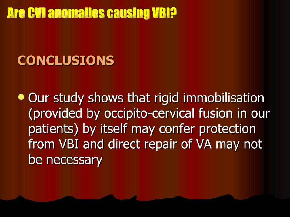
CONCLUSIONSCONCLUSIONS
Our study shows that rigid immobilisation Our study shows that rigid immobilisation (provided by occipito-cervical fusion in our (provided by occipito-cervical fusion in our patients) by itself may confer protection patients) by itself may confer protection from VBI and direct repair of VA may not from VBI and direct repair of VA may not be necessary be necessary
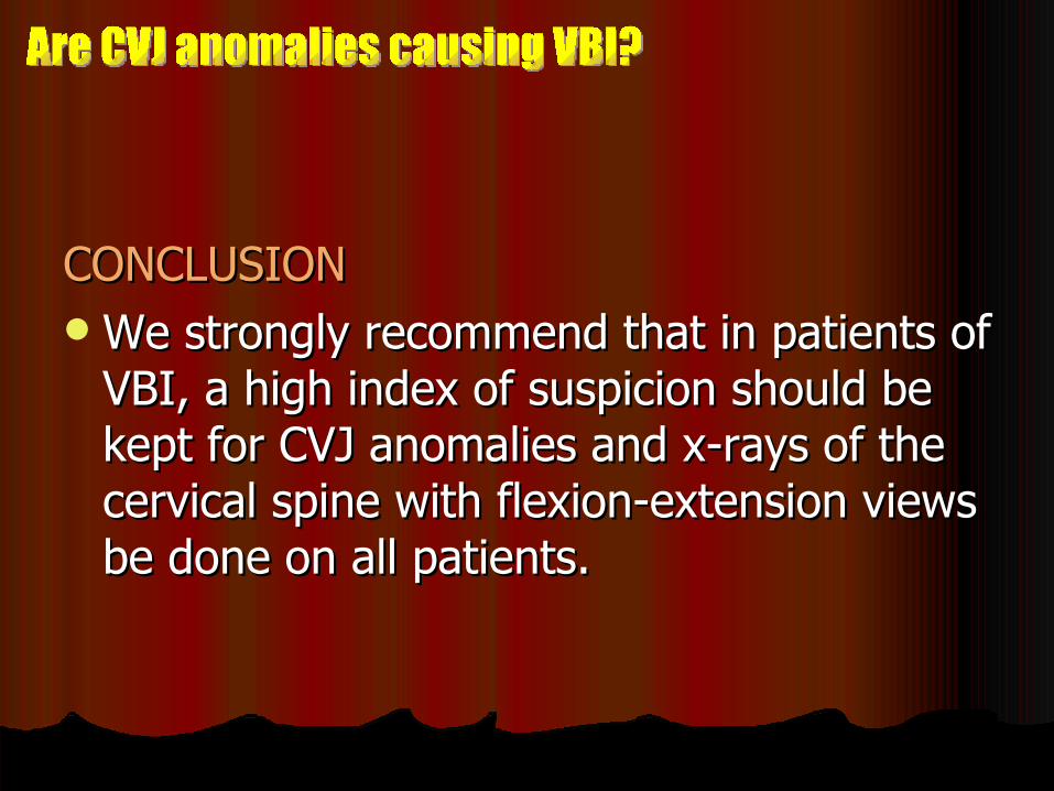
CONCLUSIONCONCLUSION We strongly recommend that in patients of We strongly recommend that in patients of
VBI, a high index of suspicion should be VBI, a high index of suspicion should be kept for CVJ anomalies and x-rays of the kept for CVJ anomalies and x-rays of the cervical spine with flexion-extension views cervical spine with flexion-extension views be done on all patients. be done on all patients.

THANK YOUTHANK YOU

![[PPT]ESTADO DE COMA · Web viewALTERACIONES DE LA CONCIENCIA DRA. MARIA BEATRIZ RODRIGUEZ GONZALEZ R2 MF DR. CANTÚ La oclusión vertebro-basilar por trombosis o embolia puede causar](https://img.pdfslide.net/doc/110x75/5aa6bf2c7f8b9ac8748eaf4d/pptestado-de-coma-viewalteraciones-de-la-conciencia-dra-maria-beatriz-rodriguez.jpg)