Embed Size (px)
Citation preview
Heart Failure -Overview
Dr.Elamaran.ESenior resident
Department Of CTVS
Definition
• Clinical syndrome
– Symptoms & signs typical of heart failure
– Objective evidence –abnormality of heart


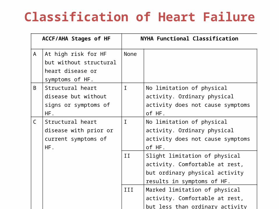
Classification of Heart FailureACCF/AHA Stages of HF NYHA Functional Classification
A At high risk for HF but without structural heart disease or symptoms of HF.
None
B Structural heart disease but without signs or symptoms of HF.
I No limitation of physical activity. Ordinary physical activity does not cause symptoms of HF.
C Structural heart disease with prior or current symptoms of HF.
I No limitation of physical activity. Ordinary physical activity does not cause symptoms of HF.
II Slight limitation of physical activity. Comfortable at rest, but ordinary physical activity results in symptoms of HF.
III Marked limitation of physical activity. Comfortable at rest, but less than ordinary activity causes symptoms of HF.
IV Unable to carry on any physical activity without symptoms of HF, or symptoms of HF at rest.D Refractory HF requiring
specialized interventions.

A thorough history and physical examination should beobtained/performed in patients presenting with HF toidentify cardiac and noncardiac disorders or behaviors that might cause or accelerate the development or progression of HF.
In patients with idiopathic DCM, a 3-generational family history should be obtained to aid in establishing the diagnosis of familial DCM.
Volume status and vital signs should be assessed at each patient encounter. This includes serial assessment of weight, as well as estimates of jugular venous pressure and the presence of peripheral edema or Orthopnea.
History and Physical Examination
I IIa IIb III
I IIa IIb III
I IIa IIb III

Diagnostic TestsInitial laboratory evaluation of patients presenting with HF should include complete blood count, urinalysis, serum electrolytes (including calcium and magnesium), blood urea nitrogen, serum creatinine, glucose, fasting lipid profile, liver function tests, and thyroid-stimulating hormone.
Serial monitoring, when indicated, should include serum electrolytes and renal function.
A 12-lead ECG should be performed initially on all patients presenting with HF.
I IIa IIb III
I IIa IIb III
I IIa IIb III

Recommendations for Noninvasive Imaging
Recommendation COR LOE
Patients with suspected, acute, or new-onset HF should undergo a chest x-ray
I C
A 2-dimensional echocardiogram with Doppler should be performed for initial evaluation of HF
I C
Repeat measurement of EF is useful in patients with HF who have had a significant change in clinical status or received treatment that might affect cardiac function, or for consideration of device therapy
I C
Noninvasive imaging to detect myocardial ischemia and viability is reasonable in HF and CAD
IIa C
Viability assessment is reasonable before revascularization in HF patients with CAD
IIa B
Radionuclide ventriculography or MRI can be useful to assess LVEF and volume
IIa C
MRI is reasonable when assessing myocardial infiltration or scar IIa B
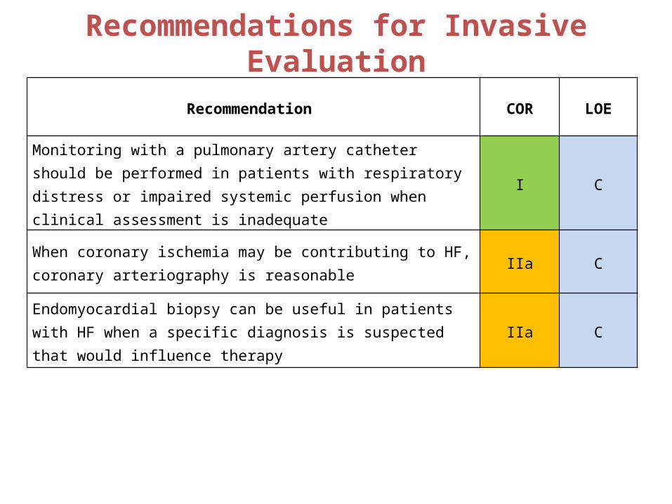
Recommendations for Invasive Evaluation
Recommendation COR LOE
Monitoring with a pulmonary artery catheter should be performed in patients with respiratory distress or impaired systemic perfusion when clinical assessment is inadequate
I C
When coronary ischemia may be contributing to HF, coronary arteriography is reasonable
IIa C
Endomyocardial biopsy can be useful in patients with HF when a specific diagnosis is suspected that would influence therapy
IIa C
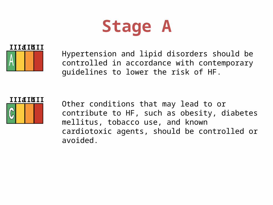
Stage AHypertension and lipid disorders should be controlled in accordance with contemporary guidelines to lower the risk of HF.
Other conditions that may lead to or contribute to HF, such as obesity, diabetes mellitus, tobacco use, and known cardiotoxic agents, should be controlled or avoided.
I IIa IIb III
I IIa IIb III
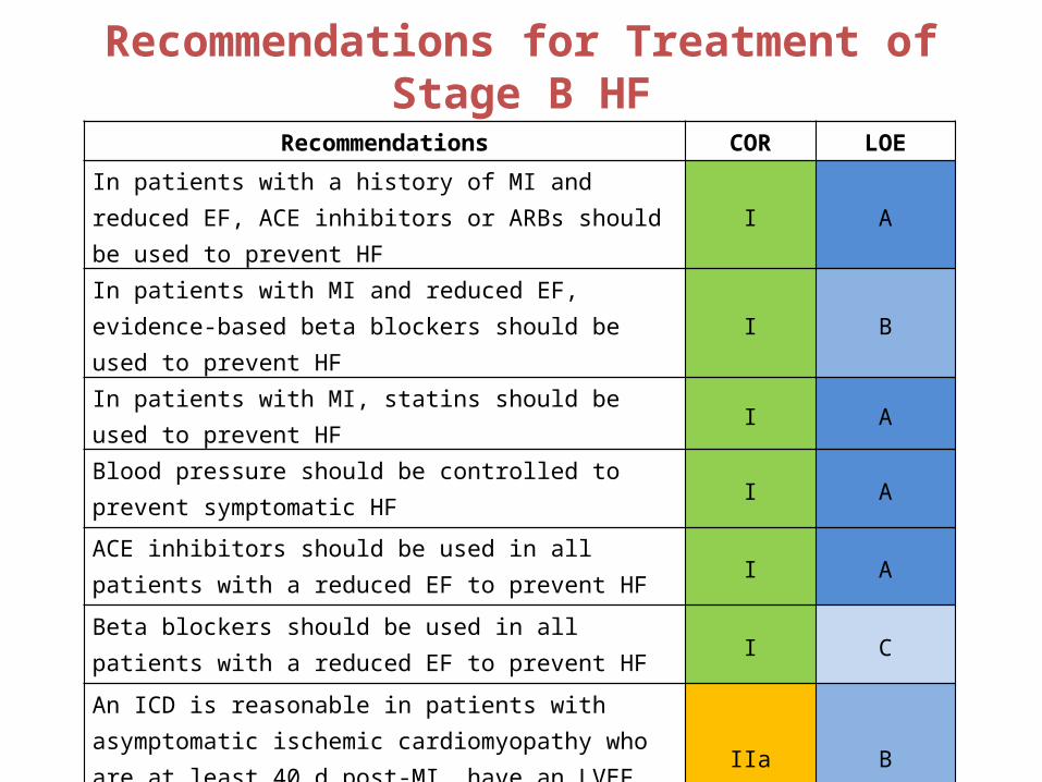
Recommendations for Treatment of Stage B HF
Recommendations COR LOE
In patients with a history of MI and reduced EF, ACE inhibitors or ARBs should be used to prevent HF I A
In patients with MI and reduced EF, evidence-based beta blockers should be used to prevent HF I B
In patients with MI, statins should be used to prevent HF I A
Blood pressure should be controlled to prevent symptomatic HF I A
ACE inhibitors should be used in all patients with a reduced EF to prevent HF I A
Beta blockers should be used in all patients with a reduced EF to prevent HF I C
An ICD is reasonable in patients with asymptomatic ischemic cardiomyopathy who are at least 40 d post-MI, have an LVEF ≤30%, and on GDMT
IIa B
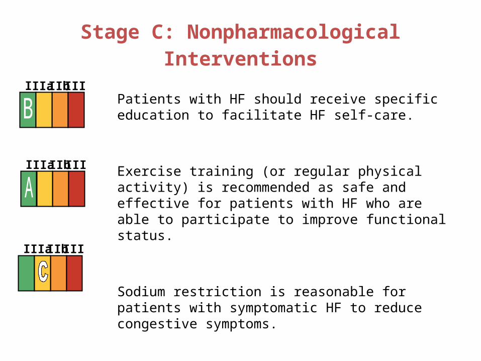
Stage C: Nonpharmacological Interventions
Patients with HF should receive specific education to facilitate HF self-care.
Exercise training (or regular physical activity) is recommended as safe and effective for patients with HF who are able to participate to improve functional status.
Sodium restriction is reasonable for patients with symptomatic HF to reduce congestive symptoms.
I IIa IIb III
I IIa IIb III
I IIa IIb III
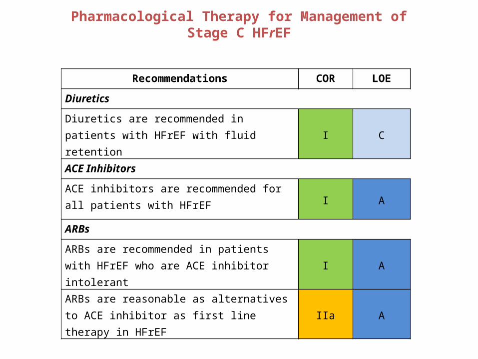
Pharmacological Therapy for Management of Stage C HFrEF
Recommendations COR LOE
Diuretics
Diuretics are recommended in patients with HFrEF with fluid retention I C
ACE Inhibitors
ACE inhibitors are recommended for all patients with HFrEF I A
ARBs
ARBs are recommended in patients with HFrEF who are ACE inhibitor intolerant I A
ARBs are reasonable as alternatives to ACE inhibitor as first line therapy in HFrEF IIa A
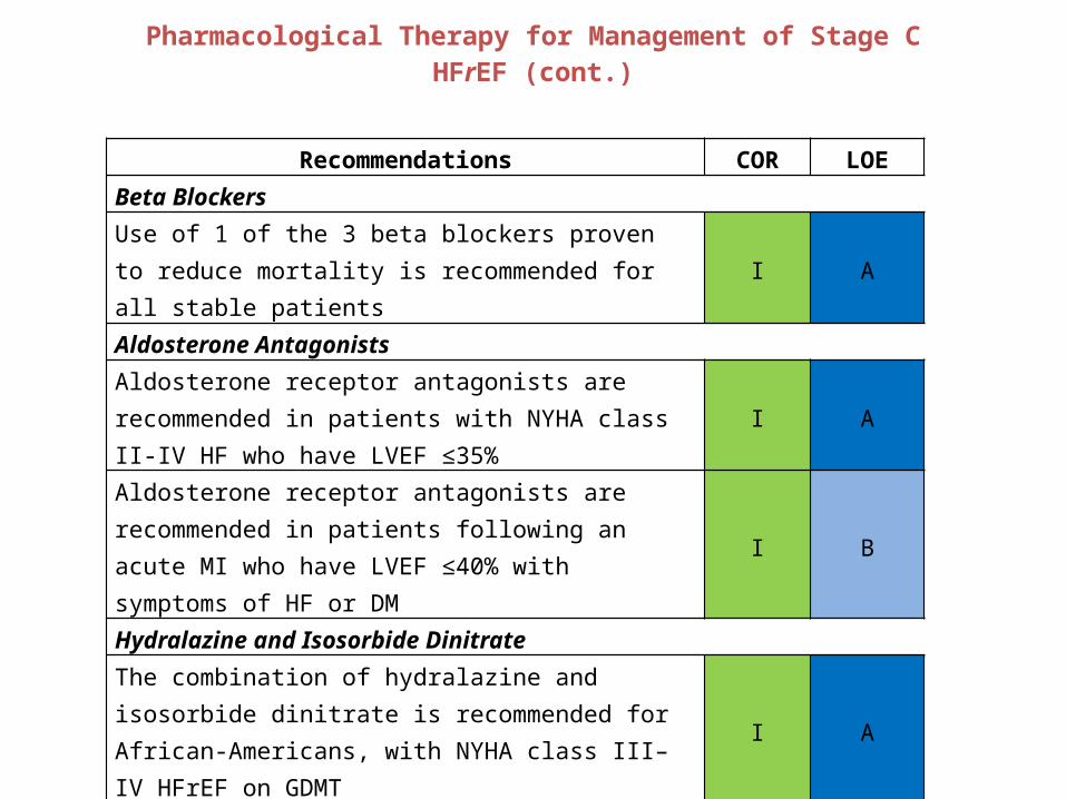
Pharmacological Therapy for Management of Stage C HFrEF (cont.)
Recommendations COR LOEBeta BlockersUse of 1 of the 3 beta blockers proven to reduce mortality is recommended for all stable patients I A
Aldosterone AntagonistsAldosterone receptor antagonists are recommended in patients with NYHA class II-IV HF who have LVEF ≤35% I A
Aldosterone receptor antagonists are recommended in patients following an acute MI who have LVEF ≤40% with symptoms of HF or DM
I B
Hydralazine and Isosorbide Dinitrate
The combination of hydralazine and isosorbide dinitrate is recommended for African-Americans, with NYHA class III–IV HFrEF on GDMT
I A
A combination of hydralazine and isosorbide dinitrate can be useful in patients with HFrEF who cannot be given ACE inhibitors or ARBs
IIa B
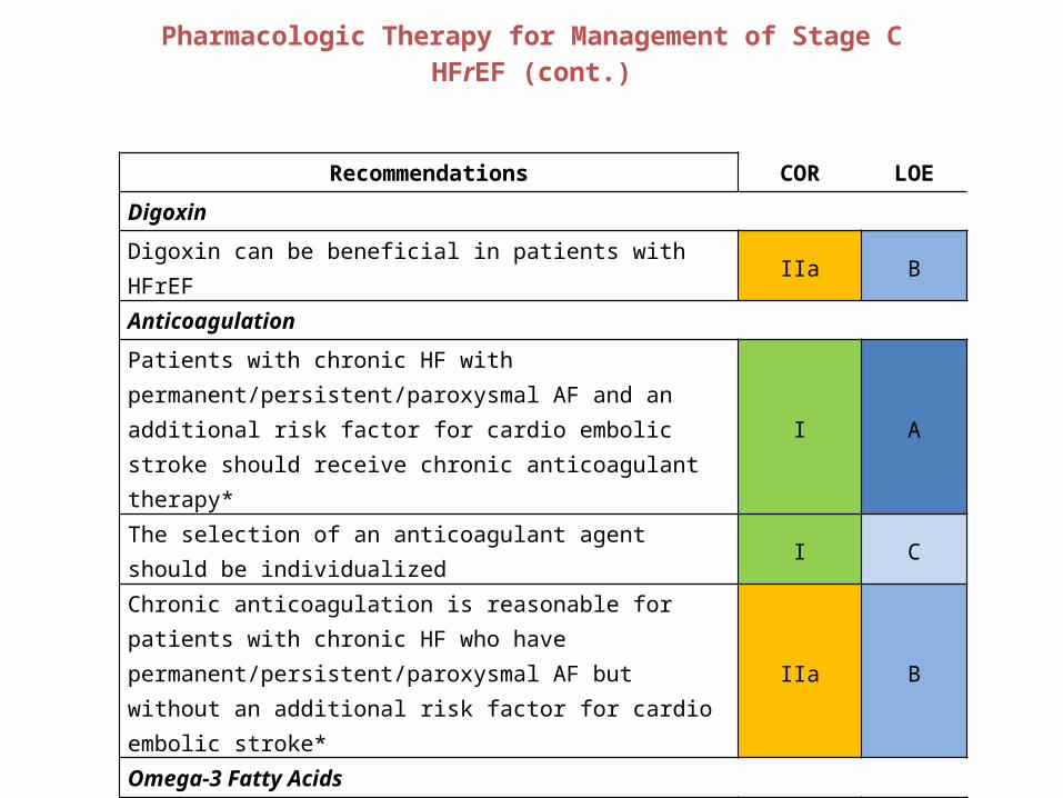
Pharmacologic Therapy for Management of Stage C HFrEF (cont.)
Recommendations COR LOE
Digoxin
Digoxin can be beneficial in patients with HFrEF IIa B
Anticoagulation
Patients with chronic HF with permanent/persistent/paroxysmal AF and an additional risk factor for cardio embolic stroke should receive chronic anticoagulant therapy* I A
The selection of an anticoagulant agent should be individualized I C
Chronic anticoagulation is reasonable for patients with chronic HF who have permanent/persistent/paroxysmal AF but without an additional risk factor for cardio embolic stroke* IIa B
Omega-3 Fatty Acids
Omega-3 PUFA supplementation is reasonable to use as adjunctive therapy in HFrEF or HFpEF patients IIa B

Treatment of HFpEFRecommendations COR LOE
Systolic and diastolic blood pressure should be controlled according to published clinical practice guidelines
I B
Diuretics should be used for relief of symptoms due to volume overload I C
Coronary revascularization for patients with CAD in whom angina or demonstrable myocardial ischemia is present despite GDMT
IIa
C
Management of AF according to published clinical practice guidelines for HFpEF to improve symptomatic HF
IIa C
Use of beta-blocking agents, ACE inhibitors, and ARBs for hypertension in HFpEF IIa C

Device Therapy for Stage C HFrEF
Recommendations COR LOE
ICD therapy is recommended for primary prevention of SCD in selected patients with HFrEF at least 40 days post-MI with LVEF ≤35%, and NYHA class II or III symptoms on chronic GDMT, who are expected to live ≥1 year* I A
CRT is indicated for patients who have LVEF ≤35%, sinus rhythm, LBBB with a QRS ≥150 ms
I
A (NYHA class
III/IV)
B (NYHA class II)
ICD therapy is recommended for primary prevention of SCD in selected patients with HFrEF at least 40 days post-MI with LVEF ≤30%, and NYHA class I symptoms while receiving GDMT, who are expected to live ≥1 year*
I B
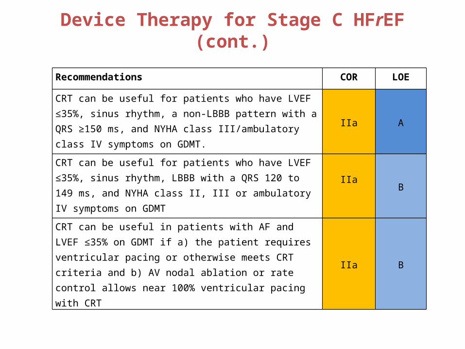
Device Therapy for Stage C HFrEF (cont.)
Recommendations COR LOE
CRT can be useful for patients who have LVEF ≤35%, sinus rhythm, a non-LBBB pattern with a QRS ≥150 ms, and NYHA class III/ambulatory class IV symptoms on GDMT. IIa A
CRT can be useful for patients who have LVEF ≤35%, sinus rhythm, LBBB with a QRS 120 to 149 ms, and NYHA class II, III or ambulatory IV symptoms on GDMT IIa
B
CRT can be useful in patients with AF and LVEF ≤35% on GDMT if a) the patient requires ventricular pacing or otherwise meets CRT criteria and b) AV nodal ablation or rate control allows near 100% ventricular pacing with CRT IIa B

Water Restriction
Fluid restriction (1.5 to 2 L/d) is reasonable in stage D, especially in patients with hyponatremia, to reduce congestive symptoms.
I IIa IIb III
Management of stage A to D
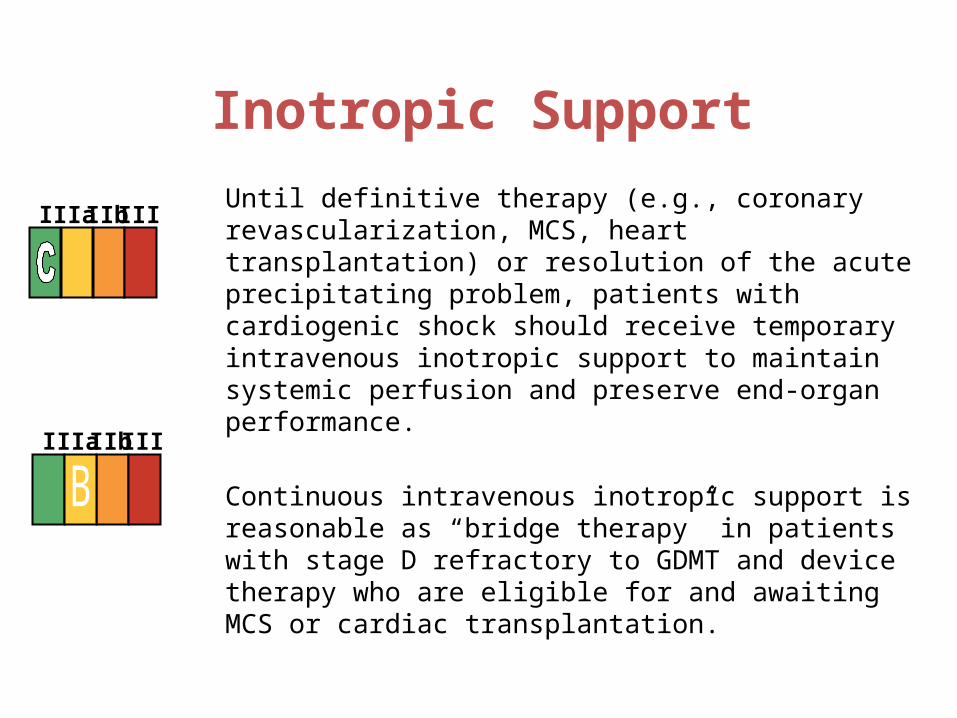
Inotropic Support
Until definitive therapy (e.g., coronary revascularization, MCS, heart transplantation) or resolution of the acute precipitating problem, patients with cardiogenic shock should receive temporary intravenous inotropic support to maintain systemic perfusion and preserve end-organ performance.
Continuous intravenous inotropic support is reasonable as “bridge therapy” in patients with stage D refractory to GDMT and device therapy who are eligible for and awaiting MCS or cardiac transplantation.
I IIa IIb III
I IIa IIb III
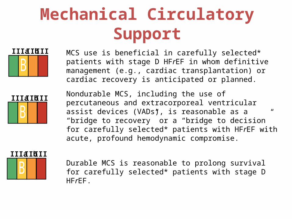
Mechanical Circulatory Support
MCS use is beneficial in carefully selected* patients with stage D HFrEF in whom definitive management (e.g., cardiac transplantation) or cardiac recovery is anticipated or planned.
Nondurable MCS, including the use of percutaneous and extracorporeal ventricular assist devices (VADs), is reasonable as a “bridge to recovery” or a “bridge to decision” for carefully selected* patients with HFrEF with acute, profound hemodynamic compromise.
Durable MCS is reasonable to prolong survival for carefully selected* patients with stage D HFrEF.
I IIa IIb III
I IIa IIb III
I IIa IIb III
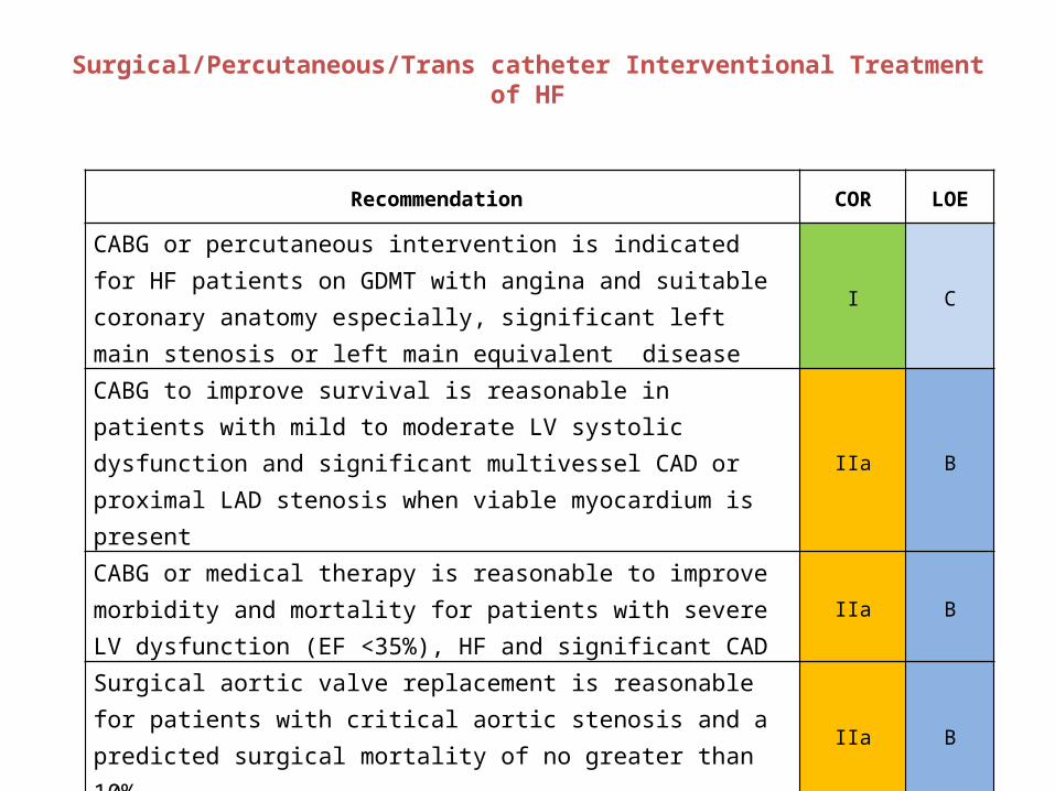
Surgical/Percutaneous/Trans catheter Interventional Treatment of HF
Recommendation COR LOE
CABG or percutaneous intervention is indicated for HF patients on GDMT with angina and suitable coronary anatomy especially, significant left main stenosis or left main equivalent disease
I C
CABG to improve survival is reasonable in patients with mild to moderate LV systolic dysfunction and significant multivessel CAD or proximal LAD stenosis when viable myocardium is present
IIa B
CABG or medical therapy is reasonable to improve morbidity and mortality for patients with severe LV dysfunction (EF <35%), HF and significant CAD
IIa B
Surgical aortic valve replacement is reasonable for patients with critical aortic stenosis and a predicted surgical mortality of no greater than 10%
IIa B
Trans catheter aortic valve replacement is reasonable for patients with critical aortic stenosis who are deemed inoperable
IIa B
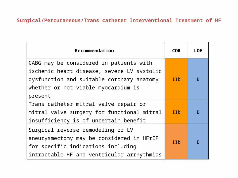
Surgical/Percutaneous/Trans catheter Interventional Treatment of HF
Recommendation COR LOE
CABG may be considered in patients with ischemic heart disease, severe LV systolic dysfunction and suitable coronary anatomy whether or not viable myocardium is present
IIb B
Trans catheter mitral valve repair or mitral valve surgery for functional mitral insufficiency is of uncertain benefit
IIb B
Surgical reverse remodeling or LV aneurysmectomy may be considered in HFrEF for specific indications including intractable HF and ventricular arrhythmias
IIb B
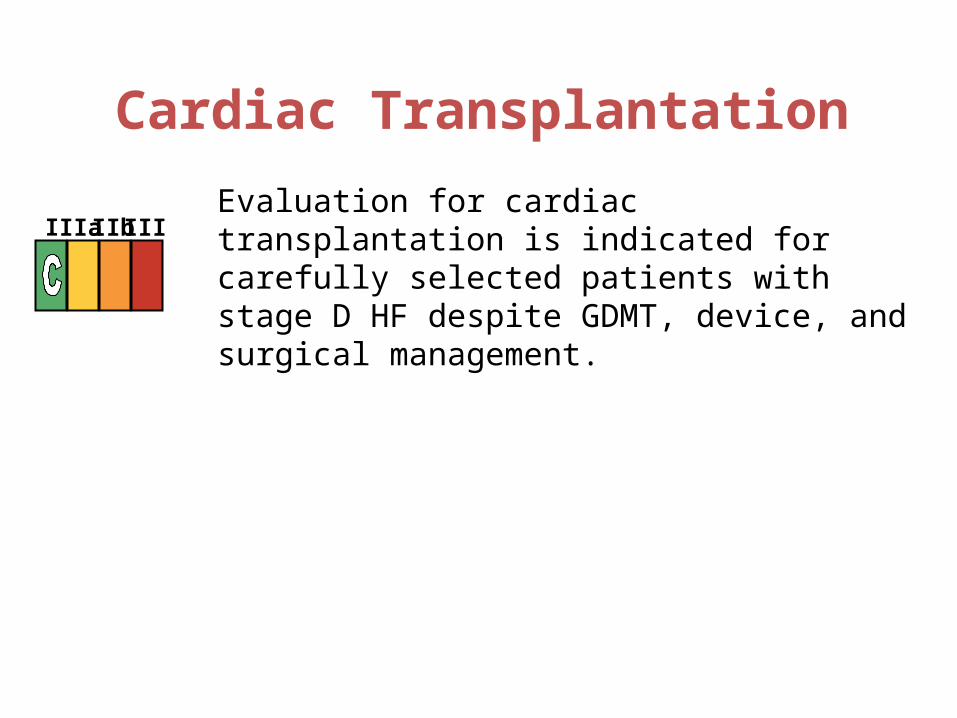
Cardiac Transplantation
Evaluation for cardiac transplantation is indicated for carefully selected patients with stage D HF despite GDMT, device, and surgical management.
I IIa IIb III
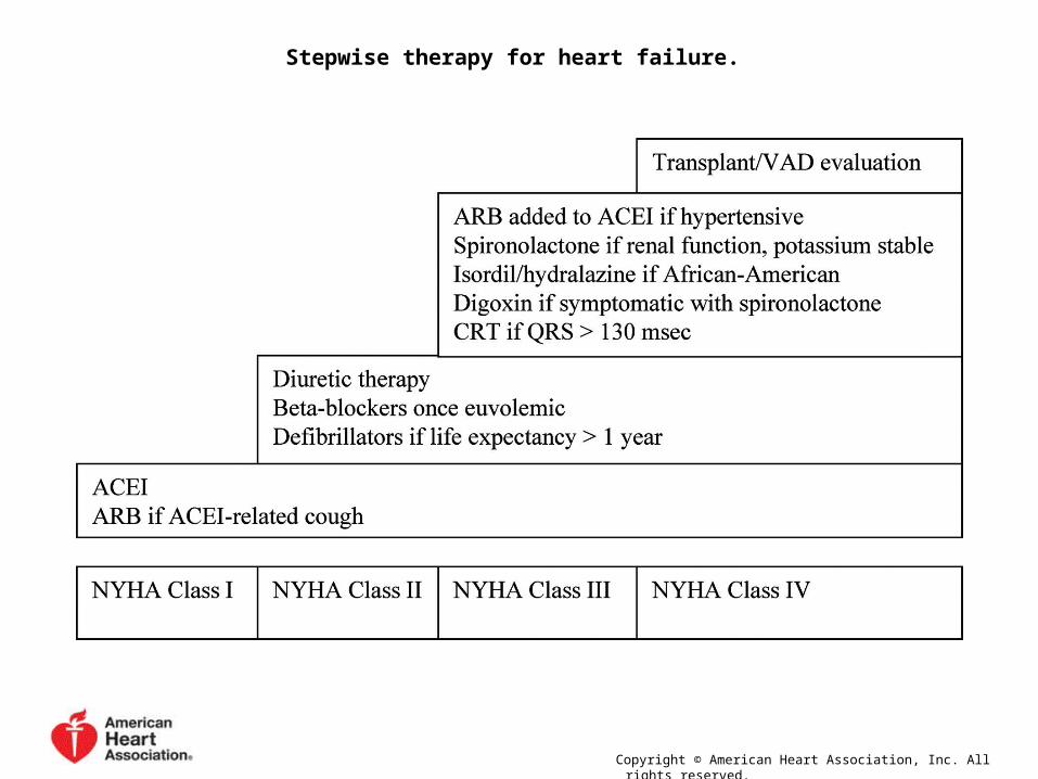
Stepwise therapy for heart failure.
Copyright © American Heart Association, Inc. All rights reserved.

Surgical Interventions

Theory
• Systolic HF leads to an enlarged LV volume to maintain stroke volume
• This leads to increase in wall stress due to Laplace's law stress = pressure x radius ÷ 2 x wall thickness
• The ventricular geometry becomes less ellipsoid and more spherical leading to progression of left ventricular dysfunction and worsening heart failure.
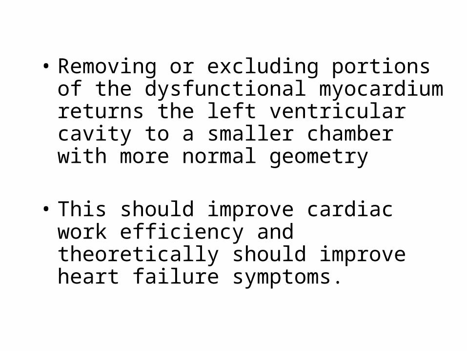
• Removing or excluding portions of the dysfunctional myocardium returns the left ventricular cavity to a smaller chamber with more normal geometry
• This should improve cardiac work efficiency and theoretically should improve heart failure symptoms.
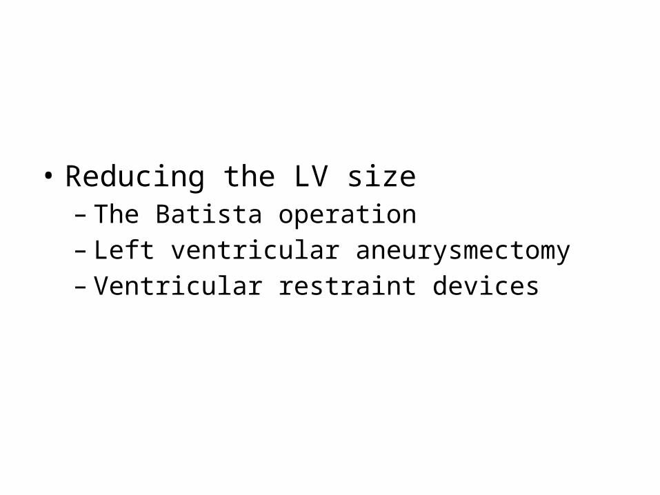
• Reducing the LV size– The Batista operation– Left ventricular aneurysmectomy– Ventricular restraint devices
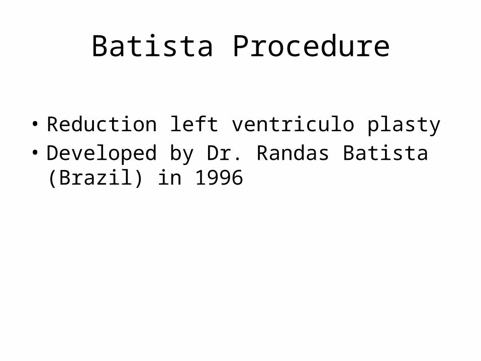
• Reduction left ventriculo plasty• Developed by Dr. Randas Batista
(Brazil) in 1996
Batista Procedure

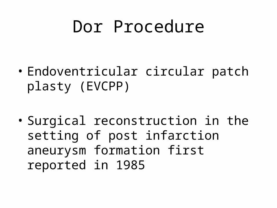
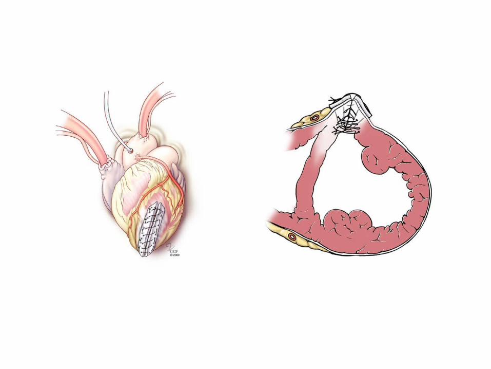
Dor Procedure
• Endoventricular circular patch plasty (EVCPP)
• Surgical reconstruction in the setting of post infarction aneurysm formation first reported in 1985

Considered Criteria for Surgical Repair
• Previous Anterior MI, • Dilated left ventricle (end-diastolic
volume index >100 mL/m2)• Depressed LVEF • Left ventricular regional dyskinesis
or akinesis >30 percent of the ventricular perimeter
• Symptoms of HF
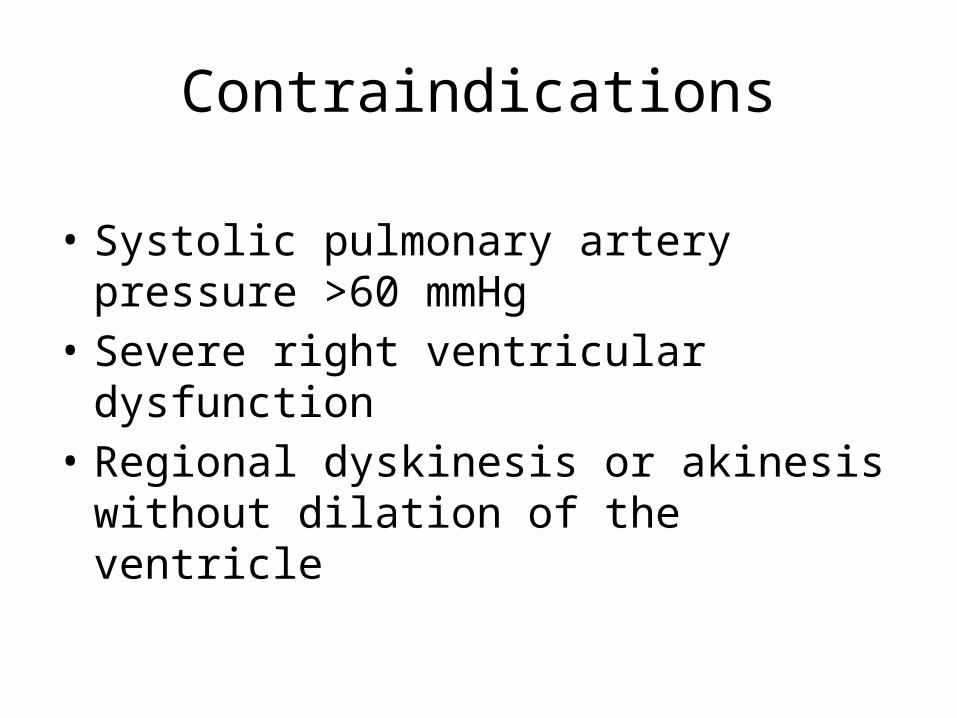
Contraindications
• Systolic pulmonary artery pressure >60 mmHg
• Severe right ventricular dysfunction • Regional dyskinesis or akinesis
without dilation of the ventricle
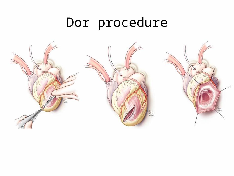
Dor procedure


Positioning of the Apicorn CorCap cardiac support device.
© 2005 European Society of Cardiology
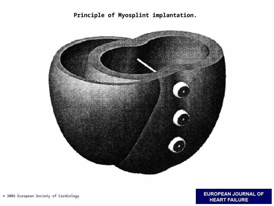
Principle of Myosplint implantation.
© 2005 European Society of Cardiology
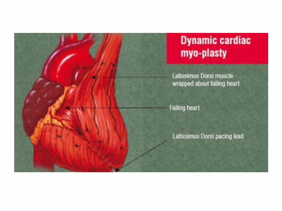
• Re-power the failing heart– Heart Transplantation– Dynamic cardio-myoplasty– Ventricular assist devices and Total
Artificial heart

• Improve the cardiac output by reducing ventricular afterload– IABP– Resection of LVOT obstruction