Embed Size (px)
Citation preview

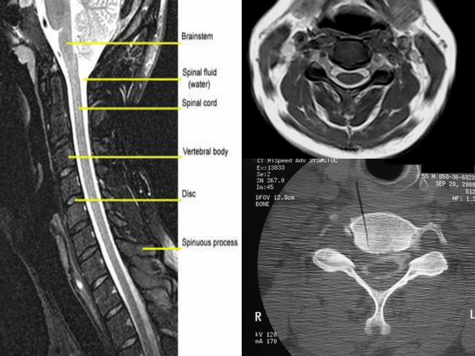
How to Read a Cervical MRI
Spine ConferenceUpper Chesapeake Medical Center
Friday May 1, 2015

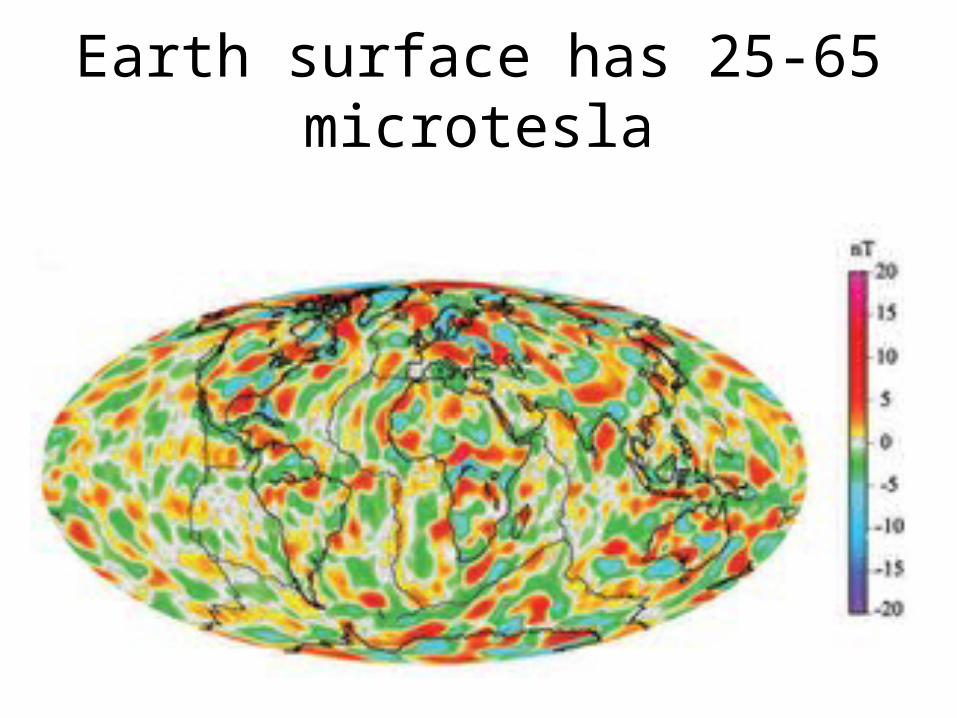
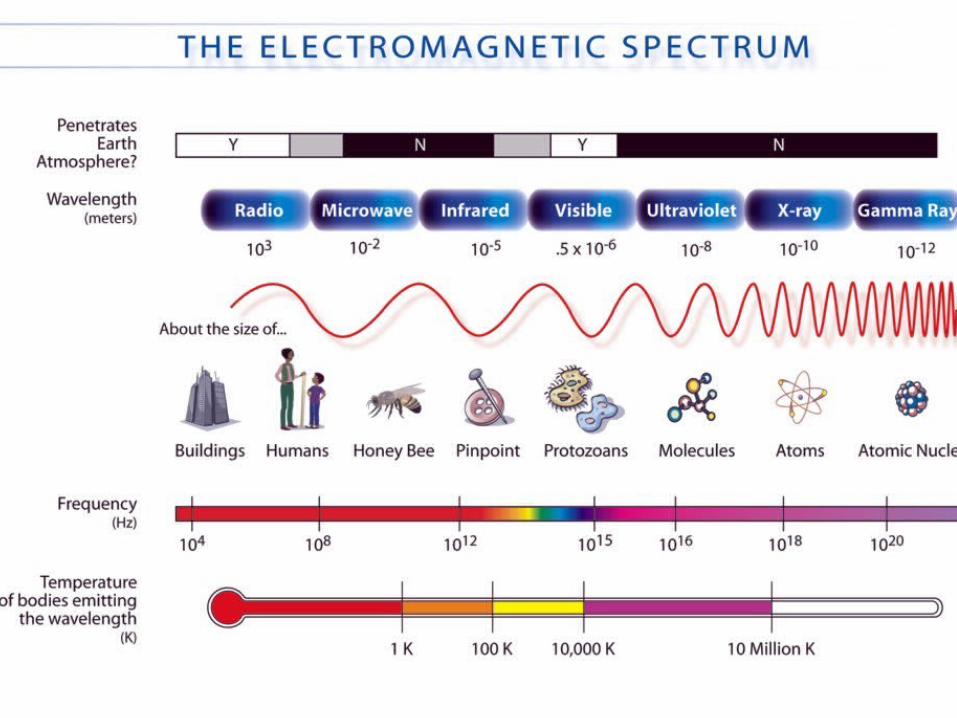
Earth surface has 25-65 microtesla

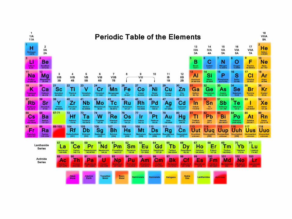
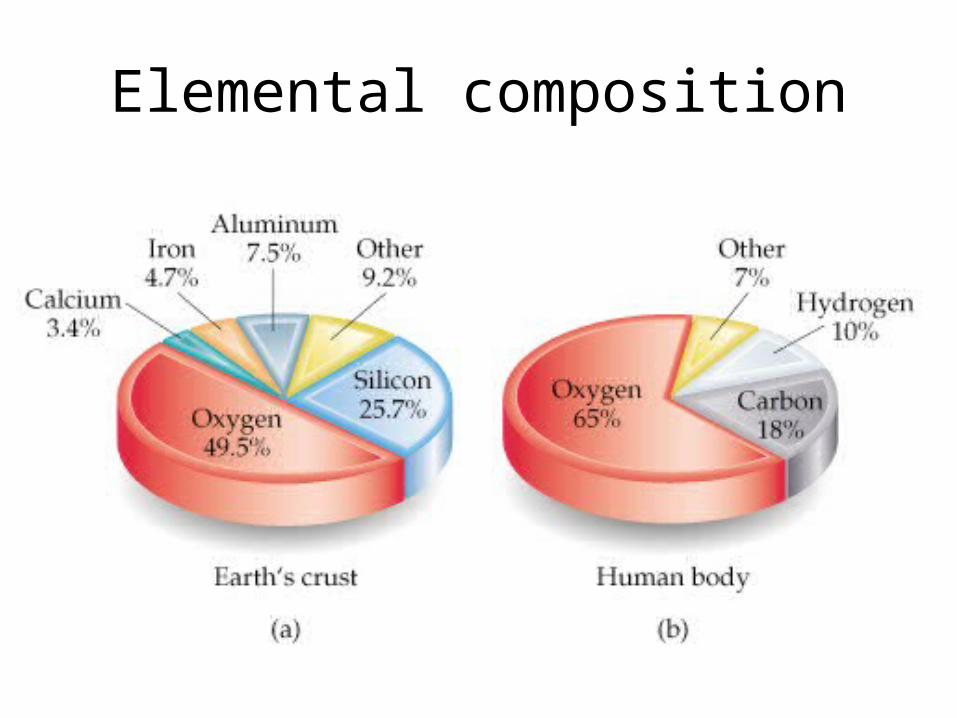
Elemental composition


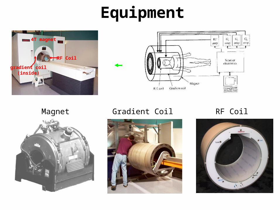
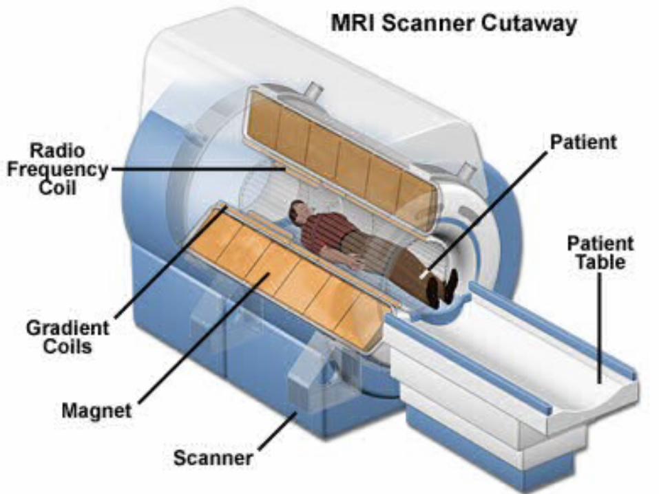
Equipment
Magnet Gradient Coil RF Coil
RF Coil
4T magnet
gradient coil(inside)
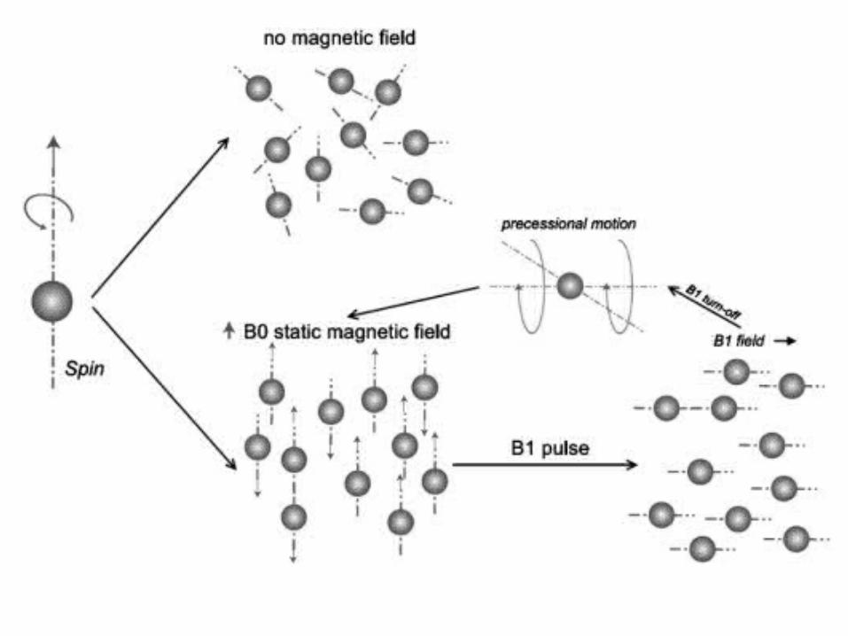
B0
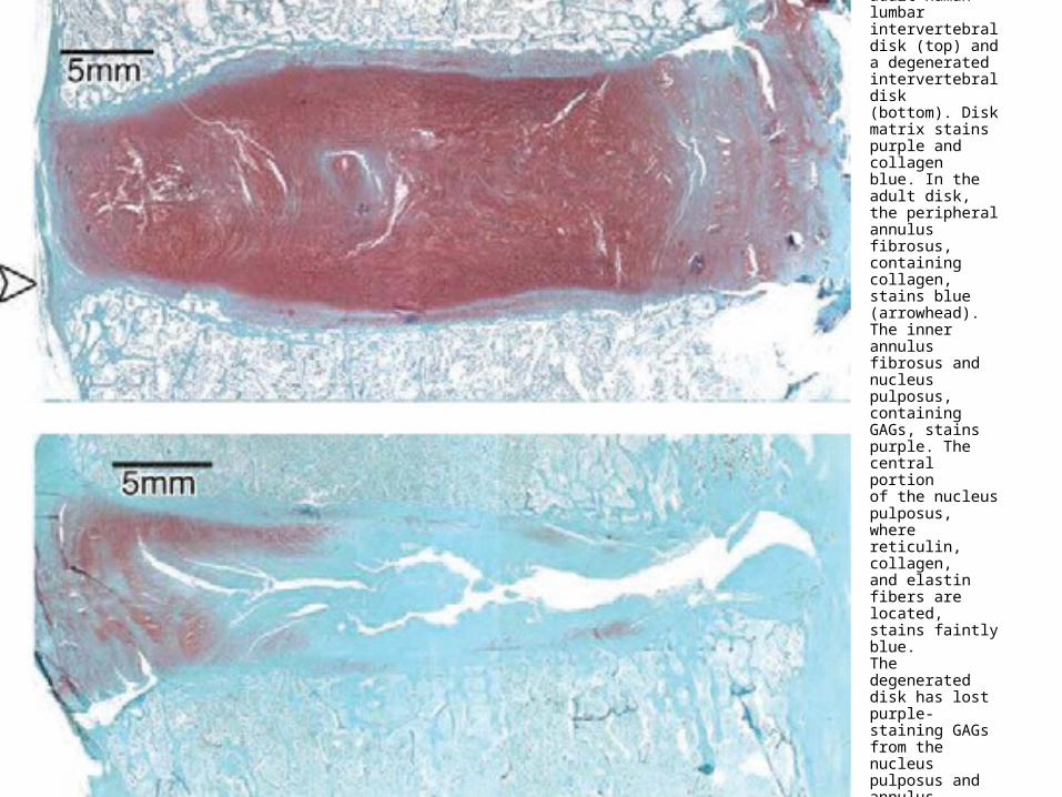
Sagittally oriented safranin-O–stainedsections of an adult human lumbar intervertebraldisk (top) and a degenerated intervertebral disk(bottom). Disk matrix stains purple and collagenblue. In the adult disk, the peripheral annulus fibrosus,containing collagen, stains blue (arrowhead).The inner annulus fibrosus and nucleus pulposus,containing GAGs, stains purple. The central portionof the nucleus pulposus, where reticulin, collagen,and elastin fibers are located, stains faintly blue.The degenerated disk has lost purple-staining GAGsfrom the nucleus pulposus and annulus fibrosus. Itconforms to Pfirrmann grade IIII degeneration.


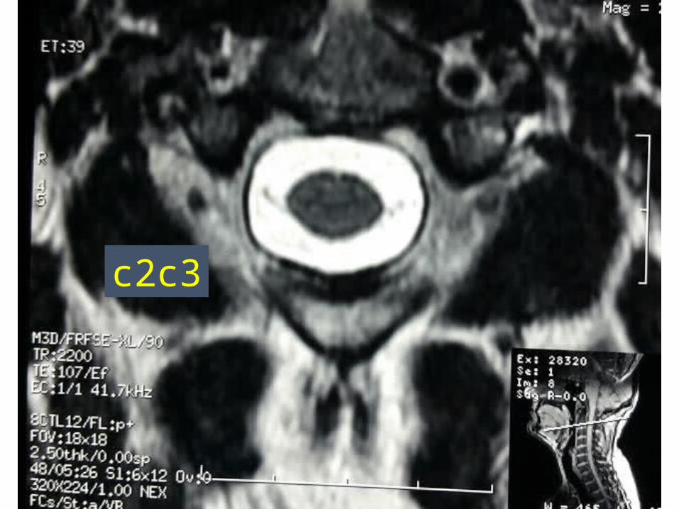
c2c3
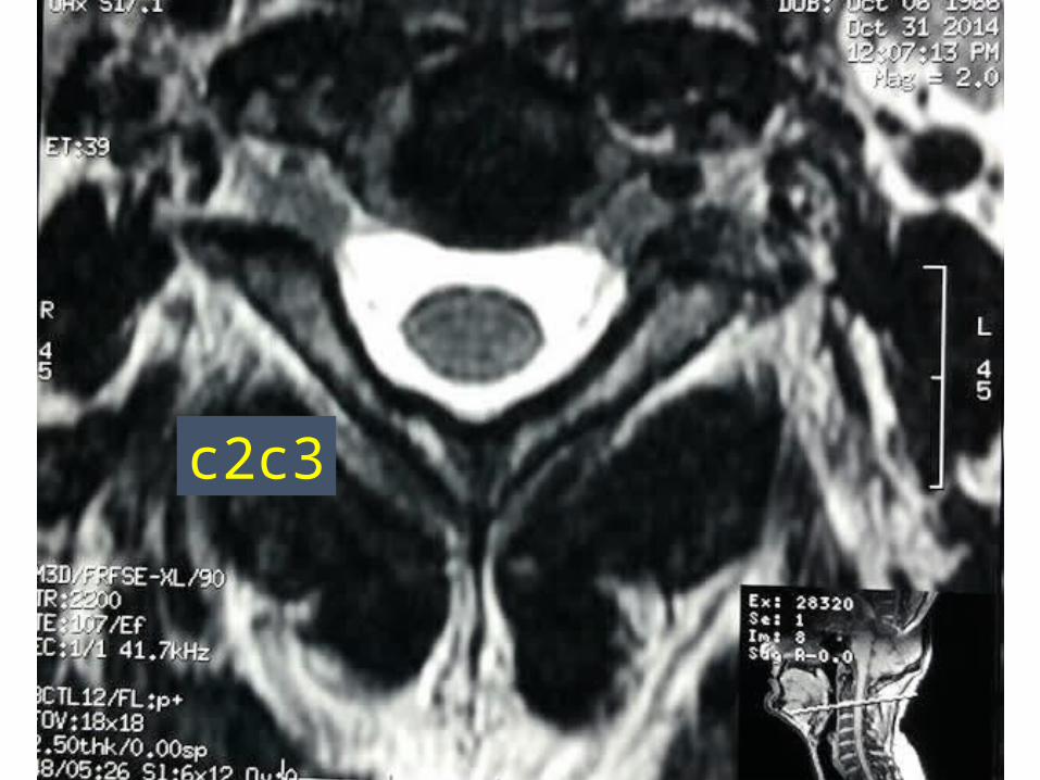
c2c3
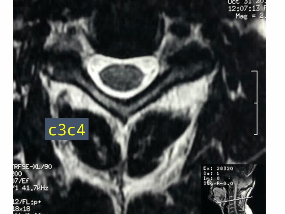
c3c4
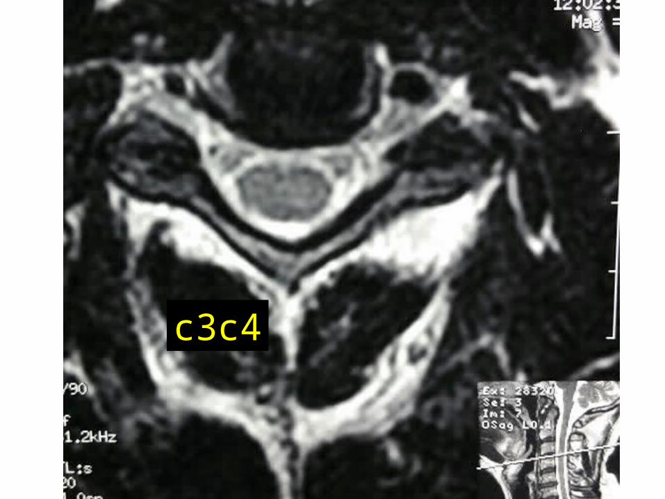
c3c4
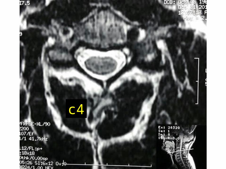
c4
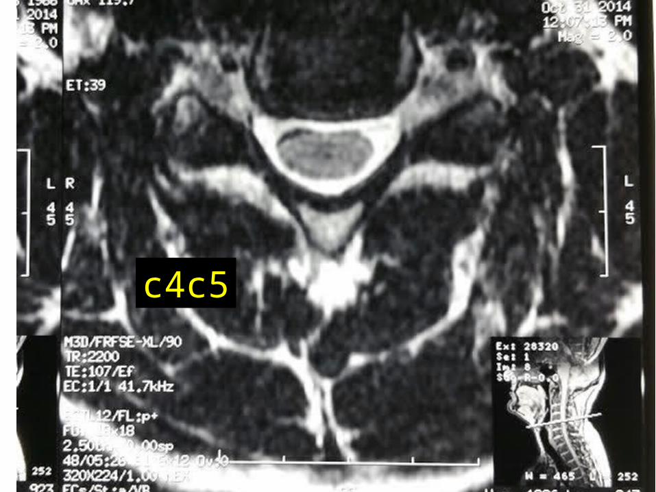
c4c5

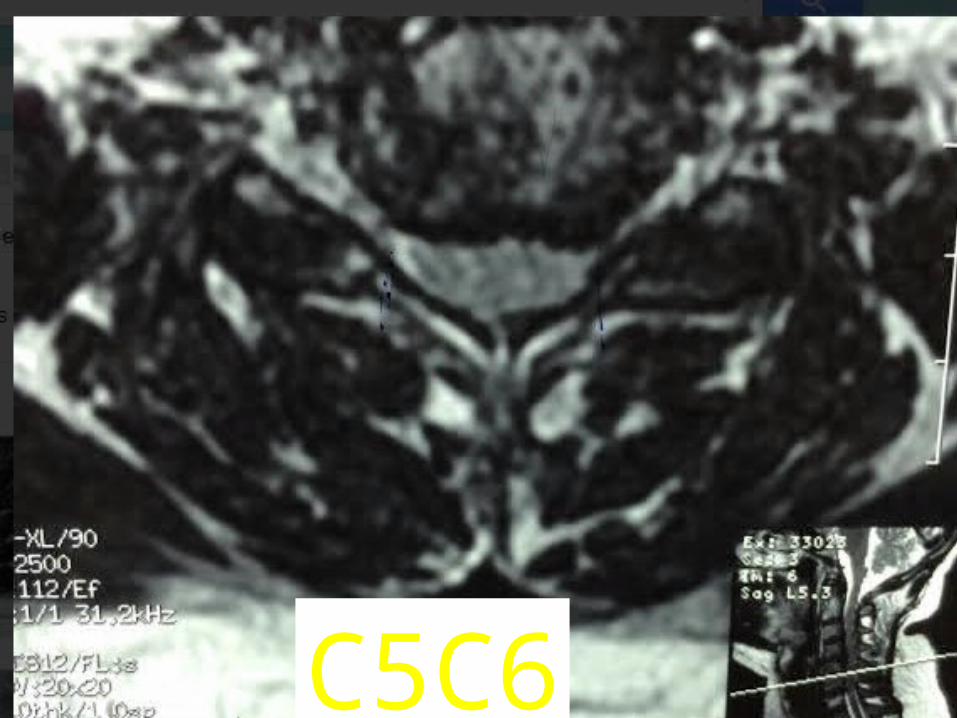
c5c6
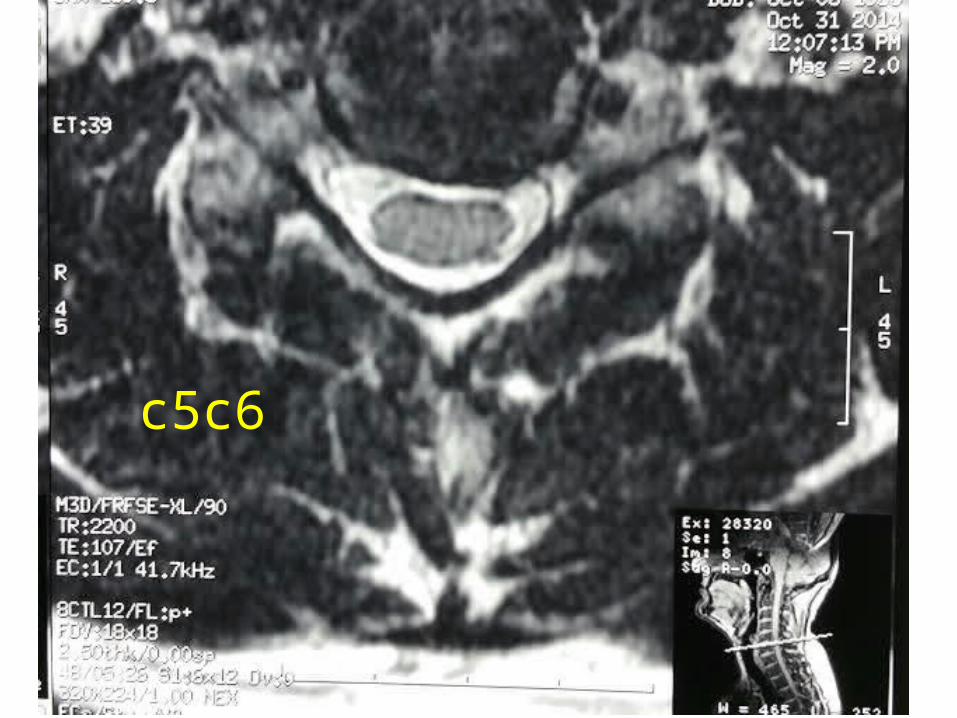
c5c6
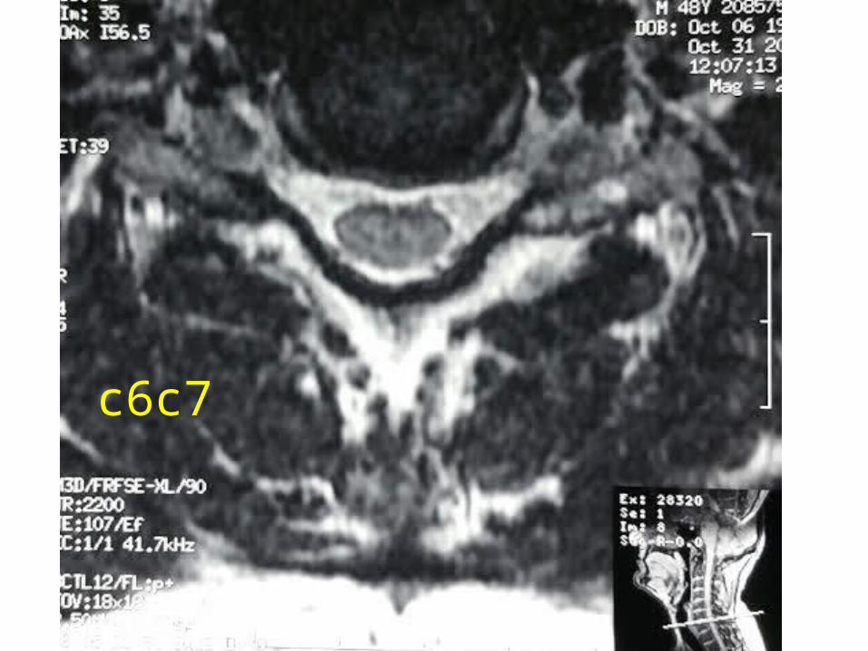
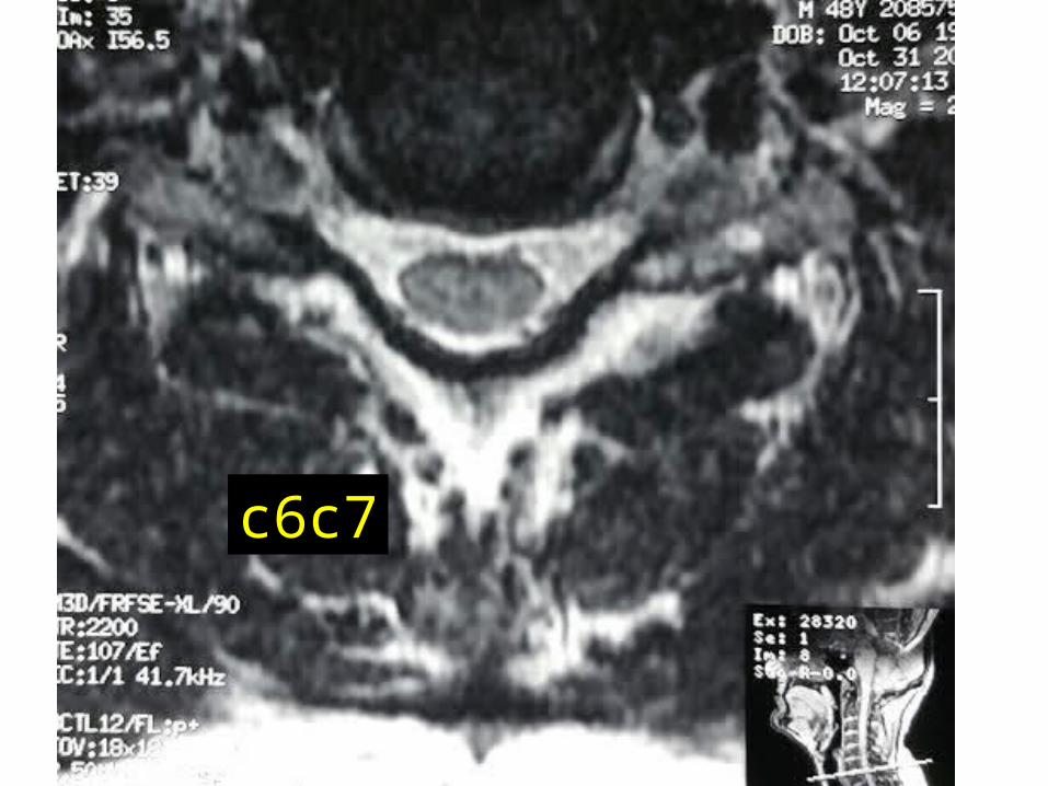
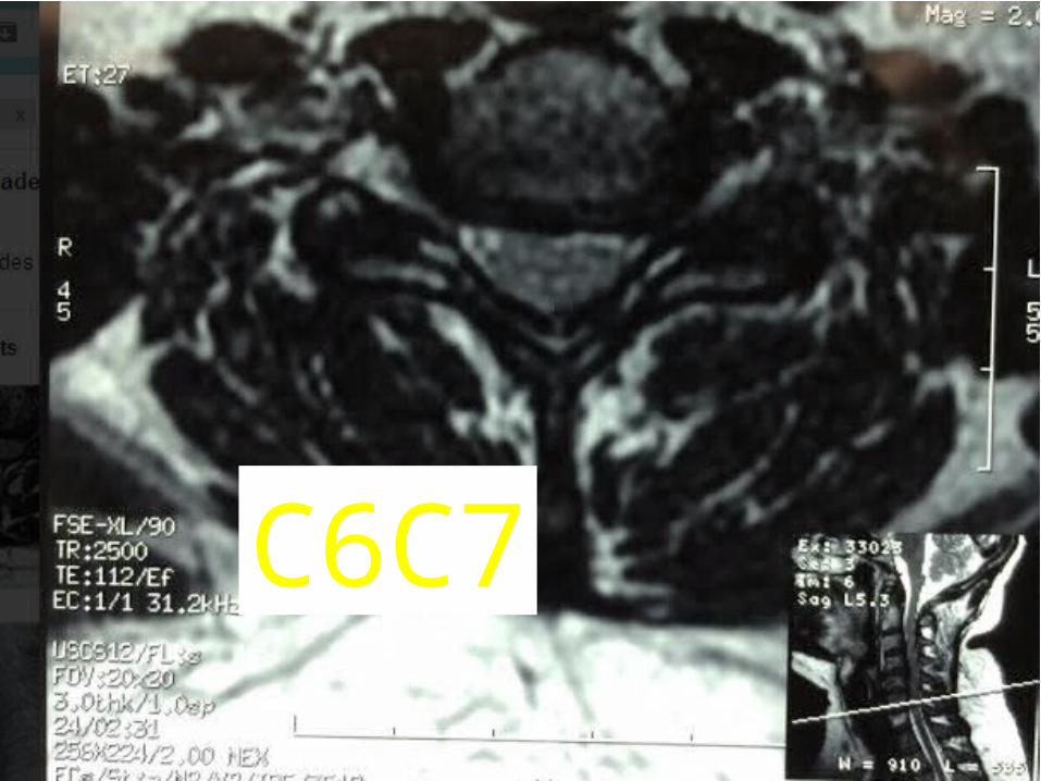
c6c7
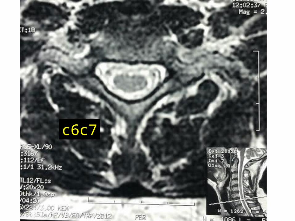
c6c7
c6c7

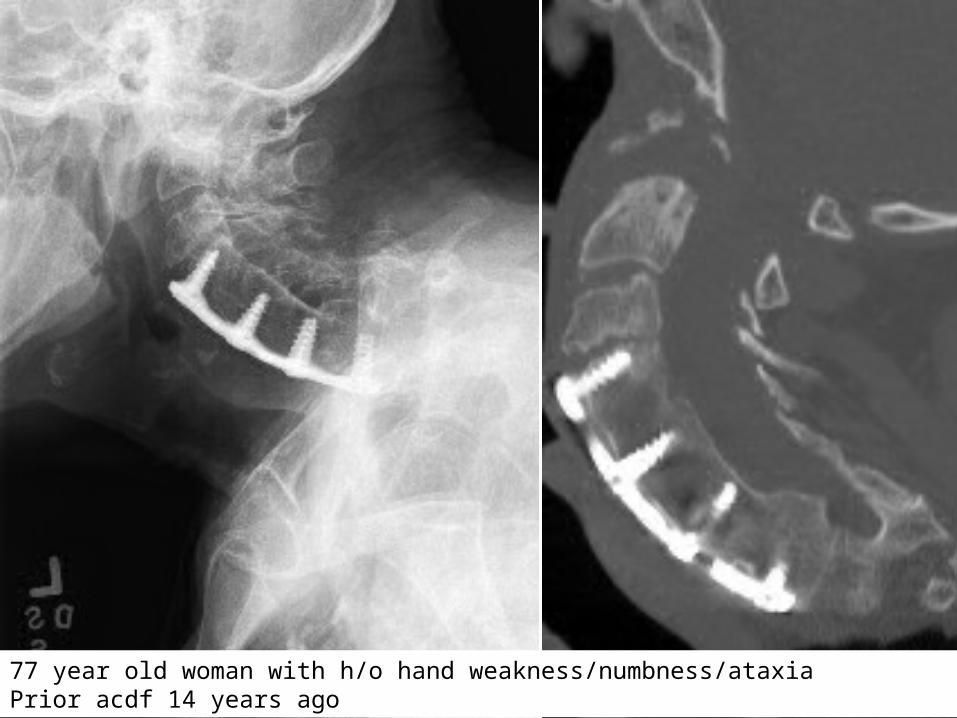
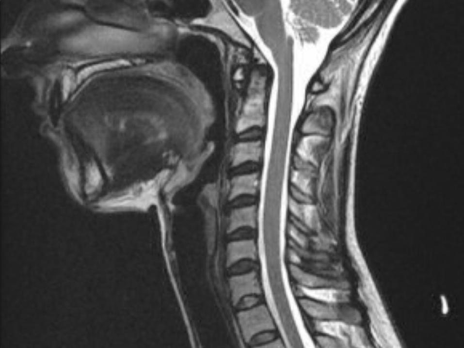
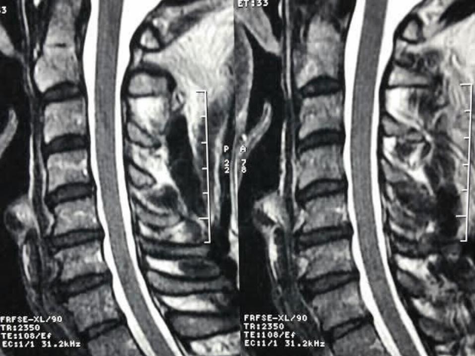
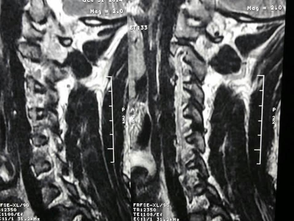
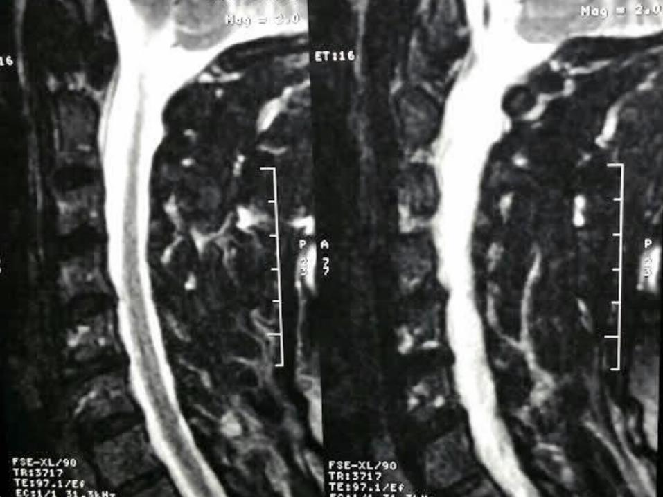
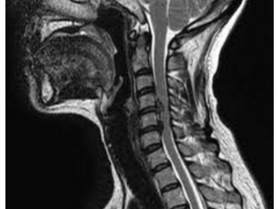
77 year old woman with h/o hand weakness/numbness/ataxiaPrior acdf 14 years ago
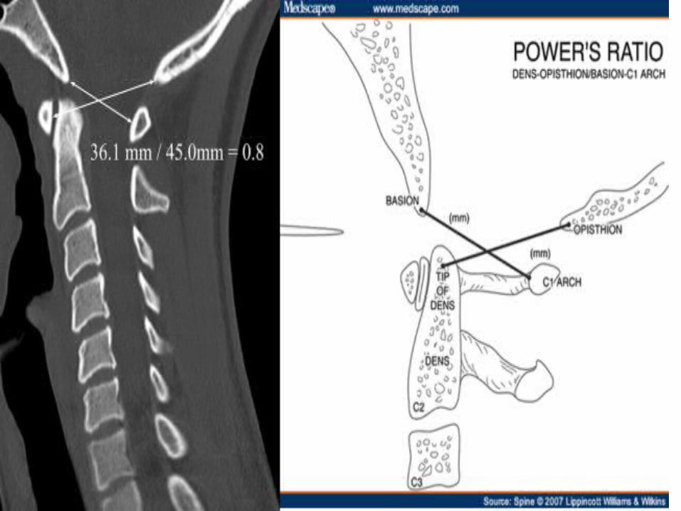
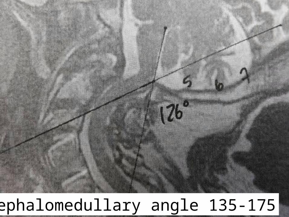
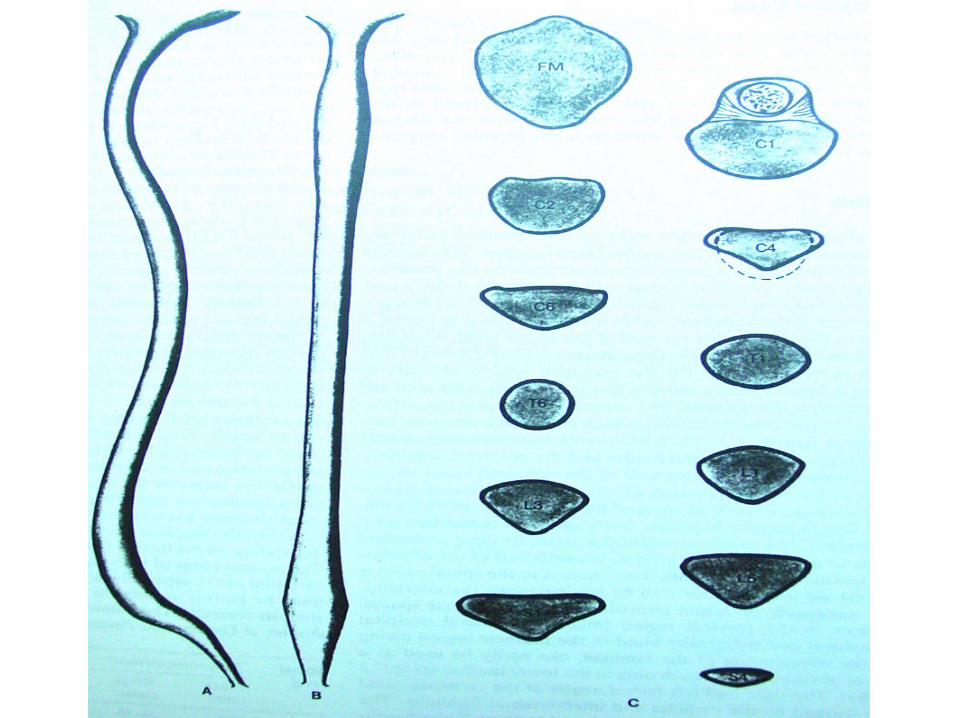
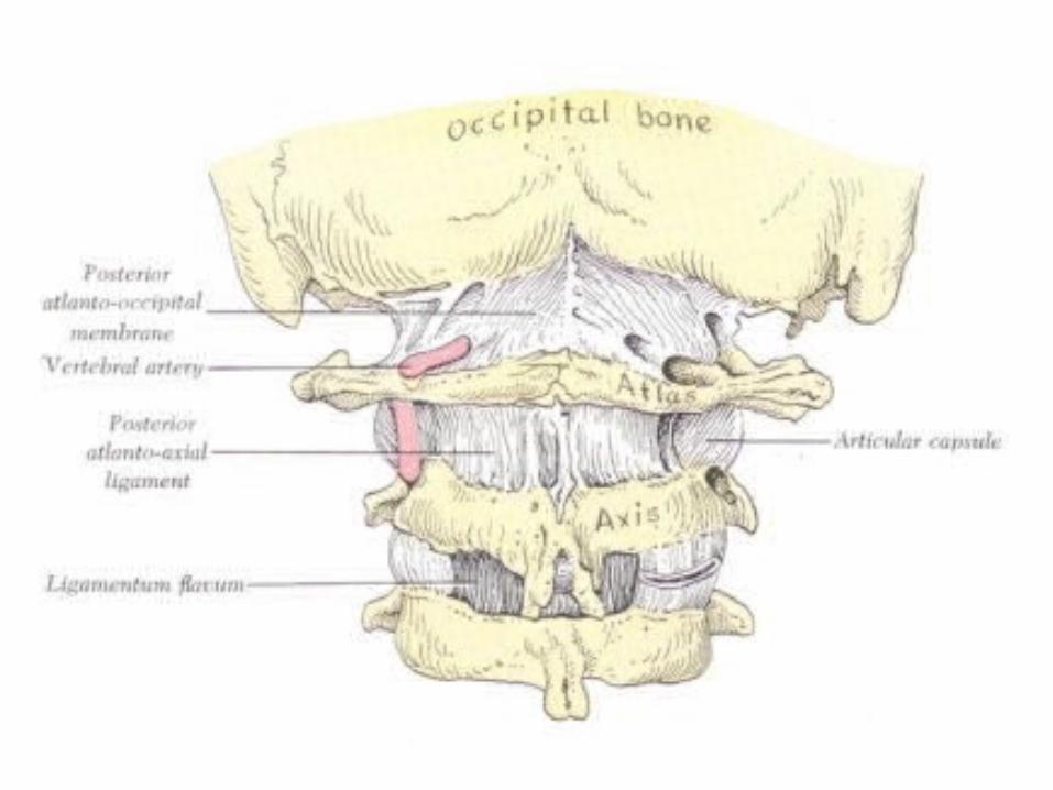
Cephalomedullary angle 135-175

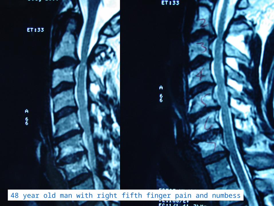
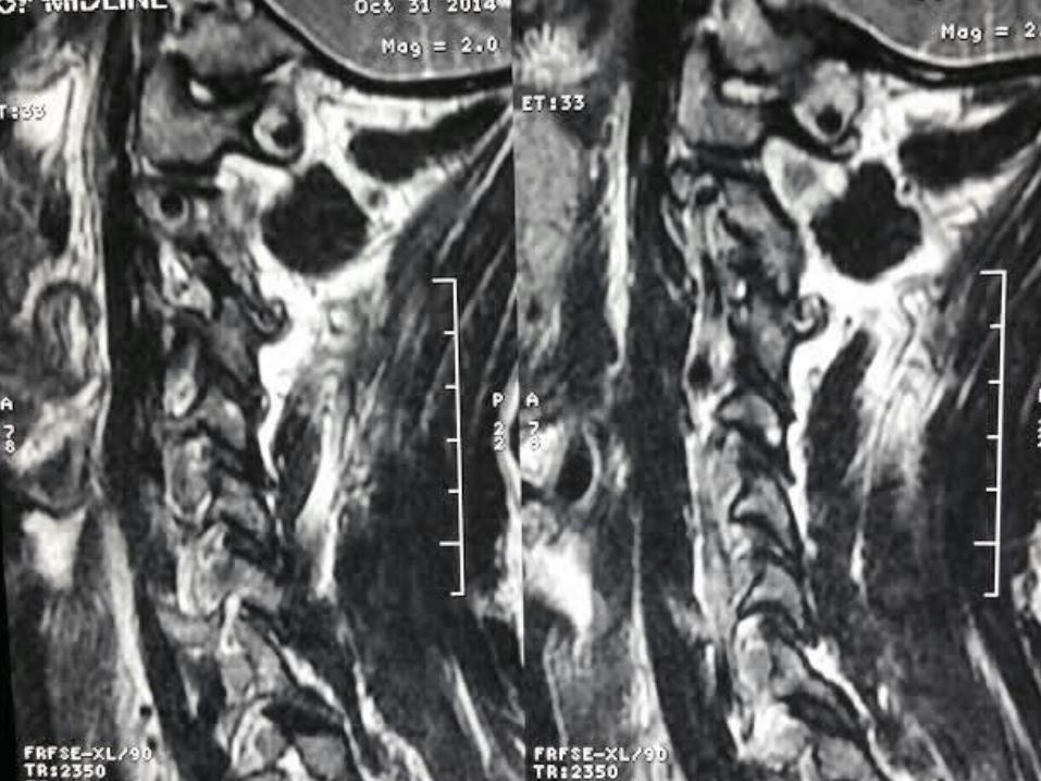
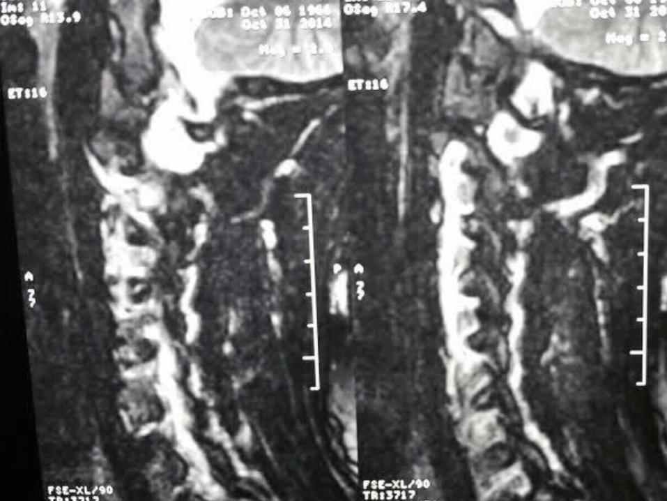
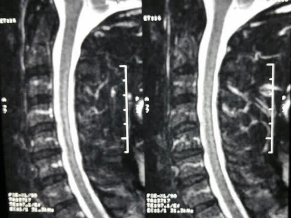
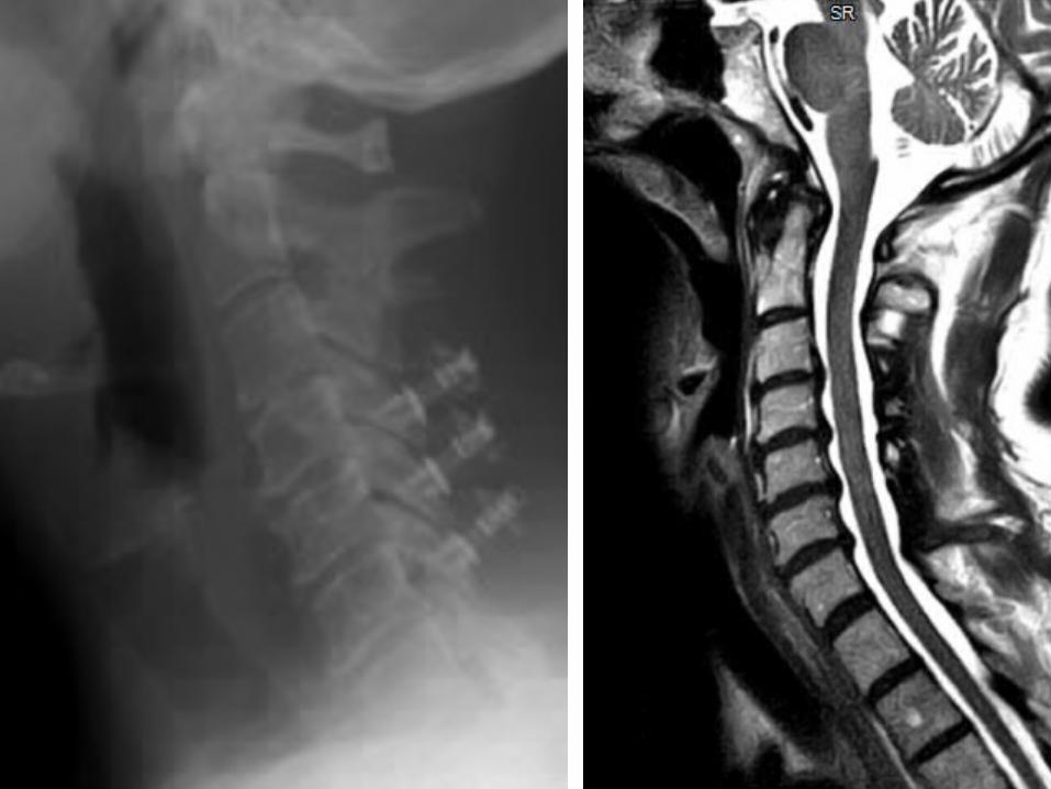
48 year old man with right fifth finger pain and numbess
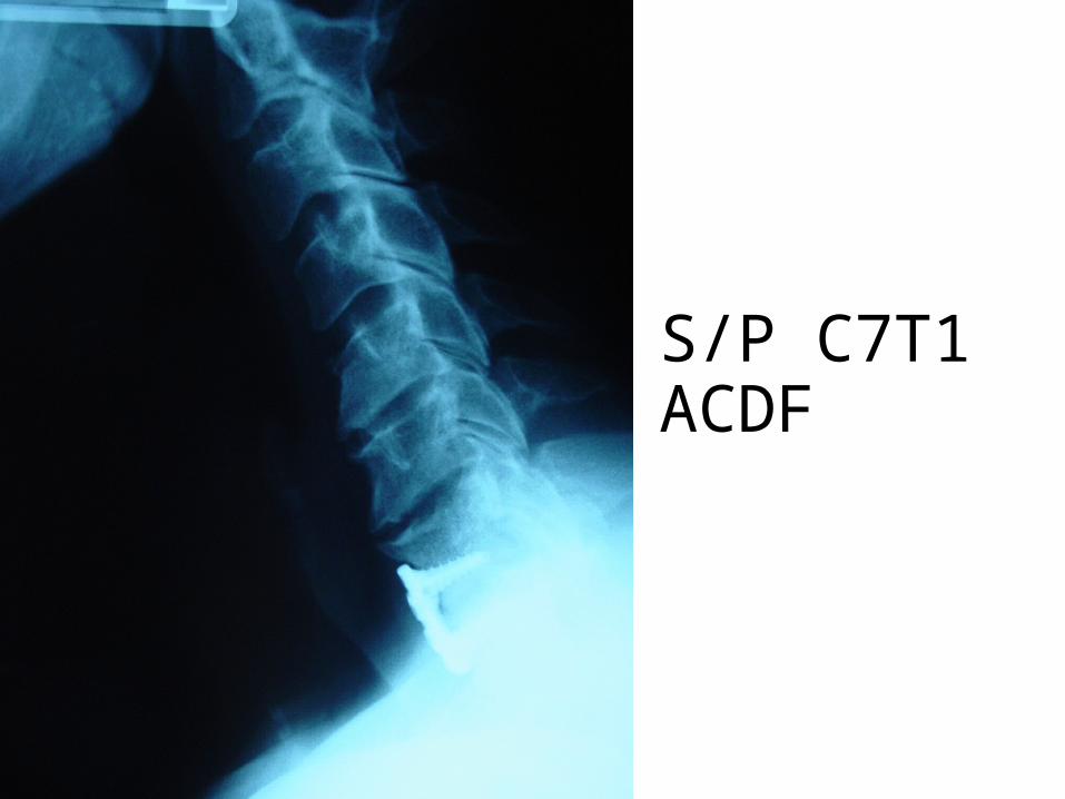
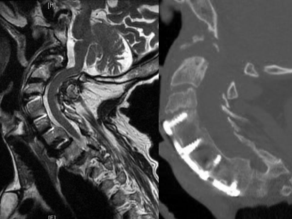
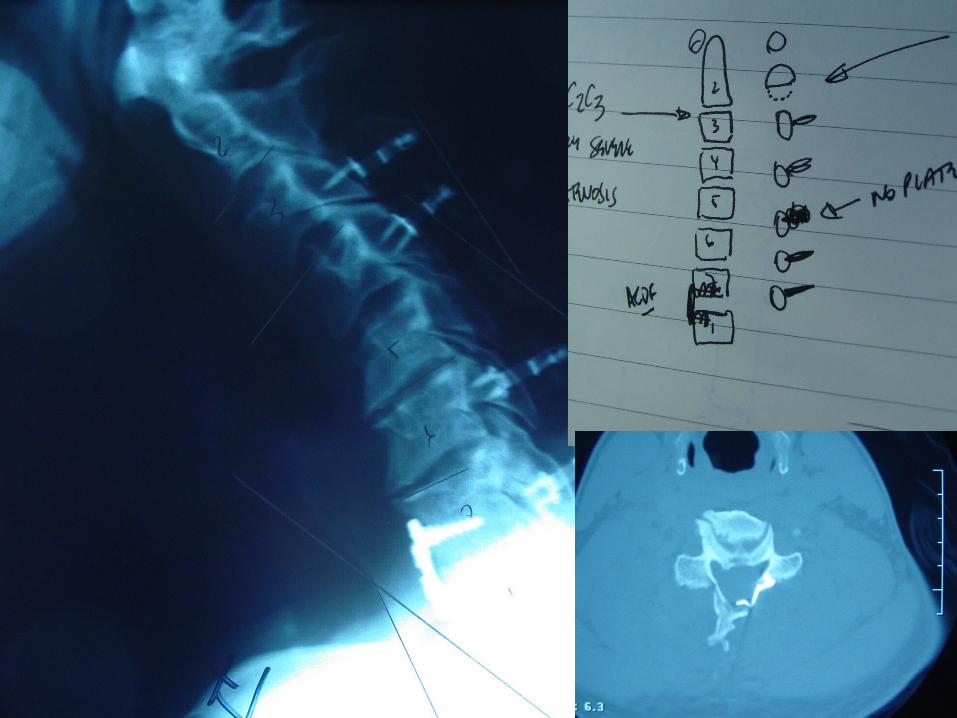
S/P C7T1ACDF
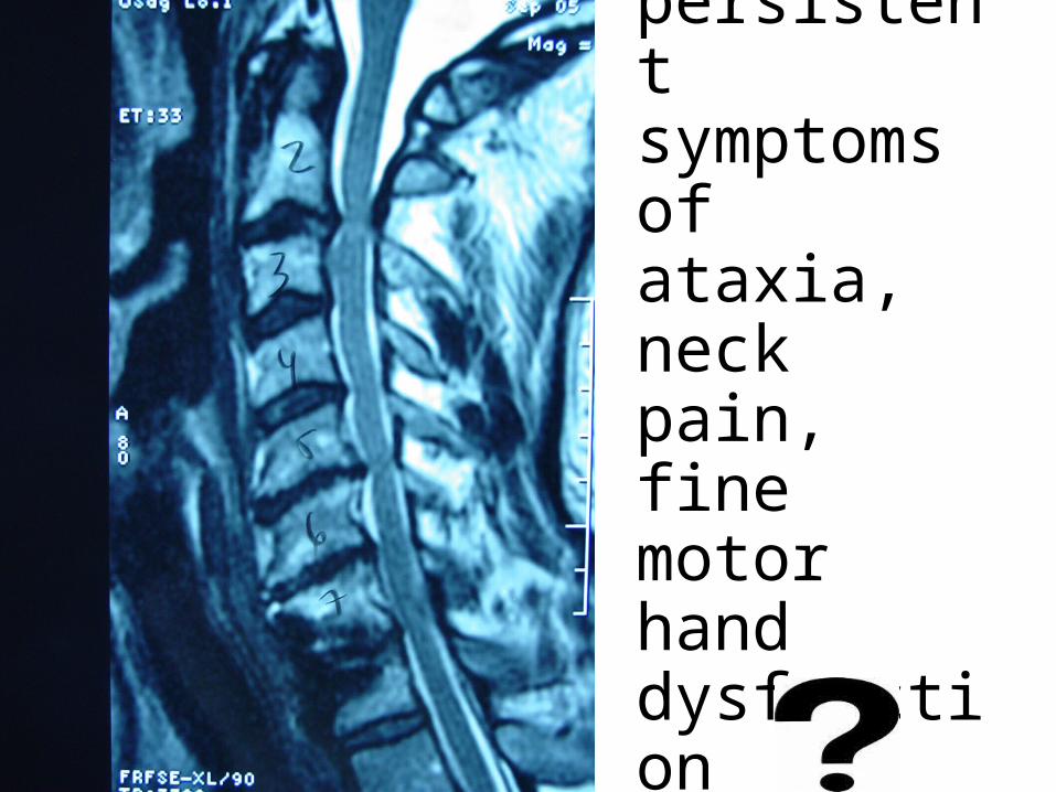
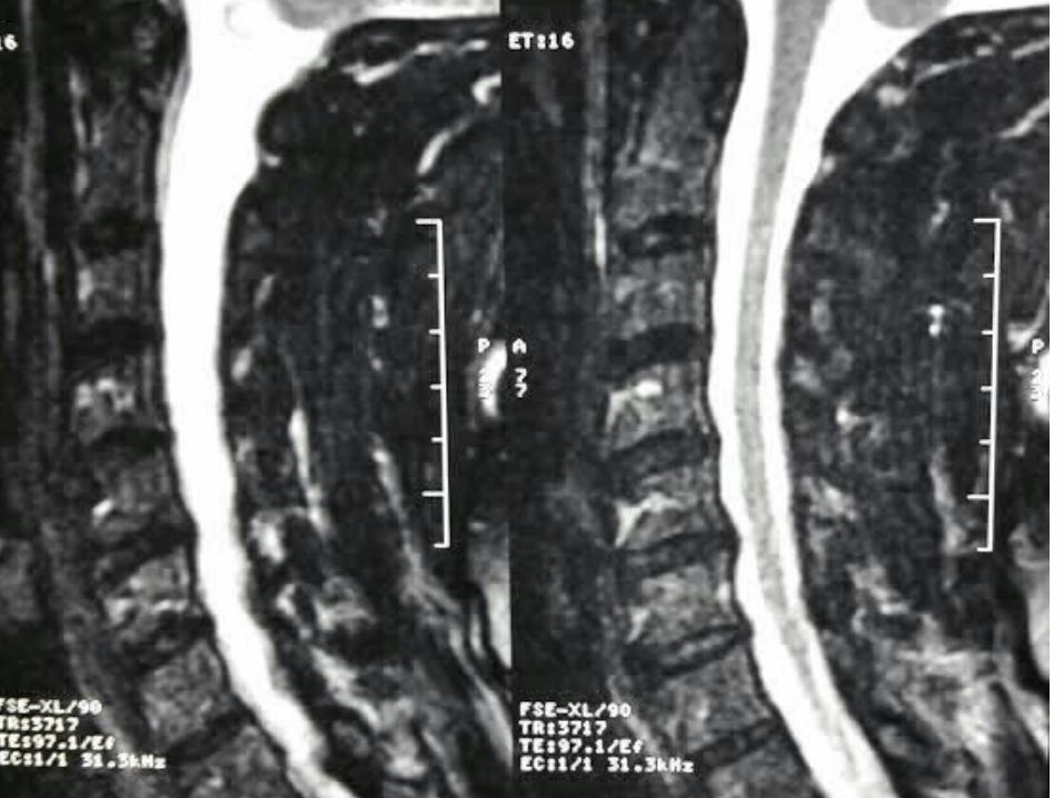
persistent symptoms of ataxia, neck pain, fine motor hand dysfunction
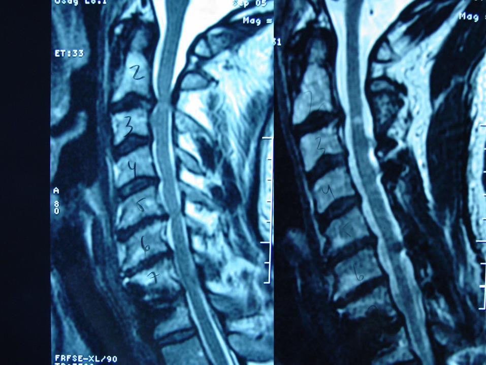
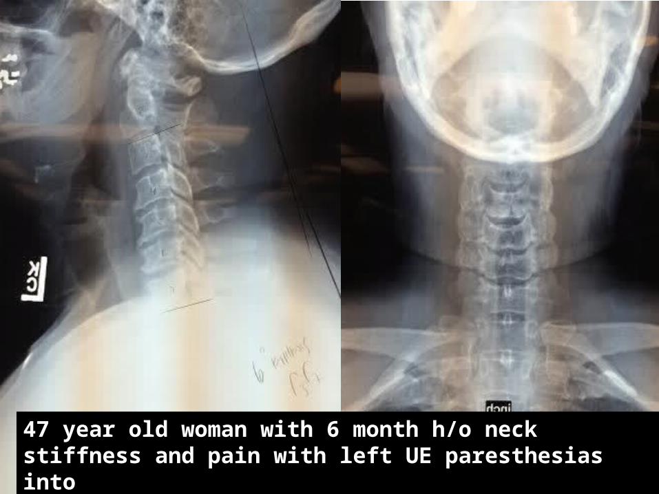
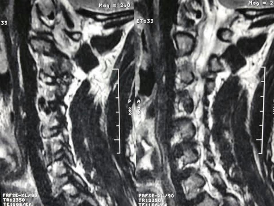
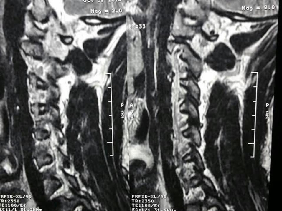
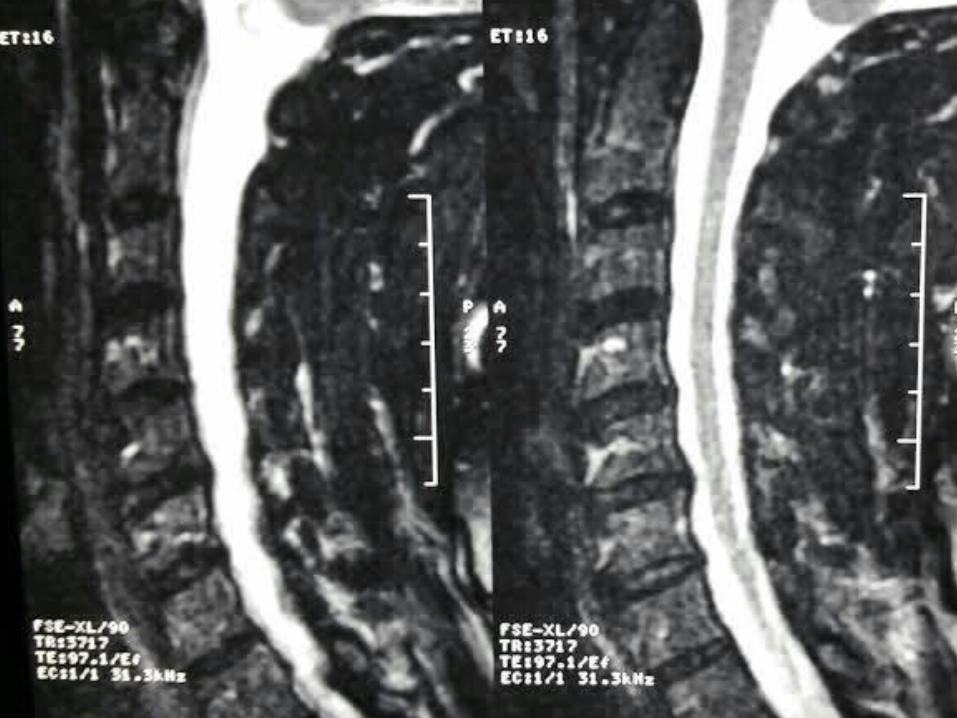
47 year old woman with 6 month h/o neck stiffness and pain with left UE paresthesias intoThe elbow forearm and hand especially IF/thumb
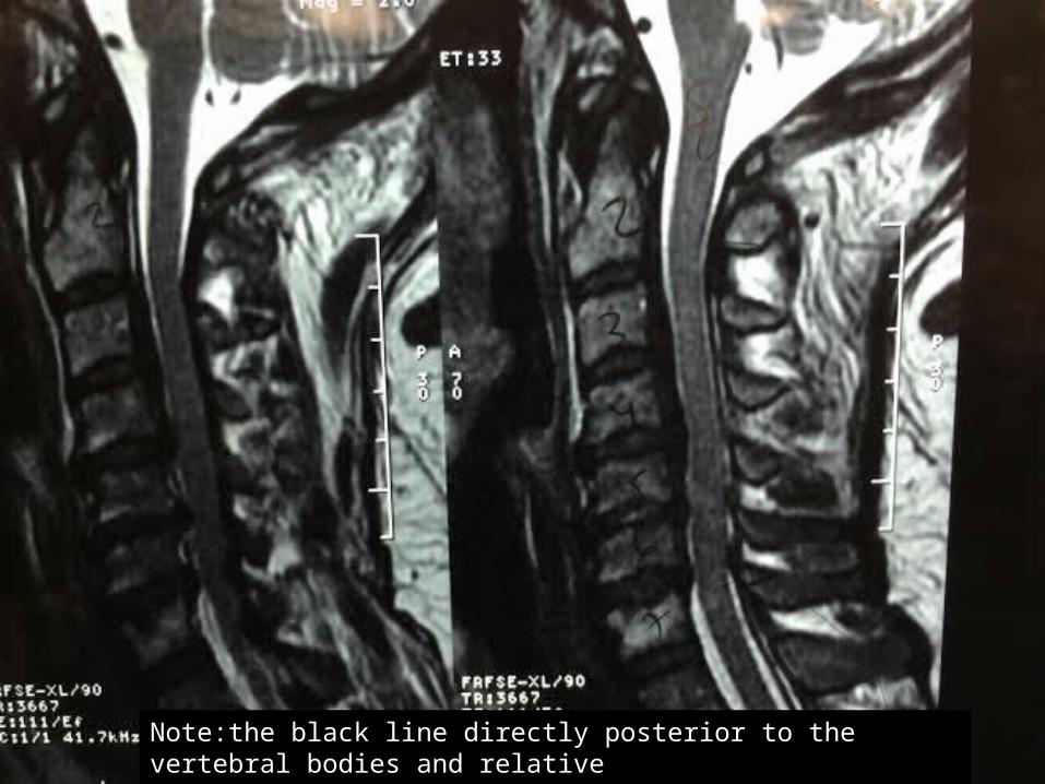
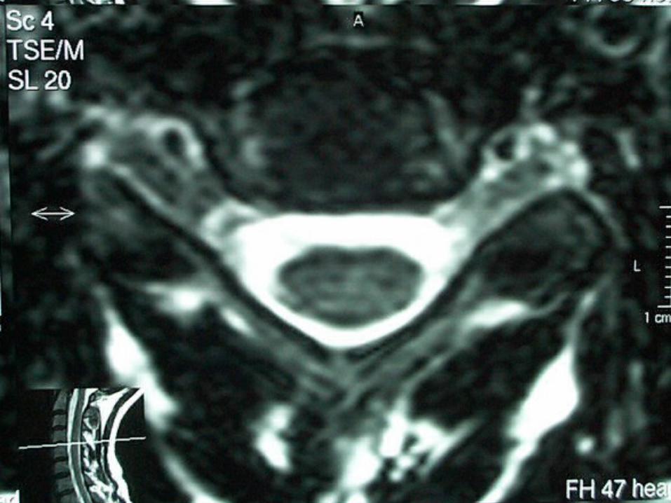
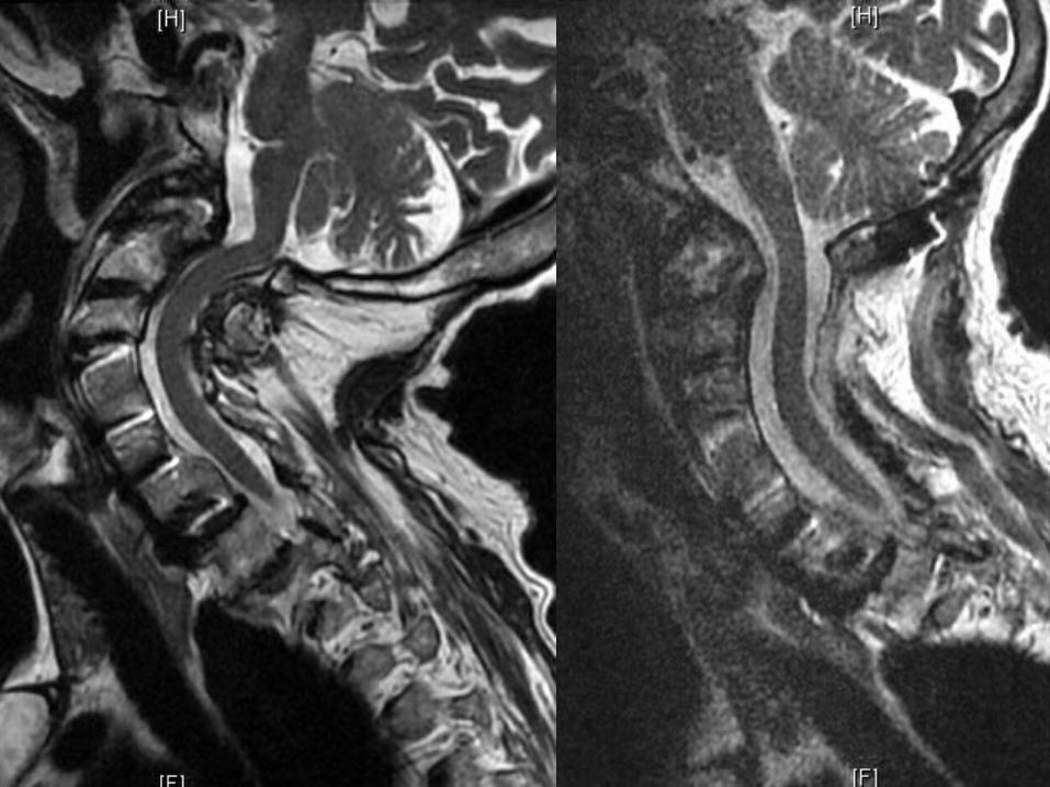
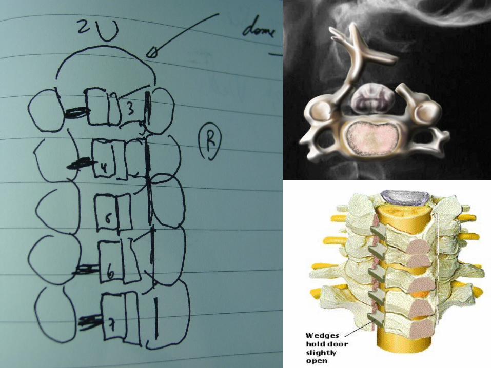
Note:the black line directly posterior to the vertebral bodies and relativeThinning of spinal cord, note normal disc height
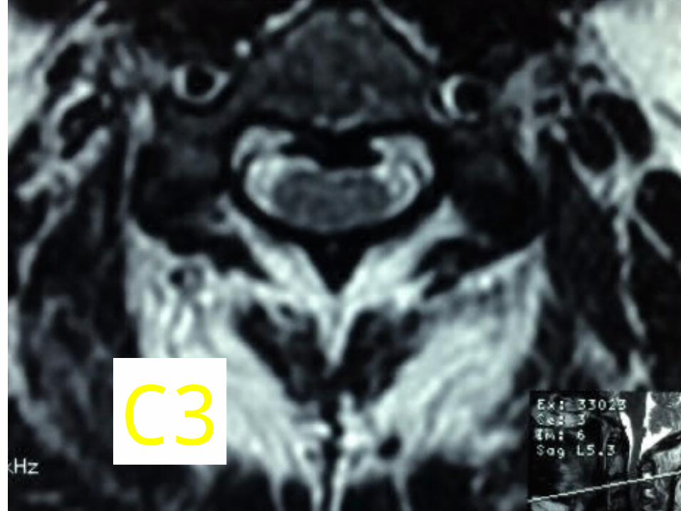
C3
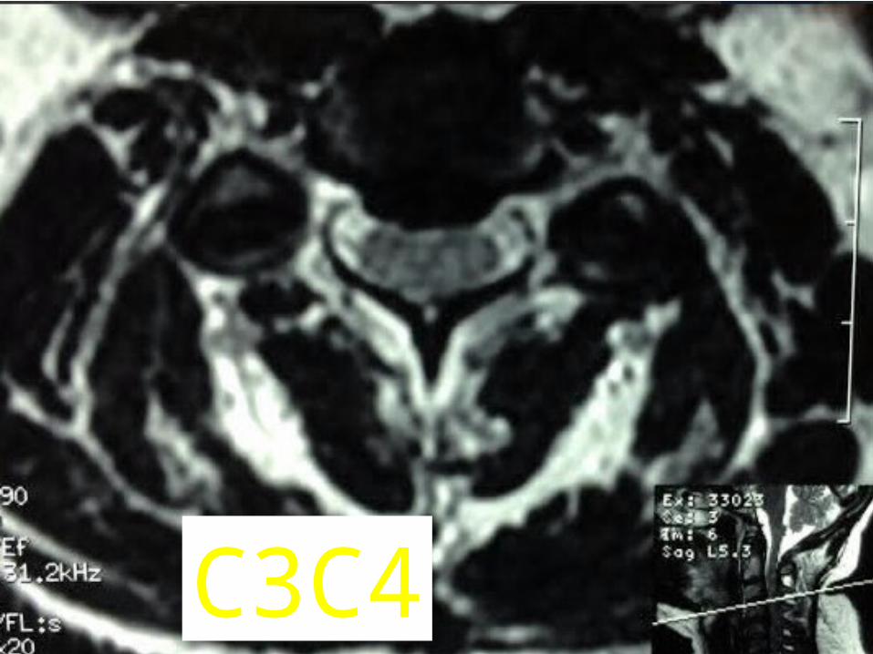
C3C4

C4C5
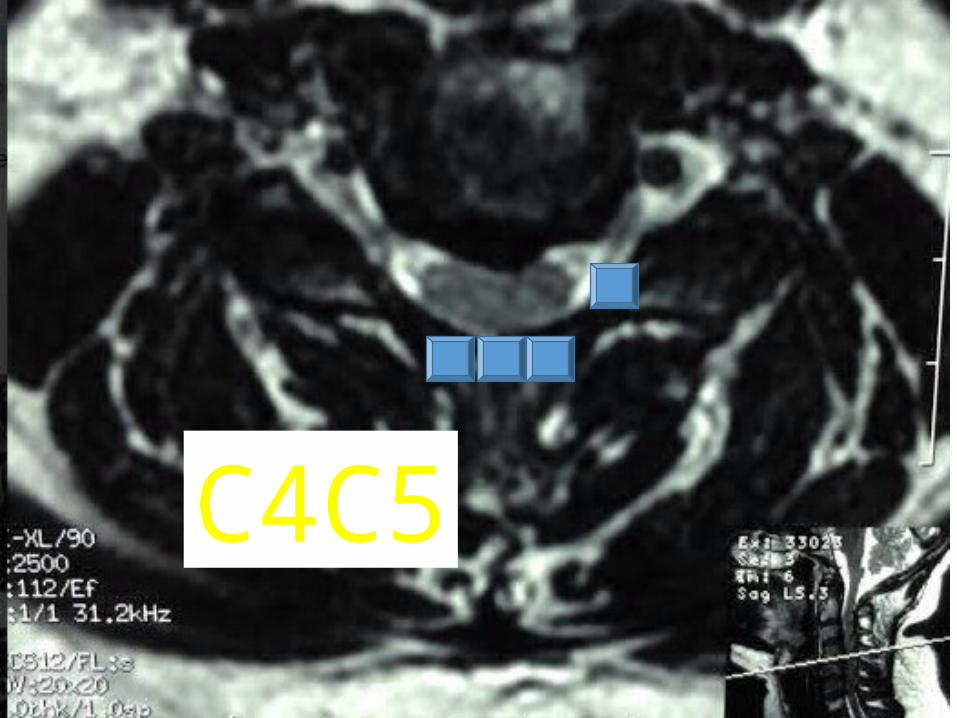
C4C5
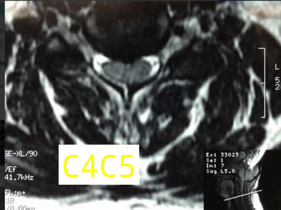
C4C5
C5C6
C6C7
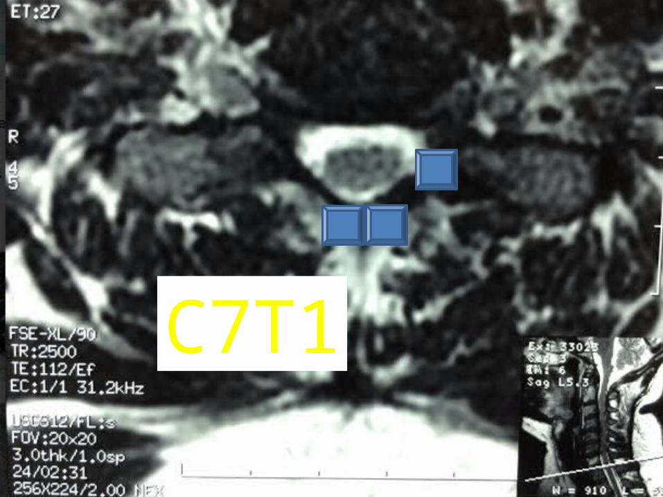
C7T1
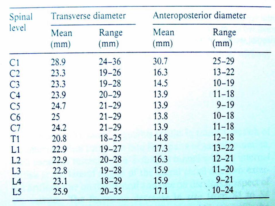
Mid-sagittal diameter: width1:5 ratio leads to necrosis of gray matter• Ogino H: Canal diameter, anteroposterior compression ratio
and spondylotic myelopathy of the cervical spine. Spine 1983; 8:1-15• Cord compression causes ischemia and direct mechanical
trauma


Thanks


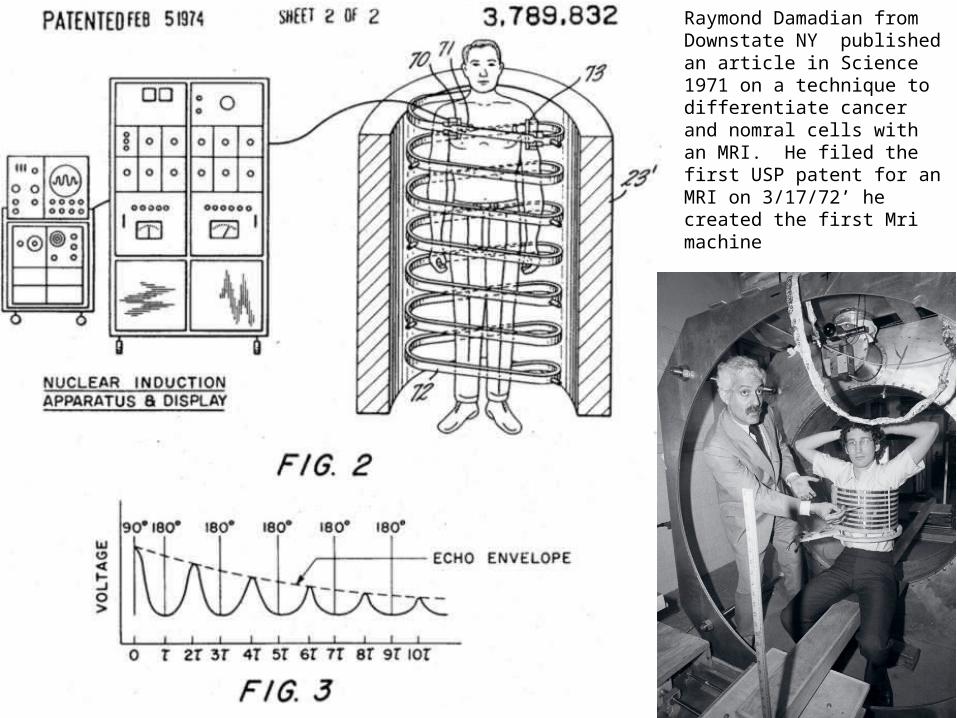
• Raymond Damadian from Downstate NY published an article in Science 1971 on a technique to differentiate cancer and nomral cells with an MRI. He filed the first USP patent for an MRI on 3/17/72’ he created the first Mri machine

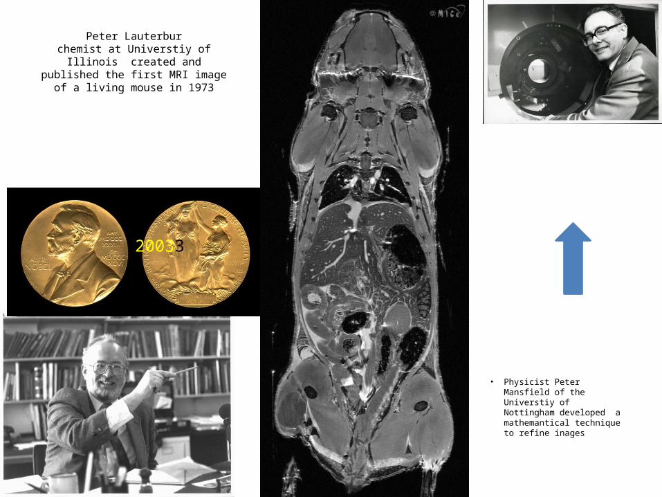
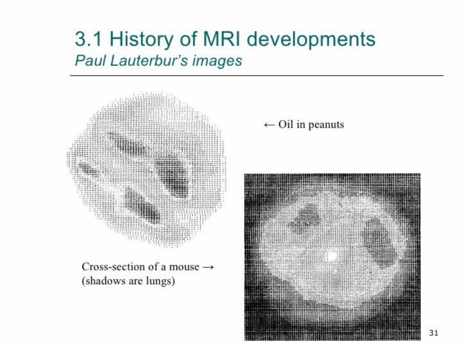
Peter Lauterburchemist at Universtiy of Illinois
created and published the first MRI image of a living mouse in 1973
• Physicist Peter Mansfield of the Universtiy of Nottingham developed a mathemantical technique to refine inages
20033
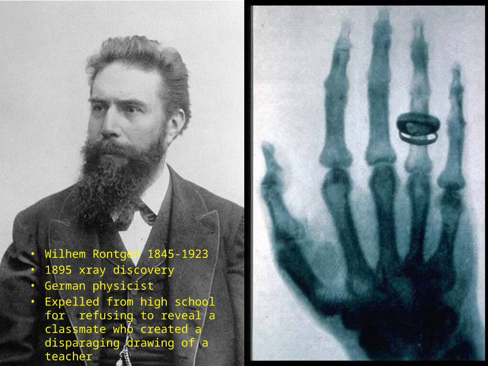
• Wilhem Rontgen 1845-1923• 1895 xray discovery• German physicist• Expelled from high school for
refusing to reveal a classmate who created a disparaging drawing of a teacher