Embed Size (px)
Citation preview
Indolent NK/T Cell Lymphoproliferative
Disorders of GI Tract
Chung-Che (Jeff) Chang, M.D., Ph.D.
Medical Director, Hematology and Molecular Pathology
Florida Hospital
Professor of Pathology, College of Medicine
University of Central Florida
407-303-1879
Important!
Many of these cases had been diagnosed as
aggressive T/NK cell lymphomas or as
celiac disease or inflammatory bowel
disease and then needlessly, and usually
ineffectively, treated.

30 year old asymptomatic white
male triathlete and marathon
runner underwent endoscopy ex.
because of a positive family
history of colon cancer and
colonic polyps.
Case report
Am J Surg Pathol 2006;30:539
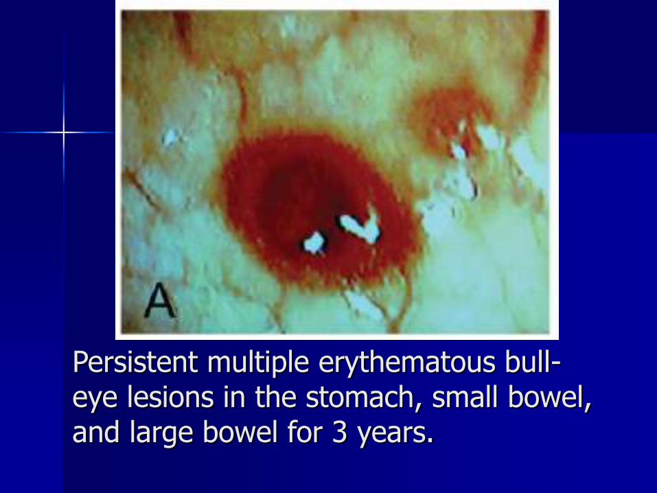
Persistent multiple erythematous bull-eye lesions in the stomach, small bowel, and large bowel for 3 years.
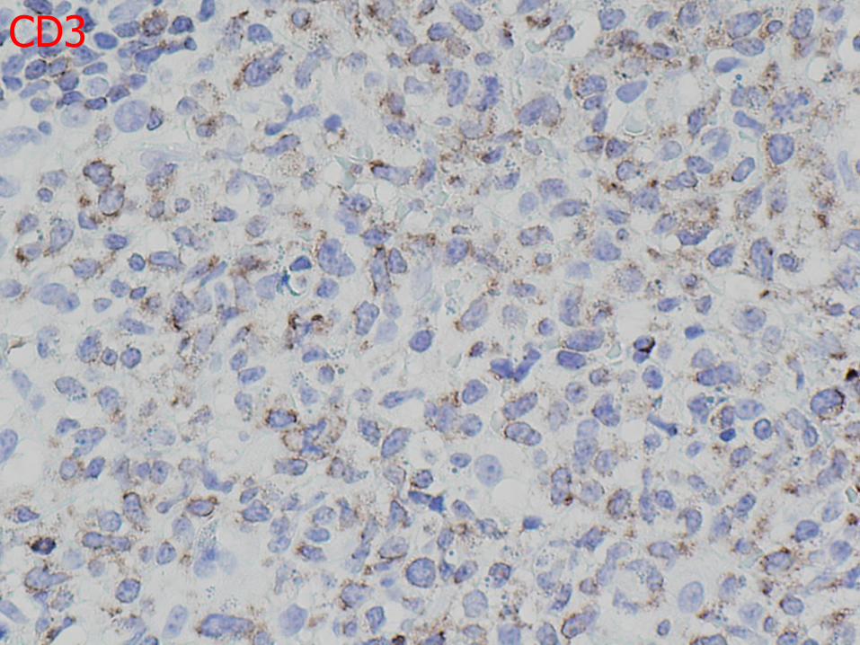
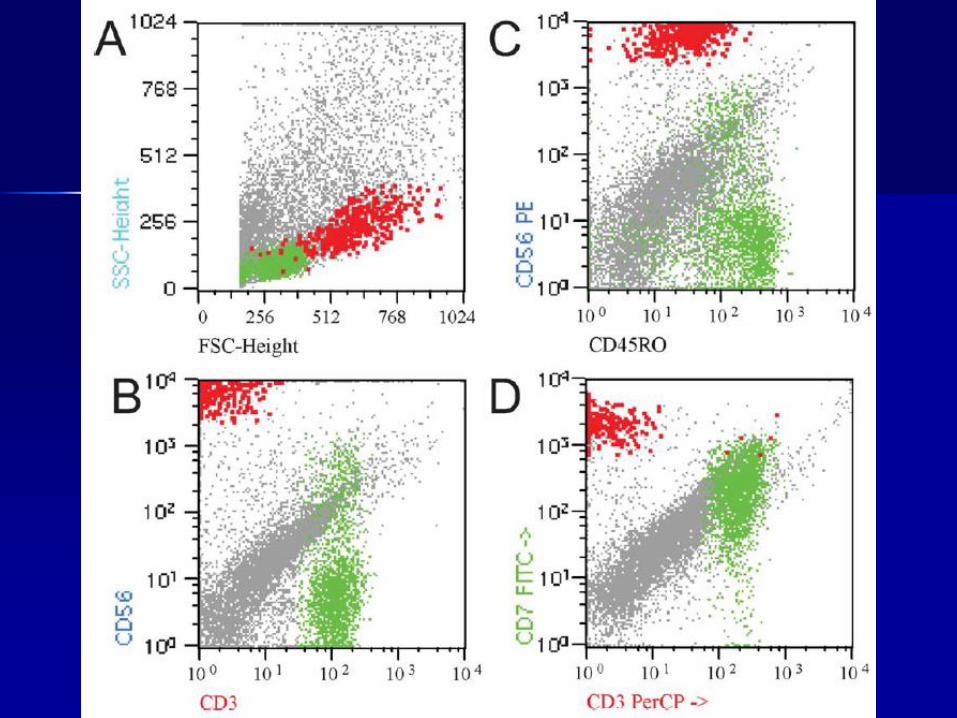
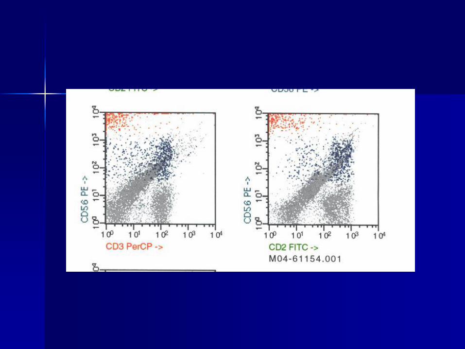
CD3
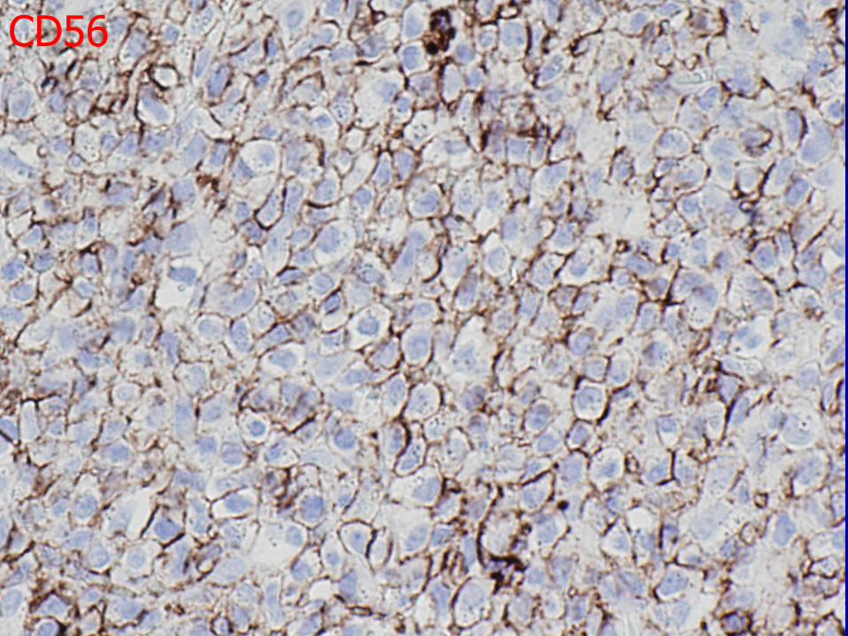
CD56
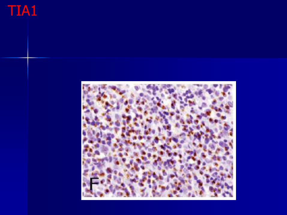
TIA1
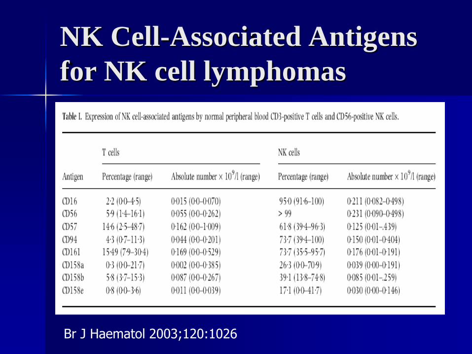
Br J Haematol 2003;120:1026
NK Cell-Associated Antigens
for NK cell lymphomas
NK cells with heterogeneous expression of
the immunoglobulin-like receptors CD158
isoforms suggesting a non-clonal process.
PCR for rearrangement of the TCR-
gamma gene showed no evidence of a
clonal T-cell population.
In situ hybridization for Epstein-Barr virus
encoded RNA (EBER) was negative.
There was no evidence of the involvement
beyond GI tract.
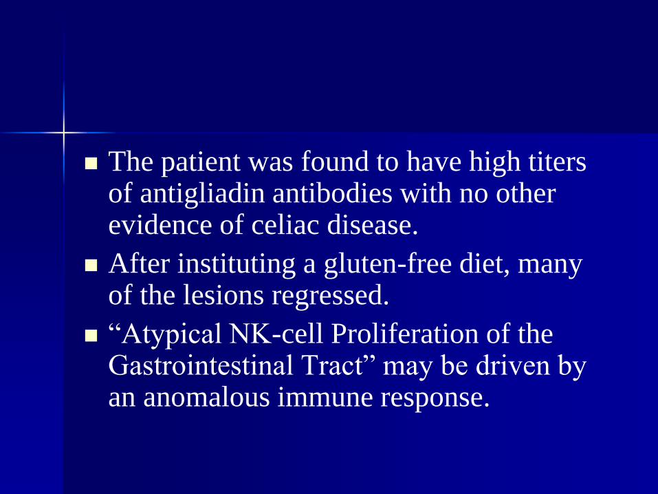
The patient was found to have high titers of antigliadin antibodies with no other evidence of celiac disease.
After instituting a gluten-free diet, many of the lesions regressed.
“Atypical NK-cell Proliferation of the Gastrointestinal Tract” may be driven by an anomalous immune response.
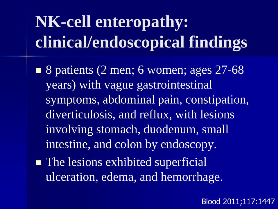
NK-cell enteropathy:
clinical/endoscopical findings
8 patients (2 men; 6 women; ages 27-68
years) with vague gastrointestinal
symptoms, abdominal pain, constipation,
diverticulosis, and reflux, with lesions
involving stomach, duodenum, small
intestine, and colon by endoscopy.
The lesions exhibited superficial
ulceration, edema, and hemorrhage.
Blood 2011;117:1447
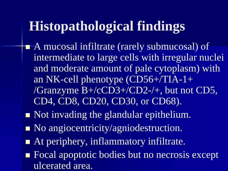
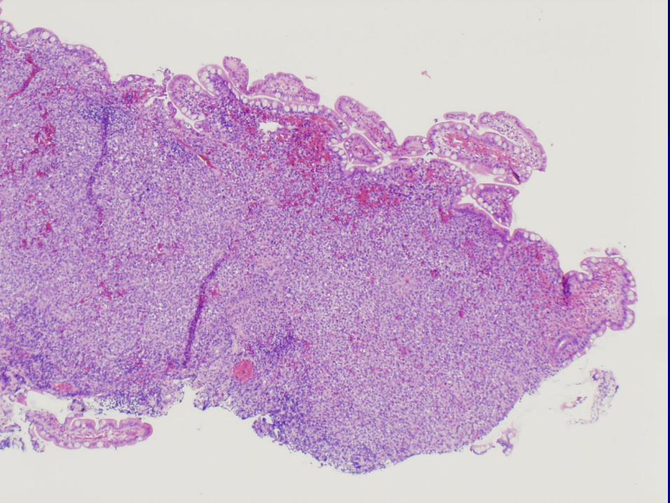
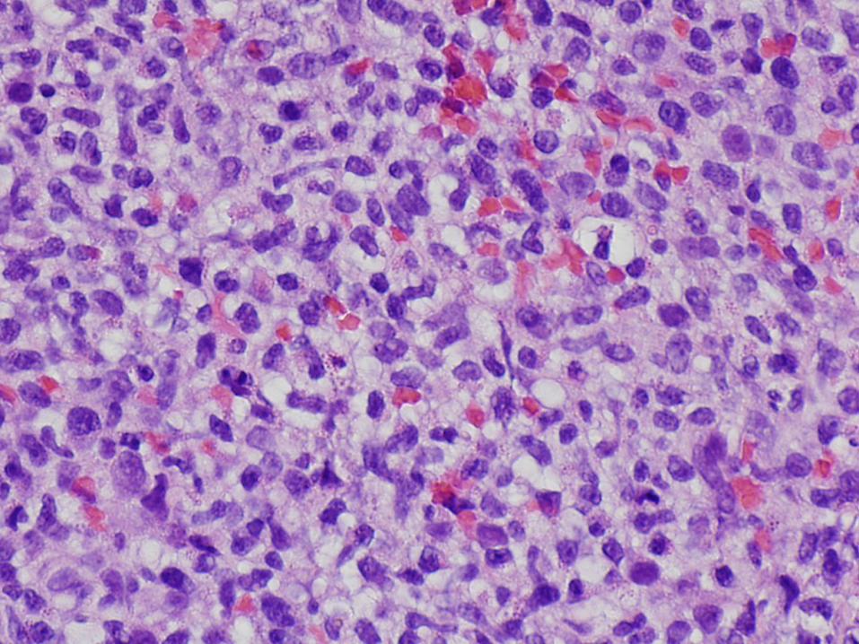
Histopathological findings
A mucosal infiltrate (rarely submucosal) of intermediate to large cells with irregular nuclei and moderate amount of pale cytoplasm) with an NK-cell phenotype (CD56+/TIA-1+ /Granzyme B+/cCD3+/CD2-/+, but not CD5, CD4, CD8, CD20, CD30, or CD68).
Not invading the glandular epithelium.
No angiocentricity/agniodestruction.
At periphery, inflammatory infiltrate.
Focal apoptotic bodies but no necrosis except ulcerated area.
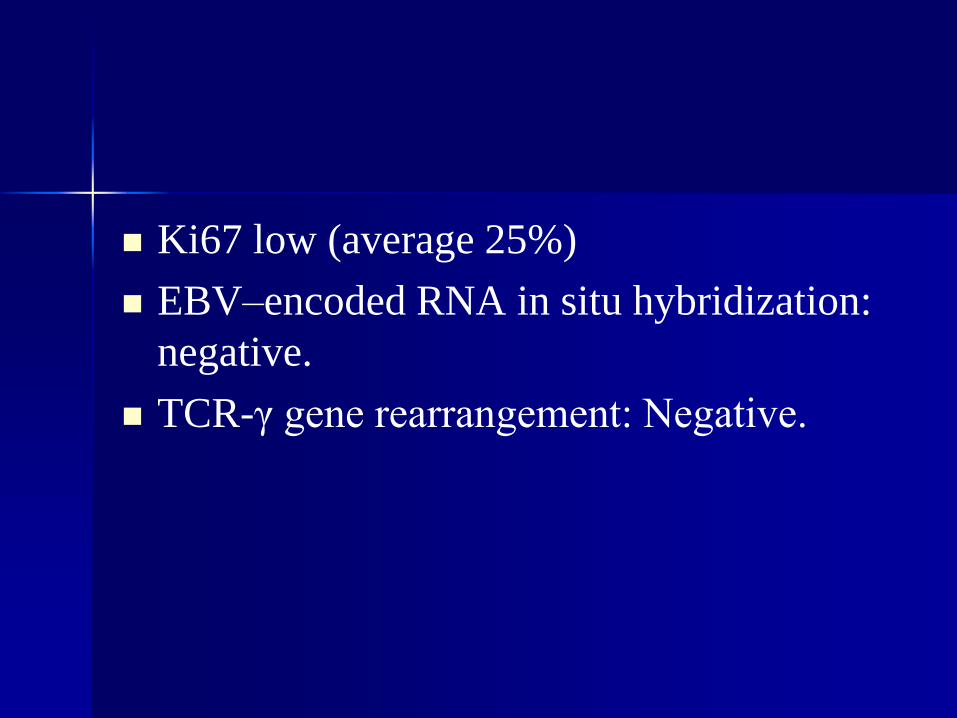
Ki67 low (average 25%)
EBV–encoded RNA in situ hybridization:
negative.
TCR-γ gene rearrangement: Negative.
Clinical outcomes
Three patients received aggressive chemotherapy followed by autologous bone marrow transplantation in 2.
Five patients were followed without treatment with no patient developed progressive disease or died of lymphoma (median follow-up, 30 months).
Repeat endoscopies in 6 of 8 patients showed persistence or recurrence of superficial gastrointestinal lesions.
“NK-cell enteropathy” of as yet unknown etiology.
Relatively high expression of Granzyme B and/or TIA-1 suggesting that the NK cells in these lesions were primed for the cytotoxic function, most probably responding to local inflammation, autoimmunity, or viruses.
Lymphomatoid gastropathy: a distinct
clinicopathologic entity of self-limited
pseudomalignant NK-cell proliferation
10 cases of unrecognized self-limited natural
killer–cell proliferation in the stomach
5 men and 5 women
Age; 46-75 years
No gastric symptoms.
Gastroscopy for screening for gastric cancer.
Gastroscopy showed ulcers or flat elevations
with a shallow depression (diameter, ∼ 1 cm).(Blood, 116:5631, 2010)
(Blood, 116:5631, 2010)
Lymphomatoid gastropathy
• Diffuse “atypical” infiltrate of medium to
large lymphoid cells, some with prominent
nucleoli, in the lamina propria.
• Necrosis present in some, not angiocentric or
angiodestructive, no apoptotic bodies.
• Occasionally extends into glands
• All cases with variable % (20 to 90%) with
eos. granules
• 9/10 H. pylori.
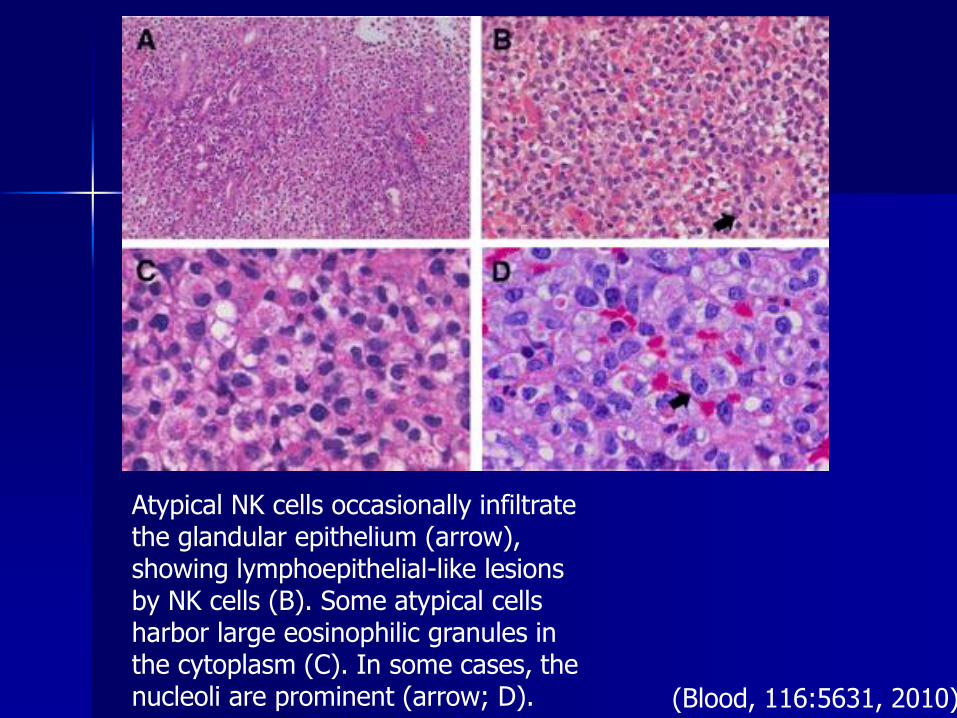
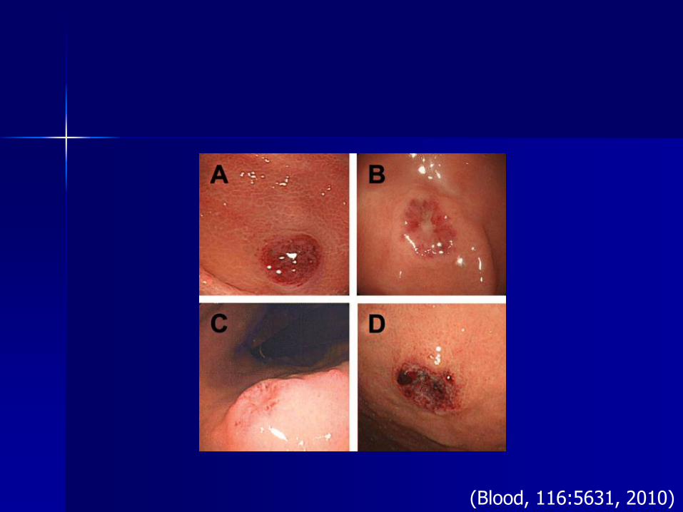
Atypical NK cells occasionally infiltrate the glandular epithelium (arrow), showing lymphoepithelial-like lesions by NK cells (B). Some atypical cells harbor large eosinophilic granules in the cytoplasm (C). In some cases, the nucleoli are prominent (arrow; D). (Blood, 116:5631, 2010)
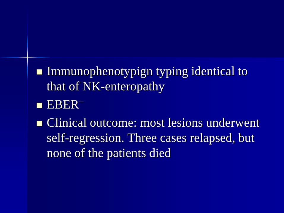
Immunophenotypign typing identical to
that of NK-enteropathy
EBER−
Clinical outcome: most lesions underwent
self-regression. Three cases relapsed, but
none of the patients died
Differential diagnosis
Extranodal natural killer (NK)-/T-cell lymphomas: angiocentric and angiodestructive growth pattern with frequent necrosis and apoptosis; EBER+;
CD56+ T-cell neoplasm with extensive loss of T-cell markers may be considered: enteropathy-associated T-cell lymphoma (EATL type II): Clonal TCR gene rearrangement
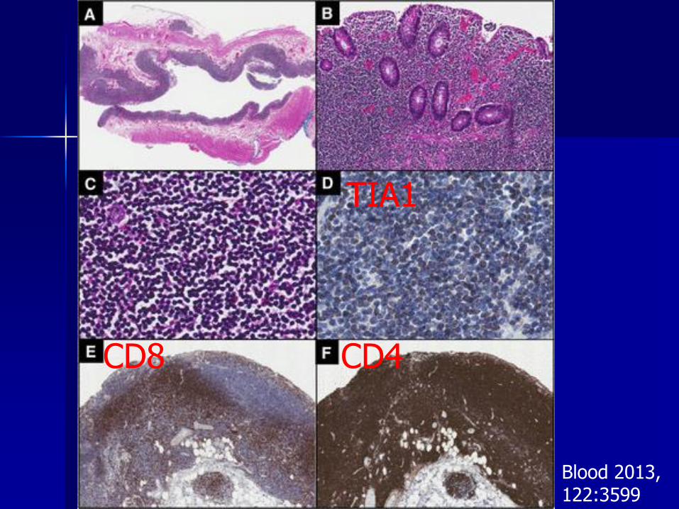
Indolent T-cell LPD
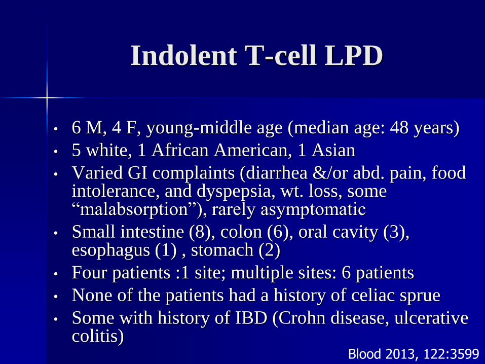
• 6 M, 4 F, young-middle age (median age: 48 years)
• 5 white, 1 African American, 1 Asian
• Varied GI complaints (diarrhea &/or abd. pain, food intolerance, and dyspepsia, wt. loss, some “malabsorption”), rarely asymptomatic
• Small intestine (8), colon (6), oral cavity (3), esophagus (1) , stomach (2)
• Four patients :1 site; multiple sites: 6 patients
• None of the patients had a history of celiac sprue
• Some with history of IBD (Crohn disease, ulcerative colitis)
Blood 2013, 122:3599
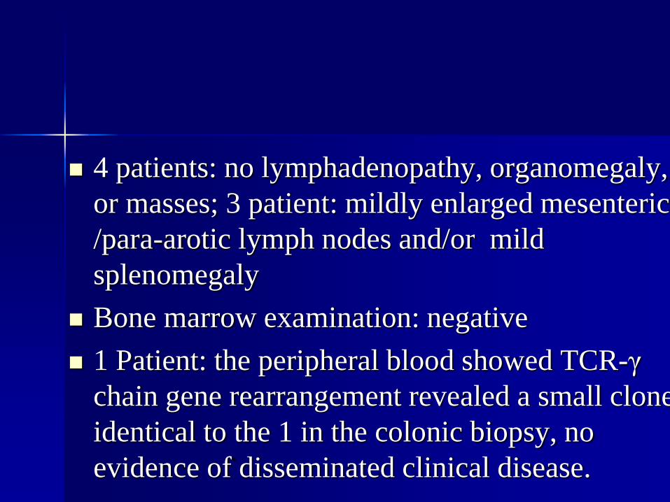
4 patients: no lymphadenopathy, organomegaly,
or masses; 3 patient: mildly enlarged mesenteric
/para-arotic lymph nodes and/or mild
splenomegaly
Bone marrow examination: negative
1 Patient: the peripheral blood showed TCR-γ
chain gene rearrangement revealed a small clone
identical to the 1 in the colonic biopsy, no
evidence of disseminated clinical disease.
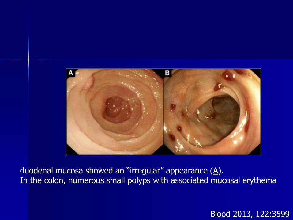
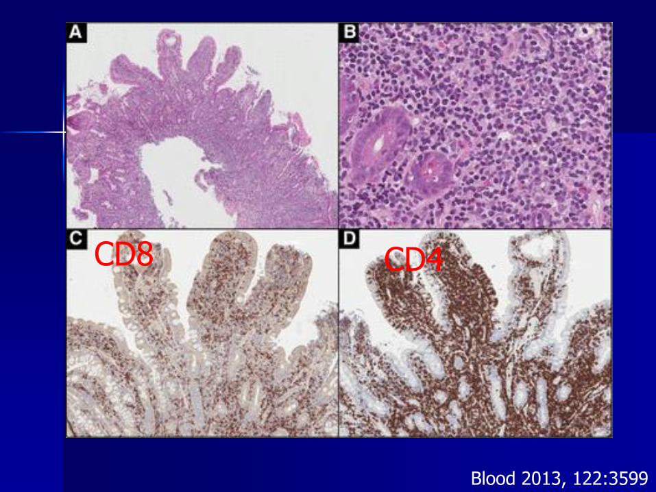
duodenal mucosa showed an “irregular” appearance (A). In the colon, numerous small polyps with associated mucosal erythema
Blood 2013, 122:3599
Indolent T-cell LPD
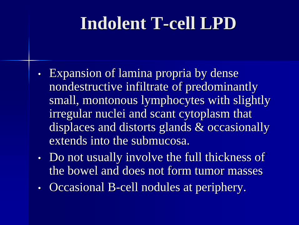
• Expansion of lamina propria by dense nondestructive infiltrate of predominantly small, montonous lymphocytes with slightly irregular nuclei and scant cytoplasm that displaces and distorts glands & occasionally extends into the submucosa.
• Do not usually involve the full thickness of the bowel and does not form tumor masses
• Occasional B-cell nodules at periphery.
• Some pleomorphism.
• Deeper extension but still with predominantly superficial involvement.
• May have some villous atrophy, crypt hyperplasia described by some, some infiltration into epithelium, 1 with “lymphocytic gastritis”.
• Some have granulomas & numerous eosinophils, other inflammatory cells.
• Diffuse LN & interstitial BM involvement (1 case)
TIA1
CD4CD8
Blood 2013, 122:3599
CD4CD8
Blood 2013, 122:3599
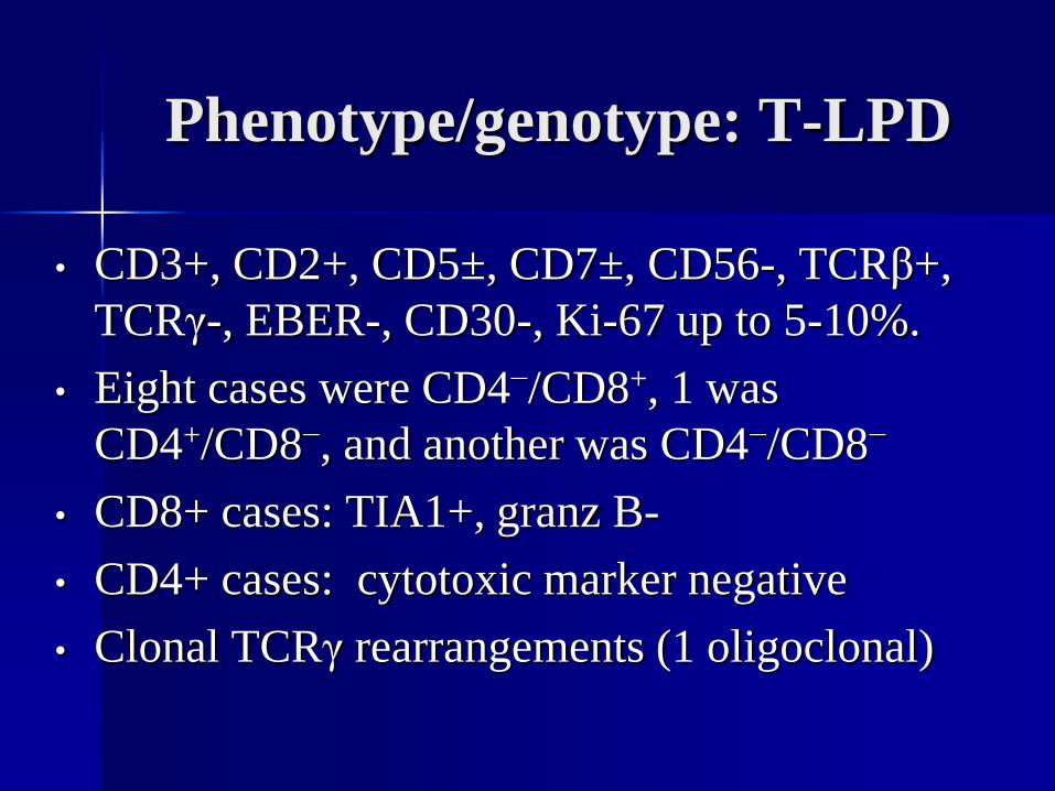
Phenotype/genotype: T-LPD
• CD3+, CD2+, CD5±, CD7±, CD56-, TCRβ+,
TCRγ-, EBER-, CD30-, Ki-67 up to 5-10%.
• Eight cases were CD4−/CD8+, 1 was
CD4+/CD8−, and another was CD4−/CD8−
• CD8+ cases: TIA1+, granz B-
• CD4+ cases: cytotoxic marker negative
• Clonal TCRγ rearrangements (1 oligoclonal)
Clinical outcome
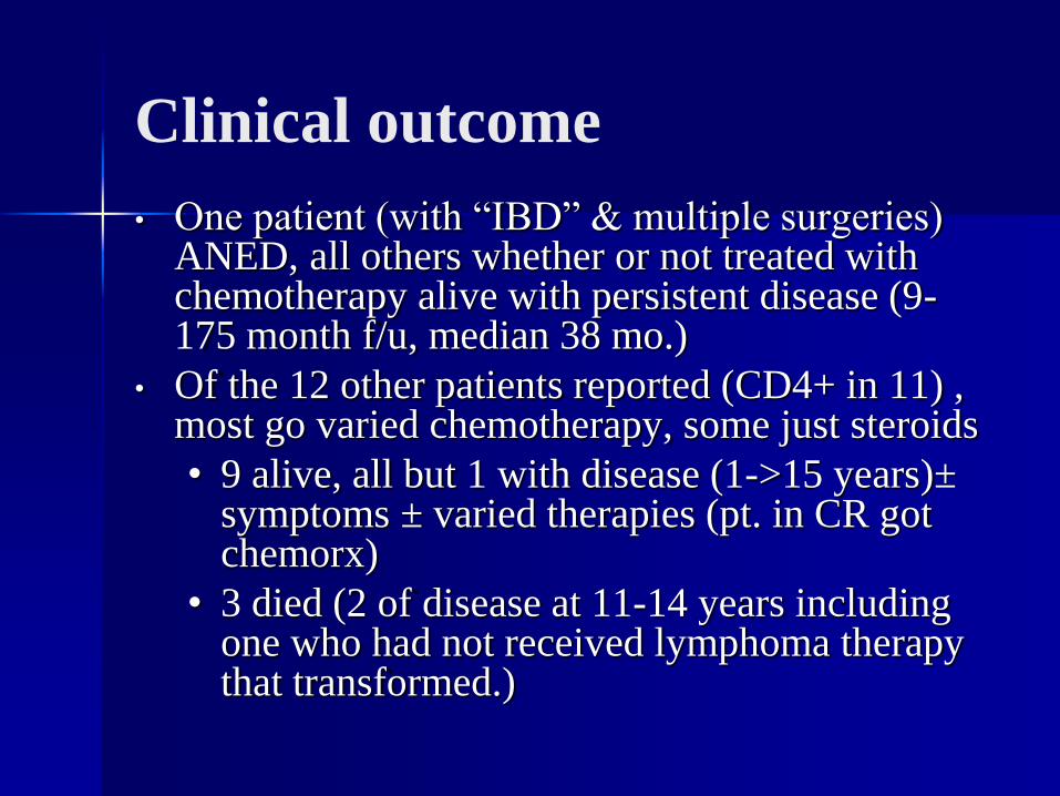
• One patient (with “IBD” & multiple surgeries) ANED, all others whether or not treated with chemotherapy alive with persistent disease (9-175 month f/u, median 38 mo.)
• Of the 12 other patients reported (CD4+ in 11) , most go varied chemotherapy, some just steroids
• 9 alive, all but 1 with disease (1->15 years)±symptoms ± varied therapies (pt. in CR got chemorx)
• 3 died (2 of disease at 11-14 years including one who had not received lymphoma therapy that transformed.)
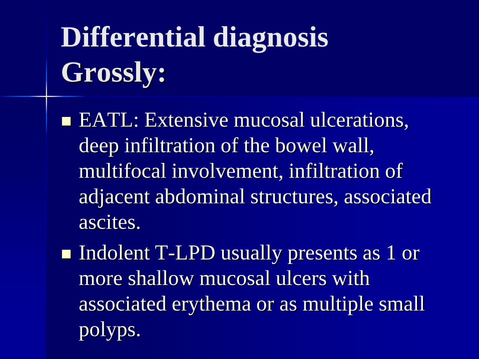
Differential diagnosis
Grossly:
EATL: Extensive mucosal ulcerations,
deep infiltration of the bowel wall,
multifocal involvement, infiltration of
adjacent abdominal structures, associated
ascites.
Indolent T-LPD usually presents as 1 or
more shallow mucosal ulcers with
associated erythema or as multiple small
polyps.
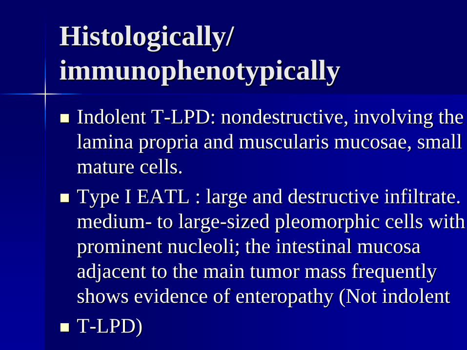
Histologically/
immunophenotypically
Indolent T-LPD: nondestructive, involving the
lamina propria and muscularis mucosae, small
mature cells.
Type I EATL : large and destructive infiltrate.
medium- to large-sized pleomorphic cells with
prominent nucleoli; the intestinal mucosa
adjacent to the main tumor mass frequently
shows evidence of enteropathy (Not indolent
T-LPD)
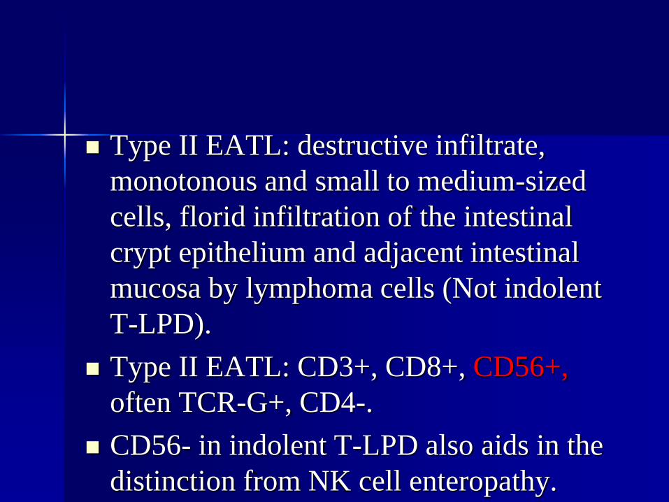
Type II EATL: destructive infiltrate,
monotonous and small to medium-sized
cells, florid infiltration of the intestinal
crypt epithelium and adjacent intestinal
mucosa by lymphoma cells (Not indolent
T-LPD).
Type II EATL: CD3+, CD8+, CD56+,
often TCR-G+, CD4-.
CD56- in indolent T-LPD also aids in the
distinction from NK cell enteropathy.
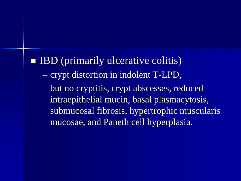
IBD (primarily ulcerative colitis)
– crypt distortion in indolent T-LPD,
– but no cryptitis, crypt abscesses, reduced
intraepithelial mucin, basal plasmacytosis,
submucosal fibrosis, hypertrophic muscularis
mucosae, and Paneth cell hyperplasia.
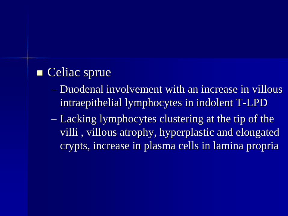
Celiac sprue
– Duodenal involvement with an increase in villous
intraepithelial lymphocytes in indolent T-LPD
– Lacking lymphocytes clustering at the tip of the
villi , villous atrophy, hyperplastic and elongated
crypts, increase in plasma cells in lamina propria
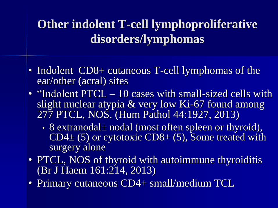
Other indolent T-cell lymphoproliferative
disorders/lymphomas
• Indolent CD8+ cutaneous T-cell lymphomas of the ear/other (acral) sites
• “Indolent PTCL – 10 cases with small-sized cells with slight nuclear atypia & very low Ki-67 found among 277 PTCL, NOS. (Hum Pathol 44:1927, 2013)
• 8 extranodal± nodal (most often spleen or thyroid), CD4± (5) or cytotoxic CD8+ (5), Some treated with surgery alone
• PTCL, NOS of thyroid with autoimmune thyroiditis(Br J Haem 161:214, 2013)
• Primary cutaneous CD4+ small/medium TCL
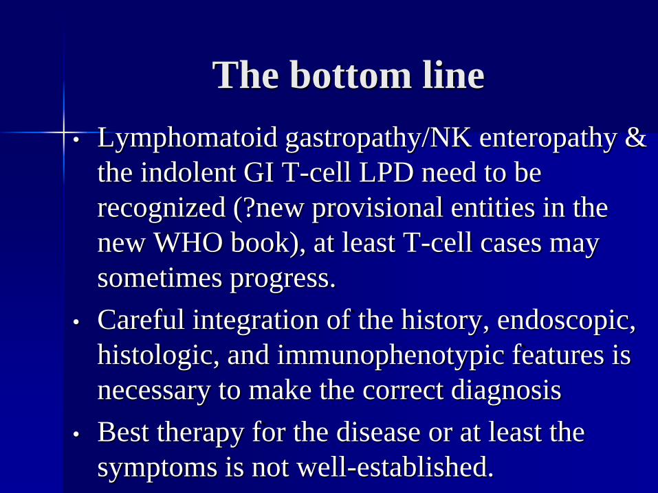
The bottom line
• Lymphomatoid gastropathy/NK enteropathy &
the indolent GI T-cell LPD need to be
recognized (?new provisional entities in the
new WHO book), at least T-cell cases may
sometimes progress.
• Careful integration of the history, endoscopic,
histologic, and immunophenotypic features is
necessary to make the correct diagnosis
• Best therapy for the disease or at least the
symptoms is not well-established.