Embed Size (px)
Citation preview
Infections of Oral & Para-Oral Tissues
Dr.Hossam ElmalahDr.Hossam Elmalahprof. of oral pathologyprof. of oral pathology
ASUASU
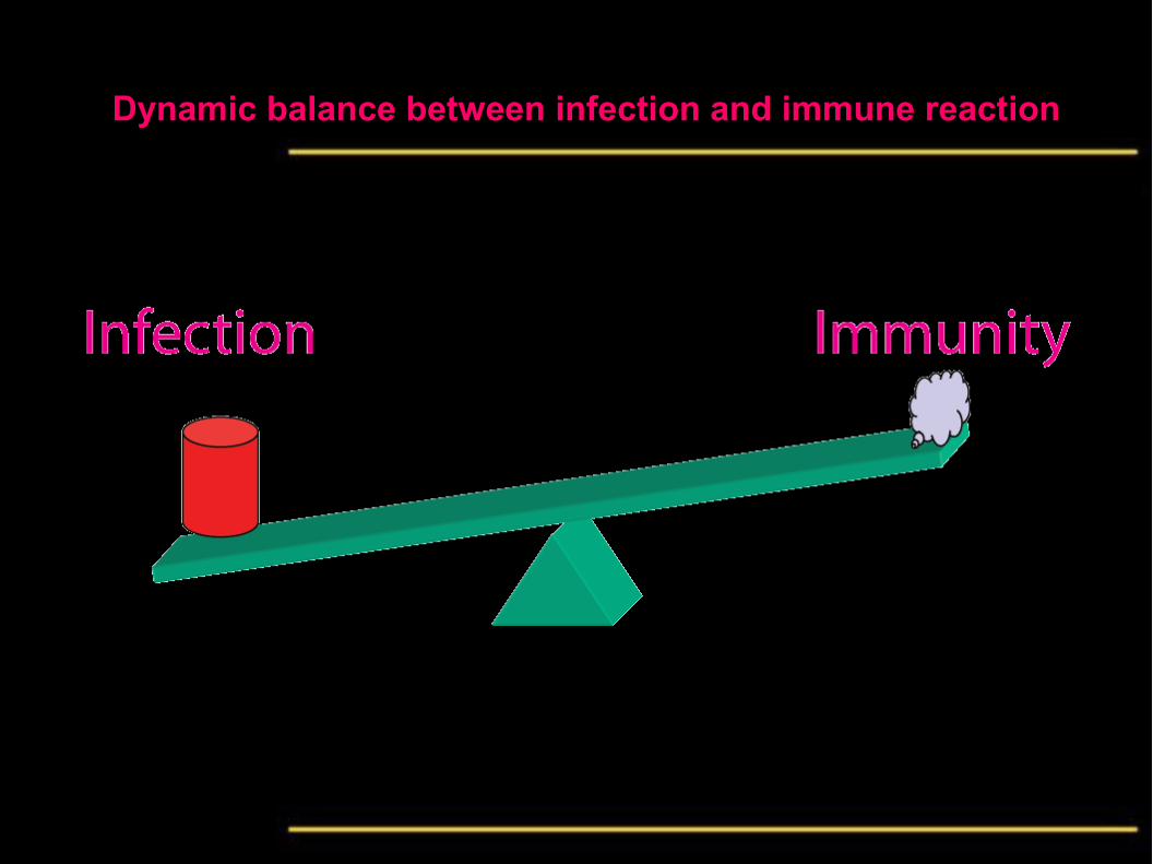
Dynamic balance between infection and immune reaction

Dynamic balance between infection and immunity
Infection Immunity
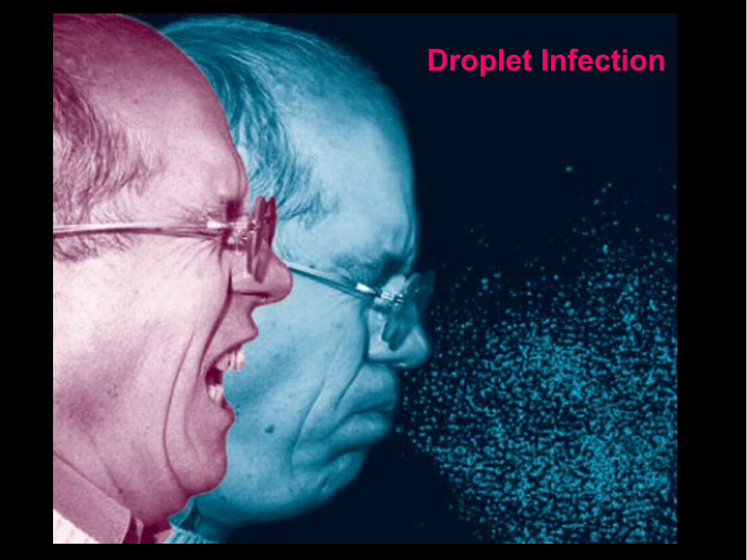
Droplet Infection

Classification of infectious agents
1. Metazoa
2. Protozoa
3. Fungi
4. Bacteria
5. Viruses
6. Prions
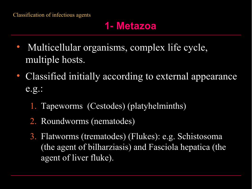
1- Metazoa
• Multicellular organisms, complex life cycle, multiple hosts.
• Classified initially according to external appearance e.g.:
1. Tapeworms (Cestodes) (platyhelminths)
2. Roundworms (nematodes)
3. Flatworms (trematodes) (Flukes): e.g. Schistosoma (the agent of bilharziasis) and Fasciola hepatica (the agent of liver fluke).
Classification of infectious agents
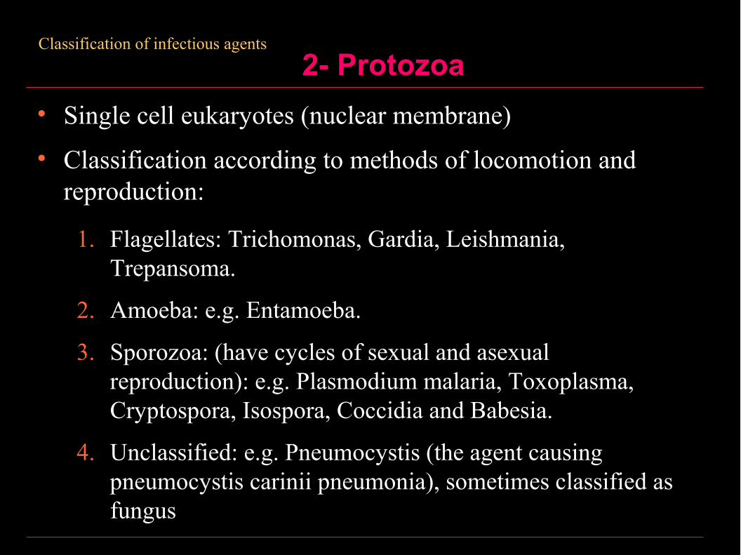
2- Protozoa
• Single cell eukaryotes (nuclear membrane)
• Classification according to methods of locomotion and reproduction:
1. Flagellates: Trichomonas, Gardia, Leishmania, Trepansoma.
2. Amoeba: e.g. Entamoeba.
3. Sporozoa: (have cycles of sexual and asexual reproduction): e.g. Plasmodium malaria, Toxoplasma, Cryptospora, Isospora, Coccidia and Babesia.
4. Unclassified: e.g. Pneumocystis (the agent causing pneumocystis carinii pneumonia), sometimes classified as fungus
Classification of infectious agents
3- Fungi
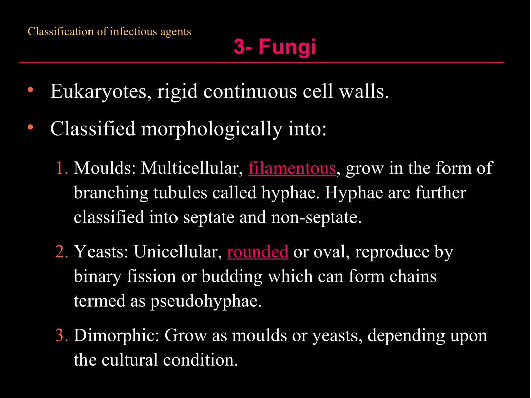
• Eukaryotes, rigid continuous cell walls.
• Classified morphologically into:
1. Moulds: Multicellular, filamentous, grow in the form of branching tubules called hyphae. Hyphae are further classified into septate and non-septate.
2. Yeasts: Unicellular, rounded or oval, reproduce by binary fission or budding which can form chains termed as pseudohyphae.
3. Dimorphic: Grow as moulds or yeasts, depending upon the cultural condition.
Classification of infectious agents
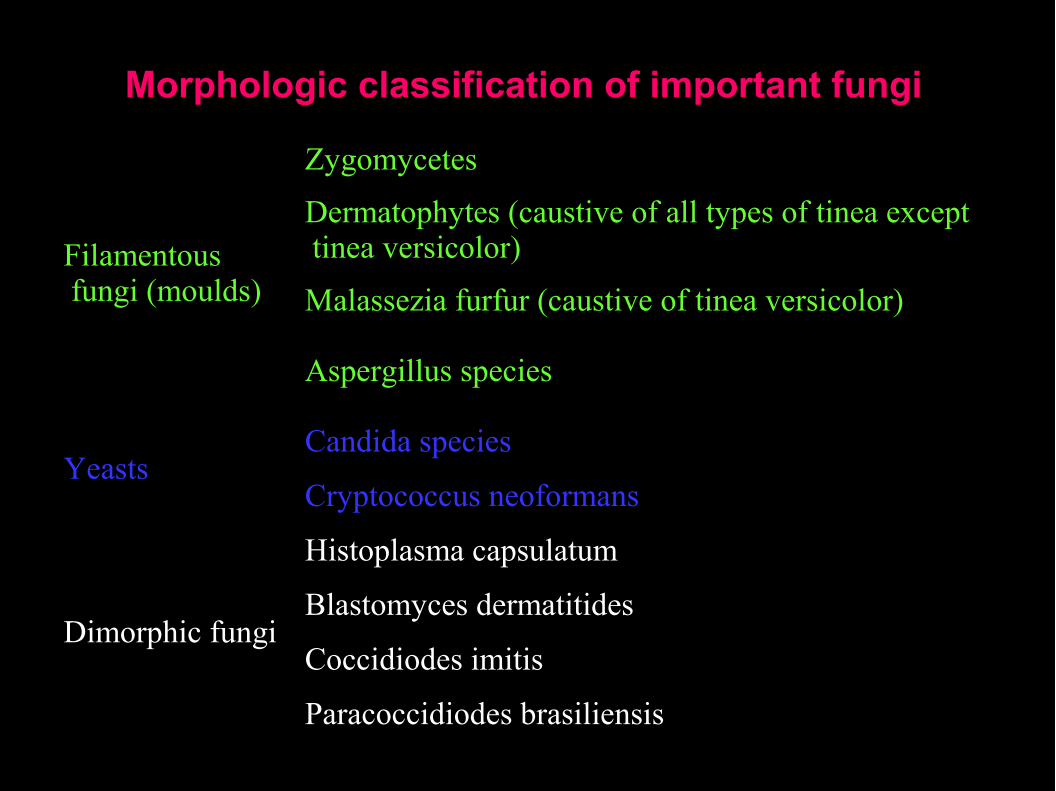
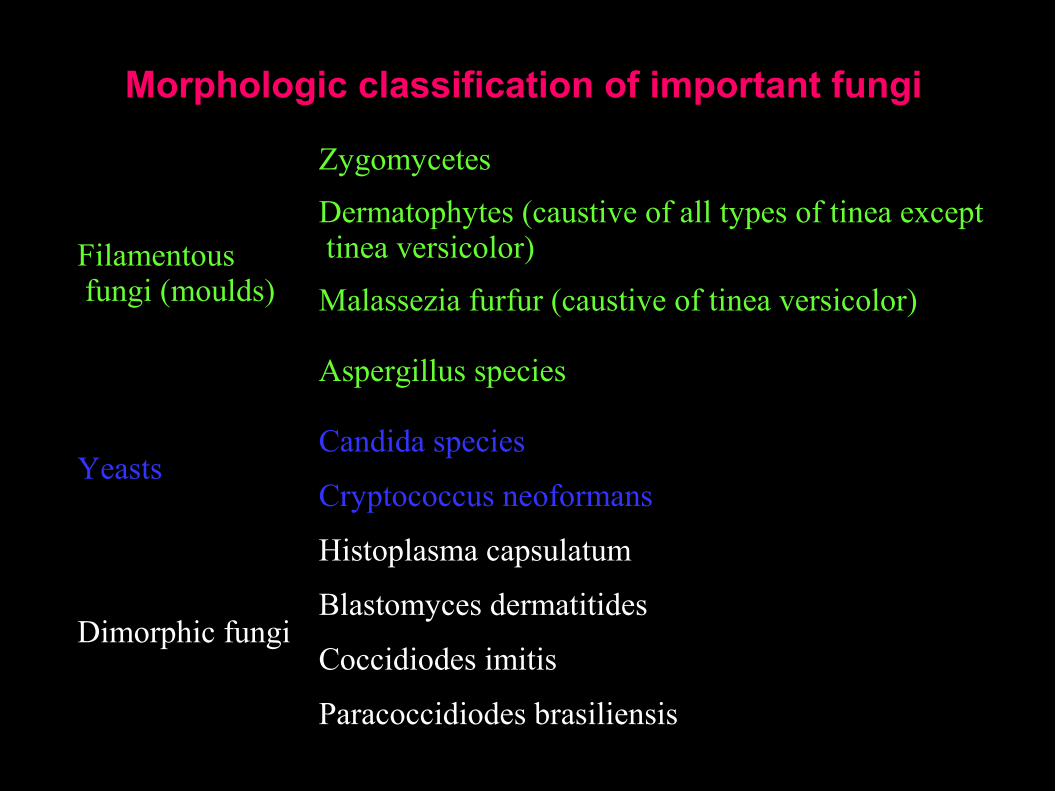
Morphologic classification of important fungi
Filamentous fungi (moulds)
Zygomycetes
Dermatophytes (caustive of all types of tinea except tinea versicolor)
Malassezia furfur (caustive of tinea versicolor)
Aspergillus species
YeastsCandida species
Cryptococcus neoformans
Dimorphic fungi
Histoplasma capsulatum
Blastomyces dermatitides
Coccidiodes imitis
Paracoccidiodes brasiliensis
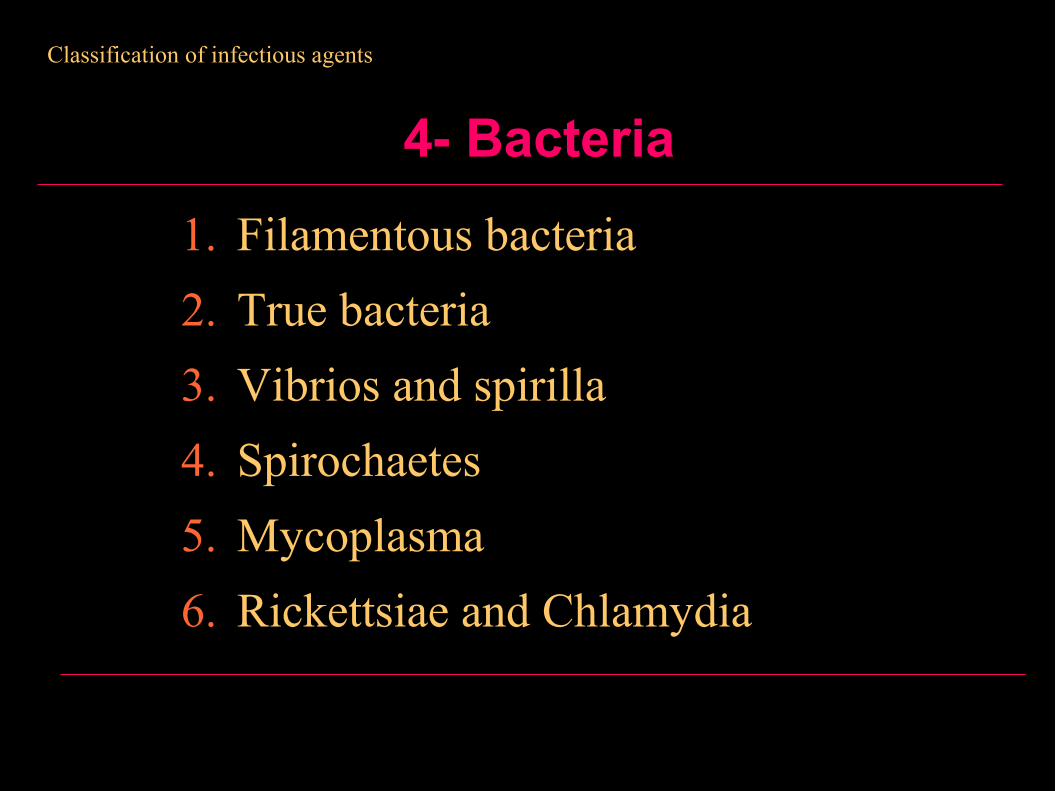
4- Bacteria
1. Filamentous bacteria
2. True bacteria
3. Vibrios and spirilla
4. Spirochaetes
5. Mycoplasma
6. Rickettsiae and Chlamydia
Classification of infectious agents
4- Bacteria
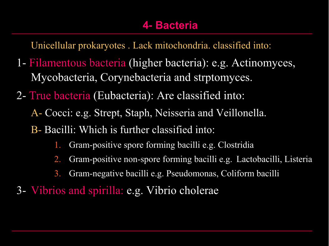
Unicellular prokaryotes . Lack mitochondria. classified into:
1- Filamentous bacteria (higher bacteria): e.g. Actinomyces, Mycobacteria, Corynebacteria and strptomyces.
2- True bacteria (Eubacteria): Are classified into:
A- Cocci: e.g. Strept, Staph, Neisseria and Veillonella.
B- Bacilli: Which is further classified into:1. Gram-positive spore forming bacilli e.g. Clostridia
2. Gram-positive non-spore forming bacilli e.g. Lactobacilli, Listeria
3. Gram-negative bacilli e.g. Pseudomonas, Coliform bacilli
3- Vibrios and spirilla: e.g. Vibrio cholerae
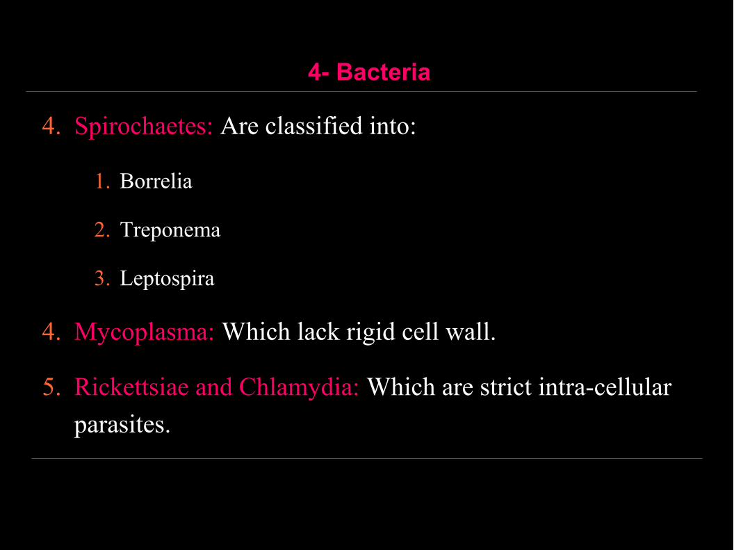
4- Bacteria
4. Spirochaetes: Are classified into:
1. Borrelia
2. Treponema
3. Leptospira
4. Mycoplasma: Which lack rigid cell wall.
5. Rickettsiae and Chlamydia: Which are strict intra-cellular
parasites.
Classification of streptococci
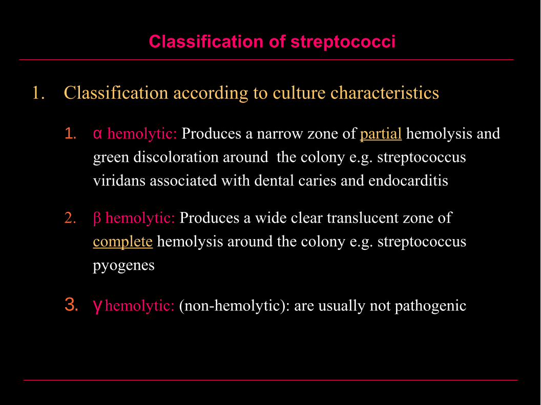
1. Classification according to culture characteristics
1. α hemolytic: Produces a narrow zone of partial hemolysis and
green discoloration around the colony e.g. streptococcus
viridans associated with dental caries and endocarditis
2. β hemolytic: Produces a wide clear translucent zone of
complete hemolysis around the colony e.g. streptococcus
pyogenes
3. γ hemolytic: (non-hemolytic): are usually not pathogenic
Classification of streptococci
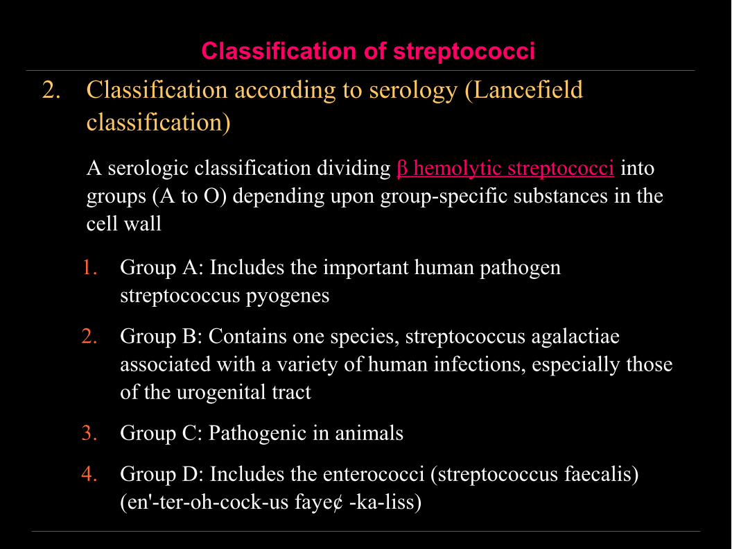
2. Classification according to serology (Lancefield classification)
A serologic classification dividing β hemolytic streptococci into groups (A to O) depending upon group-specific substances in the cell wall
1. Group A: Includes the important human pathogen streptococcus pyogenes
2. Group B: Contains one species, streptococcus agalactiae associated with a variety of human infections, especially those of the urogenital tract
3. Group C: Pathogenic in animals
4. Group D: Includes the enterococci (streptococcus faecalis) (en'-ter-oh-cock-us faye¢ -ka-liss)

Classification of infectious agents
5. Viruses
Consist of DNA or RNA (never both) and enclosed in a protein shell known as capsid. Sometimes the nucleocapsid may be enclosed in a lipoprotein envelope largely derived from the host.
Classification of infectious agents
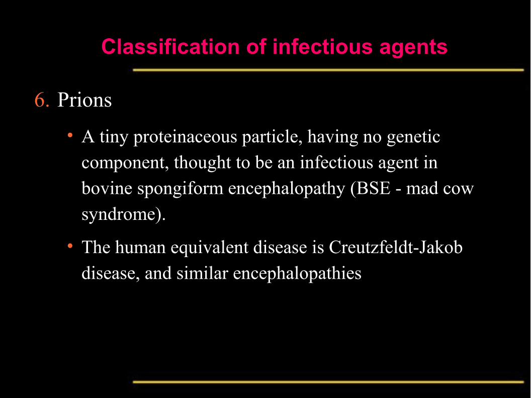
6. Prions
• A tiny proteinaceous particle, having no genetic component, thought to be an infectious agent in bovine spongiform encephalopathy (BSE - mad cow syndrome).
• The human equivalent disease is Creutzfeldt-Jakob disease, and similar encephalopathies
Bacterial Infections
1. Acute Necrotising Ulecerative Gingivitis, ANUG
2. Vincent Angina
3. Maxillofacial Gangrene (Noma, Cancrum Oris)
4. Acute Streptococcal Gingivitis
5. Scarlet Fever
6. Diphtheria
7. Anthrax
8. Syphilis
9. Tuberculosis of the Oral Cavity
10. Actinomycosis
11. Orofacial granulomatosis
Mycotic (Fungal) Infections
1. Moniliasis (Candidiasis)
2. Systemic Mycoses
1. Histoplasmosis
2. Blastomycosis
3. Mucormycosis (Zygomycosis, Phycomycosis)
4. Coccidioidomycosis
5. Paracoccidioidomycosis (Paracoccidioides brasiliensis)
6. Cryptococcosis
Parasitic Diseases
• Leishmaniasis
• Cutaneous Leishmaniasis
• Mucocutaneous Leishmaniasis
• Visceral Leishmaniasis
Viral diseases of the Oral Cavity
• Introduction
• Viral Structure
• Herpes Simplex I (Labialis)• I- Primary Herpetic Gingivostomatitis• II- Recurrent Herpetic Infection
• Varicella - Zoster Virus• I- Chicken Pox (Varicella)• II- Herpes Zoster Infection (Shingles)
• Hand-Foot and Mouth Disease
• Herpangina
• AIDS

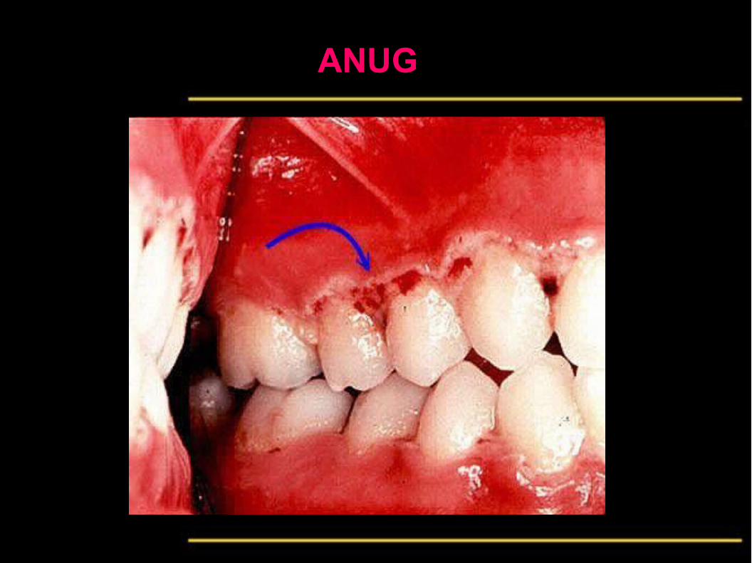
Acute Necrotising Ulecerative Gingivitis, ANUG
Definition
Acute necrotizing inflammation of the marginal gingiva and interdental papilla.
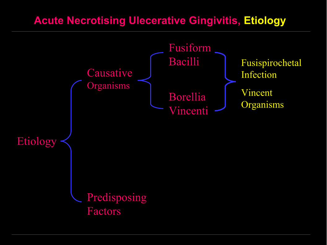
Acute Necrotising Ulecerative Gingivitis, Etiology
Etiology
Causative Organisms
Predisposing Factors
Fusiform Bacilli
Borellia Vincenti
Fusispirochetal Infection
Vincent Organisms
Acute Necrotising Ulecerative Gingivitis, Etiology
Multifactorial, 2 factors play a role:
• The causative organisms: fusispirochetal infection, which consists of a Gram negative fusiform bacilli (fusobacterium fusiforms, fusobacterium necrophorum) and the spirochete borellia vincenti, (both fusiform bacilli and borellia vincenti are called Vincent organisms).
• Decreased tissue resistance due to:
1.Local factors e.g. calculus, plaque or poor oral hygiene.
2.Psychological or physiological stresses (trench mouth).
3.Immunosuppression
Acute Necrotising Ulecerative Gingivitis, Clinically
• Rarely there is mild general malaise or slight fever.
• Necrosis and ulceration of the marginal gingiva and interdental papilla. The ulcers are covered by grayish pseudomembrane which can be wiped off leaving a red bleeding surface.
• The interdental papillae are eroded giving the characteristic crater shaped appearance.
• The attached gingiva is normal or show redness and edema.
• There is bleeding and foul odor.
• No significant regional lymphadenopathy.
• Extension of infection to produce Vincent angina or Noma.
Acute Necrotising Ulecerative Gingivitis, ANUG
Histologically
• Non-specific, necrosis of surface epithelium and
infiltration of the connective tissue with inflammatory
cells.
Treatment
• Removal of the local irritating factors, plaque or calculus.
• Antibiotics: Metronidazol (Flagyl) 200 mg taken by
mouth, after food 3 times a day for 3 days.

ANUG

ANUG
ANUG

Vincent Angina
• Vincent angina is the sever form of ANUG, usually occurring in immunocompromised patients.
• There is extension of infection to the throat with necrosis, edema and formation of pseudomembranes.
• The disease responds well to metronidazol but shows poor response if penicillin is to be used alone.
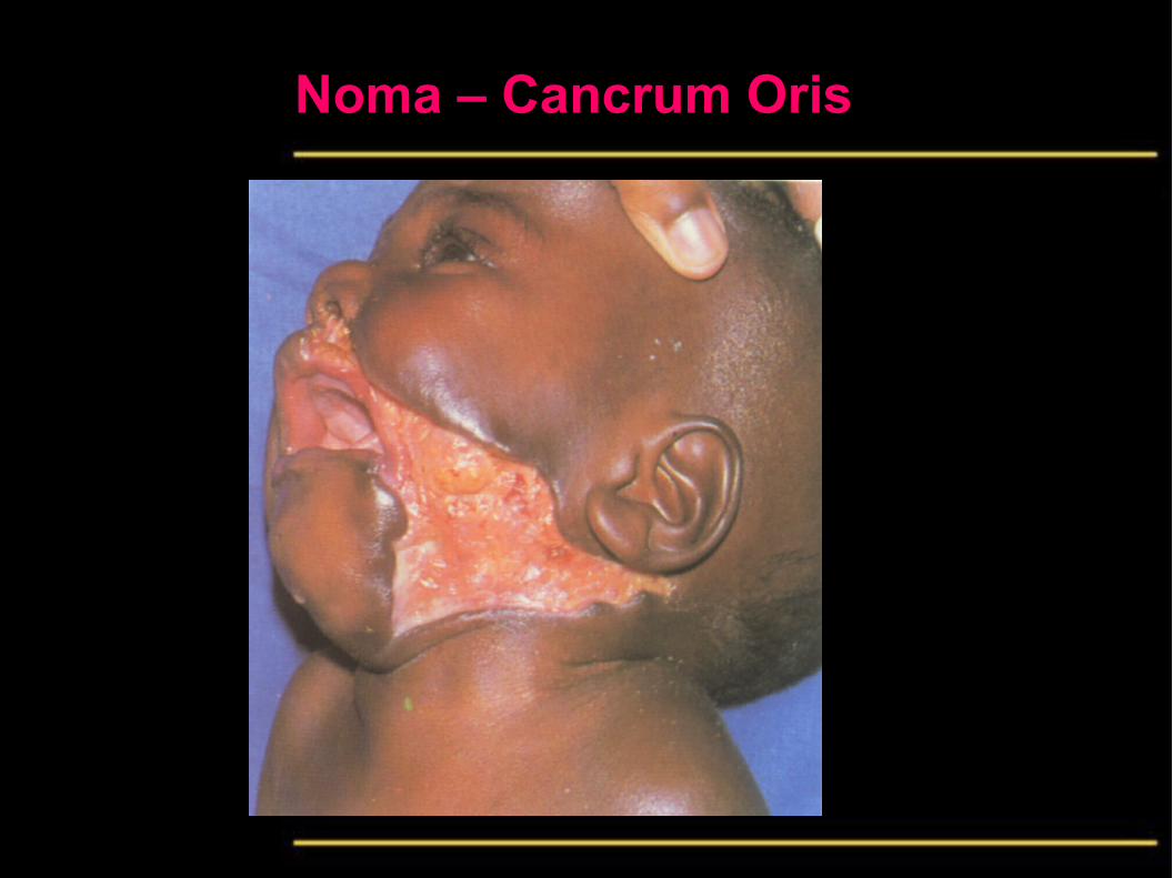
Maxillofacial Gangrene (Noma, Cancrum Oris)
• Noma is an overwhelming severe necrotizing inflammation of the oro-facial region.
• Was common among starving prisoners in Nazi concentration camps.
• Noma has disappeared from developed countries and for decades the disease has been ignored.
• However it becomes widespread again in sub-Saharan Africa as to become a subject for international and World Health Organization concern.
• This results from the widespread expansion of Political unrests, poverty, malnutrition, poor oral hygiene and debilitating diseases as AIDS.
Maxillofacial Gangrene (Clinically)
• Starts from an acute necrotizing ulcerative gingivitis associated with extensive edema, but extends outwards rapidly destroying soft tissues and bone.
• The gangrene starts as a painful, small reddish-purple spot or indurated papule which ulcerates.
• The ulcer spreads to involve the adjacent mucosa, skin and bone associated with diffuse edema of the face.
• As the overlying tissues become ischemic, the skin turns blue-black. The gangrenous area becomes sharply demarcated and ultimately sloughs away. As the slough separates, the bone dies with sequestration and exfoliation of teeth. A gaping facial defect is left.
• The disease is fatal if inadequately treated
Maxillofacial Gangrene (Etiology)
1. Causative organisms:
• Anaerobes including Fusobacterium necrophorum and spirochaetes. Fusobacterium necrophorum is a commensal in the gut of herbivores and also a cause of necrotizing infections in animals.
2. Predisposing factors:
• Are usually severe malnutrition, poor oral hygiene, poor general hygiene, AIDS and debilitating diseases.
Maxillofacial Gangrene (Treatment)
• Treatment:• Correction of the predisposing factor, if possible
• A combination of penicillin or an aminoglycoside and metronidazole will usually control the local infection.
• Debridement of the necrotic tissues is also required.
• Reconstructive surgery is needed to correct the deformity.
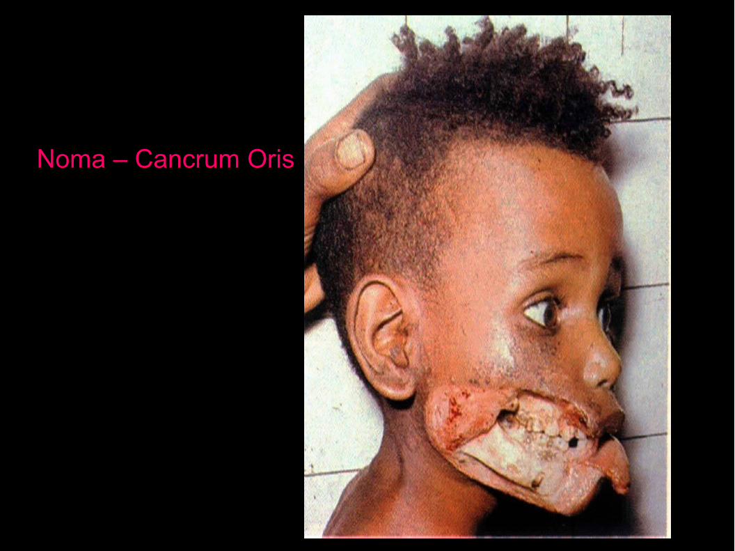
Noma – Cancrum Oris
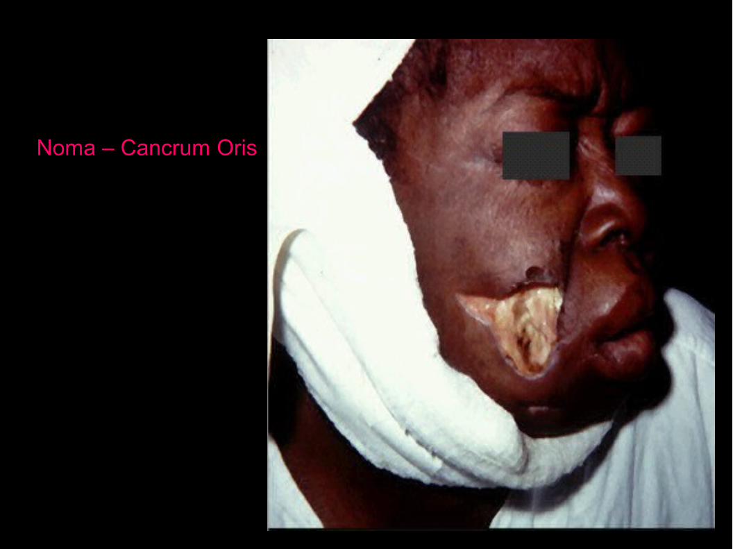
Noma – Cancrum Oris
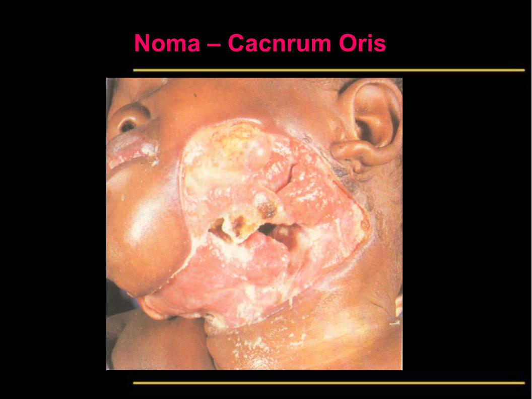
Noma – Cacnrum Oris
Noma – Cancrum Oris

Pericoronitis - Definition
• Inflammation of the soft tissue around the crown of partially or completely erupted teeth.
• Usually occurs lower third molars or lower second molars if they are the most distal tooth in the arch
• May be acute or chronic depending on the clinical characteristics

Pericoronitis - Etiology
• Accumulation of dental plaque and food debris under the operculum.
• This causes inflammation and edema of the surrounding soft tissues.
• When edema occurs, the operculum becomes mechanically traumatized by the opposing teeth, this aggravates the existing infection.
Pericoronitis - Clinically
• Coverage of the occlusal surface by a flap of tissue called the operculum. (formed during tooth eruption).
• Swelling and redness of the operculum.
• Traumatized and ulcerated operculum.
• Painful and tenderness in the affected area.
• Pus formation under the operculum.
• Trismus
• Foul odor
• Bad taste

Pericoronitis - Clinically
• Regional lymph node enlargement
• Possible fever, malaise and other toxic systemic manifestations.
• Possible spread of infection to form pericoronal abscess or into fascial spaces.
Pericoronitis - Radiographically
• In the acute stage, no changes can be noted.
• In the chronic cases, radiolucency can be detected around the crown of the affected tooth.
Pericoronitis - Treatment• Gentle debridement with warm water
• The area can be swabbed with antiseptic solution.
• Extensive curettage and surgical procedure should be avoided in the first appointment.
• If drainage needs to be established, an incision may be made or iodoform gauze may be inserted under the operculum.
• Systemic antibiotic treatment is recommended.
• After the infection subsides, a decision must be made as to whether operculectomy or extraction of the tooth is to be performed.
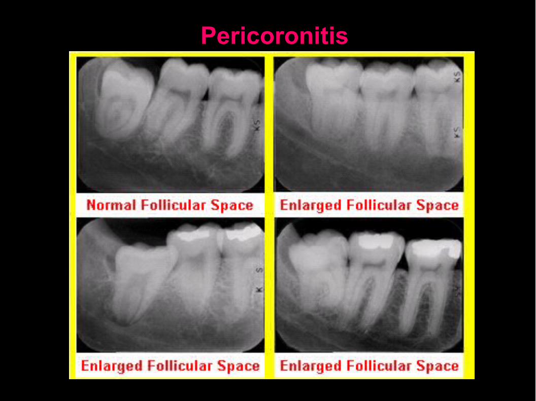
Pericoronitis
Acute Streptococcal Gingivitis• Acute inflammation of the gingiva usually as a result of
spread from acute streptococcal tonsillitis.
• Etiology: β Hemolytic streptococci, group A.
• Clinically
• Malaise and fever.
• Redness, edema and bleeding of the gingival
• No ulceration or necrosis.
• Regional lymphadenopathy.
• Treatment: Penicillin or erythromycin - Should be treated as soon as possible to avoid post-streptococcal complications (rheumatic fever and glomerulonephritis).
Scarlet Fever
• Definition: This is an acute pharyngitis accompanied by skin rash.
• Etiology: β Hemolytic streptococci, Group A. which produces erythrogenic exotoxins that attacks the blood vessels and produces the characteristic skin rash.
Scarlet Fever, Clinically
• Incubation period 2 - 3 days.• Fever and malaise.• The lesions are painful and consists of:• Erythematous skin rash with the characteristic
circumoral pallor, which is an area of pallor around the mouth corresponding to the anatomy of orbicularis oris muscle.
• Edema of the pharynx and oral mucosa• The tongue shows a white coating through which
only the fungiform papillae can be seen giving the characteristic strawberry tongue
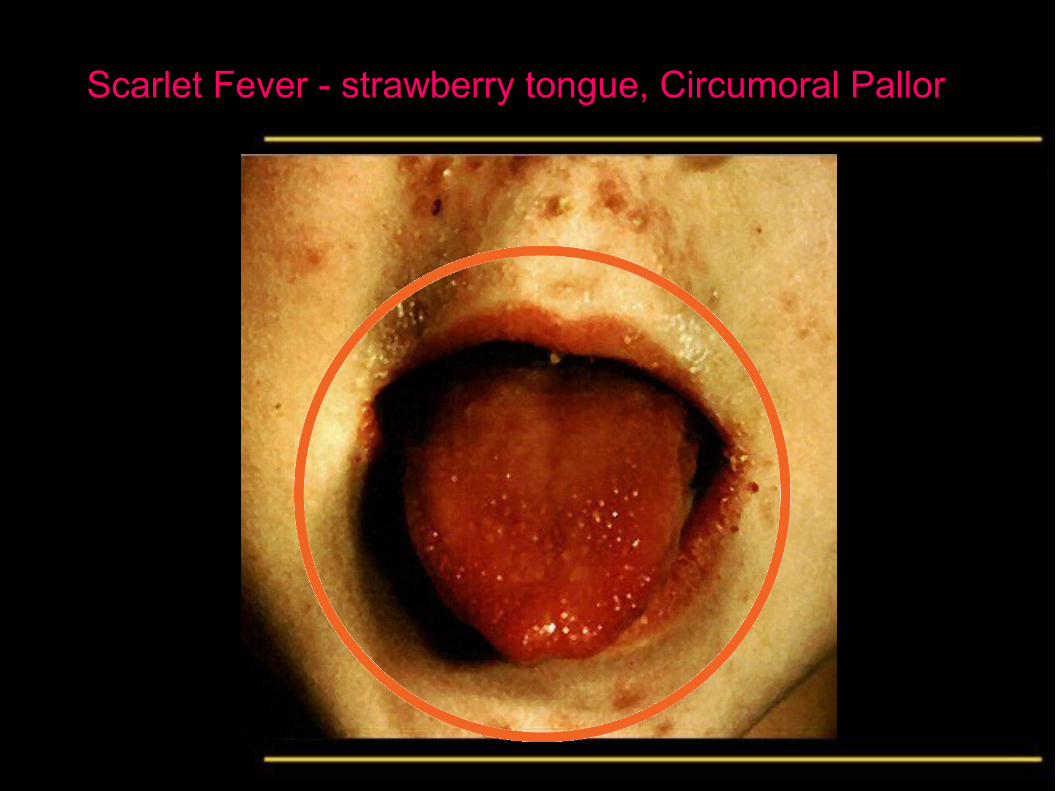
Scarlet Fever - strawberry tongue, Circumoral Pallor
Diphtheria
• Acute necrotizing inflammation of the pharynx
• Etiology: Corynebacterium diphtheria, which is Gram-positive bacilli. The organisms elaborate a potent exotoxin which causes severe systemic manifestations on the heart and nervous tissue.
• Clinically:
• High fever.
• Necrotic lesions in the pharynx covered by grayish white psuedomembrane which may extend to the mouth.

Anthrax
• A disease caused by bacillus anthracis affecting usually herbivores (cattle and sheep). The disease is rare in man usually affecting farmers, veterinarians and butchers.
• Etiology: Gram +ve bacillus anthracis - a soil organism.
• Clinically:
• Cutaneous lesions: necrotic lesions surrounded by edema and erythema.
• Mucous membrane lesions: necrotic lesions surrounded by edema and erythema.
• Pulmonary lesions: pneumonia, which is often fatal.
• Treatment: Penicillin - Vaccination in high risk.

Syphilis
• A chronic specific granulomatous inflammation
• Etiology: The spirochete Treponema Pallidium, could be detected by dark field illumination.
• Classified into:
1. Acquired (postnatal)
2. Congenital (prenatal)
• This classification is misleading since the congenital syphilis is acquired during embryonic life.

Acquired syphilis - Stages
1. Primary Stage
2. Secondary Stage
3. Tertiary Stage
Acquired syphilis, Primary stage
• Characterized by the formation of chancre.
• Occurs at the site of introduction of the treponema pallidium. In the oral cavity, the lips and tip of the tongue are the most affected site.
• Appears as a painless nodule, which soon ulcerates forming an indurated ulcer.
• Regional lymphadenopathy.
• Highly contagious.
• The incubation period is 2-3 weeks.
• Heals with minimum scar formation after 2-3 weeks.
Acquired syphilis, Secondary stage
• Characterized by the formation of mucous patches.
• Appears 2-3 months after disappearance of chancre.
• Appears as irregular painless ulcers covered by grayish psuedomembrane which when removed leaves a raw red surface. Neighboring ulcers coalesce producing the typical snail track appearance.
Acquired syphilis, Tertiary stage
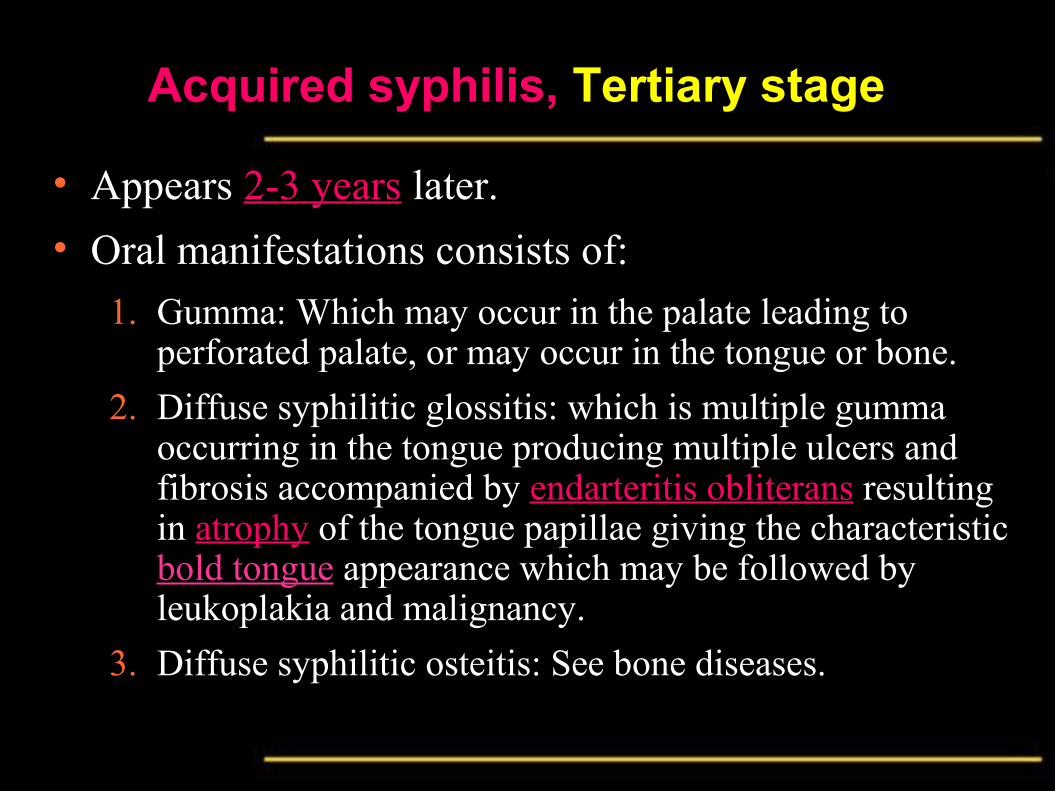
• Appears 2-3 years later.
• Oral manifestations consists of:
1. Gumma: Which may occur in the palate leading to perforated palate, or may occur in the tongue or bone.
2. Diffuse syphilitic glossitis: which is multiple gumma occurring in the tongue producing multiple ulcers and fibrosis accompanied by endarteritis obliterans resulting in atrophy of the tongue papillae giving the characteristic bold tongue appearance which may be followed by leukoplakia and malignancy.
3. Diffuse syphilitic osteitis: See bone diseases.

Syphilis, Congenital
1- Hutchinson’s teeth
2- Moon’s molar
3- Mulberry molar
4- Enamel hypoplasia
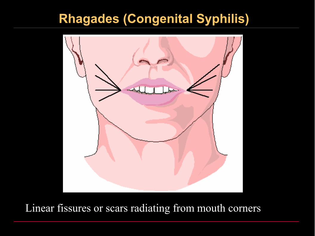
5- Rhagedes: which are linear fissures or scars radiating from mouth corners.
6- Perforated palate.

Syphilis - Treponema
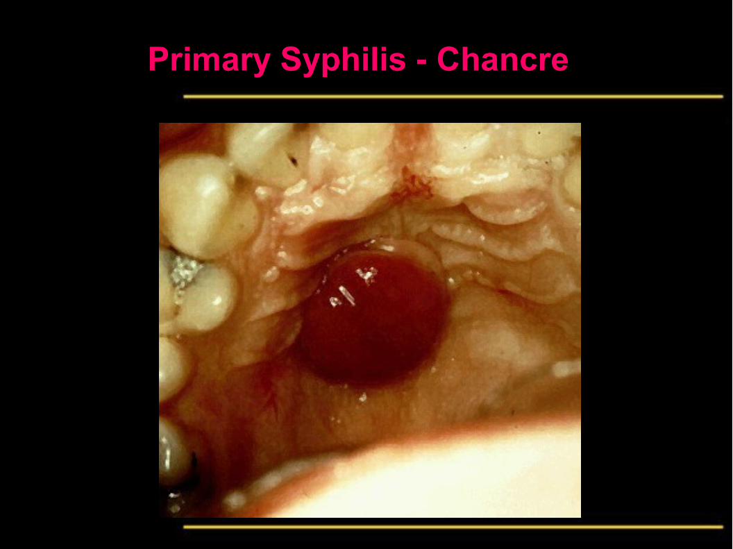
Primary Syphilis - Chancre
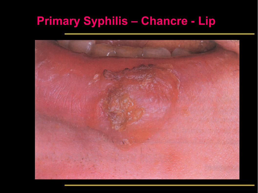
Primary Syphilis – Chancre - Lip
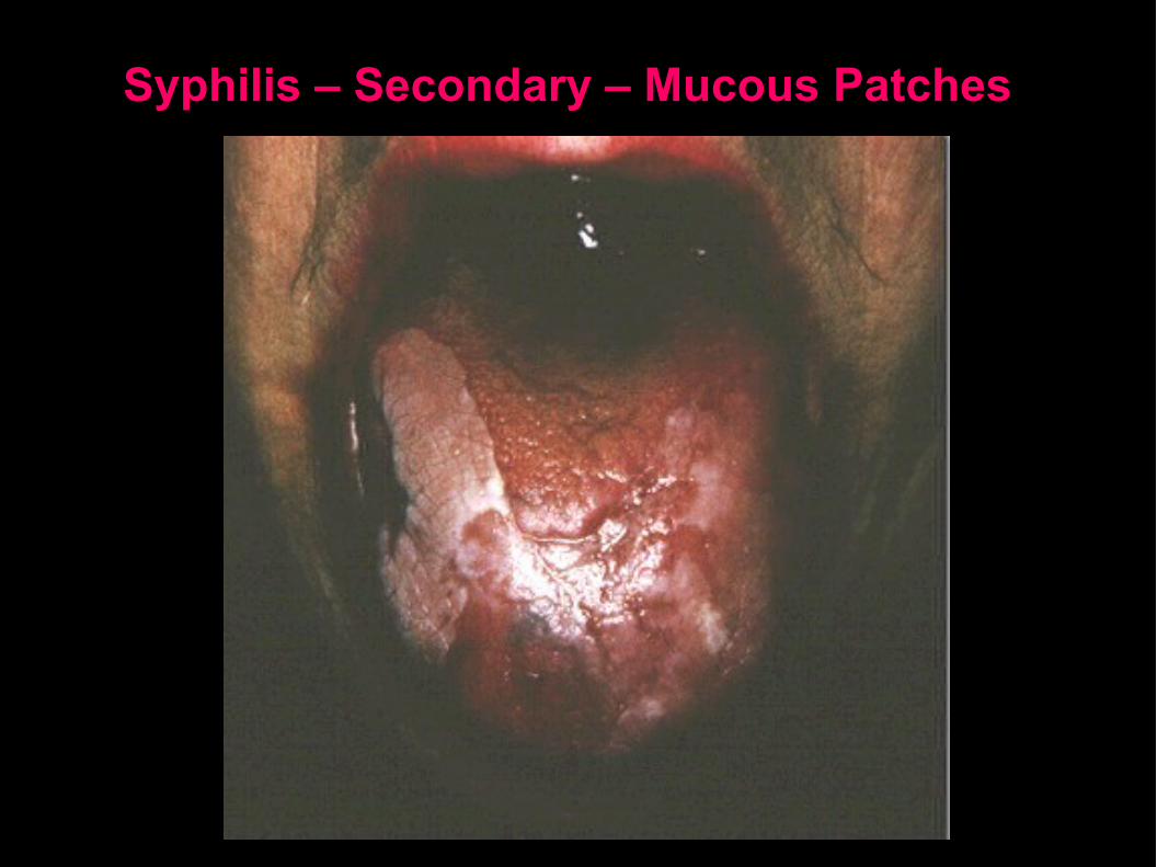
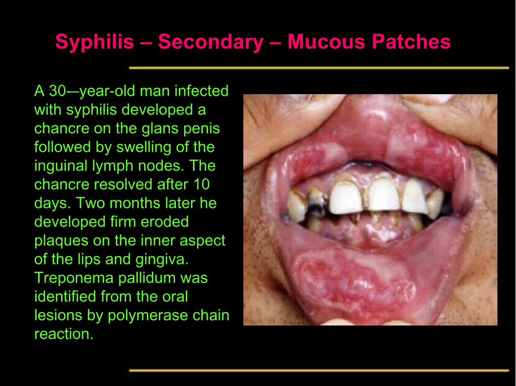
Syphilis – Secondary – Mucous Patches
Syphilis – Secondary – Mucous Patches
A 30-–year-old man infected with syphilis developed a chancre on the glans penis followed by swelling of the inguinal lymph nodes. The chancre resolved after 10 days. Two months later he developed firm eroded plaques on the inner aspect of the lips and gingiva. Treponema pallidum was identified from the oral lesions by polymerase chain reaction.
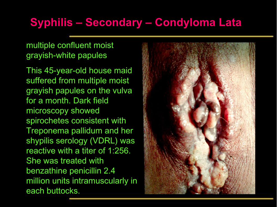
Syphilis – Secondary – Condyloma Lata
multiple confluent moist grayish-white papules
This 45-year-old house maid suffered from multiple moist grayish papules on the vulva for a month. Dark field microscopy showed spirochetes consistent with Treponema pallidum and her shypilis serology (VDRL) was reactive with a titer of 1:256. She was treated with benzathine penicillin 2.4 million units intramuscularly in each buttocks.
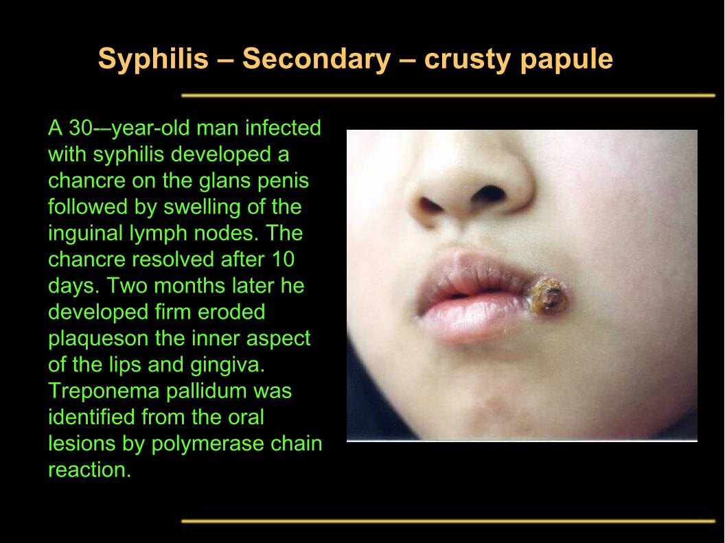
Syphilis – Secondary – crusty papule
A 30-–year-old man infected with syphilis developed a chancre on the glans penis followed by swelling of the inguinal lymph nodes. The chancre resolved after 10 days. Two months later he developed firm eroded plaqueson the inner aspect of the lips and gingiva. Treponema pallidum was identified from the oral lesions by polymerase chain reaction.
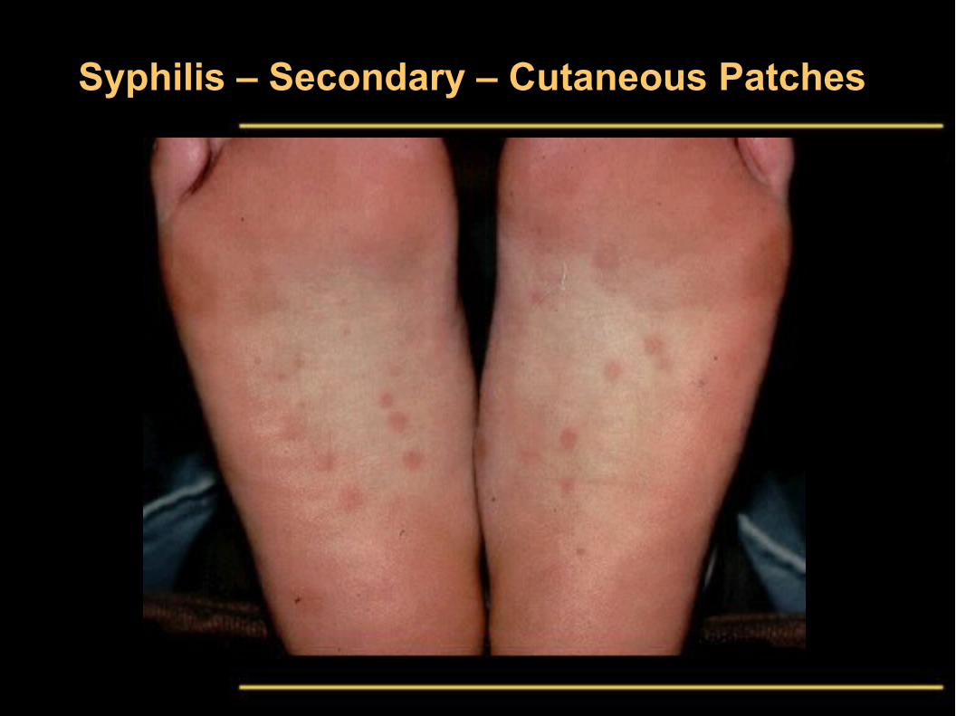
Syphilis – Secondary – Cutaneous Patches
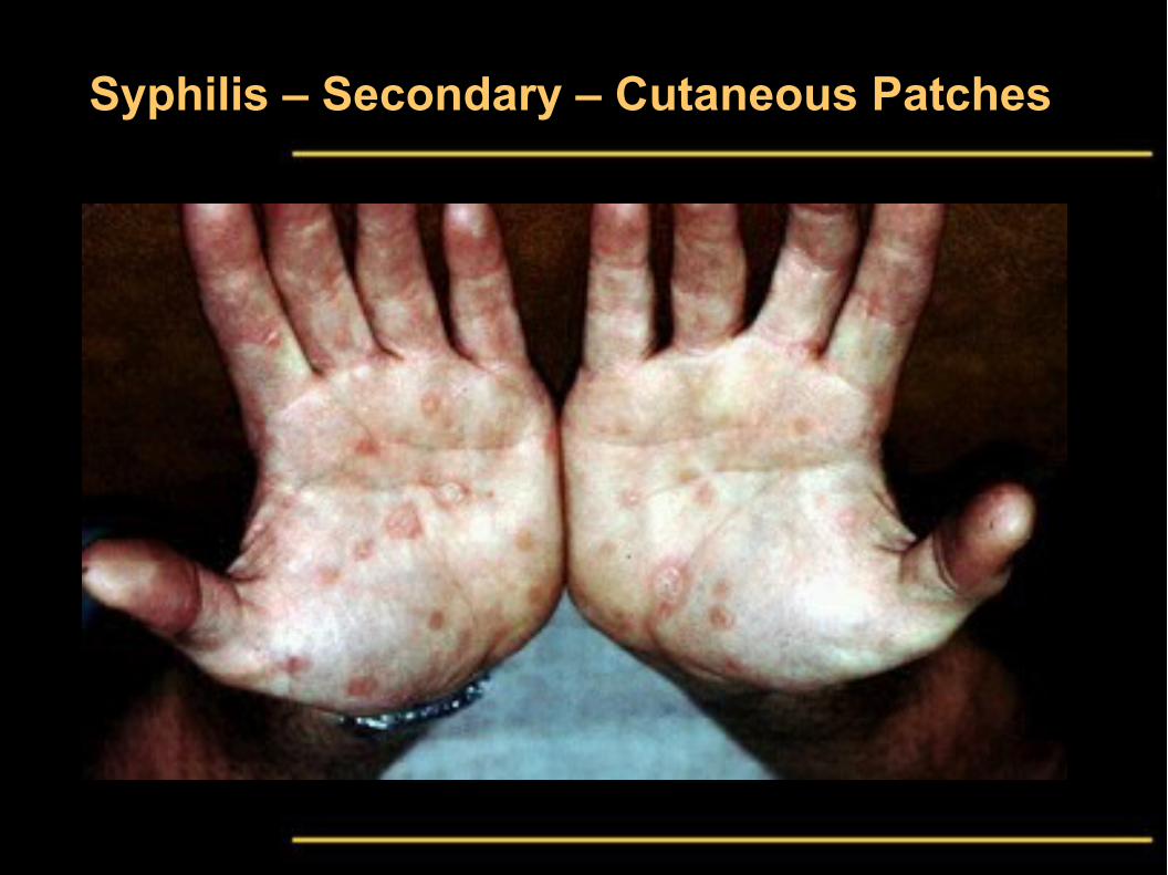
Syphilis – Secondary – Cutaneous Patches
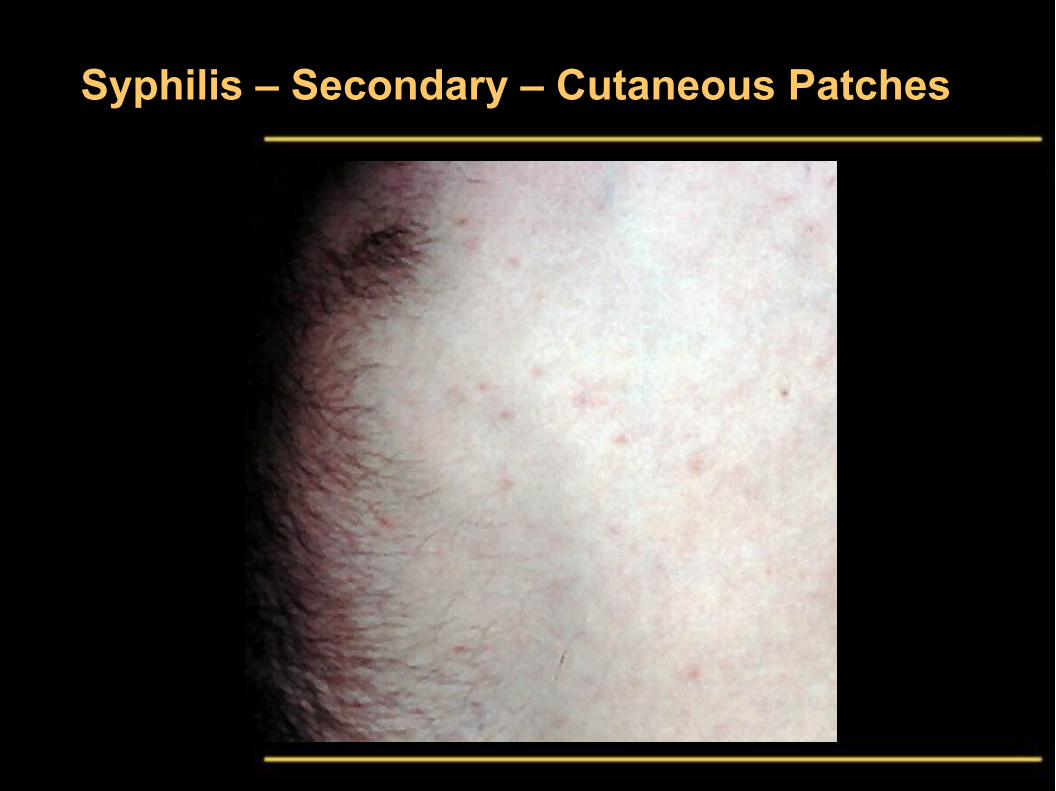
Syphilis – Secondary – Cutaneous Patches
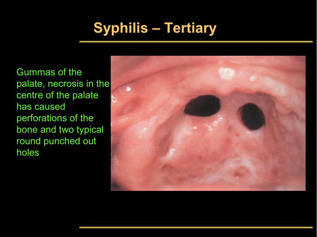
Syphilis – Tertiary
Gummas of the palate, necrosis in the centre of the palate has caused perforations of the bone and two typical round punched out holes

Congenital (Prenatal) Syphilis
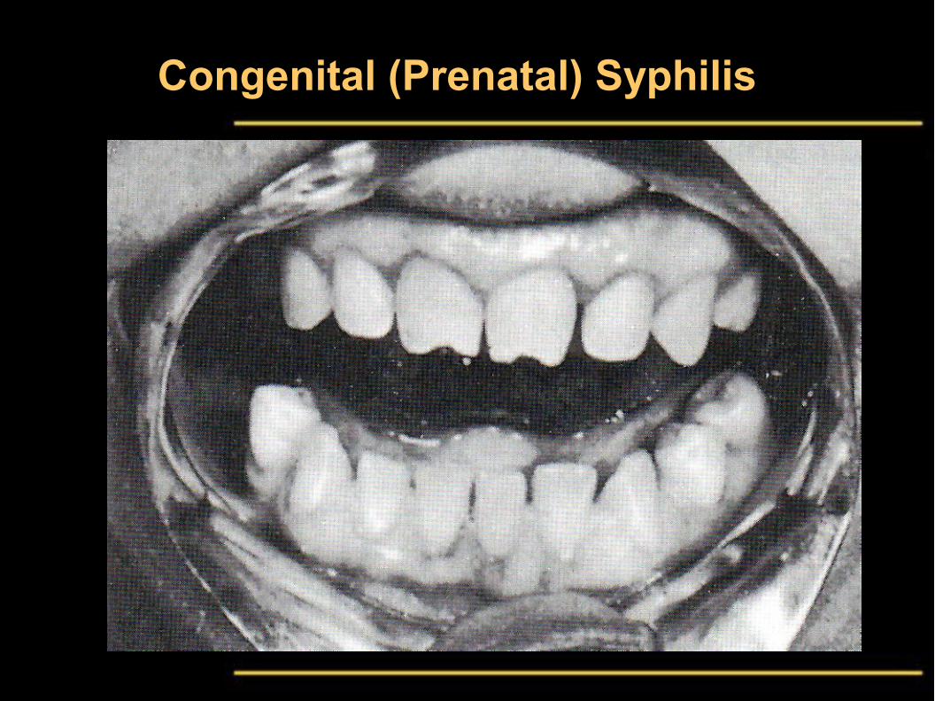
Congenital (Prenatal) Syphilis

Congenital (Prenatal) Syphilis – Gumma + Mulberry Molar
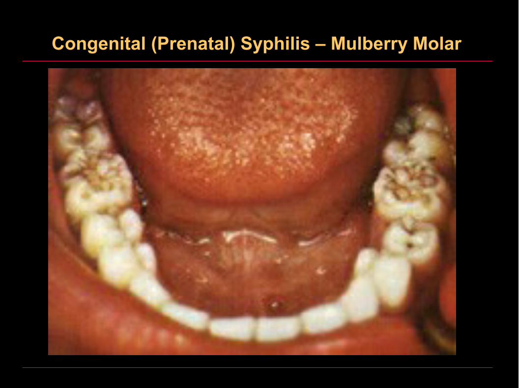
Congenital (Prenatal) Syphilis – Mulberry Molar
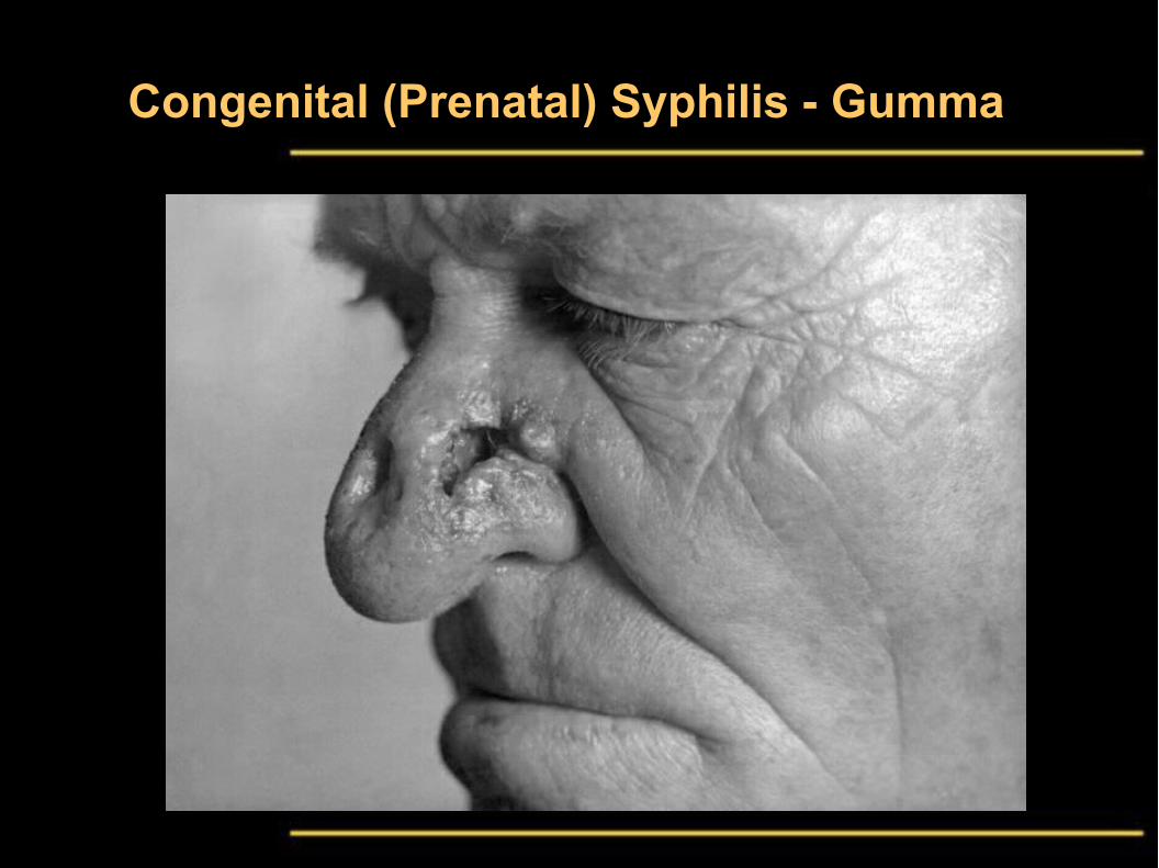
Congenital (Prenatal) Syphilis - Gumma

Congenital (Prenatal) Syphilis - Gumma
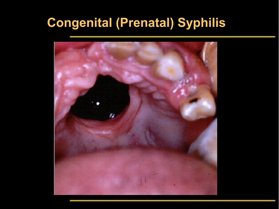
Congenital (Prenatal) Syphilis
Rhagades (Congenital Syphilis)
Linear fissures or scars radiating from mouth corners

Rhagades (Congenital Syphilis)
Linear fissures or scars radiating from mouth corners

Syphilis - Histologically
Leprosy (Hansen’s Disease)
• A chronic specific granulomatous inflammation of low infectivity caused by the acid-fast Mycobacterium Leprae.
• Leprosy is only moderately contagious; transmission of the disease requires frequent direct contact with an infected individual for a long period.
• Route of infection:
1. By inhalation of droplet infection
2. Through intact skin by direct contact. When infection develops in the skin, the initial lesion is the indeterminate macule.
Leprosy (Clinical Forms)
1- Lepromatous leprosy
2- Tuberculoid leprosy
Leprosy (Clinical Forms)
1. Lepromatous leprosy
• This occurs in patients with low cell-mediated immunity to the organism and produces widespread infection.
1. Tuberculoid leprosy
• This develops when there is a high cell-mediated immunity and the infection is limited to skin and nerves and the lesions contain very few organisms.
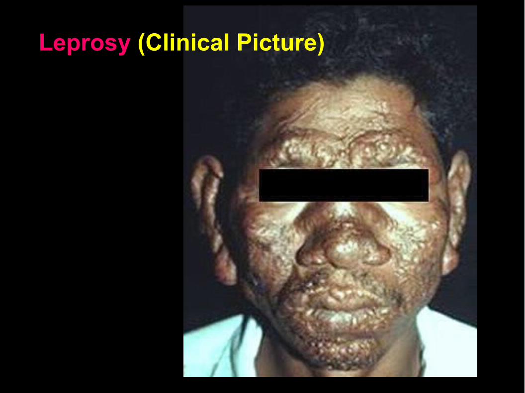
Leprosy (Clinical Picture)
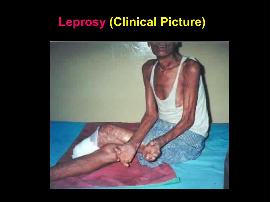
Leprosy (Clinical Picture)
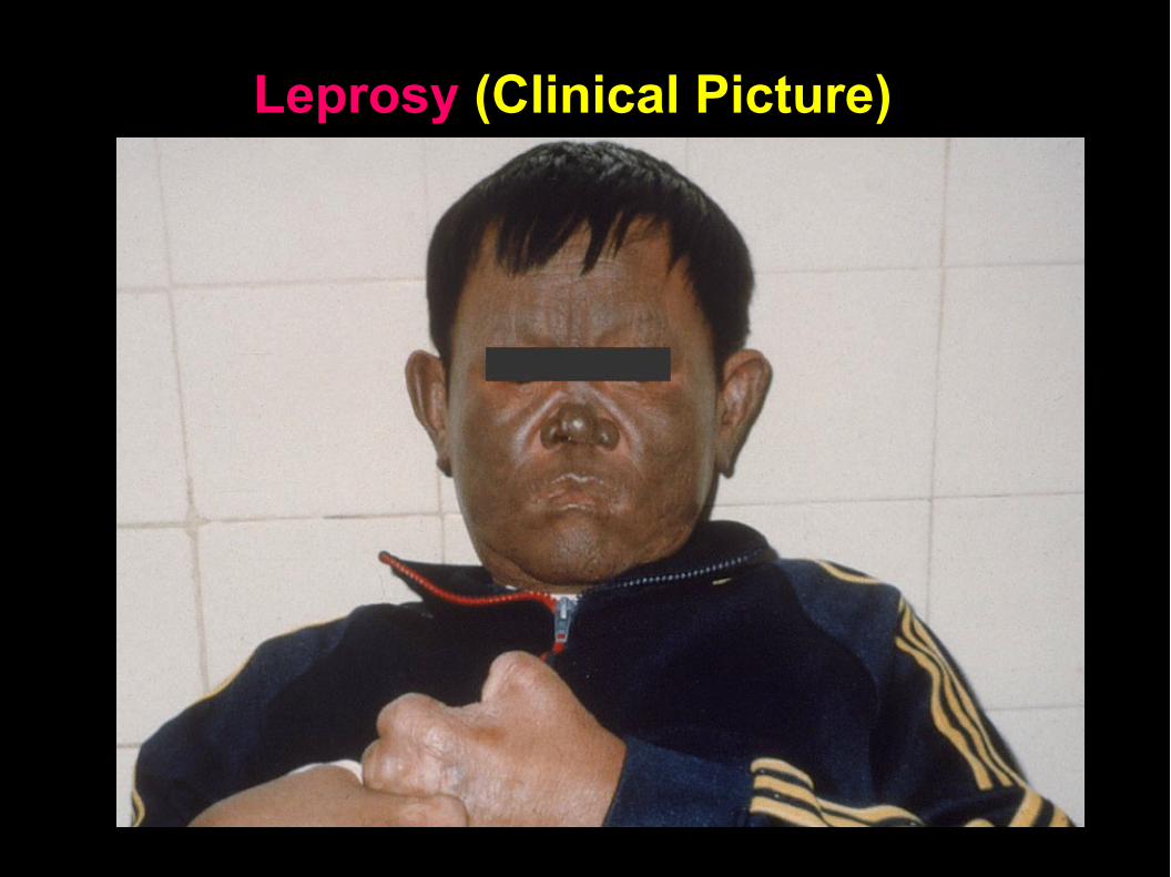
Leprosy (Clinical Picture)
Lepromatous leprosy
1. Skin involvement
2. Nerve involvement
3. Mucous membranes
4. Eyes
5. Testes
6. Death
7. Oral manifestations
Lepromatous leprosy
1. Skin involvement
• Papules
• Nodules covered by greasy skin
• Diffuse thickening, e.g. leonine facies
• Loss of hair
• On histology the skin shows
• Dermal granulomata rich in histiocytes
• Mycobacterium leprae in large numbers within histiocytes. These can be best demonstrated using the Ziehl-Neelsen method without acid differentiation.
Lepromatous leprosy
2. Nerve involvement
• Edema and ischemic necrosis
• Progressive fibrosis
• Peripheral neuritis which is symmetrical
• Anesthesia may lead to neuropathic arthropathy (Charcot’s joints) and trophic ulcers

Lepromatous leprosy
3. Mucous membrane involvement
• Nasal blockage and epistaxis
• Ulceration of the nasal septum
• Ulceration and stenosis of the larynx
Lepromatous leprosy
4. Eyes
• Keratitis
• Iritis
• Corneal ulceration
Lepromatous leprosy
5. Testes• Testicular atrophy leading to sterility and
gynaecomastia
5. Death may result from
• Respiratory infection; pneumonia, tuberculosis
• Septicaemia from chronic osteomyelitis following infection of bone marrow
• Renal failure due to chronic glomerulonephritis or amyloidosis
Lepromatous leprosy
7. Oral manifestations
• Multiple non-caseating granulomas are present in the
connective tissue
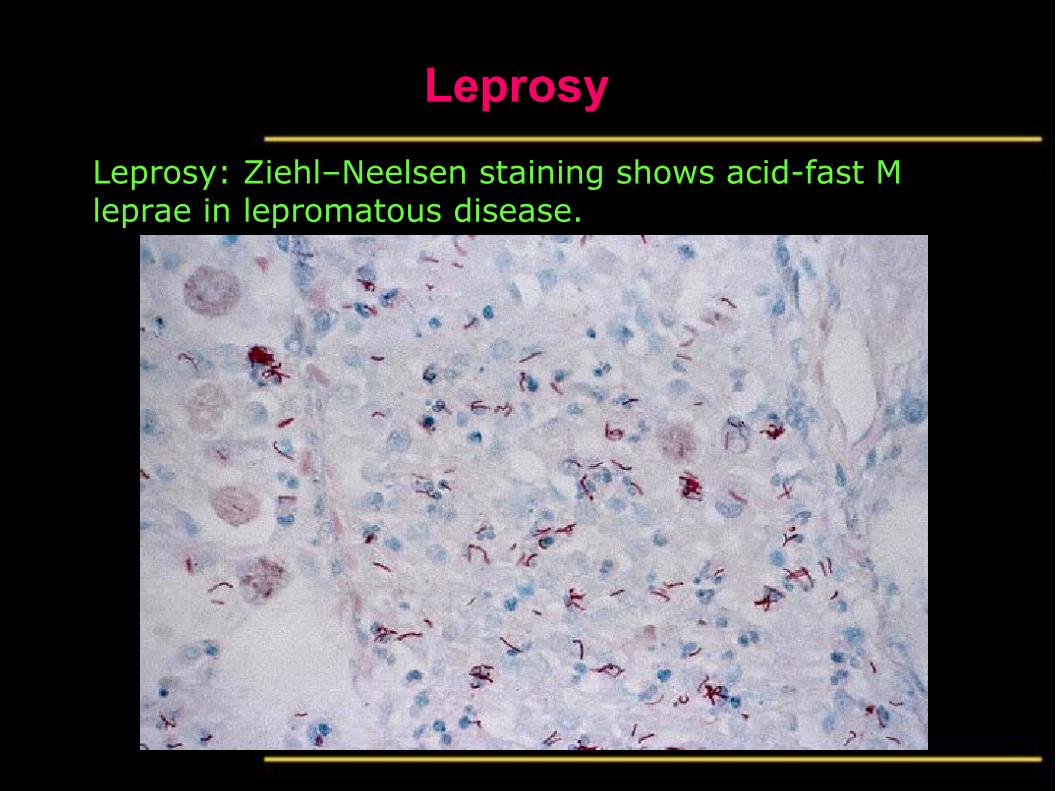
• Ziehl–Neelsen staining shows acid-fast lepra bacilli.
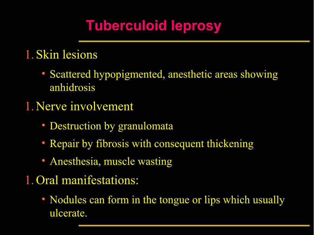
Tuberculoid leprosy
1. Skin lesions
• Scattered hypopigmented, anesthetic areas showing anhidrosis
1. Nerve involvement
• Destruction by granulomata
• Repair by fibrosis with consequent thickening
• Anesthesia, muscle wasting
1. Oral manifestations:
• Nodules can form in the tongue or lips which usually ulcerate.
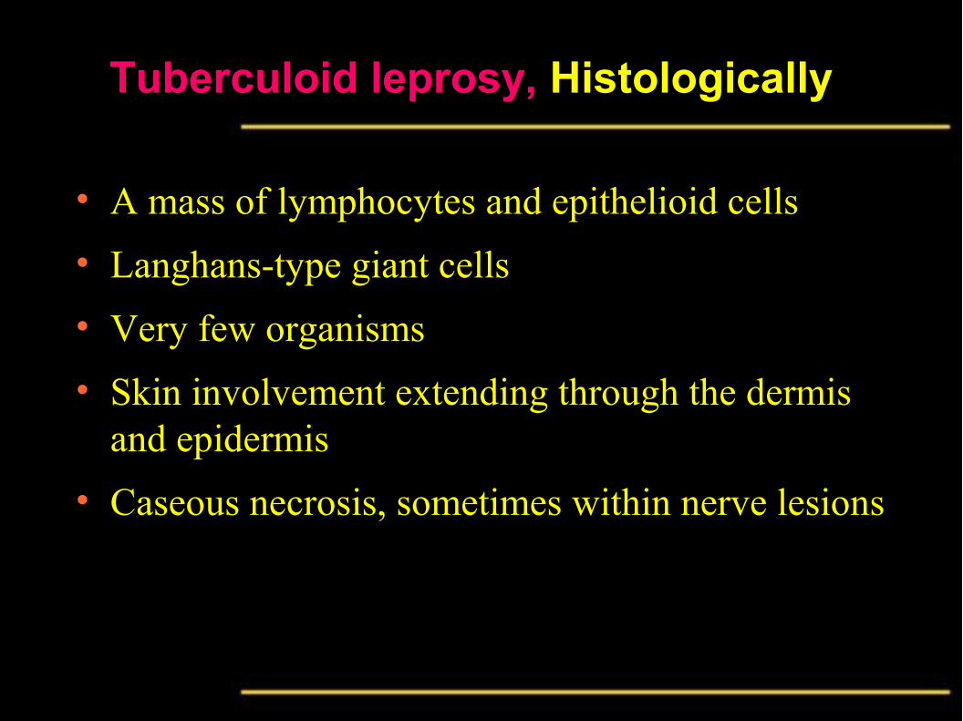
Tuberculoid leprosy, Histologically
• A mass of lymphocytes and epithelioid cells
• Langhans-type giant cells
• Very few organisms
• Skin involvement extending through the dermis and epidermis
• Caseous necrosis, sometimes within nerve lesions
Tuberculoid leprosy, Treatment
Multi-drug therapy, dapsone, rifampin and minocycline
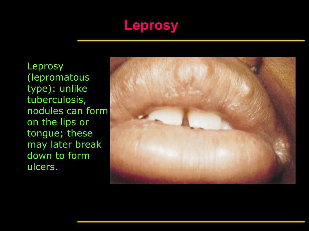
Leprosy
Leprosy (lepromatous type): unlike tuberculosis, nodules can form on the lips or tongue; these may later break down to form ulcers.
Leprosy
Leprosy: Ziehl–Neelsen staining shows acid-fast M leprae in lepromatous disease.
Leprosy
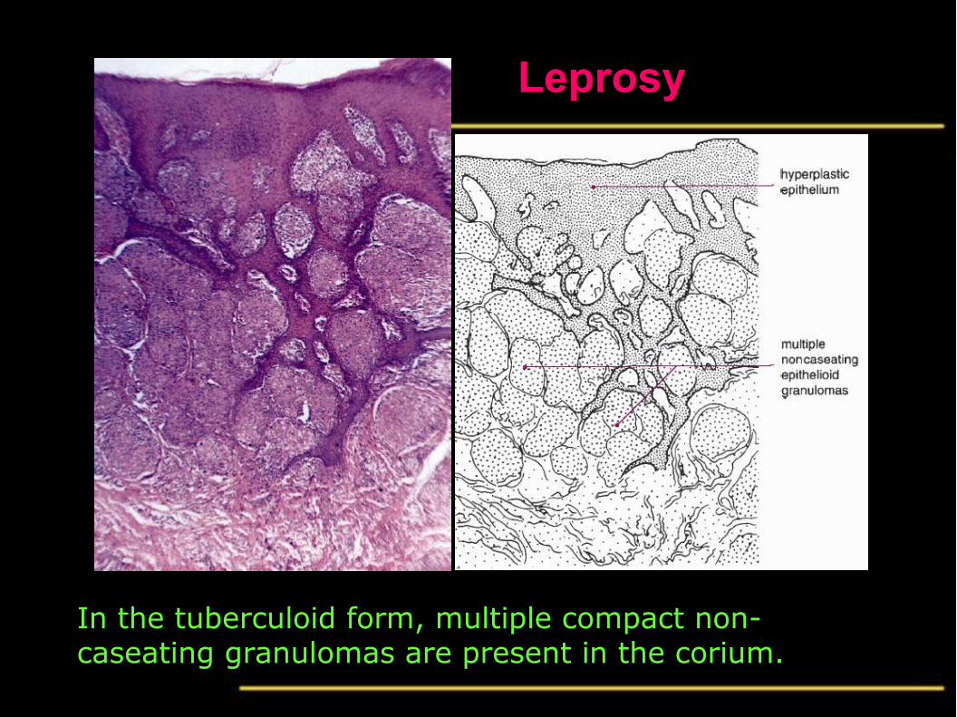
In the tuberculoid form, multiple compact non-caseating granulomas are present in the corium.
Tuberculosis
• A chronic specific granulomatous disease. The incidence of tuberculosis is increasing and multiple drug-resistant strains are becoming widespread.
• Etiology:
• Mycobacterium tuberculosis or mycobacterium bovis.
• Clinically:
• Tuberculosis of the oral cavity is rare.
• Usually secondary to pulmonary tuberculosis.
• Occurs in the oral mucosa, jaw bones or as tuberculous lymphadenitis may also affect submandibular or cervical lymph nodes.

Natural History of Pulmonary Tuberculosis
D e c r e a s e d i m m u n i t y
+P r i m a r y i n f e c t i o nu s u a l l y i n c h i l d r e n
G h o n ' s F o c u sM i d z o n e o f l u n g
H i l a r l y m p h n o d e
P r i m a r y C o m p l e x
F a t e
L o w i m m u n i t y
S p r e a d
G e n e r a l t oa l m o s t a l l
o r g a n s( M i l i a r y T B
I n d i v i d u a l o r g a n s e . g . :T B l y m p h a d e n i t i sT B m e n i n g i t i sT B o s t e o m y e l i t i s ( P o t t ' sd i s e a s e )
G o o d i m m u n i t y
H e a l i n g+ c a l c i f i c a t i o n o f G h o n ' s f o c u s
a n d h i l a r l y m p h n o d e
R e a c t i v a t i o n o r r e i n f e c t i o n ?
S p r e a d t o t h e a p e x o f t h e l u n g( T B b r o n c h o p n e u m o n i a )
Prim
aryP
ost-Prim
ary (A
dult)
Tuberculosis
1. Tuberculosis of the oral mucosa:• The most common site is the lip and the tip of tongue.
• The lesion consists of granulating ulcers, which are punched out.
1. Tuberculous osteomyelitis of the jaws:• TB of bone is usually secondary to pulmonary TB
• Common in the vertebrae (Pott’s disease), rare in the jaws
1. Tuberculous lymphadenitis:• May be presented to the dentist and he should be aware of
its features and management

Characteristics of Tuberuclous Lymphadenitis
• Firm or rubbery swelling, usually of a group of nodes
• Nodes typically become matted
• Abscess or sinus formation if neglected
• Calcified nodes from past healed disease may occur
• Diagnosis may be by Ziehl-Neelsen staining or PCR of fine-needle aspiration biopsy or by microscopic examination of surgical biopsy
• Ziehl-Neelsen staining may give false negative result
• Incision and drainage is contraindicated, affected nodes should be excised intact
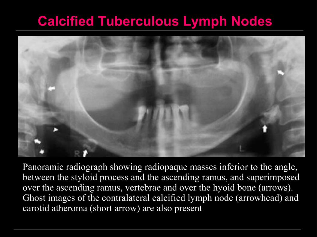
Calcified Tuberculous Lymph Nodes
Panoramic radiograph showing radiopaque masses inferior to the angle, between the styloid process and the ascending ramus, and superimposed over the ascending ramus, vertebrae and over the hyoid bone (arrows). Ghost images of the contralateral calcified lymph node (arrowhead) and carotid atheroma (short arrow) are also present
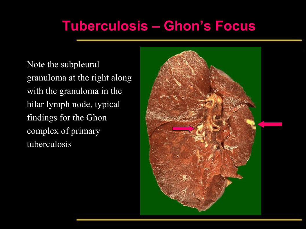
Tuberculosis – Ghon’s Focus
Note the subpleural
granuloma at the right along
with the granuloma in the
hilar lymph node, typical
findings for the Ghon
complex of primary
tuberculosis
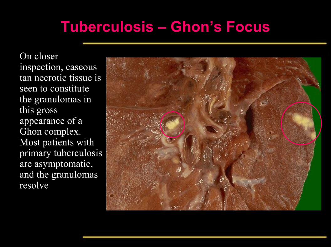
Tuberculosis – Ghon’s Focus
On closer inspection, caseous tan necrotic tissue is seen to constitute the granulomas in this gross appearance of a Ghon complex. Most patients with primary tuberculosis are asymptomatic, and the granulomas resolve

Tuberculosis GranulomaNote the rounded, focal nature of these granulomas at
low power magnification
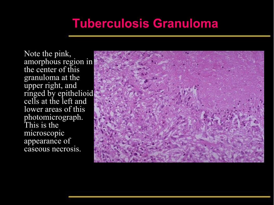
Tuberculosis Granuloma
Note the pink, amorphous region in the center of this granuloma at the upper right, and ringed by epithelioid cells at the left and lower areas of this photomicrograph. This is the microscopic appearance of caseous necrosis.
Tuberculosis Granuloma
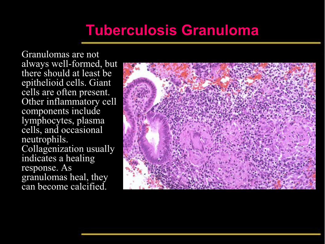
Granulomas are not always well-formed, but there should at least be epithelioid cells. Giant cells are often present. Other inflammatory cell components include lymphocytes, plasma cells, and occasional neutrophils. Collagenization usually indicates a healing response. As granulomas heal, they can become calcified.
Tuberculosis Granuloma
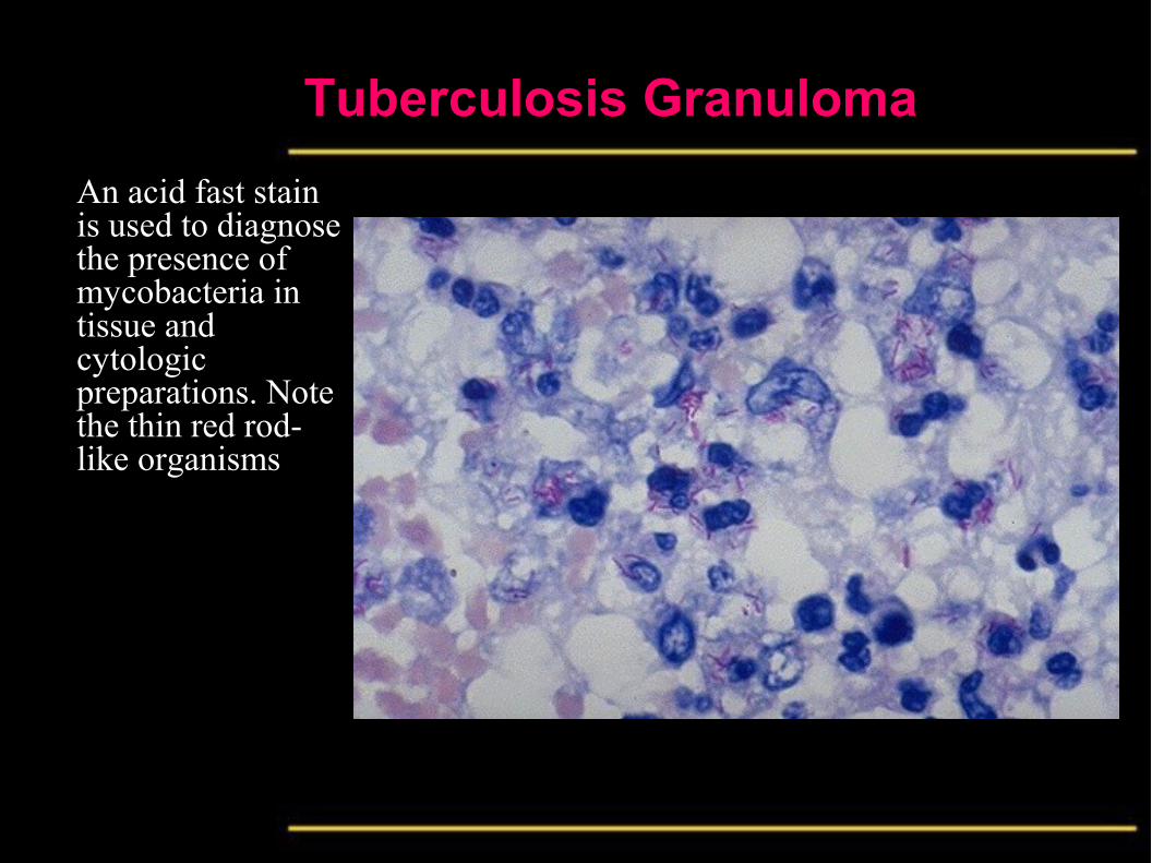
An acid fast stain is used to diagnose the presence of mycobacteria in tissue and cytologic preparations. Note the thin red rod-like organisms

Tuberculosis
Tuberculosis
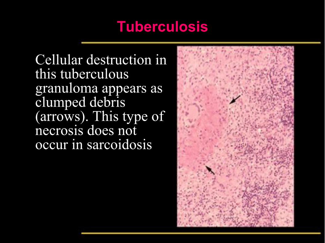
Cellular destruction in this tuberculous granuloma appears as clumped debris (arrows). This type of necrosis does not occur in sarcoidosis
Tuberculosis
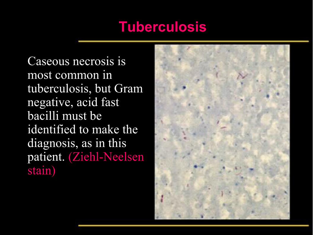
Caseous necrosis is most common in tuberculosis, but Gram negative, acid fast bacilli must be identified to make the diagnosis, as in this patient. (Ziehl-Neelsen stain)
Actinomycosis
Definition
• A type of chronic specific granulomatous infection,
which usually occurs at the soft tissues at the angle of
the mandible
• Involvement of other soft tissues exists, although is
uncommon
• In cattle the disease is known as lumpy jaw
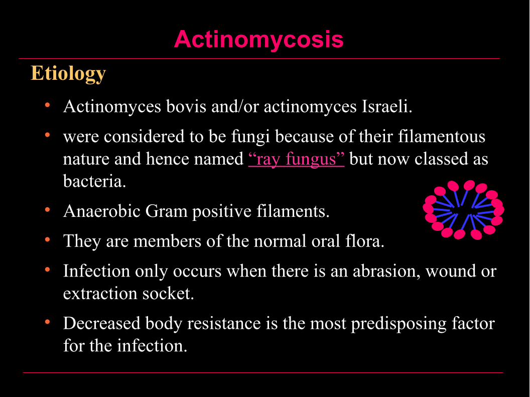
ActinomycosisEtiology
• Actinomyces bovis and/or actinomyces Israeli.
• were considered to be fungi because of their filamentous nature and hence named “ray fungus” but now classed as bacteria.
• Anaerobic Gram positive filaments.
• They are members of the normal oral flora.
• Infection only occurs when there is an abrasion, wound or extraction socket.
• Decreased body resistance is the most predisposing factor for the infection.
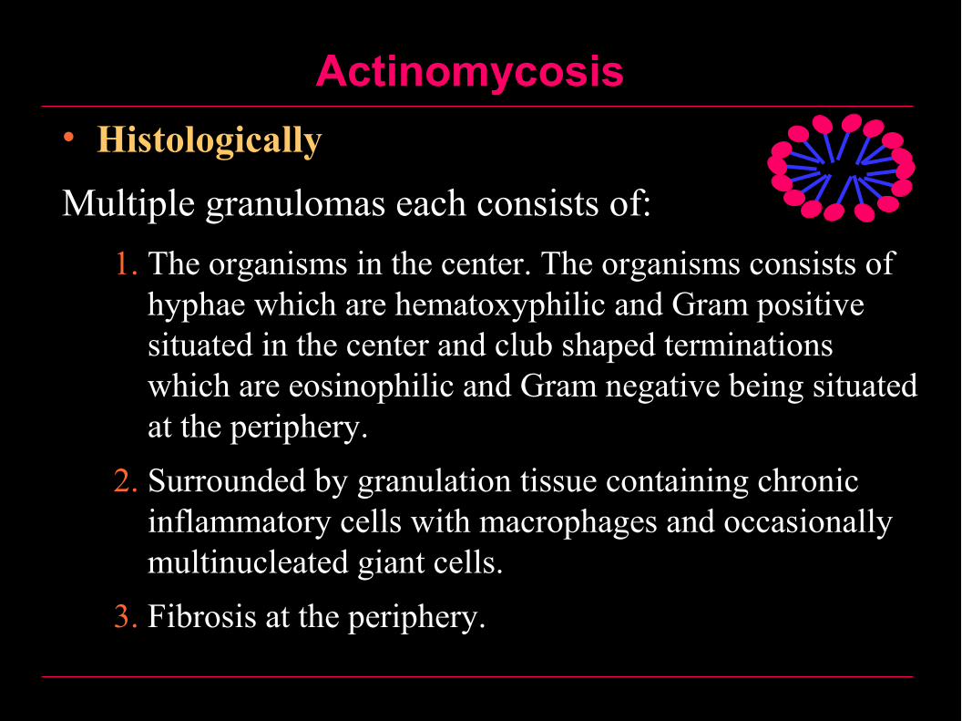
Actinomycosis
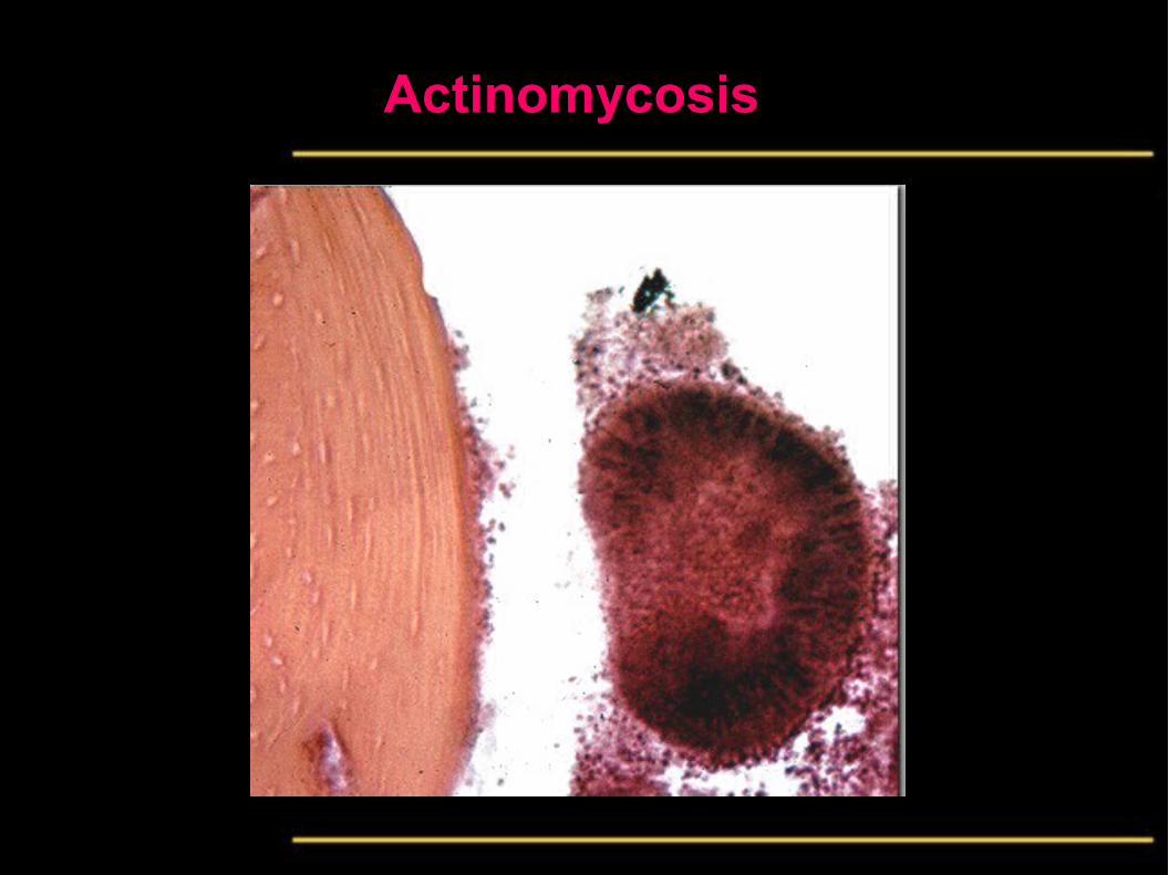
• Histologically
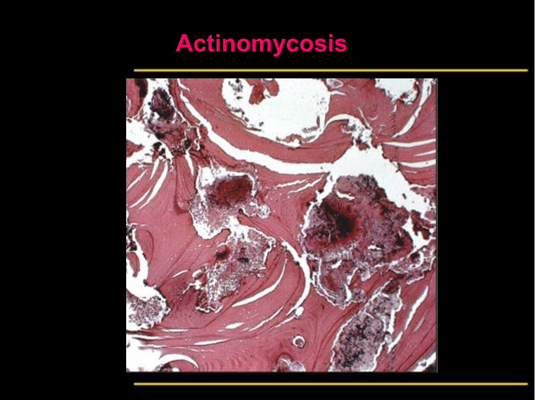
Multiple granulomas each consists of:
1. The organisms in the center. The organisms consists of hyphae which are hematoxyphilic and Gram positive situated in the center and club shaped terminations which are eosinophilic and Gram negative being situated at the periphery.
2. Surrounded by granulation tissue containing chronic inflammatory cells with macrophages and occasionally multinucleated giant cells.
3. Fibrosis at the periphery.
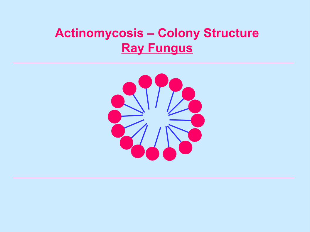
Actinomycosis – Colony StructureRay Fungus
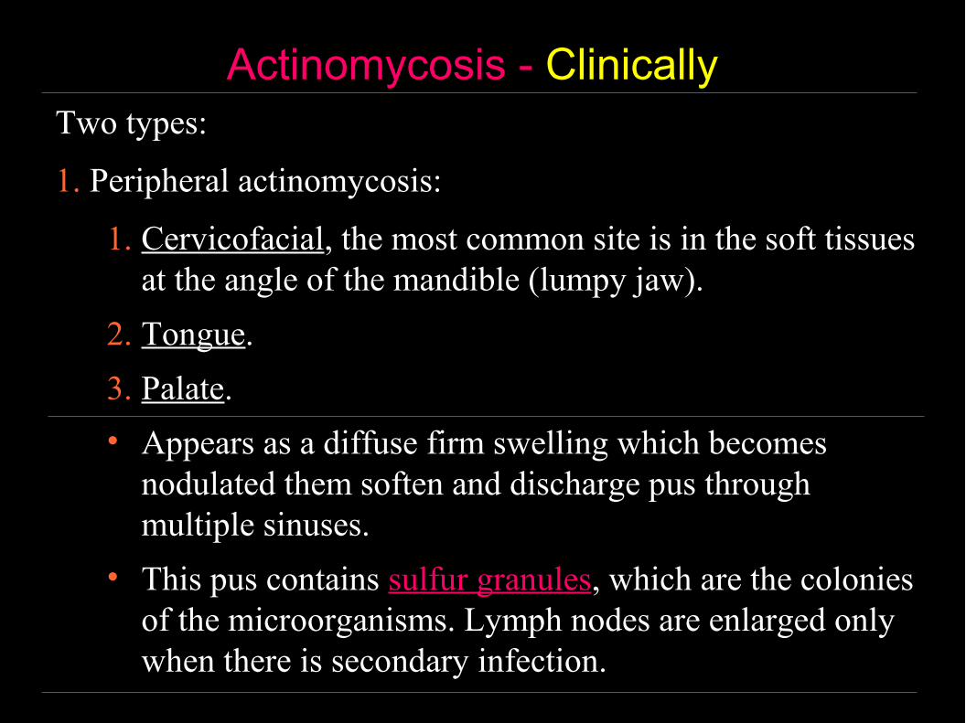
Actinomycosis - ClinicallyTwo types:
1. Peripheral actinomycosis:
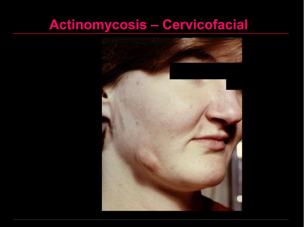
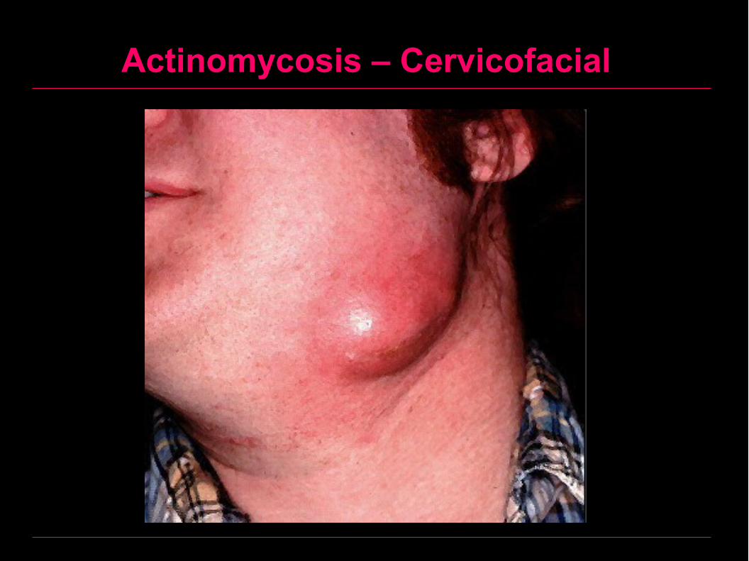
1. Cervicofacial, the most common site is in the soft tissues at the angle of the mandible (lumpy jaw).
2. Tongue.
3. Palate.
• Appears as a diffuse firm swelling which becomes nodulated them soften and discharge pus through multiple sinuses.
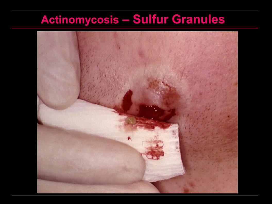
• This pus contains sulfur granules, which are the colonies of the microorganisms. Lymph nodes are enlarged only when there is secondary infection.
Actinomycosis
Clinically
2. Central actinomycosis:
• A very rare type of actinomycosis. The mandible is affected more than maxilla.
• In the central type the lesion runs a chronic course with multiple sinuses discharging pus
• Appears in x ray as a radiolucent area.
Actinomycosis
Diagnosis
• Any inflammatory swelling persists for more than 6
weeks or more without evidence of dental pathology
or osteomyelitis should arouse suspicion of
actinomycosis.
• Culture should be taken by incising a non- ruptured
nodule to avoid secondary infection, which destroys
sulfur granules.

Actinomycosis
Treatment
• Sensitivity test and giving the most appropriate antibiotic, which is usually penicillin
• Penicillin should be continued for 4-6 weeks or sometimes longer as surviving organisms may persist in the depth of the lesions to cause relapse.
• Abscesses should be drained surgically as they form.

Actinomycosis – Key Facts
Actinomyces are anaerobic bacteria
Were thought to be fungi for many years because they have filamentous forms, 0.5 to 0.8 microns in diameter, which appear to branch
The most common cause of actinomycosis is the organism Actinomyces israelii which infects both man and animals
In cattle, the disease is called "lumpy jaw" because of the huge abscess formed in the angle of the jaw
Actinomycosis – Key Facts
The organisms are Gram-positive hematoxyphilic non-acid fast branching filaments one micron in diameter
They become coated in clubs of Splendore-Hoeppli protein when the organisms are in tissues
Splendore-Hoeppli granules (phenomenon) are radiating or annular eosinophilic deposits of host derived protein and possibly of parasitic antigens which form around some fungi, helminths or bacterial colonies in tissues
Actinomycosis – Key Facts
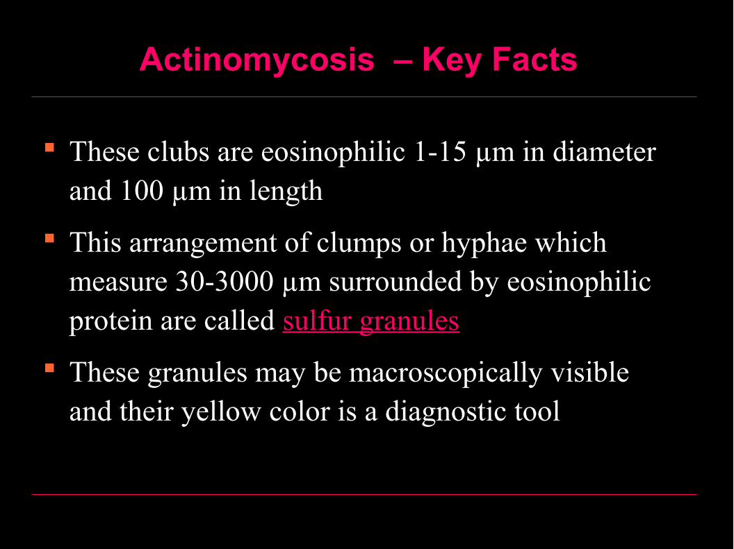
These clubs are eosinophilic 1-15 µm in diameter and 100 µm in length
This arrangement of clumps or hyphae which measure 30-3000 µm surrounded by eosinophilic protein are called sulfur granules
These granules may be macroscopically visible and their yellow color is a diagnostic tool
Actinomycosis – Cervicofacial
Actinomycosis – Cervicofacial
Actinomycosis – Sulfur Granules
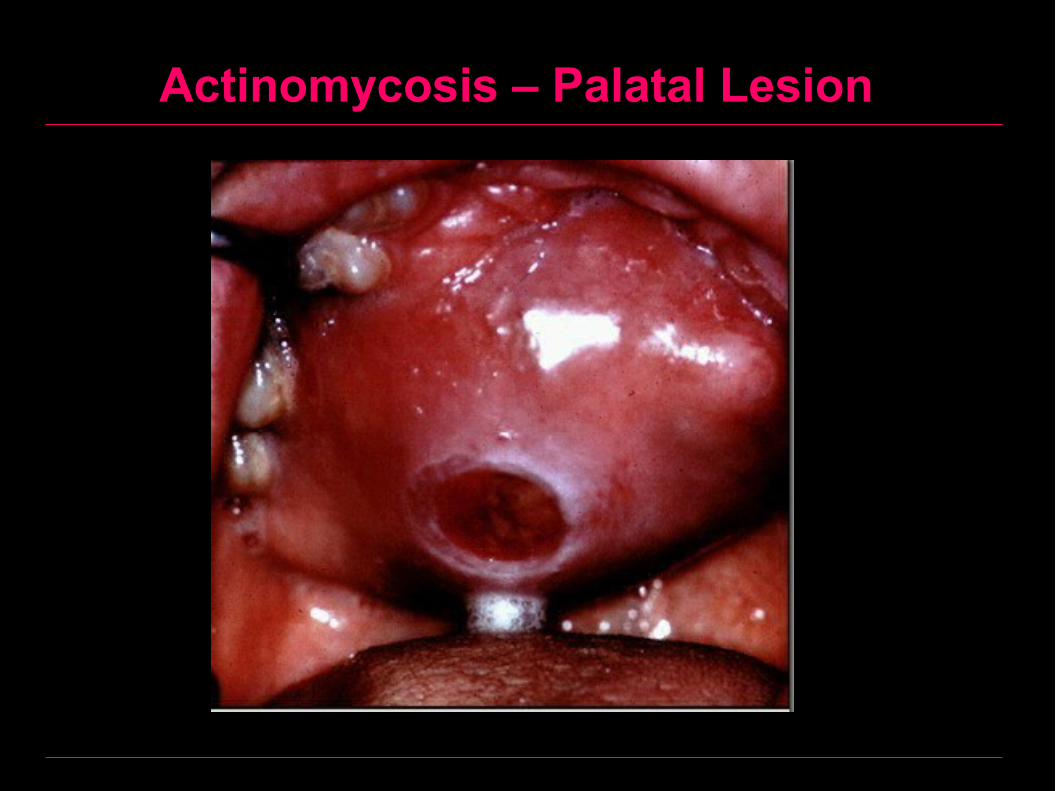
Actinomycosis – Palatal Lesion
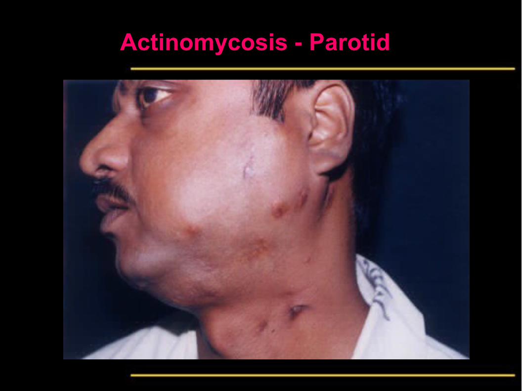
Actinomycosis - Parotid
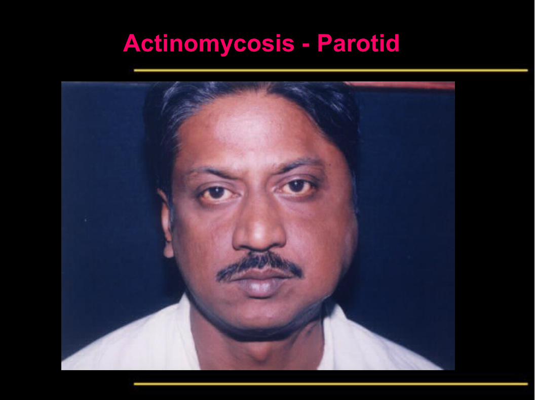
Actinomycosis - Parotid

Actinomycosis – Colonies from Abscess
Actinomycosis
Actinomycosis
Orofacial Granulomatosis
• A group of lesions characterized by orofacial swellings associated with non-caseating and non-infective granulomas.
• Although non-infective, they are mentioned herein for their relevance to the subject of granulomatous infections.
• Respond to systemic or intralesional injections of corticosteroids.
• Should not be confused with midfacial granulomas which are lethal diseases.

Orofacial Granulomatosis
• These lesions include:
1. Cheilitis granulomatosa
2. Melkersson Rosenthal syndrome
3. Sarcoidosis
4. Oral lesions of Crohn’s disease (inflammatory bowel disease)
Orofacial Granulomatosis - Sarcoidosis
• A multi-system granulomatous disease of unknown etiology which can affect lungs, eye, spleen, kidney, lymph nodes, skeleton or nervous tissues
• Etiology, unknown, the following causes were postulated:
1. Unidentified infectious agent
2. Autoimmunity
3. Allergy
4. Genetic factors
Orofacial Granulomatosis - Sarcoidosis
• Oral manifestations
1. Salivary glands enlargement
2. Intraoral submucosal nodules
3. Submandibular lymph node enlargement
4. Heerfordt’s syndrome which includes:
1. Uveal tract inflammation
2. Lacrimal gland enlargement
3. Parotid enlargement
4. Facial palsy
5. Fever
Orofacial Granulomatosis - Sarcoidosis
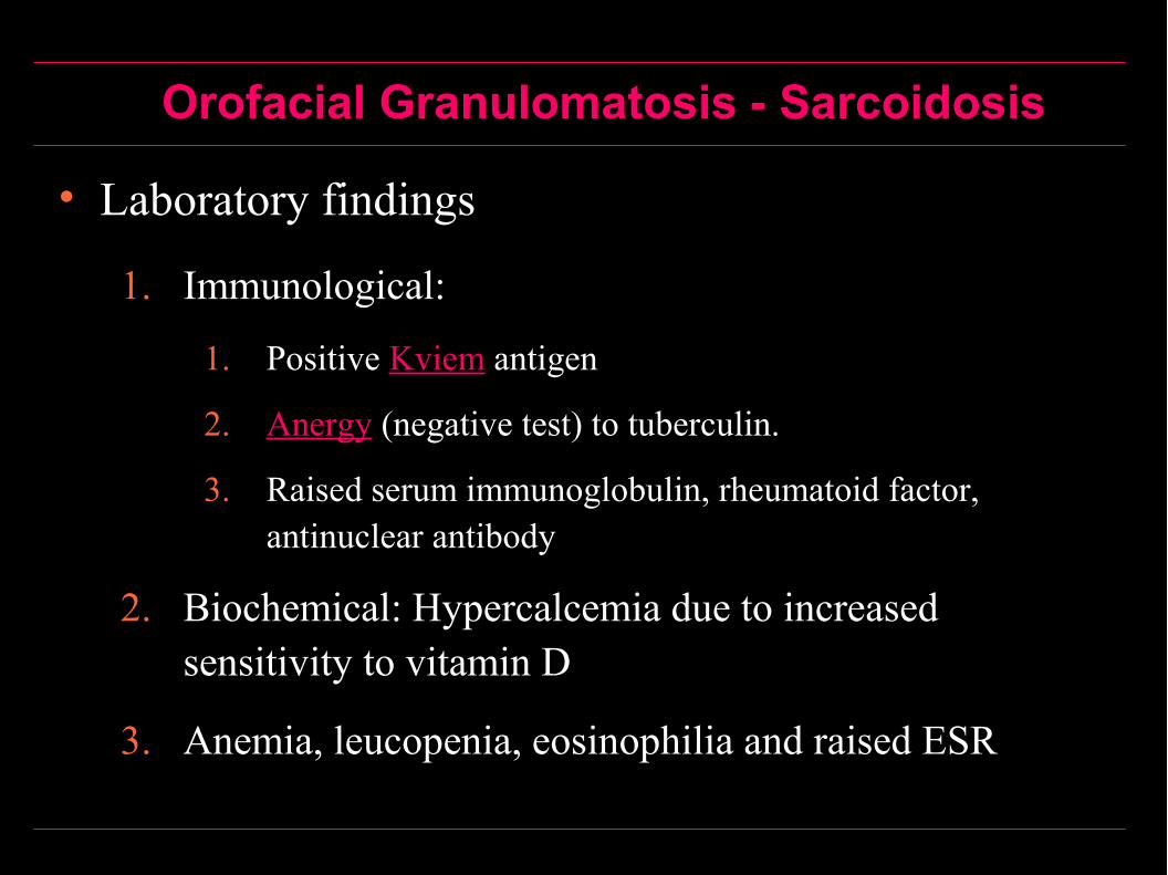
• Laboratory findings
1. Immunological:
1. Positive Kviem antigen
2. Anergy (negative test) to tuberculin.
3. Raised serum immunoglobulin, rheumatoid factor, antinuclear antibody
2. Biochemical: Hypercalcemia due to increased sensitivity to vitamin D
3. Anemia, leucopenia, eosinophilia and raised ESR
Sarcoidosis
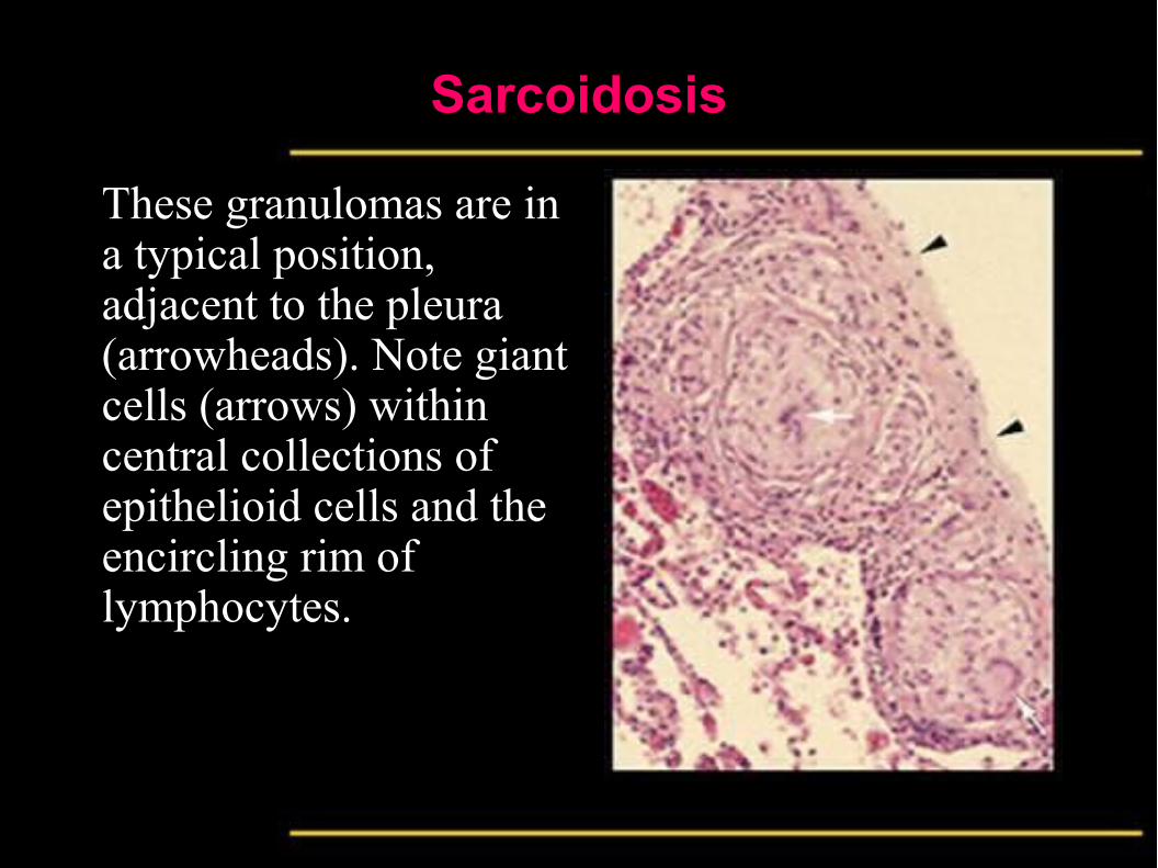
These granulomas are in a typical position, adjacent to the pleura (arrowheads). Note giant cells (arrows) within central collections of epithelioid cells and the encircling rim of lymphocytes.
Sarcoidosis
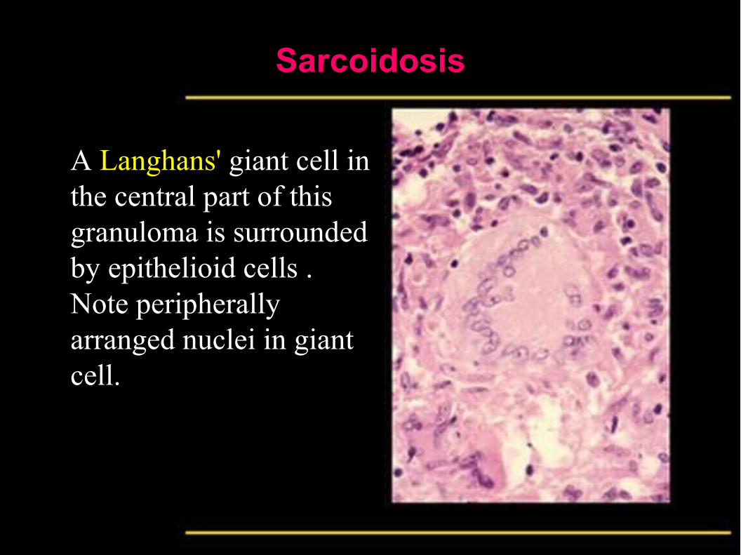
A Langhans' giant cell in the central part of this granuloma is surrounded by epithelioid cells . Note peripherally arranged nuclei in giant cell.
Sarcoidosis
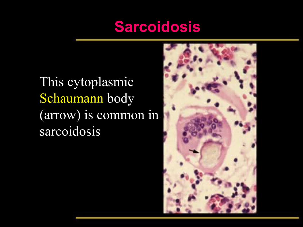
This cytoplasmic Schaumann body (arrow) is common in sarcoidosis
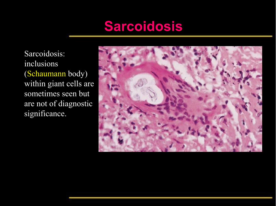
Sarcoidosis
Sarcoidosis: inclusions (Schaumann body) within giant cells are sometimes seen but are not of diagnostic significance.
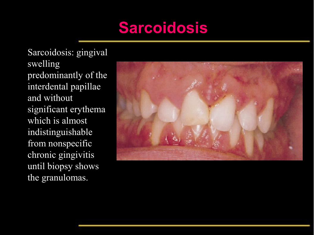
Sarcoidosis
Sarcoidosis: gingival swelling predominantly of the interdental papillae and without significant erythema which is almost indistinguishable from nonspecific chronic gingivitis until biopsy shows the granulomas.
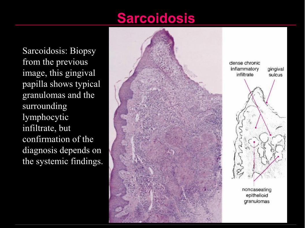
Sarcoidosis
Sarcoidosis: Biopsy from the previous image, this gingival papilla shows typical granulomas and the surrounding lymphocytic infiltrate, but confirmation of the diagnosis depends on the systemic findings.
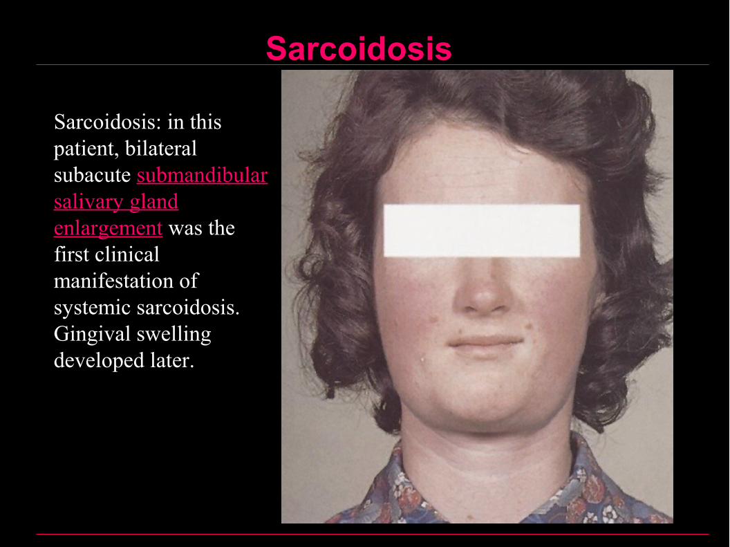
Sarcoidosis
Sarcoidosis: in this patient, bilateral subacute submandibular salivary gland enlargement was the first clinical manifestation of systemic sarcoidosis. Gingival swelling developed later.
Sarcoidosis
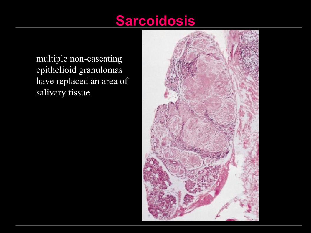
multiple non-caseating epithelioid granulomas have replaced an area of salivary tissue.

Crohn’s Disease (inflammatory bowel disease)
• A granulomatous disease affecting any part of the gastrointestinal tract from the mouth to the anus but typically affects the ileum
• Characterized by diarrhea, cramping, loss of appetite and weight with local abscesses and scarring
• Etiology: Unknown, the following causes were postulated:
1. Autoimmunity
2. Infective etiology due to Escherichia coli or viruses
3. About 20% of cases of Crohn's disease appear to run in families.

Orofacial Granulomatosis – Crohn’s Disease
Oral manifestations
1. Recurrent aphthous ulcers
2. Diffuse swellings of lips, cheek or gingiva
3. Cobblestone swellings of the oral mucosa with fissuring and hyperplastic growths
4. Mucosal tags
Treatment:
• Oral lesions may resolve when intestinal Crohn’s disease is controlled with corticosteroids or sulfasalazine
• Alternatively, oral lesions may respond to oral sulfasalazine or to intralesional injections of corticosteroids

Crohn’s DiseaseCrohn’s disease: soft nodular proliferation of the oral mucosa is a typical feature and, in this case, was associated with facial swelling and intermittent diarrhea. Cobblestone swellings

Crohn’s DiseaseCrohn’s disease: gross labial swelling and intraoral mucosal proliferation with typical histological changes led to the finding of extensive intestinal involvement.
Crohn’s Disease
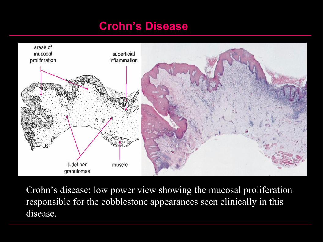
Crohn’s disease: low power view showing the mucosal proliferation responsible for the cobblestone appearances seen clinically in this disease.
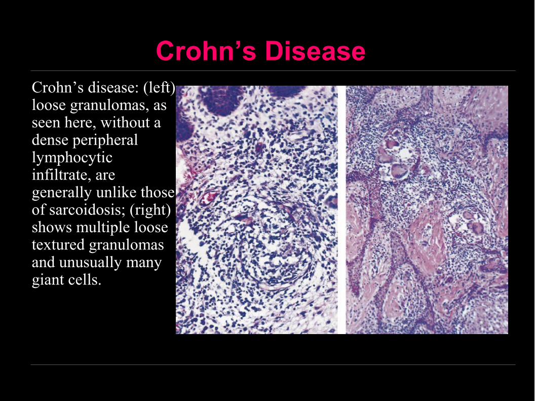
Crohn’s DiseaseCrohn’s disease: (left) loose granulomas, as seen here, without a dense peripheral lymphocytic infiltrate, are generally unlike those of sarcoidosis; (right) shows multiple loose textured granulomas and unusually many giant cells.
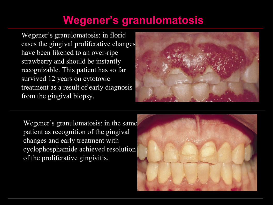
Wegener’s granulomatosisWegener’s granulomatosis: in florid cases the gingival proliferative changes have been likened to an over-ripe strawberry and should be instantly recognizable. This patient has so far survived 12 years on cytotoxic treatment as a result of early diagnosis from the gingival biopsy.
Wegener’s granulomatosis: in the same patient as recognition of the gingival changes and early treatment with cyclophosphamide achieved resolution of the proliferative gingivitis.
Wegener’s granulomatosis
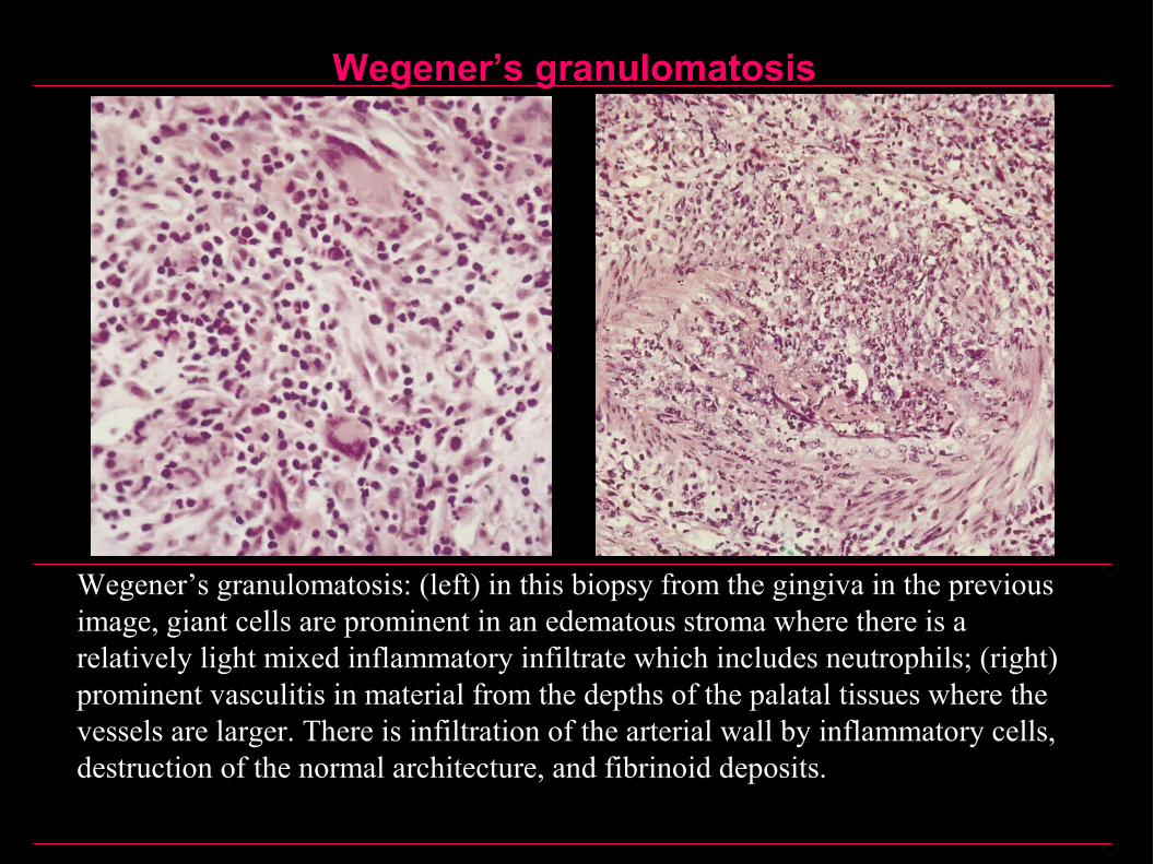
Wegener’s granulomatosis: (left) in this biopsy from the gingiva in the previous image, giant cells are prominent in an edematous stroma where there is a relatively light mixed inflammatory infiltrate which includes neutrophils; (right) prominent vasculitis in material from the depths of the palatal tissues where the vessels are larger. There is infiltration of the arterial wall by inflammatory cells, destruction of the normal architecture, and fibrinoid deposits.
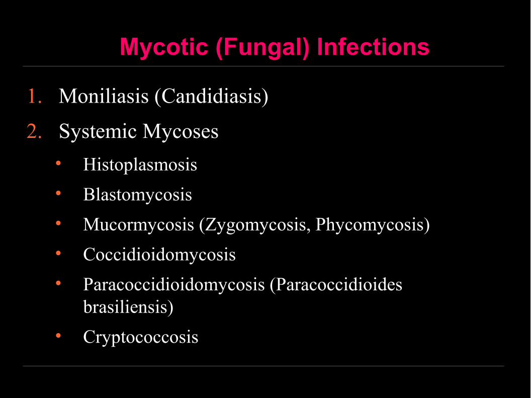
Mycotic (Fungal) Infections
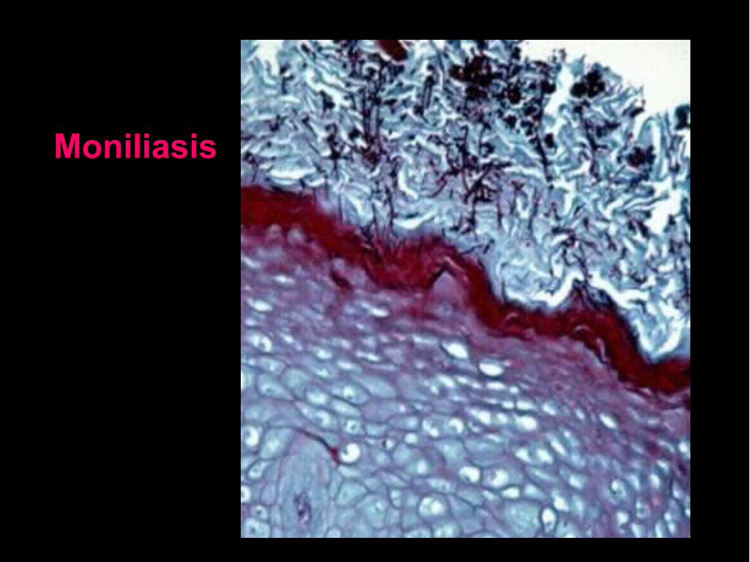
1. Moniliasis (Candidiasis)
2. Systemic Mycoses
• Histoplasmosis
• Blastomycosis
• Mucormycosis (Zygomycosis, Phycomycosis)
• Coccidioidomycosis
• Paracoccidioidomycosis (Paracoccidioides brasiliensis)
• Cryptococcosis
Candidiasis
Candidiasis
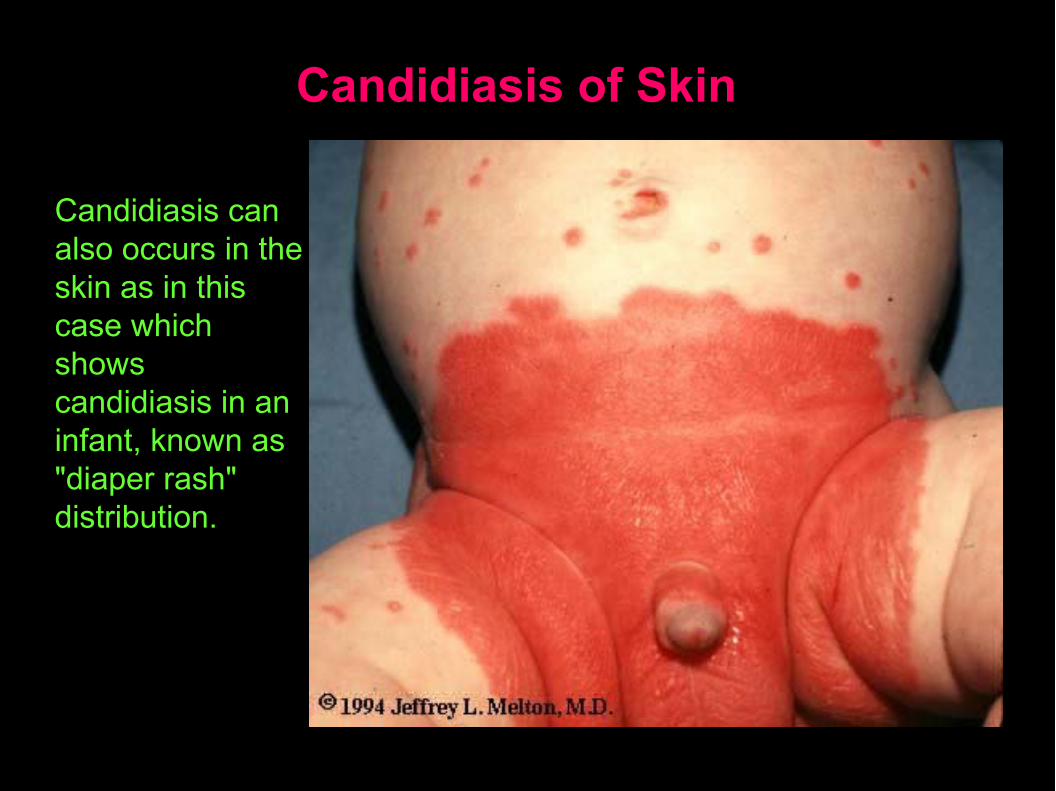
Candidiasis of Skin
Candidiasis can also occurs in the skin as in this case which shows candidiasis in an infant, known as "diaper rash" distribution.
Moniliasis (Candidiasis)
Definition:
• This is a fungal infection of the oral mucosa caused by candida albicans.
• Candida albicans is one of the normal oral flora
• Oral candidiasis and candidosis are synonyms
• It is disease of the diseased
Moniliasis (Etiology)
Local Predisposing Factors:
• Loss of integrity of the oral mucosa as in thin or ulcerated epithelium, resulting from an ill-fitting denture, or after chemotherapy or radiotherapy, ect., allows for Candida to adhere on the epithelial cells, penetrate the mucosa, and cause infection.
• Heavy smoking, leukoplakia and other oral mucosal diseases
• Xerostomia.
• Topical corticosteroids.
• Excessive moisture, particularly at mouth corners.
Moniliasis (Etiology)
Systemic Predisposing Factors:
• Immunodeficiency and immunosuppression.
• Corticosteroid therapy due to immunosuppression.
• Broad-spectrum antibiotics.
• Pregnancy.
• Dietary deficiency, such as poor nutrition, iron and vitamin deficiencies.
• Endocrine disorders, such as diabetes mellitus, hematologic dyscrasias, malignant diseases, age (neonatal, elderly)
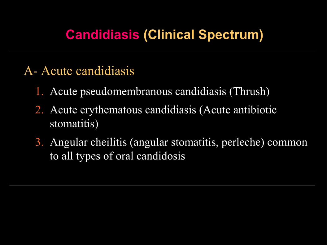
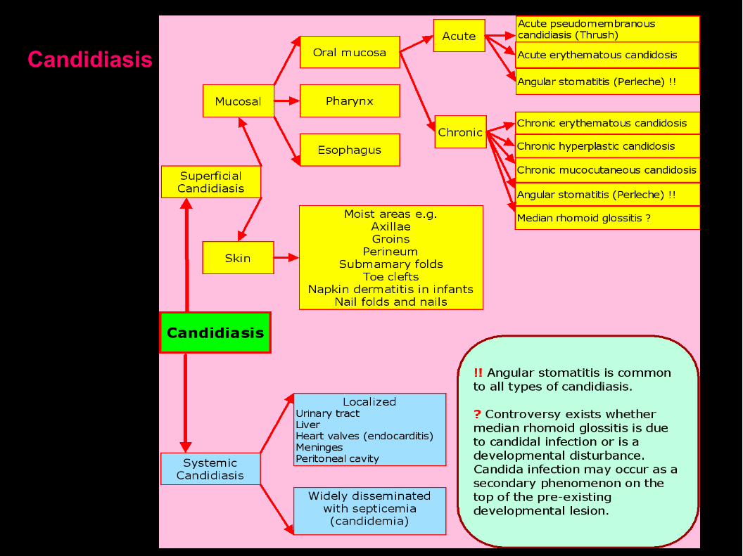
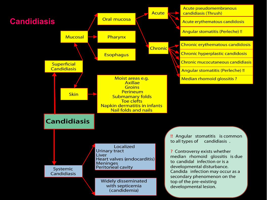
Candidiasis (Clinical Spectrum)
A- Acute candidiasis
1. Acute pseudomembranous candidiasis (Thrush)
2. Acute erythematous candidiasis (Acute antibiotic stomatitis)
3. Angular cheilitis (angular stomatitis, perleche) common to all types of oral candidosis
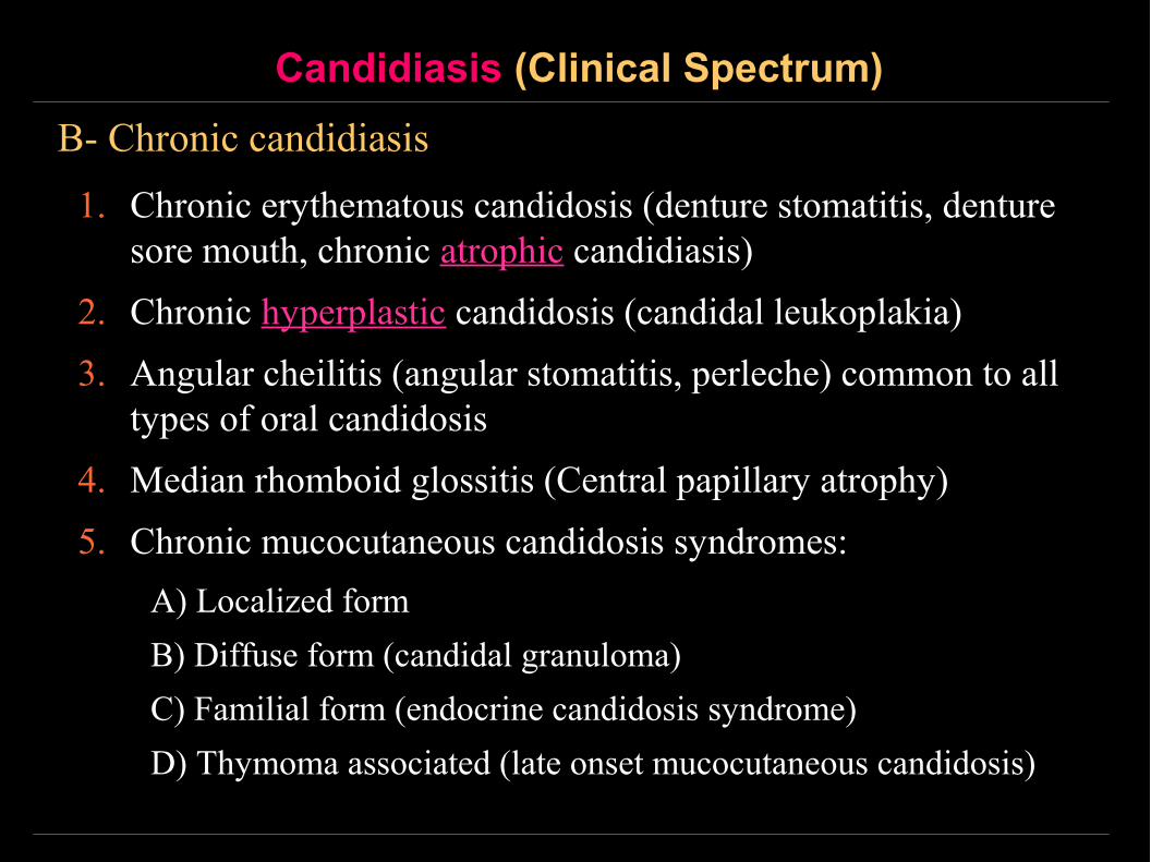
Candidiasis (Clinical Spectrum)
B- Chronic candidiasis
1. Chronic erythematous candidosis (denture stomatitis, denture sore mouth, chronic atrophic candidiasis)
2. Chronic hyperplastic candidosis (candidal leukoplakia)
3. Angular cheilitis (angular stomatitis, perleche) common to all types of oral candidosis
4. Median rhomboid glossitis (Central papillary atrophy)
5. Chronic mucocutaneous candidosis syndromes:
A) Localized form
B) Diffuse form (candidal granuloma)
C) Familial form (endocrine candidosis syndrome)
D) Thymoma associated (late onset mucocutaneous candidosis)

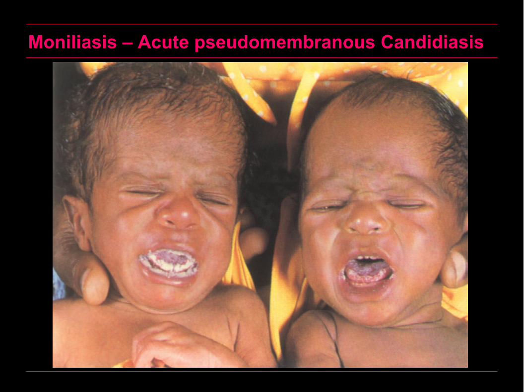
Acute pseudomembranous candidiasis (Thrush)
Any part of the oral mucosa may be affected especially the tongue, palate and buccal mucosa, mucobuccal folds, oropharynx and tongue.
Appears as white creamy patches similar to milk curds that can be wiped off leaving a raw red bleeding surface.
Pseudomembranous candidosis is rarely painful, however, some cases complain of burning sensation.
Extension of infection to the area of the oral commissures may occur and is known as angular stomatitis or perleche
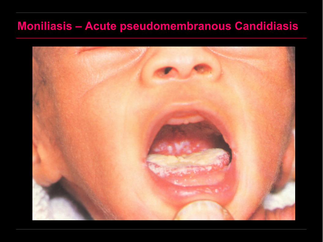
Moniliasis – Acute pseudomembranous Candidiasis
Moniliasis – Acute pseudomembranous Candidiasis
Candidiasis - Pseudomembranous Candidosis
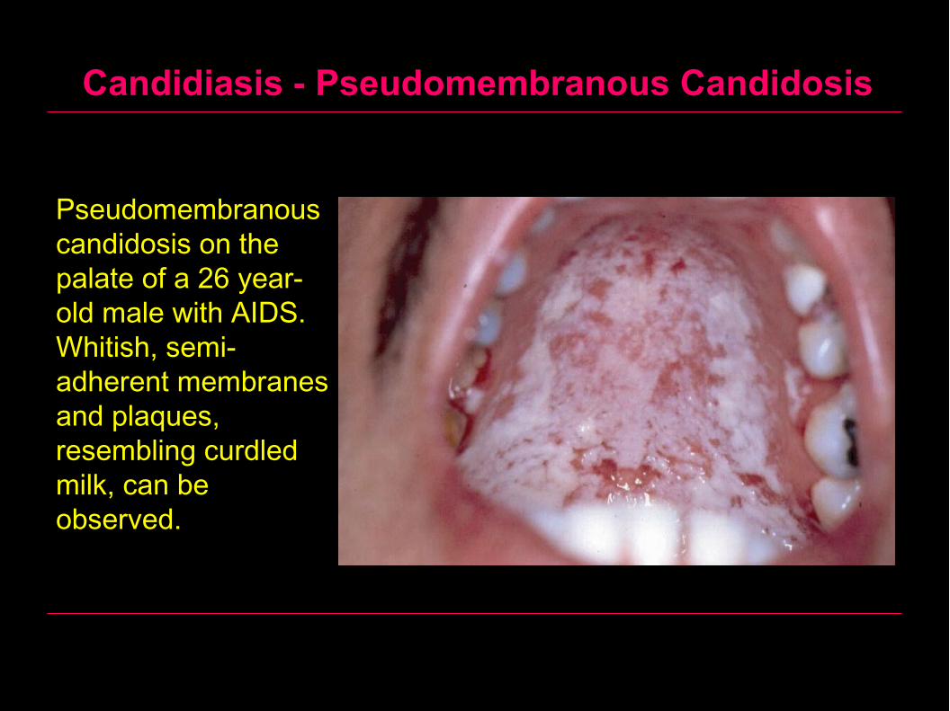
Pseudomembranous candidosis on the palate of a 26 year-old male with AIDS. Whitish, semi-adherent membranes and plaques, resembling curdled milk, can be observed.
Acute erythematous candidiasis
Persistence of acute pseudomembranous candidiasis may result in loss of the pseudomembrane with presentation of a red lesion known as acute erythematous candidiasis.
Remnants of the pseudomembrane may be noted in some areas.
There are also multiple erosions and intense inflammation associated with burning sensation.
This form was known as antibiotic stomatitis because of its association with broad-spectrum antibiotic treatment.
Withdrawal of the antibiotic, if possible and institution of oral hygiene lead to improvement.
Candidiasis – Acute Erythematous
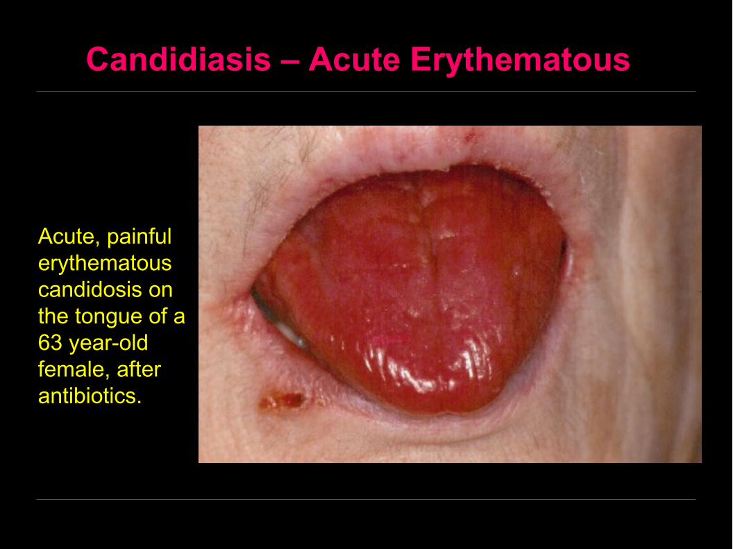
Acute, painful erythematous candidosis on the tongue of a 63 year-old female, after antibiotics.

Chronic Candidiasis
Chronic erythematous candidiasis
This form usually occurs in patients who wear complete upper dentures. There is a distinct predilection for the palatal mucosa as compared with the mandibular alveolar arch.
Chronic low-grade trauma resulting from poor denture fit and failure to remove the denture at night may contribute to the development of this condition.
Clinically, the lesion appears as a bright red, velvety surface with little keratinization.
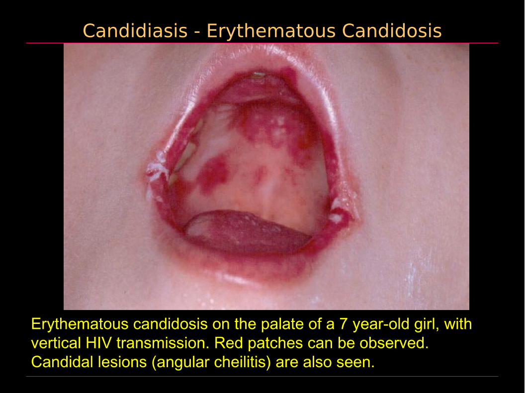
Candidiasis - Erythematous Candidosis
Erythematous candidosis on the palate of a 7 year-old girl, with vertical HIV transmission. Red patches can be observed. Candidal lesions (angular cheilitis) are also seen.

Candidiasis – Chronic Erythematous Candidosis
Angular cheilitis and erythematous candidosis on the tongue of a 69 year-old male. Patient wore dentures. Red fissures radiate from both comissures, while a red patch can be seen on the dorsum of the tongue.
Chronic hyperplastic candidiasis
A hyperplastic tissue response against chronic candidal infection.
It occurs in adults with no apparent predisposition to infection by candida albicans and it is believed by some clinicians to represent a premalignant lesion.
When occurring in the retro-commissural area, the lesion resembles speckled leukoplakia and in some classifications is known as candidal leukoplakia.

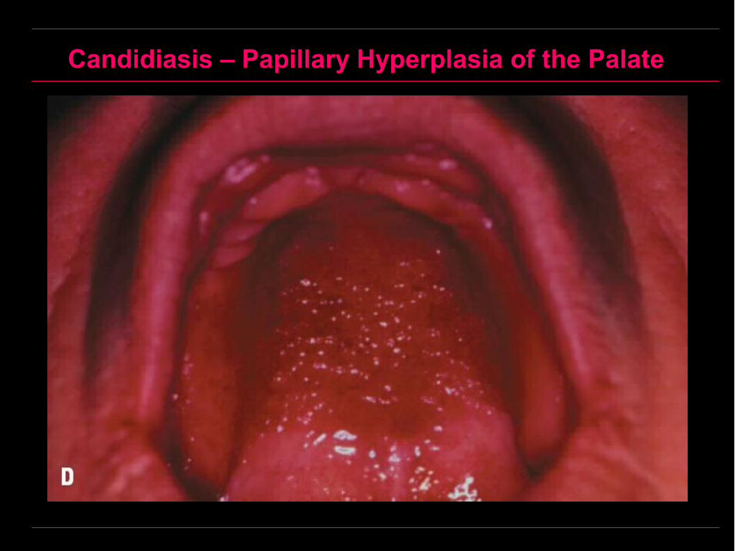
Chronic hyperplastic candidiasis
Papillary Hyperplasia of the Palate is considered to be a subtype of chronic hyperplastic candidiasis. These are individual nodules or papules measuring 2 to 3 mm in diameter on an erythematous background found usually under ill-fitting upper dentures.
Differential diagnosis of candidal leukoplakia and leukoplakia on the basis of clinical picture only is difficult. Differentiation should therefore depends on:
1- Finding PAS positive hyphae in histological smears or in tissue sections.
2- High candidal antibody titre in serum and saliva.

Chronic Hyperplastic Candidosis

Chronic Hyperplastic Candidosis
Candidiasis – Papillary Hyperplasia of the Palate
Angular cheilitis
Angular stomatitis is typically caused by leakage of candida infected saliva at the angles of the mouth.
It can be seen in infantile thrush, in denture wearers or in association with chronic hyperplastic candidiasis.
Hence, angular cheilitis can associate other types of candidiasis and is a characteristic feature of candidal infection.
The condition may occur in patients wearing denture with deep over-bite resulting form loss of vertical dimensions of dentures.
Angular cheilitis
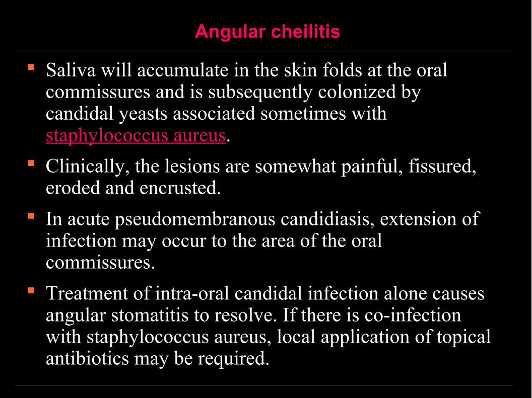
Saliva will accumulate in the skin folds at the oral commissures and is subsequently colonized by candidal yeasts associated sometimes with staphylococcus aureus.
Clinically, the lesions are somewhat painful, fissured, eroded and encrusted.
In acute pseudomembranous candidiasis, extension of infection may occur to the area of the oral commissures.
Treatment of intra-oral candidal infection alone causes angular stomatitis to resolve. If there is co-infection with staphylococcus aureus, local application of topical antibiotics may be required.
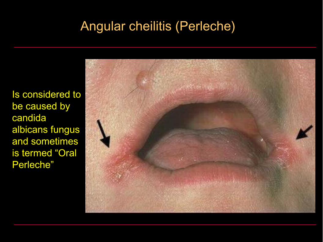
Angular cheilitis (Perleche)
Is considered to be caused by candida albicans fungus and sometimes is termed “Oral Perleche”
Median rhomboid glossitis(Central papillary atrophy)
A developmental disturbance of the tongue which sometimes may be secondarily infected with candida albicans.
Since MRG is never seen in children, its developmental etiology is denied by some authors.
The role of candida albicans as the primary etiology of the condition is still controversial because of the fixed site and shape of the lesion.
Therefore, the etiology of median rhomboid glossitis remains speculative.
Chronic mucocutaneous candidiasis
Chronic mucocutaneous candidiasis is a persistent candidal infection of the skin and mucous membranes secondary to defective immune response.
Four forms have been recognized:
1. Localized form
2. Diffuse form
3. Familial form (endocrine candidosis syndrome)
4. Thymoma associated
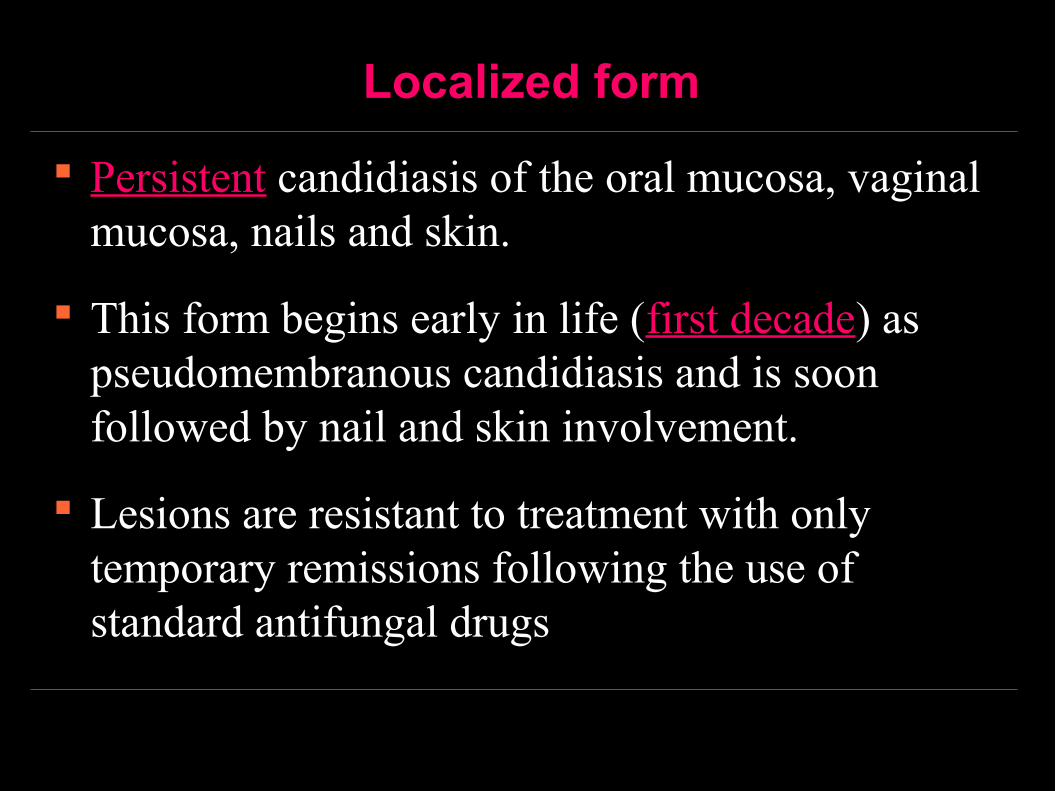
Localized form
Persistent candidiasis of the oral mucosa, vaginal mucosa, nails and skin.
This form begins early in life (first decade) as pseudomembranous candidiasis and is soon followed by nail and skin involvement.
Lesions are resistant to treatment with only temporary remissions following the use of standard antifungal drugs
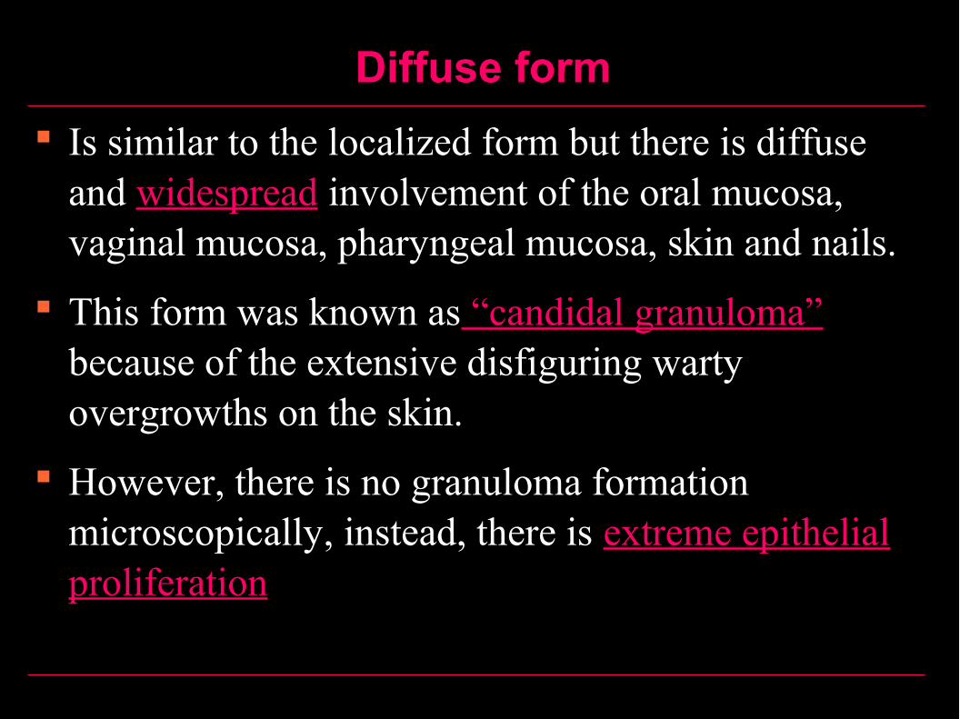
Diffuse form
Is similar to the localized form but there is diffuse and widespread involvement of the oral mucosa, vaginal mucosa, pharyngeal mucosa, skin and nails.
This form was known as “candidal granuloma” because of the extensive disfiguring warty overgrowths on the skin.
However, there is no granuloma formation microscopically, instead, there is extreme epithelial proliferation
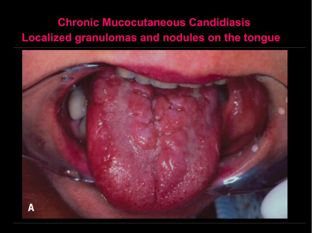
Chronic Mucocutaneous CandidiasisLocalized granulomas and nodules on the tongue
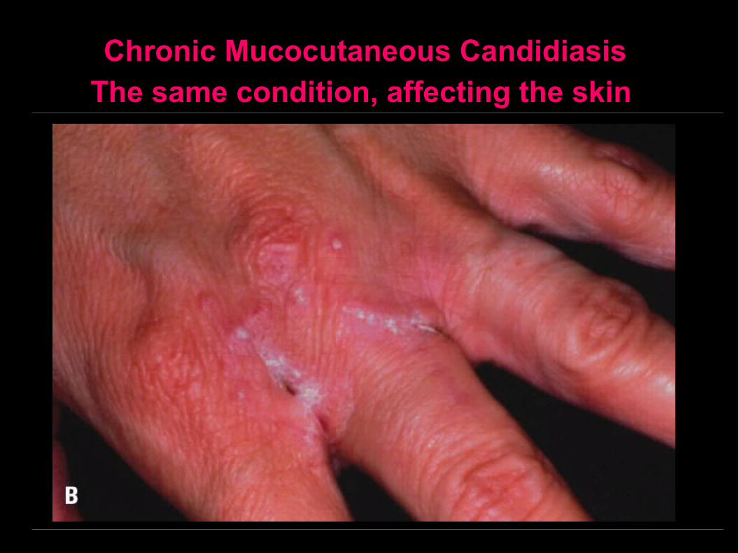
Chronic Mucocutaneous CandidiasisThe same condition, affecting the skin
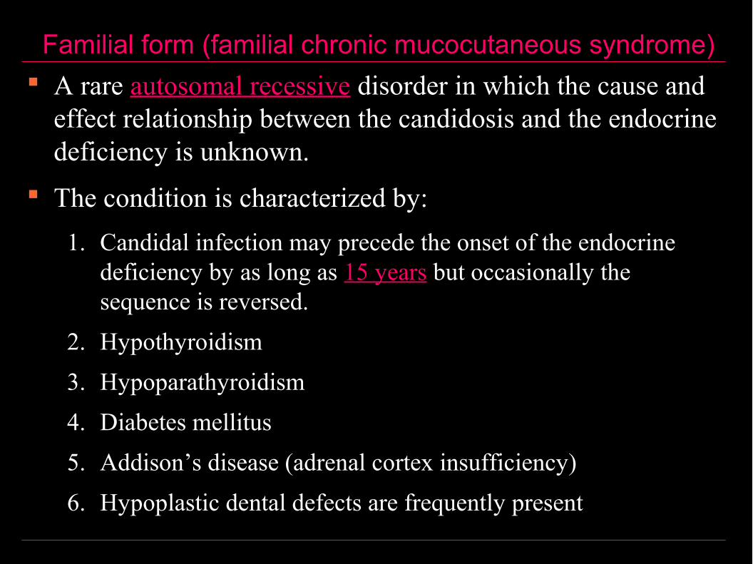
Familial form (familial chronic mucocutaneous syndrome)
A rare autosomal recessive disorder in which the cause and effect relationship between the candidosis and the endocrine deficiency is unknown.
The condition is characterized by:
1. Candidal infection may precede the onset of the endocrine deficiency by as long as 15 years but occasionally the sequence is reversed.
2. Hypothyroidism
3. Hypoparathyroidism
4. Diabetes mellitus
5. Addison’s disease (adrenal cortex insufficiency)
6. Hypoplastic dental defects are frequently present
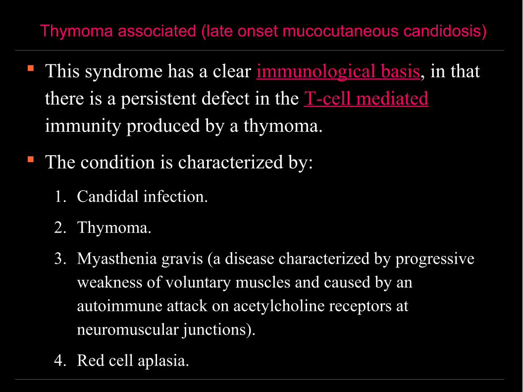
Thymoma associated (late onset mucocutaneous candidosis)
This syndrome has a clear immunological basis, in that there is a persistent defect in the T-cell mediated immunity produced by a thymoma.
The condition is characterized by:
1. Candidal infection.
2. Thymoma.
3. Myasthenia gravis (a disease characterized by progressive weakness of voluntary muscles and caused by an autoimmune attack on acetylcholine receptors at neuromuscular junctions).
4. Red cell aplasia.

Candidosis (Histologically)
Destruction of the surface epithelium and replacement by the pseudomembrane which consists of masses of monilial organisms, bacterial, desquamated epithelial cells and fibrin.
Destruction of the epithelium may be due to the combined effects of toxins and enzymes (lipases and proteases) produced by the monilial organisms.
PNL aggregation and chronic inflammatory cell infiltration of the connective tissue.
Pseudohyphae are best demonstrated using PAS or Grocott methenamine silver stains.
Epithelial atrophy is a characteristic feature of chronic erythematous candidiasis.
Epithelial hyperplasia is a characteristic feature of chronic hyperplastic candidiasis.
Candidosis (Treatment)
Correction of the predisposing factors, if possible.
Mild cases, topical application of nystatin.
Severe cases, ketoconazole 400 mg daily in a single dose by mouth.
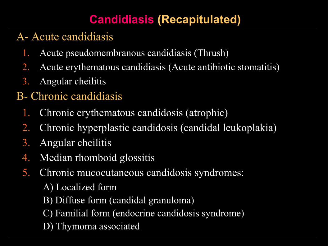
Candidiasis (Recapitulated)
A- Acute candidiasis1. Acute pseudomembranous candidiasis (Thrush)
2. Acute erythematous candidiasis (Acute antibiotic stomatitis)
3. Angular cheilitis
B- Chronic candidiasis
1. Chronic erythematous candidosis (atrophic)
2. Chronic hyperplastic candidosis (candidal leukoplakia)
3. Angular cheilitis
4. Median rhomboid glossitis
5. Chronic mucocutaneous candidosis syndromes:A) Localized formB) Diffuse form (candidal granuloma)C) Familial form (endocrine candidosis syndrome)D) Thymoma associated
Moniliasis
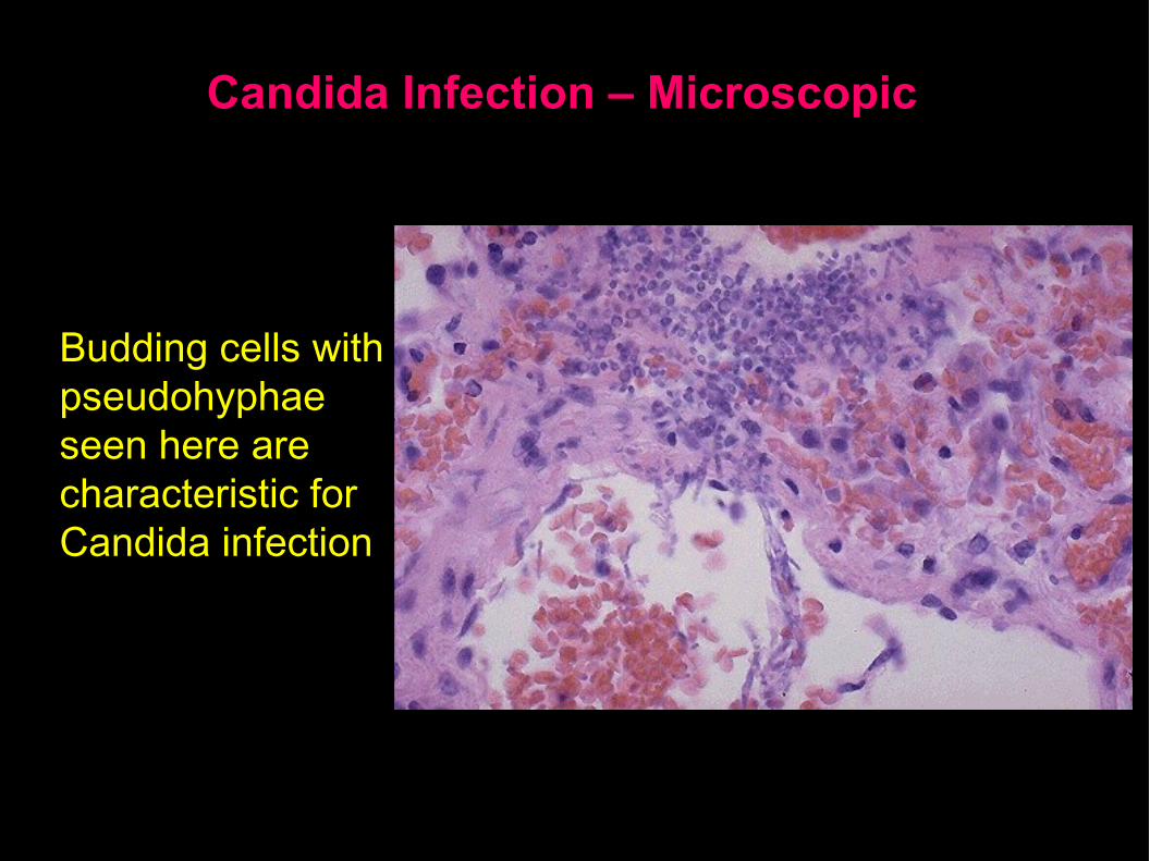
Candida Infection – Microscopic
Budding cells with pseudohyphae seen here are characteristic for Candida infection
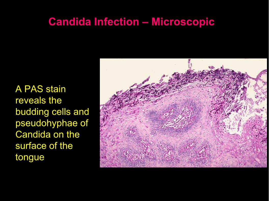
Candida Infection – Microscopic
A PAS stain reveals the budding cells and pseudohyphae of Candida on the surface of the tongue
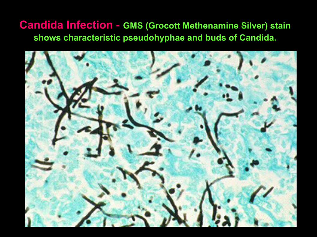
Candida Infection - GMS (Grocott Methenamine Silver) stain shows characteristic pseudohyphae and buds of Candida.

Systemic Mycoses
The Systemic mycoses are that type of infection which involve deep viscera with wide dissemination.
It is more common in immunocompromised patients.
Clinically most of the systemic mycoses can cause oral lesions.
Oral lesions are usually nodular granulomas with ulcerations which can be tumor-like in appearance.

Systemic Mycoses
The most interesting systemic mycoses include:
1. Histoplasmosis
2. Blastomycosis
3. Mucormycosis
4. Coccidioidomycosis
5. Paracoccidioidomycosis
6. Cyptococcosis
Morphologic classification of important fungi
Filamentous fungi (moulds)
Zygomycetes
Dermatophytes (caustive of all types of tinea except tinea versicolor)
Malassezia furfur (caustive of tinea versicolor)
Aspergillus species
YeastsCandida species
Cryptococcus neoformans
Dimorphic fungi
Histoplasma capsulatum
Blastomyces dermatitides
Coccidiodes imitis
Paracoccidiodes brasiliensis
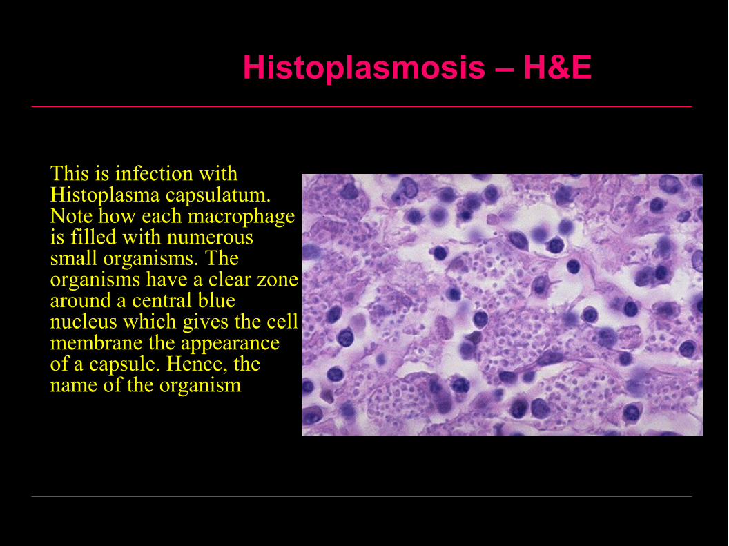
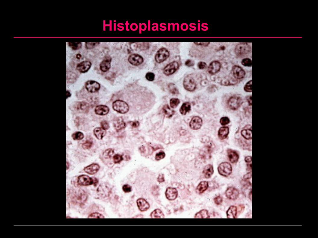
Histoplasmosis
Caused by histoplasma capsulatum, which is a dimorphic soil fungus.
The organism is an intracellular parasite, which parasitize macrophages (histiocytes).
The organisms have a clear zone around a central blue nucleus which gives the cell membrane the appearance of a capsule. Hence, the name of the organism
Mode of infection is by inhalation of the dust contaminated by the fungus.

Histoplasmosis
Clinically
In the respiratory tract the fungus is engulfed by macrophages and the infection is asymptomatic in most cases.
In rare cases clinical pneumonia occurs which heals spontaneously.
In immuno-compromised patients, wide dissemination of the fungus occurs to involve the reticuloendothelial system producing fever, anemia, spleenomegally, hepatomegally and lymphadenopathy.
Oral manifestations consist off multiple ulcers due to breakdown of multiple granulomas.

Histoplasmosis
Histologically
Multiple granulomas showing large numbers of macrophages.
The macrophages show the organism histoplasma capsulatum in their cytoplasm (intracellular parasite).
Diagnosis
Histoplasmin test: +ve result.
Treatment
Amphotericin B 0.5 mg/kg IV or Ketoconazole 400 mg as a single daily dose by mouth.
Histoplasmosis – Key Facts
Histoplasmosis is a systemic disease, mostly of the reticuloendothelial system, manifesting itself in the bone marrow, lungs, liver, and the spleen
Hepatosplenomegaly is the primary sign in children, while in adults, histoplasmosis more commonly appears as pulmonary disease
Histoplasmosis generally occurs in one of three forms: acute pulmonary, chronic pulmonary or disseminated
Histoplasmosis – Case Report
33 year old male had growth of a tumor over the last 3 – 4 months in the floor of the mouth. He lost approximately 50 pounds in the last 4 – 5 months. At examination, there was purulence associated with the lesion and limited range of motion of the tongue
This tumor reveals sections of mucosa with a chronic inflammatory infiltrate and swollen histiocytes.
Within the histiocytes are microorganisms that are small with a clear zone.
Special stains with GMS and PAS revealed the organisms.
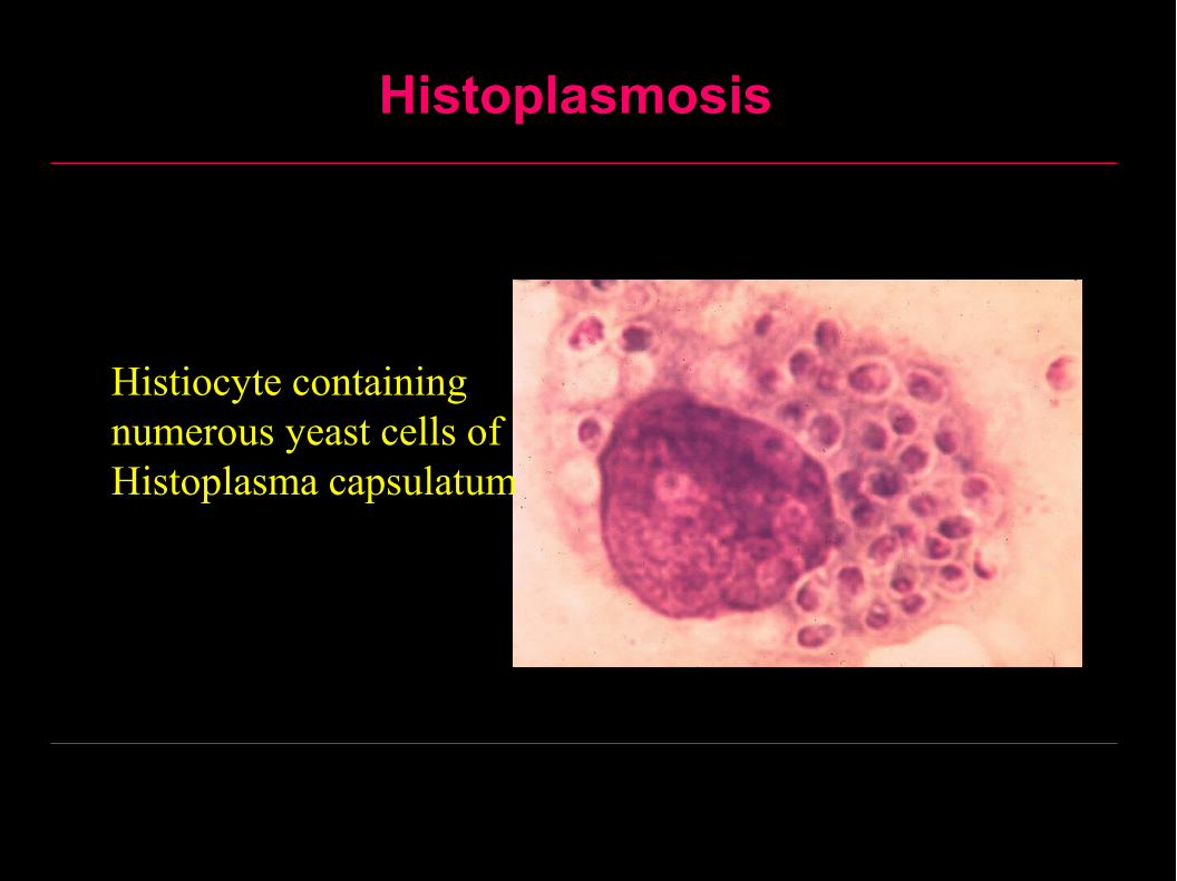
Histoplasmosis
Histiocyte containing numerous yeast cells of Histoplasma capsulatum
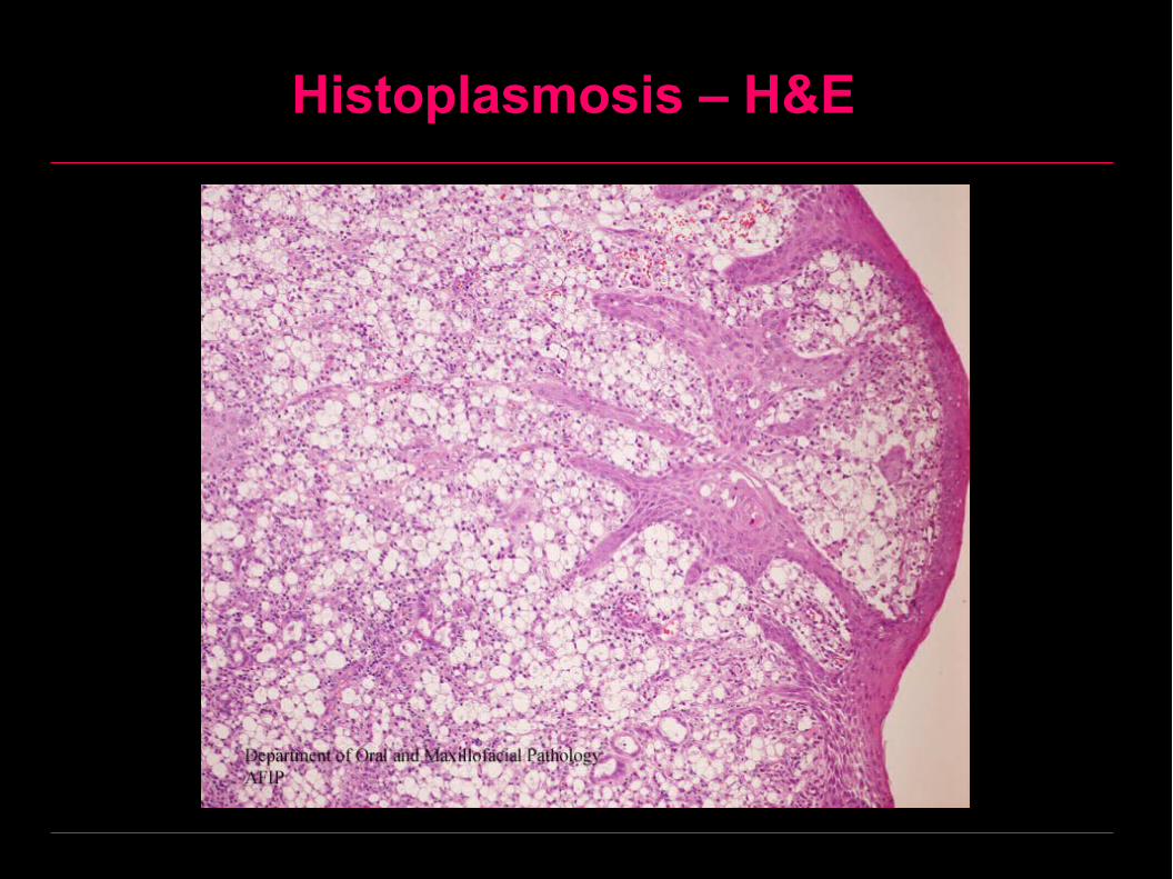
Histoplasmosis – H&E
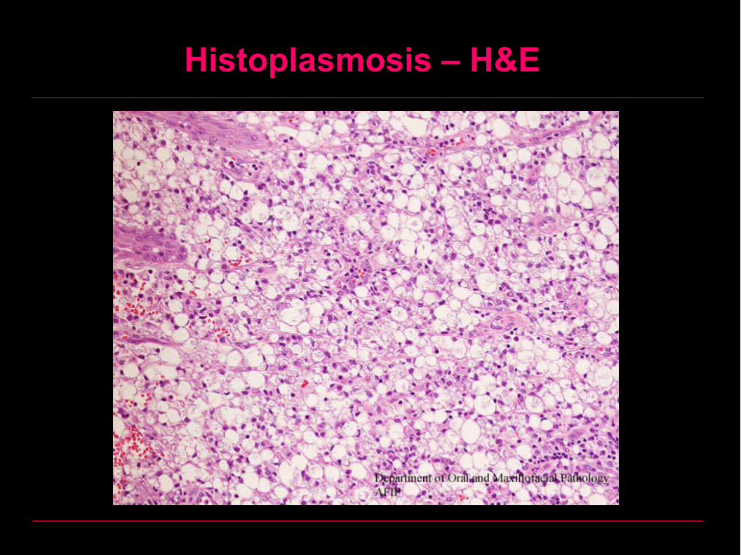
Histoplasmosis – H&E
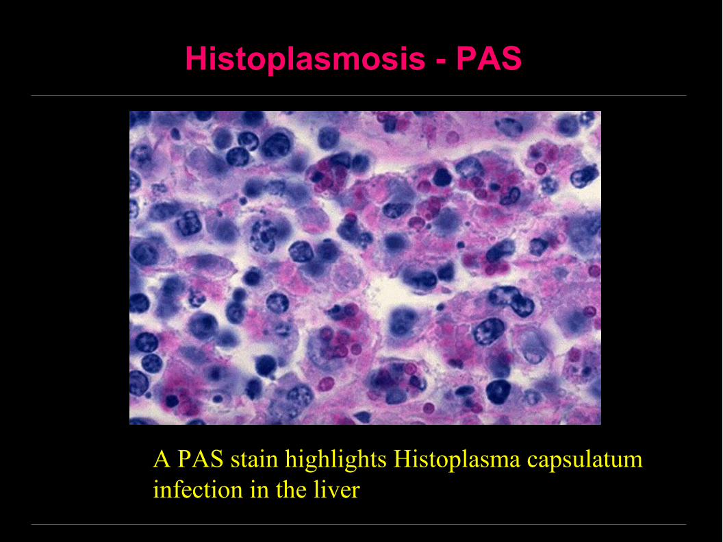
Histoplasmosis - PAS
A PAS stain highlights Histoplasma capsulatum infection in the liver
Histoplasmosis – H&E
This is infection with Histoplasma capsulatum. Note how each macrophage is filled with numerous small organisms. The organisms have a clear zone around a central blue nucleus which gives the cell membrane the appearance of a capsule. Hence, the name of the organism
Histoplasmosis

Histoplasmosis

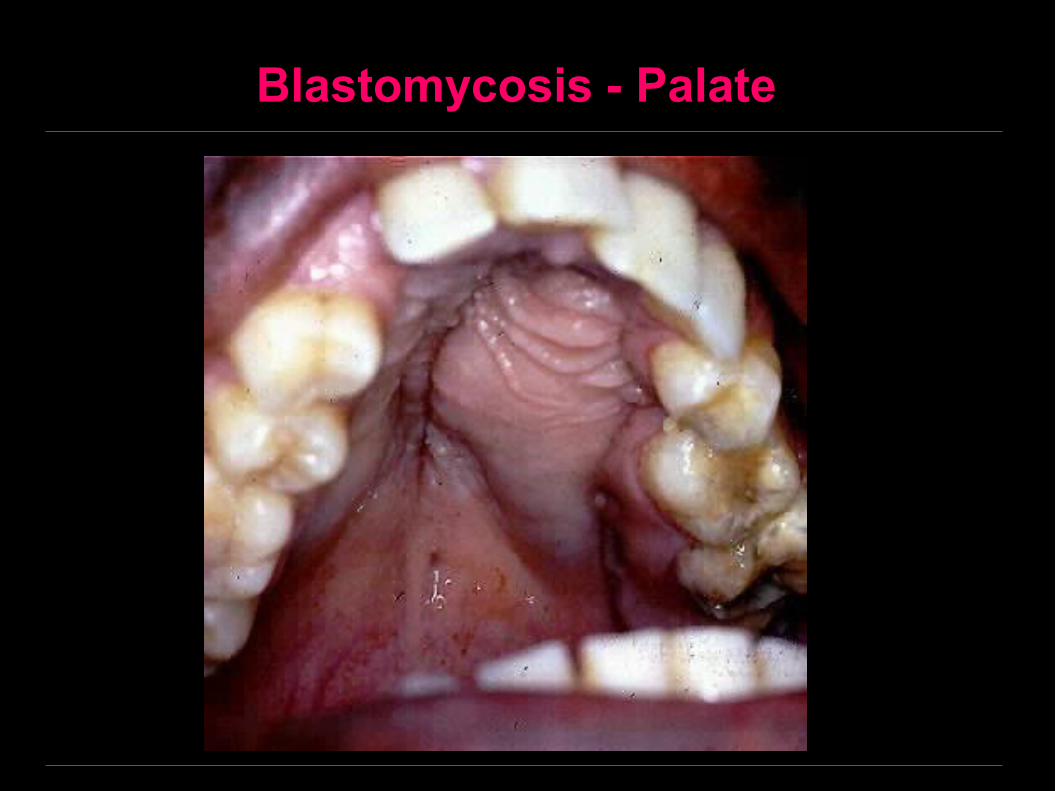
Blastomycosis
Caused by a dimorphic fungus known as blastomyces dermatitidis, which is a soil fungus.
The fungus is PAS (periodic acid Schiff) positive.
Mode of infection is by inhalation or rarely through the oral mucosa or extraction socket.
BlastomycosisClinically Respiratory tract infection usually passes unnoticed. In immunocompromised patients wide dissemination into bone, skin
and mucosa occurs. Lesion consists of multiple granulomas which breakdown forming
ulcers. The periphery of the ulcers shows psuedoepitheliomatous hyperplasia.
Histologically Multiple granulomas similar to that of actinomycosis. The spherical fungi could be detected by PAS stain. Psuedoepitheliomatous hyperplasia could be detected around the
ulcers.
Treatment Amphotericin B 0.5 mg/kg IV or Ketoconazole 400 mg as a single
daily dose by mouth.
Blastomycosis - Palate

Blastomycosis – Nodular Skin Lesion
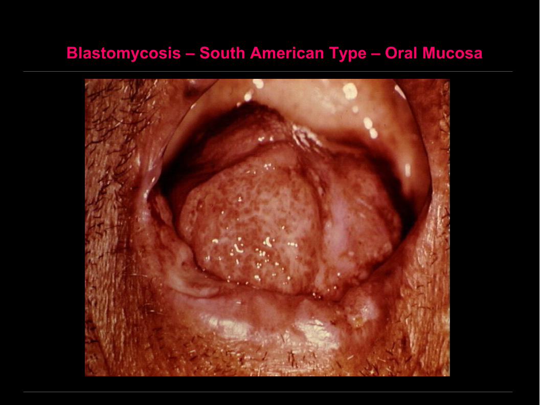
Blastomycosis – South American Type – Oral Mucosa
Blastomycosis
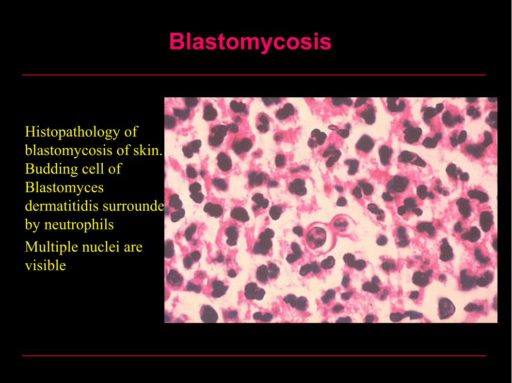
• Histopathology of blastomycosis of skin. Budding cell of Blastomyces dermatitidis surrounded by neutrophils
• Multiple nuclei are visible
Blastomycosis
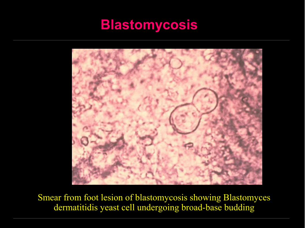
Smear from foot lesion of blastomycosis showing Blastomyces dermatitidis yeast cell undergoing broad-base budding
Coccidioidomycosis
Etiology Caused by a dimorphic fungus known as coccidioides
immitis which is a soil fungus endemic in arid regions.
Clinically Mode of infection is by inhalation of the contaminated
dust. In most people, influenza like fever results which is self-limiting and disappears spontaneously.
In immunocompromised patients, wide dissemination of the organisms occurs through the blood in a similar fashion to that of miliary TB to involve most systems of the body especially bones, skin and oral mucosa.
Lesions take the form of multiple granulomas with suppuration and ulcerations.
Coccidioidomycosis
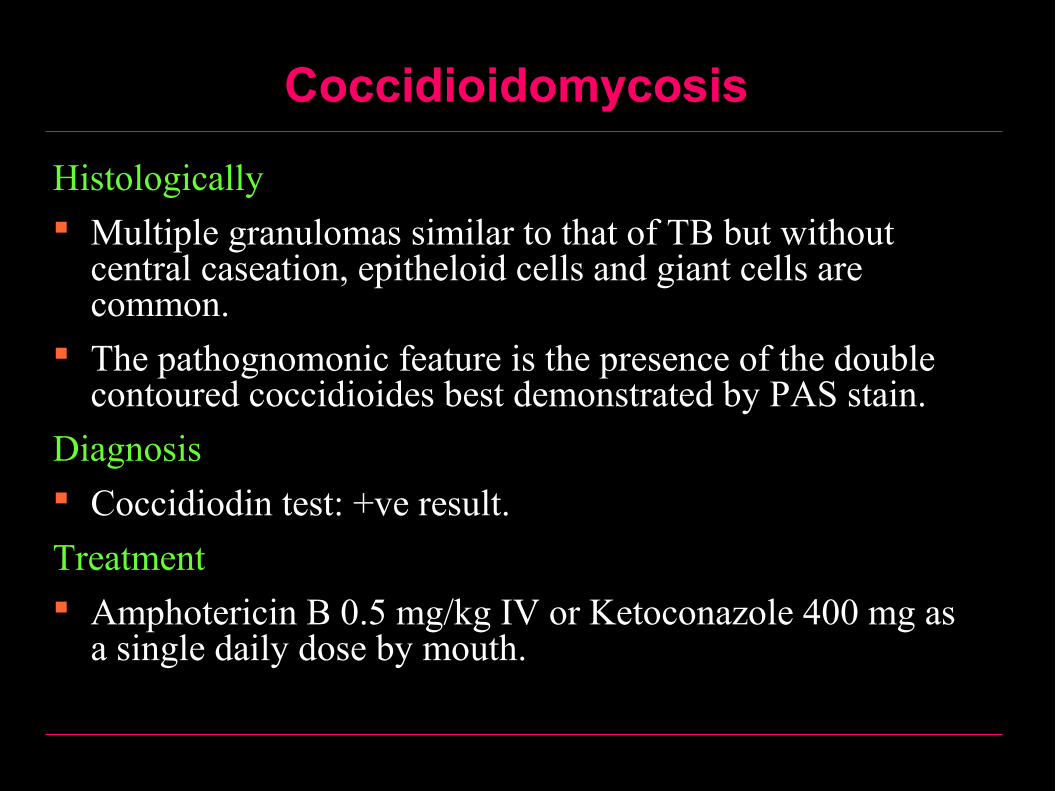
Histologically Multiple granulomas similar to that of TB but without
central caseation, epitheloid cells and giant cells are common.
The pathognomonic feature is the presence of the double contoured coccidioides best demonstrated by PAS stain.
Diagnosis Coccidiodin test: +ve result.
Treatment Amphotericin B 0.5 mg/kg IV or Ketoconazole 400 mg as
a single daily dose by mouth.

Coccidioidomycosis
Coccidioidomycosis
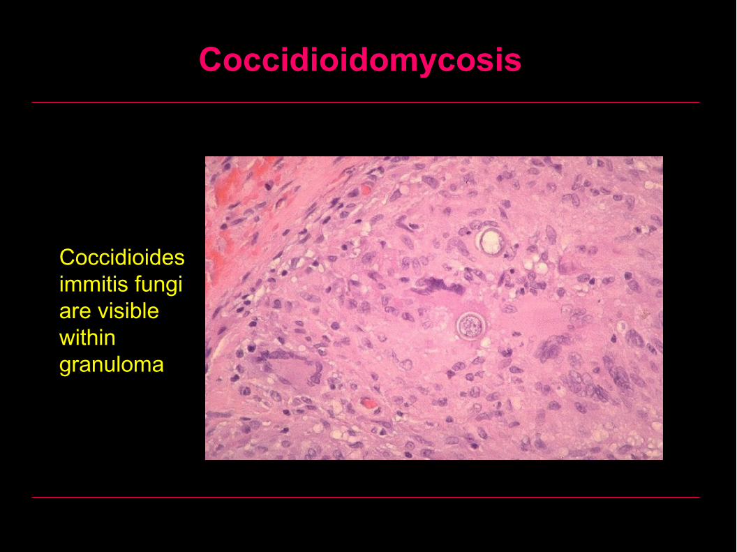
Coccidioides immitis fungi are visible within granuloma
Coccidioidomycosis
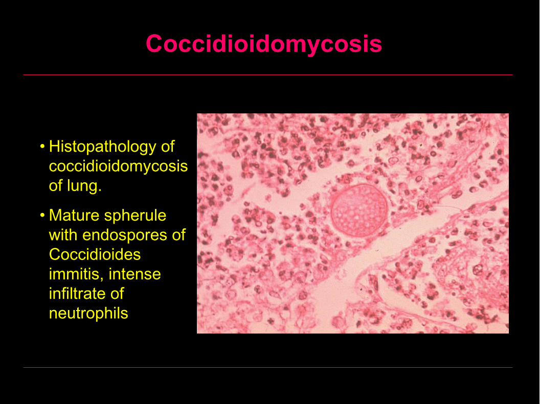
• Histopathology of coccidioidomycosis of lung.
• Mature spherule with endospores of Coccidioides immitis, intense infiltrate of neutrophils
Coccidioidomycosis
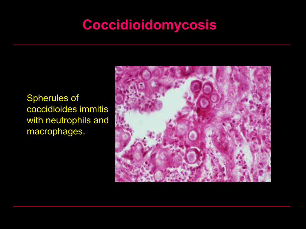
Spherules of coccidioides immitis with neutrophils and macrophages.
Coccidioidomycosis
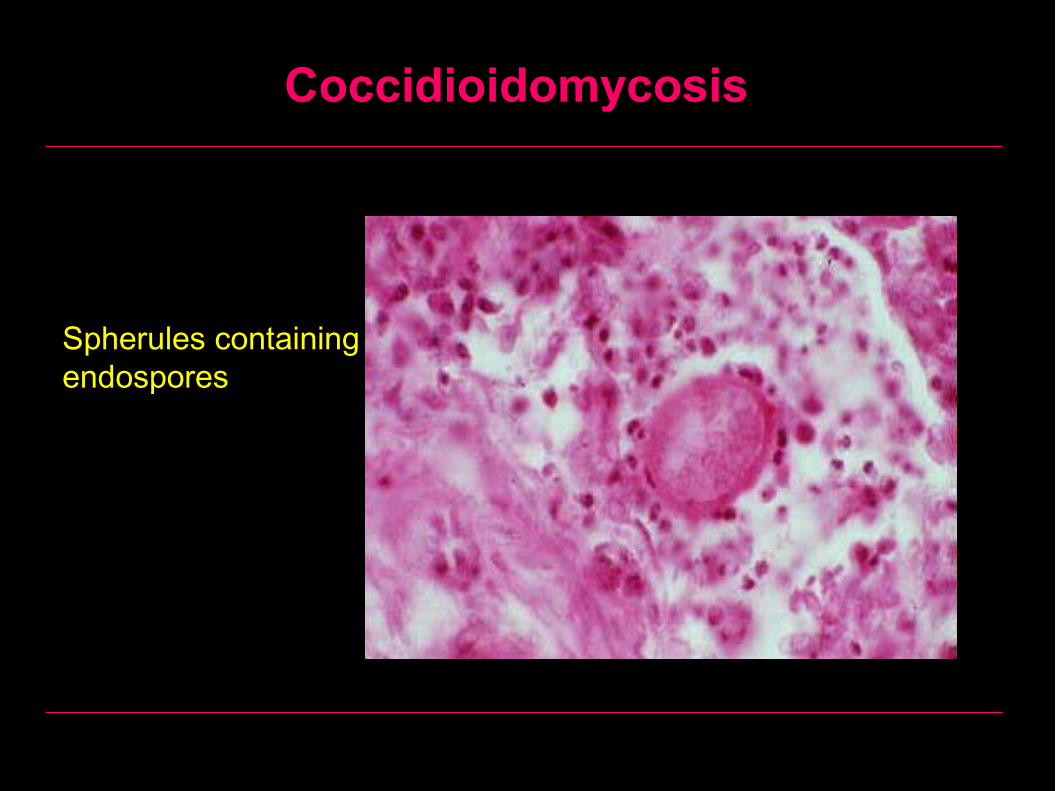
Spherules containing endospores
Paracoccidioidomycosis
Definition
A chronic granulomatous disease of mucous membranes, skin, and pulmonary system. Most cases are reported from Brazil.
Etiology
Caused by the dimorphic paracoccidioides brasiliensis which is a soil fungus.
Paracoccidioidomycosis
Clinically:
A common triad of symptoms is pulmonary lesions, edentulous mouth, and cervical lymphadenopathy.
Prior to the recognition of this disease, patients with paracoccidioidomycosis were often sent to TB sanitariums, just as patients with histoplasmosis.
The organisms invade the mucous membranes of the mouth causing looseness and loss of teeth.
White plaques are also found in the buccal mucosa.
Has a long latency period. 10-20 years may pass between infection and manifestation of the infection in the non-endemic areas of the world.
Paracoccidioidomycosis
Histologically:
Histologically, multiple granulomas containing multiple buds forming a "Captain's wheel." This is diagnostic of paracoccidioidomycosis. In this case, the mother cell is 40-50 microns in diameter and the buds are 2-5 microns in size.
Treatment:
Amphotericin B 0.5 mg/kg IV or Ketoconazole 400 mg as a single daily dose by mouth.
Paracoccidioidomycosis
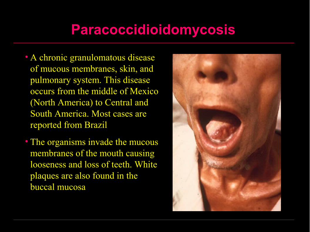
• A chronic granulomatous disease of mucous membranes, skin, and pulmonary system. This disease occurs from the middle of Mexico (North America) to Central and South America. Most cases are reported from Brazil
• The organisms invade the mucous membranes of the mouth causing looseness and loss of teeth. White plaques are also found in the buccal mucosa

Paracoccidioidomycosis – Tongue
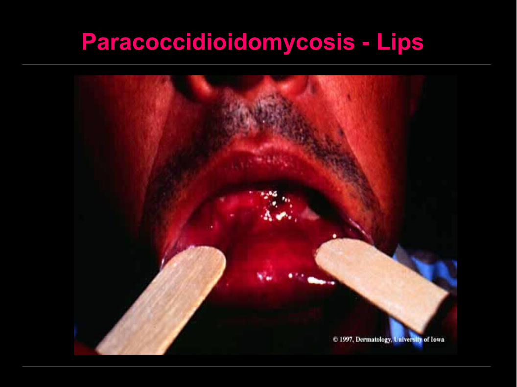
Paracoccidioidomycosis - Lips
Paracoccidioidomycosis
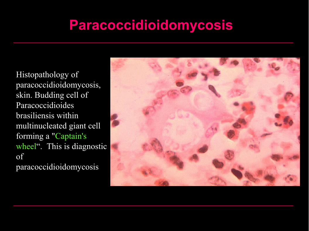
Histopathology of paracoccidioidomycosis, skin. Budding cell of Paracoccidioides brasiliensis within multinucleated giant cell forming a "Captain's wheel“. This is diagnostic of paracoccidioidomycosis

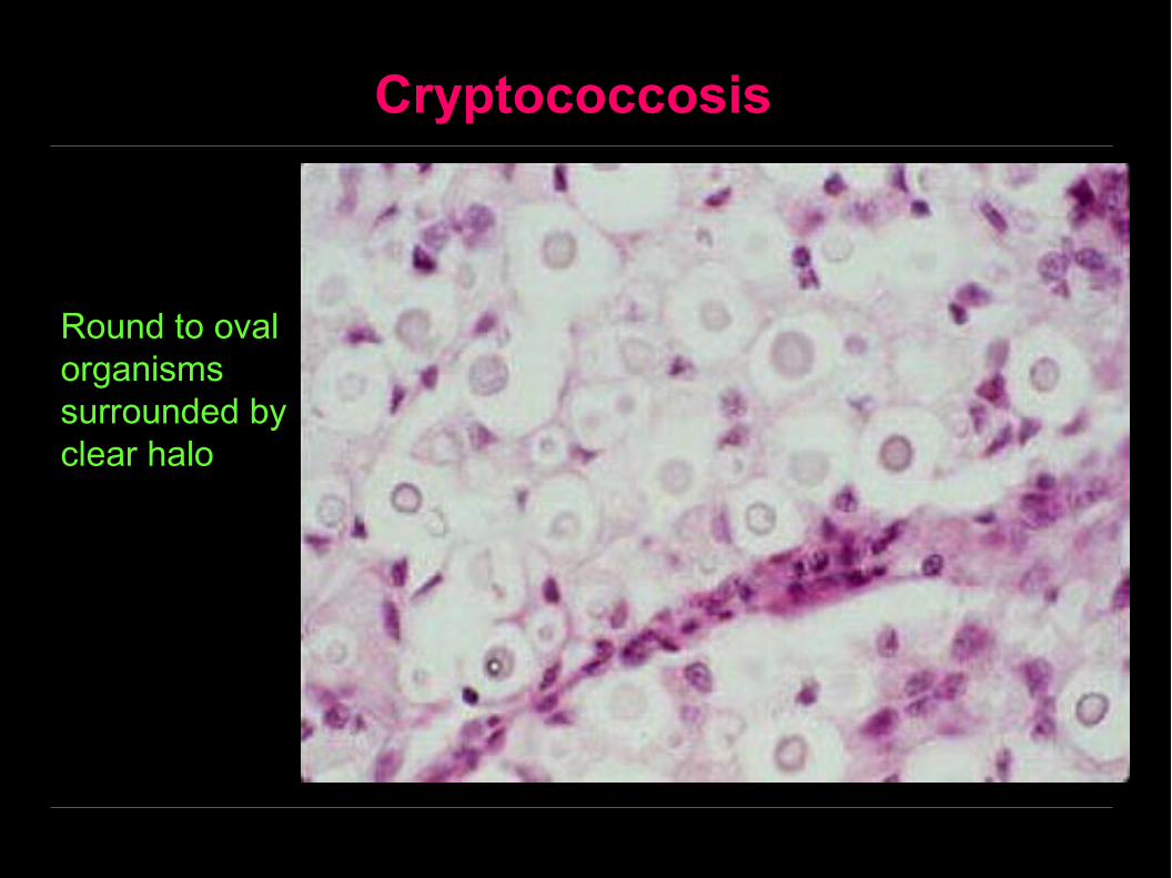
Cryptococcosis
Caused by the yeast Cryptococcus neoformans.
Immunosuppression is a predisposing factor.
Rare in the oral cavity. May have membranous nasopharyngitis, meningitis, hearing loss.
Diagnosis is by fluorescent antibody.
Treatment with amphotericin B.
Cryptococcosis
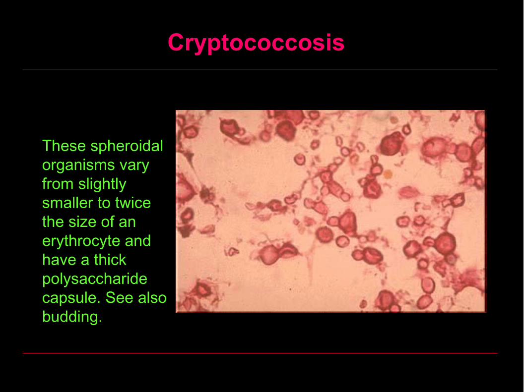
These spheroidal organisms vary from slightly smaller to twice the size of an erythrocyte and have a thick polysaccharide capsule. See also budding.
Cryptococcosis
Round to oval organisms surrounded by clear halo
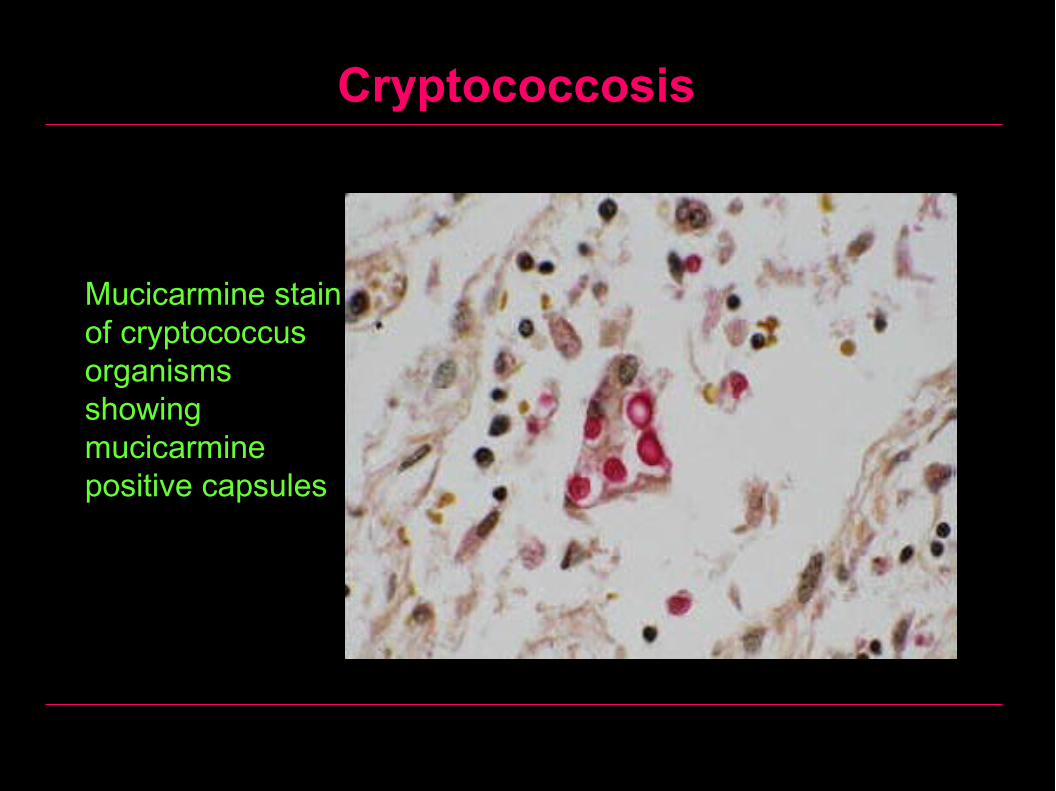
Cryptococcosis
Mucicarmine stain of cryptococcus organisms showing mucicarmine positive capsules
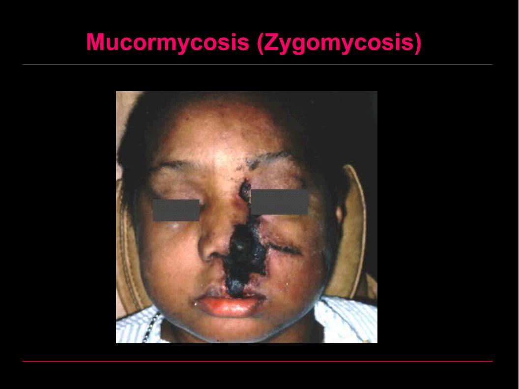
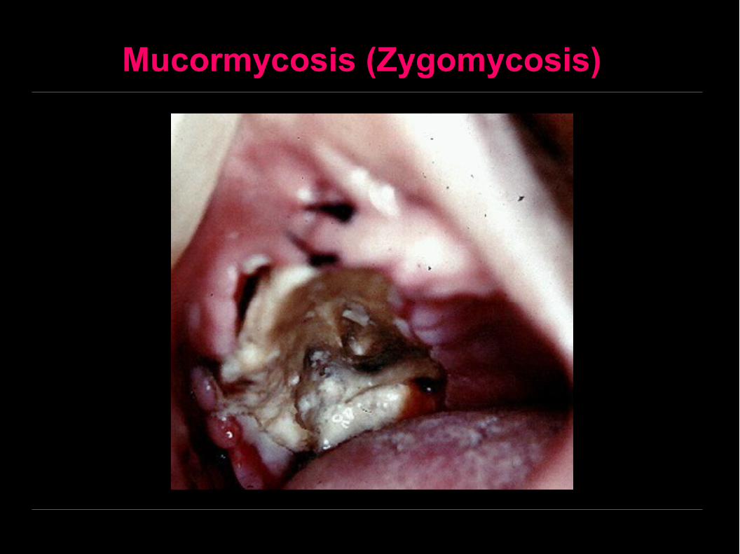
Mucormycosis (Zygomycosis)
Is a chronic fungal infection of the midface, nasal cavity and sinuses (fungus is a mould of the genus mucor)
Common in immunocompromised patients
The mode of infection is through the respiratory tract
Can result in blockage of the sinuses and nasal discharge
The organism invade and occlude blood vessels thus causing necrosis and gangrene
Diagnosis is by biopsy (nonseptate hyphae).
Treatment respond to amphotericin B, and aggressive surgical debridement.
Mucormycosis (Zygomycosis)
Mucormycosis (Zygomycosis)
Mucormycosis (Zygomycosis)
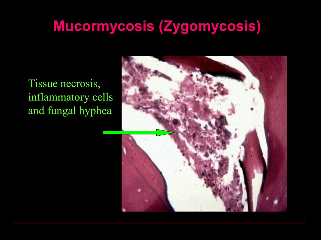
Tissue necrosis, inflammatory cells and fungal hyphea

Mucormycosis (Zygomycosis)

Mucormycosis (Zygomycosis)

Mucormycosis (Zygomycosis)
Inflammation containing the broad, nonseptate hyphae with 90 degree branching and hollow appearance of mucor.
Mucormycosis (Zygomycosis)
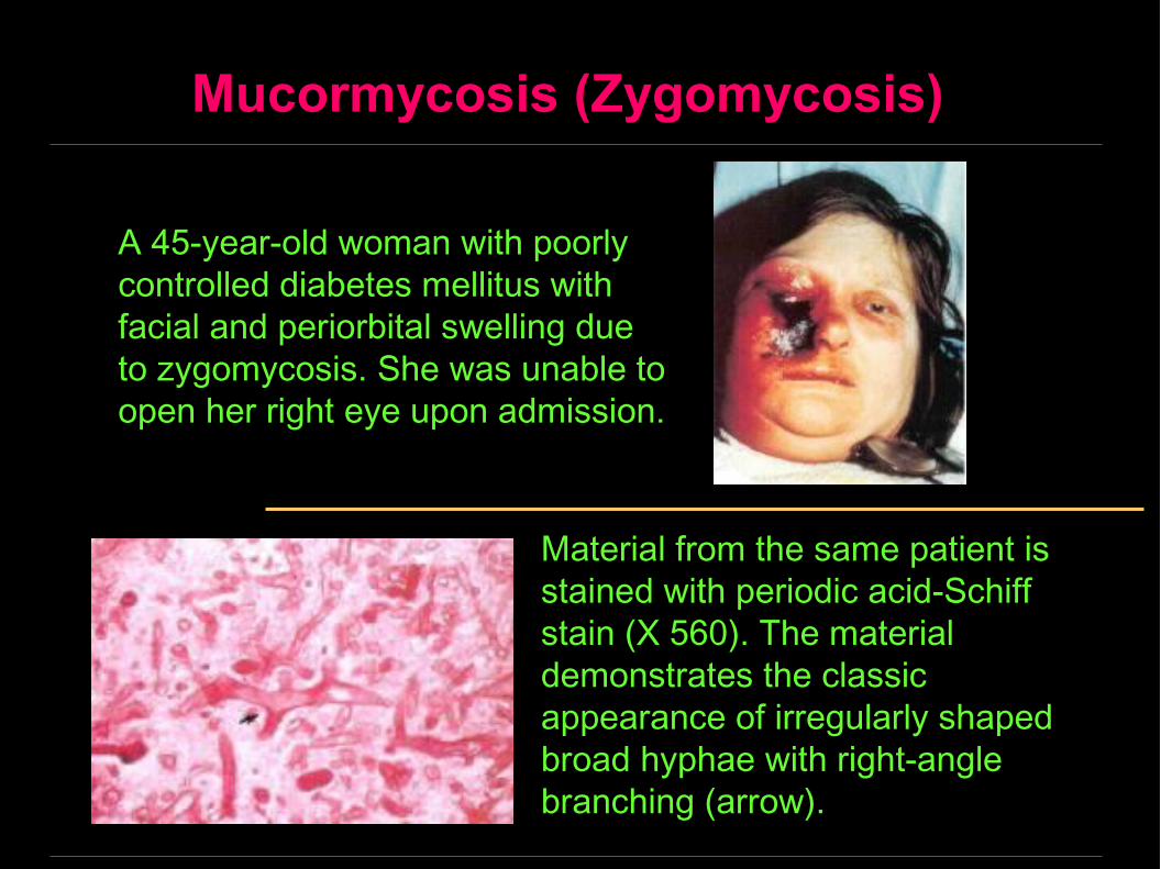
A 45-year-old woman with poorly controlled diabetes mellitus with facial and periorbital swelling due to zygomycosis. She was unable to open her right eye upon admission.
Material from the same patient is stained with periodic acid-Schiff stain (X 560). The material demonstrates the classic appearance of irregularly shaped broad hyphae with right-angle branching (arrow).

Aspergillus Infection, Lung
• This is a fungal abscess of the lung. Note the yellow tan material in the abscess.
• It is very firm material, because it is composed of fungal hyphae. This one is due to Aspergillus.
• Aspergillus has a habit of colonizing previously formed cavities, such as those with tuberculosis
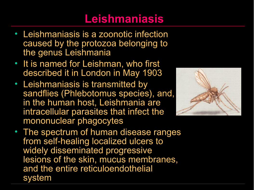
Leishmaniasis• Leishmaniasis is a zoonotic infection
caused by the protozoa belonging to the genus Leishmania
• It is named for Leishman, who first described it in London in May 1903
• Leishmaniasis is transmitted by sandflies (Phlebotomus species), and, in the human host, Leishmania are intracellular parasites that infect the mononuclear phagocytes
• The spectrum of human disease ranges from self-healing localized ulcers to widely disseminated progressive lesions of the skin, mucus membranes, and the entire reticuloendothelial system
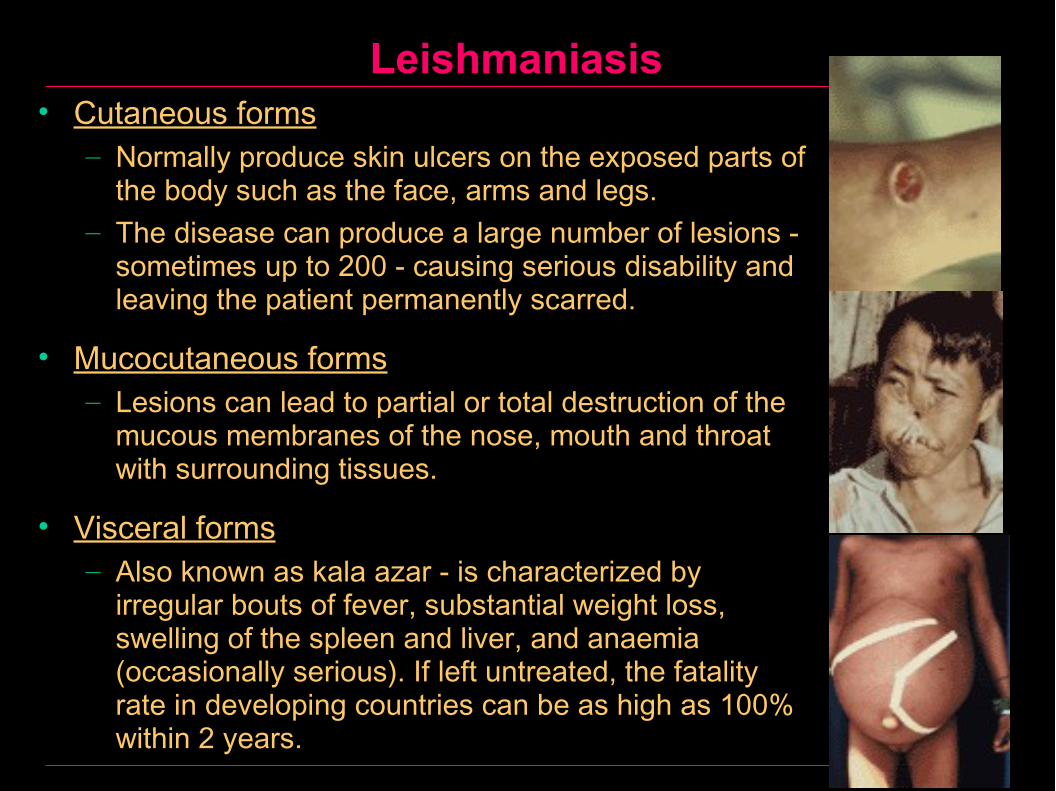
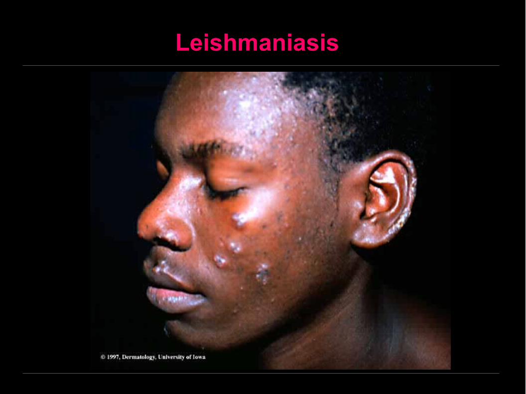
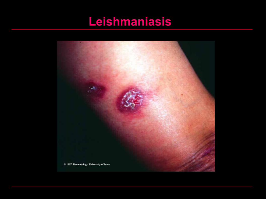
Leishmaniasis• Cutaneous forms
– Normally produce skin ulcers on the exposed parts of the body such as the face, arms and legs.
– The disease can produce a large number of lesions - sometimes up to 200 - causing serious disability and leaving the patient permanently scarred.
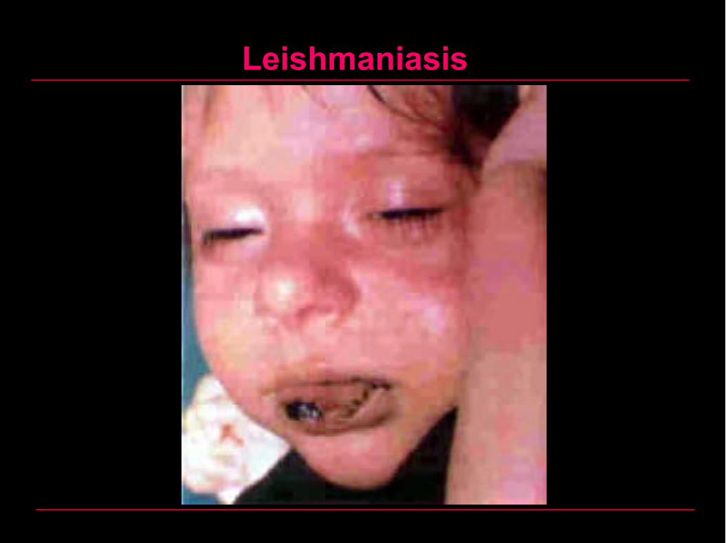
• Mucocutaneous forms – Lesions can lead to partial or total destruction of the
mucous membranes of the nose, mouth and throat with surrounding tissues.
• Visceral forms – Also known as kala azar - is characterized by
irregular bouts of fever, substantial weight loss, swelling of the spleen and liver, and anaemia (occasionally serious). If left untreated, the fatality rate in developing countries can be as high as 100% within 2 years.
Cutaneous Leishmaniasis (Oriental Sore)
• Most frequent on the extremities.• Head and neck involvement does occur.• Papules which undergo ulceration with crust
formation (1-3 cm).• Ulcers may heal completely, but usually leave
permanent scars.• Diagnosis is by biopsy which reveals granulomas
with typical intracellular parasites inside macrophages best stained with Giemsa stain.
• Treatment is with pentostam.
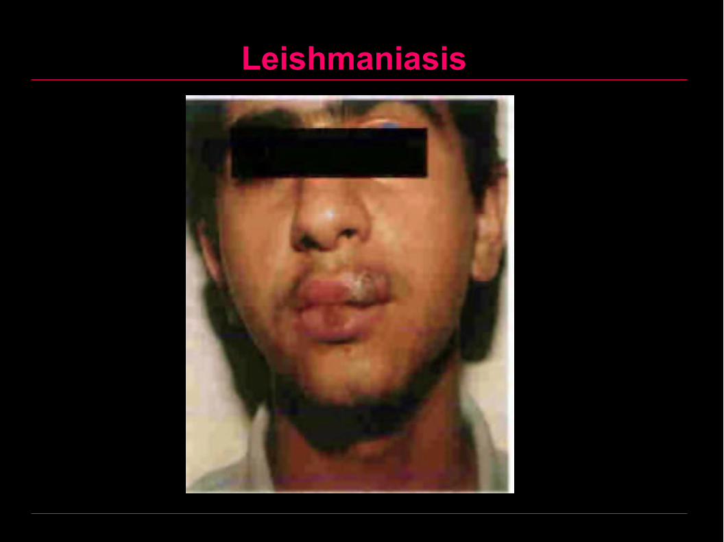
Mucocutaneous Leishmaniasis (Espundia)
• Involves macrophages of the skin around the mucous membranes.
• The disease starts with a cutaneous lesion that enlarges to involve the mouth, nose, and soft palate leading to severe tissue destruction.
• Lesions are papules which ulcerate and often become secondary infected.
• Diagnosis is by biopsy which reveals granulomas with typical intracellular parasites inside macrophages best stained with Giemsa stain.
• Treatment with pentostam.

Visceral Leishmaniasis
• Also known as kala azar - is characterized by irregular bouts of fever, substantial weight loss, swelling of the spleen and liver, and anemia (occasionally serious).
• If left untreated, the fatality rate in developing countries can be as high as 100% within 2 years.
Leishmaniasis
Leishmaniasis
Leishmaniasis
Leishmaniasis
Viral Diseases - Introduction
Viral Structure– A virus (virion) consists of a nucleic acid core
surrounded by a protein coat (capsid).– Some viruses have an additional protective layer, the
envelope.– Viruses without an envelope are known as naked
viruses.
Viral Diseases - Introduction
Nucleic Acid Core:– Known as viral genome and contains either DNA or
RNA.– Can exist as a single strand (as in the case of most
RNA viruses, except reoviruses) or double strand (the case of most DNA viruses, except parvoviruses).
– Most viruses have a linear genomes, with the exception of papovaviruses which have a circular genomes.
Viral Diseases - Introduction
Viral Capsid:– Functions:
• Protects the nucleic acid core.• Responsible for viral adsorption and penteration of host
cells throught interaction with host cell membrane receptors.
– Composition:• Viral capsids are composed of varying numbers of
capsomeres each of which is made up of one or more polypeptide chains known as protomeres.
Viral Diseases - Introduction
Viral Envelope:– Derived from the host cell membrane and is acquired
by the virus during the late stages of budding.– Consists of a lipid bilayer (derived from host cell) and
viral coded glycoproteins, which are inserted into the lipid bilayer and projects to the outer surface. Its serve two basic functions:
– Promote interaction with nucleocapsid proteins essential for the final stages of viral assembly.
– Mediate attachment to the host cell receptors essential for infectivity of the virus.

Viral Diseases of the Oral Mucosa
Virus type Primary infection Reactivation
Herpes simplex Primary herpetic gingivostomatitis
Herpes labialis
Recurrent intra-oral herpes
Herpes zoster Herpetic whitlow; chickenpox
Shingles
Epstein–Barr Glandular fever No
Coxsackie group Herpangina; Hand, foot and mouth disease No
Human papillomavirus
Squamous cell papilloma
Verruca vulgaris
Condyloma accuminatum
Squamous cell papilloma
Verruca vulgaris
Condyloma accuminatum
Pox virus Molluscum contagiosum No
Paramyxovirus Mumps; measles No
Herpes Simplex I (Labialis)• There are 2 types of herpes simplex virus• The first one is called herpes simplex type I (herpes simplex
libialis) which affects the upper part of the body• The second is called herpes simplex II (herpes simplex
genitalis) which usually affects the lower part of the body.• Herpes simplex infection occurs in two forms. The first form
which occurs as a result of the primary attack by the virus and is known as primary herpetic gingivostomatitis, after which the virus remains latent in the sensory ganglia or epithelial cells waiting for the chance of decreased body resistance and then reactivates and produce the secondary attack known as recurrent herpes labialis.
• Herpetic whitlow refers to viral infection of fingers.
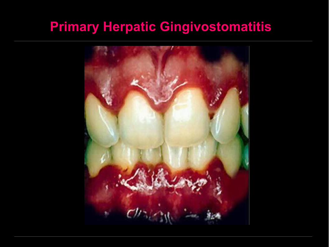
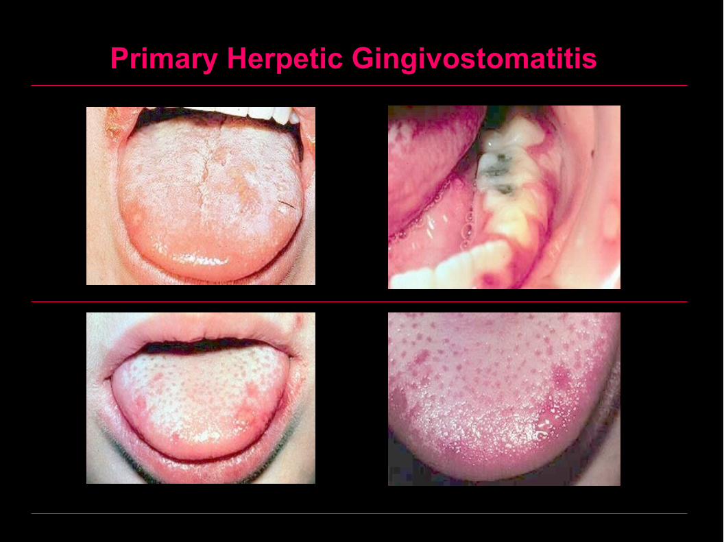
I- Primary Herpetic Gingivostomatitis
DefinitionThis is the primary attack of herpes simplex virus.Clinically• Usually occurs in infants or young children.• Fever and malaise. Then vesicles appear in the oral
cavity. These vesicles rupture forming painful ulcers.
• The disease is self limiting and heals in 10-14 days.
I- Primary Herpetic Gingivostomatitis
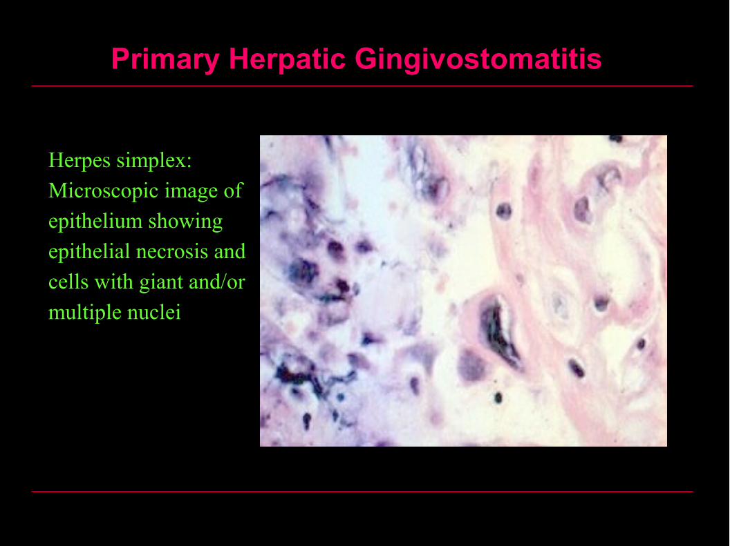
Histologically
• Intraepithelial vesicles, which result from edema (hydropic degeneration) of epithelial cells.
• Nuclei of epithelial cells show viral inclusion bodies (Lipschutz bodies).
• Infiltration of the underlying connective tissue by PNL, lymphocytes and plasma cells.
I- Primary Herpetic Gingivostomatitis
Treatment• Symptomatic and includes administration of
antipyretic drugs and mild sedation at bedtime.• Plenty of fluids and soft diet.• Acyclovir 20 mg/kg BW every 8 hours iv for 10 days.
Recommended only in immunocompromised patients.• The efficiency of topical acyclovir ointments has not
been proven. ComplicationsEncephalitis and/or memingitis.
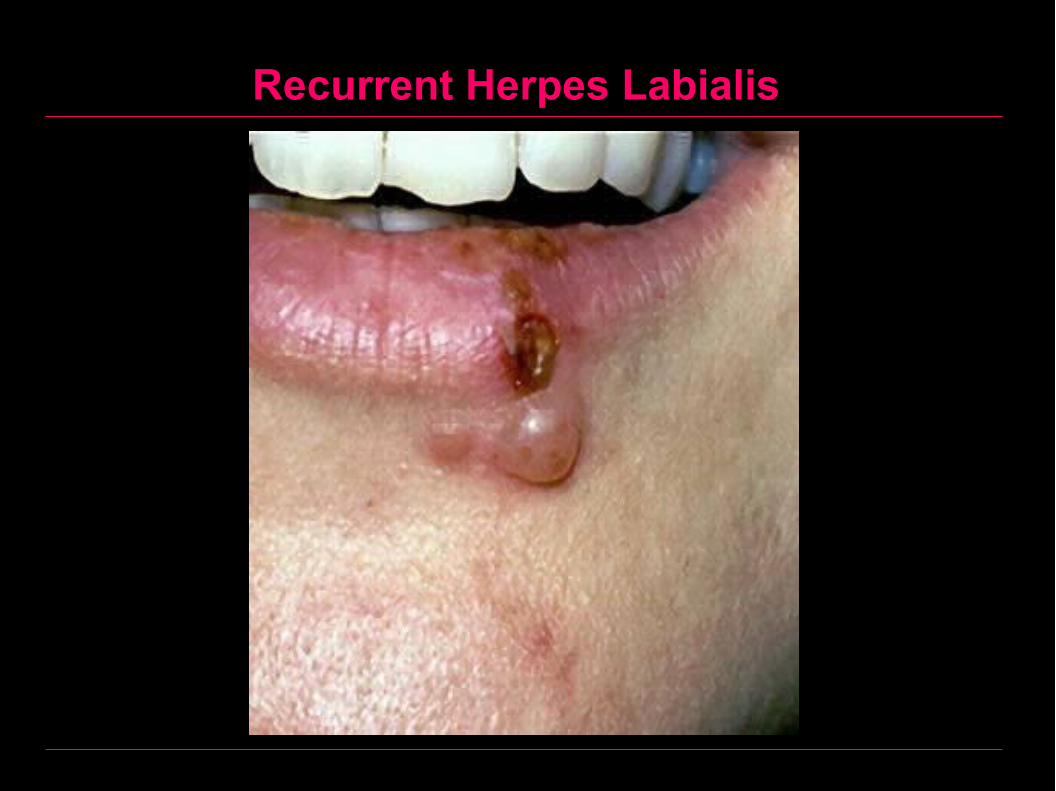
II- Recurrent Herpetic Infection
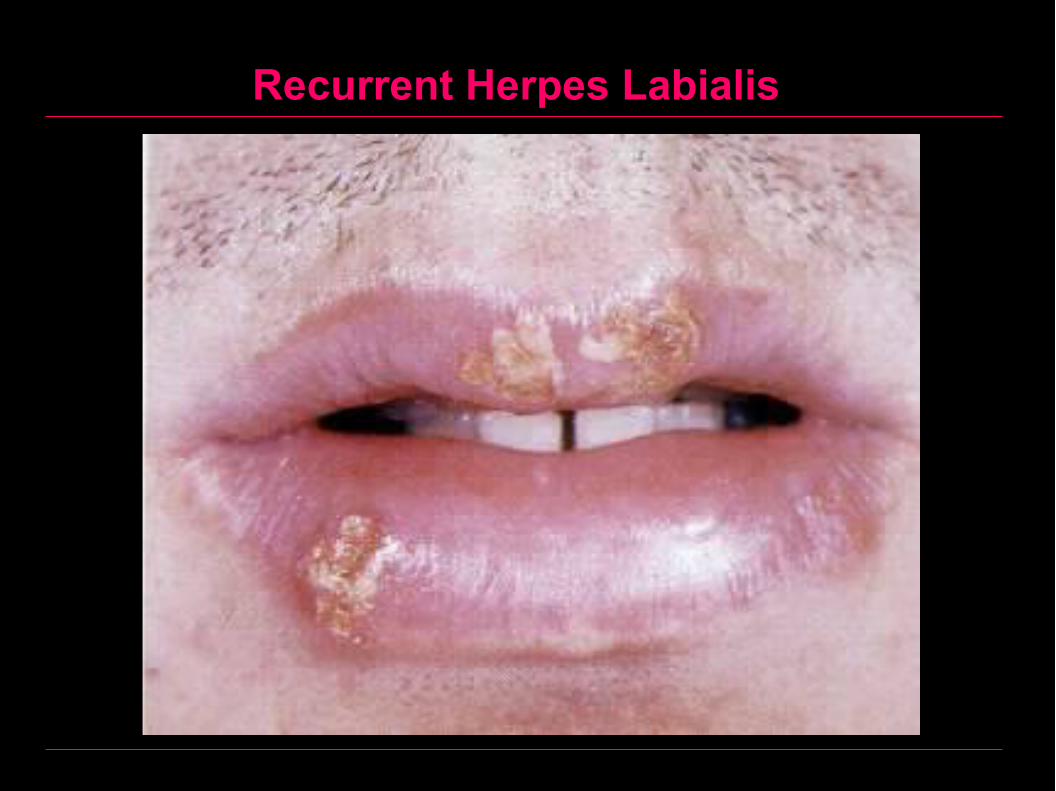
Usually called recurrent herpes labialisDefinition
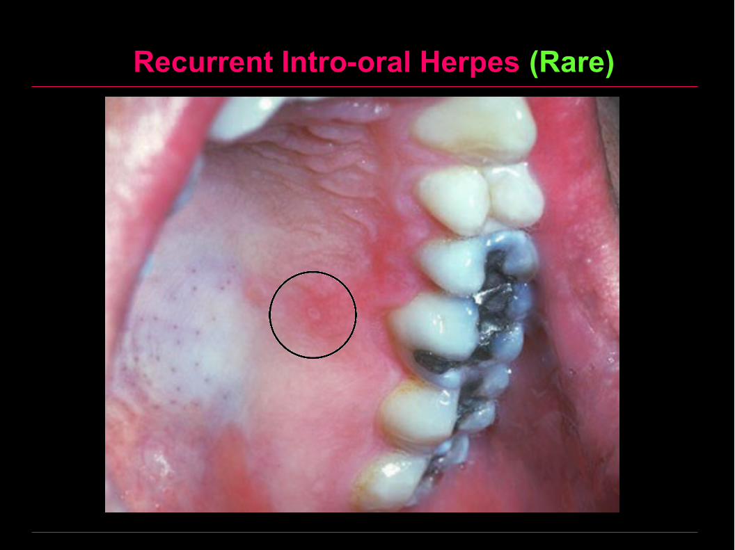
This is the secondary attack by the herpes simplex virus. The disease is usually confined to the mucocutaneous junction of the lip and rarely occurs in the oral cavity.
EtiologyThe predisposing factors for reactivation of the latent virus are usually influenza (so the disease is sometimes called cold sores), stresses or other diseases lowering the body resistance.

II- Recurrent Herpetic Infection
Clinically• Numbness or pain in the site of the subsequent eruptions.• Followed by appearance of multiple small vesicles.• Vesicles rupture forming painful ulcers covered by a crust.• The disease is self limiting and disappears within 7-10 days.
Treatment• Recommended only in immunocompromised patients.
Acyclovir 400 mg 4 times daily for 5 days given by mouth• otherwise no treatment is needed as the disease is self
limiting.
Primary Herpatic Gingivostomatitis
Primary Herpetic Gingivostomatitis
Primary Herpatic Gingivostomatitis
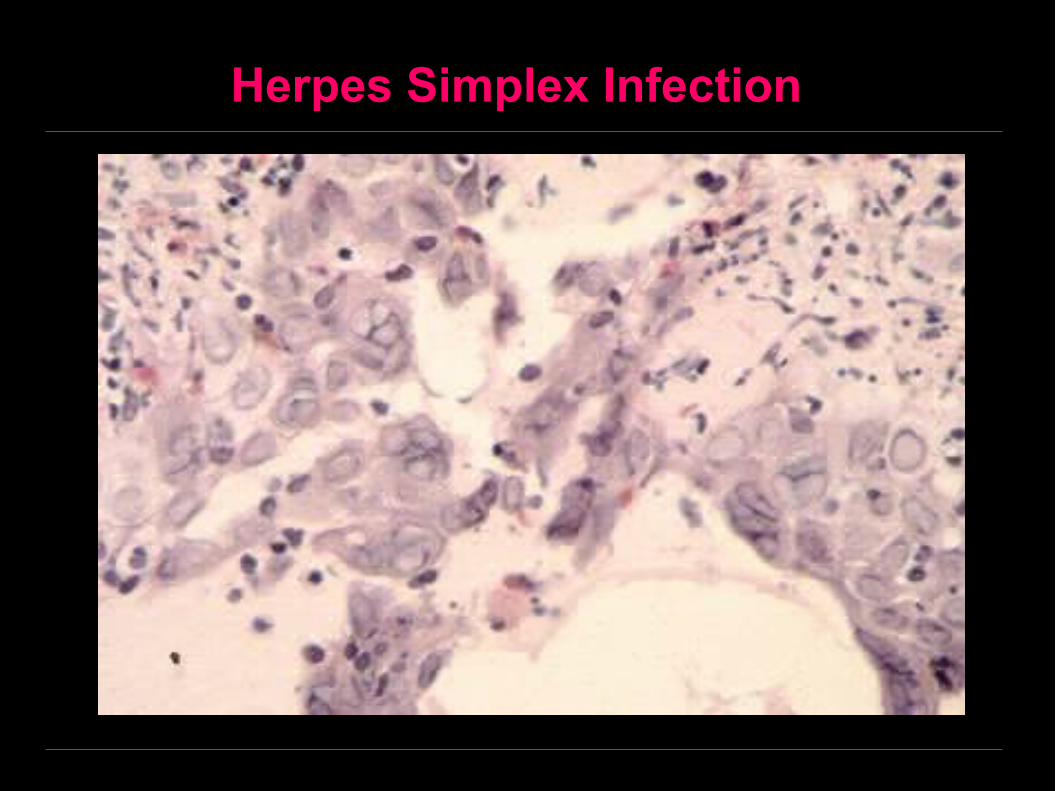
Herpes simplex: Microscopic image of epithelium showing epithelial necrosis and cells with giant and/or multiple nuclei
Recurrent Herpes Labialis
Recurrent Herpes Labialis

Recurrent Herpes Labialis

Recurrent Herpes Labialis

Recurrent Herpes Labialis

Recurrent Herpes Labialis, although rare, can occur in the skin
Recurrent Intro-oral Herpes (Rare)
Herpetic Whitlow
Herpetic Whitlow
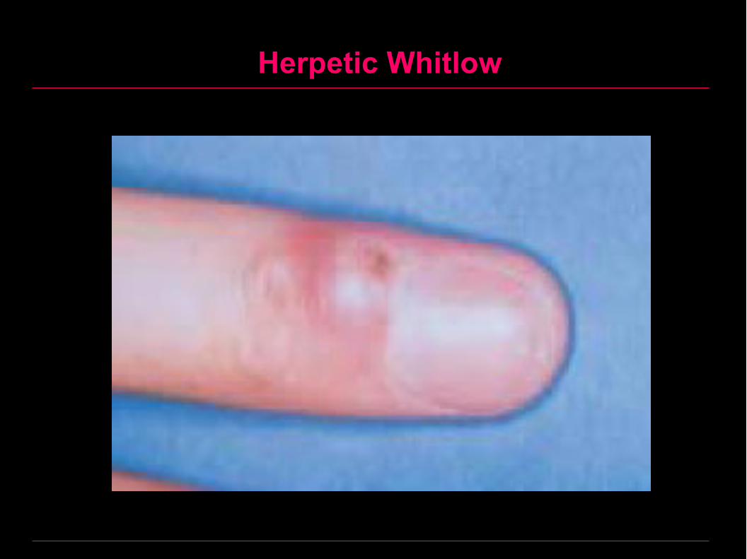
• Herpetic whitlow occurs when HSV infects the subcutaneous tissues of the finger, producing an erythematous and vesicular eruption analogous to a staphylococcal whitlow.
• This is an extremely painful condition, which will prevent affected dental professionals from undertaking their normal duties for a period of time because of the potential for spreading the virus and because of the severe pain.
• Lesions often take 2–3 weeks to heal, and the treatment available is acyclovir (200 mg tablets five times daily for 5 days). Importantly, acyclovir cannot prevent the recurrence of herpetic whitlow.
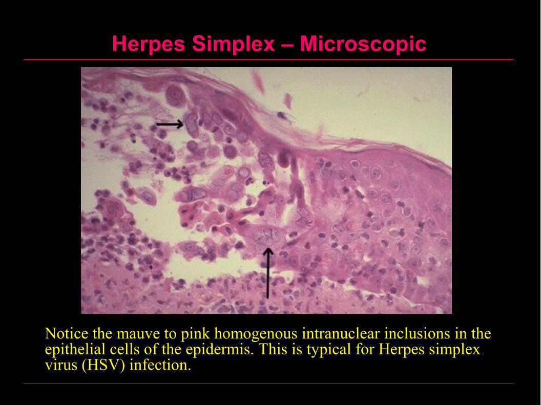
Herpes Simplex – Microscopic
Notice the mauve to pink homogenous intranuclear inclusions in the epithelial cells of the epidermis. This is typical for Herpes simplex virus (HSV) infection.
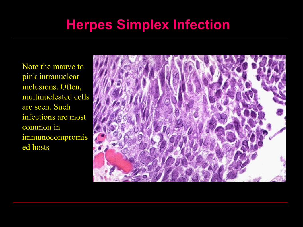
Herpes Simplex Infection
Note the mauve to pink intranuclear inclusions. Often, multinucleated cells are seen. Such infections are most common in immunocompromised hosts
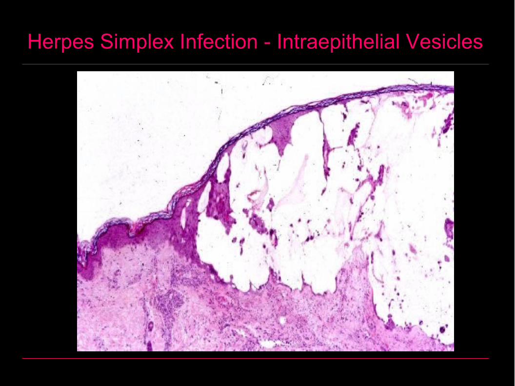
Herpes Simplex Infection - Intraepithelial Vesicles
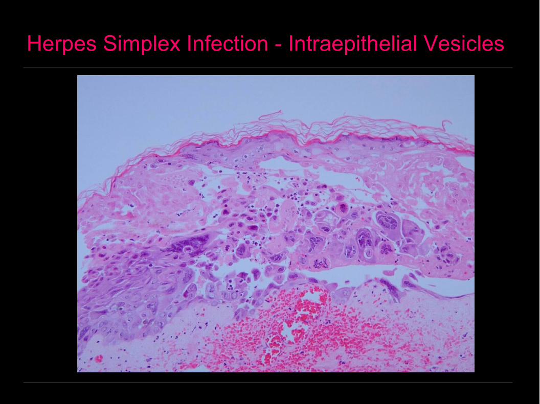
Herpes Simplex Infection - Intraepithelial Vesicles

Herpes Simplex Infection
Viral multinucleated giant cells
Herpes Simplex Infection
Varicella - Zoster Virus
• The primary attack of the virus causes varicella (chicken pox)
• while the secondary attack causes herpes zoster infection (shingles).

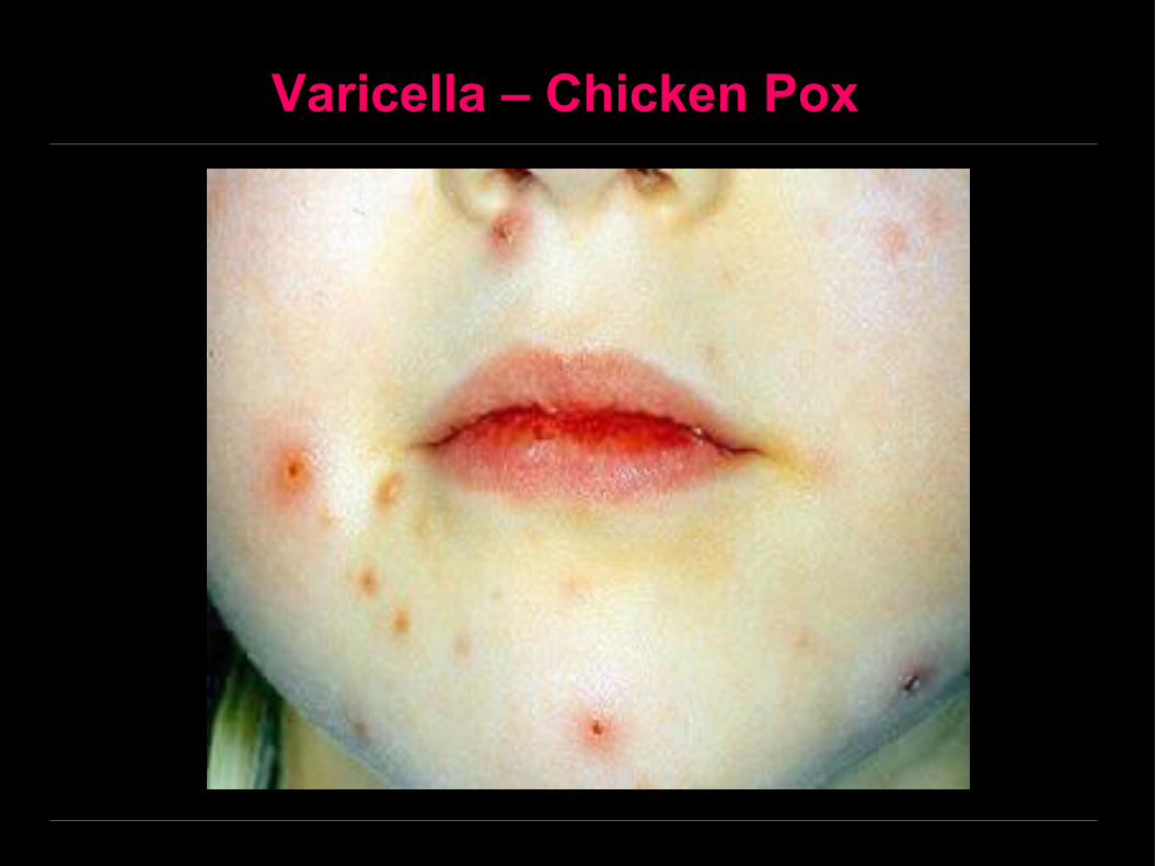
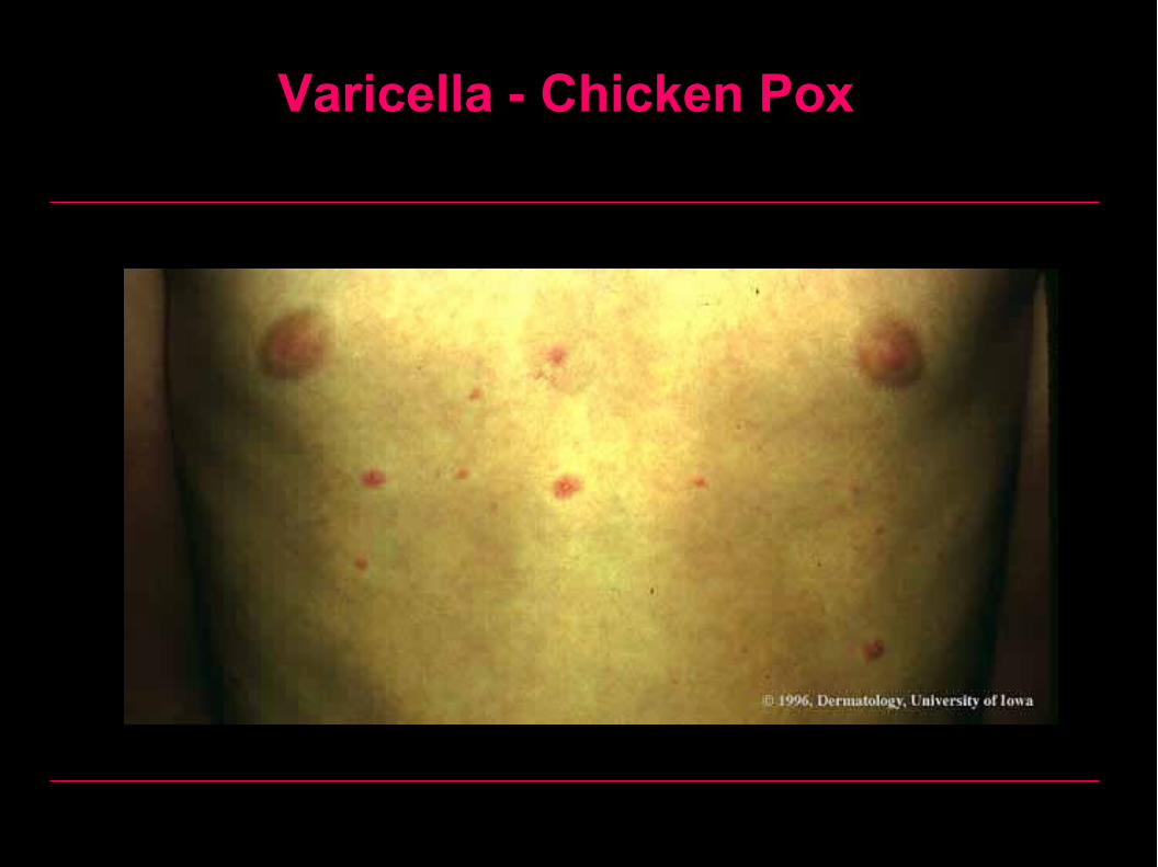
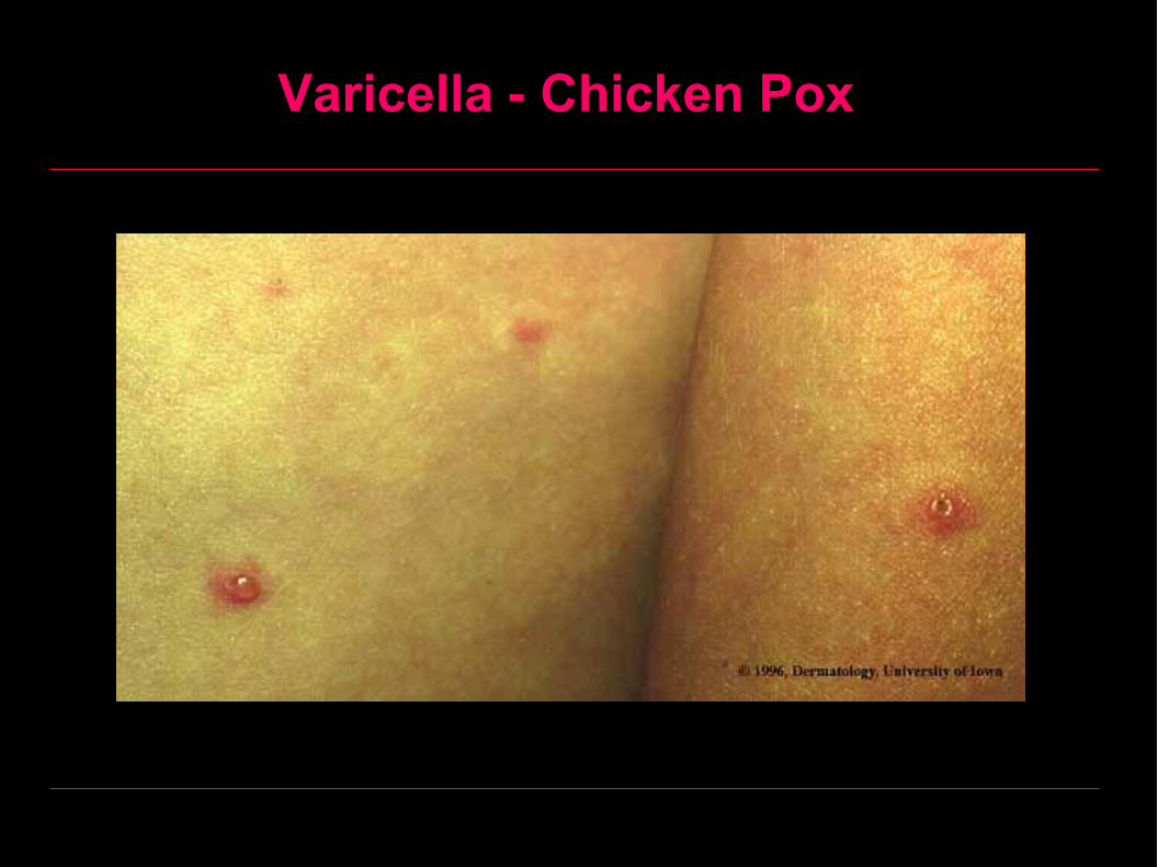
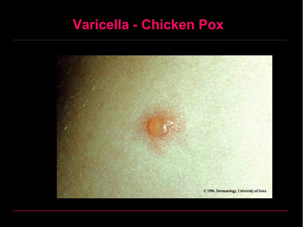
I- Chicken Pox (Varicella)
• Occurs usually in childhood.• Mild fever, followed by vesicles on the skin
and / or rarely on oral mucosa which rupture forming ulcers.
TreatmentIs indicated only in immunocompromised patients. Acyclovir 20 mg/kg body weight (800 mg maximum), 4 times daily by mouth for 5 days.
Varicella – Chicken Pox
Varicella - Chicken Pox

Varicella - Chicken Pox
Varicella - Chicken Pox
Varicella - Chicken Pox

II- Herpes Zoster Infection (Shingles)
• This is the secondary attack of herpes zoster.
• After the primary attack (chicken pox) the virus remains latent in the cells of the sensory ganglia. Decreased body resistance produces reactivation and migration of the virus to affect the skin and mucous membrane supplied by the affected nerve.
• When the mandibular or maxillary branches of the trigeminal nerve are affected, oral lesions may appear as a grouped vesicles which rupture forming painful ulcers occurring on the skin and oral mucosa supplied by the affected nerve.
TreatmentAcyclovir 20 mg/kg body weight (800 mg maximum), 4 times daily by mouth for 7 -10 days.
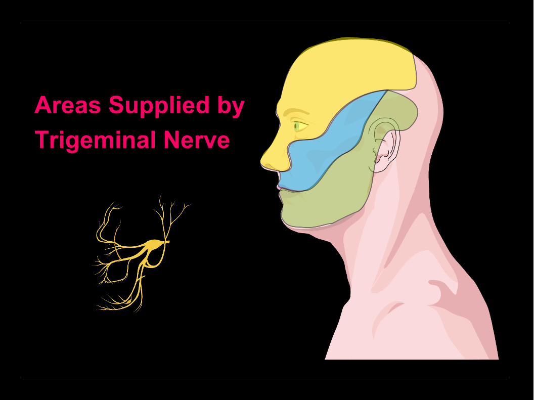
Areas Supplied by
Trigeminal Nerve
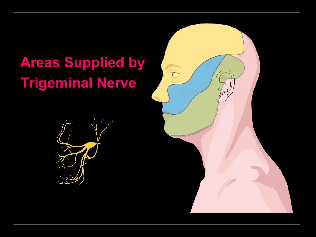
Areas Supplied by
Trigeminal Nerve
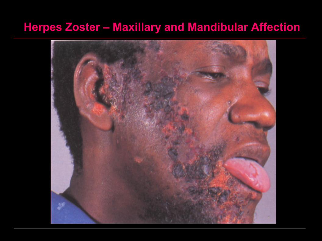
Herpes Zoster – Maxillary and Mandibular Affection
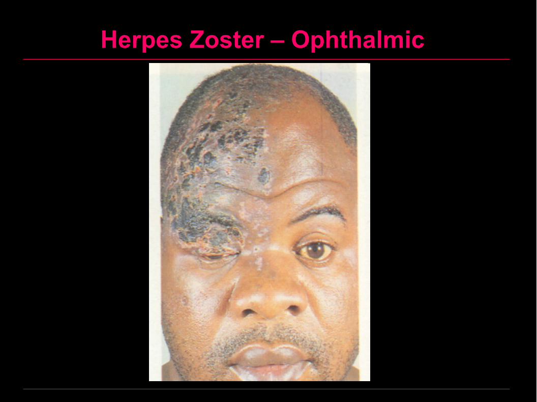
Herpes Zoster – Ophthalmic
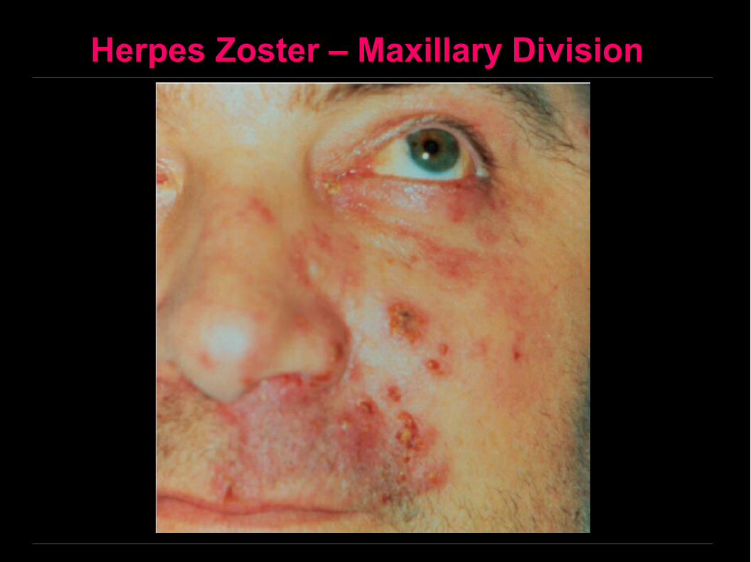
Herpes Zoster – Maxillary Division

Ramsay–Hunt Syndrome - Definition
A special form of herpes zoster affecting the facial nerve via the geniculate ganglion.
Ramsay–Hunt Syndrome - Clinically
1- Predromal stage:– Headache, malaise and fever– Sometimes, pain localized to the ear or
radiating to the jaws and neck
2- Herpetic oticus:– Vesicles on the tragus of the ear, on the
external auditory canal or on the tympanic membrane
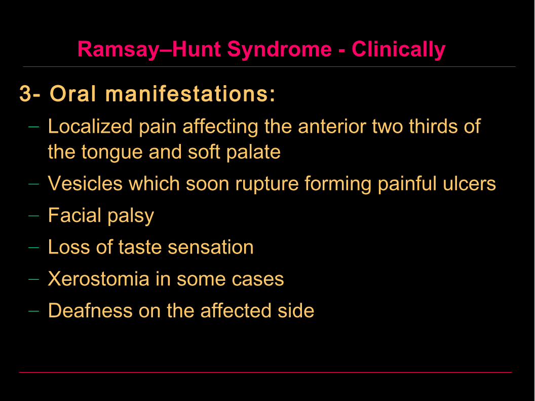
Ramsay–Hunt Syndrome - Clinically
3- Oral manifestations:– Localized pain affecting the anterior two thirds of
the tongue and soft palate– Vesicles which soon rupture forming painful ulcers – Facial palsy– Loss of taste sensation– Xerostomia in some cases– Deafness on the affected side

Ramsay–Hunt Syndrome - Treatment
Treatment
– Acyclovir 20 mg/kg body weight (800 mg maximum), 4 times daily by mouth for 7 -10 days.
– Corticosteroid therapy may be used in addition to acyclovir. Prednisone, 60 mg/daily with gradually withdraw in 10 days.
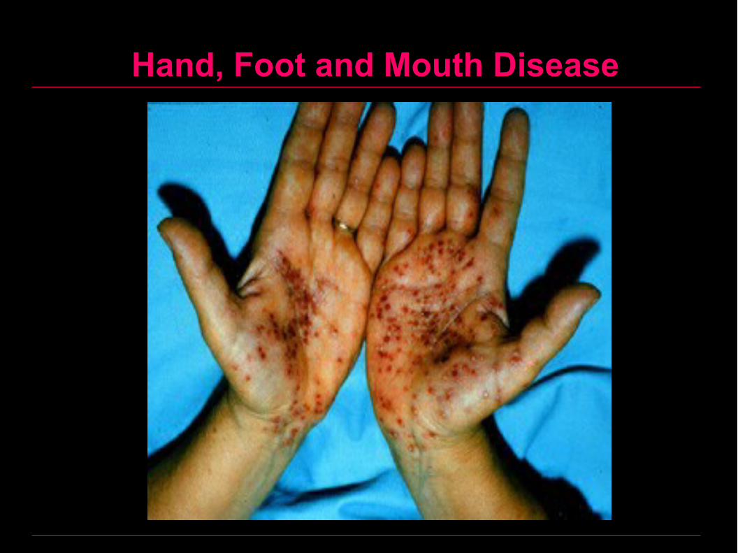
Hand-Foot and Mouth Disease
Definition– Highly contagious disease caused by Coxsackie virus
type A16 (belonging to the family of picornaviruses).
Clinically• Fever and malaise.• Appearance of vesicles, which rupture forming painful
ulcers on the palms, soles and oral mucosa.• The disease lasts only for few days (self limiting) and
complications are rare.
Treatment• Symptomatic.
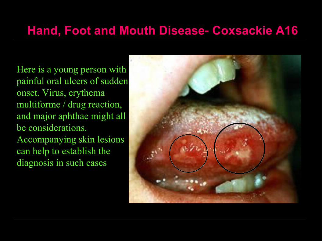
Hand, Foot and Mouth Disease- Coxsackie A16
Here is a young person with painful oral ulcers of sudden onset. Virus, erythema multiforme / drug reaction, and major aphthae might all be considerations. Accompanying skin lesions can help to establish the diagnosis in such cases
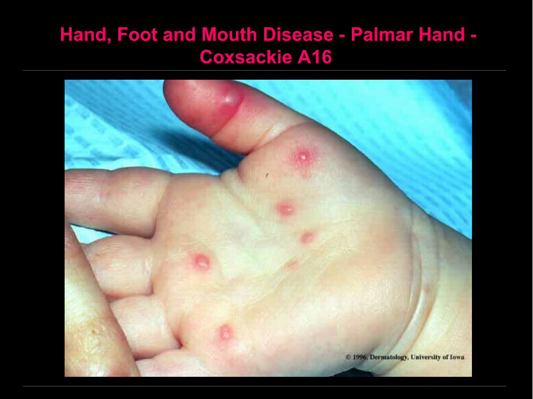
Hand, Foot and Mouth Disease - Palmar Hand - Coxsackie A16
Hand, Foot and Mouth Disease

Hand, Foot and Mouth Disease
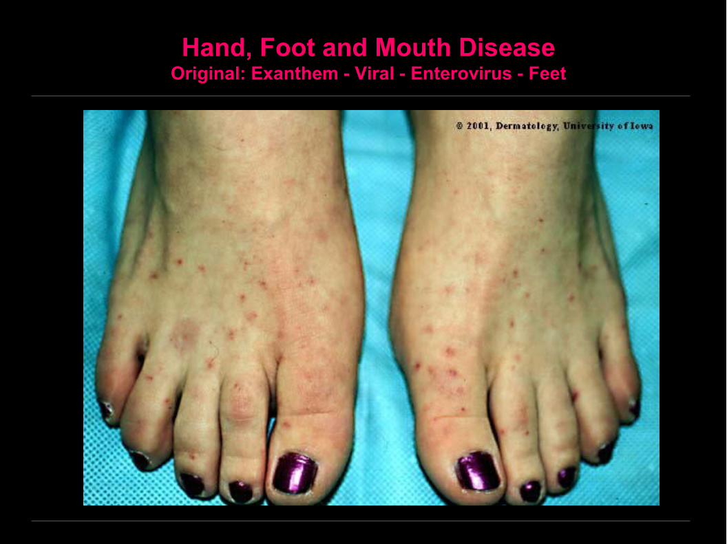
Hand, Foot and Mouth DiseaseOriginal: Exanthem - Viral - Enterovirus - Feet
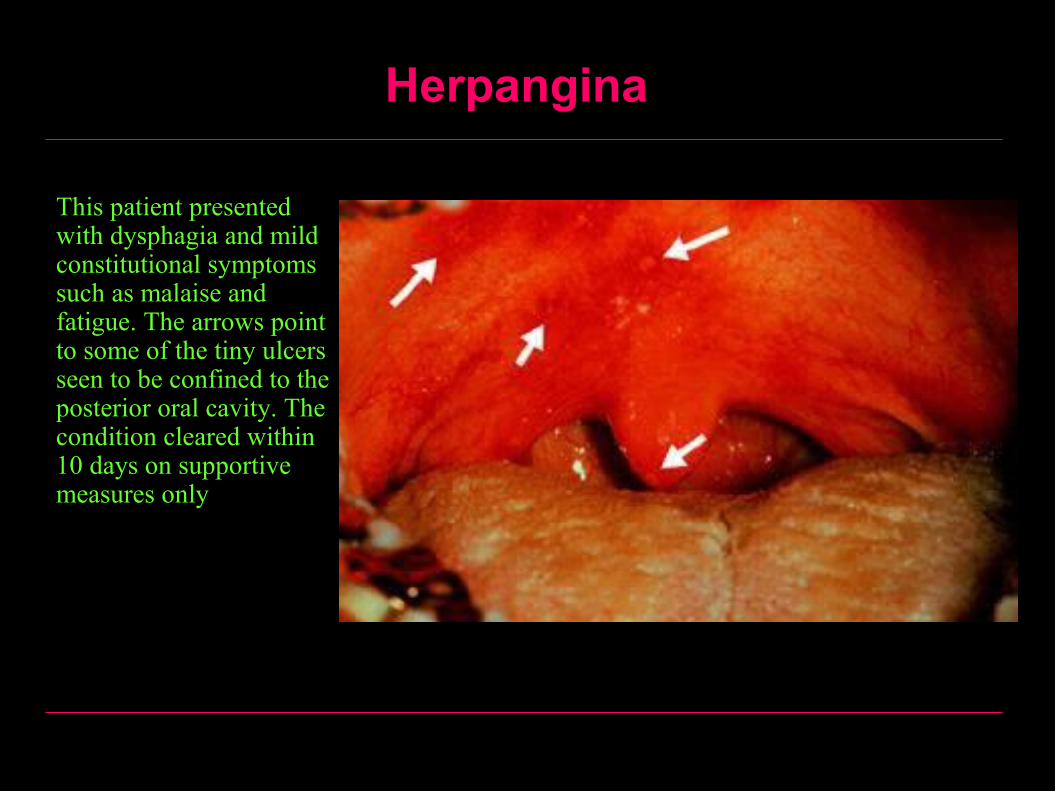
Herpangina
Definition– Highly contagious disease caused by Coxsackie virus type A10
(belonging to the family of picornaviruses).
Clinically• Fever and malaise and sometimes headache, nausea and vomiting.
• Appearance of vesicles, which rupture forming painful ulcers.
• Ulcers characteristically affect the soft palate near the tonsils.
• The rarity of involvement of the anterior mucosa or the gingiva helps differentiate herpangina from herpes simplex. Herpangina also differs from herpes simplex by its brief evolution, the lesions last only 2 - 4 days, while herpes lesions lasts 7 - 10 days.
Treatment• Symptomatic and complications are rare.
Herpangina
This patient presented with dysphagia and mild constitutional symptoms such as malaise and fatigue. The arrows point to some of the tiny ulcers seen to be confined to the posterior oral cavity. The condition cleared within 10 days on supportive measures only
Infectious Mononucleosis
Definit ion– An acute febrile illness caused by the Epstein-
Barr virus– A member of the Herpesviridae family– Infectious mononucleosis is the primary
infection of EBV seen usually in children and young adults
– The virus remains latent for years (possibly for life) in the B lymphocytes of infected individuals.
Infectious Mononucleosis
Pathogenesis• The incubation period is 30 – 50 days• The mode of infection is by droplet infection• EBV receptors (CD21 molecule) are expressed on
mature resting B lymphocytes and stratified squamous epithelium of the oropharynx and salivary glands
• In the host, the virus invades and replicates within epithelial cells of salivary glands and nasopharynx and then invade B-lymphocytes which possesses receptors for the virus

Infectious Mononucleosis
Clinically• The infection is usually subclinical in children, but symptomatic
in young adults.• Fever and malaise with headache and sweating.• Sore throat and pharyngitis which may be severe and
accompanied by a grayish white membrane and gross tonsillar enlargement.
• Pinpoint petechiae in the palate.• Cervical lymphadenopathy which may become generalized,
often with splenic enlargement and tenderness.• Mild hepatomegaly in some cases and clinical jaundice in 5-
10%.• The illness can last several weeks.
Infectious Mononucleosis
Diagnosis• Complete blood picture reveals atypical
lymphocytes.• Epstein-Barr virus-specific antibody test.
Infectious Mononucleosis
Complications• Are rare but some are serious.• Acute air way obstruction may occur as a result of the
lymphoid enlargement and edema which respond to corticosteroid therapy and rarely needs tracheostomy.
• Splenic rupture is also rare.• Neurological complications include meningitis and
encephalitis.
Treatment• Supportive treatment. Acyclovir therapy does reduce EBV
shedding in acute infections and may be used in immunocompromized patients.
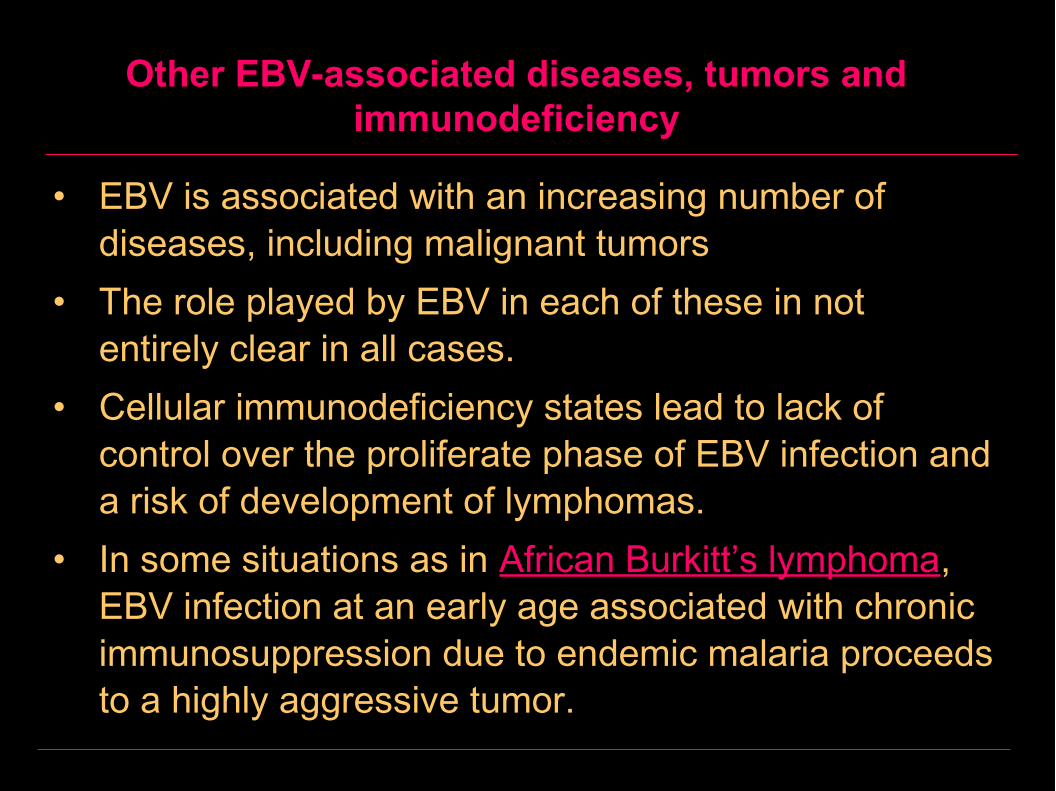
Other EBV-associated diseases, tumors and immunodeficiency
• EBV is associated with an increasing number of diseases, including malignant tumors
• The role played by EBV in each of these in not entirely clear in all cases.
• Cellular immunodeficiency states lead to lack of control over the proliferate phase of EBV infection and a risk of development of lymphomas.
• In some situations as in African Burkitt’s lymphoma, EBV infection at an early age associated with chronic immunosuppression due to endemic malaria proceeds to a highly aggressive tumor.
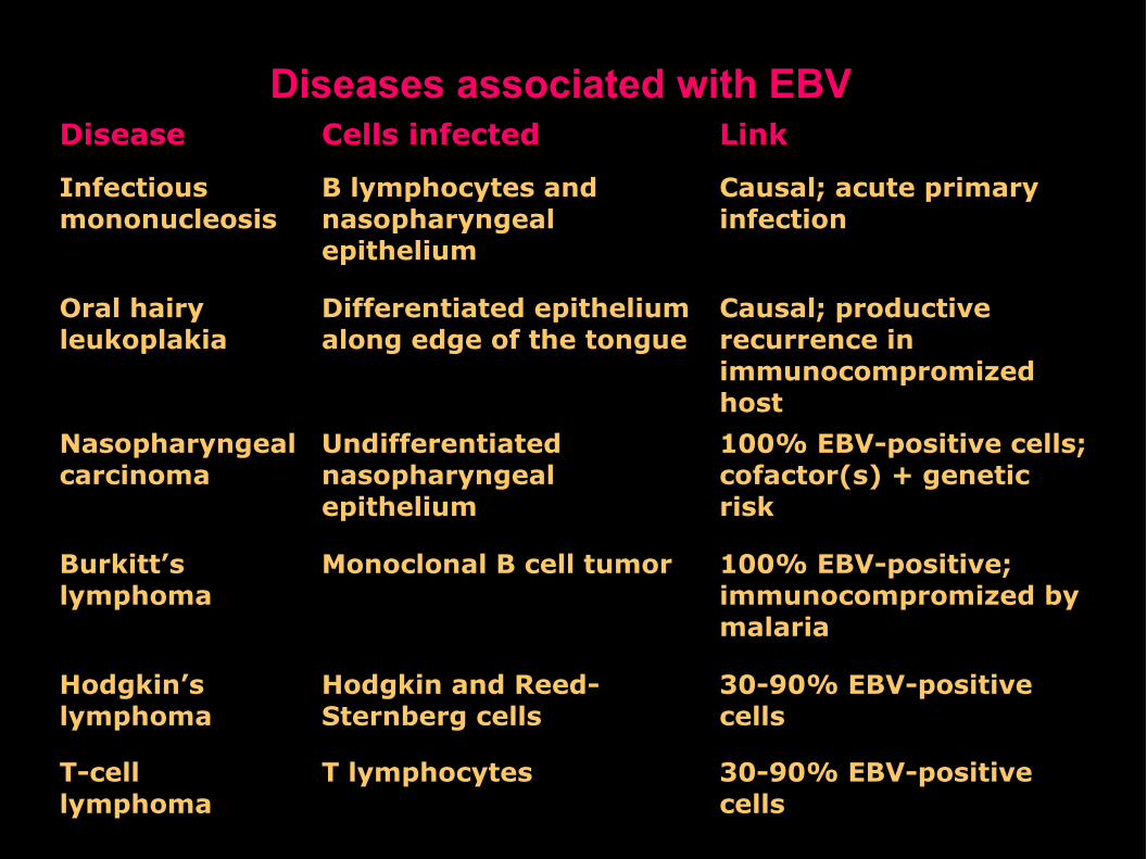
Diseases associated with EBVDisease Cells infected Link
Infectious mononucleosis
B lymphocytes and nasopharyngeal epithelium
Causal; acute primary infection
Oral hairy leukoplakia
Differentiated epithelium along edge of the tongue
Causal; productive recurrence in immunocompromized host
Nasopharyngeal carcinoma
Undifferentiated nasopharyngeal epithelium
100% EBV-positive cells; cofactor(s) + genetic risk
Burkitt’s lymphoma
Monoclonal B cell tumor 100% EBV-positive; immunocompromized by malaria
Hodgkin’s lymphoma
Hodgkin and Reed-Sternberg cells
30-90% EBV-positive cells
T-cell lymphoma
T lymphocytes 30-90% EBV-positive cells
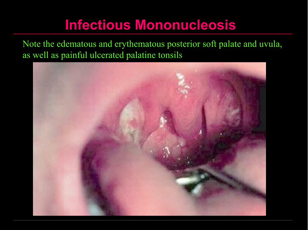
Infectious MononucleosisNote the edematous and erythematous posterior soft palate and uvula, as well as painful ulcerated palatine tonsils
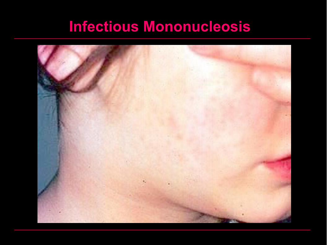
Infectious Mononucleosis
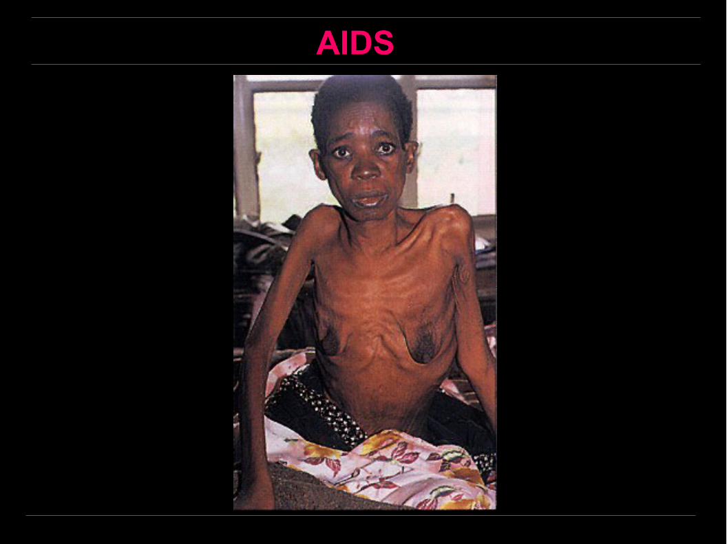
AIDS
AIDS
Definit ionAIDS is an acquired immunodeficiency syndrome.
EtiologyThe disease is caused by the human immunodeficiency virus (HIV), which belong to the family of lentiviruses.
PathogenesisThe virus binds to the CD4 receptor on T lymphocyts and thus produces elimination of the entire population of T4 cells (helper T lymphocytes).
AIDS
Transmission1- Sexual2- Parenteral
1. Blood transfusion2. Shared needles in drug addicts3. Accidental needle stick
3- From mother to fetus (vertical transmission)
Incubation Period3-7 years
AIDS
Clinically1- An acute viral like illness in 50% of patients2- Persistent generalized lymphadenopathy with mild illness3- AIDS related complex consists of: Persistent fever, Weight
loss, Diarrhea4- Full blown AIDS: Appear after 5-7 years and consists of:
• Opportunistic infections e.g. pneumocystis carinii pneumonia
• Multiple malignancies• Neurologic involvement:
- Encephalopathy- Dementia
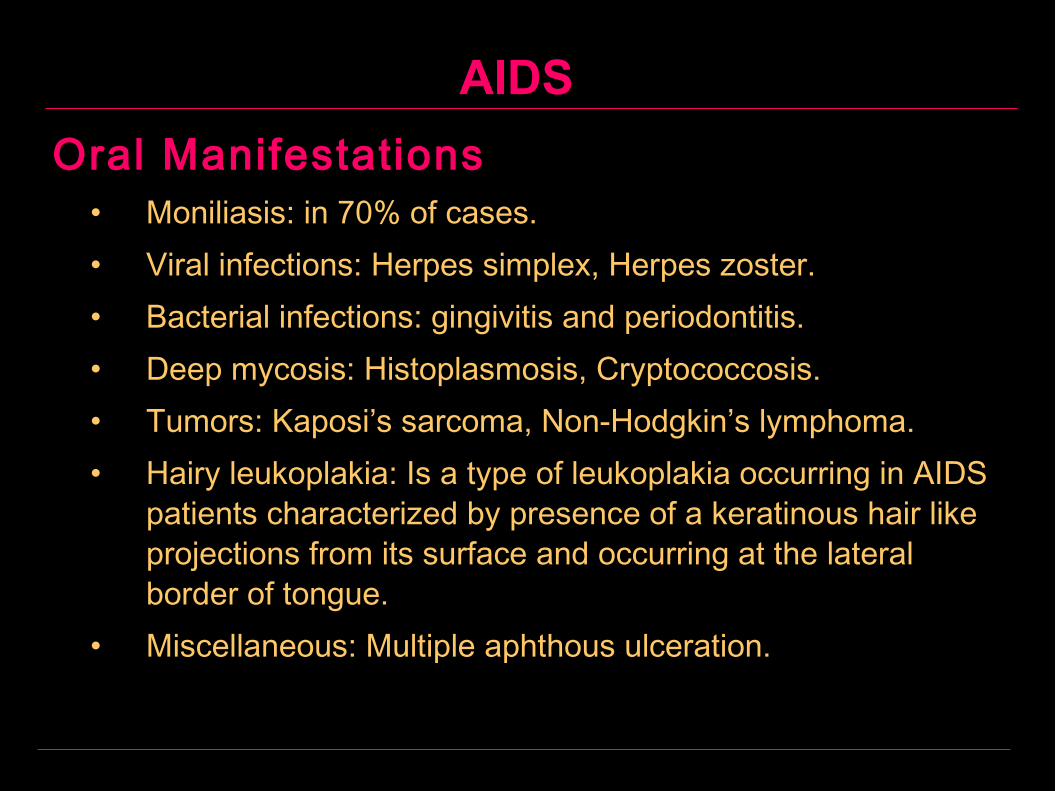
AIDS
Oral Manifestations• Moniliasis: in 70% of cases.• Viral infections: Herpes simplex, Herpes zoster.• Bacterial infections: gingivitis and periodontitis.• Deep mycosis: Histoplasmosis, Cryptococcosis.• Tumors: Kaposi’s sarcoma, Non-Hodgkin’s lymphoma.• Hairy leukoplakia: Is a type of leukoplakia occurring in AIDS
patients characterized by presence of a keratinous hair like projections from its surface and occurring at the lateral border of tongue.
• Miscellaneous: Multiple aphthous ulceration.
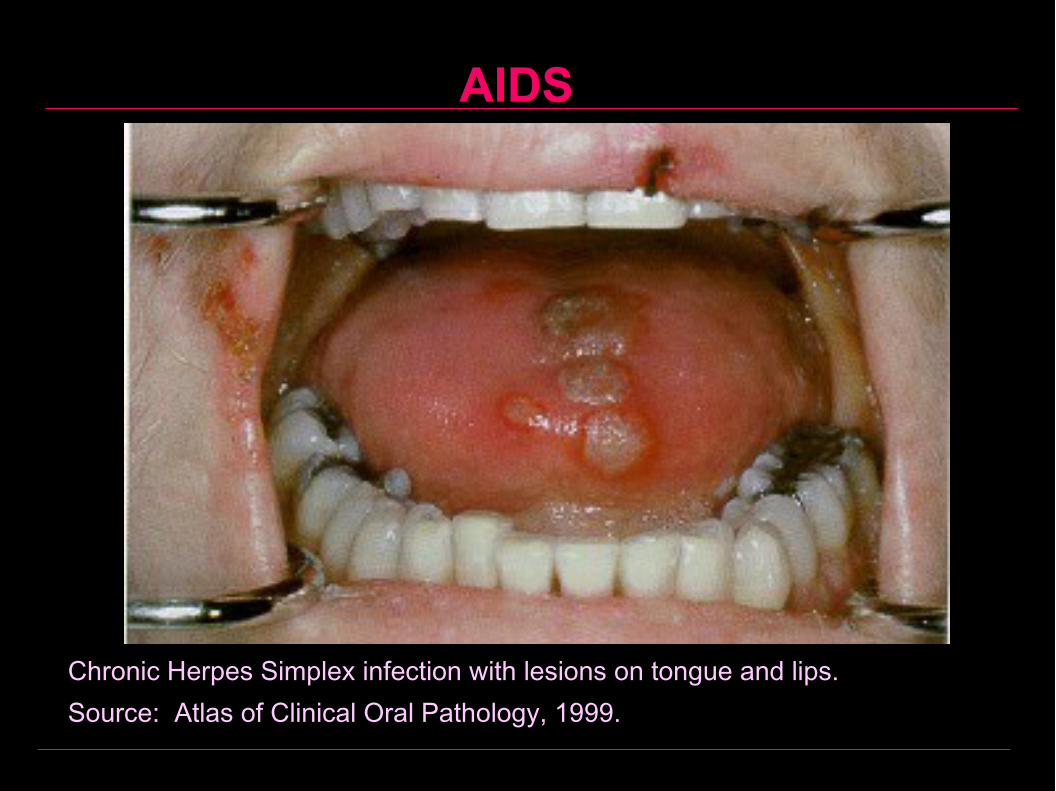
AIDS
Chronic Herpes Simplex infection with lesions on tongue and lips.Source: Atlas of Clinical Oral Pathology, 1999.
AIDS
PrognosisWith full blown AIDS, the 5 year survival is 85%.
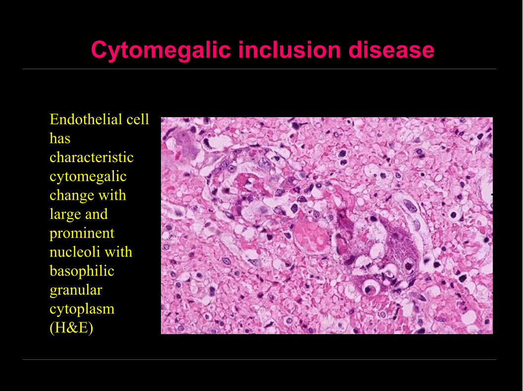
Cytomegalic inclusion disease
Endothelial cell has characteristic cytomegalic change with large and prominent nucleoli with basophilic granular cytoplasm (H&E)
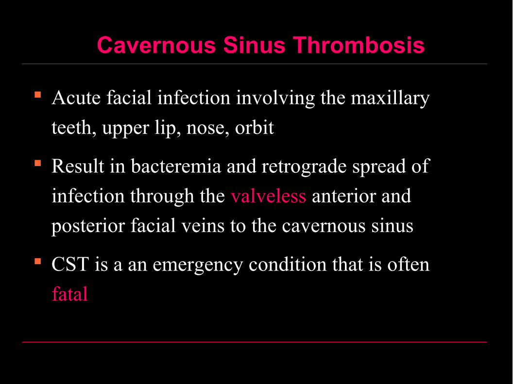
Cavernous Sinus Thrombosis
Acute facial infection involving the maxillary
teeth, upper lip, nose, orbit
Result in bacteremia and retrograde spread of
infection through the valveless anterior and
posterior facial veins to the cavernous sinus
CST is a an emergency condition that is often
fatal
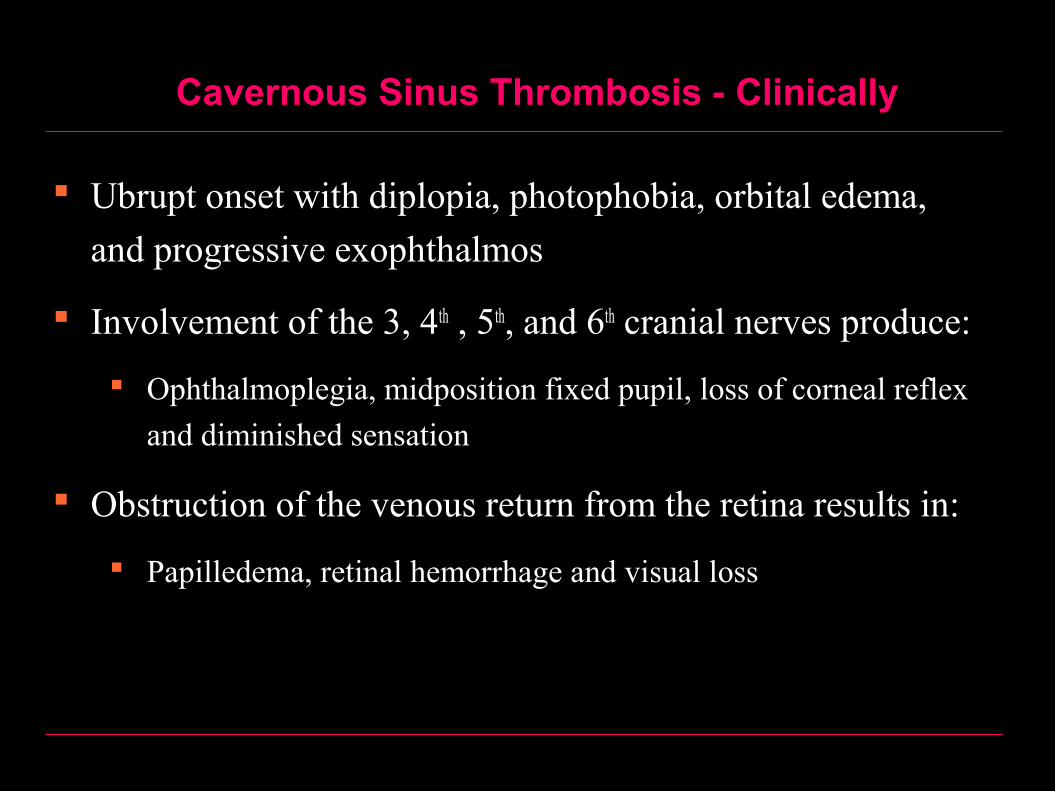
Cavernous Sinus Thrombosis - Clinically
Ubrupt onset with diplopia, photophobia, orbital edema,
and progressive exophthalmos
Involvement of the 3, 4th , 5th, and 6th cranial nerves produce:
Ophthalmoplegia, midposition fixed pupil, loss of corneal reflex
and diminished sensation
Obstruction of the venous return from the retina results in:
Papilledema, retinal hemorrhage and visual loss
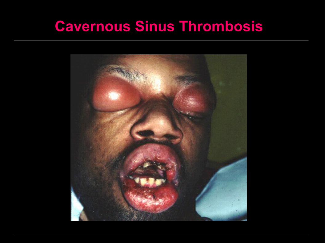
Cavernous Sinus Thrombosis
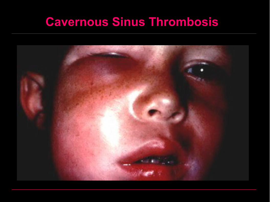
Cavernous Sinus Thrombosis
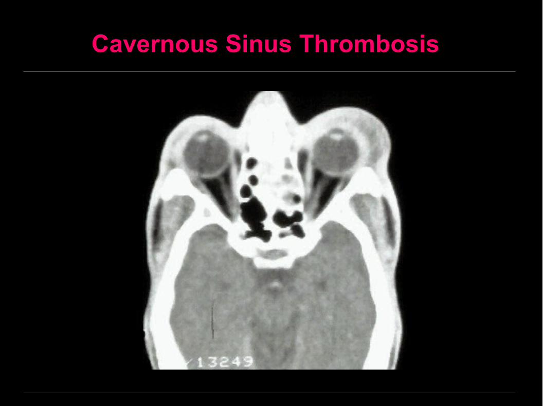
Cavernous Sinus Thrombosis