Embed Size (px)
Citation preview

EXAMINATIONSOF ENDOCRINE DISORDERS
AL AUF JALALUDEEN
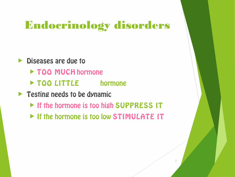
Endocrinology disorders
Diseases are due to TOO MUCH hormone TOO LITTLE hormone
Testing needs to be dynamic If the hormone is too high SUPPRESS IT If the hormone is too low STIMULATE IT
2

3

Endocrine disorders
(a) Central level (Hypothalamic / pituitary disease)
(b) Peripheral level (Dysfunction of peripheral gland)
(c) Receptor / postreceptor level (Target cell
insufficiency - low sensitivity to hormone action)
Manifestation of endocrine disordersManifestation of endocrine disorders

Laboratory tests•Plasma hormone levels•Hormone diurnal rhythm•U-hormones / metabolites•Stimulatory / inhibitory test•Standard biochemistry (Na, K, glc...)
Graphic procedures (imaging)Ultrasonography•CT / MRI•Scintigraphy
Other•Endoscopy•Perimeter
Examination methods

Functional tests
Basal hormonal concentration very often doesn´t allow to establish a diagnosis of hypo- or hyperfunction.
Suspect hypofunction → Stimulatory tests= quantification of functional reserve of endocrine gland
Suspect hyperfunction → Inhibitory tests= quantification of responsibility of endocrine gland to inhibitory factors
Principles:• negative feedback inhibition / stimulation• direct stimulation / inhibition

Insulin hypoglycemia test
i.v. aplic. insulin (O,1 IU/kg)to cause hypoglycaemia (2 mmol / L)
stimulation of ACTH + STH secretionNormal response: STH > 10 ng/mL, P-cortisol > 18 µg / dLContraind.: diabetes mellitus
Stimulatory tests of pituitary function

Methyrapone (Methopyrone) test
• Blocade of cortisol synthesis by metyrapone• negative feedback elevation of ACTH secretion• Secondary elevation of adrenal cortisosteroids (11-deoxycortisol) in plasma• normal: 11-deoxycorticosteroids > 7 µg / dL
Levodopa test
• Physiological elevation of STH secretion in pituitary• Normal: STH > 6 ng /mL• (Test is safer than hypoglycemia test)

Arginin infusion testPhysiol.: elevation of STH secretion in pituitarynormal: GH > 6 ng / mL
TRH testi.v. application of TRH evokes TSH and PRL response
GnRH testi.v. application of GnRH (LHRH) stimulates LH elevation (+ slow FSH elevation)
CRH testi.v. application of corticoliberin stimulates POMC response+ combination with sinus petrosus inferior cathetrization

Dopaminergic drugs test
Dopamin = prolactin inhibitory factor
Physiol. inhibition of PRL (+ STH) secretion
Inhibitory tests of pituitary function

Dexamethazone test
Dexamenthazone = synthetic glucocorticoid
Principle: Peroral administration of DEX via negative feedback inhibits ACTH and cortisol production
Basic test variants:- overnight test (onetime application of 1 or 2 mg p.o.)- 7-day test (2 days basal cortisol levels, 2 days DEX 2
mg/day, 2 days DEX 8 mg/day)

Venous catheterization with selective blood sample collection
1. Catheterization of sinus petrosus inferiorSinus p.i. = venous drenage of pituitary glandPrinciple: Local concentration of ACTH (before and after stimulation with CRH) may distinguish pituitary and paraneoplastic Cushing syndrome)
2. Catheterization of vena cava inferiorStep by step blood sample collection from abdom. veinsPrinciple: Localization of small (CT/MRI undetectable) abdominal tumor (carcinoid, insulinoma etc.) due to high local concentration of hormone.
Local hormonal concentrations

Imaging methods
Indications:
1. Localization of endocrine active tumors, hyperplasia,
ectopic hormonal production
2. Evaluation of systemic complications
Native X-ray exams
Ultrasonography
CT / MRI
Scintigraphy
Angiography

Osteolysis of sella turcica as a late manifestation of the large pituitary tumor.Notice: The standard method for this diagnosis is MRI !
X-ray examination

X-ray examination

Arachnodactylia
X-ray examination

„Salt and peper“
scull
Increased parathyroid activity leading to characteristic
subperiosteal resorption
X-ray examination
Hyper-PTH

Ultrasonography
Indications:1. Thyroid gland, parathyroid glands
- most important imaging method2. Abdominal endocrinopathy (adrenal gland, endocrine
pancreas)- gives only rough picture, replaced now with CT / MRI
Techniques:2D USG: Cystic changes and solid conditions as small as 3 to 5
mm can be detected.Doppler USG: Blood-flow is present.USG + Biopsy: USG guided removal of tissue samples

USG: Normal thyroid gland
USG

Thyroid glandColor USG showing blood flow(hirger perfussion typical e.g.for GB disease
USG

CT / MRI
Computed Tomography (CT)Magnetic Resonance Imaging (MRI)The better degree of contrast in the imaging than in USG.
The comparison of CT and MRI
CT advantages MRI advantages
Lower costBetter availabilityBeter resolution of bone structures(e.g. osteolysis)
High resolution of vascular abnorm.(e.g. differentiation of pituit. tumorsand hemangiomas)No radiation load

CT

Nodular goiter
MRI

Scintigraphy
131I
125I
99mTc-MIBI
131I-MIBEG
99mTc-octreotide
γ-emitter
β+γ emitter
γ-emitter
β+γ emitter
γ-emitter
Application of isotope and its uptake in functional parenchyma of endocrine gland. Extracorporal detection of γ-emission.

Thyroid gland - Fine needleaspiration biopsy (FNAB)
Biopsy
1. Thyroid gland - unclear solitary nodule, tumors2. Adrenal glands - rarely

Thyroid Tests
1. Thyroid Function
2. Iodine Kinetics
3. Thyroid Structure
4. Thyroid Antibodies
5. Thyroglobulin

Adrenal Hormone TestsTo evaluate patients with suspected dysfunction of the adrenal glands
To aid in the diagnosis and evaluation of adrenal abnormalities, such as Cushing’s syndrome, Addison’s disease, adrenal adenoma, or adrenal hyperplasia
27

28