Embed Size (px)
Citation preview

Journal club

Ultrasound-Guided Percutaneous Thyroid NoduleCore Biopsy: Clinical Utility in Patients
with Prior Non-diagnostic Fine-Needle Aspirate
Introduction
• Nodular thyroid disease is common. Palpable thyroid nodules are present in 4%–7% adults.
• Non palpable thyroid nodules are even more common, found in up to 65% of subjects in sonographic and autopsy studies.
• Five percent to 15% of isolated thyroid nodules or nodular thyroids will ultimately harbor a thyroid malignancy.
• Consequently, the accurate diagnosis of nodular thyroid disease is a common and important clinical problem.

• Fine-needle aspiration (FNA) is widely utilized for the detection of malignancy in thyroid nodules and has been extensively validated for this purpose.
• Sonographically guided FNA biopsy of neck masses has high sensitivity.
• The low cost, ease of the procedure, ready availability, and safety make FNA the method of choice for nodule sampling.
• However, FNA of thyroid nodules is nondiagnostic in 5%–20% of patients, even with the use of ultrasound (US) guidance.
• A nondiagnostic thyroid nodule FNA does not assure a benign nodule.
• Therefore, uncertainty about the presence of thyroid malignancy remains a common clinical problem after nondiagnostic FNA.

• Although core needle biopsies are widely used in other anatomic regions, relatively few series of US-guided core needle biopsy (CB) of thyroid nodules have been reported.
• Current American Thyroid Association (ATA) thyroid nodule management guidelines and the Society of Radiologists in Ultrasound Consensus Statement do not discuss the role of thyroid nodule core biopsy (CB) in the evaluation of thyroid nodules.
• In fact, several practice guidelines suggest that surgical excision may be appropriate after several non-diagnostic FNAs.

• The recent American Association of Clinical Endocrinologists (AACE), Associazione Medici Endocrinologi (AME), and European Thyroid Association (ETA) thyroid nodule guidelines mention the potential use of CNB but do not specifically recommend its use after nondiagnostic biopsies.
• Therefore the study is done to review experience to determine the diagnostic utility of combined FNA and CB (CFNACB) in patients with prior nondiagnostic thyroid nodule FNA.

Materials and Methods
Medical record review
• Three authors (M.K.S., W.C.F., and A.E.S.) retrospectively identified all patients who underwent CFNACB of a thyroid nodule at the Massachusetts General Hospital (MGH) between January 1, 2006, and December 31, 2008, by searching the interventional radiology divisional database, and manually reviewing all thyroid-related cytology and histopathology reports during this 3-year period.
• During this time period 5542 thyroid FNAs were performed; 762 of these were nondiagnostic.
• Our analysis is restricted to those patients with non-diagnostic FNAs who subsequently had a CFNACB

• The following parameters were obtained from each• medical record: • (i) nodule size, • (ii) patient age and sex,• (iii)number and result of prior FNA procedures,• (iv) result of CFNACB, • (v) subsequent management, and • (vi) outcome.• FNA and CB needle size and the number of passes
wererecorded when available.

Inclusion criteria
• Inclusion criteria included the following: • (i) a prior non-diagnostic thyroid nodule FNA
performed and interpreted at their institution and
• (ii) subsequent CFNACB of the same nodule performed and interpreted.

CFNACB procedure• All CFNACB procedures were performed by a staff interventional
radiologist with an interventional radiology fellow or radiology resident using real-time US guidance. (25 gauge, 5 cm in length) were used for all FNA procedures, and (20 gauge, 6 cm in length) (Cardinal Healthcare, Dublin, OH) with a 10 or 20mm adjustable needle throw were used for all CBs.
• After obtaining informed consent, each patient was placed in the supine position with a pillow under the shoulders to slightly extend the neck.
• Initial US examination was performed to identify the thyroid nodule of interest. Images of the thyroid nodule were then obtained. After the administration of subcutaneous and peri-thyroidal local anesthesia (5– 10 cc of 1% lidocaine), four to six freehand US-guided FNAs were performed.
• Two to four CBs were subsequently obtained under US guidance.
• When performing thyroid CBs, our goal is to obtain a visually estimated total core length of at least 20 mm; however, total CB length is not routinely documented.
• A post procedure sonogram was performed to assess for hematoma.
• The patient was discharged at the conclusion of the procedure.
Specimen preparation
• FNA specimens were collected and smears were prepared by the radiologist.
• The departmental protocol is to smear two aspirates on glass slides, which are then immediately fixed in 95% ethanol.
• Two to four additional, US-guided aspirates were performed and placed in their entirety into a CytoLyt (Cytyc Corp., Marlborough, methanol+water) liquid preservative solution.
• The specimens were stained in the cytology laboratory using the modified Papanicolaou procedure and submitted for cytologic assessment.

• While in the US suite, the CB samples were placed in normal saline and transported to the cytology laboratory on the same day.
• Upon arrival in the cytology laboratory, the CB samples were immediately decanted into formalin and after being embedded into paraffin blocks using standard protocols, cut into sections and stained with hematoxylin and eosin prior to histopathologic assessment.

Specimen interpretation
• A board-certified attending staff cytopathologist reviewed all FNA specimens.
• Specimens were considered nondiagnostic if insufficient cellular material (fewer than six groups of cells containing > 10 cells each) was present and no evidence of cellular atypia was found.
• A board-certified attending staff pathologist reviewed all CB samples.
• Cytology and histopathology results were available to the pathologist and cytopathologist, respectively, at the time of their interpretations.

Results interpretation
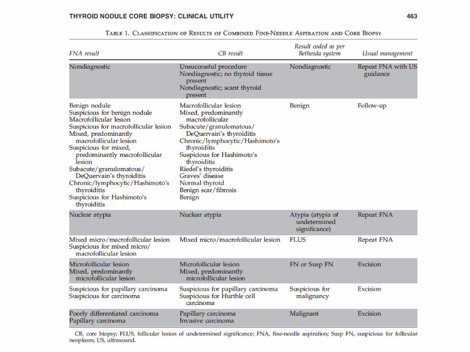
• Each FNA and CB result was recorded, and then classified into one of seven categories according to the Bethesda classification scheme.
• Each result was then classified as either diagnostic or nondiagnostic, based on the cytopathology report and clinical notes.

Diagnostic accuracy of CFNACB
• The diagnostic accuracy of CFNACB was determined by a review of surgical pathology in cases that underwent thyroidectomy and by change in nodule size by US or clinical assessment in patients who did not have surgery.

Results
Demographics and indications for CFNACB
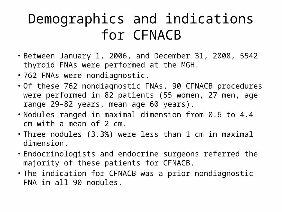
• Between January 1, 2006, and December 31, 2008, 5542 thyroid FNAs were performed at the MGH.
• 762 FNAs were nondiagnostic. • Of these 762 nondiagnostic FNAs, 90 CFNACB procedures were
performed in 82 patients (55 women, 27 men, age range 29–82 years, mean age 60 years).
• Nodules ranged in maximal dimension from 0.6 to 4.4 cm with a mean of 2 cm.
• Three nodules (3.3%) were less than 1 cm in maximal dimension. • Endocrinologists and endocrine surgeons referred the majority of
these patients for CFNACB.• The indication for CFNACB was a prior nondiagnostic FNA in all 90
nodules.

• There was one prior FNA in 77% of nodules (69/90), two prior FNAs in 20% of nodules (18/90), and three prior FNAs in 3%of nodules (3/90).
• Thirteen additional thyroid nodule CFNACBs were excluded from the study as the prior FNA was performed and interpreted outside our institution.

Complication rate of CFNACB
• The radiology databases were reviewed for any documented complications.
• Procedure reports and follow-up visit notes were available for all patients.
• There were no reported complications

Diagnostic yield of CFNACB
• Overall, the combined procedure (CFNACB) yielded a diagnostic result in 87% of nodules (78/90).
• Both FNA and CB were diagnostic in 37% of nodules. • In 40% of nodules, CB was diagnostic when FNA was not. • In 10% of nodules, FNA was diagnostic when CB was not
For the entire subject group, CB was diagnostic in 77% of nodules, whereas repeat FNA was diagnostic in only 47%.
• The diagnostic yield of CFNACB was further analyzed with respect to prior FNA procedures.

Relative yield of CB versus FNA following one priornondiagnostic FNA
• Sixty-nine nodules had only one prior nondiagnostic biopsy.
• Of these 69 nodules, CFNACB was diagnostic in 87% (60/69), CB was diagnostic in 74% (51/69), and repeat FNA was diagnostic in 52% (36/69) (Table 2).
• In this subgroup, the diagnostic performance of CB was significantly superior to that of concurrent FNA ( p = 0.0135).

Relative yield of CB versus FNA following two or moreprior nondiagnostic FNA procedures
• Twenty-one nodules had at least two prior nondiagnostic FNAs.
• Of these 21 nodules, CFNACB was diagnostic in 86% (18/21), CB was diagnostic in 86% (18/21), and FNA was diagnostic in 29% (6/21).
• Of those with two prior nondiagnostic biopsies, there were no instances where the FNA was diagnostic and the CB was not.

• In this subgroup, the diagnostic performance of CB was significantly superior to concurrent FNA ( p = 0.0005).
• Three nodules had previously had three nondiagnostic FNAs. • The CB was diagnostic in all three cases whereas none of the FNAs
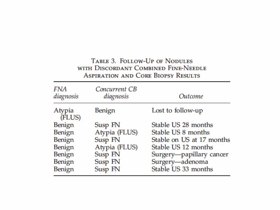
were diagnostic. Discordant cytology and pathology findings for the FNA and CB components of CFNACB were uncommon, occurring in 8 of 90 nodules (9%).
• There are currently no data to guide management in this situation. • Therefore our approach is to err on the side of caution by
accepting the most (rather than the least) concerning diagnosis. • In some of these cases patients declined surgery. • The outcome of these eight cases is tabulated in Table 3.
Diagnostic accuracy of CFNACB
• CFNACB yielded a benign diagnosis in 40% of nodules (36/90). • Of these 36 nodules, follow-up data were available in 22
(61%). Seventeen (47% were followed with US (mean follow-up 18 months, range 4–37 months).
• Sixteen of 17 nodules (94%) were stable or decreased in size; one nodule (6%) had a minimal increase in size after 12 months and is awaiting a repeat US.
• Of the three nodules followed with clinical examination, all were stable.
• One patient with Reidel’s thyroiditis was treated with glucocorticoids and one patient with Graves’ disease was treated with radioactive iodine.

• Follow-up data were not available for 14 patients (39%).
• CFNACB yielded an atypical diagnosis (atypia of undetermined significance or follicular lesion of undetermined significance) in 10% of nodules (9/90).
• Follow-up data were available for all of these nodules. • Five of these nodules underwent surgery and all were
benign follicular adenomas• (Table 4).


• Four nodules were followed with serial USs and were stable (mean follow-up 16.5 months, range 2.8–28.9 months).
• CFNACB yielded a diagnosis of follicular neoplasm or suspicious for a follicular neoplasm in 31% of nodules (28/90).
• Follow-up data were available in 26 of these 28 nodules (93%).• Twenty-two were removed surgically and four were followed with
sonography. • Of these, 2 of 22 (9%) were malignant, 18 of 22 (82%) were benign
follicular adenomas, and 2 of 22 (9%) were nodular Hashimoto’s thyroiditis.
• Four of 26 nodules (15%) were stable on serial US examination (mean follow-up 21 months, range 10–39 months).
• Two subjects were lost to follow-up.
• CFNACB yielded a suspicious for malignancy or malignant diagnosis in 5.5% of nodules (5/90).
• Follow-up was available in all cases and all were malignant at surgery.
• CFNACB yielded a non diagnostic result in 13.3% of nodules (12/90).• Follow-up data were available for 11 of these 12 subjects (92%). • Five of these patients had surgery and all had benign pathology (four
follicular adenomas, and oneHashimoto’s thyroiditis. • Five nodules were stable on serial US (mean followup 18.5months,
range 5–26months). • One was stable on followup physical examination and one was lost
to follow-up.


Discussion• Recent ATA thyroid nodule guidelines and the Society of
Radiologists consensus statement on thyroid nodules and the National Cancer Institute (NCI) State of the Art conference do not discuss the utility of thyroid CB.
• The ATA guidelines note: ‘‘Partially cystic nodules that repeatedly yield nondiagnostic aspirates need close observation or surgical excision.
• Surgery should be more strongly considered if the cytologically nondiagnostic nodule is solid.
• Recommendation rating: B’’ (8). Baloch et al. summarized the NCI State of the Art conference position on this point by noting:
• ‘‘If repeat smears are ‘nondiagnostic,’ surgery ought to be considered’’.

• The recent AACE/AME/ETA guidelines do note: ‘‘CNB, performed under US guidance with a 20- to 21-gauge cutting needle by experienced operators, may offer additional information to FNA biopsy in selected cases of thyroid or neck masses with repeated inadequate FNA biopsy cytology’’.
• These guidelines do not provide a specific recommendation for the use of core needle biopsies.

• CFNACB of the thyroid gland is a safe technique with a very low rate of complications.
• In a systematic review of clinical complications following thyroid FNA, the authors reported that the rate of major complications ranged from 0.036%–1% (34).
• In this series there were no complications from CFNACB (0%; 95% confidence interval 0%, 4.02%).
• This is concordant with the low rate of complications reported in other CB series.
• For example, in a series of 209 CBs, Screaton et al. reported no major complications, and four self-limited minor complications—three small post biopsy hematomas and one episode of hemoptysis (21). In a series of 377 CBs, Renshaw and Pinnar reported a single post biopsy hematoma that was described as ‘‘large,’’ but which did not require hospitalization (32).
• Although the complication rate of CFNACB is low, the procedure results in the acquisition and interpretation of additional specimens and is therefore more expensive, more time consuming, and requires additional expertise.
• There is no evidence that CFNACB is superior to FNA for the initial diagnosis of thyroid nodules.
• Therefore, FNA continues to be the preferred initial biopsy technique for thyroid nodules.

• This study has several potential limitations. The results of a cohort of patients who were referred for CFNACB after a nondiagnostic FNA, they have not studied all patients with an initial nondiagnostic FNA.
• Many of the referring physicians choose to repeat a single nondiagnostic FNA, and only refer cases considered to be more difficult for CFNACB.
• It is possible that an unrecognized selection bias influenced the outcome of CFNACB in our cohort.

• However, this selection bias appears to have made little difference to the overall diagnostic yield of CFNACB, which was similar in subjects with one or two prior nondiagnostic FNAs.
• Another limitation is that this study is retrospective and the post procedural follow-up is incomplete.

• However, this should not impact the diagnostic rate of CFNACB.
• Finally, the interpreting cytologists and histopathologists were not blinded to each other’s assessments: it is possible that this has biased the diagnostic rate of FNA or CB.
• However, it is likely that this potential bias would decrease the apparent additional benefit of the CB component compared with FNA alone

• This is the first study to systematically assess the diagnostic yield of CFNACB after prior nondiagnostic FNA for thyroid nodules.
• This study provides strong evidence for the high diagnostic yield and safety of CFNACB after prior nondiagnostic FNA.
• Results suggest that CFNACB has the potential to reduce the number of repeat biopsies and unnecessary thyroidectomies in patients with one or more nondiagnostic prior FNA procedures.
• Based on these data, the study strongly recommend CB or CFNACB as an alternative to surgical excision for nodules with two prior nondiagnostic FNAs.
• The role of CFNACB as an alternative to repeat FNA after a single nondiagnostic FNA requires additional study with prospective randomized trials.
References• 1. Mazzaferri EL 1992 Thyroid cancer in thyroid nodules:• finding a needle in the haystack. Am J Med 93:359–362.• 2. Wang C, Crapo LM 1997 The epidemiology of thyroid disease• and implications for screening. Endocrinol Metab Clin• North Am 26:189–218.• 3. Brander A, Viikinkoski P, Nickels J, Kivisaari L 1991 Thyroid• gland: US screening in a random adult population. Radiology• 181:683–687.• 4. Mazzaferri EL 1993 Management of a solitary thyroid nodule.• N Engl J Med 328:553–559.• 5. Hegedus L 2004 Clinical practice. The thyroid nodule. N• Engl J Med 351:1764–1771.

Does Core needle Biopsy Add Pain to theThyroid Nodule Evaluation?
Angelo Carpi1*, Giuseppe Rossi2, Andrea Nicolini1, Giorgio Iervasi3, Matteo Russo4, Jeffrey Mechanick5
• In conclusion, the study suggests that concerns of discomfort or pain following Core needle biopsy of thyroid nodules may not be generally applicable and therefore in light of the demonstrable benefits, CB should be considered as part of the diagnostic algorithm for thyroid nodule evaluation.
The use of core needle biopsy as first-line indiagnosis of thyroid nodules reduces false
negative and inconclusive data reported byfine-needle aspiration in thyroid nodules at high risk of
malignancy detected by ultrasound elastography
WORLD JOURNAL OFSURGICAL ONCOLOGY

• In conclusion, findings indicate a higher rate of diagnostic accuracy with first-line use of CNB than FNA for assessing at-risk thyroid nodules identified by US.
• The false negatives and inconclusive results of FNA may also be reduced making CNB the preferred method in this setting.

• The presence of at least one US risk factor (hypoechogenicity, microcalcifications, irregular margins, intranodular vascularization, and taller than wide shape) had 85% sensitivity and 91% negative predictive value. When elastography was combined with US, the presence of at least one of the six parameters had 97% sensitivity and 97% negative predictive value.
• Ultrasound elastography is a valuable tool for detecting malignant thyroid lesions with a sensitivity similar to traditional US and doppler features.
• By adding Elastography evaluation, the sensitivity for malignancy of US findings is markedly increased and the selection of nodules that do not need cytology is made more reliable.

What is Elastography?
• Elastography is an imaging technique to measure the stiffness of tissues.
• The main idea is that whether the tissue is hard or soft will give diagnostic information about the presence or status of disease.
• For example, cancerous tumours will often be harder than the surrounding tissue, and diseased livers are stiffer than healthy ones
• Images are acquired before and after soft compression of tissues and the deformation is evaluated.
• Initially elastography used manual compression and was only qualitative, now some methods appears to apply a non operator dependant compression.

Elastography and US
• Elastography was developed first in the US field.
• Three step approach:– Organs mechanically stressed by either external or
internal forces.–Measurement of tissues movement induced.– Qualitative or quantitative evaluation of tissue
elastic properties from the measured displacement of tissues.


• Comparison of Ultrasound Elastography, Mammography, and Sonography in the Diagnosis of Solid Breast Lesions
• Invasive ductal carcinoma in a 55-year-old woman. A, Mammogram showing several masses suspected to be benign lesions. B, Right, Sonographic image. A hypoechoic mass with a round shape was suspected to be a benign lesion. Left, Ultrasound elastographic image. Both the entire hypoechoic lesion and its surrounding area were blue. The blue area on UE was larger than the lesion area on sonography. It was scored 5 and was diagnosed as a malignant lesion.


Natural resolution or intervention for fluid collections in acute severe pancreatitis

Introduction
• Knowledge of disease behaviour and temporal evolution, better intensive care and ubiquitous use of advanced radiological imaging have changed the classification and management of acute pancreatitis.
• A considerable proportion of patients with acute pancreatitis develop fluid collections in or near the pancreas early in the course of the disease.

• These are defined as acute fluid collections in the original Atlanta classification. They lack a definite wall, occur in 30-50 per cent of patients, mostly resolve spontaneously and have prognostic implications.
• Acute fluid collections that persist for more 4weeks and develop a definite wall are called acute pseudocysts in the original Atlanta classification.
• They may resolve spontaneously or become complicated and require drainage.
• Advances in non-surgical drainage of pseudocysts have revealed that they are not all the same.

• Those containing necrotic debris, usually developing in a background of necrotizing pancreatitis, have higher morbidity and therapeutic failure rates.
• Thus the revised Atlanta classification divides acute pancreatitis into two historadiological categories:
1) Interstitial oedematous pancreatitis and 2) acute necrotizing pancreatitis.
• Fluid collections that develop in these two situations are described as acute peripancreatic fluid collection (APFC) and acute necrotic collection (ANC) respectively, and persistent collections that develop a definite wall are called pancreatic pseudocyst and walled-off necrosis (WON) respectively.

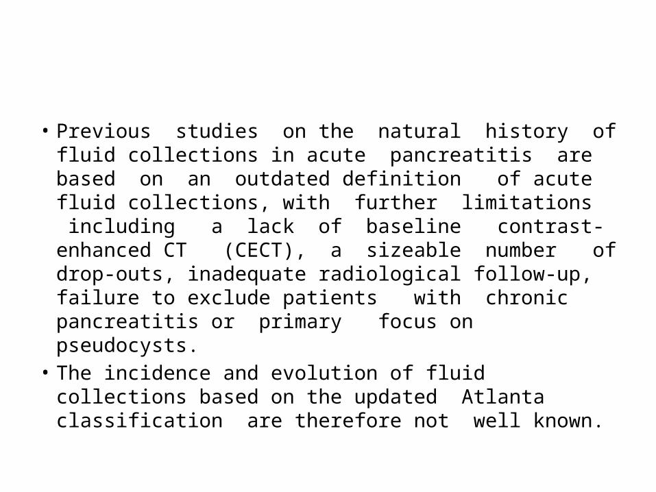
• Previous studies on the natural history of fluid collections in acute pancreatitis are based on an outdated definition of acute fluid collections, with further limitations including a lack of baseline contrast-enhanced CT (CECT), a sizeable number of drop-outs, inadequate radiological follow-up, failure to exclude patients with chronic pancreatitis or primary focus on pseudocysts.
• The incidence and evolution of fluid collections based on the updated Atlanta classification are therefore not well known.

• The aim of the present study was to investigate fluid collections ill acute pancreatitis with a focus on the frequency of development in either interstitial oedematous pancreatitis or acute necrotizing pancreatitis.
• Baseline risk factors for their development; frequency and risk factors for their evolution into pseudocysts or WON; and the eventual out come of pseudocysts and WONs.


Methods
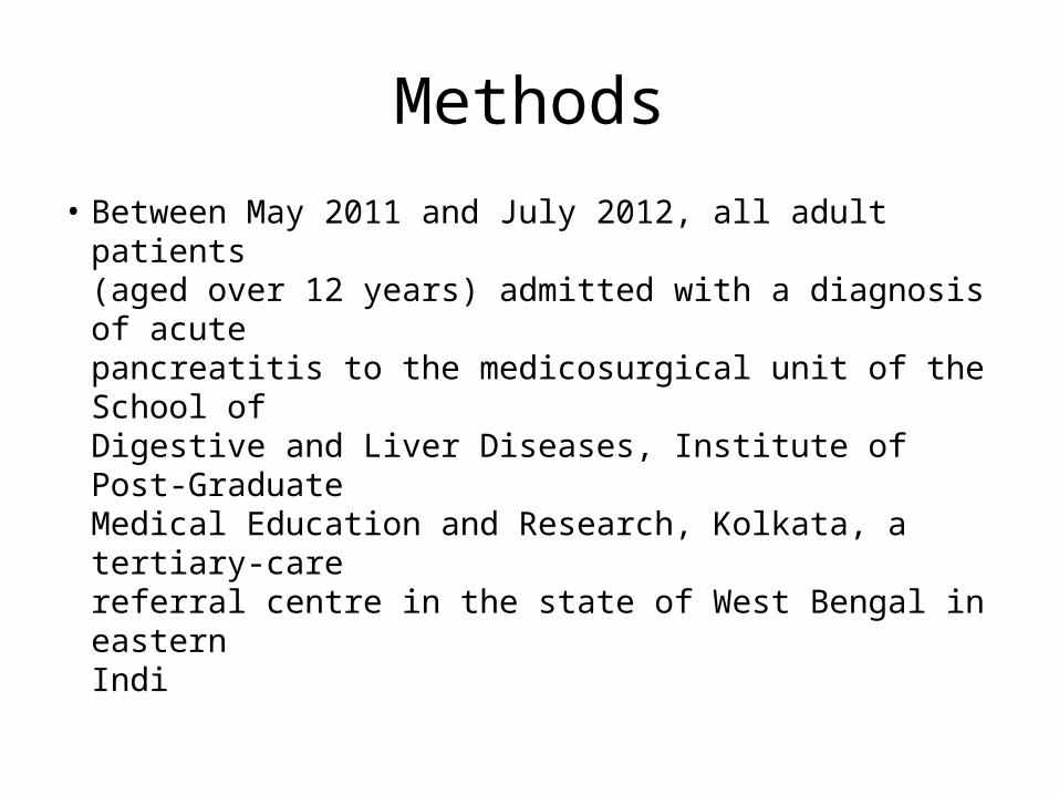
• Between May 2011 and July 2012, all adult patients (aged over 12 years) admitted with a diagnosis of acute pancreatitis to the medicosurgical unit of the School of Digestive and Liver Diseases, Institute of Post-Graduate Medical Education and Research, Kolkata, a tertiary-care referral centre in the state of West Bengal in eastern Indi
Exclusion criteria
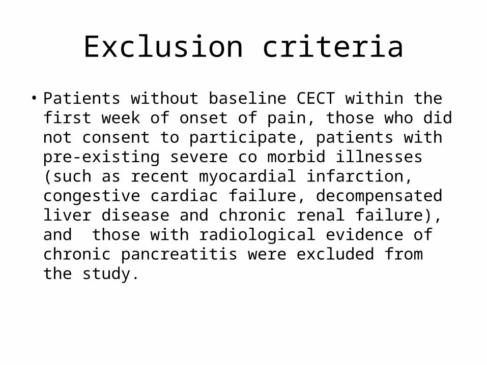
• Patients without baseline CECT within the first week of onset of pain, those who did not consent to participate, patients with pre-existing severe co morbid illnesses (such as recent myocardial infarction, congestive cardiac failure, decompensated liver disease and chronic renal failure), and those with radiological evidence of chronic pancreatitis were excluded from the study.

Diagnosis• A diagnosis of acute pancreatitis was made when two of the following
three criteria were present: • 1) compatible clinical symptoms; • 2) raised serum levels of amylase and/or lipase at least three times the
upper limit; • 3) and radiological evidence of acute pancreatitis. • Biliary aetiology was established
when there were compatible biochemical and/or radiological features. • Alcohol was considered as the aetiology when the patient or relatives
reported a history of pro-longed and substantial abuse of alcohol and/or if there was a recent history of binge consumption before the attack.

• Other aetiologies were established based on specific history (for example post-endoscopic retrograde cholangiopancre-atography, drugs or trauma) and biochemical abnormali-ties.
• Pancreatitis was thought to be idiopathic if any other aetiologies were absent.
• Complete blood count, estimation of packed cell volume (PCV), liver function tests, measurement of levels of blood urea nitrogen, serum creatinine, calcium and phosphate, and fasting blood glucose, arterial blood gas analysis and transabdominal ultrasonography were done on the day of admission.
• CECT of the abdomen was performed according to a standard protocol 5 - 7 days after the onset of pain, with calculation of the baseline CT Severity Index (CTSI).

CT & evaluation of fluid collections
• All scans were evaluated in a structured reporting format by a single gastroradiologist who was blinded to the patient clinical data.
• The number, size (maximum transverse diam- eter of largest) and location of fluid , and the extent/degree of pancreatic parenchymal necrosis were recorded on baseline CFC T.
• Patients were classified as having either oedematous or necrotizing pancreatitis, according to the absence or presence of parenchymal (with or without extraparenchymal) necrosis on baseline CECT, confirmed independently by a second radiologist.


Follow up• Patients with fluid collection(s) were followed up prospectively with
an initial ultrasound examination at discharge or 2 weeks after admission, and then every 4weeks.
• The follow-up examinations were done by two radiologists. Abdominal CECT was repeated after 8+/-2 weeks in those with persistent fluid collection(s) on ultrasonography.
• The nature, with special attention to whether there was a well defined wall, and site of persistent fluid collections were recorded. All patients were followed up until January 2013.

Management• Patients received standard medical care throughout the
study interval. • Further investigations and decisions on intervention were the
prerogative of the treating physician. • Infected pancreatic necrosis (infected ANC) was treated by
either surgical and/or radiological drainage, and any symp- tomatic fluid collection was drained surgically, endoscopi- cally or radiologically.
• The fluid collections were followed up ultrasonographically until drainage, spontaneous reso- lution or for up to 6 months from the onset of symptoms.
• Spontaneous resolution was defined by the absence of any fluid collection on two consecutive ultrasound examinations 4 weeks apart

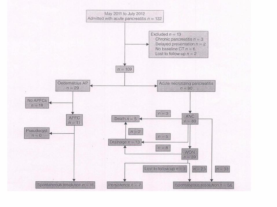
Results


Conclusions
• This study showed that, although APFCs rarely evolve into pseudocysts, almost one-half of ANCs developed into WON.
• Admission blood urea nitrogen was identified as an important predictor of the development of these collections as well as of WONs
• Only one-quarter of AN Cs either required intervention or persisted beyond 6 months.
• More than one-half of WONs resolved spontaneously.

• A baseline ANC diameter exceeding 6 cm was an important independent predictor of the persistence of a collection or the need for intervention.
• Although all patients with necrotizing pancreatitis had ANCs at baseline, approximately 40 per cent of these collections resolved spontaneously before evolving into WONs.
• An admission blood urea nitrogen level of at least 20 mg/dl was found to be an independent predictor of the development of WON in these patients with necrotizing pancreatitis.

References• Vege SS, Chari ST. Organ failure as an indicator of severity of acute pancreatitis: time to revisit the Atlanta• Classification. Gastroenterology 2005; 128: 113 3-113 5.• 2 Bollen TL, van Santvoort HC, Besselink MG, van Leeuwen MS, Horvarth KD, Freeny PC et al., Dutch Acute Pancreatitis
Study Group. The Atlanta classification of acute pancreatitis revisited. Br J Surg 2008; 95: 6-21.• 3 Petrov MS, Vege SS, Wrndsor JA. Global survey of controversies in classifying the severity of acute pancreatitis. Eur J
Gastroenterol Hepatol 2012; 24: 715-721.• 4 Banks PA, Bollen TL, Dervenis C, Gooszen HG,Johnson CD, Sarr MG et al.; Acute Pancreatitis Classification Working
Group. Classification of acute pancreatitis - 2012: revision of the Atlanta classificationand definitions by international consensus. Gut 2012; 62: 102-111.
• 5 Bradley EL ill. A clinically based classification system for acute pancreatitis. Summary of the International Symposium on Acute Pancreatitis, Atlanta, Ga, September
• 11through13, 1992.Arch Surg 1993; 128: 586-590.• 6 Cannon]W, Callery MP, Vollmer CMJr. Diagnosis and management of pancreatic pseudocysts:what is the evidence? J Am
Coll Surg 2009; 209: 385-393.• 7 Baron TH, Hare~ood GC, Morgan DE, YatesMR.• Outcome differences after endoscopic drainage of pancreatic necrosis, acute pancreatic pseudocysts, and chronic pancreatic
pseudocysts. Gastrointest Endosc 2002; 56: 7-17.• 8 Hookey LC, Debroux S, Delhaye M, ArvanitakisM, Le• Moine 0, Deviere ]. Endoscopic drainage of• pancreatic-fluid collections in 116 patients: a comparison of etiologies, drainage techniques, and outcomes. Gastrointest
Endosc 2006; 63: 635-643.• 9 Schulze S, Baden H, Brandenhoff P, Larsen T, Burcharth F. Pancreatic pseudocysts during first attack of acute pancreatitis.
Scand] Gastroenterol 1986; 21: 1221-1223.• 10 Gonzalez AC, Bradley EL, ClementsJLJr. Pseudocyst formation in acute pancreatitis: ultrasonographic evaluation of 99
cases. AJRAm J Roentgenol 1976; 127: 315-317.• 11 Bradley EL, Gonzalez AC, Clements JL Jr. Acute pancreatic pseudocysts: incidence and implications.Ann Surg 197 6;• 184: 734-738.• 12 Siegelman SS, Copeland BE, Saba GP, CameronJL,• Sanders RC, Zerhouni FA. CT of fluid collections associated with pancreatitis. AJR Am J Roentgenol 1980; 134:• 1121-1132.• 13 Balthazar EJ, Robinson DL, Megibow AJ, RansonJHC.• Acute pancreatitis: value of CT in establishing prognosis.• Radiology 1990; 174: 331-336.• 14 Balthazar EJ, RansonJH, Naidich DP, Megibow AJ, CaccavaleR, Cooper MM. Acute pancreatitis: prognostic value of CT.
Radiology 1985; 156: 767-772.