Embed Size (px)
Citation preview
25-Feb-15
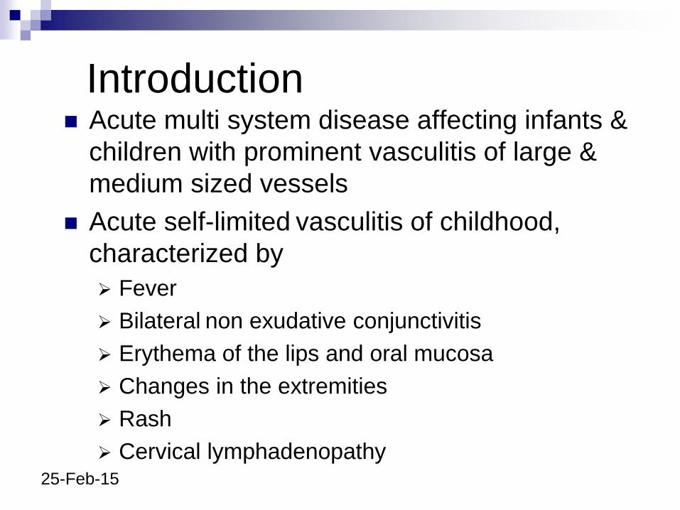
Introduction Acute multi system disease affecting infants &
children with prominent vasculitis of large &
medium sized vessels
Acute self-limited vasculitis of childhood,
characterized by
Fever
Bilateral non exudative conjunctivitis
Erythema of the lips and oral mucosa
Changes in the extremities
Rash
Cervical lymphadenopathy
25-Feb-15
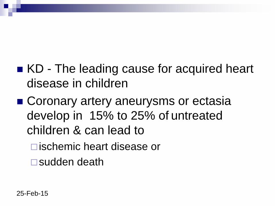
KD - The leading cause for acquired heart
disease in children
Coronary artery aneurysms or ectasia
develop in 15% to 25% of untreated
children & can lead to
ischemic heart disease or
sudden death
25-Feb-15
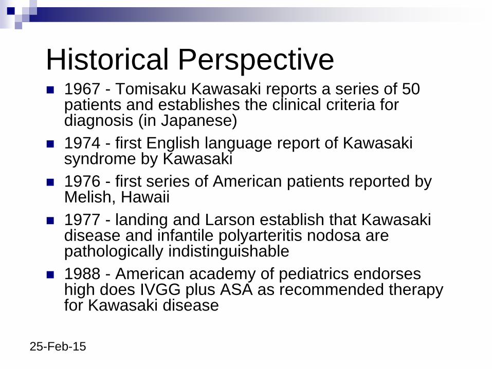
Historical Perspective 1967 - Tomisaku Kawasaki reports a series of 50
patients and establishes the clinical criteria for diagnosis (in Japanese)
1974 - first English language report of Kawasaki syndrome by Kawasaki
1976 - first series of American patients reported by Melish, Hawaii
1977 - landing and Larson establish that Kawasaki disease and infantile polyarteritis nodosa are pathologically indistinguishable
1988 - American academy of pediatrics endorses high does IVGG plus ASA as recommended therapy for Kawasaki disease
25-Feb-15
Epidemiology
More prevalent in Japan and in children of
Japanese ancestry (annual incidence of 112
cases per 100 000 children <5 years old)
Age of onset -
Peak age - 2 to 5 yrs
80 - 85 % < 5 yrs
Rare > 11 yrs
25-Feb-15
Aetiology and Pathogenesis
Aetiology of KD remains unknown, (although
clinical and epidemiological features strongly
suggest an infectious cause)
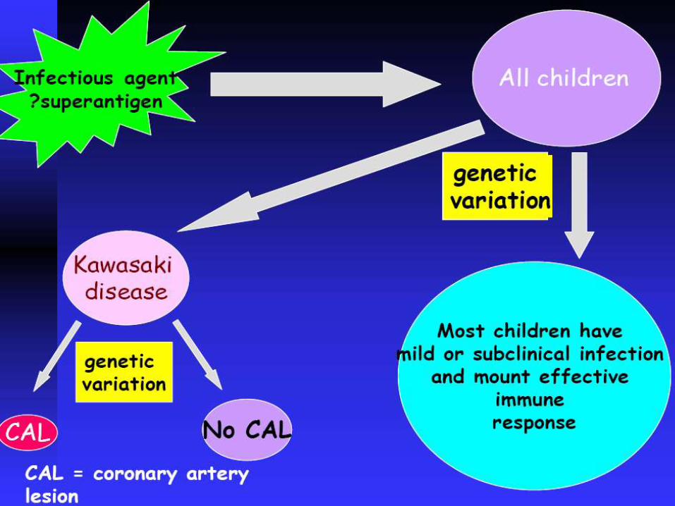
Hypothesis - KD is caused by a ubiquitous
infectious agent that produces clinically
apparent disease only in certain genetically
predisposed individuals, particularly Asians
25-Feb-15
Its rarity in the first few months of life and in adults suggests an agent to which the latter are immuneand from which very young infants are protected by passive maternal antibodies
Little evidence exists of person-to-persontransmission
Hypothesis assumes that most infected childrenexperience asymptomatic infection with only a small fraction developing overt clinical features of Kawasaki disease
The genetic basis of susceptibility is currently unknown
25-Feb-15
Pathology Generalized systemic vasculitis involving
blood vessels throughout the body
Aneurysms may occur in other extraparenchymal muscular arteries (celiac, mesenteric, femoral, iliac, renal, axillary, and brachial arteries)
Media of affected vessels demonstrate edematous dissociation of the smooth muscle cells
25-Feb-15
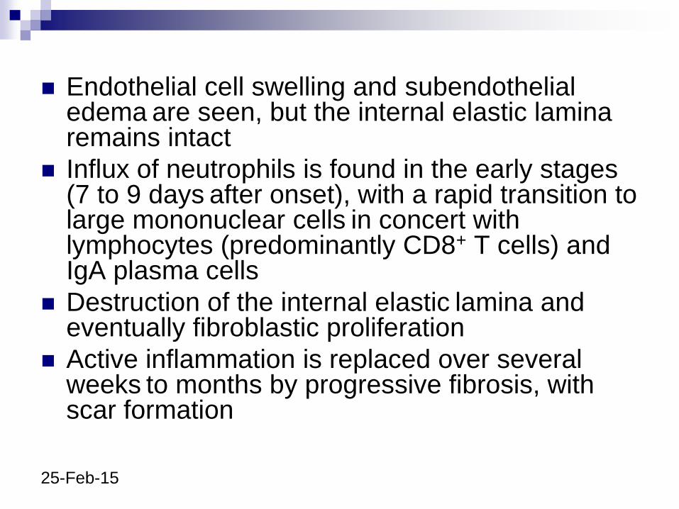
Endothelial cell swelling and subendothelialedema are seen, but the internal elastic lamina remains intact
Influx of neutrophils is found in the early stages (7 to 9 days after onset), with a rapid transition to large mononuclear cells in concert with lymphocytes (predominantly CD8+ T cells) andIgA plasma cells
Destruction of the internal elastic lamina and eventually fibroblastic proliferation
Active inflammation is replaced over several weeks to months by progressive fibrosis, with scar formation
25-Feb-15
25-Feb-15
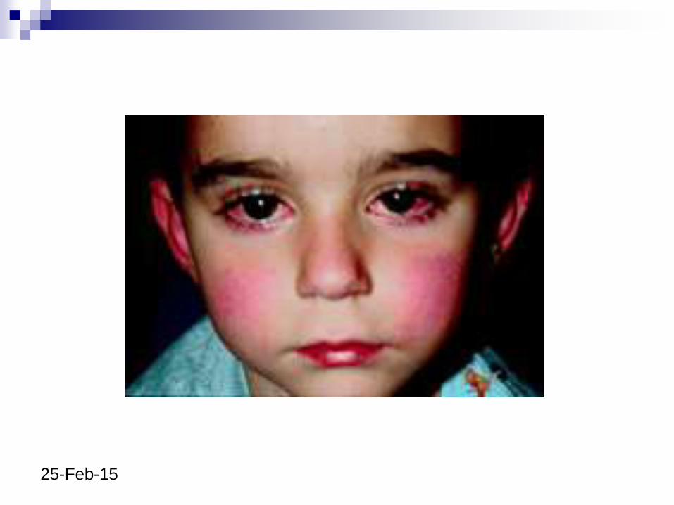
Clinical Features
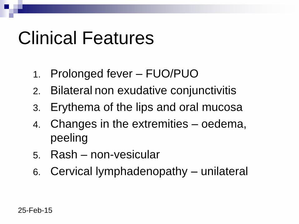
1. Prolonged fever – FUO/PUO
2. Bilateral non exudative conjunctivitis
3. Erythema of the lips and oral mucosa
4. Changes in the extremities – oedema,
peeling
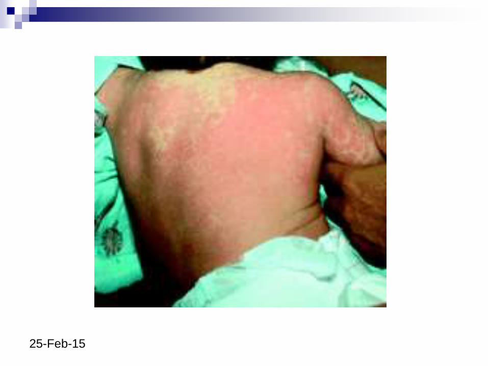
5. Rash – non-vesicular
6. Cervical lymphadenopathy – unilateral
25-Feb-15
25-Feb-15
25-Feb-15
25-Feb-15
25-Feb-15
1: Fever in KD First day of fever is considered first day of illness,
although other features may develop first
High-spiking (~40 C) and remittent
Fever of ≥5 days generally distinguishes Kawasaki
disease from self-limiting viral infections (don't have to
wait 5 days before starting treatment)
Untreated the fever usually lasts 1-2 weeks
Defervescence within 1-2 days of treatment with IVIG
25-Feb-15
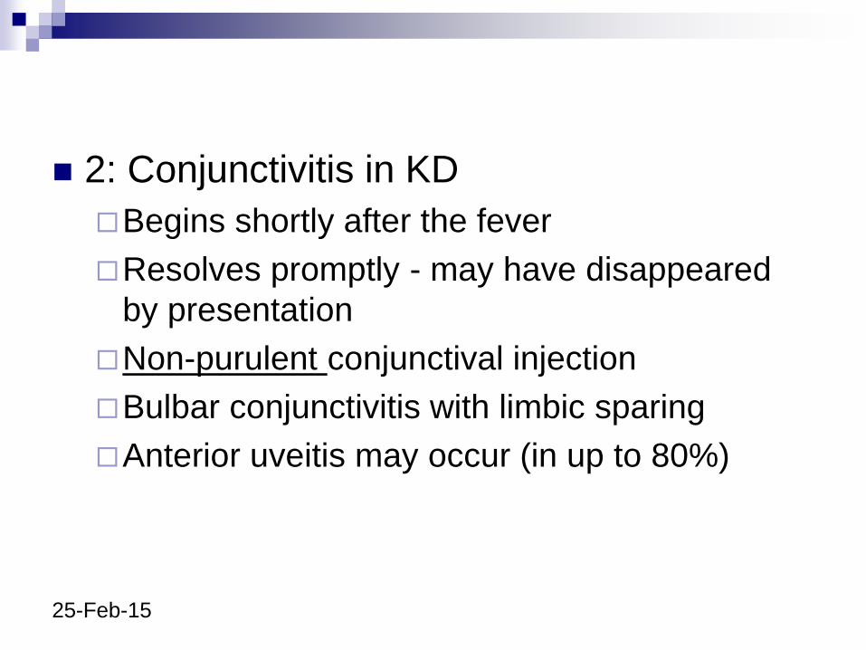
2: Conjunctivitis in KD
Begins shortly after the fever
Resolves promptly - may have disappeared
by presentation
Non-purulent conjunctival injection
Bulbar conjunctivitis with limbic sparing
Anterior uveitis may occur (in up to 80%)
25-Feb-15
25-Feb-15
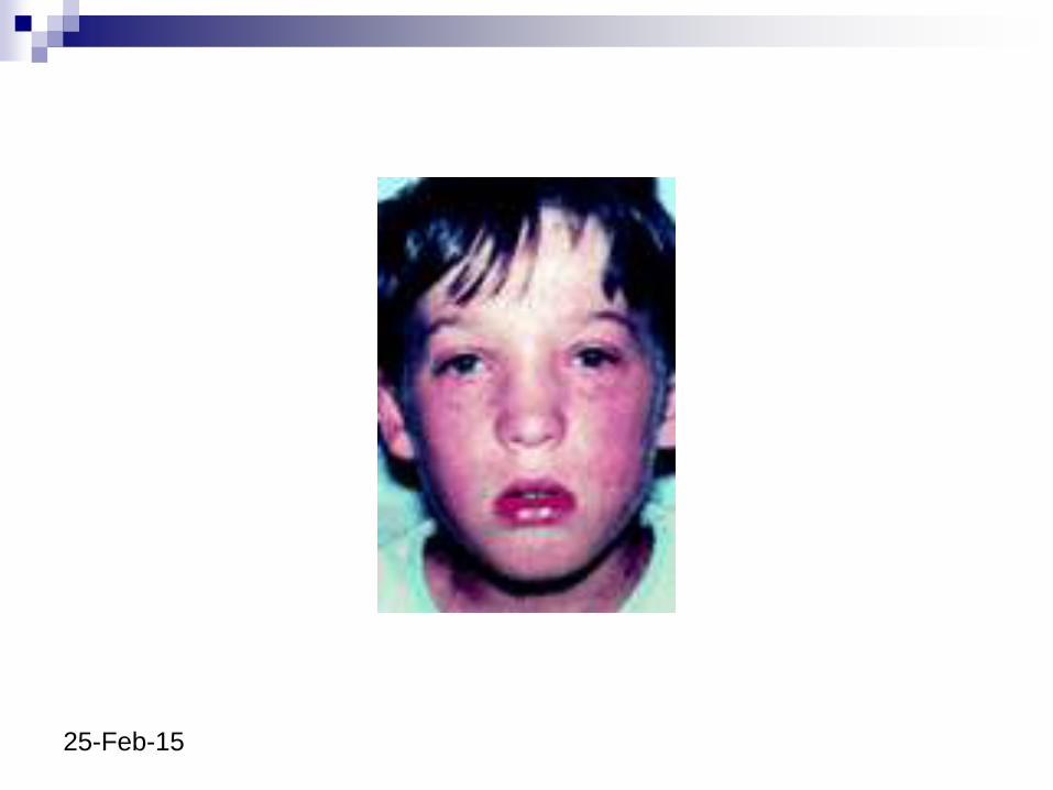
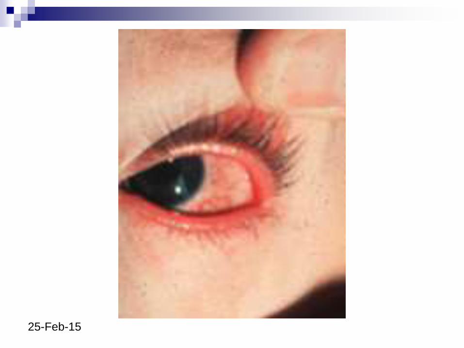
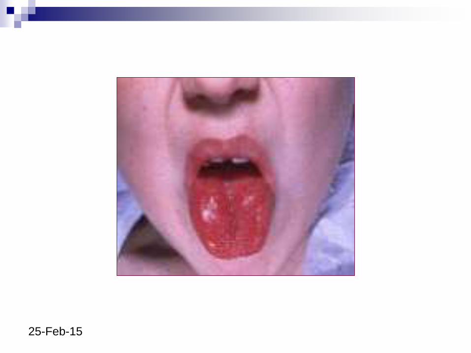
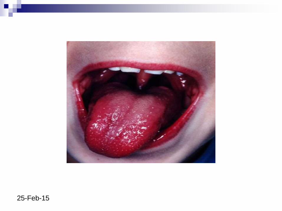
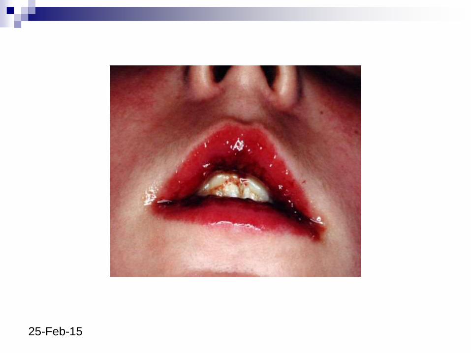
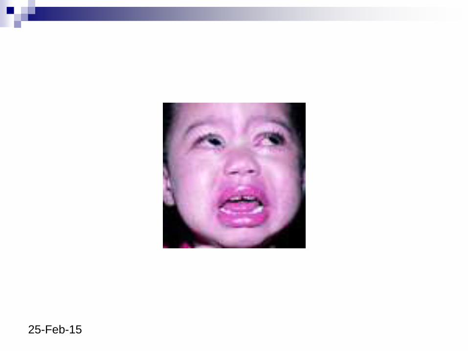
3: Oropharyngeal changes
Erythema, dryness, swelling and peeling of
lips - lipstick sign
Lips may bleed
Erythema of oropharyngeal mucosa
Strawberry tongue
No Koplik’s spots or oral ulceration or
exudates in KD
25-Feb-15
25-Feb-15
25-Feb-15
25-Feb-15
25-Feb-15
25-Feb-15
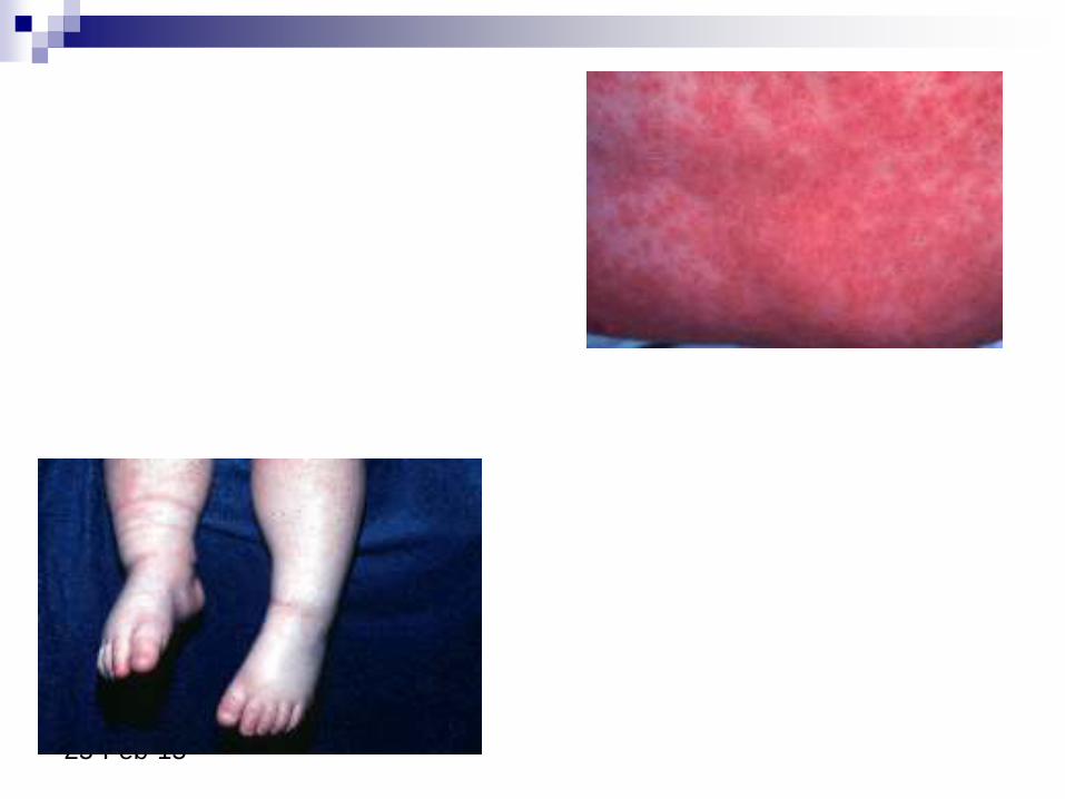
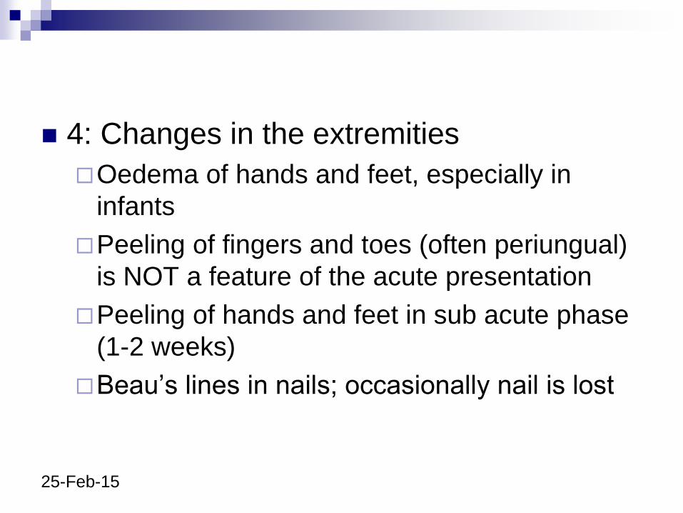
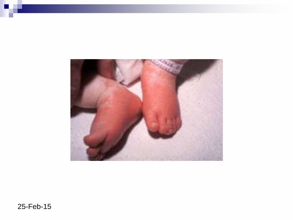
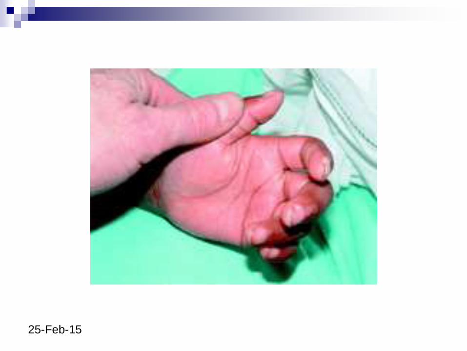
4: Changes in the extremities
Oedema of hands and feet, especially in
infants
Peeling of fingers and toes (often periungual)
is NOT a feature of the acute presentation
Peeling of hands and feet in sub acute phase
(1-2 weeks)
Beau’s lines in nails; occasionally nail is lost
25-Feb-15
25-Feb-15
25-Feb-15
25-Feb-15
25-Feb-15
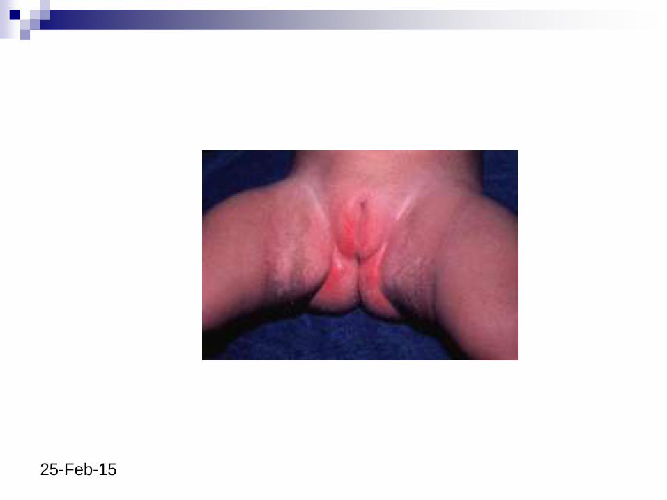
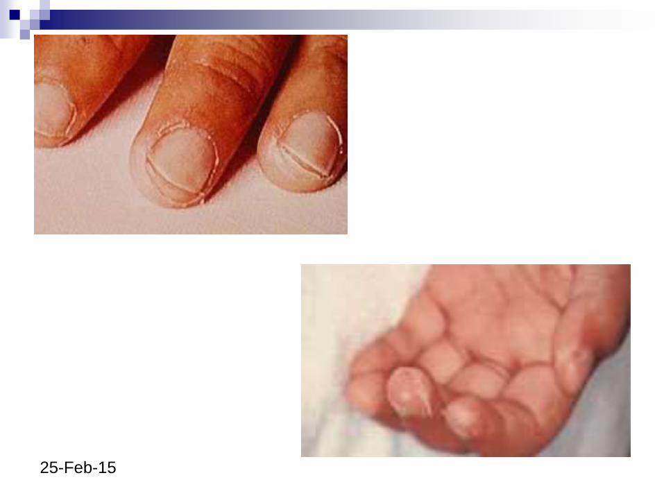
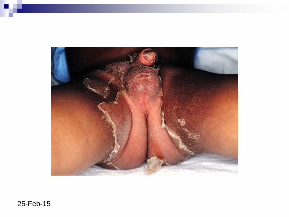
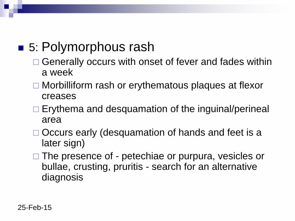
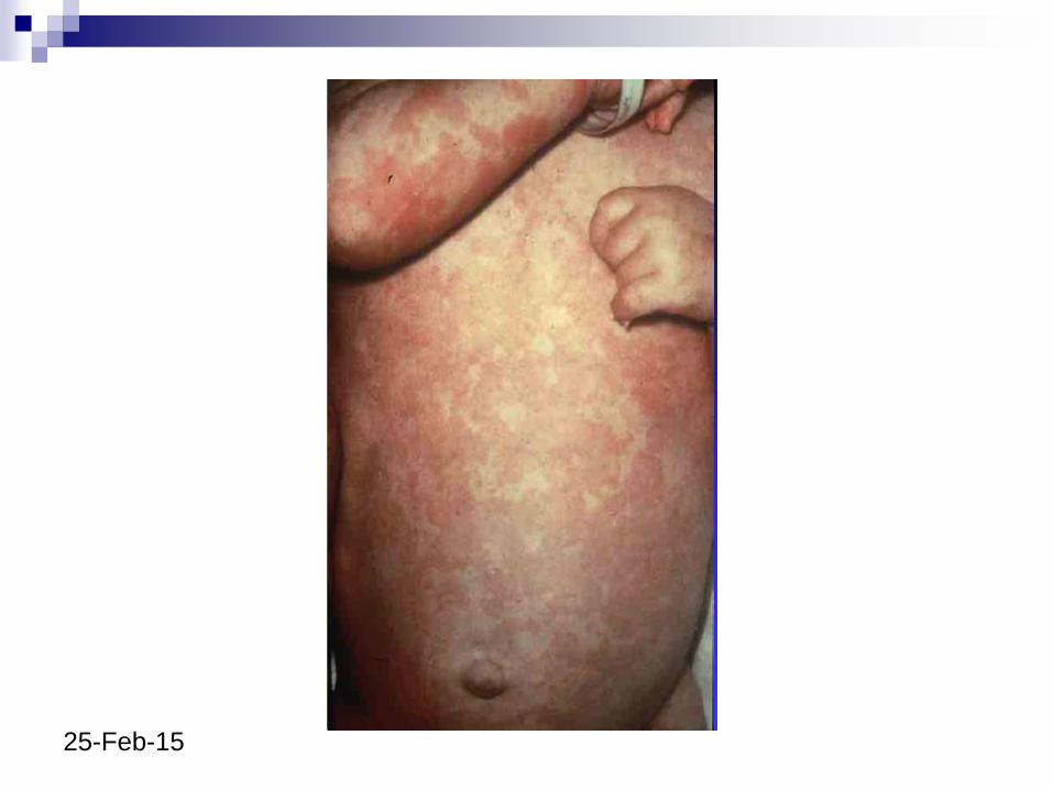
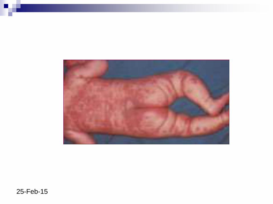
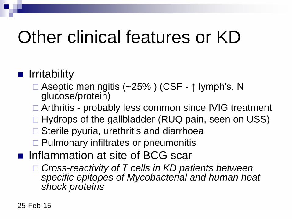
5: Polymorphous rash Generally occurs with onset of fever and fades within
a week
Morbilliform rash or erythematous plaques at flexor creases
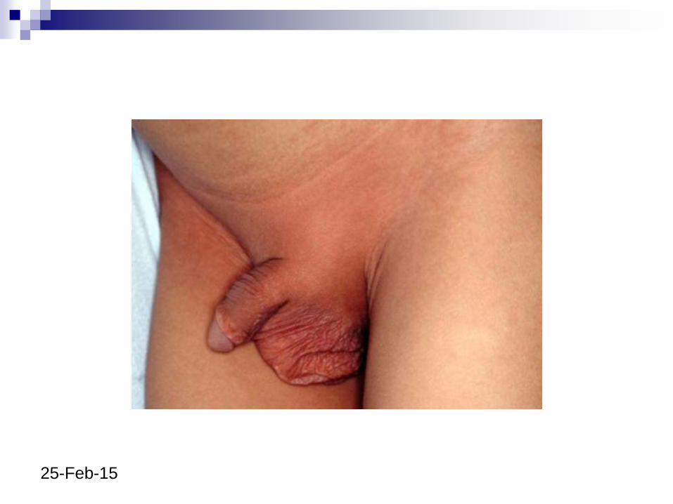
Erythema and desquamation of the inguinal/perineal area
Occurs early (desquamation of hands and feet is a later sign)
The presence of - petechiae or purpura, vesicles or bullae, crusting, pruritis - search for an alternative diagnosis
25-Feb-15
25-Feb-15
25-Feb-15
25-Feb-15
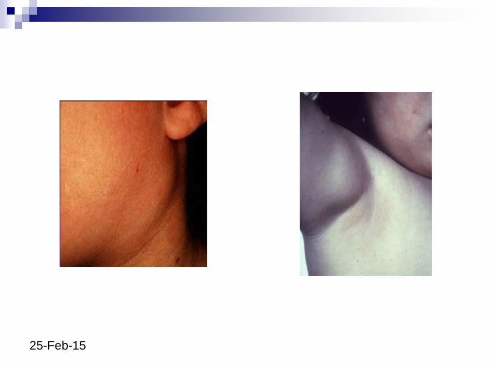
5: Lymphadenopathy in KD
50-80% of cases
>1.5cm, usually more obvious
May be unilateral single node
May be erythematous, but non-fluctuant and
no pus
25-Feb-15
25-Feb-15
25-Feb-15
25-Feb-15
Other clinical features or KD
Irritability Aseptic meningitis (~25% ) (CSF - ↑ lymph's, N
glucose/protein)
Arthritis - probably less common since IVIG treatment
Hydrops of the gallbladder (RUQ pain, seen on USS)
Sterile pyuria, urethritis and diarrhoea
Pulmonary infiltrates or pneumonitis
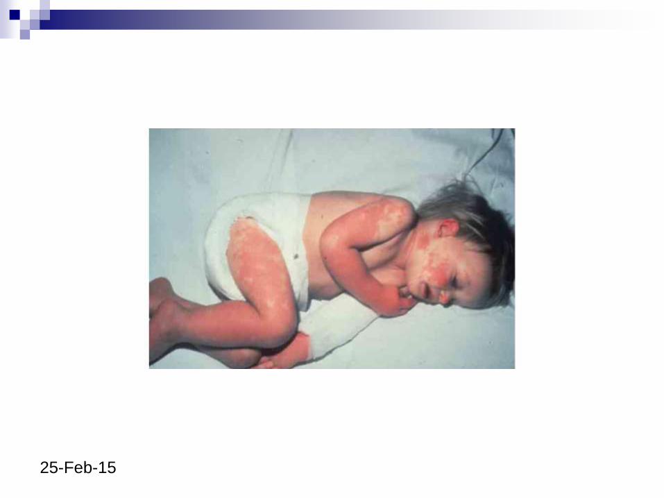
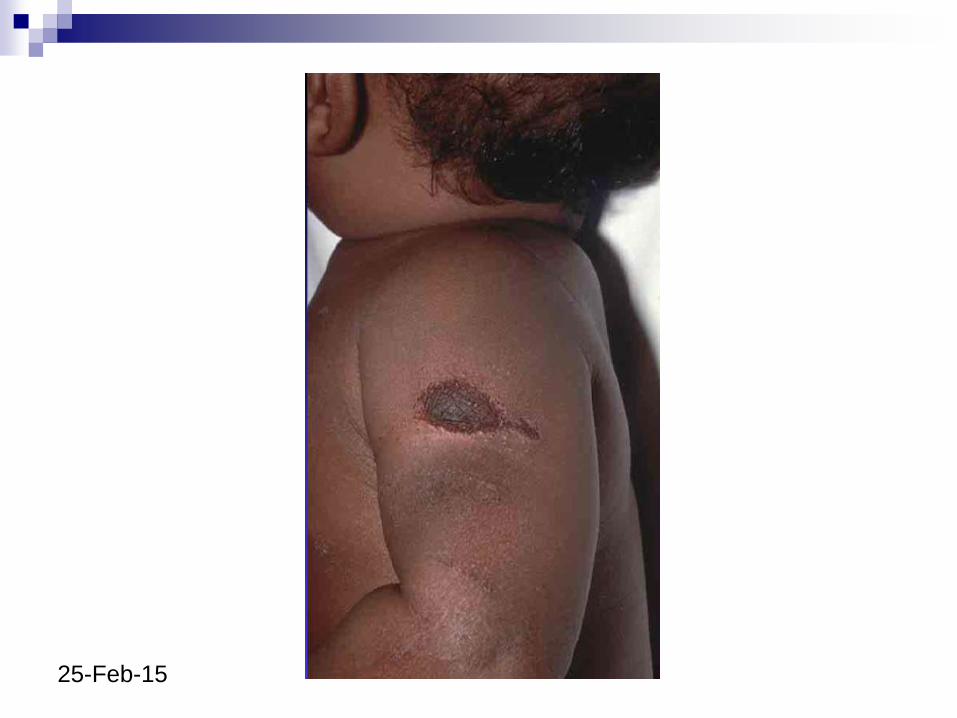
Inflammation at site of BCG scar Cross-reactivity of T cells in KD patients between
specific epitopes of Mycobacterial and human heat shock proteins
25-Feb-15
25-Feb-15
25-Feb-15
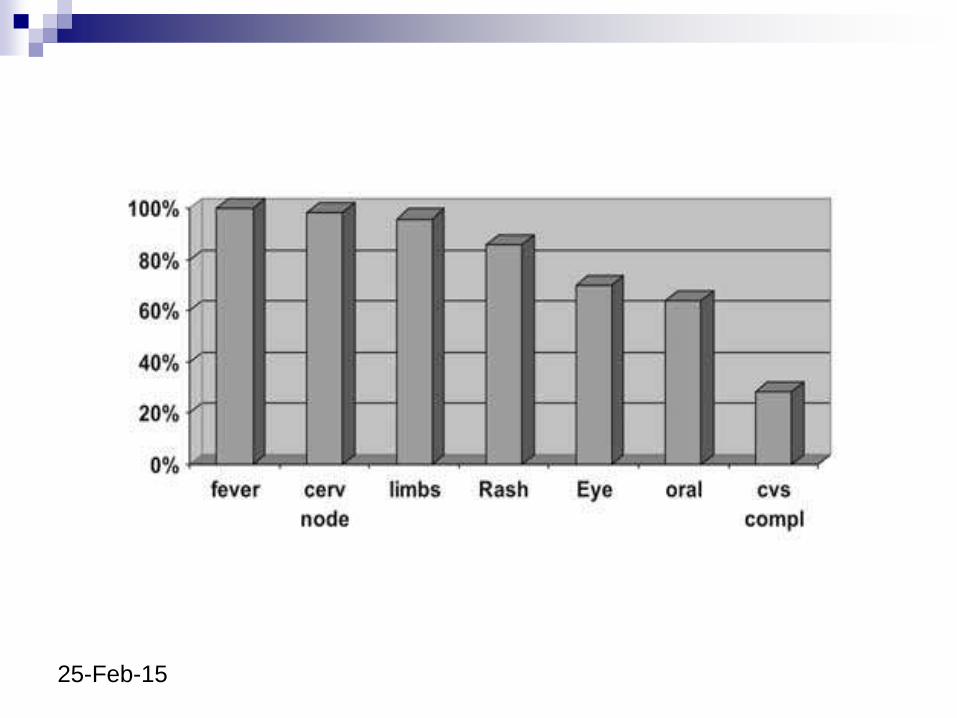
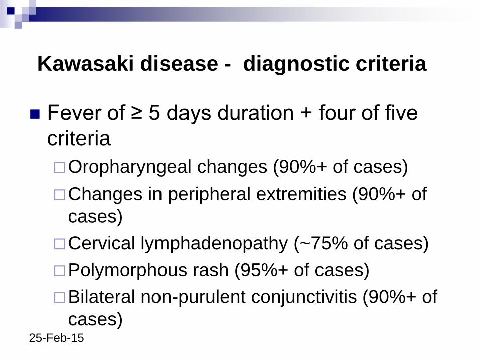
Kawasaki disease - diagnostic criteria
Fever of ≥ 5 days duration + four of five
criteria
Oropharyngeal changes (90%+ of cases)
Changes in peripheral extremities (90%+ of
cases)
Cervical lymphadenopathy (~75% of cases)
Polymorphous rash (95%+ of cases)
Bilateral non-purulent conjunctivitis (90%+ of
cases)
25-Feb-15
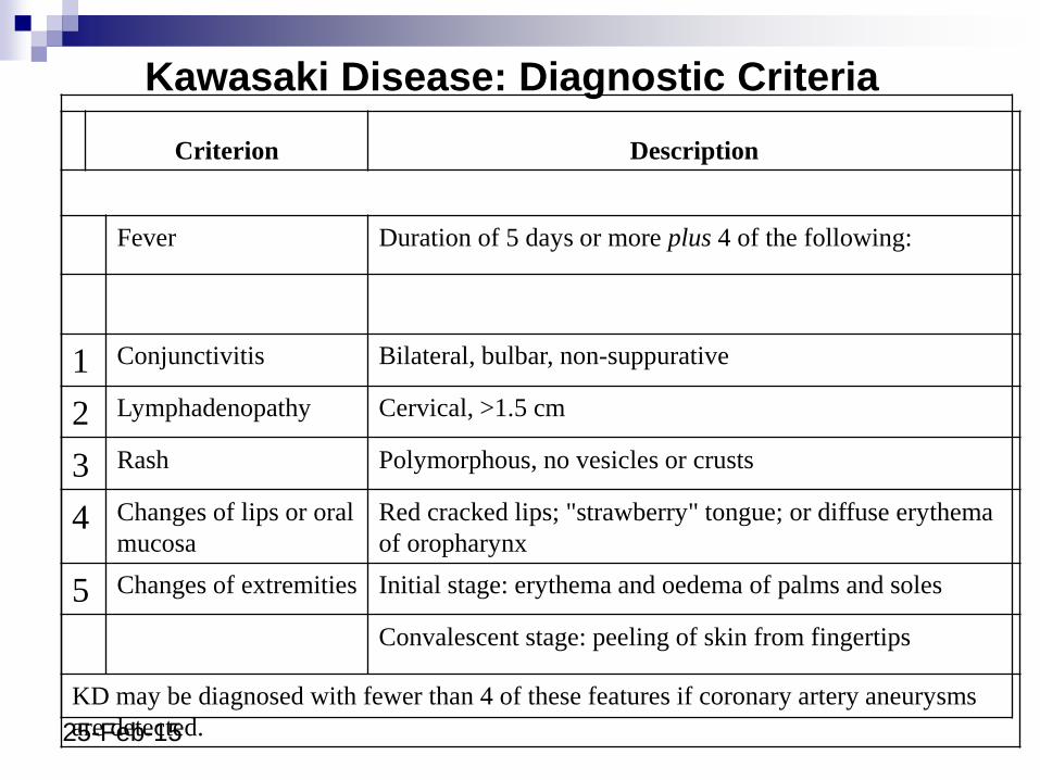
Kawasaki Disease: Diagnostic Criteria
Criterion Description
Fever Duration of 5 days or more plus 4 of the following:
1 Conjunctivitis Bilateral, bulbar, non-suppurative
2 Lymphadenopathy Cervical, >1.5 cm
3 Rash Polymorphous, no vesicles or crusts
4 Changes of lips or oral
mucosa
Red cracked lips; "strawberry" tongue; or diffuse erythema
of oropharynx
5 Changes of extremities Initial stage: erythema and oedema of palms and soles
Convalescent stage: peeling of skin from fingertips
KD may be diagnosed with fewer than 4 of these features if coronary artery aneurysms
are detected.
25-Feb-15
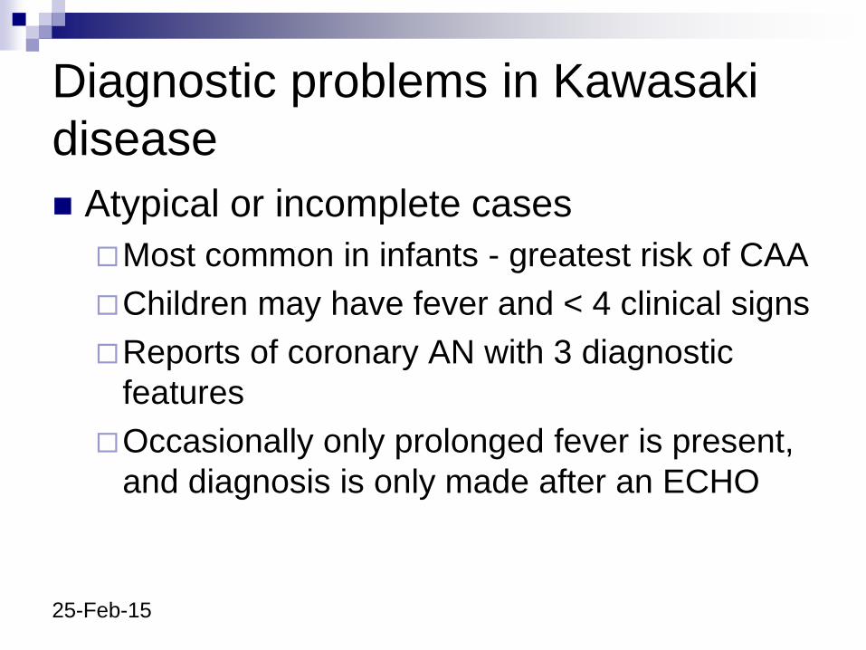
Diagnostic problems in Kawasaki
disease
Atypical or incomplete cases
Most common in infants - greatest risk of CAA
Children may have fever and < 4 clinical signs
Reports of coronary AN with 3 diagnostic
features
Occasionally only prolonged fever is present,
and diagnosis is only made after an ECHO
25-Feb-15
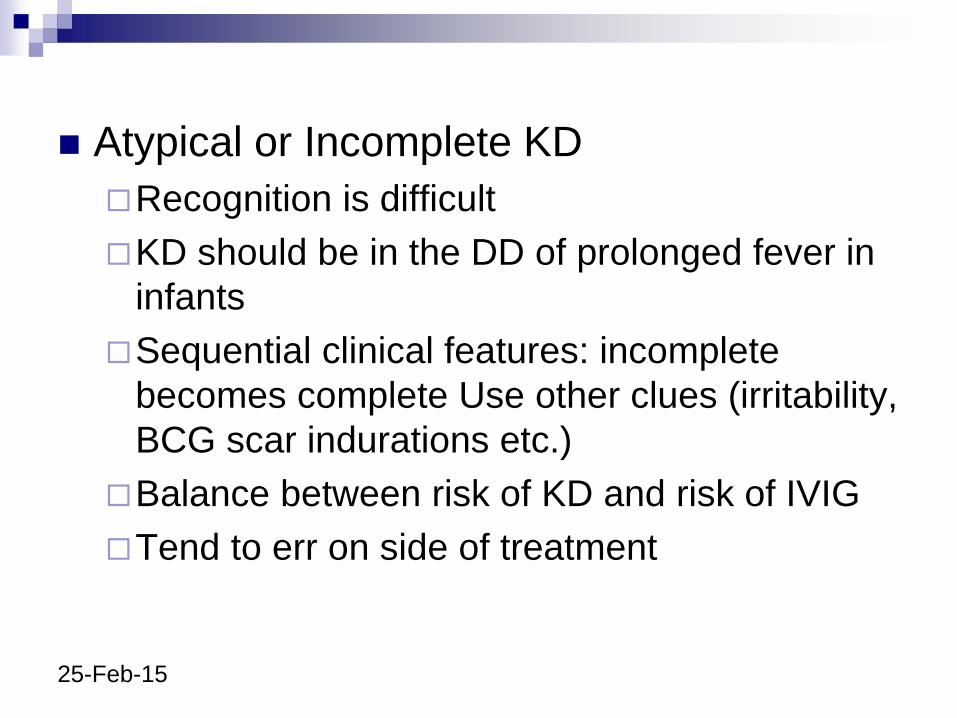
Atypical or Incomplete KD
Recognition is difficult
KD should be in the DD of prolonged fever in
infants
Sequential clinical features: incomplete
becomes complete Use other clues (irritability,
BCG scar indurations etc.)
Balance between risk of KD and risk of IVIG
Tend to err on side of treatment
25-Feb-15
Recurrent Kawasaki Disease
Much rarer than parents or clinicians think
2% in Japanese; ~<1% in UK and N America
Must fulfil diagnostic criteria again in full
Skin peeling with subsequent febrile illnesses
is common
Increased rate of heart damage in second
episode of KD
25-Feb-15
DD Streptococcal infection (scarlet fever, toxic
shock-like syndrome)
Staphylococcal infection (toxic shock syndrome, scalded skin syndrome)
Viral exanthemas (Measles, rubella, roseola infantum, Epstein Barr virus, influenza A and B, adenovirus)
Mycoplasma pneumonia
Stevens-Johnson syndrome
Systemic idiopathic juvenile arthritis
25-Feb-15
Initial Investigations for Suspected
Kawasaki Disease
Full blood count and film
Erythrocyte sedimentation rate
C reactive protein
Blood cultures
ASOT and anti DNase B
Nose and throat swab, stool culture (superantigen toxin typing if staphylococcus aureus and/or ß haemolytic streptococci detected)
Renal and liver function tests
25-Feb-15
Coagulation screen
Autoantibody profile (antinuclear antibodies; Extractable nuclear antibodies; Rheumatoid factor; antineutrophil cytoplasmic antibodies)
Serology (IgG and IgM) for mycoplasma pneumoniae, enterovirus, adenovirus, measles, parvovirus, Epstein–Barr virus, cytomegalovirus
Urine microscopy and culture
Dip test of urine for blood and protein
Electrocardiogram and echocardiogram
Consider serology for rickettsiae and leptospirosis if history suggestive
Consider chest x ray
25-Feb-15
Complications Irritability and aseptic meningitis
Gallbladder hydrops
Hepatitis
Otitis media
Pancreatitis
Myositis
Pericarditis and myocarditis
Aneurysm formation can lead to peripheral gangrene, cerebral infarction and cardiac arterial aneurysm (this may lead to thrombosis, myocardial infarction and dysrrhythmia)
25-Feb-15
Cardiac complications 20–40% of untreated KD patients develop coronary
artery abnormalities
50% of these lesions regress within five years, and in most with mild CAA (3–4 mm) regression occurs within two years
Giant aneurysms (>8 mm) are unlikely to resolve, and some may develop stenosis with risk of coronary thrombosis, myocardial infarction, and death
In 1993, a report from the BPSU indicated a mortality rate of 3.7% in the UK for KD
25-Feb-15
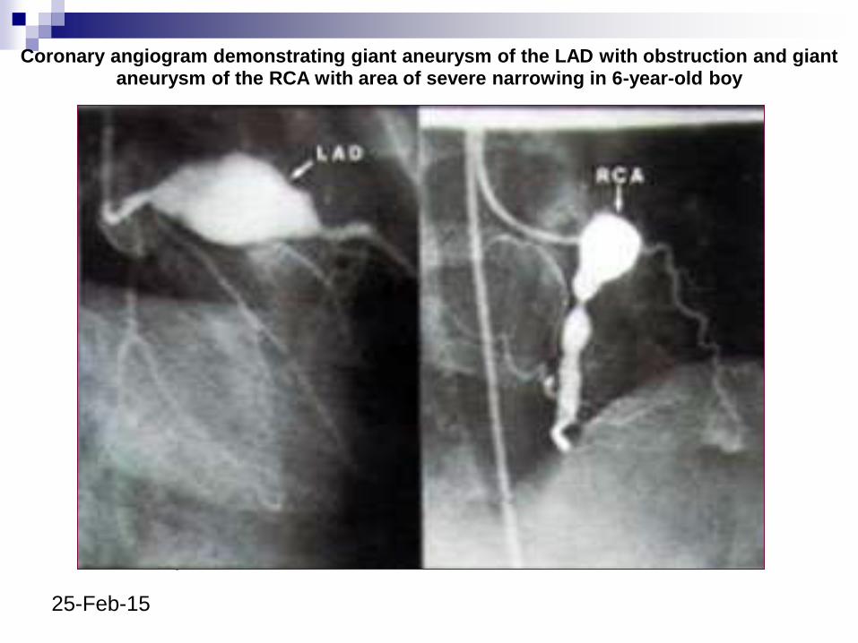
Newburger, J. W. et al. Circulation 2004;110:2747-2771
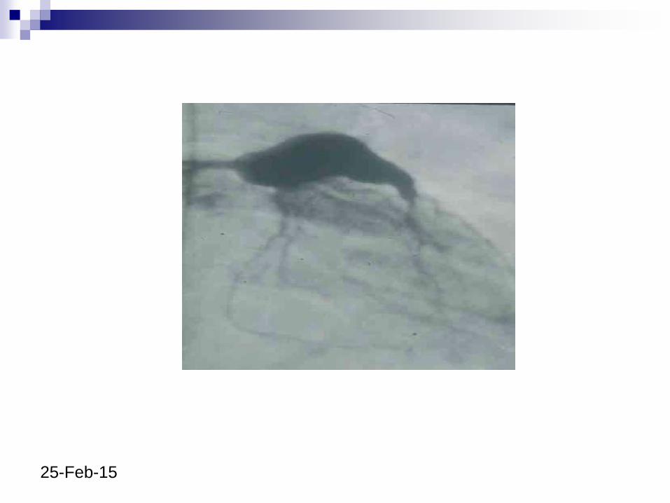
Coronary angiogram demonstrating giant aneurysm of the LAD with obstruction and giant aneurysm of the RCA with area of severe narrowing in 6-year-old boy
25-Feb-15
25-Feb-15
25-Feb-15
25-Feb-15
25-Feb-15
25-Feb-15
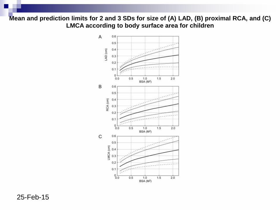
Mean and prediction limits for 2 and 3 SDs for size of (A) LAD, (B) proximal RCA, and (C) LMCA according to body surface area for children
25-Feb-15
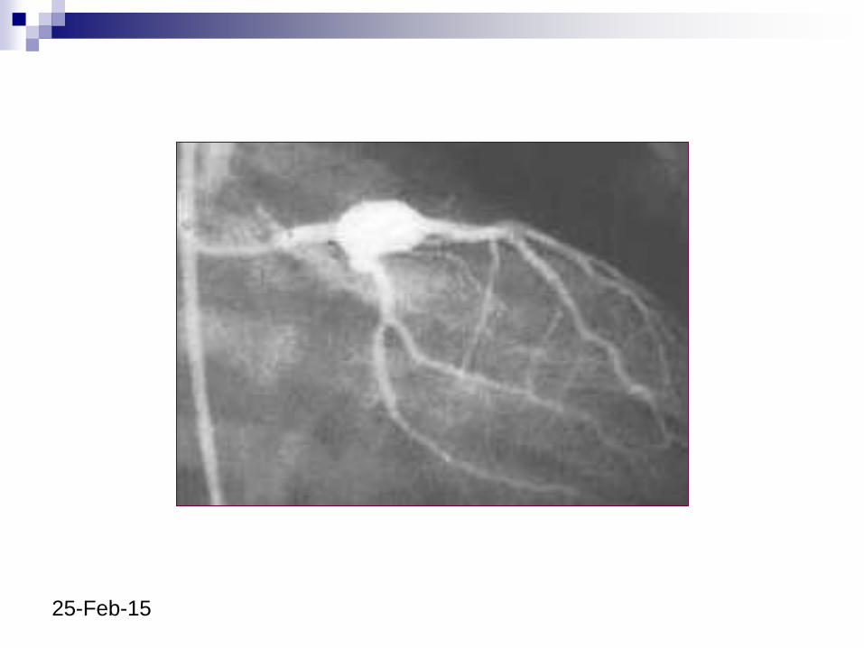
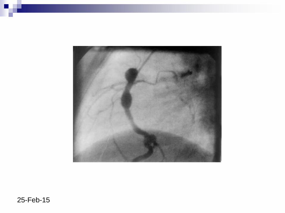
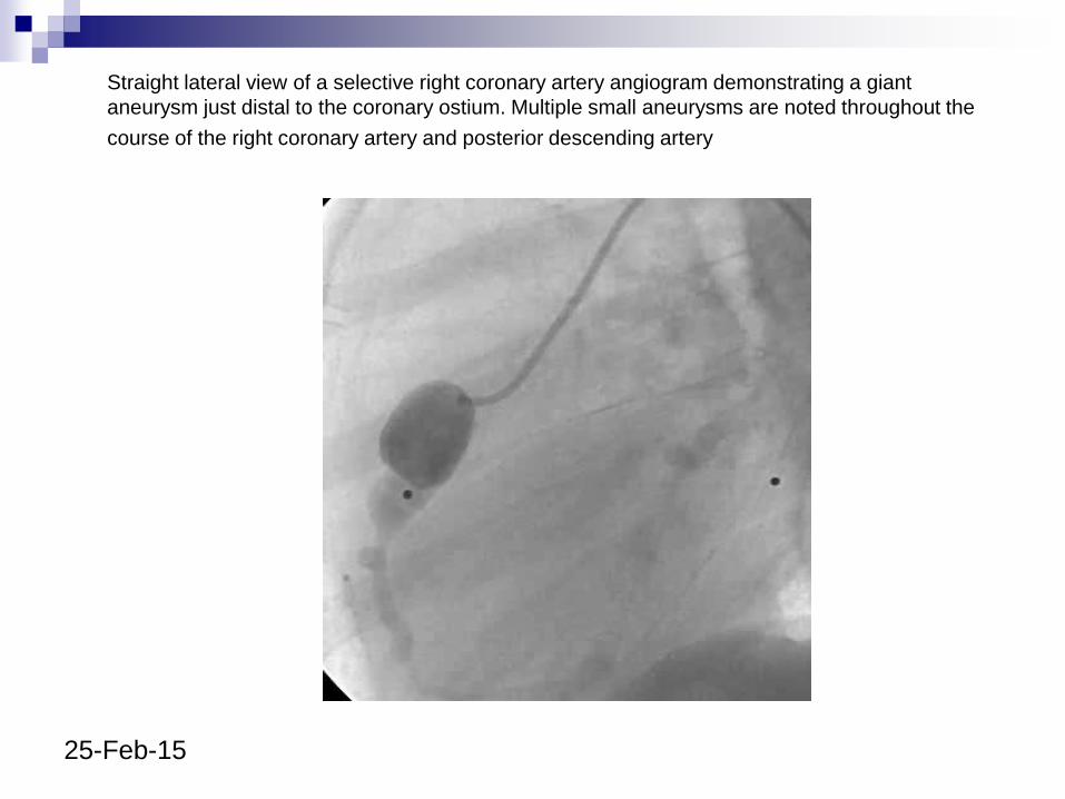
Straight lateral view of a selective right coronary artery angiogram demonstrating a giant
aneurysm just distal to the coronary ostium. Multiple small aneurysms are noted throughout the
course of the right coronary artery and posterior descending artery
25-Feb-15
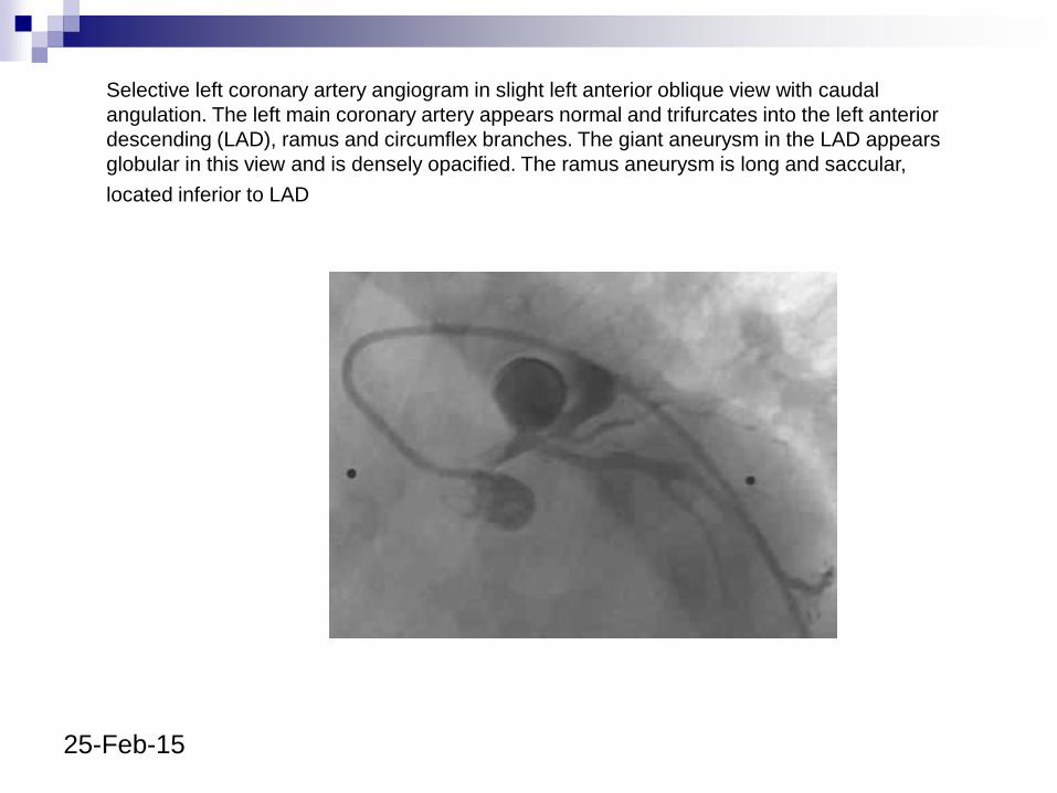
Selective left coronary artery angiogram in slight left anterior oblique view with caudal
angulation. The left main coronary artery appears normal and trifurcates into the left anterior
descending (LAD), ramus and circumflex branches. The giant aneurysm in the LAD appears
globular in this view and is densely opacified. The ramus aneurysm is long and saccular,
located inferior to LAD
25-Feb-15
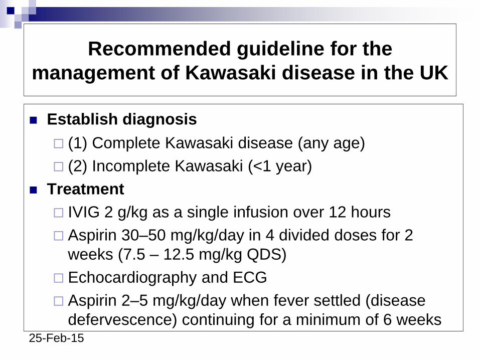
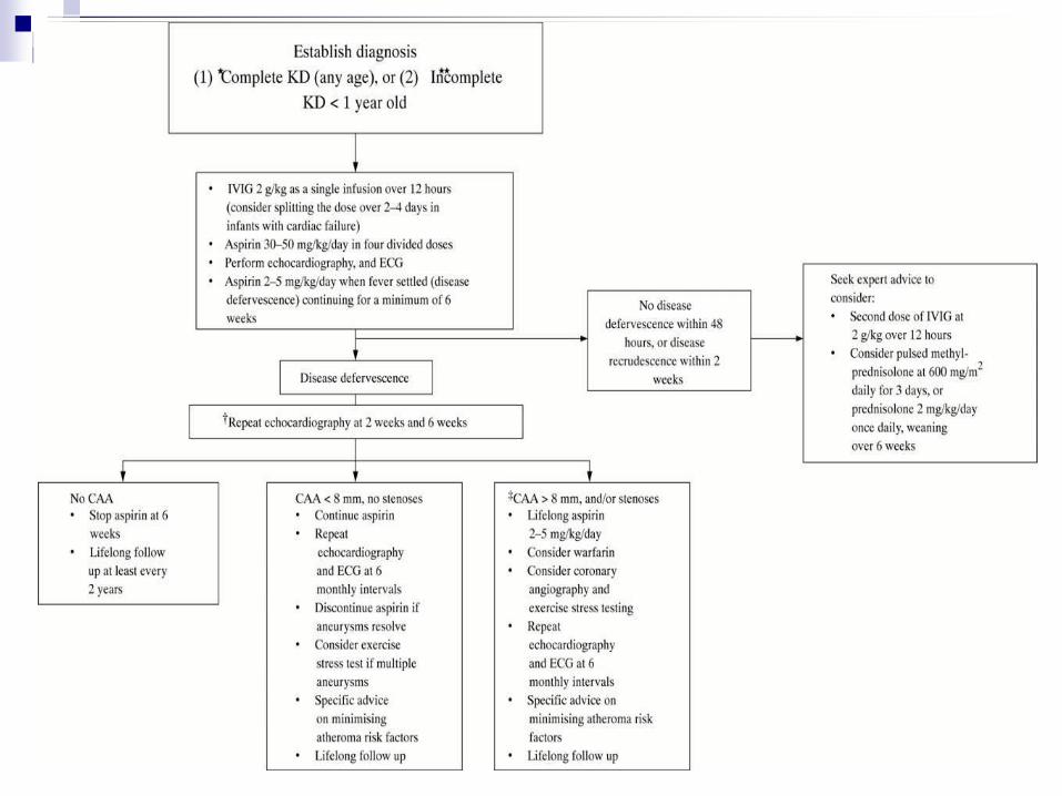
Recommended guideline for the
management of Kawasaki disease in the UK
Establish diagnosis
(1) Complete Kawasaki disease (any age)
(2) Incomplete Kawasaki (<1 year)
Treatment
IVIG 2 g/kg as a single infusion over 12 hours
Aspirin 30–50 mg/kg/day in 4 divided doses for 2
weeks (7.5 – 12.5 mg/kg QDS)
Echocardiography and ECG
Aspirin 2–5 mg/kg/day when fever settled (disease
defervescence) continuing for a minimum of 6 weeks
25-Feb-15
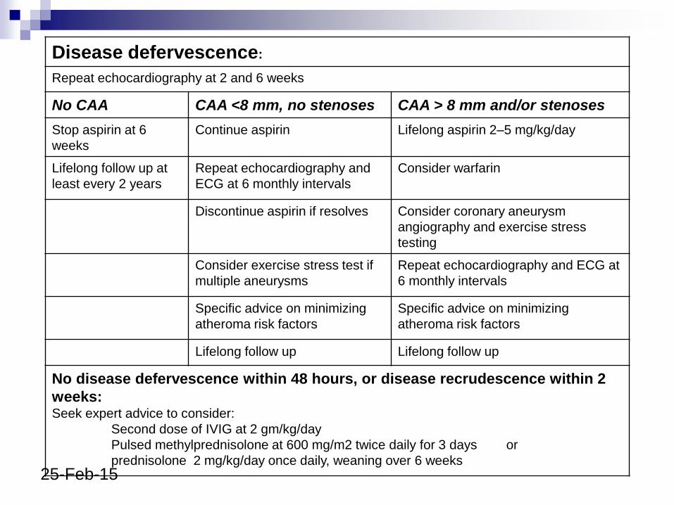
Disease defervescence:
Repeat echocardiography at 2 and 6 weeks
No CAA CAA <8 mm, no stenoses CAA > 8 mm and/or stenoses
Stop aspirin at 6
weeks
Continue aspirin Lifelong aspirin 2–5 mg/kg/day
Lifelong follow up at
least every 2 years
Repeat echocardiography and
ECG at 6 monthly intervals
Consider warfarin
Discontinue aspirin if resolves Consider coronary aneurysm
angiography and exercise stress
testing
Consider exercise stress test if
multiple aneurysms
Repeat echocardiography and ECG at
6 monthly intervals
Specific advice on minimizing
atheroma risk factors
Specific advice on minimizing
atheroma risk factors
Lifelong follow up Lifelong follow up
No disease defervescence within 48 hours, or disease recrudescence within 2
weeks:Seek expert advice to consider:
• Second dose of IVIG at 2 gm/kg/day
• Pulsed methylprednisolone at 600 mg/m2 twice daily for 3 days or
prednisolone 2 mg/kg/day once daily, weaning over 6 weeks
25-Feb-15
25-Feb-15
● Treatment can be commenced before full 5 days of fever if sepsis excluded
● Treatment should also be given if the presentation is >10 days from fever onset
● Incomplete cases >1 year old treated at discretion of clinician--seek expert advice
● Refer to paediatric cardiologist● Other specific interventions such as positron
emission tomography scanning, addition of calcium channel blocker therapy, and coronary angioplasty at discretion of paediatric cardiologist
25-Feb-15
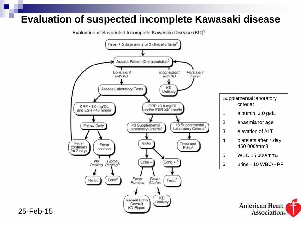
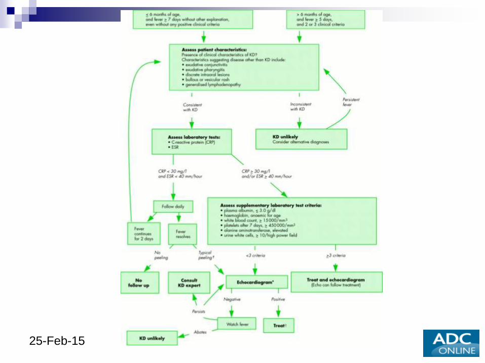
Evaluation of suspected incomplete Kawasaki disease
Supplemental laboratory
criteria:
1. albumin 3.0 g/dL
2. anaemia for age
3. elevation of ALT
4. platelets after 7 day
450 000/mm3
5. WBC 15 000/mm3
6. urine - 10 WBC/HPF
25-Feb-15
25-Feb-15
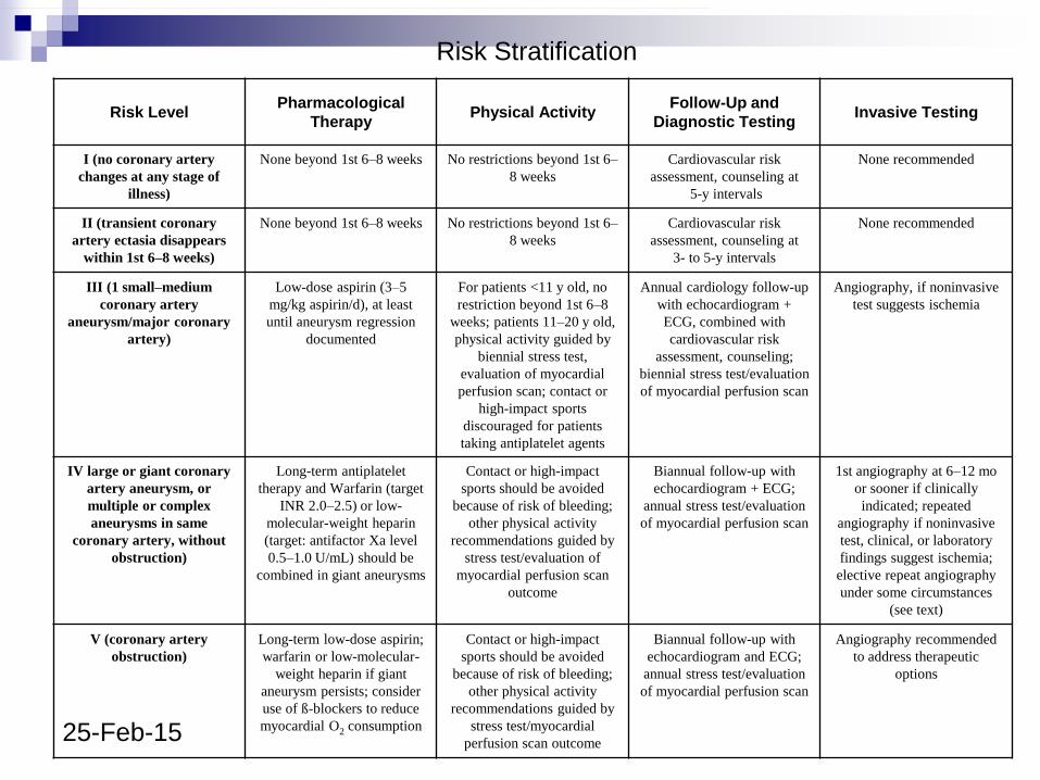
Risk Level Pharmacological
Therapy Physical Activity
Follow-Up and
Diagnostic Testing Invasive Testing
I (no coronary artery
changes at any stage of
illness)
None beyond 1st 6–8 weeks No restrictions beyond 1st 6–
8 weeks
Cardiovascular risk
assessment, counseling at
5-y intervals
None recommended
II (transient coronary
artery ectasia disappears
within 1st 6–8 weeks)
None beyond 1st 6–8 weeks No restrictions beyond 1st 6–
8 weeks
Cardiovascular risk
assessment, counseling at
3- to 5-y intervals
None recommended
III (1 small–medium
coronary artery
aneurysm/major coronary
artery)
Low-dose aspirin (3–5
mg/kg aspirin/d), at least
until aneurysm regression
documented
For patients <11 y old, no
restriction beyond 1st 6–8
weeks; patients 11–20 y old,
physical activity guided by
biennial stress test,
evaluation of myocardial
perfusion scan; contact or
high-impact sports
discouraged for patients
taking antiplatelet agents
Annual cardiology follow-up
with echocardiogram +
ECG, combined with
cardiovascular risk
assessment, counseling;
biennial stress test/evaluation
of myocardial perfusion scan
Angiography, if noninvasive
test suggests ischemia
IV large or giant coronary
artery aneurysm, or
multiple or complex
aneurysms in same
coronary artery, without
obstruction)
Long-term antiplatelet
therapy and Warfarin (target
INR 2.0–2.5) or low-
molecular-weight heparin
(target: antifactor Xa level
0.5–1.0 U/mL) should be
combined in giant aneurysms
Contact or high-impact
sports should be avoided
because of risk of bleeding;
other physical activity
recommendations guided by
stress test/evaluation of
myocardial perfusion scan
outcome
Biannual follow-up with
echocardiogram + ECG;
annual stress test/evaluation
of myocardial perfusion scan
1st angiography at 6–12 mo
or sooner if clinically
indicated; repeated
angiography if noninvasive
test, clinical, or laboratory
findings suggest ischemia;
elective repeat angiography
under some circumstances
(see text)
V (coronary artery
obstruction)
Long-term low-dose aspirin;
warfarin or low-molecular-
weight heparin if giant
aneurysm persists; consider
use of ß-blockers to reduce
myocardial O2 consumption
Contact or high-impact
sports should be avoided
because of risk of bleeding;
other physical activity
recommendations guided by
stress test/myocardial
perfusion scan outcome
Biannual follow-up with
echocardiogram and ECG;
annual stress test/evaluation
of myocardial perfusion scan
Angiography recommended
to address therapeutic
options
Risk Stratification
25-Feb-15
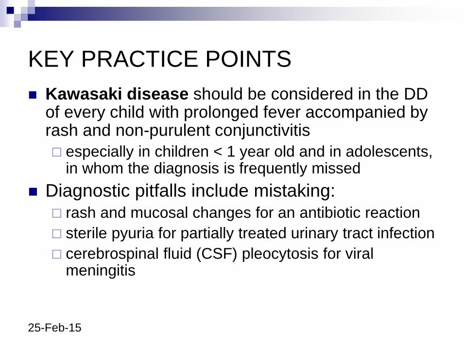
KEY PRACTICE POINTS
Kawasaki disease should be considered in the DD of every child with prolonged fever accompanied by rash and non-purulent conjunctivitis
especially in children < 1 year old and in adolescents, in whom the diagnosis is frequently missed
Diagnostic pitfalls include mistaking: rash and mucosal changes for an antibiotic reaction
sterile pyuria for partially treated urinary tract infection
cerebrospinal fluid (CSF) pleocytosis for viral meningitis
25-Feb-15
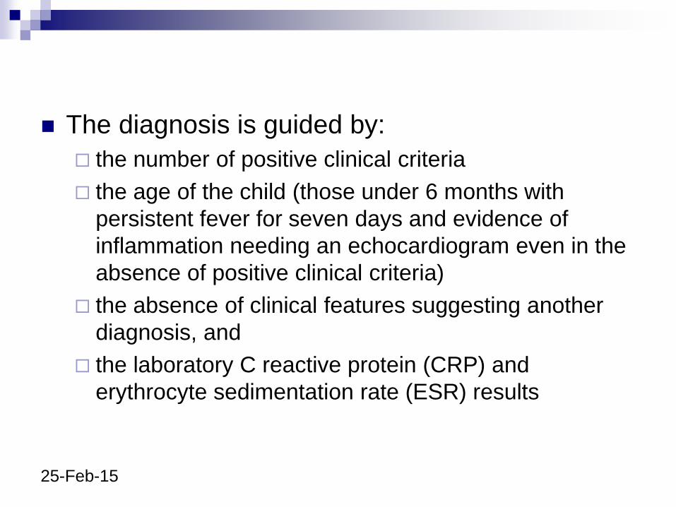
The diagnosis is guided by:
the number of positive clinical criteria
the age of the child (those under 6 months with
persistent fever for seven days and evidence of
inflammation needing an echocardiogram even in the
absence of positive clinical criteria)
the absence of clinical features suggesting another
diagnosis, and
the laboratory C reactive protein (CRP) and
erythrocyte sedimentation rate (ESR) results
25-Feb-15
In those with raised CRP and/or,do other
supplementary investigations contribute to the
decision as to whether to treat with IVIG
WBC
PLT
S albumin
ALT
Urine –WBC
Echocardiography
25-Feb-15
Vaccination post KD
IVIG can block replication of live viral vaccines & subsequent actively acquired immunity
Current recommendation - live vaccines be deferred for at least three months following treatment with IVIG
Autoimmune diseases including the systemic vasculitides flare in response to live and non-live vaccine preparations
Defer immunisation with all vaccines for at least three months following an episode of KD
25-Feb-15
Areas of Future Research Epidemiology including incidence, management, complication rate,
and case fatality rate
Investigation of superantigen dependent or independent pathways of T cell activation in the acute and convalescent phases of the illness
Investigation of a viral aetiopathogenesis, in particular herpes viruses, using degenerate polymerase chain reaction methodology
Investigation of host genetic determinants of susceptibility and outcome in KD
Correlation of coronary and/or peripheral arterial involvement with clinical presentation
Evaluation of the potential of circulating endothelial microparticles as a laboratory diagnostic test and predictor of those at risk of coronary arterial aneurysm formation
Examination of B lymphocyte homing receptor expression of circulating antibody secreting B lymphocytes in acute and convalescent KD.
Areas of Future Research
Inflixumab – TNF alpha antagonist- in the
RX of KD
Biomarker for diagnosis of KD –
Urinary protiens – Flamin C & Mephrin A
THRIL – activated macrophage

Thank you