Embed Size (px)
Citation preview

Complete Muscle Histology
By- Dr. Armaan SinghBy- Dr. Armaan Singh
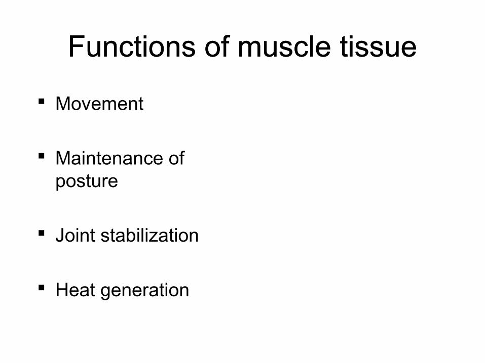
Functions of muscle tissueFunctions of muscle tissue
Movement
Maintenance of posture
Joint stabilization
Heat generation
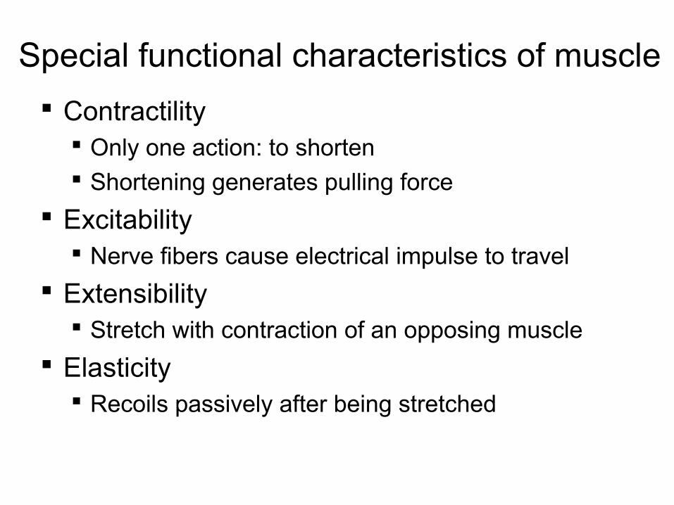
Special functional characteristics of muscle
Contractility Only one action: to shorten Shortening generates pulling force
Excitability Nerve fibers cause electrical impulse to travel
Extensibility Stretch with contraction of an opposing muscle
Elasticity Recoils passively after being stretched

Types of Muscle Tissue
Skeletal muscle
Cardiac muscle
Smooth muscle

Types of Muscle TissueSkeletal
•Attach to and move skeleton•40% of body weight•Fibers = multinucleate cells (embryonic cells fuse)•Cells with obvious striations•Contractions are voluntary
Cardiac: only in the wall of the heart
•Cells are striated•Contractions are involuntary (not voluntary)
Smooth: walls of hollow organs•Lack striations•Contractions are involuntary (not voluntary)

Similarities…
Their cells are called fibers because they are elongated
Contraction depends on myofilaments Actin Myosin
Plasma membrane is called sarcolemma Sarcos = flesh Lemma = sheath

Skeletal muscle
Epimysium: surrounds whole muscle
Perimysium is around fascicle
Endomysium is around each muscle fiber

Skeletal Muscle
Each muscle: one nerve, one artery, one vein Branch repeatedly
Attachments One bone to another Cross at least one movable joint Origin: the less movable
attachment Insertion: is pulled toward the
origin Usually one bone moves while the
other remains fixed In muscles of the limb, origin lies
proximal to the insertion (by convention)
Note: origin and insertion may switch depending on body position and movement produced
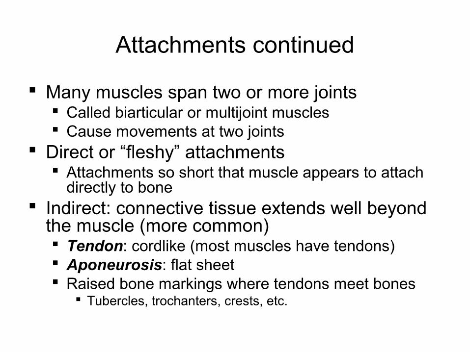
Attachments continued
Many muscles span two or more joints Called biarticular or multijoint muscles Cause movements at two joints
Direct or “fleshy” attachments Attachments so short that muscle appears to attach
directly to bone Indirect: connective tissue extends well beyond
the muscle (more common) Tendon: cordlike (most muscles have tendons) Aponeurosis: flat sheet Raised bone markings where tendons meet bones
Tubercles, trochanters, crests, etc.
Some sites showing animations of muscle contraction
http://entochem.tamu.edu/MuscleStrucContractswf/index.html
http://www.brookscole.com/chemistry_d/templates/student_resources/shared_resources/animations/muscles/muscles.html

Skeletal muscle
Fibers (each is one cell) have striations
Myofibrils are organelles of the cell: these are made up of filaments
Sarcomere Basic unit of
contraction Myofibrils are long
rows of repeating sarcomeres
Boundaries: Z discs (or lines)
This big cylinder is a fiber: 1 cell -an organelle
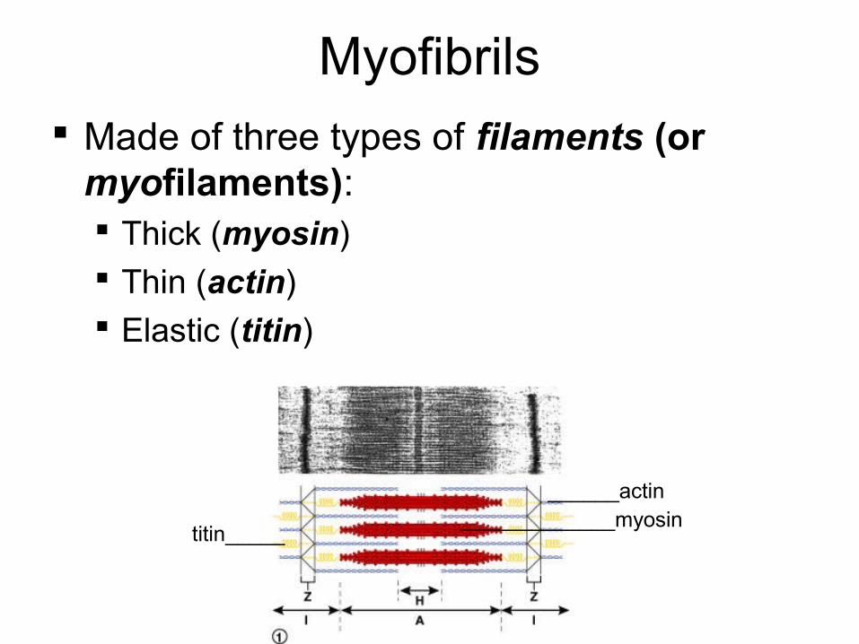
Myofibrils Made of three types of filaments (or
myofilaments): Thick (myosin) Thin (actin) Elastic (titin)
______actin_____________myosin
titin_____

Sliding Filament Model
__relaxed sarcomere__ _partly contracted_
fully contracted
“A” band constant because it is caused by myosin, which doesn’t change length
Sarcomere shortens because actin pulled towards its middle by myosin cross bridges
Titin resists overstretching
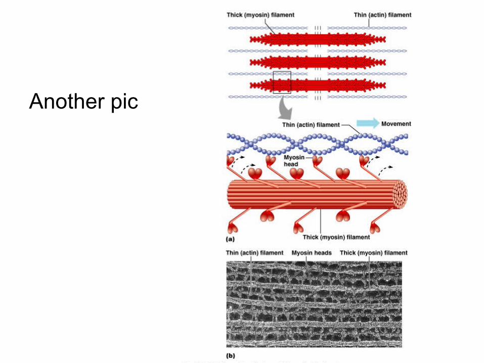
Another pic

EM (electron microscope): parts of 2 myofibrils
Labeled and unlabeled
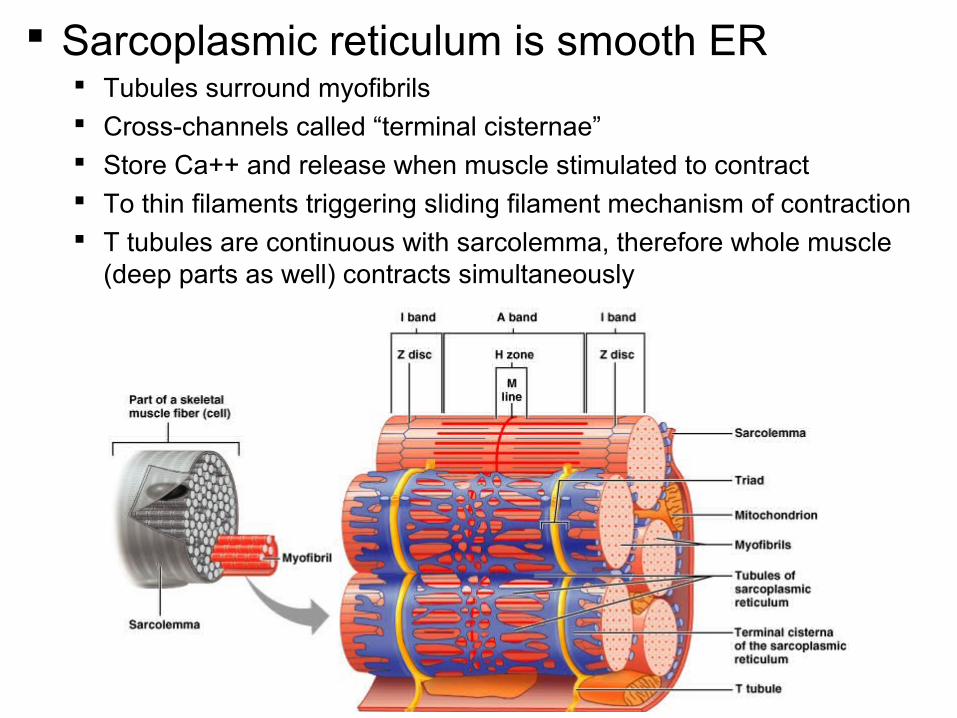
Sarcoplasmic reticulum is smooth ER Tubules surround myofibrils Cross-channels called “terminal cisternae” Store Ca++ and release when muscle stimulated to contract To thin filaments triggering sliding filament mechanism of contraction T tubules are continuous with sarcolemma, therefore whole muscle
(deep parts as well) contracts simultaneously
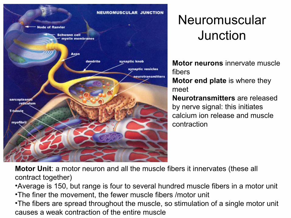
Neuromuscular Junction
Motor neurons innervate muscle fibersMotor end plate is where they meetNeurotransmitters are released by nerve signal: this initiates calcium ion release and muscle contraction
Motor Unit: a motor neuron and all the muscle fibers it innervates (these all contract together)•Average is 150, but range is four to several hundred muscle fibers in a motor unit•The finer the movement, the fewer muscle fibers /motor unit•The fibers are spread throughout the muscle, so stimulation of a single motor unit causes a weak contraction of the entire muscle


Types of skeletal muscle fibers Fast, slow and intermediate Whether or not they predominantly use oxygen to
produce ATP (the energy molecule used in muscle contraction) Oxidative – aerobic (use oxygen) Glycolytic – make ATP by glycolysis (break down of sugars
without oxygen=anaerobic) Fast fibers: “white fibers” – large, predominantly
anaerobic, fatigue rapidly (rely on glycogen reserves); most of the skeletal muscle fibers are fast
Slow fibers: “red fibers” – half the diameter, 3X slower, but can continue contracting; aerobic, more mitochondria, myoglobin
Intermediate: in between
A skeletal muscle contracts when its motor units are stimulated
Amount of tension depends on1. the frequency of stimulation2. the number of motor units involved
Single, momentary contraction is called a muscle twitch
All or none principle: each muscle fiber either contracts completely or not at all
Amount of force: depends on how many motor units are activated
Muscle tone Even at rest, some motor units are active: tense the
muscle even though not causing movement: “resting tone”
Muscle hypertrophy Weight training (repeated intense workouts): increases diameter and
strength of “fast” muscle fibers by increasing production of Mitochondria Actin and myosin protein Myofilaments containing these contractile proteins The myofibril organelles these myofilaments form
Fibers enlarge (hypertrophy) as number and size of myofibrils increase[Muscle fibers (=muscle cells) don’t increase in number but increase in diameter producing large muscles]
Endurance training (aerobic): doesn’t produce hypertrophy Muscle atrophy: loss of tone and mass from lack of
stimulation Muscle becomes smaller and weaker
Note on terminology: in general, increased size is hypertrophy; increased number of cells is hyperplasia

Cardiac muscle
Bundles form thick myocardium
Cardiac muscle cells are single cells (not called fibers)
Cells branch Cells join at intercalated
discs 1-2 nuclei in center Here “fiber” = long row of
joined cardiac muscle cells Inherent rhythmicity: each
cell! (muscle cells beat separately without any stimulation)
Intercalated disc__________

Smooth muscle
•Muscles are spindle-shaped cells•One central nucleus•Grouped into sheets: often running perpendicular to each other•Peristalsis•No striations (no sarcomeres)•Contractions are slow, sustained and resistant to fatigue•Does not always require a nervous signal: can be stimulated by stretching or hormones
6 major locations: 1. inside the eye 2. walls of vessels 3. respiratory tubes 4. digestive tubes 5. urinary organs 6. reproductive organs
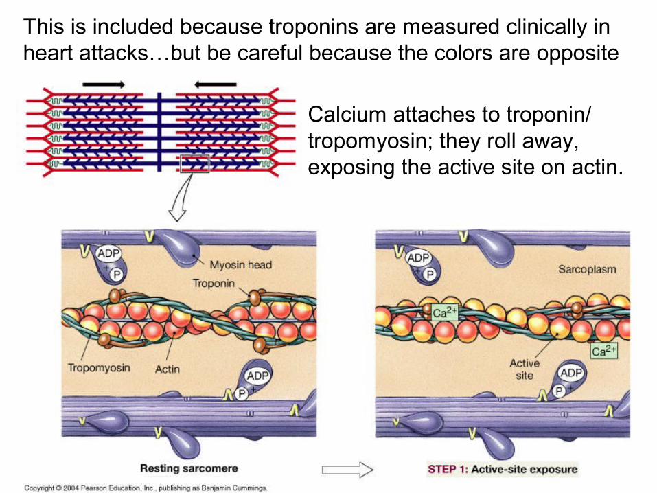
This is included because troponins are measured clinically in heart attacks…but be careful because the colors are opposite
Calcium attaches to troponin/ tropomyosin; they roll away, exposing the active site on actin.