Embed Size (px)
Citation preview

Mycobacteria
• To know various species of the Mycobacteria
• To know the various diseases caused by the Mycobacteria
• To know the pathogenesis of Mycobacteria
• To know various tests for diagnosis of diseases caused by
Mycobacteria
• To know the treatment of diseases caused by Mycobacteria

Mycobacteria Classification:
A: Tubercle bacilli• Human: M. tuberculosis• Bovine: M. bovis• Murine : M. microti• Avian: M. marinum
B: Lepra bacilli• Human: M.leprae• Rat: M.leprae marium
C: Mycobacteria causing skin ulcers: M. ulcerans, M. balnei
D: Atypical Mycobacteria: – Photochromogen – Scotochromogen – Nonphotochromogen– Rapid growers
E: JOHNE’s bacillus: M. paratuberculosis
F: Saprophytic Mycobacteria: M. butyricum, M. pheli, M. stercoris, M. smegmatis

Mycobacteria are either: a) Obligate pathogens:
M. tuberculosisM. bovisM. africanum
b) Opportunistic pathogen: Atypical Mycobacteria or MOTT: M. kansasii M.avium
c) Saprophytic Mycobacteria:M. smegmatis M. pheli M. stercorisM. butyrijcum

Mycobacterium tuberculosis
• Fungus like bacteria: filamentous forms resembling fungal mycelium, acid fast, aerobic, non motile, non capsulated & non sporing
• Morphology: 2-3um x 0.4 um, straight or slightly curved rods with rounded ends, branching & filamentous forms , beaded, arranged singly or in small clumps,

Cultural characteristic: – an obligate aerobe, – 370c, pH- 7,– slow growth – take 2-8 weeks to grow – generation time- 14-15 hrs,– grows well on enriched media containing
serum, potato, blood & egg– M. bovis is microaerophilic

1) Solid media:
LJ, Dorset egg medium• Containing blood: Tarshis medium• Containing serum: Loeffler’s serum slope• Containing potato: Pawolosky’s medium
2) Liquid medium: Dubbo’s, Middle brook’s(7H-10 & H-12) , proskaur’s medium
Colony characters: on solid media: dry, rough, raised wrinkled, irregular
• Liquid media: growth occurs as bottom creeps up the sides, forms prominent surface pellicle, extends along sides above the medium
• New technique: BACTEC system

Biochemical tests:
• Niacin test• Neutral red test• Aryl sulphtase test• Nitrate reduction test• Amidase test• Catalase & peroxidase test• Susceptibility to pyrazinamide• Susceptibility to thiophen 2-carboxylic acid hydrazide• Tween 80 hydrolysis

Resistance: • viable in sputum for 20-30 hrs• in droplet nuclei for 8-10 hrs• in culture for 6-8 months • resistant to disinfectant• sensitive to formaldehyde & Glutaraldehyde• killed by sunlight for 2-3 hrs exposure• sensitive to ethanol

Pathogenesis: Inhalation of M. tuberculosis
Reach lung
Ingested by alveolar macrophages Multiplication in macrophages
Ghon’s focus in lower lobe

Hilar lymph node involvement
Primary complex
healing & calcification haematogeneous Cause latent infection milliary TB
Reactivation or exogenous infection
Secondary tuberculosis
Usually pulmonary TB

• Tubercle formation: avascular granuloma composed of central zone containing giant cells with or without caseation & necrosis, surrounded by a zone of epitheloid cells, with a peripheral zone of lymphocytes & fibroblast
• Pathogenicity due to biological activities of the bacteria
– Cell wall: delayed type of hypersensitivity
– Lipid: formation of macrophages, monocyte, epitheloid, giant cell
– Polysccaharide : immediate type of hypersensitivity
– Phospitidase: helps in tubercle formation by forming epitheloid cell & giant cell

• Tubercle lesion : 1) exudative 2) productive
• Infection depends on: – dose of organism– virulence– mode of entry– age– resistance– hypersensitivity of patient
• Antigens present in mycobacterium spp:
– Group specific polysaccharide antigen– Type specific protein antigen : used in detection of antibodies
• Phage types of mycobacterium: A, B, C, type I

Various systems involved in TB: • Pulmonary ,renal, tubercular meningitis, bone
marrow & joint , miliary, intestinal, skin tuberculosis
• Clinical symptoms:
fever, cough with expectoration, haemoptysis, weight loss, loss of appetite, signs of pleural effusion/consolidation/cavity

LAB diagnosis: • Specimens:
SputumGastric lavageCSF
• Microscopy: Ziehl - Neelsen staining: grading (RNTCP)
3+ = > 10 AFB / Oil immersion field
2+ = 1 - 10 AFB / Oil immersion field
1+ = 10 - 99 AFB / 100 Oil immersion field
Exact = 1 – 9 AFB / 100 Oil immersion field

• Petroff’s method for sputum and oxalic acid for urine
• Culture on Lowenstein Jensen (LJ) medium
• ELISA, RIA, latex agglutination
• Nucleic acid detection: PCR,LCR,DNA probes
• BACTEC, VersaTrek, MGIT
• Animal inoculation in guinea pig
• Antibiotic Sensitivity test

Tuberculin test: Mantoux test• Type IV hypersensitivity test
• Purified protein derivative (PPD) intradermal inoculation on forearm
• Site observed after 72 hrs for erythema & induration
• Interpretation:
– 10 mm indicates positive– 5 mm negative, – between 5-9 mm indicates doubtful
• Use: to diagnose active infection in infants

Treatment :
• Bactericidal : rifampicin, isoniazide, pyrizinamide, streptomycin
• Bacteriostatic: ethambutol, cycloserine, capreomycin, kanamycin, ciprofloxacin
• DOTS,RNTCP
Prophylaxis:
– BCG: live attenuated vaccine– Strains used: M.bovis– Route of inoculation: intradermal– Age: at birth,shedule: single dose
• Immunity: 10- 12 yrs, protects individuals from complicated forms of tuberculosis
• Adverse effects : local, regional, systemic
• Contraindicated in AIDS, measles
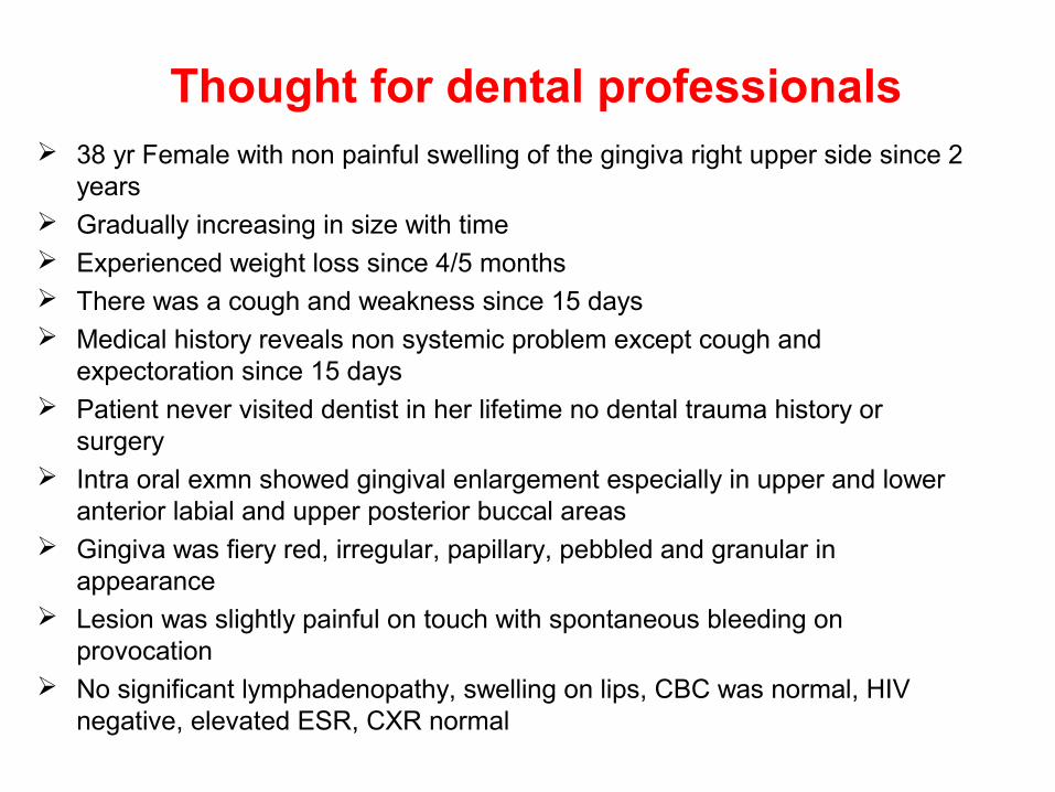
Thought for dental professionals 38 yr Female with non painful swelling of the gingiva right upper side since 2
years Gradually increasing in size with time Experienced weight loss since 4/5 months There was a cough and weakness since 15 days Medical history reveals non systemic problem except cough and
expectoration since 15 days Patient never visited dentist in her lifetime no dental trauma history or
surgery Intra oral exmn showed gingival enlargement especially in upper and lower
anterior labial and upper posterior buccal areas Gingiva was fiery red, irregular, papillary, pebbled and granular in
appearance Lesion was slightly painful on touch with spontaneous bleeding on
provocation No significant lymphadenopathy, swelling on lips, CBC was normal, HIV
negative, elevated ESR, CXR normal


• Gingival tuberculosis Published in J Ind Soc Periodontol Aug 2009
• Conclusion: – It can be occupational risk for dentists– It is rare entity– Consider for diff diagnosis– Diff Diag :
• Gingival enlargement due to drugs• Infections (bacerial, fungal and viral)• Malignancy – leukemia• Traumatic ulcer• Squamous cell carcinoma
• A major concern for dentists so Infection prevention practices meticulously

Atypical Mycobacteria• Mycobacteria other than mammalian tubercle bacilli
• Occasionally cause human disease resembling TB
• Are opportunistic pathogens
• Also referred as Nontuberculous mycobacteria or MOTT • Mycobacteria other than tubercle
• Classifed by Runyon (1959) based on pigment production and rate of growth

Classification of atypical mycobacteria
• Photochromogens: – pigments in sunlight, – M. kansasii, M.simiae, M.marinum
• Scotochromogen : – pigments in light as well as in dark, – M. scrofulaceum, M.szulgai

Classification of atypical mycobacteria
• Nonchromogen: – not produce pigment in light also,
M.avium,M.intracillularae,M.xenopi, M. ulcerans
• Rapid growers: – grow within 7 days,– M. fortuitum, M. chelonei

Atypical mycobacteria• Opportunistic pathogen• Some are rapid growers• Some produce
pigments• Niacin: negative• Aryl sulphtase: positive• Strong catalase positive• Non pathogenic to
guinea pig• R to antituberculous
drugs• Some grow at 250 C
&some at 450 C
Typical • Obligate pathogen• Slow growers• Non pigmented• Niacin : positive• Aryl sulphtase :
negative• Weak catalase positive• Pathogenic to guinea
pig
• S to antituberculous drugs
• Growth does not occur below 250C & above 440C

• Disease• Pulmonary infection like
tuberculosis
• Lymphadenopathy usually cervical
• Cutaneous ,subcutaneous lesions
• a. chronic ulcers• b. Abscess• c. Swimming pool
granuloma• d.Buruli ulcer• e. Surgical wound infection
• Systemic disseminated disease
• Usual atypical agents• M.kansasii, M.avium, M.
intracellulare, M.simiae
• M.szulgae, M.xenopi, M.chelonei, M. avium-intracellulare, M.scrofulaceum, M.kansasii
• M. marinum• M.fortuitum, M.chelonei• M.marinum
• M.ulcerans M. chelonei, M.fortuitum
• M.avium, M.intracellularae

Laboratory diagnosis
• Microscopy
– Ziehl Neelsen staining
• Culture
– on Lowenstein Jensen (LJ)
– Incubated at 250C, 370C, 450C,
– slants observed for pigment production

Identification
• Identification by different tests such as – Nitrate reduction– Aryl sulphtase– Tween 80 hydrolysis– growth at different temperatures
• Antibiotic susceptibility testing• Treatment :
– prolonged treatment with INH, – rifampicin, – ethambutol, clofazimine, – quinolones, – macrolides

Mycobacterium leprae
• Leprosy is a old disease since vedic times
• Probably originated in the tropics and spread to the rest of the world
• Lepra bacilli was first observed by Hansen in 1868

Morphology
• L. leprae is a straight or slightly curved rod• 1-8 um x 0.2-0.5 um in size• Polar bodies and other intracellular elements
present• Clubbed forms, lateral buds or branching• It is Gram positive readily stain than Tb bacilli• Acid fast but less than M. tuberculosis• 5% H2SO4 is used as decolorizer• Bacilli are seen singly and in groups intracellular or
lying free outside the cells

Cultivation:• Not been able to cultivate in artificial media• Can be cultivated in mice, nine banded armadillo• Generation time 12-13 days
Resistance: Remain viable in warm humid envt. For 9-16 days For 46 days in moist soils Survive exposure to direct sunlight for 2 hrs and UV rays for
30 min

Leprosy
• Is a chronic granulomatous disease primarily of skin, peripheral nerves and nasal mucosa
• Classified as– Lepromatous – Tuberculoid– Dimorphous– Indeterminate
Type of disease is reflection of immune status of the host

Lepromatous:• Lepromatous is seen in where the host immune response os
low• Bacilli are seen in large numbers (multibacillary stage)• Bacilli invade nose, mouth and upper respiratory tract and
are shed in large numbers in nasal and oral secretions• More infective and prognosis is very poor
• Cell mediated immunity is deficient and lepromin test is negative
• Humoral immunity is more

Tuberculoid: Seen in patients with high degree of resistance Skin lesions are few and sharply demarcated Bacilli are scanty (paucibacillary) Cell mediated immunity is adequate Lepromin test is positive Prognosis is good but humoral immune response is poor

Borderline or dimorphous: Lesions possessing both tuberculoid and lepromatous
typesI
Indeterminate: Is early unstable tissue reaction Which is not characteristics of either It may undergo healing spontaneously

Ridely and jopling classification
• 1966
• Five groups1. Tuberculoid (TT)
2. Borderline tuberculoid (BT)
3. Borderline (BT)
4. Borderline lepromatous (BL)
5. Lepromatous (LL)

epidemiology• Exclusively human disease• Source – patient• Mode of transmission is still not clear
• Large no. of bacilli shed in nasal secretions
• Patients may discharge 8x10⁸ bacilli in one nose blow• Mode of entry
– Respiratory tract– Skin It is not highly communicable Disease develops in 5% in spouses Incubation period is 2-5 yrs Worldwide in distribution India – Orisa and Bihar (highest prevalence)

• Lepromin test: – skin test for delayed hypersensitivity & used to study immunity in
leprosy patients, antigen is Mitsuda’s Ag or bacillary lepromin
• Procedure : 0.1ml injected intradermally on forearm
• Two reactions: 1. early or Fernandez reaction: is an acute inflammatory area of more than
10 mm in diameter appearing in 24-48 hrs & disappears in 3-4 days2. Late or Mitsuda’s reaction: appears 3-4 weeks after injection in the form
of nodule,may ulcerate or subside in few weeks
• Mitsuda’s reaction is more meaningful is a manifestation of CMI,induced by lepromin while Fernandez reaction indicates past reaction
• Uses: 1. classify lesions of leprosy2. to assess prognosis & response to treatment3. to assess resistance of an individual to leprosy4. to verify candidate lepra bacillus

Lab diagnosis:
• Specimens: skin clippings, nasal samples,skin nerve biopsy, lymph node puncture
• Microscopy: – ZN staining with 5% sulphuric acid
• Gradings: Ridely scale• 1+: 1-10 bacilli/ 100field• 2+: 1-10 bacilli/ 10 field• 3+: 1-10 bacilli/field• 4+: 10-100/ field• 5+: 100-1000/field• 6+: more than 1000, clumps/field

• Morphological & bacteriological index calculated
• Culture: footpads of mouse, granuloma developes in 1-6 months
• Serological: latex agglutination, ELISA
• Lepromin test not for diagnosis but to resitance of patient
• Treatment: – for paucibacillary leprosy: rifampicin 600mg once in a month &
Dapsone (4’,4’- diaminodiphenyl sulphone,DDS)100mg daily for 6 months
– For multibacillary leprosy : rifampicin, 600mg once a month Dapsone 100mg daily &clofazimine 50mg daily for 2 years

Department of Microbiology
Mr. Mukhit Kazi, Lecturer


![Detection of clinically important non tuberculous mycobacteria … · 2020. 8. 26. · atypical or non-tuberculous mycobacteria (NTM) [2]. NTM, also known as environmental mycobacteria](https://img.pdfslide.net/doc/110x75/60d3deeff170c737ef603bcb/detection-of-clinically-important-non-tuberculous-mycobacteria-2020-8-26-atypical.jpg)