Embed Size (px)
DESCRIPTION
Slides with topics that are covered and were tested in the recent Absite exams.Nir Hus MD., PhD.http://www.nirhus.com
Citation preview
Absite Topic Review
Nir Hus
Nir Hus
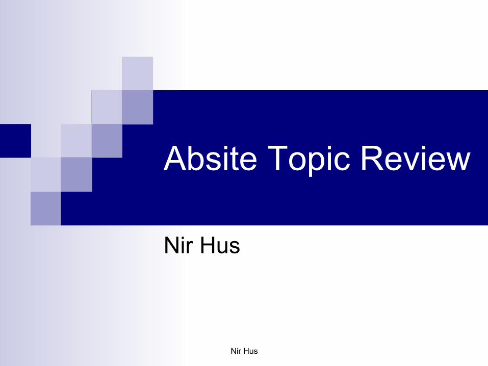
Predict Increased risk BRCA-1,2 mutation
BRCA I Ovarian CA – 40% lifetime risk Male Breast CA – 1% lifetime risk
BRCA II Ovarian CA – 10% lifetime risk Male Breast CA – 10% lifetime risk
Nir Hus
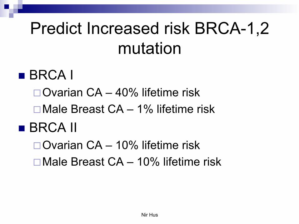
BRCA cont.
BRCA gene + family Hx = 60% lifetime risk of breast CA (autosomal dominant)
Bi prophylactic mastectomy w/ BRCA gene results in decreased risk of breast CA by 90% Add TAH & BSO decrease risk 95%
CA in a person w/ BRCA I,II carries a 50% risk of developing CA in contralateral breast.
Nir Hus
A newborn infant fails to pass meconium in the 1st 24 hrs of life & subsequently gets progressive abdominal distention. Plain films show a distended colon. On exam, the child has an anus located in the proper position and on rectal exam there is explosive release of watery stool. The most appropriate next step is?
A. Upper GI series B. Barium Enema C. Enteroclysis D. Rectal Bx.
Nir Hus
Hirschsprung’s disease
More common in Males #1 cause of colonic obstruction in infants Most common sign infants fail to pass
meconium is 1st 24hrs. Can also present in older individuals as chronic
constipation. Results in ABD distention and colitis. Anorectal exam explosive stool release. Barium enema can be normal, but shows a
spastic distal segment and dilated proximal segment.
Nir Hus

Hirschsprung’s disease cont.
Rectal Bx is the gold standard in diagnosis of the condition. Will show absence of ganglion cells in myenteric plexus.
Tx colon resection until proximal to the area where we see ganglionic cells.
Nir Hus
Gastroschisis
Associated congenital anomalies are uncommon and occur in about 10% of cases, most commonly intestinal atresia or stenosis.
These anomalies are thought to reflect mechanical or vascular compromise to the herniated bowel.
Rarely, infants with gastroschisis will have complete loss of small bowel secondary to in utero volvulus.
Nir Hus
Nir Hus

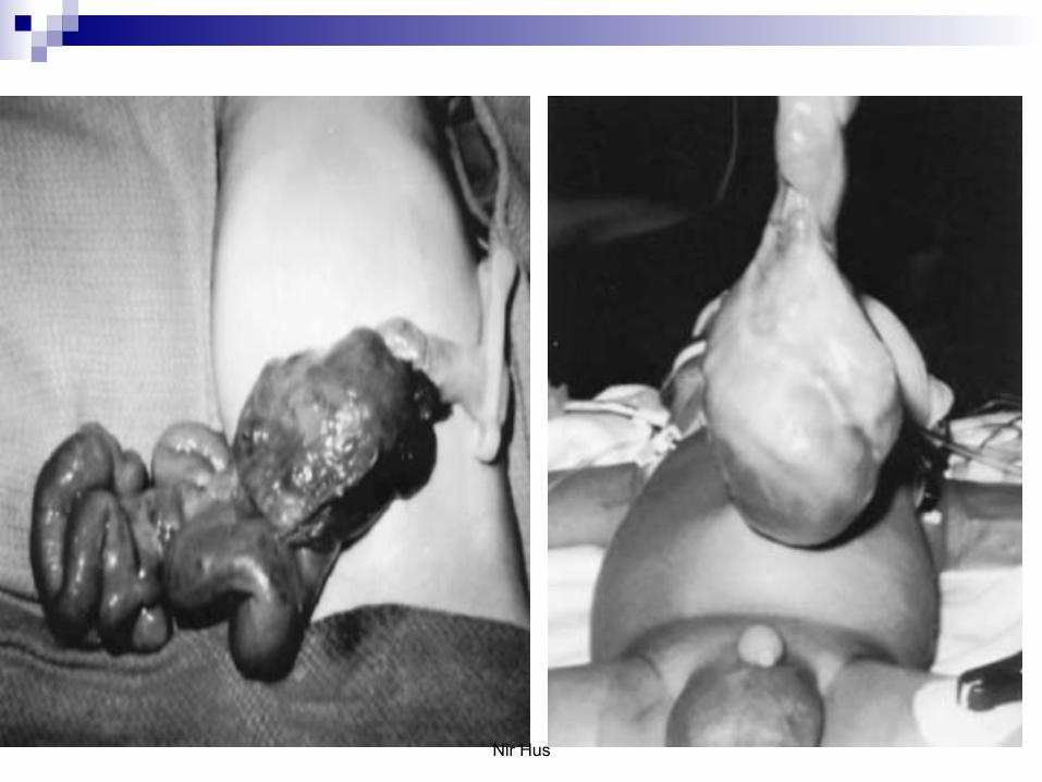
Gastroschisis Pathophysiology. In normal fetal development, there are two paired
umbilical veins. Intestine returns to the abdominal cavity through the
umbilicus. The right umbilical vein undergoes resorption, leaving
the left umbilical vein intact. Weakness of the umbilical membrane at the site of
umbilical vein resorption may evolve into a hernia, and, in the case of membrane rupture, evisceration of the intestine through the defect may occur.
This explanation is consistent with the clinical observation that the abdominal wall defect in gastroschisis nearly always is located to the right of the umbilicus.
Nir Hus