Embed Size (px)
DESCRIPTION
Citation preview

Potassium Management
Marica A. Lazo, MD

Potassium PearlsO Potassium is the major intracellular cation.O A healthy adult has roughly 50 mEq/Kg of K+ in his/her
body.O 70 Kg man = 70x50 = 3500 mEq in body
O Only 2% is found outside the cells and of this only 0.4% of your K+ is found in the plasma. O Thus serum K+ measurements have limitations at
reflecting TOTAL body K+ stores.O A 1 mEq/L drop in K+ reflects between 200-400 mEq
total body K+ deficitO Example: a K+ of 2.5 means that someone is
roughly 300 mEq in the negative. This would require 7 boluses of 40 mEQ of K+ to make up for this!

HypokalemiaO Clinical consequences of
hypokalemia usually goes unnoticed.O Common findings include weakness,
fatigue, constipation, ileus, and respiratory muscle dysfunction.
O Symptoms seldom occur unless plasma K+ is less than 3.0 mmol/L.

ECG changesO ST depressions with prominent U
waves and prolonged repolarization

ECG changes

Hypokalemia - CausesO Spurious - i.e. K+ is falsely lowO Diminished intakeO Redistribution – i.e. movement into
cellsO Extrarenal loss – usually associated
with preservation of renal K+O Renal loss – often associated with
acid-base disturbances.

Spurious HypokalemiaO Marked leukocytosis and blood tube
that has been sitting at room temp too long gives time for K+ to enter the white blood cells and thus falsely lower K+ value.
O Insulin given just prior to blood draw allows a small amount (about 0.3 mEq) to shift into cells in the blood tube.

Redistribution Hypokalemia
O Transcellular shiftO Alkalosis (response H+ out K+ in) – a
key point is that alkalosis disorders are usually involved in depletion of total body K+ in addition to redistribution.
O Increased B adrenergic effect – increases Na/K ATPase activity. Think of both medications or increased sympathetic tone like MI, head trauma, DTs, and theophylline toxicity.

Redistribution Hypokalemia
O Other causes of hypokalemia due to cell entry include risperidone, quetiapine, and cesium, hypothermia, barium intoxication, chloroquine intoxication.

Extrarenal K+ Loss Urine K+ < 20 mEq/24 hours or spot urine K+
of < 30
O Diarrhea – causes loss of HCO3 and K+ thus you get metabolic acidosis + hypokalemia.
O Chronic Laxative Abuse O Sweat – 9 mEq/L of K+ in sweat.O Fasting/inadequate diet – usually no
more than total body deficit of 300 mEq.
O Villous adenoma at rectosigmoid

Renal K+ LossUrine K+ >20 mEq/24 hours or spot urine K+
of > 30
O Renal hypokalemia with metabolic acidosisO RTA type I (distal) and type II
(proximal)O DKAO Carbonic anhydrase inhibitor therapyO ureterosigmoidostomy

Renal K+ LossUrine K+ >20 mEq/24 hours or spot urine K+
of > 30
O Renal hypokalemia with metabolic alkalosis:O Almost always occurs with
hypokalemia because virtually every cause of metabolic alkalosis also causes hypokalemia.
O The excess HCO3 acts as a poorly reabsorbable anion and carries more Na+ to the collecting tubules leading to increased Na-K exchange and urinary K loss.

Renal K+ LossUrine K+ >20 mEq/24 hours or spot urine K+
of > 30
O Renal hypokalemia with no acid-base disorder:O Recovery from ARF, postobstructive
diuresis, and osmotic diuresis, PCNs all increase Na delivery to collecting tubules resulting in increased K excretion.
O Low magnesium- think of with resistant cases. Hypomagnesemia is present in up to 40% of patients with hypokalemia


Urinary K+: > 20 mEq/L – Renal loss
Urinary K + : < 20 mEq/L – Extrarenal loss
TTKG : Transtubular Potassium Gradient ( Urine K+ / Plasma K+ ) ( Urine Osm / Plasma Osm )
TTKG : Renal loss : > 4 Extra renal loss : < 4
Renal Vs Extra renal loss
TreatmentO Therapeutic goals
O Prevent life-threatening complications (arrhythmias, respiratory failure, hepatic encephalopathy)
O Correct the K+ deficitO Minimize ongoing lossesO Treat the underlying cause

TreatmentO K+ deficit
O (4 – Actual K+) x 3002
O (4 – 2.5) x 300 = 225 meqs 2
O Estimation of K+ deficitO 3.0 meq/L= total body K+ deficit of 200-
400 meq/70kgO 2.5 meq/L = 500 meq/70kgO 2.0 meq/L = 700 meq/70kg

TreatmentO Oral therapy
O Generally saferO Degree of K+ depletion does not
correlate well with the plasma K+O KCl is usually the preparation of choiceO Kalium durule: 1 durule = 10 meqs KClO KCl syrup: 1meq/mLO Ie. Kalium durule 750mg TID PO x 2-
3daysor KCl syrup 15-30cc TID

TreatmentO IV therapy
O For severe hypokalemia or those who are unable to take anything by mouth
O Maximum rate at which potassium is infused into peripheral veins is usually 10 meq/hr
O Central – 20 meq/hrO Rate of infusion should not exceed 20
meq/hour unless paralysis or malignant ventricular arrhythmias are present
O Ie. 40 meqs KCl in 230cc PNSS x 5meq/hr (32cc/hr) OR 20 meqs KCl in 100cc PNSS x 1hr

HyperkalemiaO Remember that total body K+ is roughly 50
mEq/kg and only a small fraction if found outside the cells.
O Contrary to struggling to try to replace a low K+ with mEq after mEq and watching it slowly climb into the normal range; only a small shift of intracellular K+ to the extracellular space or a small amount of K+ given to a person with a bad kidney can cause quick problems.
O To get a serum K+ rise by 1 meq/L you only need to give 100-200 meq of extra K+.
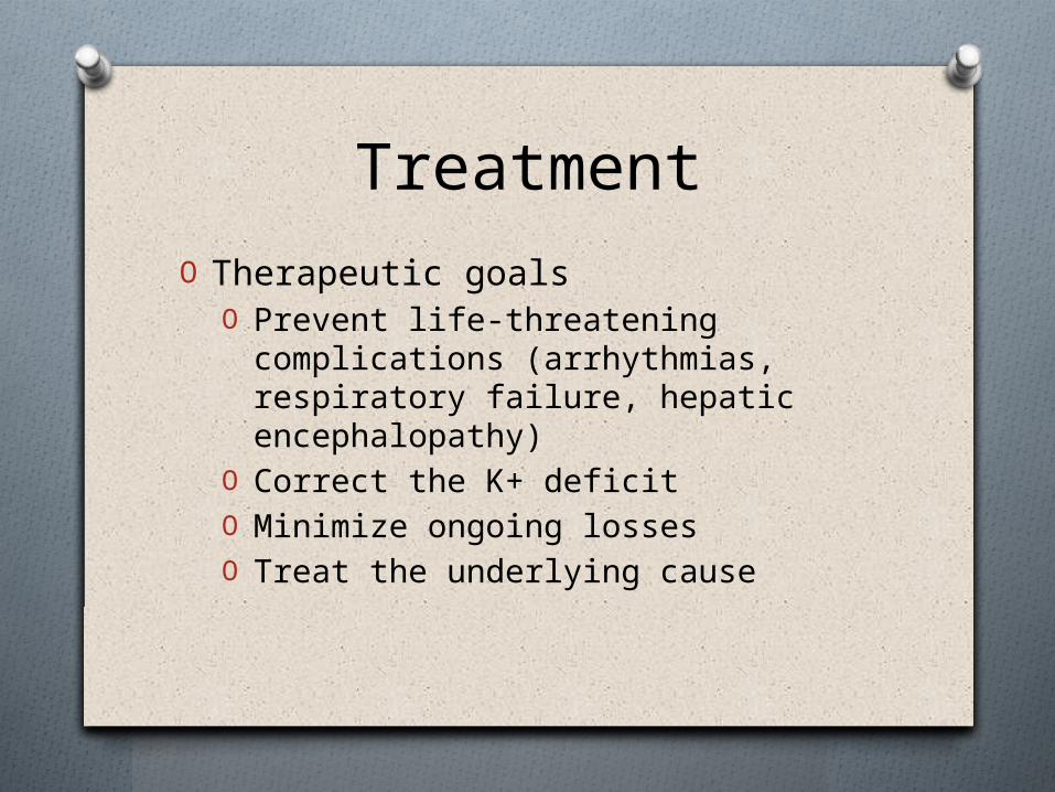
HyperkalemiaO The most serious effect of
hyperkalemia is cardiac toxicityO Hyperkalemia partially depolarizes
the cell membrane, which impairs membrane excitability and is manifest as weakness that may progress to flaccid paralysis and hypoventilation if the respiratory muscles are involved

Hyperkalemia - CausesO Increased K+ intake
O Rarely the sole causeO Iatrogenic hyperkalemia may result
from overzealous parenteral K+ replacement or in patients with renal insufficiency
O PseudohyperkalemiaO Artificially elevated plasma K+ due to
K+ movement out of the cells immediately before or following venipuncture

Hyperkalemia - CausesO Transcellular shift
O Tumor lysis syndrome and rhabdomyolysis lead to K+ release from cells
O Metabolic acidosis can be associated with mild hyperkalemia resulting from intracellular buffering of H+
O Insulin deficiency and hypertonicity promote K+ shift from the ICF to the ECF

HYPERKALEMIA
PSEUDOHYPERK K RETENTION REDISTRIBUTION
GFR < 20 ml/min GFR > 20 ml/min
Hemolysis Thrombocytosis Leukocytosis Mononucleosis
Renal failure
Aldosterone deficiency Addison’s disease RTA Type 4 Drugs Heparin NSAIDs ACE inhibitors Cyclosporine
Tubular hyperK Acquired SLE Obstr. Uro. Amyloidosis AIDS TID Drugs Trimethoprim K sparers
Acidosis Insulin deficiency/DKA Beta blockers Periodic paralysis Digitalis intoxication Succinylcholine Exercise Tissue damage

EKG Changes
Note the “tented” or “pinched” shape to Twaves

Acute Treatment O Calcium Gluconate 10 ml of 10% solution
(1gram) IV slowly over 5-10 min.O Decreases membrane excitabilityO Temporarily (1 hour) antagonizes cardiac
effects of hyperkalemia while more definitive therapy is begun.
O Warning: may induce Digitalis toxicity!O May precipitate if given with NaHCO3.O May repeat after 5 min. if ECG does not
improve.

Acute TreatmentO Glucose/Insulin – 100 ml of 25% glucose
solution with 10 units of Regular insulin. Infuse over 15-30 minutes.O Insulin stimulates cellular uptake of K+ by
activating Na+K+ATPase ( decreasing plasma K+ )
O Temporarily translocates K+ into cells. O Effect occurs w/in 30-60 min and lasts
about 1 hr.O May induce hyperglycemia, thus if
already hyperglycemic just use insulin.

Acute TreatmentO Beta 2 agonists (Albuterol) - 10-20
mg over 15 minutes via nebulizer.O Promotes cellular uptake of K+O Onset 30 minutes.O Lowers plasma K+ by 0.5-1.5 mmol/L
and the effect lasts for 2-4 hoursO Potentially dangerous in patients with
coronary artery disease!

Acute TreatmentO Lasix – 40 to 80 mg IV.
O Especially helpful in aldosterone deficiency states and renal failure.
O NaHCO3 – 1 standard amp (50mEq) IV over 5-10 min.O Can shift K+ into the cells.O Mostly used with acidemic states.O Will precipitate with Calcium!!!! Thus
don’t give while using calcium gluconate.

Acute TreatmentO Kayexalate (Sodium Polystyrene
Sulfonate) – 15 g ORALLY 1 to 4 times daily as a slurry in water or syrup.O Onset 1-2 hours with duration of 4-6 hours.O Effect—In the intestine (mostly the large
intestine), Na ions are released and are replaced by K+ and other cations before the resin is passed from the body.
O Each gram may remove 1 mEq K+ in exchange for 1-2 mEq Na+ thus may cause ECF volume overload.

TREATMENT OF HYPERKALEMIA
MEDICATION MECHANISM OF ACTION
DOSAGE PEAK EFFECT
Calcium gluconate Insulin and glucose Sodium bicarbonate Albuterol
Antagonism of membrane actions Increased K entry to cells Increased K entry to cells Increased K entry to cells
10-30 ml of 10% solution IV over 10 minutes 10 units insulin plus 50 ml D20 50 meq IV over 5 minutes 10-20 mg IV or nebulized
5 minutes 30-60 min. 30-60 min. 30-60 min.

Thank you!