Embed Size (px)
Citation preview

By-Mr. ASHOK BISHNOI
Lecturer

Excretory System – removes excess water, urea, carbon dioxide, and other wastes from our blood.
Kidneys (Nephron)– filter out excess water and urea
Lungs (Alveoli) – filter out carbon dioxide,from the blood.
Skin (Sweat glands) – excretes water, as sweat, which contains some trace chemical wastes, including urea.
Liver-ammonia
Introduction:-

Types of metabolic wastes:-
Waste Produced from
Carbon Dioxide Aerobic Respiration
Water Aerobic Respiration
Salts Metabolic activities
Nitrogenous wastes Breakdown of excess
Amino Acids & Proteins
Types of nitrogenous wastes toxicity
Ammonia (NH3) Highly Toxic
Urea Moderately Toxic
Uric Acid Crystals Minimally Toxic
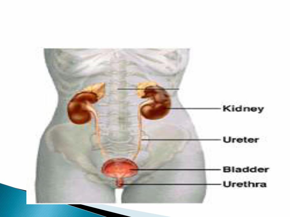

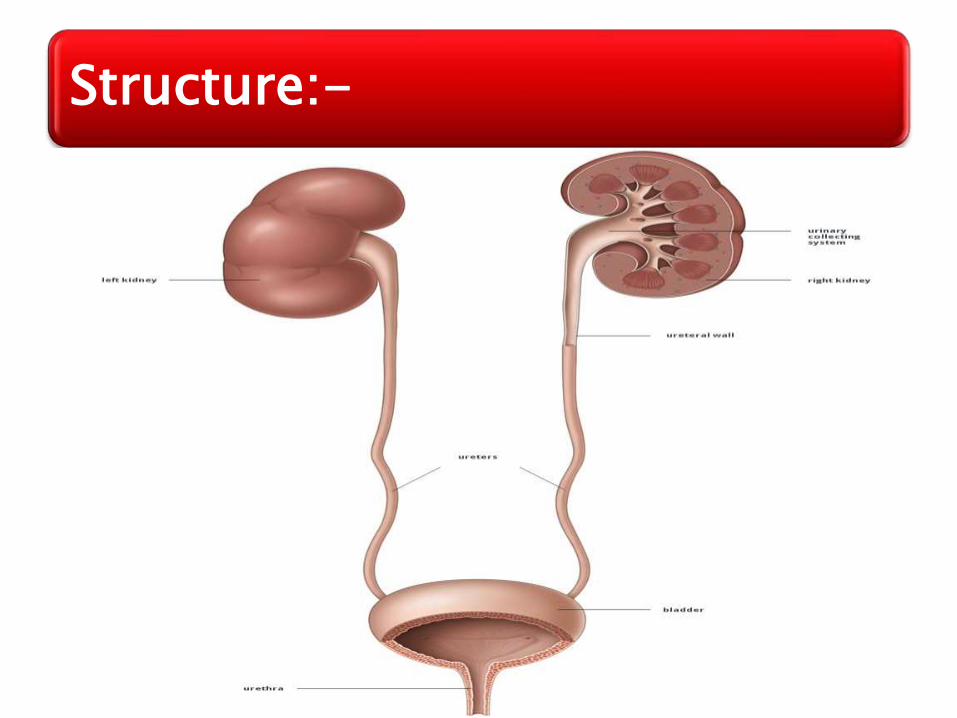
The urinary system is the main excretory system & consist of following organs...
2 Kidneys:-Which secrete urine.
2 Ureters:- Which convey urine from the kidney to urinary bladder.
1 Urinary bladder:- Where urine collect & temporary stored.
1 Urethra:- Through which the urine is discharge from the urinary bladder to the exterior.

Filter 200 liters of blood daily, allowing toxins, metabolic wastes, and excess ions to leave the body in urine.
Regulate volume and chemical makeup of the blood.
Maintain the proper balance between water & salts, acids & bases
Function:-
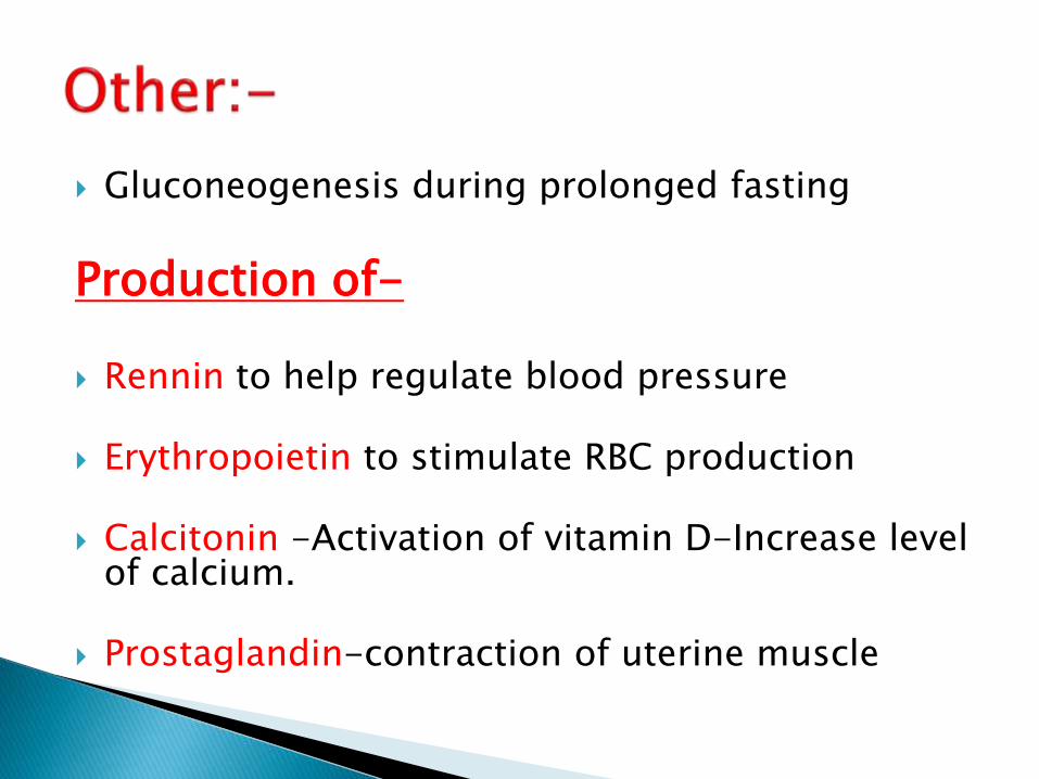
Gluconeogenesis during prolonged fasting
Production of-
Rennin to help regulate blood pressure
Erythropoietin to stimulate RBC production
Calcitonin -Activation of vitamin D-Increase level of calcium.
Prostaglandin-contraction of uterine muscle
Location:- It occupy the Epigastric, Hypochondriac,
lumber & umbilical regions.
Vertically they extend from the upper boarder of 12th thoracic vertebra to the centre of the body of 3rd lumber vertebra
The right kidney is lower than the left because of liver
Kidney (2)-Renal, Nephron:-
Shape:- It is Bean shaped organ
Size:- 11cm long 6 cm wide 3 cm thick
Weight:- 150gm in male 135gm in female
Colour:- Raddish brown in colour

The Parts of the Kidney:-

Each kidney is composed of three sections:
◦ The outer cortex,◦ The middle part medulla◦ & the inner pelvis.
The cortex (cone-shaped) is where the blood is filtered.
The medulla (funnel-shaped )contains the collecting ducts which carry filtrate (filtered substances) to the pelvis.
The pelvis is a hollow cavity where urine accumulates and drains into the ureter.
Nephron
The filtering units of the kidneys are the nephrons.
There are approximately “1” million nephronsin each kidney.
The nephrons are located within the cortex and medulla of each kidney.
The tubes of the nephron are surrounded by cells and a network of blood vessels spreads throughout the tissue.
Therefore, material that leaves the nephronenters the surrounding cells and returns to the bloodstream by a network of vessels.
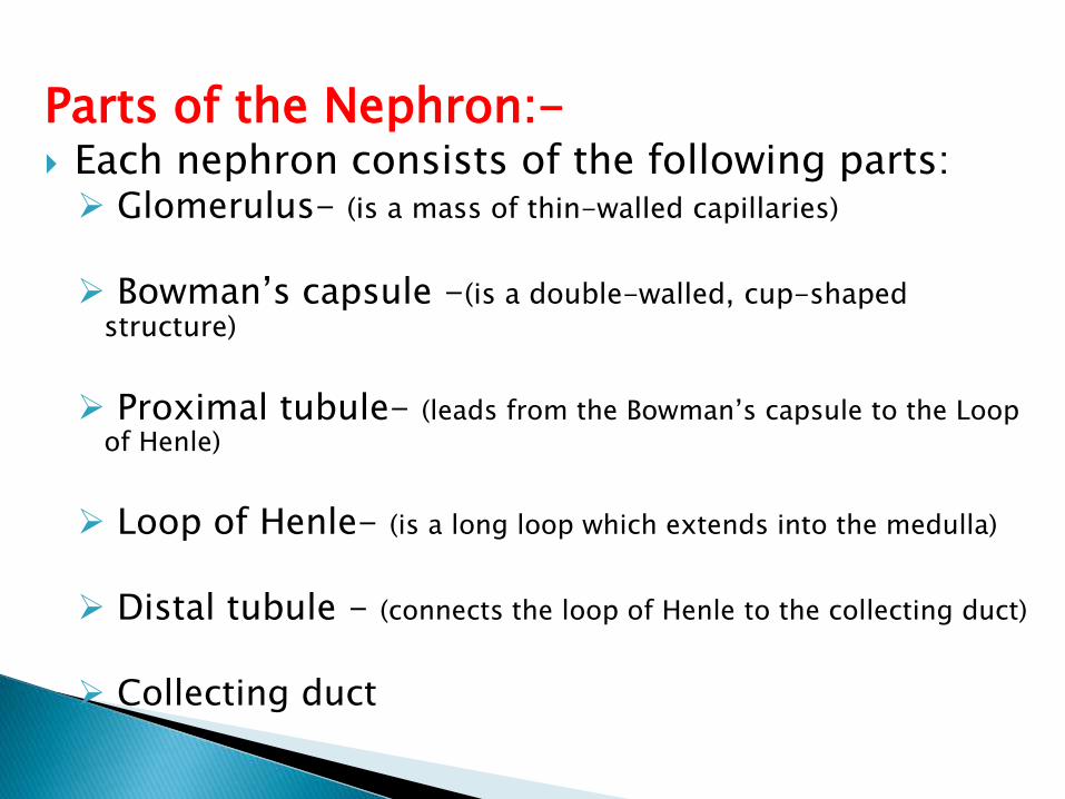
Parts of the Nephron:- Each nephron consists of the following parts: Glomerulus- (is a mass of thin-walled capillaries)
Bowman’s capsule -(is a double-walled, cup-shaped structure)
Proximal tubule- (leads from the Bowman’s capsule to the Loop of Henle)
Loop of Henle- (is a long loop which extends into the medulla)
Distal tubule - (connects the loop of Henle to the collecting duct)
Collecting duct
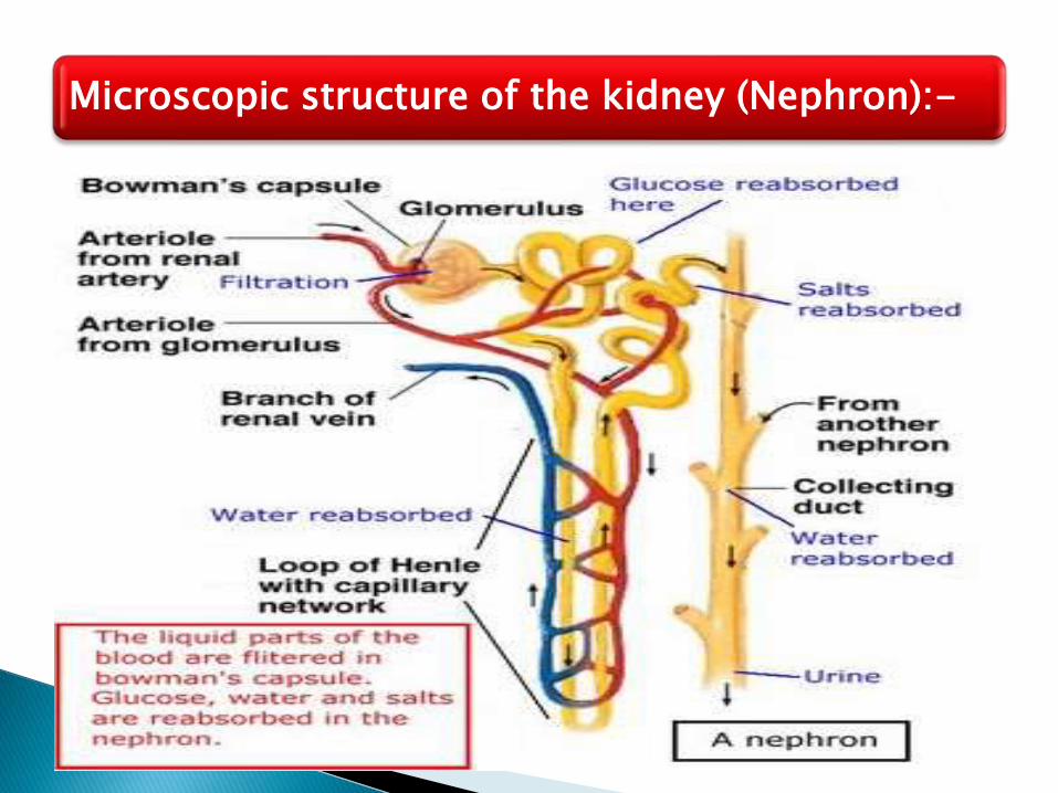
Microscopic structure of the kidney (Nephron):-

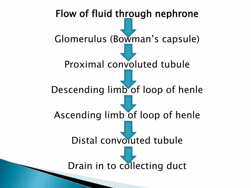
Flow of fluid through nephrone
Glomerulus (Bowman’s capsule)
Proximal convoluted tubule
Descending limb of loop of henle
Ascending limb of loop of henle
Distal convoluted tubule
Drain in to collecting duct

Blood supply in the kidney
Renal artery
Segment artery
Inter lobular artery
Afferent arteriole
Glomerular capillaries
Efferent arterioles
Inter lobule vein
Segmental vein
Renal vein

19
Blood and Nerve Supply:-
Approximately one-fourth (1200 ml) of systemic cardiac output flows through the kidneys each minute.
Arterial flow into and venous flow out of the kidneys follow similar paths
The nerve supply is via the renal plexus
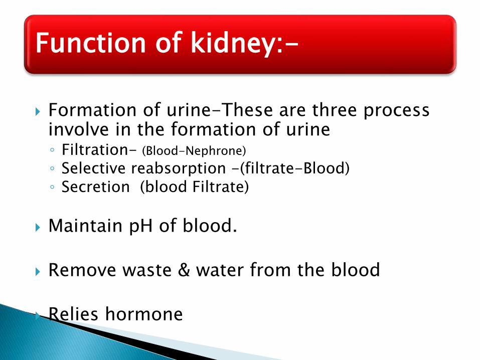
Formation of urine-These are three process involve in the formation of urine◦ Filtration- (Blood-Nephrone)
◦ Selective reabsorption -(filtrate-Blood)◦ Secretion (blood Filtrate)
Maintain pH of blood.
Remove waste & water from the blood
Relies hormone
Function of kidney:-
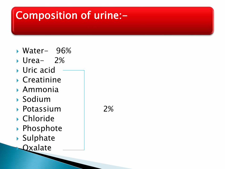
Water- 96%
Urea- 2%
Uric acid
Creatinine
Ammonia
Sodium
Potassium 2%
Chloride
Phosphote
Sulphate
Oxalate
Composition of urine:-
23
Glomerular Filtration Rate (GFR)
“
The total amount of filtrate formed per minute by the kidneys”

Ureters (2)
25
Introduction:-
Slender tubes that convey urine from the kidneys to the urinary bladder
Ureters enter the base of the bladder through the posterior wall.
It is about 25-30 cm long
It is about 3mm in diameter
It is continuous with funnel shaped renal pelvis.
It passes downwards through the abdominal cavity, behind the peritoneum in front of the psoas muscle in to the pelvic cavity & passessobliquely through the posterior wall of the bladder
Structure:-

28
Wall of ureters is consist of three layers;-
Outer layer-Adventitia- of fibrous tissue continuous with the fibrous capsule of the kidney.
Middle layer –Muscular-consisting of smooth muscles fiber
Inner layer - Mucosa -composed of transitional epithelium.
Blood supply by:-Ureter receives its arterial blood supply in three different parts, as explained below.
Upper part receives its blood supply from renal artery
Middle part receives its blood supply from testicular or ovarian artery
Pelvic part receives its blood supply from the superior vesical artery
Venous drainage by:- The venous blood is drained by veins that correspond to the arteries explained above.

Lymph drainage by:-
Lymph from the ureters drains into the lateral aortic nodes and the iliac nodes.
Nerve supply by:-
sympathetic nerves
Propel urine to the bladder via response to Peristaltic contraction of smooth muscle layer.
Function of ureter:-

Urinary bladder (1)
It is reservoir of urine
It is pear shaped but become more oval as it fills with the urine.
It is a Smooth, collapsible, muscular sac that temporarily stores urine
It lies in the Pelvic cavity
Total capacity is about 600ml
Introduction:-
It lies retroperitoneal on the pelvic floor posterior to the pubic symphysis
◦ Males – prostate gland surrounds the neck inferiorly
◦ Females – Anterior to the vagina and uterus
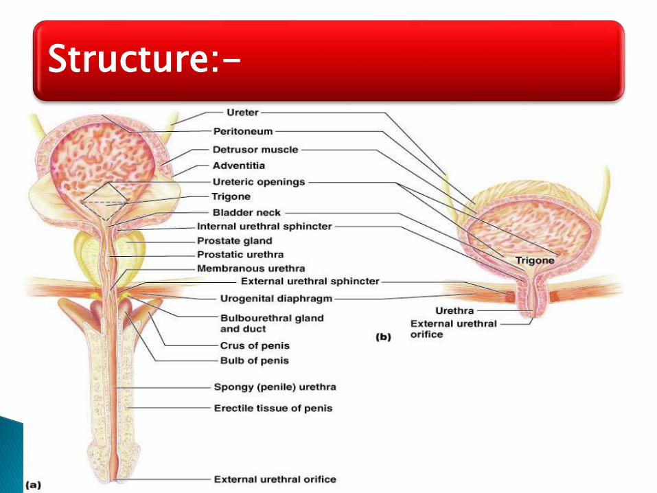
Structure:-
36
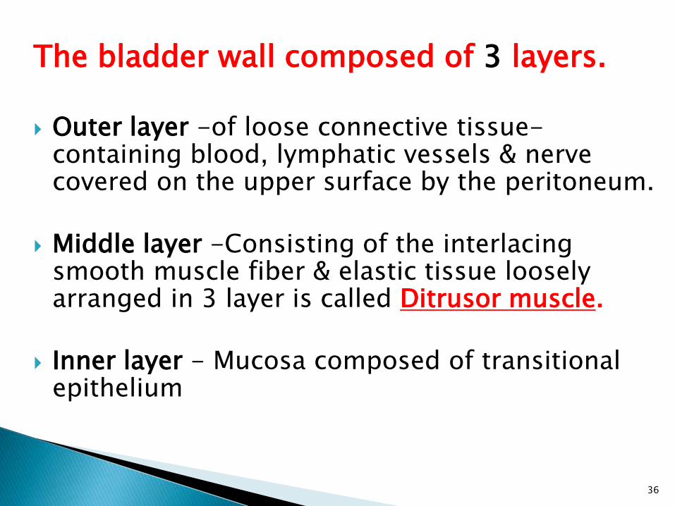
The bladder wall composed of 3 layers.
Outer layer -of loose connective tissue-containing blood, lymphatic vessels & nerve covered on the upper surface by the peritoneum.
Middle layer -Consisting of the interlacing smooth muscle fiber & elastic tissue loosely arranged in 3 layer is called Ditrusor muscle.
Inner layer - Mucosa composed of transitional epithelium
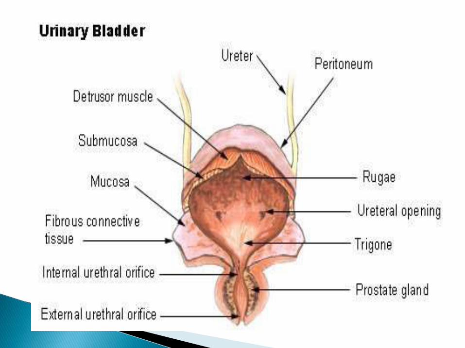
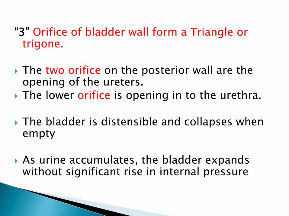
“3” Orifice of bladder wall form a Triangle or trigone.
The two orifice on the posterior wall are the opening of the ureters.
The lower orifice is opening in to the urethra.
The bladder is distensible and collapses when empty
As urine accumulates, the bladder expands without significant rise in internal pressure

Blood Supply by:-Superior & inferior vesicalarteries
Venous drainage by : Veins from the vesicalvenous plexus that drain into the internal iliac vein
Lymphatic drainage by : Into internal & external iliac lymph nodes.
Nerve supply by:- Sympathetic & parasympathetic nerve
Urethra (1)
It is a canal extending from the neck of the bladder to the exterior, at the external urethral orifice.
It is a longer in male then the female
The male urethra has three named regions◦ Prostatic urethra – runs within the prostate gland◦ Membranous urethra – runs through the urogenital
diaphragm◦ Spongy (penile) urethra – passes through the penis
and opens via the external urethral orifice
Introduction:-
Structure:-
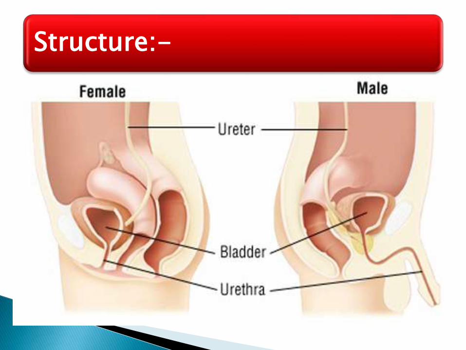
Male :- It is about “19-20” cm long
Female :- it is about “4” cm long & “6” mm in diameter.
To transport urine from the bladder.
To transport the semen (sperm cells and fluid from the seminal vesicles and the prostate) out the tip of the penis
Function in male urethra:-

26-46
Disorders of Urinary System
Renal calculi
Urinary tract infections
Glomerular disease
Renal failure
Polycystic kidney disease
The kidney has other functions but it is usually associated with the excretion of cellular waste such as :
1) urea (a nitrogenous waste produced in the liver from the breakdown of protein. It is the main component of urine) ;
2) uric acid (usually produced from breakdown of DNA or RNA) and
3) creatinine (waste product of muscle action).

All of these compounds have nitrogen as a major component.
The kidneys are more than excretory organs.
They are one of the major homeostatic organs of the body.
They control water pH, secrete erythropoietin (a hormone that stimulates red blood cell production) and activate vitamin D production in the skin.
That is why a doctor can tell so much from a urine sample.