Embed Size (px)
Citation preview

Dr. Mohit Goel,
JR II ,
REFERENCES :
An imaging checklist for pre-FESS CT: framing a
surgically relevant report. S. Vaid,*, N. Vaid , S.Rawat , A.T.
Ahuja
Anatomical Principles of Endoscopic Sinus Surgery: A
Step by Step Approach Author: Bradoo Renuka


A significant percentage of patients have septal deviations that need surgical intervention
as they impede endoscopic access to the middle meatus.
Nasal septum

The anterior most attachment to the cribriform plate is vertically oriented and is
best seen on coronal images.
Posteriorly, the middle turbinate is obliquely oriented, forming the basal lamella,
which is commonly attached to the lamina papyracea. This lamella is seen
separating the anterior ethmoid cells from the posterior ethmoid cells.
More posteriorly, it is horizontally oriented
and attached to the medial wall of the
maxillary sinus.
Middle turbinate

Sagittal image with arrow showing vertical attachment of basal lamellae to anterior skull base
separating the anterior ethmoid (AE) and posterior ethmoid (PE) sinuses.
(FS: frontal sinus, AG: agger nasi cell, SpS: sphenoid sinus, MT: middle turbinate)

Ground / Basal Lamella
• It may show dehiscences or be partially deficient in
which case infection can pass from anterior to posterior
ethmoids.
• It may itself be pneumatized and split into multiple
septae.
• The ground lamella usually attaches to the lamina
papyracea.
Rarely it may, however, turn inferiorly in which case it
“misses” the lamina papyracea and attaches to the
lateral wall of the maxillary sinus.

Normal variations that can cause a narrowing of the middle meatus are concha
bullosa, interlamellar cell of Grunwald and paradoxical curvature of the middle
turbinate.
Concha bullosa, -- pneumatized from either the frontal recess, the agger nasi cell, anterior ethmoid cells or
the middle meatus.
It may have septations .
It may compromise ventilation and drainage of secretions to produce chronic infection of the paranasal
sinuses.


The middle turbinate may show a sharp bend laterally instead of its usual smooth medial
curvature. This is the paradoxically bent middle turbinate.
It is quite often bilateral and can block the infundibulum

The uncinate process is a thin, crescent-shaped bone, attached anteriorly to the lacrimal
bone and inferiorly to the inferior turbinate.
Superiorly, it may be attached to the anterior skull base, the lamina papyracea, or the
middle turbinate. The pattern of attachment determines the FSDP.
Coronal CT image shows medially draining FSDP (arrowheads) into the middle meatus (MM)
when the uncinate process (asterisk) attaches to the skull base/lamina papyracea (arrows)
Uncinate process

A laterally draining FSDP (arrowheads) into the ethmoidal infundibulum (EE) when the uncinate process
attaches to the middle turbinate (arrow).

An acute angle of attachment between the uncinate process and the lamina papyracea is of
significance as it increases the chances of orbital penetration during FESS.

The uncinate process can, at times, be laterally positioned against the orbit, as seen in maxillary
sinus hypoplasia and silent sinus syndrome.
(a) Coronal and (b) axial CT images show left maxillary sinus hypoplasia (asterisk) with laterally positioned
and atelectatic uncinate process (arrows).

Occasionally the upper end of the
uncinate process may lie free within
the middle meatus and not attach to
any adjacent bony structure.
The uppermost portion of the
uncinate process may be
pneumatized and compromise the
infundibulum.

All six components of the OMC, i.e., the maxillary ostium, the middle meatus, the ethmoidal
infundibulum, the bulla ethmoidalis, the uncinate process, and the hiatus semilunaris, are well
visualized on coronal CT.
OMC and maxillary sinuses
Coronal CT image depicting all six
components of the OMC.
MO, maxillary ostium;
MM, middle meatus;
EE, ethmoidal infundibulum;
BE, bulla ethmoidalis;
UP, uncinate process;
HSL, hiatus semilunaris
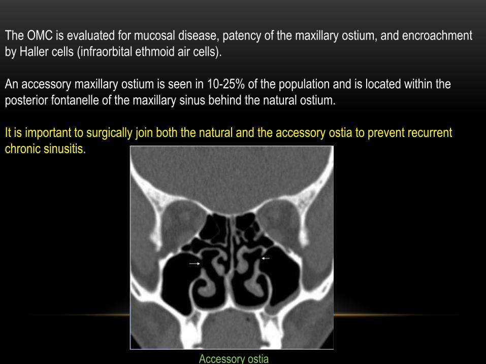
The OMC is evaluated for mucosal disease, patency of the maxillary ostium, and encroachment
by Haller cells (infraorbital ethmoid air cells).
An accessory maxillary ostium is seen in 10-25% of the population and is located within the
posterior fontanelle of the maxillary sinus behind the natural ostium.
It is important to surgically join both the natural and the accessory ostia to prevent recurrent
chronic sinusitis.
Accessory ostia

The size of the maxillary sinus and the presence of any intrasinus septae need to be
documented.
In maxillary sinus hypoplasia, the medial wall of the sinus is closely related to the medial wall of
the orbit. This may increase the risk of orbital penetration during endoscopic sinus surgery.
In extensive pneumatization there may only be a thin soft tissue layer between the dental roots
and the sinus. This may result in recurrent maxillary sinusitis due to dental disease and tooth
extraction in these patients,
and it may cause oroantral fistulae.
The infraorbital nerve (a branch of the
maxillary division of the trigeminal nerve)
runs over the roof of the sinus and exits
through this foramen.
This nerve may be dehiscent here, an
important Preoperative finding for the
avoidance of intraoperative nerve injury.Left infraorbital foramen is dehiscent into the
maxillary sinus (arrowhead)

The frontal process of the maxilla extends superiorly to form the frontonasal process or frontal
beak.
It is seen on coronal views but it is best evaluated on parasagittal images. The thickness of
the frontal beak influences the size of the frontal ostium.
FSDP and frontal sinuses
Parasagittal CT image (a) shows superior extension of the frontal process of the maxilla forming the
frontal beak (arrow).
Coronal CT image in the same patient (b) shows the frontal beak (arrow) separating the frontal sinus
(asterisk) above it from the frontal recess (curved arrow) below it.

Parasagittal CT image shows a thin frontal beak (arrow) with resultant wide frontal ostium
(asterisk).
(b) A thick frontal beak with a narrow frontal ostium.

The FSDP is well delineated on all three orthogonal planes. However, it is well seen in the
parasagittal plane as an hour-glass shape.
The “waist” of the hour glass corresponds to the frontal ostium located at the level of the frontal
beak.
The frontal sinus lies above the waist and
the frontal recess can be identified below
the waist.
This configuration makes the
frontal recess an anatomical “tight spot”,
implicated as a cause of sinusitis.
Parasagittal CT image showing typical “hour-
glass” configuration of the FSDP (outlined by
black and white arrows) draining inferiorly into
the middle meatus.

Agger Nasi cell
The agger nasi cell is the most anterior ethmoidal cell, present in 93% of the population,
lying in the FSDP. The anterior wall of this cell forms the anterior
boundary of the frontal recess, making it an important surgical
landmark.
On coronal images, the agger nasi cell is
identified as an air cell below the frontal beak, before the
antero-superior attachment of the middle turbinate.
However, it is best viewed on parasagittal images.

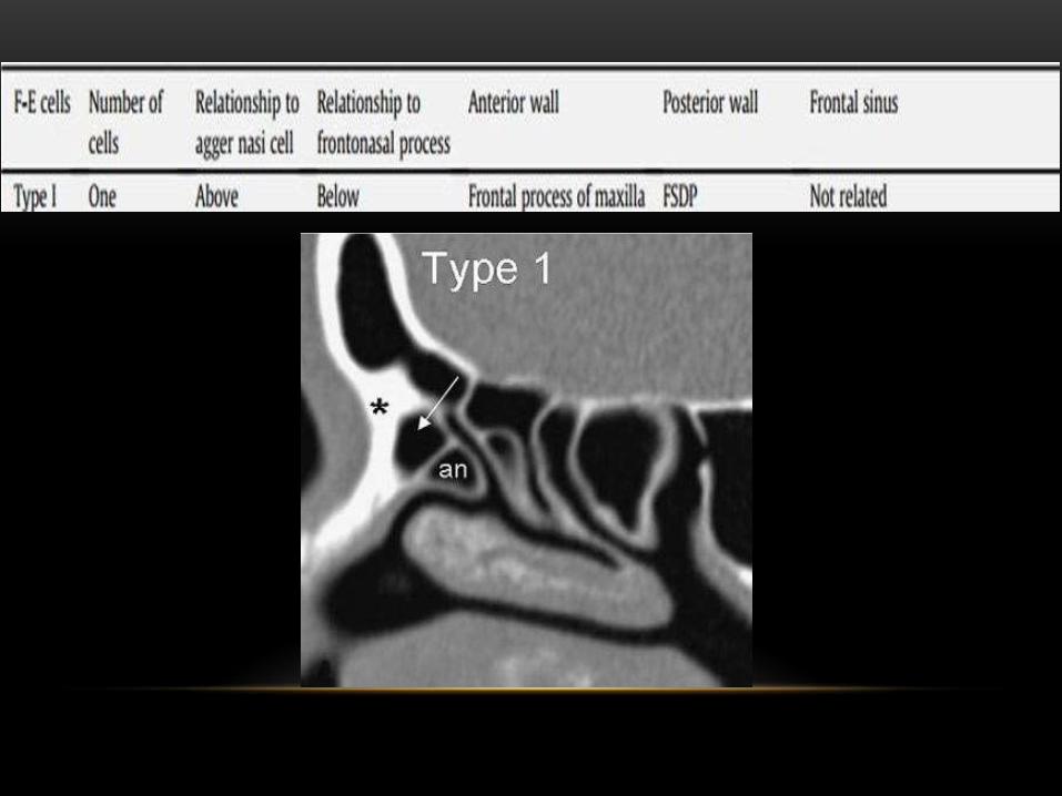
The cells of the FSDP
The classification is based on the position of the cells in relation to the agger nasi cell, the
frontal beak, and degree of superior extension into the ipsilateral frontal sinus.


Large frontal bullar cell (asterisk),
suprabullar in location (B, bulla ethmoidalis),
with an anterior margin related to the frontal sinus
(arrows) and posterior margin formed by the
anterior skull base (arrowheads).
The frontal bullar cell originates in the suprabullar region,extends superiorly into the frontal sinus
along the anterior margin of the skull base, and is best viewed on sagittal imaging.
The anterior wall of this cell is related to the frontal sinus and the posterior wall is the anterior
skull base.
Caution needs to be exercised while fracturing the frontal bullar cell posteriorly as it may cause
inadvertent trauma to the anterior skull base.

Interfrontal sinus septal cell: this cell is within the interfrontal sinus septum and may compromise
the frontal Ostium.

The extent of the superior pneumatization of the frontal sinus needs to be assessed if a frontal
trephination is planned. An underpneumatized frontal sinus may result in intracranial
penetration of the drill during this procedure.
Areas of dehiscence in the posterior wall of the frontal sinus need preoperative identification as
posterior wall disruption and meningeal trauma may occur while administering sinus washes.
Frontal sinus
Lateral and superior pneumatization of frontal sinus

Ethmoid air cells anterior to the basal lamella are termed anterior ethmoidal cells.
The largest anterior ethmoid air cell is termed the bulla ethmoidalis. It is a reliable surgical
landmark.
However, if it is small, there is a relative medial projection of the lamina papyracea, called the
torus lateralis. This may increase the chances of orbital injury.
Anterior ethmoid sinus group

Supraorbital ethmoid cell is the ethmoid cell that extends superolaterally between the middle
orbit wall and the ethmoid roof.
Supraorbital ethmoid cells may simulate multiple frontal sinuses, type III frontal cells,
suprabullar cells, frontal bulla cells or interfrontal sinus septal cells on coronal CT images.
During endoscopic sinus surgery, these cells may be mistaken for the frontal sinus and need
to be differentiated by their more lateral and posterior location as compared to the frontal
sinus.
Supraorbital ethmoid cell

This group is comprised of the posterior ethmoidal sinuses and the sphenoid
sinus.
Posterior sinus group
Posterior ethmoid sinus
These are ethmoid air cells posterior to the basal lamella which drain into the superior meatus
and border the sphenoid sinus posteriorly.
It is important to document the vertical distance from the superior margin of the maxillary
sinus to the roof of the posterior ethmoid cells, as measured on coronal CT.
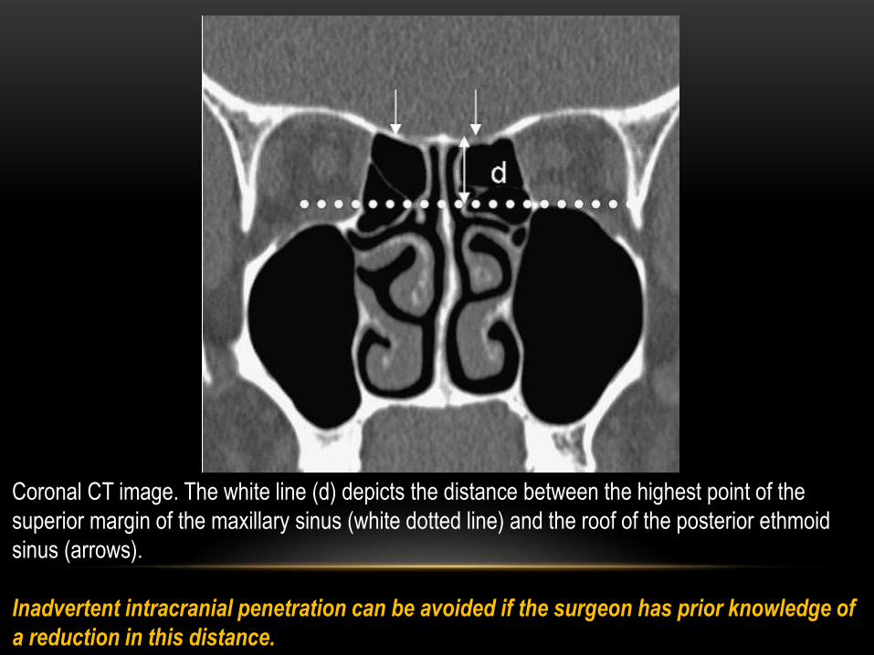
Coronal CT image. The white line (d) depicts the distance between the highest point of the
superior margin of the maxillary sinus (white dotted line) and the roof of the posterior ethmoid
sinus (arrows).
Inadvertent intracranial penetration can be avoided if the surgeon has prior knowledge of
a reduction in this distance.

Another normal variant which needs identification is the Onodi cell (lateral and posterior
pneumatization of the most posterior ethmoid cell
over the sphenoid sinus).
An Onodi cell should be suspected when coronal
images demonstrate a horizontally or
obliquely directed septum in the sphenoid sinus.
There are increased chances of injury to the optic
nerves within Onodi cells.
Onodi cell

Coronal CT image shows horizontal and obliquely oriented sphenoid sinus septae (arrows) with
Onodi cells (black arrowheads) into which the optic nerves are seen dehiscent (white
arrowheads).
An intersinus septum is seen attaching to the bone covering the right internal carotid artery
(curved arrow)

Sphenoid sinus
Vital structures such as the carotid arteries, optic nerves, maxillary branches of the trigeminal
nerves within the foramen rotundum, the Vidian canals and the cavernous sinuses are closely
related to the sphenoid sinus.
These structures are often seen as indentations on the roof and walls of the sinus and can
project into the lumen of the sinus (endosinal) in case of hyperpneumatization .
Coronal CT images. Bilateral endosinal vidian canals (arrows) and dehiscent right optic nerve with
attachment of an intersinus septum (arrowhead).
Excessive traction on these septae during surgery can result in injury to these structures.

Hyperpneumatized sphenoid sinus with an
endosinal right foramen rotundum (arrow)
and bilateral optic nerve dehiscence
(arrowheads).


Anterior skull base anatomy
Two regions that deserve special attention in the anterior skull base are the ethmoid sinus roof
and the olfactory fossa.
The roof of the anterior ethmoid sinus is made up of the cribriform bone medially and the fovea
ethmoidalis laterally.
In most patients, the plane of the fovea
ethmoidalis passes above the upper one-third of
the vertical diameter of the corresponding orbit.
A foveal plane passing through the mid-orbital
plane or below predisposes the patient to
inadvertent intracranial penetration
Coronal CT image showing a low-lying and medially sloping fovea ethmoidalis on right side (arrow) almost
reaching the mid-orbital plane (horizontal white line).
The dotted line depicts the vertical height of the right orbit.

Asymmetry in the height of the ethmoid roof needs documentation on CT. Intracranial penetration
is more likely to occur on the side with the lower ethmoid roof.
The olfactory fossa is formed by the horizontal lamella of the cribriform plate, its vertical lamellae
and a part of the orbital plate of the frontal bone.

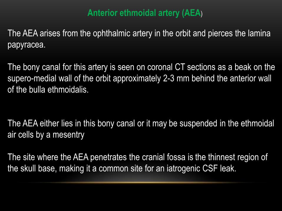
Anterior ethmoidal artery (AEA)
The AEA arises from the ophthalmic artery in the orbit and pierces the lamina
papyracea.
The bony canal for this artery is seen on coronal CT sections as a beak on the
supero-medial wall of the orbit approximately 2-3 mm behind the anterior wall
of the bulla ethmoidalis.
The AEA either lies in this bony canal or it may be suspended in the ethmoidal
air cells by a mesentry
The site where the AEA penetrates the cranial fossa is the thinnest region of
the skull base, making it a common site for an iatrogenic CSF leak.

(a) Coronal CT image shows normal bony canal for the anterior ethmoidal arteries on both
sides (arrows).
(b) The arteries suspended in a mesentry without any bone cover (arrows).

Lamina papyracea
The lamina papyracea has areas of focal dehiscence seen in 0.5% to 10% of
the population.
There is a reduction in the volume of oribital fat between the lamina papyracea
and the medial rectus muscle in the posterior orbit.
Therefore, there are increased chances of injury to the medial rectus muscle in
cases of iatrogenic posterior orbital penetration secondary to posterior lamina
payracea defects.

Coronal CT images depicting multiple focal areas of dehiscence in the lamina papyracea (LP) on both sides
(arrows). Image section through the anterior orbit (a) shows adequate thickness of the orbital fat between
the dehiscent LP and medial rectus muscle (arrowhead).
Posteriorly (b) the thickness of this fat is markedly reduced (arrowhead). Focal areas of dehiscence in the
cribriform plate are also seen on both
sides (black arrows)

Normally the lamina papyracea and the maxillary sinus ostium lie in the same sagittal plane.
Sometimes the lamina papyracea may lie medial to the plane of the maxillary ostium, resulting in
inadvertent orbital penetration .
The dotted horizontal line represents the sagittal plane of the lamina papyracea, which should normally
pass through the maxillary ostium (asterisk). A medially located lamina papyracea may predispose to
inadvertent orbital penetration.

Bony margins of the sinuses
Three features of bone involvement that need documentation are hyperostosis, destruction,
and remodelling.
Hyperostosis adjacent to a soft-tissue abnormality in the sinus is an indication of a chronic
inflammatory or granulomatous process, or a postoperative sequela due to neo-osteogenesis.
This preoperatively prepares the surgeon for extensive bone drilling during the procedure.
Malignant or aggressive infective conditions such as fungal disease can erode or destroy the
bony sinus margins and lamellae.
Remodelling deformities of the bone indicate a slow, progressive, and benign sinonasal
pathology, such as mucocoele or chronic allergic fungal sinusitis.

(a) Coronal CT image shows a focal osteolytic lesion involving the inferior wall of the left maxillary sinus
with an associated soft-tissue mass (arrows).
(b) HRCT image in bone windows reveals associated hyperostosis (asterisk). Histopathological
examination confirmed the presence of granulation tissue due to tuberculosis.

Axial (a) and sagittal (b) CT images show an extensive remodelling deformity of the anterior skull
base (arrows) due to chronic allergic fungal rhinosinusitis. Note the linear hyperdensities within
the sinus, which is characteristic of chronic fungal disease (asterisk)

The critical anatomical information provided by pre-FESS CT has an impact
on the surgical approach adopted by the surgeon and subsequently on the
postoperative benefit to the patient. A systematic checklist ensures
preoperative knowledge of all surgically relevant details.
The authors would, however, like to emphasize that communication between
the radiologist and the surgeon forms the basis of an accurate examination
report. This ensures preoperative knowledge of all surgically relevant details
and goes a long way to preventing potentially morbid intra and postoperative
complications.
Conclusion

THANK YOU