Embed Size (px)
Citation preview
GASTROINTESTINAL PROTOZOAL INFECTIONS
Chair person: Prof Dr Nagappa H
Co-chair person: Dr Narayanaswamy
Presenter: Dr Yashavanth H S
Date: 21/01/2014Time: 2.15pm
Introduction:
• Protozoan
• Greek: protos- first, and zoon- animal
• Protozoology
• A protozoan can be defined as a motile eucaryotic unicellular organism
• Entamoeba histolytica infects all age groups but has its most profound effects in adults.
• Giardia lamblia and Cryptosporidium parvumhave their major impact in childrens
Protozoa
• Moisture is necessary protozoa because they are susceptible to desiccation.
• Most protozoa are free living and inhabit freshwater or marine environments.
• Many terrestrial protozoa can be found in decaying organic matter, in soil, and even in beach sand
• Some are parasitic in plants or animals.
Nutrition of protozoa
• Holozoic nutrition : bacteria are acquired by phagocytosis and the subsequent formation of phagocytic vacuoles.
• Saprozoic nutrition: soluble nutrients such as amino acids and sugars cross the plasma membrane by pinocytosis, diffusion, or carrier mediated transport
• Significant role in the food web of nature
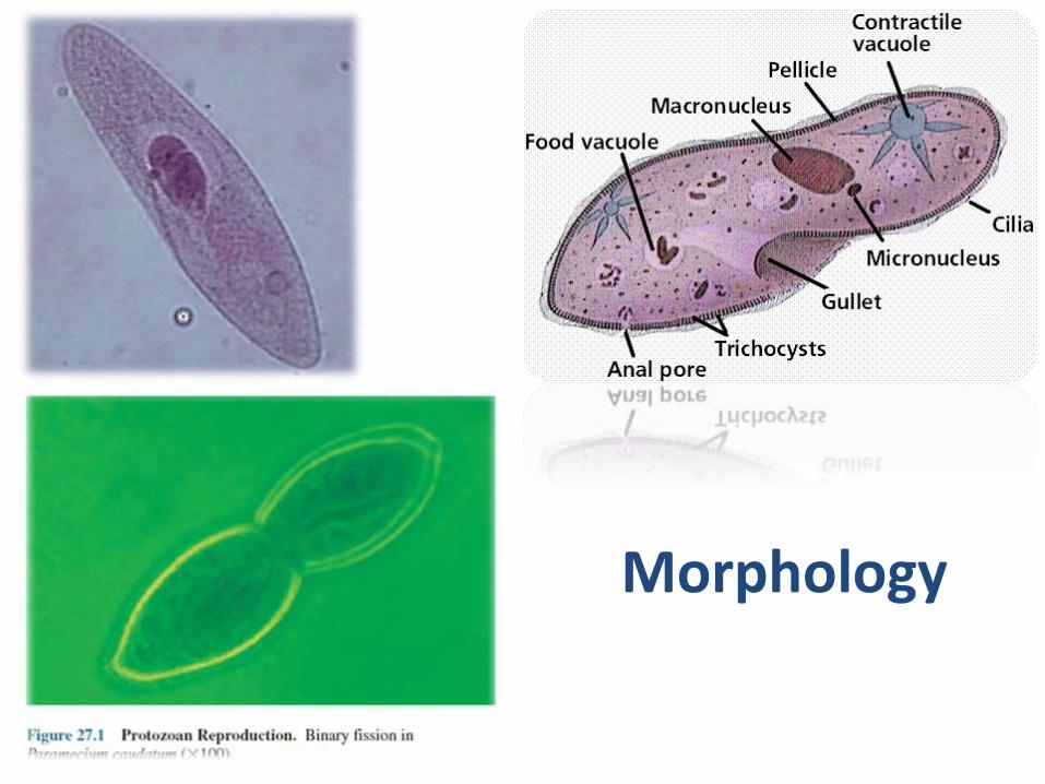
Morphology

Y
Cysts
• They protect against adverse changes in the environment
• They are sites for nuclear reorganization and
cell division (reproductive cysts)
• They serve as a means of transfer between hosts in parasitic species.
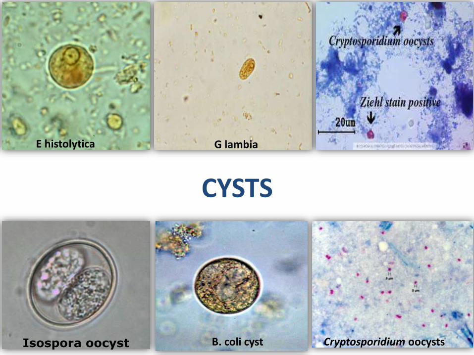
E histolytica G lambia
Isospora oocyst Cryptosporidium oocysts
CYSTS
B. coli cyst
Protozoans causing intestinal infection
• Entamoeba
• Giardia
• Cryptosporidium
• Isospora
• Balantadium
• Cyclospora
• Microsporidia

Epidemiology
• Race – not associated
• Sex – no association ( except in amoebic liver abscess M:F= 10:1 )
• 53.8% school children are infected
• 36.8% of pregnant women are infected
• In rural southern India: 23.1% was infected by one variety and 74.3% are infected by more than 1 group
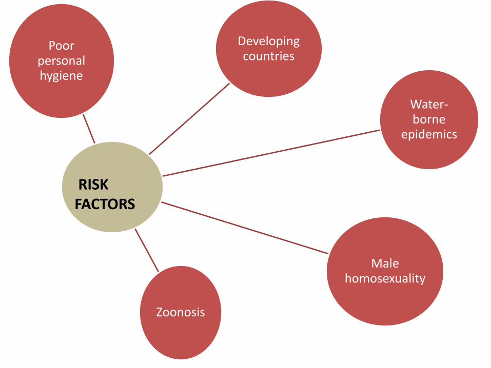
Poor personal hygiene
Developing countries
Water-borne
epidemics
Male homosexuality
Zoonosis
RISK FACTORS
In HIV
• Diarrhoea is the most common GI manifestation
• The diagnosis and management of diarrhoea in a PLHIV is a major challenge.
• There are multiple reasons for the high occurrence of diarrhoea:
• Immune dysfunction of the intestinal epithelial cells.
• Reduced IgA levels.
• Poor gastric acid secretion and nutritional deficiencies.
• Protozoa isolated from the stools of PLHIV without any symptoms, although the isolation of parasites was shown to be more common in patients with diarrhoea.
• The most common pathogen identified in those with diarrhoea was Isospora belli.
• In asymptomatic patients were more likely to shed Giardia in their stools
• Enteric pathogens in stool:
– 57.4% of diarrhoeal patients
– 40% without diarrhoea (P >0.05)
• Protozoal pathogens 71.8%
• Most commonly isolated pathogens:
– Chronic diarrhoea: Isospora belli (25%)
– Controls: Giardia lamblia (16%)
• In patients with acute diarrhoea, there is no definite prominent pathogen
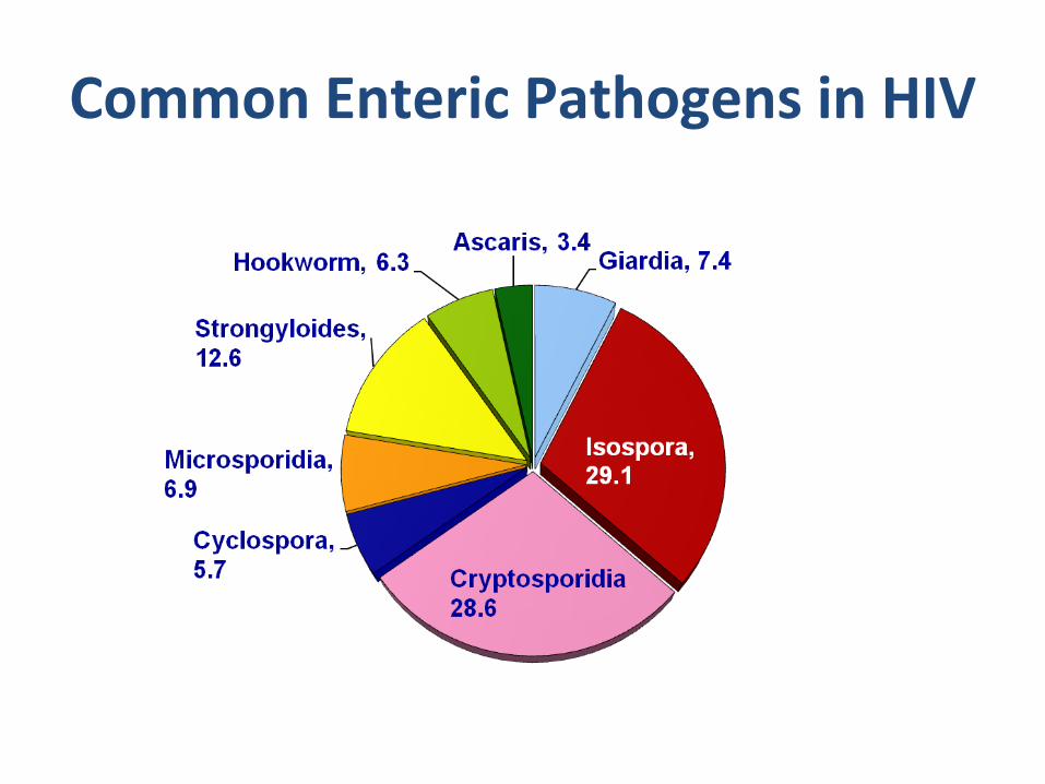
Common Enteric Pathogens in HIV
Differential diagnosis
• Irritable bowel syndrome
• Inflammatory bowel disease (Crohn's, microscopic colitis)
• Gallbladder or pancreatic disease
• Malignancy (amoeboma)
• HIV Enteropathy
Diagnosis of Intestinal Protozoa•Suspect: acute or chronic GI symptoms•Confirmed: detection of parasite in feces
•3 non-consecutive days (inconsistent excretion)
•copro-antigens or molecular probes
•Cryptosporidium•acid-fast stain
•Giardia•duoenal aspirates or biopsy•presumptive treatment in chronic cases
•Entamoeba•sigmoidoscopy (lesions, aspirates, biopsy)•extra-intestinal disease
• Sometimes even after repeated stool testing, no pathogen can be isolated.
• May not be related to technique, but due to fact that the shedding of pathogens may be intermittent.
• The relationship between the pathogen and diarrhoea is unclear.
• Many of the pathogens have been isolated from stools of asymptomatic PLHIV.

• Stool cultures
• Endoscopic studies
• Biopsies and histo-pathological studies
• Electron microscopy and other special studies
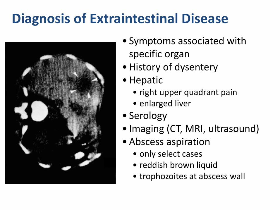
Diagnosis of Extraintestinal Disease
• Symptoms associated with specific organ
• History of dysentery• Hepatic
• right upper quadrant pain• enlarged liver
• Serology • Imaging (CT, MRI, ultrasound)• Abscess aspiration
• only select cases• reddish brown liquid• trophozoites at abscess wall
Non specific treatment
• Maintaining adequate hydration
• Adequate nutritional supplements and specific vitamins and minerals replacement are essential.
• Initiate ART : Initiation of ART is important in controlling diarrhoea especially in conditions where diarhhoea due to cryptosporiodia, isospora and microspora.
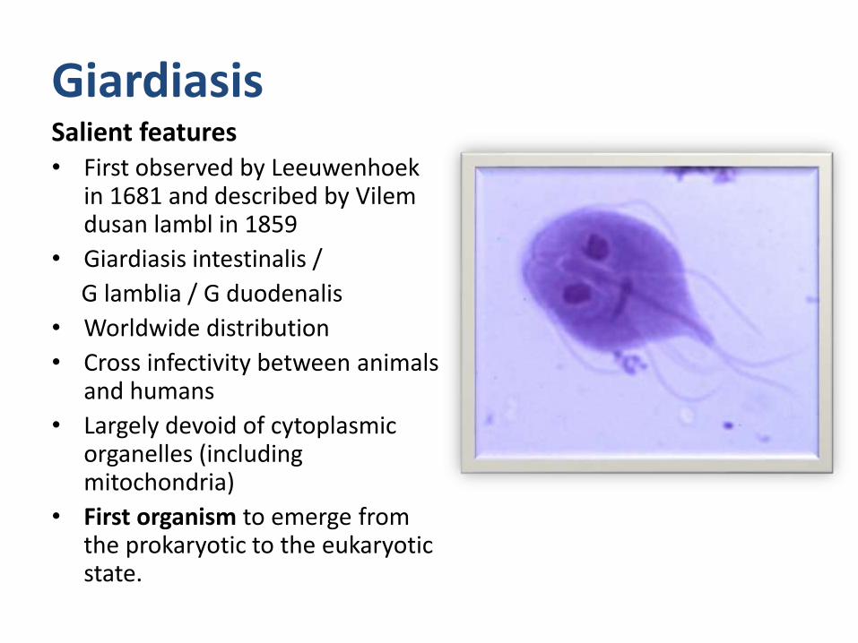
GiardiasisSalient features• First observed by Leeuwenhoek
in 1681 and described by Vilemdusan lambl in 1859
• Giardiasis intestinalis /
G lamblia / G duodenalis
• Worldwide distribution
• Cross infectivity between animals and humans
• Largely devoid of cytoplasmic organelles (including mitochondria)
• First organism to emerge from the prokaryotic to the eukaryotic state.

Risk factors
• Hypogammaglobulinemia
• AIDS
• Cystic fibrosis
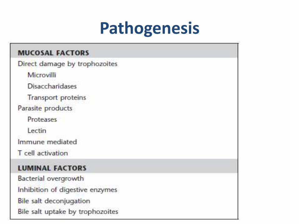
Pathogenesis
Epithelial damage
• villus blunting
• crypt cell hypertrophy
• cellular infiltration
Malabsorbtion
Enzyme deficiencies
• lactase (lactose intolerance)
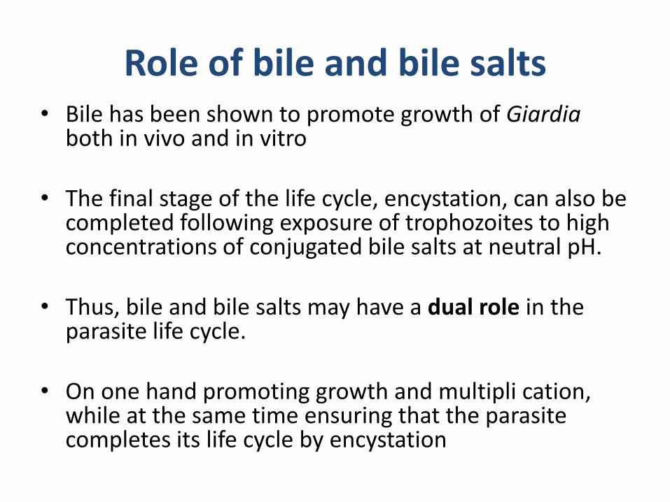
Role of bile and bile salts• Bile has been shown to promote growth of Giardia
both in vivo and in vitro
• The final stage of the life cycle, encystation, can also be completed following exposure of trophozoites to high concentrations of conjugated bile salts at neutral pH.
• Thus, bile and bile salts may have a dual role in the parasite life cycle.
• On one hand promoting growth and multipli cation, while at the same time ensuring that the parasite completes its life cycle by encystation

Range of Outcomes
Asymptomatic/latentAcute short-lasting
diarrheaChronic/nutritional
disorders
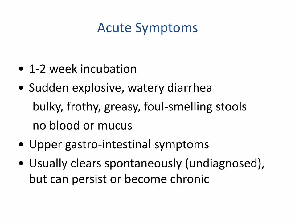
Acute Symptoms
• 1-2 week incubation
• Sudden explosive, watery diarrhea
bulky, frothy, greasy, foul-smelling stools
no blood or mucus
• Upper gastro-intestinal symptoms
• Usually clears spontaneously (undiagnosed), but can persist or become chronic
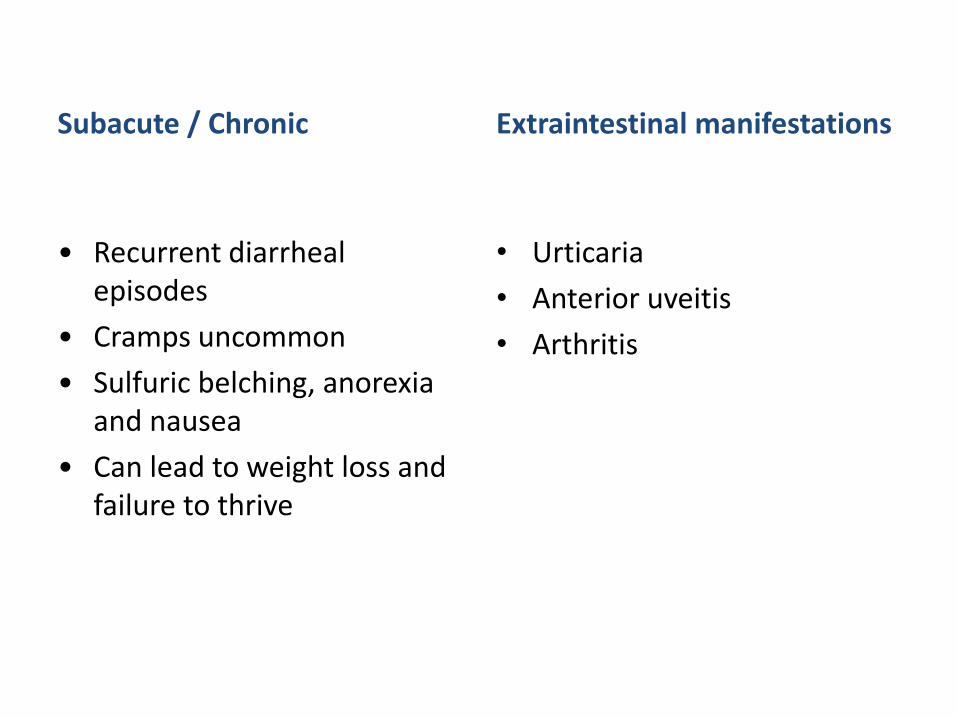
Subacute / Chronic
• Recurrent diarrheal episodes
• Cramps uncommon
• Sulfuric belching, anorexia and nausea
• Can lead to weight loss and failure to thrive
Extraintestinal manifestations
• Urticaria
• Anterior uveitis
• Arthritis
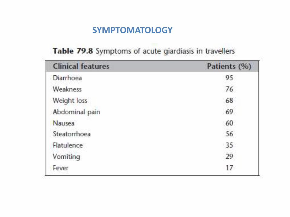
SYMPTOMATOLOGY
Complications
• Dehydration
• Weight loss
• Malnutrition
• Malabsorption
• Arthritis
• “Salt and pepper" retinal changes
Diagnosis
• The traditional diagnosis for giardiasis consists of performing an ova and parasite (O+P) exam of one to three stool specimens on non-consecutive days (sensitivity: 85-90%)
• Several days of specimen collection are needed to improve sensitivity
• Stool microscopy is relatively inexpensive, but it does require a skilled technician and may be a time consuming process
• These alternative diagnostic methods should only be used when stool examination is repeatedly negative and there is a high clinical suspicion of infection
• Duodenal aspirate biopsy and collection of duodenal fluid with the string test
Enterotest
• This test requires the patient to swallow a gelatin capsule containing a string. The proximal end of the string is taped to the patient's cheek and the distal end in the capsule moves to the duodenum after the capsule dissolves in the stomach. Several (4-6) hours later, the string is removed and microscopically examined for trophozoites.
Treatment
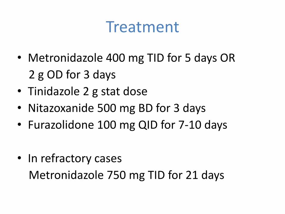
• Metronidazole 400 mg TID for 5 days OR
2 g OD for 3 days
• Tinidazole 2 g stat dose
• Nitazoxanide 500 mg BD for 3 days
• Furazolidone 100 mg QID for 7-10 days
• In refractory cases
Metronidazole 750 mg TID for 21 days
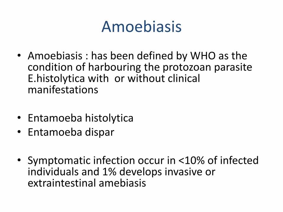
Amoebiasis
• Amoebiasis : has been defined by WHO as the condition of harbouring the protozoan parasite E.histolytica with or without clinical manifestations
• Entamoeba histolytica• Entamoeba dispar
• Symptomatic infection occur in <10% of infected individuals and 1% develops invasive or extraintestinal amebiasis
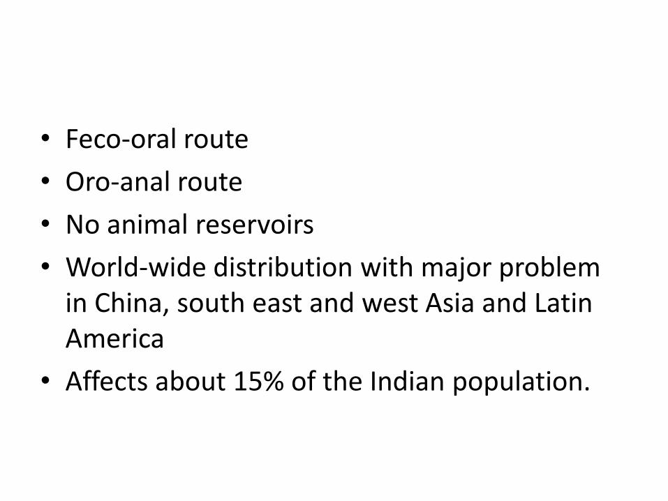
• Feco-oral route
• Oro-anal route
• No animal reservoirs
• World-wide distribution with major problem in China, south east and west Asia and Latin America
• Affects about 15% of the Indian population.
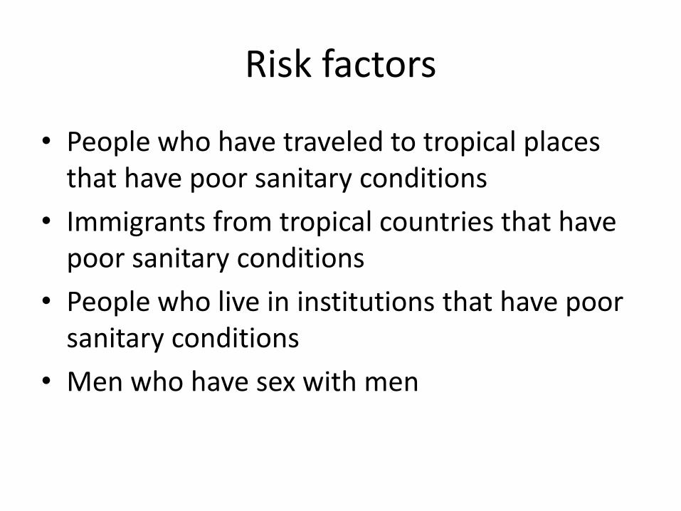
Risk factors
• People who have traveled to tropical places that have poor sanitary conditions
• Immigrants from tropical countries that have poor sanitary conditions
• People who live in institutions that have poor sanitary conditions
• Men who have sex with men
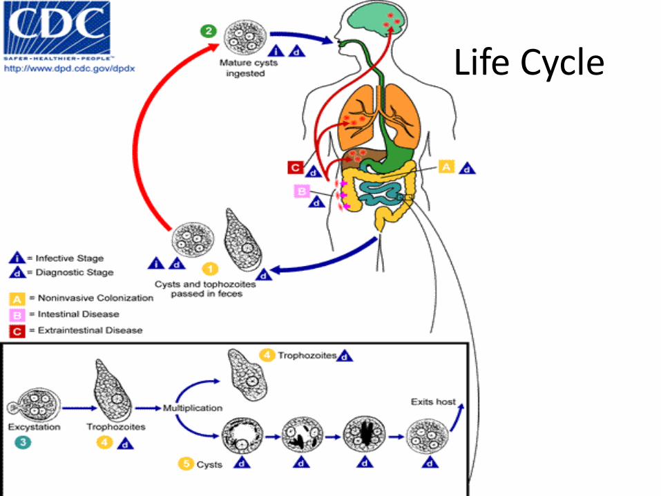
Life Cycle
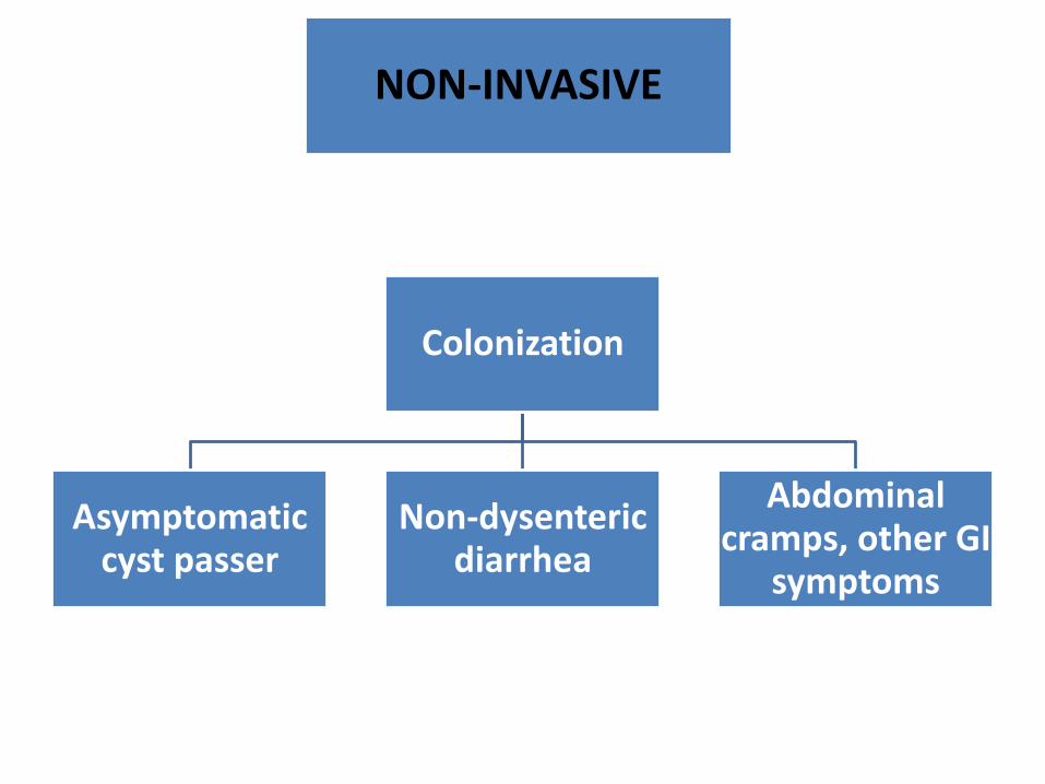
Colonization
Asymptomatic cyst passer
Non-dysenteric diarrhea
Abdominal cramps, other GI
symptoms
NON-INVASIVE
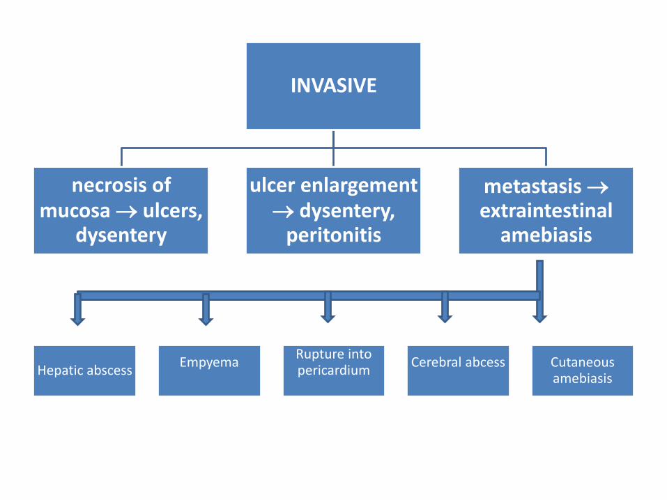
INVASIVE
necrosis of mucosa ulcers,
dysentery
ulcer enlargement dysentery,
peritonitis
metastasis extraintestinal
amebiasis
Hepatic abscessEmpyema
Rupture into pericardium
Cerebral abcess Cutaneous amebiasis
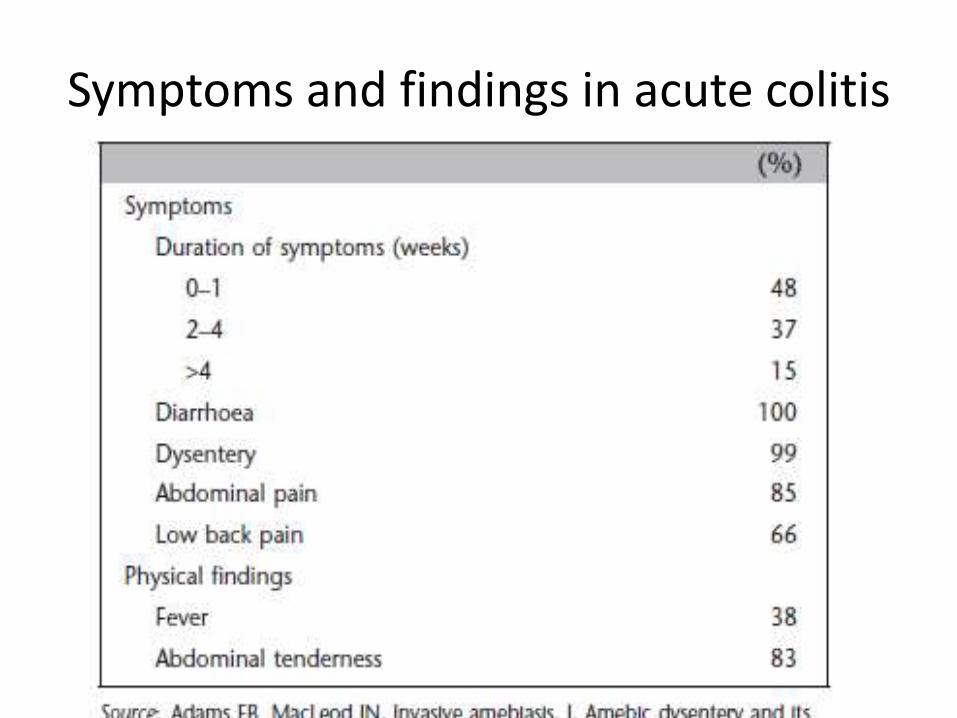
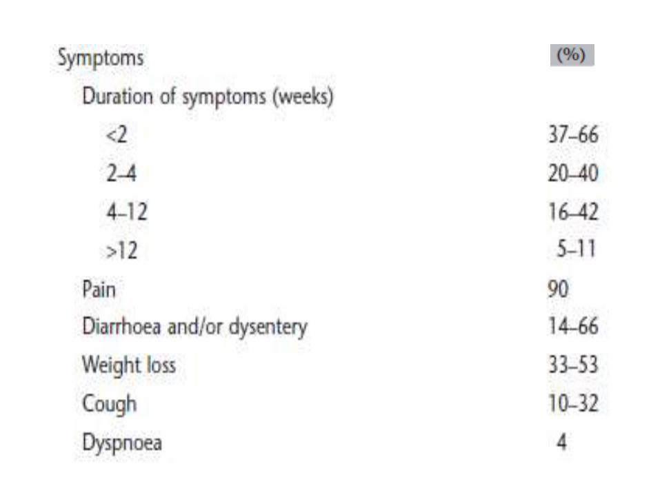
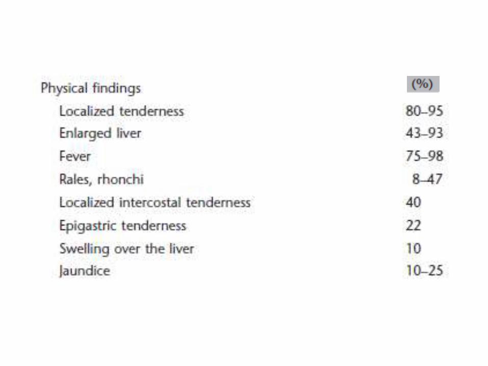
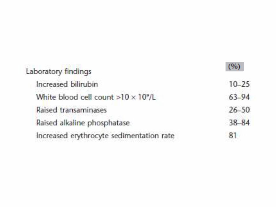
Symptoms and findings in acute colitis
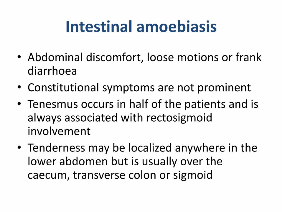
Intestinal amoebiasis
• Abdominal discomfort, loose motions or frank diarrhoea
• Constitutional symptoms are not prominent
• Tenesmus occurs in half of the patients and is always associated with rectosigmoidinvolvement
• Tenderness may be localized anywhere in the lower abdomen but is usually over the caecum, transverse colon or sigmoid
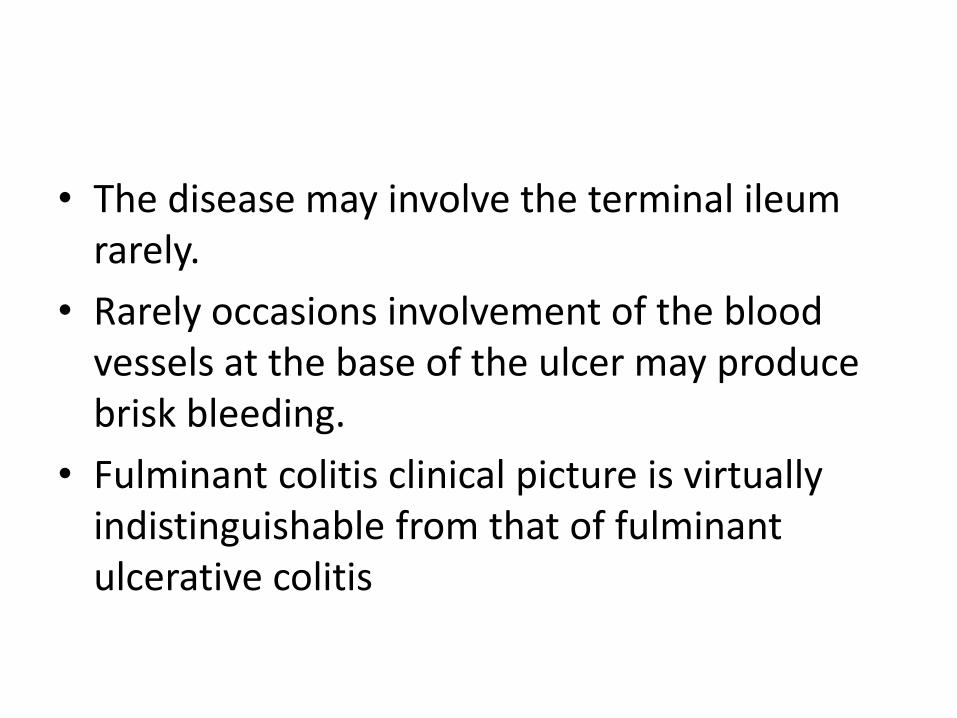
• The disease may involve the terminal ileum rarely.
• Rarely occasions involvement of the blood vessels at the base of the ulcer may produce brisk bleeding.
• Fulminant colitis clinical picture is virtually indistinguishable from that of fulminant ulcerative colitis
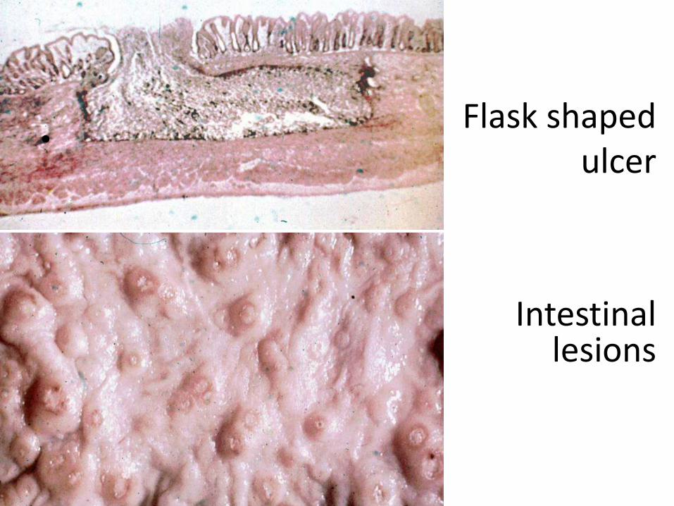
Flask shaped ulcer
•
Intestinal lesions
Amoeboma or amoebic granuloma
• Amoeboma is non-fibrotic and contains granulation tissue with lymphocytes, plasma cells, eosinophils and giant cells. There is remarkably little inflammation and most of the swelling is due to oedema.
• Repeated invasion of the colon by E. histolytica, complicated by pyogenic infection.
• Lesions are usually single and involve a short segment of the colon.
• Occurs commonly in caecum (40%) and rectosigmoidjunction (20%).
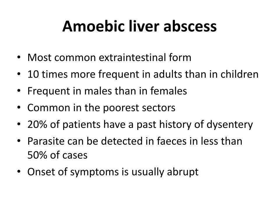
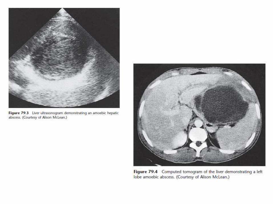
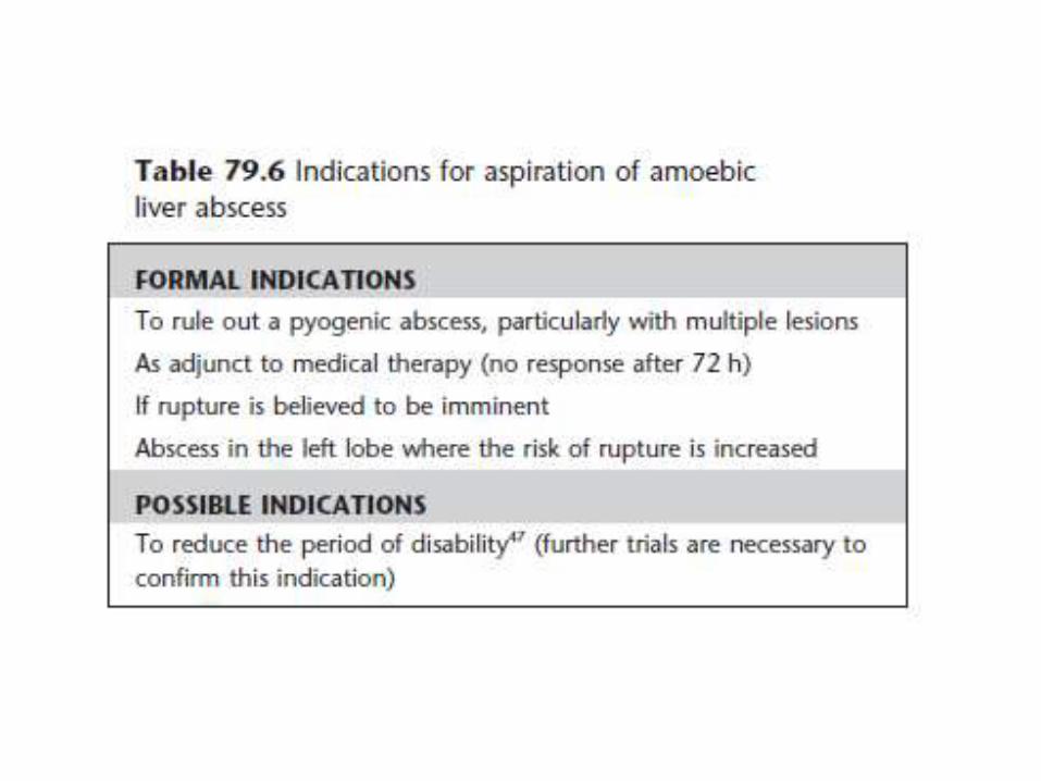
Amoebic liver abscess
• Most common extraintestinal form
• 10 times more frequent in adults than in children
• Frequent in males than in females
• Common in the poorest sectors
• 20% of patients have a past history of dysentery
• Parasite can be detected in faeces in less than 50% of cases
• Onset of symptoms is usually abrupt
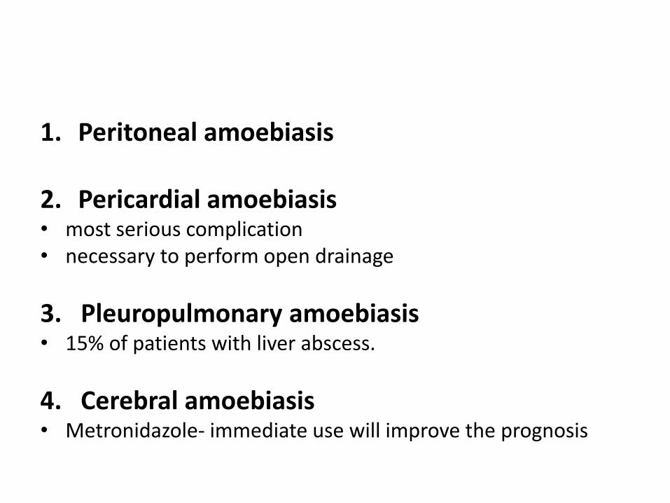
1. Peritoneal amoebiasis
2. Pericardial amoebiasis• most serious complication• necessary to perform open drainage
3. Pleuropulmonary amoebiasis• 15% of patients with liver abscess.
4. Cerebral amoebiasis• Metronidazole- immediate use will improve the prognosis
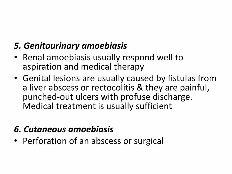
5. Genitourinary amoebiasis• Renal amoebiasis usually respond well to
aspiration and medical therapy• Genital lesions are usually caused by fistulas from
a liver abscess or rectocolitis & they are painful, punched-out ulcers with profuse discharge. Medical treatment is usually sufficient
6. Cutaneous amoebiasis• Perforation of an abscess or surgical
Diagnosis
• Stool or rectal smears for cyst and trophozoite
(within 30 min)
• Rectosigmoidoscopy and colonoscopy of mild or moderate cases usually reveals the presence of small ulcers (3–5 mm in diameter)
• Serology can be useful in the diagnosis of amoebiasis, particularly in non-endemic areas. Antibody response is present in 85–95% of patients with invasive disease.
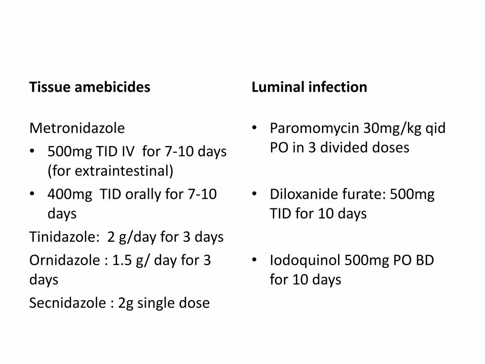
Tissue amebicides
Metronidazole
• 500mg TID IV for 7-10 days (for extraintestinal)
• 400mg TID orally for 7-10 days
Tinidazole: 2 g/day for 3 days
Ornidazole : 1.5 g/ day for 3 days
Secnidazole : 2g single dose
Luminal infection
• Paromomycin 30mg/kg qidPO in 3 divided doses
• Diloxanide furate: 500mg TID for 10 days
• Iodoquinol 500mg PO BD for 10 days
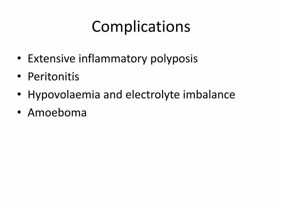
Complications
• Extensive inflammatory polyposis
• Peritonitis
• Hypovolaemia and electrolyte imbalance
• Amoeboma
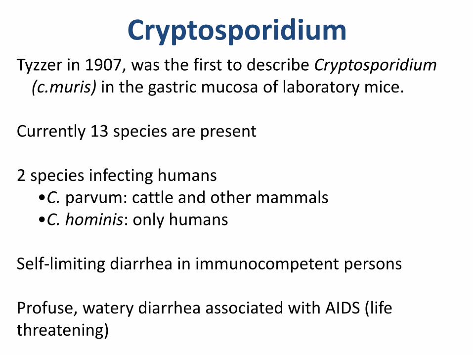
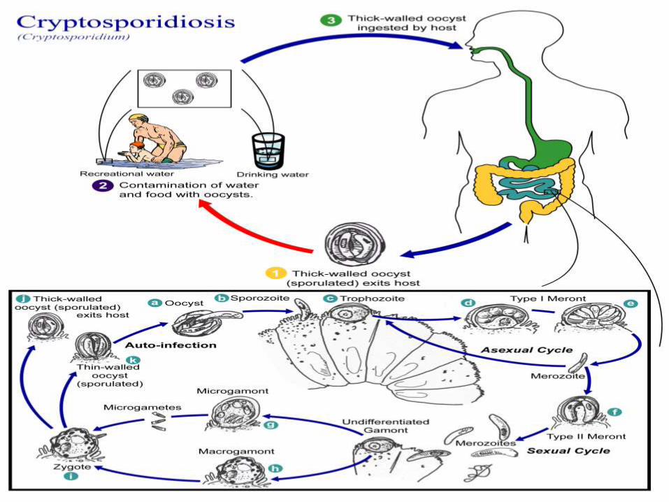
CryptosporidiumTyzzer in 1907, was the first to describe Cryptosporidium
(c.muris) in the gastric mucosa of laboratory mice.
Currently 13 species are present
2 species infecting humans •C. parvum: cattle and other mammals•C. hominis: only humans
Self-limiting diarrhea in immunocompetent persons
Profuse, watery diarrhea associated with AIDS (life threatening)
The Milwaukee OutbreakNEJM 331:161 (1994)
• Massive cryptosporidiosis outbreak following spring thaw• >400,000 people may have been affected• based on clinical symptoms (acute watery diarrhea)• ~100-fold higher prevalence of Cryptosporidium oocysts
in stools than normal
• Treated water had high levels of turbidity
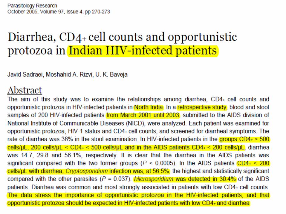
• Substantial outbreaks of water-borne diarrhoea in the immunocompetent, and for diarrhoea in travellers and in children.
• Cryptosporidiosis is an important contributor to childhood diarrhoea, with a prevalence among children with diarrhoea of 1–3% in the industrialized world and 4–17% in developing countries.
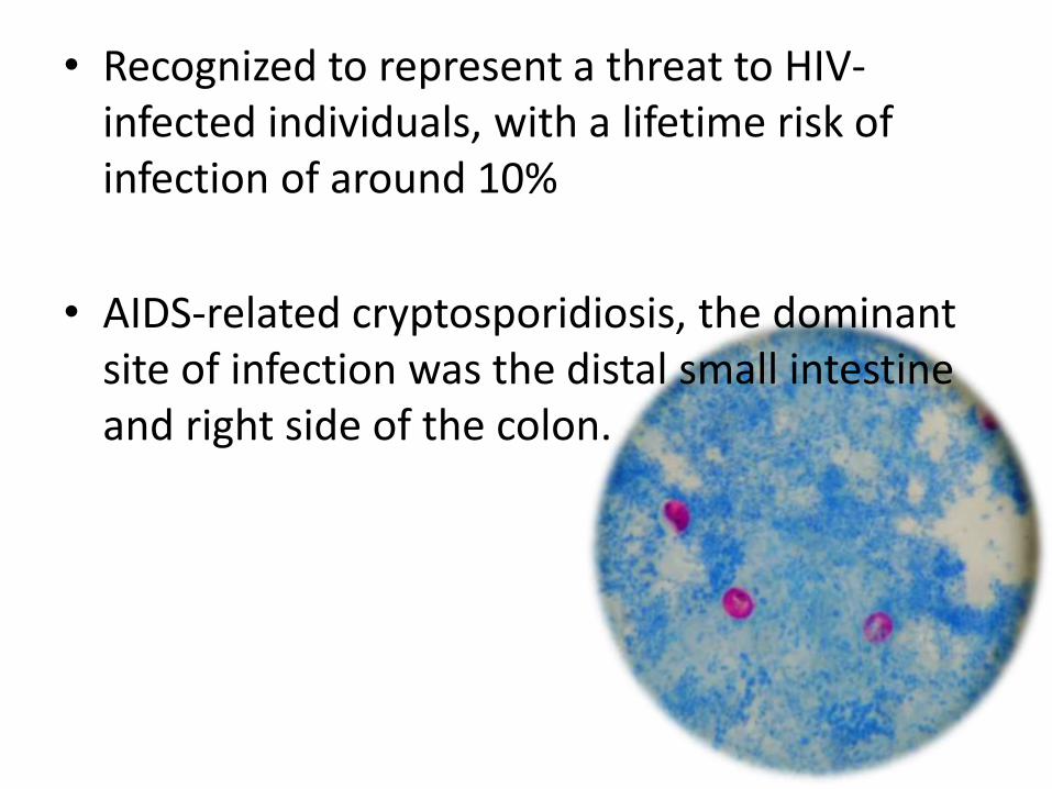
• Recognized to represent a threat to HIV-infected individuals, with a lifetime risk of infection of around 10%
• AIDS-related cryptosporidiosis, the dominant site of infection was the distal small intestine and right side of the colon.
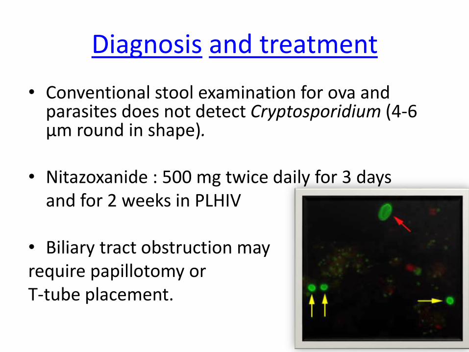
Diagnosis and treatment
• Conventional stool examination for ova and parasites does not detect Cryptosporidium (4-6 µm round in shape).
• Nitazoxanide : 500 mg twice daily for 3 daysand for 2 weeks in PLHIV
• Biliary tract obstruction mayrequire papillotomy or T-tube placement.
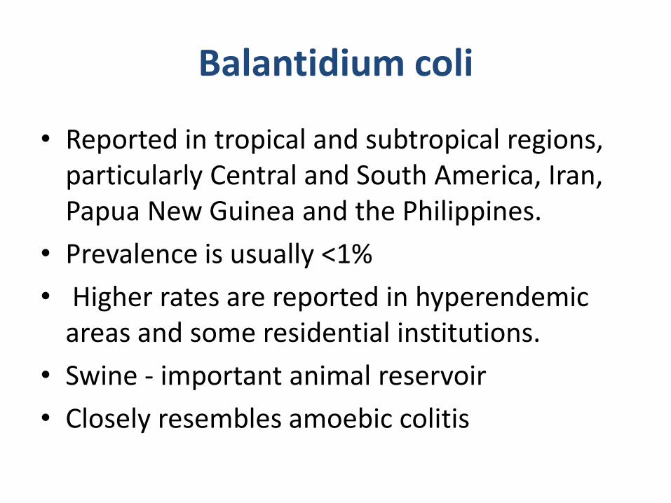
Balantidium coli
• Reported in tropical and subtropical regions, particularly Central and South America, Iran, Papua New Guinea and the Philippines.
• Prevalence is usually <1%
• Higher rates are reported in hyperendemicareas and some residential institutions.
• Swine - important animal reservoir
• Closely resembles amoebic colitis
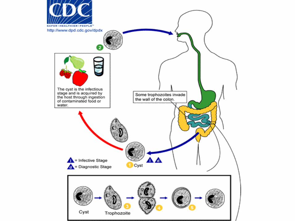
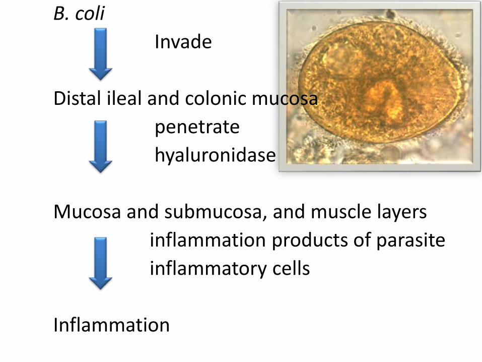
B. coli
Invade
Distal ileal and colonic mucosa
penetrate
hyaluronidase
Mucosa and submucosa, and muscle layers
inflammation products of parasite
inflammatory cells
Inflammation
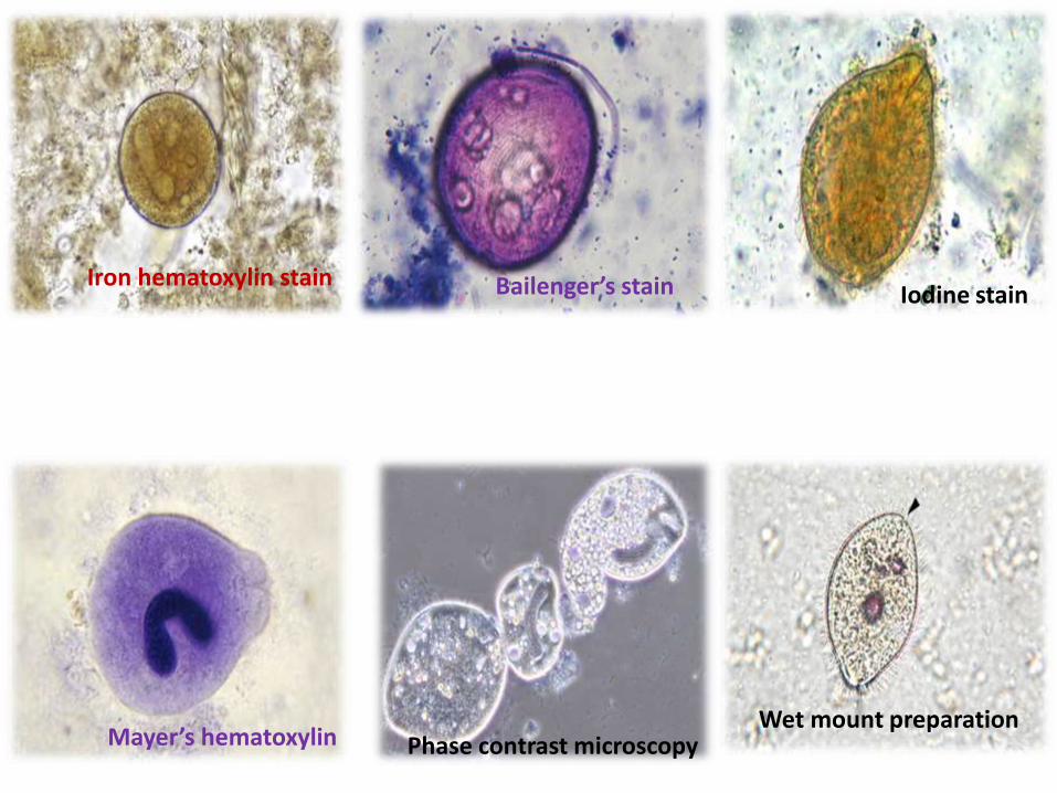
Iron hematoxylin stain Bailenger’s stain
Mayer’s hematoxylin Phase contrast microscopy
Iodine stain
Wet mount preparation
Diagnosis
• Large motile trophozoites can be detected using hand lens
• Serology : specific antibody can be detected
Management
• Tetracycline 500 mg four times daily for 10 days
Isospora belli
• Faecal-oral spread
• Associated with mild to a subtotal villous atrophy
• Inflammatory cells and eosinophils are seen in the lamina propria.
• Watery diarrhoea, cramping abdominal pain and nausea.
• Associated with wasting and dehydration
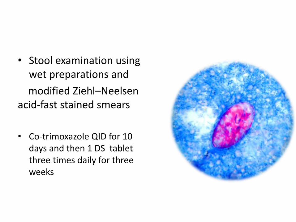
• Stool examination using wet preparations and
modified Ziehl–Neelsenacid-fast stained smears
• Co-trimoxazole QID for 10 days and then 1 DS tabletthree times daily for three weeks
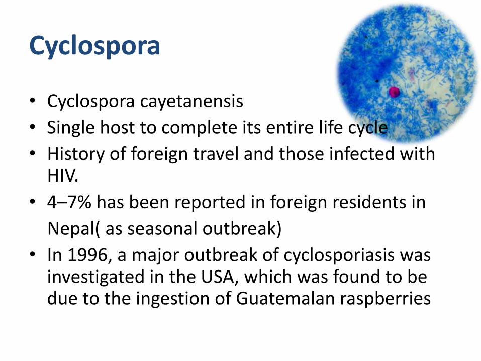
Cyclospora
• Cyclospora cayetanensis
• Single host to complete its entire life cycle
• History of foreign travel and those infected with HIV.
• 4–7% has been reported in foreign residents in
Nepal( as seasonal outbreak)
• In 1996, a major outbreak of cyclosporiasis was investigated in the USA, which was found to be due to the ingestion of Guatemalan raspberries
• Responsible for persistent diarrhoea in both immunocompetent and immunocompromisedindividuals.
• Abdominal gas , bloating and weight loss are also commonly associated features.
• Guillain–Barré syndrome.
• Cyst concentration techniques
• TMP-SMX 160–800 mg twice dailyfor 7 days
Microspora
• Found since the outbreak of the HIV
• Enterocytozoon bieneusi
• No mitochondria
• Sclerosing cholangitis like syndrome indistinguishable from cryptococcus induced
• Diagnosis : gold standard is the electron microscopy of intestinal epithelial cells
• Detection of spores using chromotope in stool
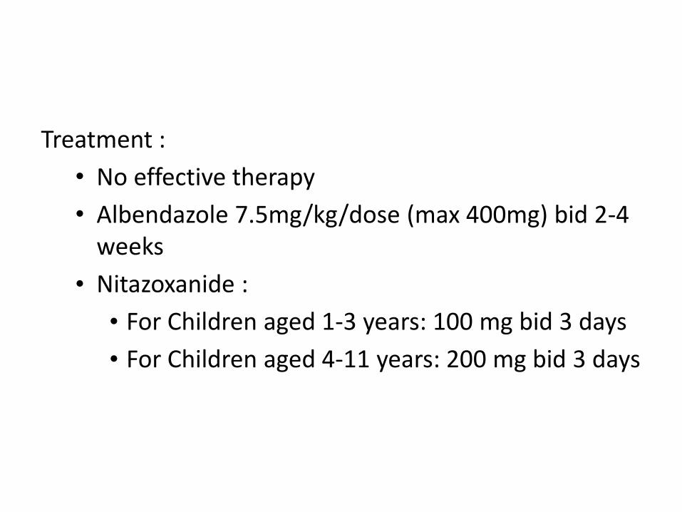
Treatment :
• No effective therapy
• Albendazole 7.5mg/kg/dose (max 400mg) bid 2-4 weeks
• Nitazoxanide :
• For Children aged 1-3 years: 100 mg bid 3 days
• For Children aged 4-11 years: 200 mg bid 3 days
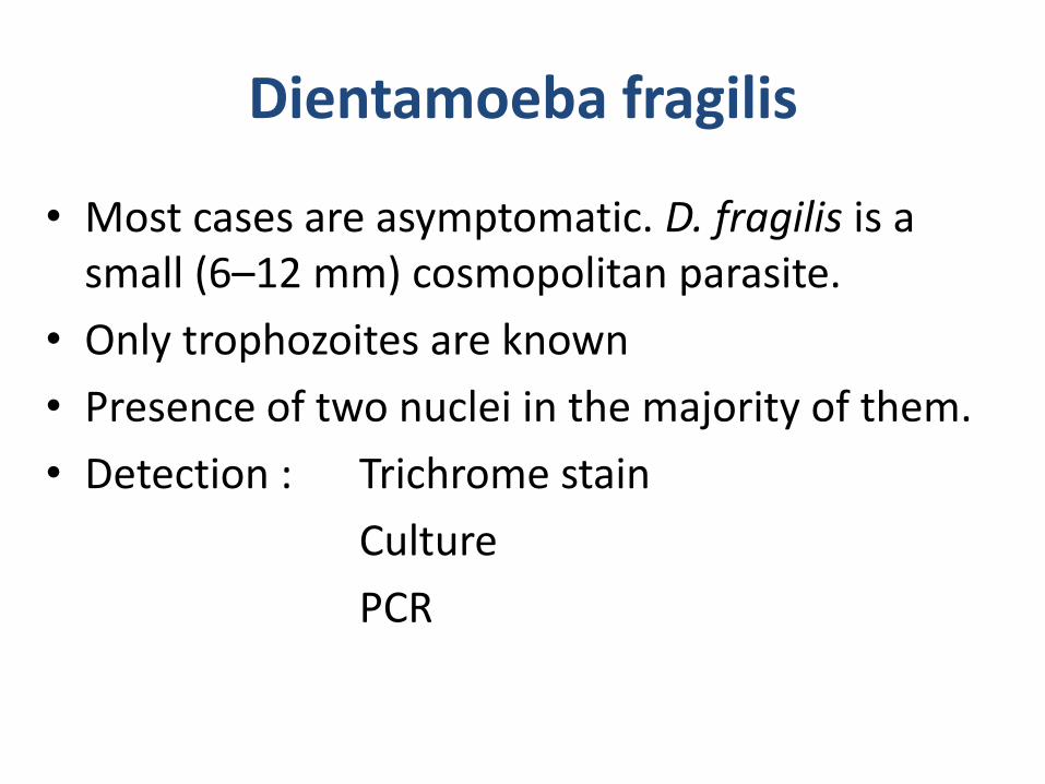
Dientamoeba fragilis
• Most cases are asymptomatic. D. fragilis is a small (6–12 mm) cosmopolitan parasite.
• Only trophozoites are known
• Presence of two nuclei in the majority of them.
• Detection : Trichrome stain
Culture
PCR
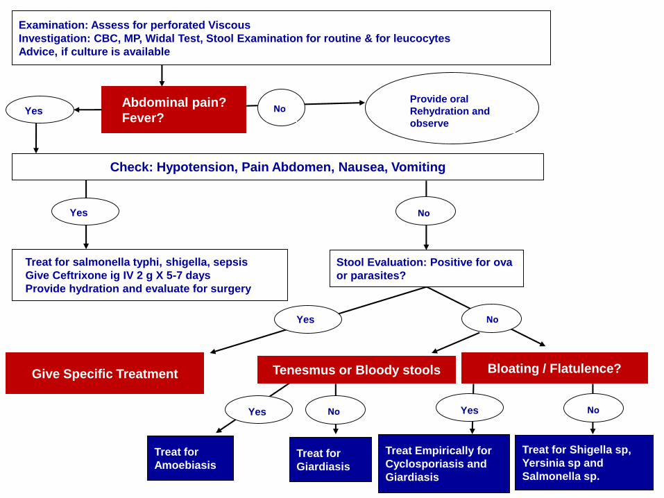
Examination: Assess for perforated Viscous
Investigation: CBC, MP, Widal Test, Stool Examination for routine & for leucocytes
Advice, if culture is available
Abdominal pain?
Fever?
Check: Hypotension, Pain Abdomen, Nausea, Vomiting
Treat for salmonella typhi, shigella, sepsis
Give Ceftrixone ig IV 2 g X 5-7 days
Provide hydration and evaluate for surgery
Provide oral
Rehydration and
observe
Stool Evaluation: Positive for ova
or parasites?
Tenesmus or Bloody stools Bloating / Flatulence?
Treat for
Giardiasis
Treat for
Amoebiasis
Treat Empirically for
Cyclosporiasis and
Giardiasis
Treat for Shigella sp,
Yersinia sp and
Salmonella sp.
NoYes
NoYes
Yes NoYes
Yes No
No
Give Specific Treatment
Problems with Diagnosis and Management
• Failure to detect pathogens
• Relationship between enteric pathogens and chronic diarrhoea is uncertain
• Pathogens may not have effective / convenient treatment
• Laboratory facilities and expertise are needed
• Quality of life and functional status have received little attention
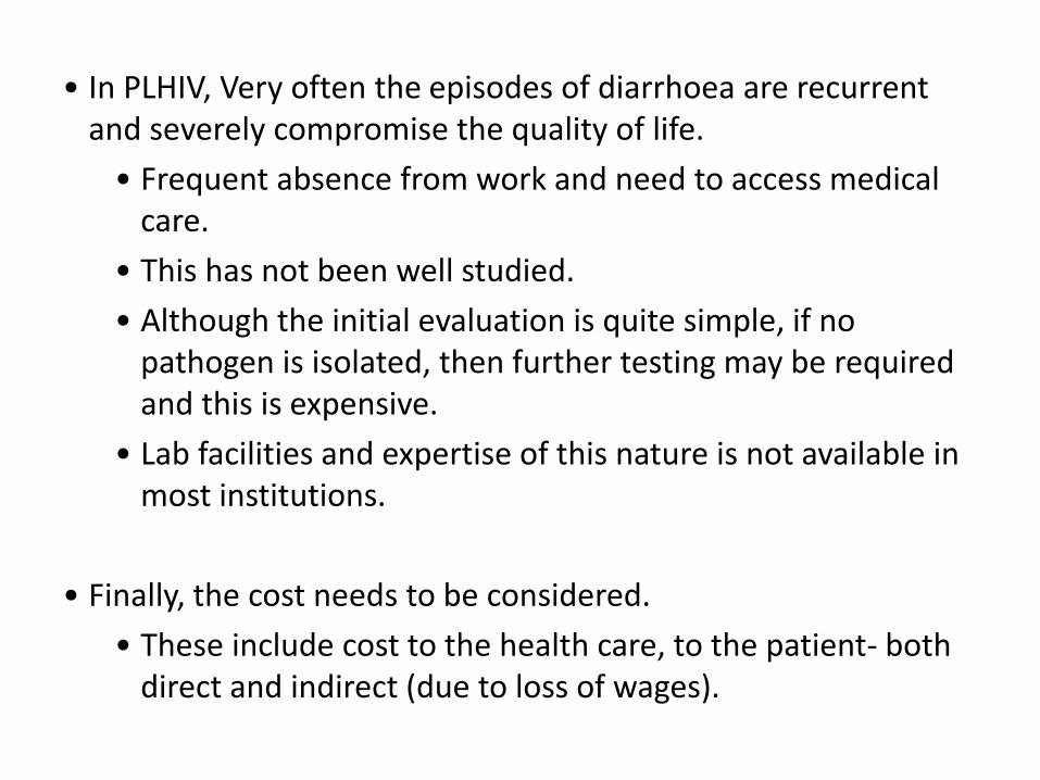
• In PLHIV, Very often the episodes of diarrhoea are recurrent and severely compromise the quality of life.
• Frequent absence from work and need to access medical care.
• This has not been well studied.
• Although the initial evaluation is quite simple, if no pathogen is isolated, then further testing may be required and this is expensive.
• Lab facilities and expertise of this nature is not available in most institutions.
• Finally, the cost needs to be considered.
• These include cost to the health care, to the patient- both direct and indirect (due to loss of wages).
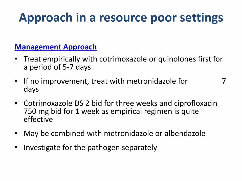
Approach in a resource poor settings
Management Approach
• Treat empirically with cotrimoxazole or quinolones first for a period of 5-7 days
• If no improvement, treat with metronidazole for 7 days
• Cotrimoxazole DS 2 bid for three weeks and ciprofloxacin 750 mg bid for 1 week as empirical regimen is quite effective
• May be combined with metronidazole or albendazole
• Investigate for the pathogen separately
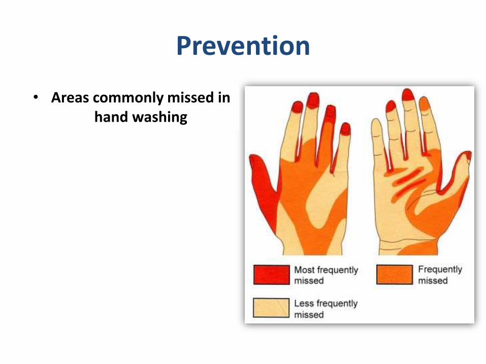
Prevention
• Areas commonly missed in hand washing
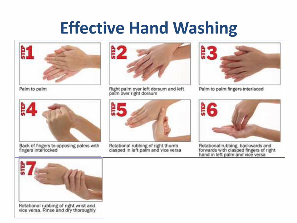
Effective Hand Washing
Prevention
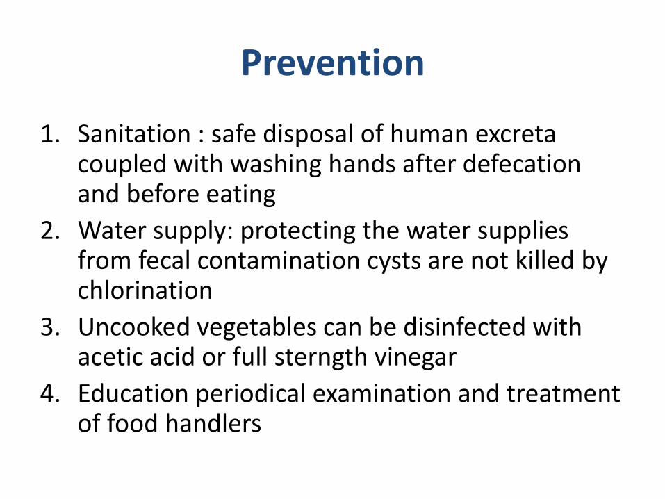
1. Sanitation : safe disposal of human excreta coupled with washing hands after defecation and before eating
2. Water supply: protecting the water supplies from fecal contamination cysts are not killed by chlorination
3. Uncooked vegetables can be disinfected with acetic acid or full sterngth vinegar
4. Education periodical examination and treatment of food handlers
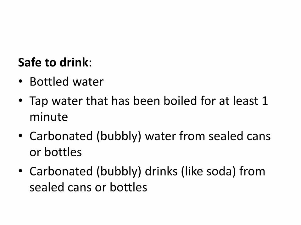
Safe to drink:
• Bottled water
• Tap water that has been boiled for at least 1 minute
• Carbonated (bubbly) water from sealed cans or bottles
• Carbonated (bubbly) drinks (like soda) from sealed cans or bottles
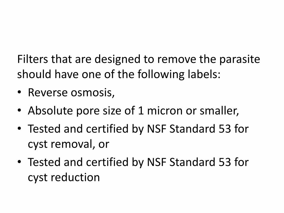
Filters that are designed to remove the parasite should have one of the following labels:
• Reverse osmosis,
• Absolute pore size of 1 micron or smaller,
• Tested and certified by NSF Standard 53 for cyst removal, or
• Tested and certified by NSF Standard 53 for cyst reduction
References • API medicine update 2013
• Harrison's Principles of Internal Medicine – 18th edition
• Davidson's Principles and Practice of Medicine 21st edition
• Manson's Tropical Diseases 22nd ed. - G. Cook, et. al., (Saunders, 2009)
• Parasitology in relation to clinical medicine by K D Chatterjee
• Park’ textbook of preventive and social medicine, 21st edition
• Parasitology Research journal, October 2005, Volume 97, Issue 4, pp 270-273
References:
• Gastrointestinal Manifestations in PLHIV, NACO guidelines-September 2013
• www.dpd.cdc.gov
• Enteric pathogens in southern Indian HIV-infected patients with & without diarrhoea, Mukhopadhya A. Indian Journal of Medical Research 1999; 109: 85-90.
• Attili SV, Gulati AK, Singh VP, Varma DV, Rai M, Sundar S. Diarrhea, CD4 counts and enteric infections in a hospital -based cohort of HIV-infected patients around Varanasi, India. BMC Infect Dis. 2006 Mar 1;6:39.


![Review of natural products with hepatoprotective effects...antipyretic, cardioprotective, antibacterial, antiviral, anti-protozoal, and anticarcinogenic capacities[15]. In addition](https://img.pdfslide.net/doc/110x75/5fff160632e6df0ff74d87d4/review-of-natural-products-with-hepatoprotective-effects-antipyretic-cardioprotective.jpg)