Embed Size (px)
Citation preview

Pleura
Air
Understand Basic Bedside Lung Ultrasound
Suthaporn Lumlertgul MD
Resus Ultrasound ©
Emergency Unit King Chulalongkorn Memorial Hospital
Bangkok, Thailand

Pleura
AirAnd monitor too much fluid loading bedside
Resus Ultrasound ©
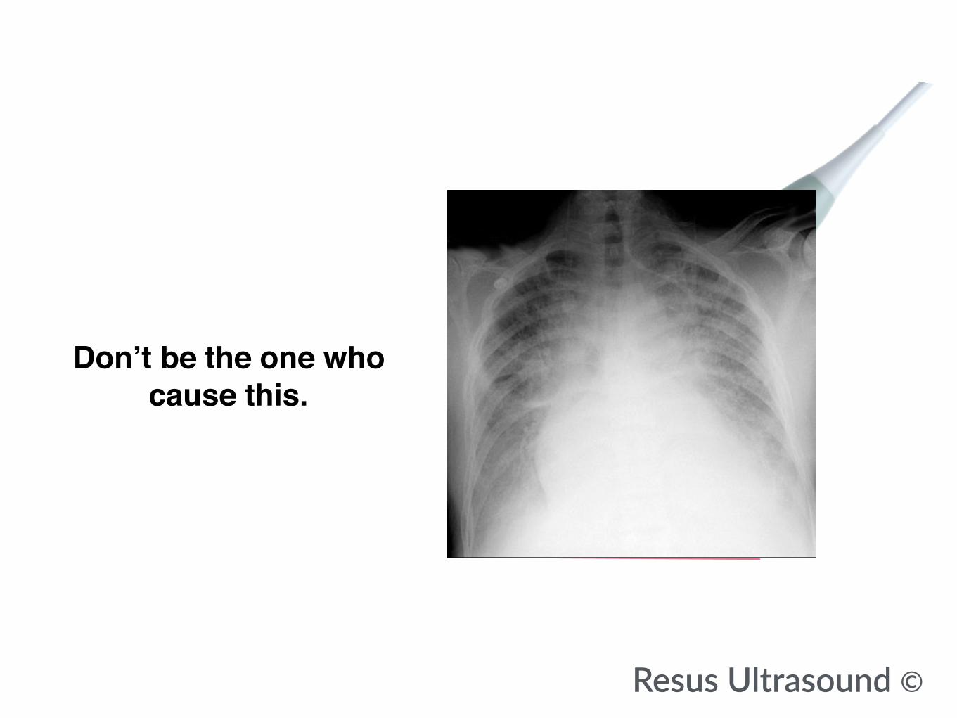
Pleura
AirDon’t be the one who cause this.
Resus Ultrasound ©

Pleura
AirNormally ultrasounds cannot
get through air in Lung
Therefore ultrasound bounce back almost 100%
Resus Ultrasound ©

Multiple reflections of pleural would appear
Pleura
Resus Ultrasound ©
Air

These reflections of pleural call A line
Pleura
Resus Ultrasound ©
Air

Horizontal white line below pleural with
repetitive same distance
Resus Ultrasound ©
rib rib
Pleural
A line
A line

Put ultrasound probe on both sides(echo or abdomen for beginners)
on midclavicular line.
On the left side avoid heart.
Resus Ultrasound ©

Resus Ultrasound ©
Four areas each side

In patient with dyspnea from diseases with no extra water.
U would see A line in bedside ultrasound
Resus Ultrasound ©
Pulmonary Embolism
COPD

In patient with dyspnea from diseases with no extra water.
U would see A line in bedside ultrasound
and in normal people as well.
Resus Ultrasound ©
Pulmonary Embolism
COPD

This helps differential dry dyspnea(black lung) from wet dyspnea(white lung)
when you have no time for X-ray
Resus Ultrasound ©
PECOPD
CHF,ARDSPneumonia
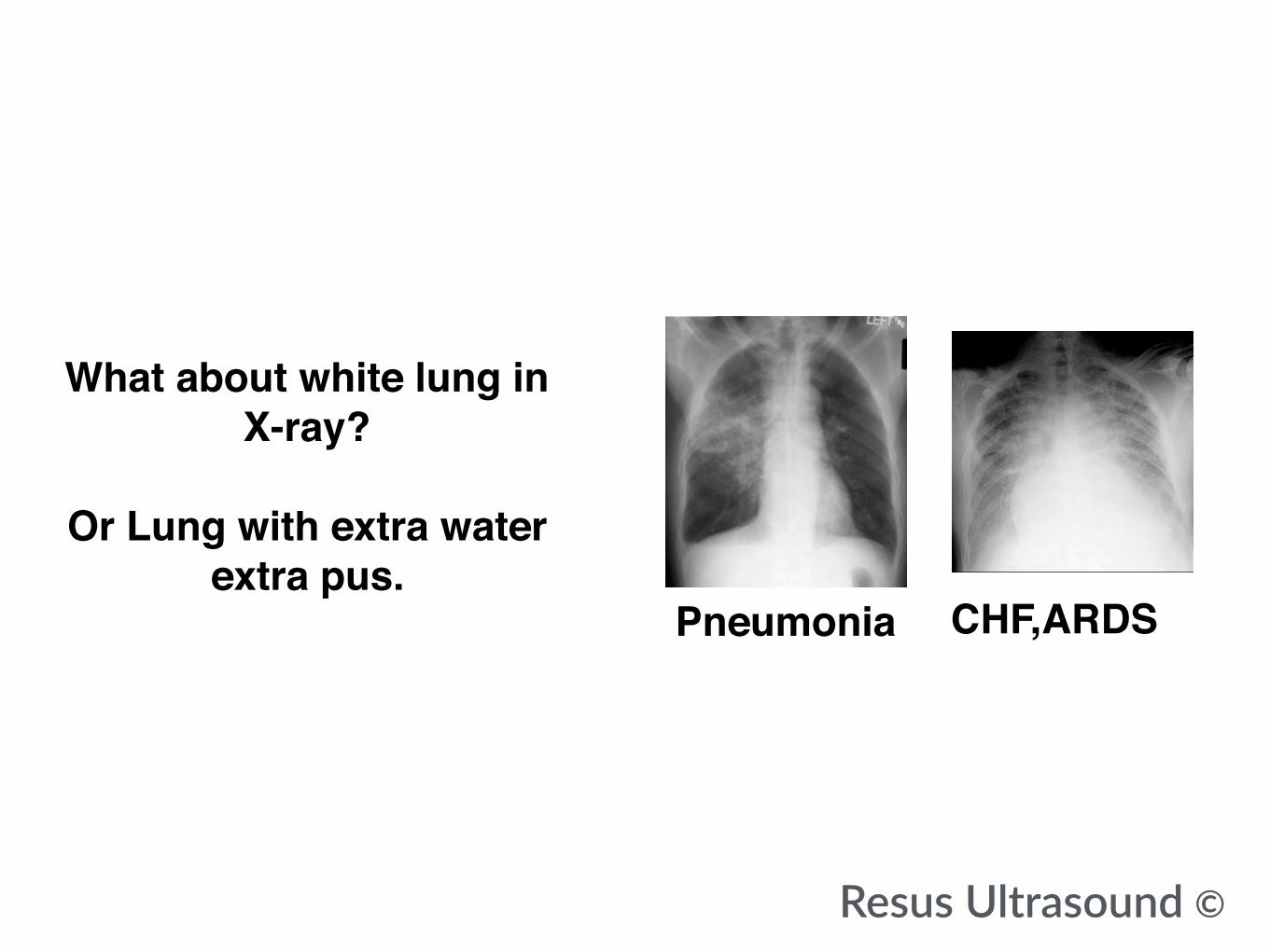
What about white lung in X-ray?
Or Lung with extra water
extra pus.
Resus Ultrasound ©
CHF,ARDSPneumonia

When there’s more water less air
Pleura
Resus Ultrasound ©
AirW
ater

The reflection of pleura will be gone
Pleura
Resus Ultrasound ©
AirW
ater

The sound would bounce between
wet alveoli insteadPleura
Resus Ultrasound ©
AirW
ater

The multiple bouncing of soundbetween wet alveoli call
B linePleura
Resus Ultrasound ©
AirW
ater

This is what B line look like.
Resus Ultrasound ©

This is what B line look like.
Resus Ultrasound ©

Long white color
start from pleural to the end of screen
and you would see no A line
Resus Ultrasound ©
B line

CHF,ARDS
So you would find B line in both lungs CHF, ARDS.
Resus Ultrasound ©

Lobar pneumonia
In pneumoniayou would find B line
in one side that have pneumonia
(extra pus instead of air)
And A line in the normal lung.
Resus Ultrasound ©

In Falls Protocolby Lichtenstein et al.
Appear of B line can help you monitor bedside
if you have load the patient too much water
Resus Ultrasound ©
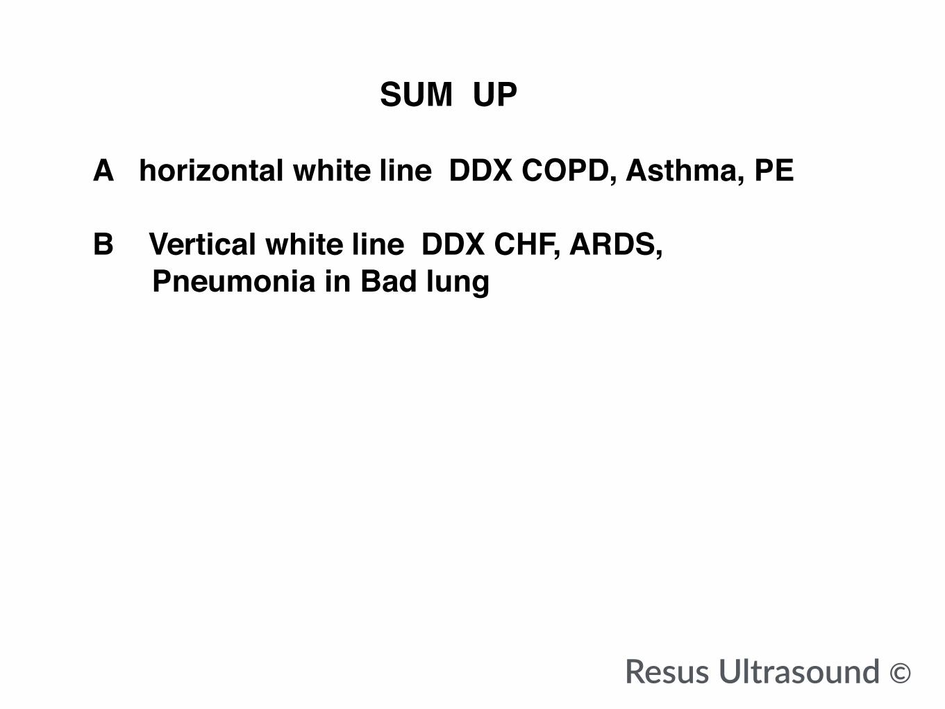
A horizontal white line DDX COPD, Asthma, PE
B Vertical white line DDX CHF, ARDS, Pneumonia in Bad lung
Resus Ultrasound ©
SUM UP

A horizontal white line DDX COPD, Asthma, PE
B Vertical white line DDX CHF, ARDS, Pneumonia in Bad lungNormally we search IVC, Heart, DVT + clinical + Lab to conclude diagnosed not pure Lung Ultrasound How to Ddx CHF VS ARDS, pneumothorax, atelectasis is not in this chapter
Resus Ultrasound ©
SUM UP

Don’t make decision only by ultrasound
Always add clinical and Labs for confirmation.
If you just start doing ultrasound in new organs.
Resus Ultrasound ©

PleuraAir
Resus Ultrasound ©
Effusion : Jelly fish sign, no Curtain
Consolidate : Shred ,C LineStatic Air Brochogram in 2D
Pneumothorax
Barcode in M modeNo lung sliding in 2D
Atelectasis
Lung Pulse in M mode Static Air Brochogram in 2D
A vs B
Common probe using Lung Ultrasound
(Abdominal probe can be used instead of Echo probe) If you trained your eye, any probe can be used for any signs
this is just recommend for the beginning.

Resus Ultrasound ©
Not Here

https://www.facebook.com/resusultrasound/
Resus ultrasound (FREE Beta Launch!, only FAST section available )
Heart Coming Soon