Embed Size (px)
Citation preview
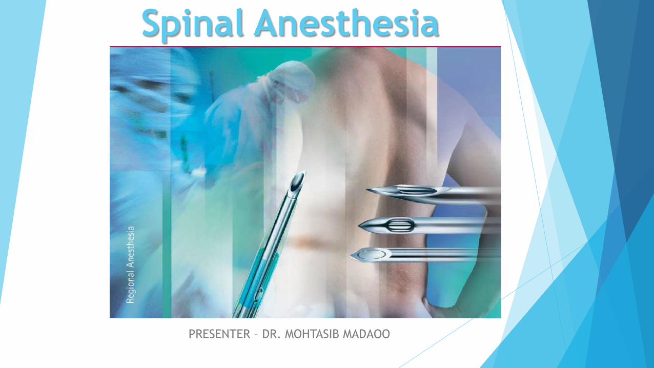
Spinal Anesthesia
PRESENTER – DR. MOHTASIB MADAOO

Introduction
Spinal anesthesia involves the use of small amounts of local
anesthetic injected into the subarachnoid space to produce a
reversible loss of sensation and motor function. The anesthesia
provider places the needle below L2 in the adult patient to avoid
trauma to the spinal cord. Spinal anesthesia provides excellent
operating conditions for:
Hernia (Inguinal or epigastric).
Haemorrhoidectomy , fistula , fissure.
Nephrectomy and cystectomy in combination with GA.
Transurethral resection of the prostate and transurethral resection of the
bladder tumors.
Abdominal and vaginal hysterectomies
Laparoscopic assisted vaginal hysterectomies(LAVH) combined with GA.
Caesarean sections.

Corning in 1885 , accidently administered cocaine
intrathecally.
Quincke in 1891 , made use of spinal puncture in
diagnosis.
August bier of Germany in 1898 , introduced the
technique of spinal anesthesia.
Pitkin popularized the method of introducing
agent's intrathecally.
History

Anatomy
A vertebra is composed of two parts
The body or base anteriorly, which bears
weight
The arch, which surrounds the cord
laterally and posteriorly consisting of
lamina and pedicles.
In addition there are seven processes or
projections
1. Three muscular processes – two transvers
and one spinous
2. Four articular processes, two upper and
two lower.
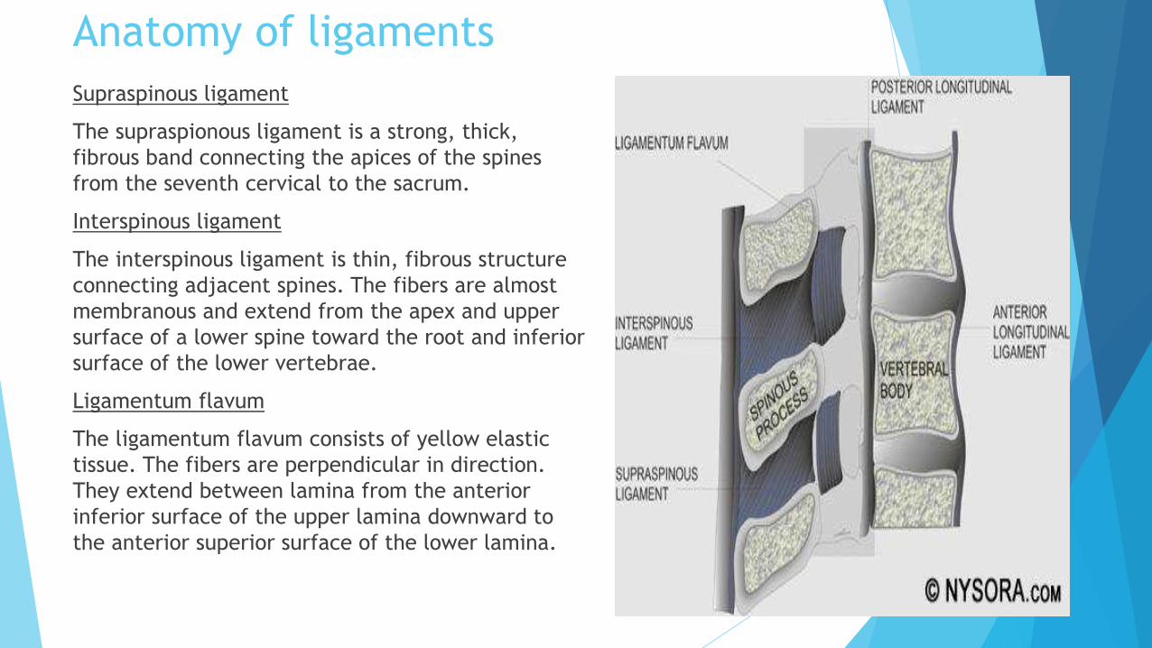
Anatomy of ligaments
Supraspinous ligament
The supraspionous ligament is a strong, thick,
fibrous band connecting the apices of the spines
from the seventh cervical to the sacrum.
Interspinous ligament
The interspinous ligament is thin, fibrous structure
connecting adjacent spines. The fibers are almost
membranous and extend from the apex and upper
surface of a lower spine toward the root and inferior
surface of the lower vertebrae.
Ligamentum flavum
The ligamentum flavum consists of yellow elastic
tissue. The fibers are perpendicular in direction.
They extend between lamina from the anterior
inferior surface of the upper lamina downward to
the anterior superior surface of the lower lamina.

Position of the Spinal Cord according to Age At 3 months of fetal life the tip of the cord is located at 2nd
coccygeal vertebrae.
At 6 months of fetal life the conus is at the level of S1.
At birth the tip of the spinal cord lies at the level of the
lower border of the L3 vertebra and the Dural sac at the S3
vertebra.
At one year of age the conus medullaris is at the lower border
of the L2 vertebra and the dural sac ends at the S2 vertebra.
By 12 to 16 years of age the adult relations are attained and
the spinal cord is located at the lower border of L1.
The average length of the spinal cord in the adult males is 45cm
and, in females, it is 42cm. The average weight approx. 30gm.
The spinal cord becomes relatively free of its dura but is
surrounded by the arachnoid and is engulfed in the CSF in the
subdural space. It also causes the downwards fanning of the
spinal nerves so that they run in an increasingly downward
oblique direction to form, at the lowest levels, the cauda
equina.
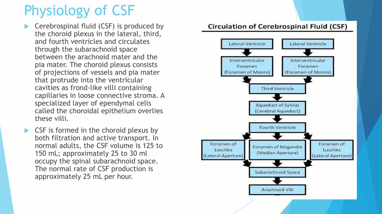
Physiology of CSF Cerebrospinal fluid (CSF) is produced by
the choroid plexus in the lateral, third, and fourth ventricles and circulates through the subarachnoid space between the arachnoid mater and the pia mater. The choroid plexus consists of projections of vessels and pia mater that protrude into the ventricular cavities as frond-like villi containing capillaries in loose connective stroma. A specialized layer of ependymal cells called the choroidal epithelium overlies these villi.
CSF is formed in the choroid plexus by both filtration and active transport. In normal adults, the CSF volume is 125 to 150 mL; approximately 25 to 30 ml occupy the spinal subarachnoid space. The normal rate of CSF production is approximately 25 mL per hour.

Dermatomes
A dermatome is an area of skin innervated by sensory fibers from a
single spinal nerve. To achieve surgical anesthesia for a given
procedure, the extent of spinal anesthesia must reach a certain
dermatomal level.
Dermatomal Levels of Spinal Anesthesia for Common Surgical
Procedures
Procedure Dermatomal Level
Upper abdominal surgery T4
Intestinal, gynecologic, and urologic surgery T6
Transurethral resection of the prostate T6
Vaginal delivery of a fetus, and hip surgery T10
Thigh surgery and lower leg amputations L1
Foot and ankle surgery L2
Perineal and anal surgery S2 to S5 (saddle block)

Positions for Spinal Tap Procedures

Approaches for Spinal Anesthesia Midline Approach. The most common approach, the needle
or introducer is placed midline, perpendicular to spinous
processes, aiming slightly cephalad.
Paramedian Approach. Indicated in patients who cannot
adequately flex because of pain or whose ligaments are
ossified, the spinal needle is placed 1.5 cm laterally and
slightly caudad to the center of the selected interspace. The
needle is aimed medially and slightly cephalad and passed
lateral to the supraspinous ligament. If the lamina is
contacted, the needle is redirected and "walked off" in a
medial and cephalad direction.
Taylor or Lumbosacral Approach. This approach is useful in
patients with calcified or fusion of higher intervertebral
spaces. The injection site is 1cm medial and 1cm caudad of
the posterior iliac spine. The needle is directed 45 degrees
medial and 45 degrees caudad, after contacting the lamina
the needle is walked upward and medially to enter the L5-S1
interspace

Technique
Midline Approach
The midline approach affords the practitioner two
advantages. Anatomic projection is only in 2 planes,
making visualization of the intended trajectory and
anatomical structures more apparent. The midline
provides a relatively avascular plane. It is important to
have the patient sitting up straight, not slumping to the
side, to minimize lumbar lordosis, and maximize the
space between the spinous processes. By proper
positioning you should have access to L2-L3, L3-L4, L4-L5,
and L5-S1. Identify the top of the iliac crest. Tuffier’s line
generally corresponds with the 4th lumbar vertebrae.
“Tuffier’s” line is a line drawn across the iliac crest
that crosses the body of L4 or L4-L5 interspace. This is
a helpful landmark for the placement of spinal or
epidural anesthetics.

Technique
Palpation in the midline should help to identify the interspinous
ligament. The extent of the space is noted by palpating the cephalad
and caudad spine. The midline is noted by moving your fingers from
medial to lateral.
Wash hands, put on sterile gloves, use sterile technique.
Prepare the tray in a sterile fashion. An assistant may help with
opening, in sterile fashion, specific items. Prepare the back with an
antiseptic. Start at the area of intended injection and move out. This
is done three times.
Place a skin wheal of local anesthetic at the intended spinous
interspace. Smaller gauge needles will require an introducer to
stabilize the needle. Place the introducer firmly into the interspinous
ligament.
Anatomical structures that will be transversed include skin,
subcutaneous fat, supraspinous ligament, interspinous ligament,
ligamentum flavum, epidural space, and dura.
Technique Hold the spinal needle like a dart/pencil. Cutting needles should be inserted
with the bevel parallel to the longitudinal fibers of the dura. This helps
reduce cutting fibers and enhances tactile sensation as anatomical structures
are crossed.
As the ligamentum flavum and dura are transversed, a change in resistance is
noted. Some will describe this as a “pop”; however, it may be a decrease in
pressure or a loss of resistance.
Once in the subarachnoid space, remove the stylet and CSF should appear. If
CSF does not appear, rotate the needle 90 degrees until it appears. If no CSF
appears then the stylet should be replaced. With smaller gauged needles it
may take 20-30 seconds for CSF to appear. Assess the needle position. Is it at
an appropriate depth? Is it midline or is its trajectory off the midline? Being
off the midline is one of the most common reasons that CSF does not come
back. If off the midline, remove the needle and start over.
If blood returns from the needle, wait to see if it clears. If it does not clear,
reassess needle position. If the needle is midline, not lateral, it may be in an
epidural vein. Advance the needle slightly further to transverse the dura. If
the needle is not midline, remove it and start over.

If bone is encountered, reassess the patient’s position and ensure the needle is midline. If bone is contacted early, the needle may be contacting the spinous process. Move the needle slightly caudad. If bone is contacted late, the needle may be contacting the lamina of the vertebrae. Move the needle slightly cephalad. Moving down an interspace may increase the chance of success since the intervertebral spaces will be larger.
After unsuccessful attempts, consider converting to a general anesthetic. The more attempts, the more trauma, increasing the risk of a spinal/epidural hematoma.
Once CSF returns, steady the needle with the dorsum of the non dominant hand against the patients back. Attach the syringe with the intended spinal anesthetic. Gently aspirate some CSF into the syringe. If a hyperbaric technique is being used, a “swirling” in the solution will be noted due to the dextrose content. Aspiration with an isobaric technique will yield additional CSF fluid into the syringe. The cerebral spinal fluid should be clear. If blood is returned with aspiration, replace the styletand start over.
Inject the local anesthetic at a rate of 0.2 ml per second. After injection aspirate 0.2 ml of CSF to confirm that the needle remains in the subarachnoid space. If the patient complains of pain during injection, stop immediately. Redirect the needle away from the side of pain and into the midline.
Place the patient in the appropriate position for the procedure and baricity of the spinal anesthetic solution.

TechniqueParamedian Approach
The advantage of the paramedian approach is a larger target. By placing the
needle laterally, the anatomical limitation of the spinous process is avoided. The
most common error when attempting this technique is being too far from the
midline, which makes encountering the vertebral lamina more likely.
Palpate the vertebral process and identify the caudad tip. Move 1 cm down
and 1 cm laterally.
Prepare the back with an antiseptic solution. Place a skin wheal of local
anesthetic at the identified area of needle insertion. A longer needle is often
required to infiltrate the tissue.
Insert the spinal needle 10-15 degrees off the sagittal plane. At this point the
most common error is inserting the needle too far cephalad, which results in
encountering the lamina of the vertebral body. If bone is contacted, redirect
the needle a little further caudad.
It may be possible to feel the characteristic change in resistance or loss of
resistance. With a lateral approach the needle is inserted further than with
the midline approach.
Once CSF is obtained, continue in the same manner as the midline approach.

Taylor’s Technique
This is a very useful method in cases of spine fusion, arthritic spine, skin
infection in the lumbar region, or in other conditions in which the usual
approach is difficult or impossible.
Largest interspace L5-S1.
A skin wheal is made 1cm medially and 1cm below the lowest prominence of
the posterior-superior spine. A 12-cm , needle is directed upward , medially
and forward at an angle of about 50degree , approximating forward at an
angle that the dorsal aspect of the sacrum makes with the skin. The needle
then is advanced so that it’s point enters the lumbosacral space between the
sacrum and the last lumbar vertebra. As the space is entered , there usually
an immediate flow of CSF , although gentle aspiration may be necessary.

PRINCIPLES IN ADMINISTRATING ANAESTHETIC
SOLUTIONS
Main aim of anaesthetists is to secure anaesthesia of
Sufficient duration
Sufficient Height
STOUT’S PRINCIPLES FOR SPREAD OF SOLUTIONS
Height of anaesthesia is
Directly proportional to concentration of the drug
Inversely proportional to rapidity of fixation
Directly to speed of injection
Directly proportional to the volume of fluid.
Inversely proportional to spinal fluid pressure.
Directly proportional to specific gravity for hyper baric solution.
With isobaric or hypobaric solutions, extent depends on position of patient.

FACTORS POSTULATED TO BE RELATED TO
SPINAL ANAESTHETIC BLOCK HEIGHT
PATIENT CHARACTERISTICS
Age, Height, Weight, Intra abdominal pressure, position, anatomic
configuration of spinal column.
TECHNIQUE OF INJECTION
Site of injection, direction of injection, rate of injection.
CHARACTERISTICS OF SPINAL FLUID
Volume, Pressure, density.
CHARACTERISTICS OF ANAESTHETIC SOLUTIONS
Density, Amount, Concentration, temperature, volume.
PATIENT FACTORSAGE
Spinal space become smaller with age - distribution greater.
OBESITY
Increase intra-abdominal pressure
increase pressure in epidural space.
Decrease subarachnoid space
PREGNANCY
Increase intra-abdominal pressure
Increase volume of epidural venous plexus - Small subarachnoid spaces.
INTRAABDOMINAL PRESSURE
Changes resulting from direct pressure of increased intra-abdominal pressure on epidural and subarachnoid spaces.
Collateral flow through epidural venous plexus expand- SA space small
SPINAL CURVATURE
Abnormal curvature have an effect on technical aspects
Changes the contour of Subarachnioid space
RATE OF INJECTION
Slow injections - low levels
Rapid injections - high level

DENSITY / SPECIFIC GRAVITY AND BARICITY
Density of any solution is the weight in grams of 1 ml of the solution at a
standard temperature. Density varies inversely with temperature.
Specific gravity is the density of a solutions compared in a ratio with the
density of water.
Baricity is a ratio comparing the density of one solution to another.
Density of normal human. CSF at 370C is 1.0001 to 1.0005
Specific gravity of spinal fluid 1.003 to 1.008
ISOBARIC SOLUTIONS
Densities between 0.9998 and 1.0008
Solutions are mixed with physiological saline
Solutions with out added glucose
Bupivacaine, ropivacaine, levobupivacaine
Spread not influenced by position

HYPOBARIC SOLUTIONS
Baricity less than 0.9998 at 370C
Prepared by diluting with distilled water
HYPERBARIC SOLUTIONS
Solutions at 37degree Celsius with baricity greater than 1.0008
Made by addition of 5-9.5% dextrose
They show bimodal spread – gravitate from the site of injection to two
different directions i.e., to the point below L3 into the lumbosacral concavity
or above L3 into the thoracic concavity to the T5 level.
Travel to the most dependent part of the subarachnoid space when there is
deviation of the patients position from the horizontal.

Barbotage
This is the technique of stirring up to increase turbulence , mixing of injected
solutions and increasing the distribution in the subarachnoid space.
The technique first was described by Bier and consists of the injection of the
anesthetic solution into the subarachnoid space, immediate withdrawal of a
portion of the solution and reinjection. This may be repeated. The to-and-fro
movement agitates the injectate in the spinal fluid, and the currents mix the
agent more completely and carry the agent more extensively and to higher
levels.
Caution must be observed and each operator must learn the results of his
barbotage

Types of Spinal Needles

Whitacre Pencil Point Spinal Needles
Help Reduce Post-Procedure Headaches
Designed to spread the dural fibers and help reduce the occurrence of post
dural puncture headache
Distinct "pop" as the pencil point penetrates the dura
Precision-formed side hole helps directional flow of anesthetic agents and
helps reduce the possibility of straddling the dura
Designed to track straight when advancing through ligaments toward the dura
Translucent window hub features contact clarity that helps allow visualization
of CSF
Needle gauges 22 to 27 G.
Needle lengths 3½ in. to 5 in.

Quincke Needles
Key/Slot arrangement of stylet and cannula hubs facilitates proper needle
bevel orientation
Translucent window hub features contact clarity that helps allow visualization
of CSF
Fitted stylet reduces tissue coring
Needle gauges 18 to 27 G.
Needle lengths 1 in. to 7 in.

Spinal Anesthesia – Physiologic
Effects

Somatic Blockade
Neuraxial anesthesia effectively stops the transmission of painful sensation
and abolishes the tone of skeletal muscle, enhancing operating conditions for
the surgeon.
Sensory blockade involves somatic and visceral painful stimulation. Motor
blockade involves skeletal muscles. Neuraxial anesthesia results in a
phenomenon known as differential blockade. This effect is due to the activity
of local anesthetics and anatomical factors.
Local anesthetic factors include the concentration and duration of contact
with the spinal nerve root. As the local anesthetic spreads out from the site of
injection the concentration becomes less, which may in turn effect which
nerve fibers are susceptible to blockade. Anatomical factors are related to
various fiber types found within each nerve root. Small myelinated fibers are
easier to block than large unmyelinated fibers.
In general, the differential blockade found after neuraxial blockade is as
follows: sympathetic blockade is 2-6 dermatome segments higher than
sensory and sensory blockade is generally 2 dermatome levels higher than
motor.
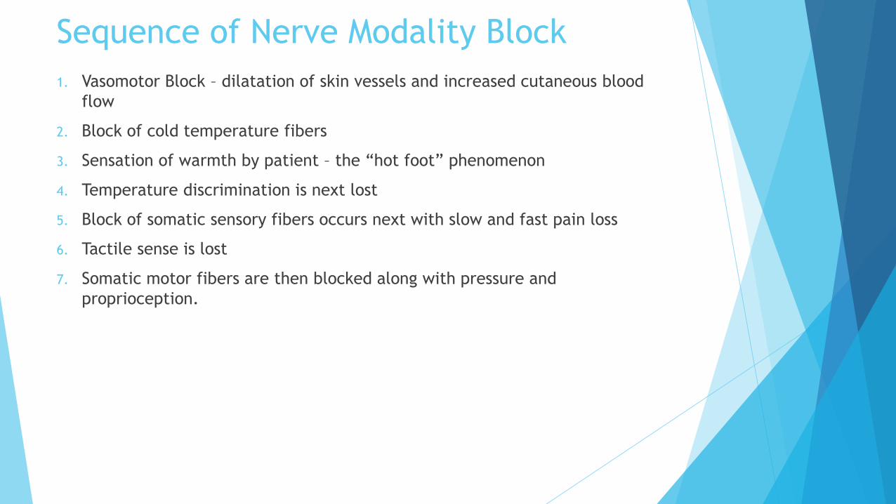
Sequence of Nerve Modality Block
1. Vasomotor Block – dilatation of skin vessels and increased cutaneous blood
flow
2. Block of cold temperature fibers
3. Sensation of warmth by patient – the “hot foot” phenomenon
4. Temperature discrimination is next lost
5. Block of somatic sensory fibers occurs next with slow and fast pain loss
6. Tactile sense is lost
7. Somatic motor fibers are then blocked along with pressure and
proprioception.
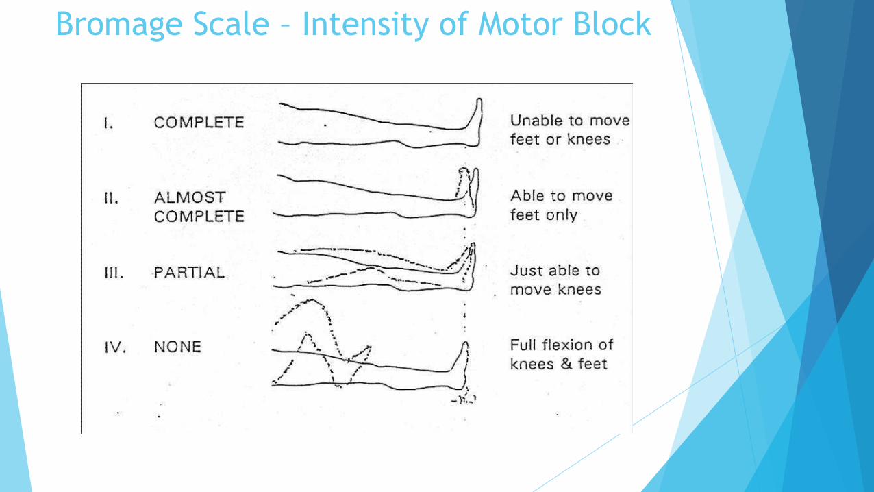
Bromage Scale – Intensity of Motor Block
Autonomic Blockade
Neuraxial blockade effectively blocks efferent autonomic transmission of the
spinal nerve roots, producing a sympathetic block and a partial
parasympathetic block.
Sympathetic fibers are small, myelinated, and easily blocked. During
neuraxial blockade, the anesthesia provider will observe a sympathetic block
prior to sensory, followed by motor.
The sympathetic nervous system (SNS) is described as thoracolumbar since
sympathetic fibers exit the spinal cord from T1 to L2.
The parasympathetic nervous system (PNS) has been described as craniosacral
since parasympathetic fibers exit in the cranial and sacral regions of the CNS.
The end result of neuraxial blockade is a decreased sympathetic tone with an
unopposed parasympathetic tone. This imbalance will result in many of the
expected alterations of normal homeostasis noted with the administration of
spinal anesthesia.

Cardiovascular Effects
Neuraxial blockade can impact the cardiovascular system by causing the
following changes:
Decrease in blood pressure (33% incidence of hypotension in non-obstetric
populations)
Decrease in heart rate (13% incidence of bradycardia in non-obstetric
populations)
Decrease in cardiac contractility

Respiratory Effects Neuraxial blockade plays a very minor role in altering pulmonary function.
Even with high thoracic levels of blockade, tidal volume is unchanged. There is a slight decrease in vital capacity. This is the result of relaxation of the abdominal muscles during exhalation.
The phrenic nerve is innervated by C3-C5 and is responsible for the diaphragm. The phrenic nerve is extremely hard to block, even with a high spinal. In fact, apnea associated with a high spinal is thought to be related to brainstem hypoperfusion and not blockade of the phrenic nerve. This is based on the fact that spontaneous respiration resumes after hemodynamic resuscitation has occurred.
The risk and benefits of neuraxial anesthesia should be carefully weighed for the patient with severe lung disease. Patients with chronic lung disease depend on intercostal and abdominal muscles to aid their inspiration and exhalation. Neuraxial blockade may reduce the function of these muscles, having a detrimental impact on the patient’s ability to breathe, as well as affect the ability to clear secretions and cough. For procedures above the umbilicus, a pure regional anesthetic may not be beneficial for the patient with chronic lung disease.
Thoracic and abdominal surgical procedures are associated with decreased phrenic nerve activity resulting in decreased diaphragmatic function and FRC (functional reserve capacity). This can lead to atelectasis and hypoxia due to ventilation/perfusion mismatching
Gastrointestinal Effects Since sympathetic outflow originates at T5-L1, neuraxial blockade results in a
sympathectomy with a predomination of parasympathetic nervous system
effects. The end result is a small, contracted gut with peristalsis.
Hepatic blood flow decreases in relation to decreases in mean arterial
pressure but does not differ significantly from other anesthetic techniques.
Renal Effects
Neuraxial blockade has little effect on the blood flow to the renal system.
Autoregulation maintains adequate blood flow to the kidneys as long as
perfusion pressure is maintained.
Neuraxial blockade effectively blocks sympathetic and parasympathetic
control of the bladder at the lumbar and sacral levels. Urinary retention can
occur due to the loss of autonomic bladder control. Detrusor function of the
bladder is blocked by local anesthetics. Normal function does not return until
sensory function returns to S3.

Complications IntraOP
and PostOP

Complications IntraOP and PostOP
IntraOP
1. Hypotension
2. Respiratory impairment
3. Nausea and Vomiting
4. Total Spinal
PostOP
1. Post Dural Puncture Headache
2. Infections
Treatment of Hypotension
Hypotension is due to vasodilation and a functional decrease in the effective
circulating volume.
1.Vasopressors
2.All hypotensive patients should be given OXYGEN by mask until the blood
pressure is restored.
Ephedrine 2.5-6mg titrated against the blood pressure. Its effect generally
lasts about 10 minutes and it may need repeating.
It can also be given intramuscularly but its onset time is delayed although its
duration is prolonged..
Phenylephrine.
Noradrenaline.
Adrenaline/Epinephrine.
Increase the rate of the intravenous infusion to maximum until the blood
pressure is restored to acceptable levels.
Complications
Respiratory impairment
Related to high spinal levels with ascending blockade of the thoracic and the
cervical segments(intercostal and phrenic nerves). A progressive ascending
paralysis of the intercostal muscles and diaphragm ensues. This leads to
respiratory insufficiency and apnea.
Treatment of Total Spinal.:
1. Hypotension - Remember that nausea may be the first sign of hypotension. Give
vasopressors.
2. Bradycardia – Administer atropine
3. Increasing anxiety - reassure.
4. Numbness or weakness of the arms and hands, indicating that the block has
reached the cervico-thoracic junction.
5. Difficulty breathing - as the intercostal nerves are blocked the patient may state
that they can't take deep breaths. As the phrenic nerves (C 3,4,5) which supply the
diaphragm are blocked, the patient will initially be unable to talk louder than a
whisper and will then stop breathing.
6. Loss of consciousness.
Ask for help - several pairs of hands may be useful!
Intubate and ventilate the patient with 100% oxygen.
Once the airway has been controlled and the circulation restored, consider
sedating the patient with a benzodiazepine.

Nausea and Vomiting
Often due to sudden change in position. Nausea and Vomiting accompany
hypotension and are related to the hypoxia., excessive rise in Blood pressure
following administration of a vasopressor is also prone to produce nausea.
ComplicationsHeadache (PDPH): .
A characteristic headache may occur following spinal anaesthesia. It begins
within 24-72 hours and may last a week or more.
It is postural, being made worse by standing or even raising the head and
relieved by lying down.
It is often occipital and may be associated with a stiff neck. Nausea,
vomiting, dizziness and photophobia frequently accompany it.
It is more common in the young, in females and especially in obstetric
patients.
It is thought to be caused by the continuing loss of CSF through the hole made
in the dura by the spinal needle. This results in traction on the meninges and
pain.
The incidence of headache is related directly to the size of the needle used. A
16 gauge needle will cause headache in about 75% of patients, a 20 gauge
needle in about 15% and a 25 gauge needle in 1-3%.
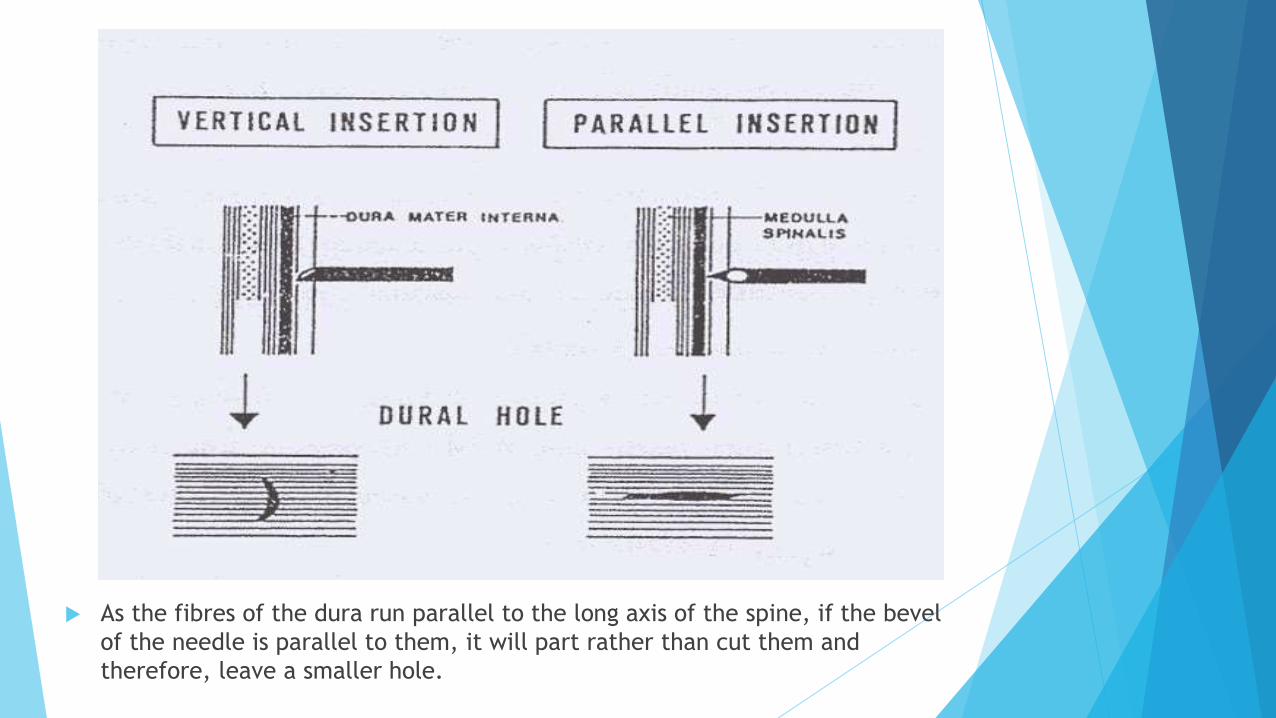
As the fibres of the dura run parallel to the long axis of the spine, if the bevel
of the needle is parallel to them, it will part rather than cut them and
therefore, leave a smaller hole.

It is widely considered that pencil-point needles (Whitacre or Sprotte) make a smaller hole in the
dura and are associated with a lower incidence of headache (1%) than conventional cutting-edged
needles (Quincke)
Treatment of PDPH
Positive reassurance of recovery
Confinement to bed. Head down position may be necessary.
Hydration therapy – Oral as well as Intravenous.
Analgesics and Antiemetics – Paracetamol, Aspirin, Codeine, Ondasentron.
Abdominal binders – Raises pressure in peridural venous plexus and thereby
increasing CSF pressure.
Oxygen inhalations.
Caffeine containing drinks such as tea, coffee are often helpful.
Prolonged or severe headaches may be treated with epidural blood patch
performed by aseptically injecting 15-20ml of the patient's own blood into the
epidural space. This then clots and seals the hole and prevents further
leakage of CSF.

Sources
Principles of Anesthesiology: General and Regional Anesthesia - Vincent J.
Collins
Morgan and Mikhail's Clinical Anesthesiology
NYSORA
IJA
BJA
Oxford Journal of Anaesthesia
http://www.pitt.edu/
Thanks for Your
Attention.