Embed Size (px)
Citation preview

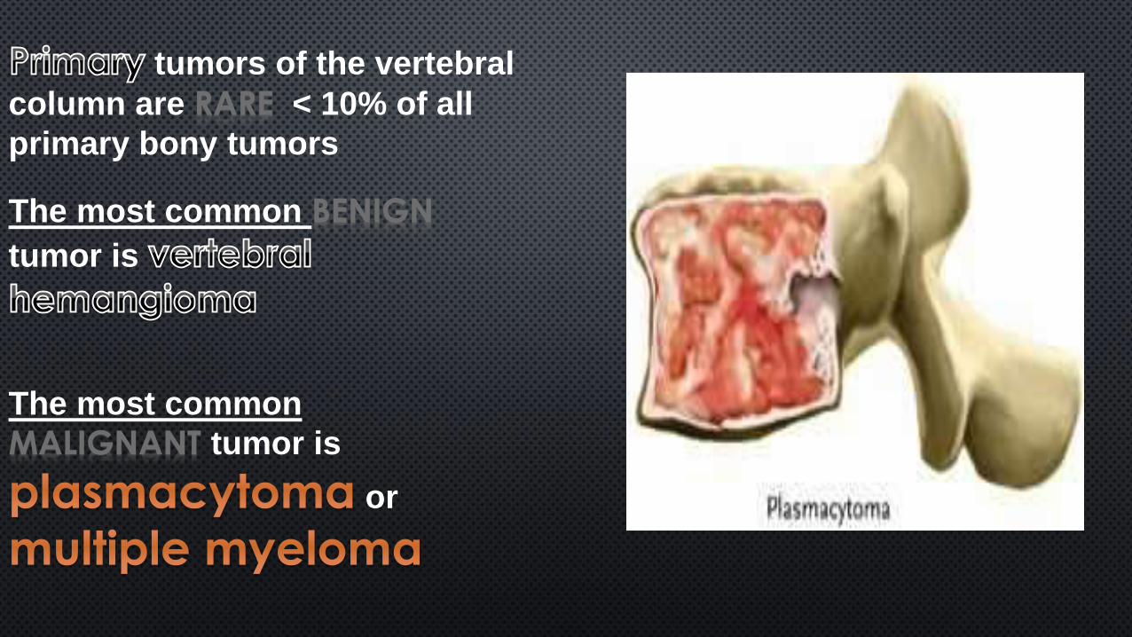
tumors of the vertebral
column are RARE < 10% of all
primary bony tumors
The most common BENIGN
tumor is
The most common
MALIGNANT tumor is
or
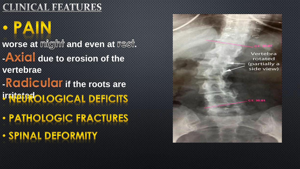
worse at and even at .
- due to erosion of the
vertebrae
- if the roots are
irritated
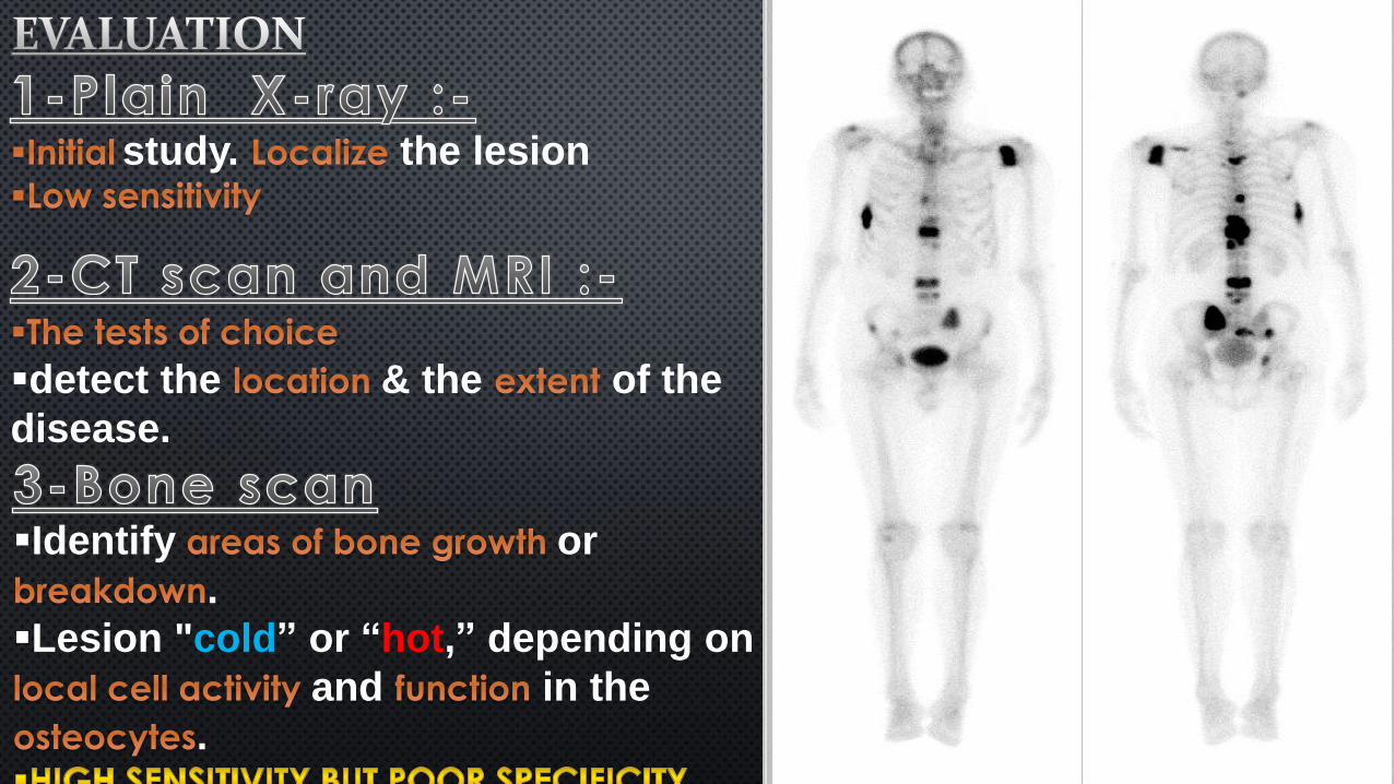
study. the lesion
detect the & the of the
disease.
Identify or
.
Lesion "cold” or “hot,” depending on
and in the
.
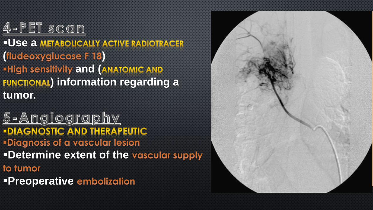
Use a
( )
and (
) information regarding a
tumor.
Determine extent of the
Preoperative
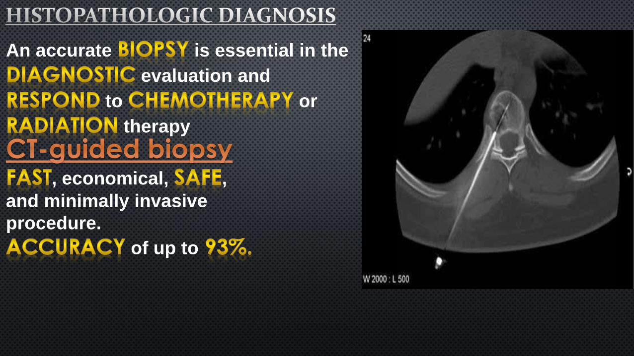
An accurate is essential in the
evaluation and
to or
therapy
, economical, ,
and minimally invasive
procedure.
of up to
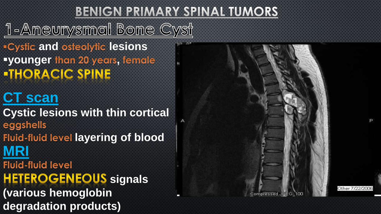
and lesions
younger ,
CT scanCystic lesions with thin cortical
layering of blood
MRI
signals
(various hemoglobin
degradation products)
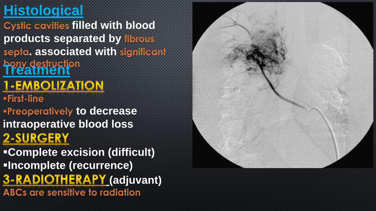
Histologicalfilled with blood
products separated by
. associated with
Treatment
to decrease
intraoperative blood loss
Complete excision (difficult)
Incomplete (recurrence)
(adjuvant)
The tumors
Incidental findings,
are
Myelopathy
Pathologic
fractures
Pain
Radiculopathy
CT scan
MRIsignals on both T1
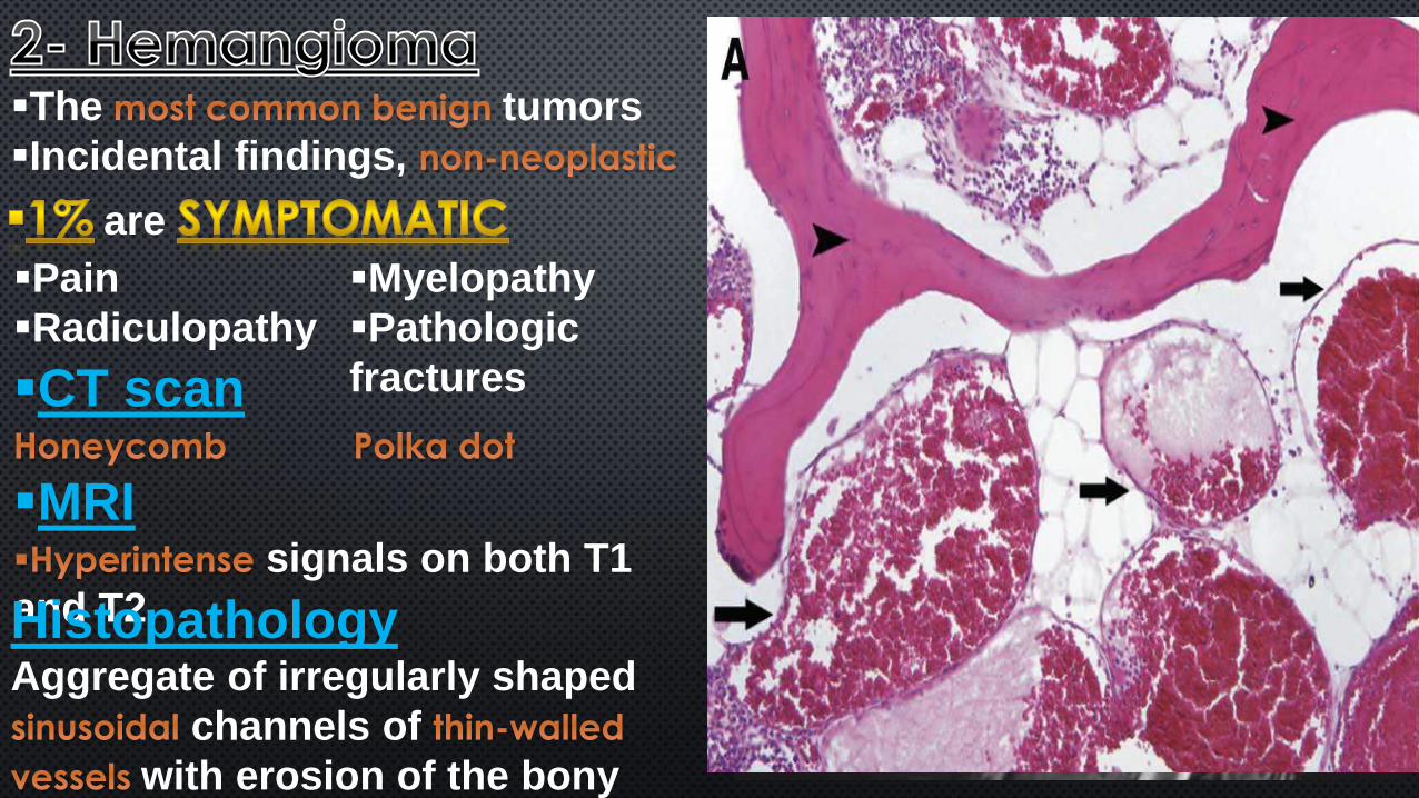
and T2HistopathologyAggregate of irregularly shaped
channels of
with erosion of the bony
architecture.
thoracic and lumbar spine.
The is the most
frequent site of
Association between
and the development of
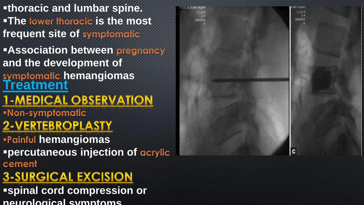
hemangiomasTreatment
hemangiomas
percutaneous injection of
spinal cord compression or
neurological symptoms
benign, ,
Teenage years,
Involve the
worse at
with
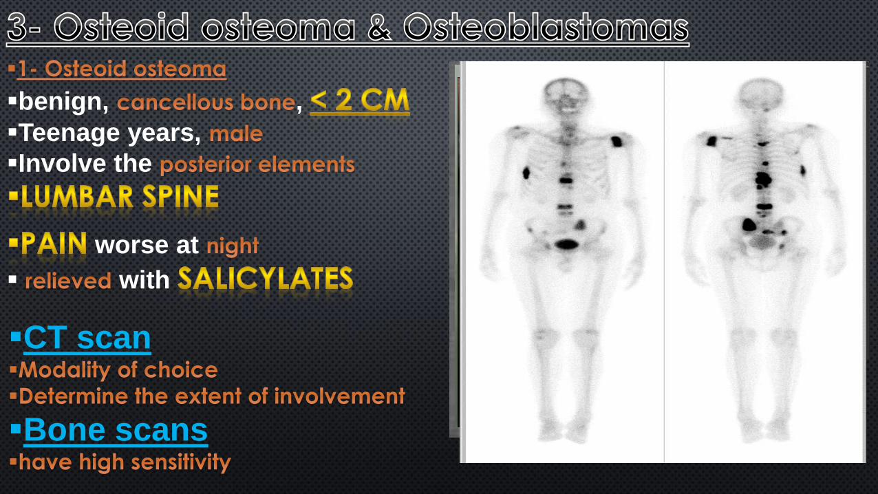
CT scan
Bone scans
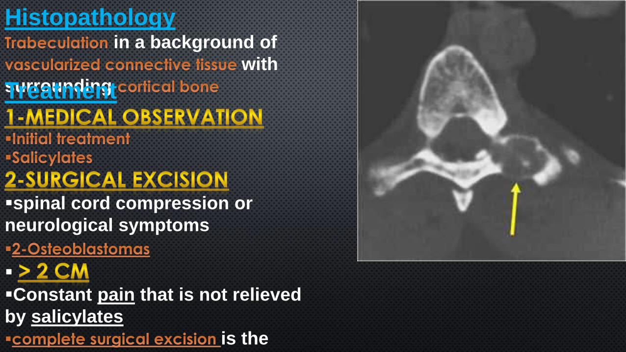
Histopathologyin a background of
with
surrounding Treatment
spinal cord compression or
neurological symptoms
Constant pain that is not relieved
by salicylates
is the
primary treatment.
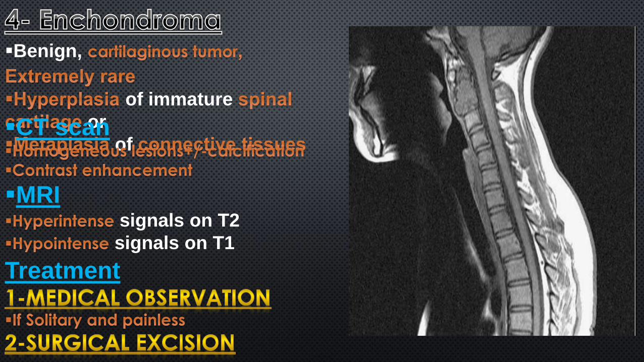
Benign,
of immature
or
of CT scan
MRIsignals on T2
signals on T1
Treatment
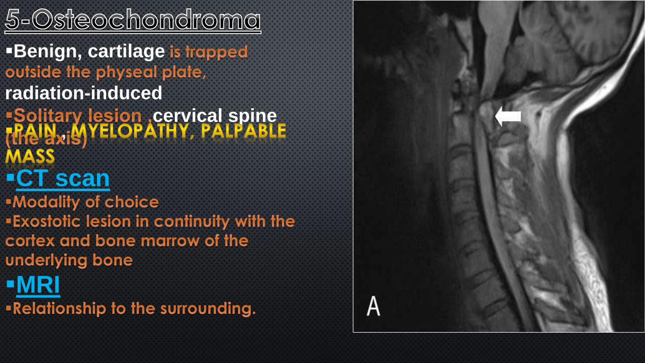
Benign, cartilage
radiation-induced
cervical spine ,
CT scan
MRI
Treatment
Symptomatic lesions, Complete
excision
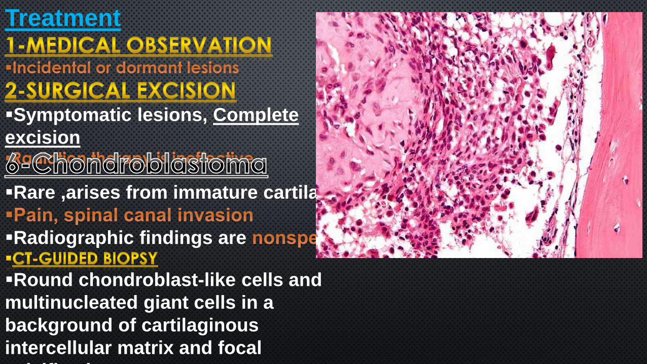
Rare ,arises from immature cartilage
Radiographic findings are
Round chondroblast-like cells and
multinucleated giant cells in a
background of cartilaginous
intercellular matrix and focal
calcification
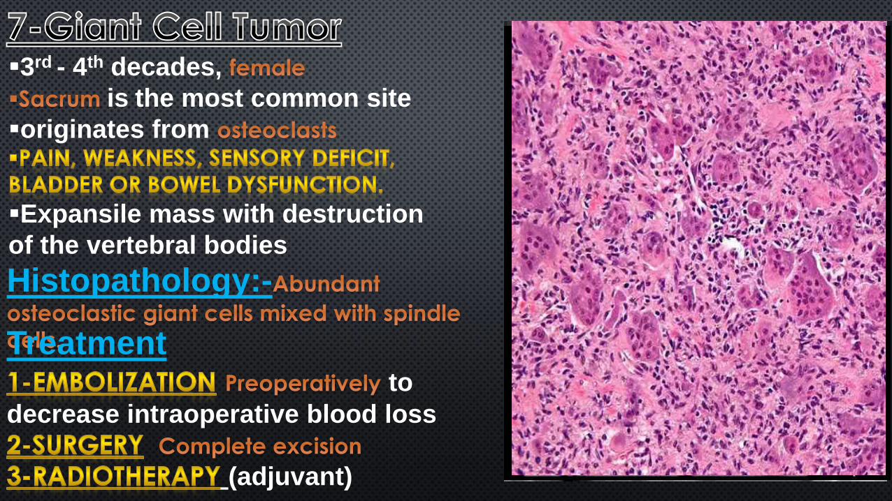
3rd - 4th decades,
is the most common site
originates from
Expansile mass with destruction
of the vertebral bodies
Histopathology:-
Treatmentto
decrease intraoperative blood loss
(adjuvant)
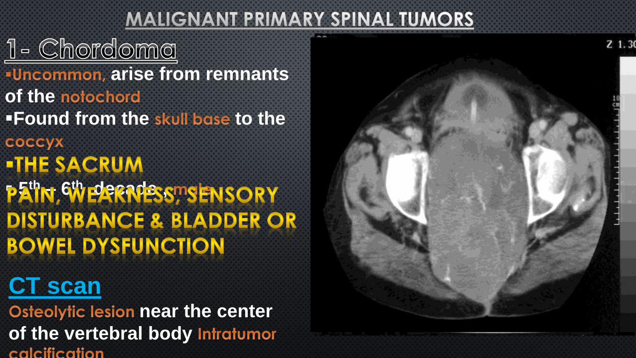
arise from remnants
of the
Found from the to the
5th – 6th decade ,
CT scannear the center
of the vertebral body
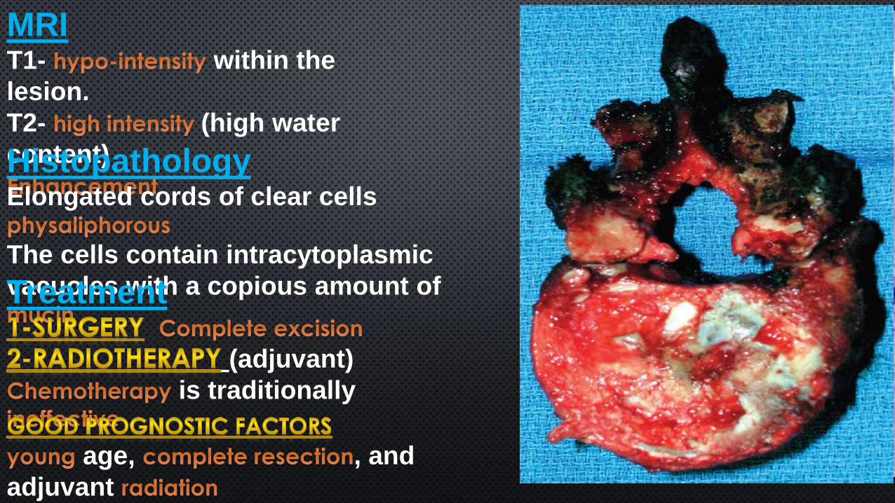
MRIT1- within the
lesion.
T2- (high water
content)HistopathologyElongated cords of clear cells
The cells contain intracytoplasmic
vacuoles with a copious amount ofTreatment
(adjuvant)
is traditionally
age, , and
adjuvant
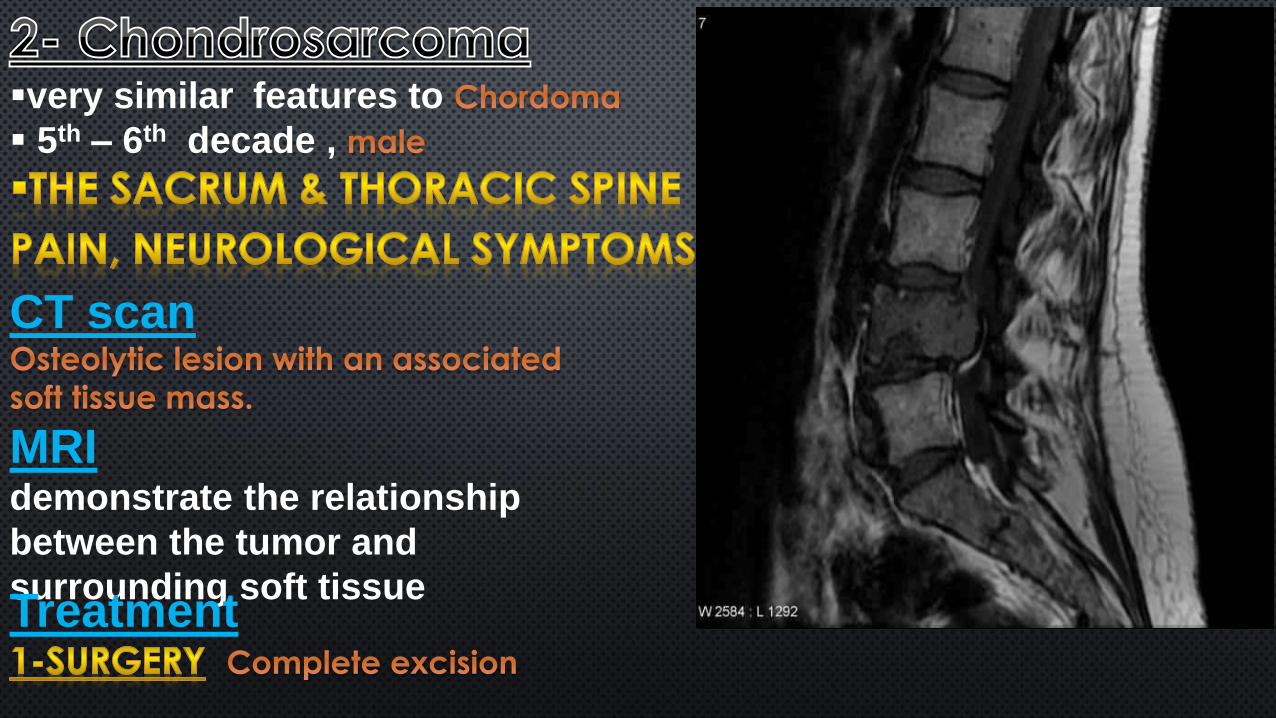
very similar features to
5th – 6th decade ,
CT scan
MRIdemonstrate the relationship
between the tumor and
surrounding soft tissueTreatment
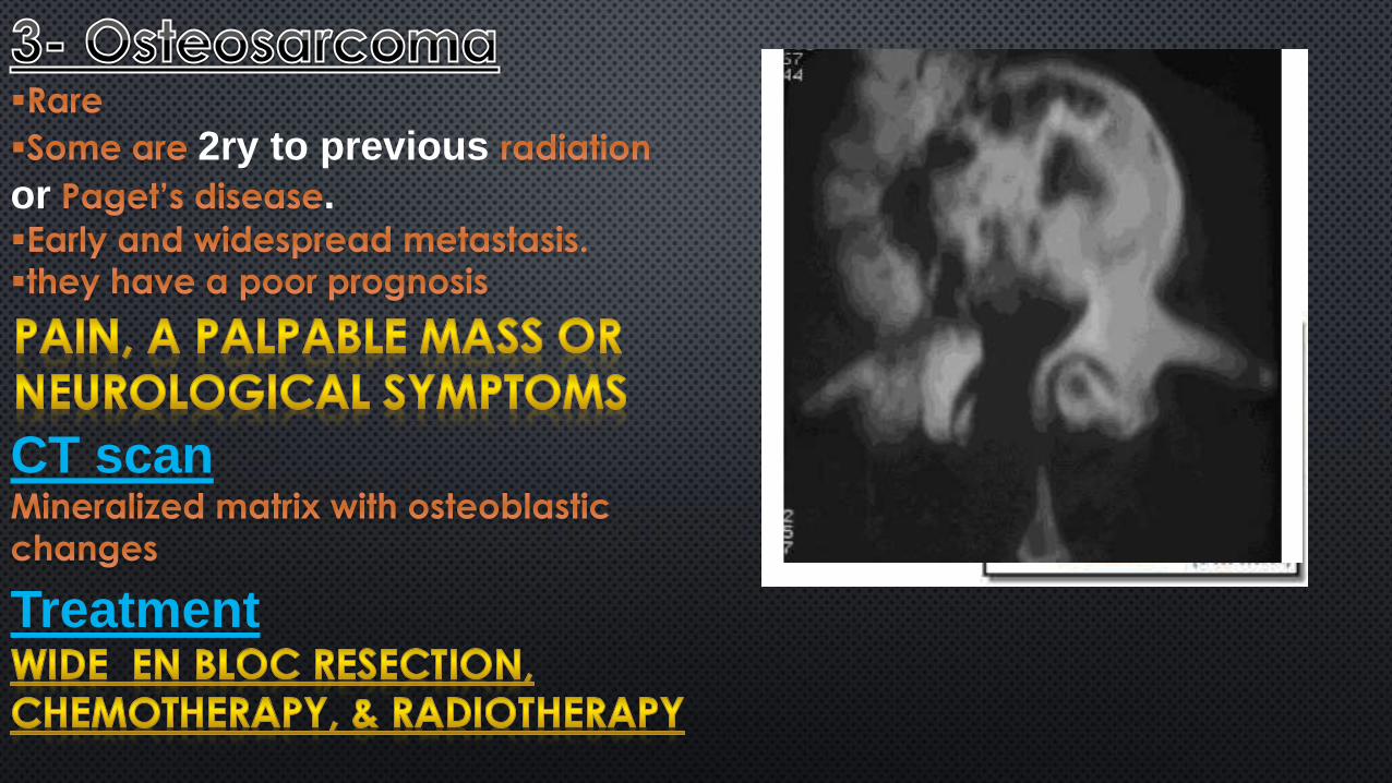
2ry to previous
or .
CT scan
Treatment
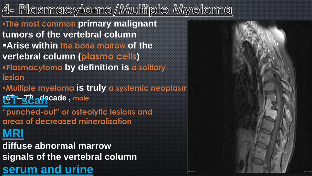
primary malignant
tumors of the vertebral column
Arise within of the
vertebral column ( )
by definition is
is truly6th – 7th decade , CT scan
MRIdiffuse abnormal marrow
signals of the vertebral column
serum and urine
electrophoresis
Treatmenten bloc spondylectomy
(adjuvant)