Embed Size (px)
Citation preview
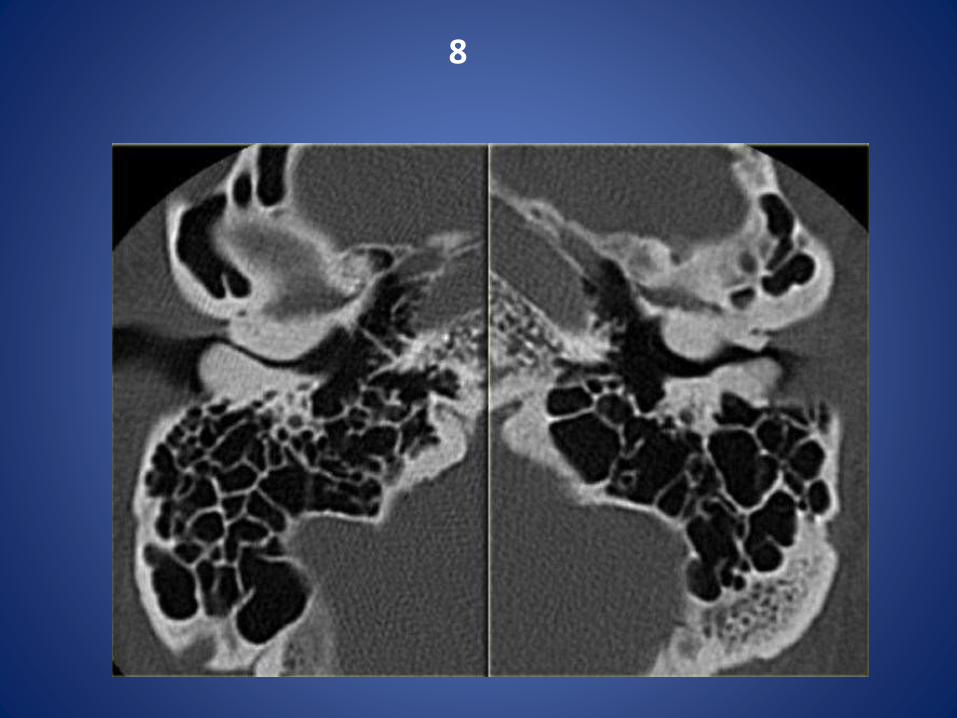
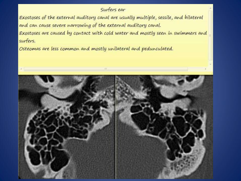
SPOTS
Dr Mohit Goel JR III, 22/4/14

1
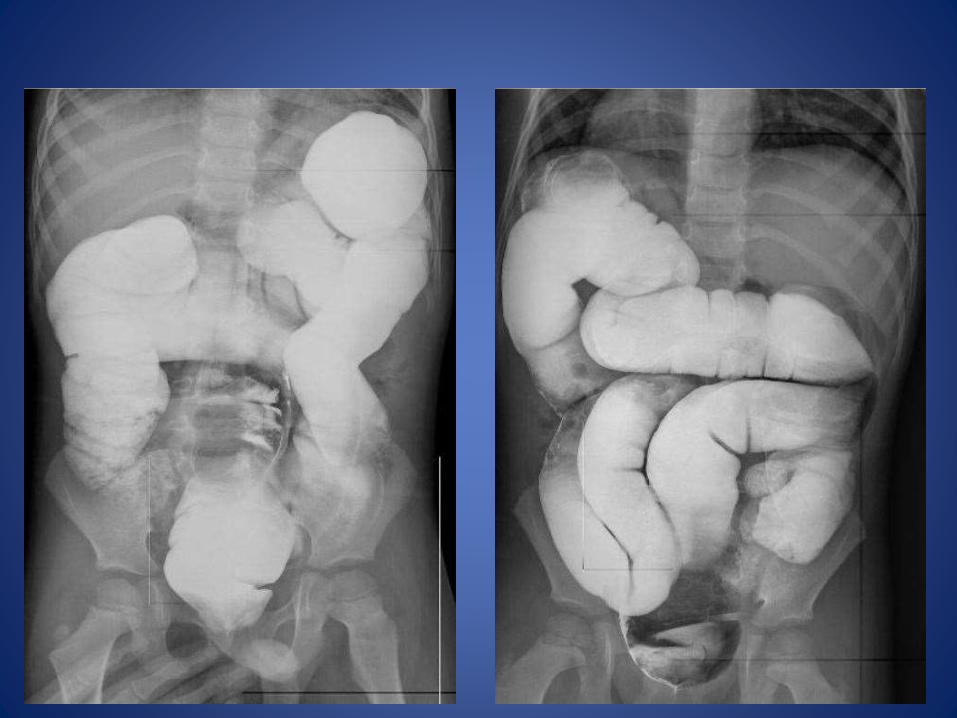
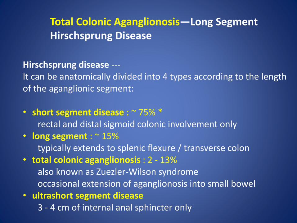
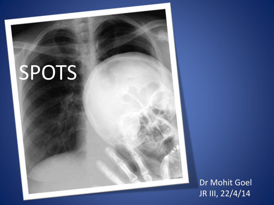
Total Colonic Aganglionosis—Long Segment Hirschsprung Disease
Hirschsprung disease ---It can be anatomically divided into 4 types according to the length of the aganglionic segment:
• short segment disease : ~ 75% *rectal and distal sigmoid colonic involvement only
• long segment : ~ 15%typically extends to splenic flexure / transverse colon
• total colonic aganglionosis : 2 - 13%also known as Zuezler-Wilson syndromeoccasional extension of aganglionosis into small bowel
• ultrashort segment disease3 - 4 cm of internal anal sphincter only
2
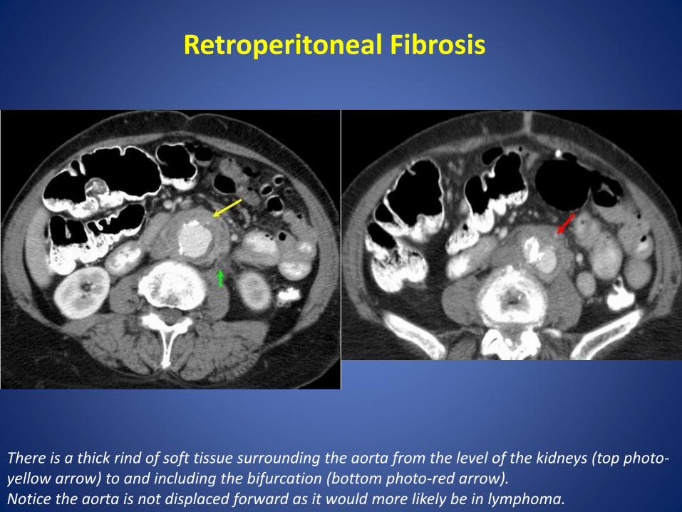
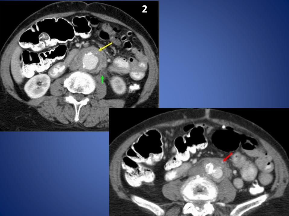
Retroperitoneal Fibrosis
There is a thick rind of soft tissue surrounding the aorta from the level of the kidneys (top photo-yellow arrow) to and including the bifurcation (bottom photo-red arrow). Notice the aorta is not displaced forward as it would more likely be in lymphoma.

Retroperitoneal fibrosis (RPF) is a condition that has previously been described as chronic periaortitis.
It is an uncommon fibrotic reaction in the retroperitoneum that typically presents with ureteral obstruction.

On excretory or CT urography§ Most retroperitoneal masses displace ureters laterally§ Tapering of ureters distal to mass
On CT scans§ Rind of soft tissue around aorta and inferior vena cava between level of
kidney and sacrum § Spreads to involve the ureters, causing varying degrees of obstruction. § Fat plane between the mass and the psoas muscle may be obliterated § Unlike adenopathy, RPF tends not to displace aorta anteriorly § Mass may show varying degrees of enhancement depending on the stage of
the disease
Imaging Findings
3
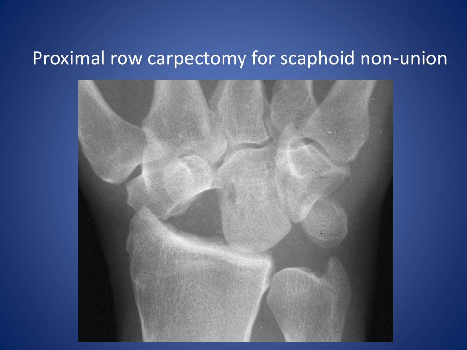
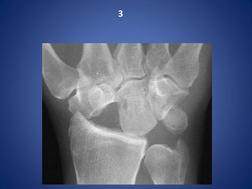
Proximal row carpectomy for scaphoid non-union

4
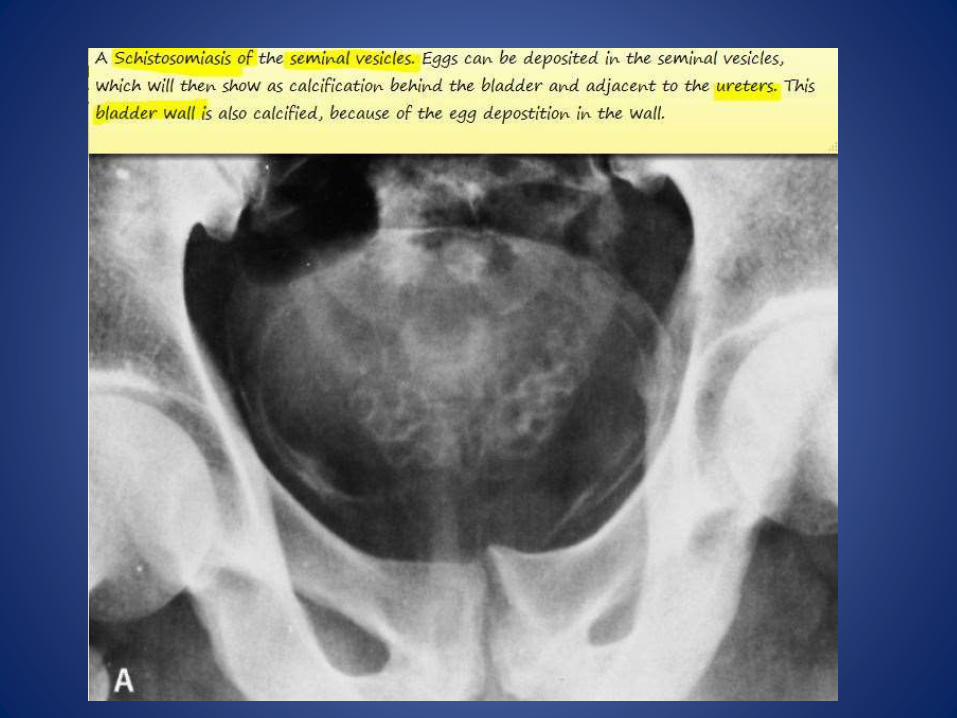

SCHISTOSOMIASIS OF UB

5

6
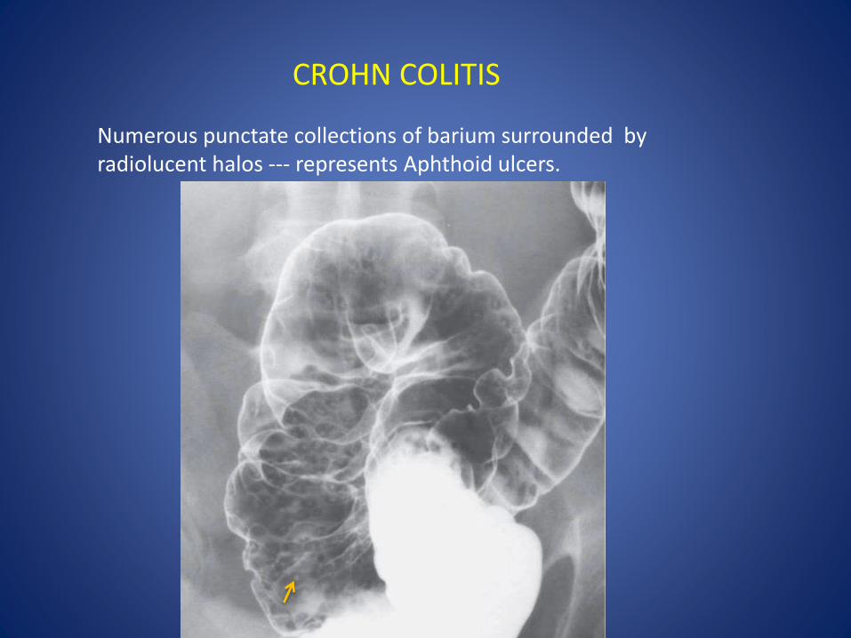
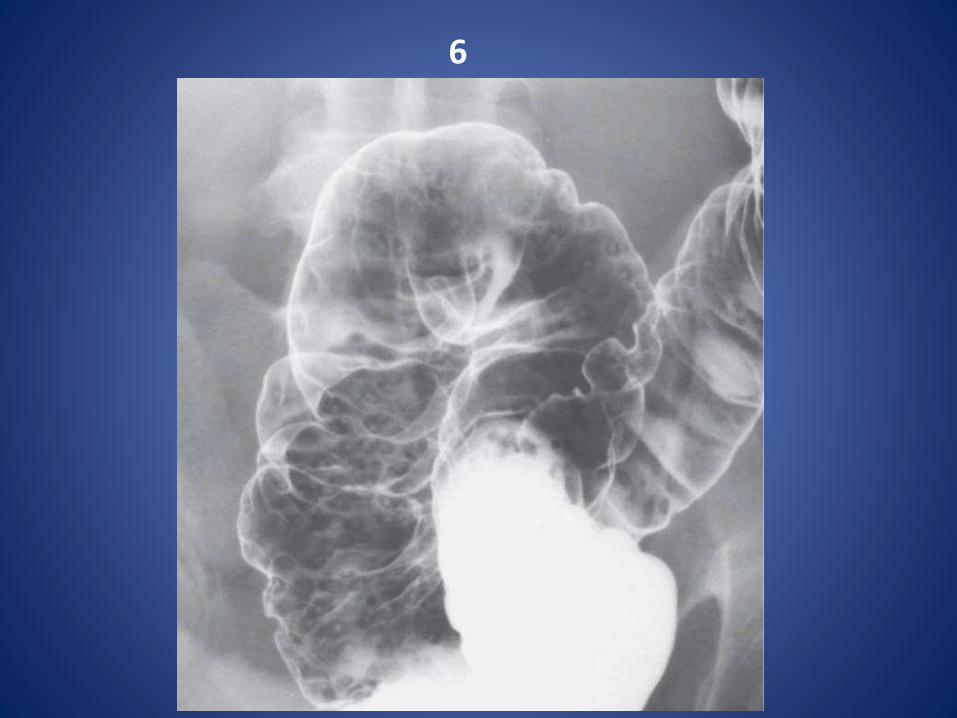
Numerous punctate collections of barium surrounded by radiolucent halos --- represents Aphthoid ulcers.
CROHN COLITIS

7
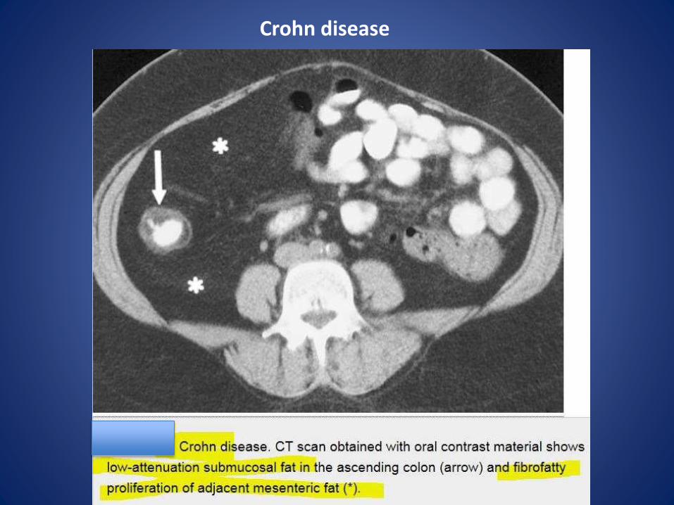
Crohn disease
8

9

Pectus excavatumPectus excavatum (or funnel chest) is a congenital chest wall deformity characterisedby concave depression of the sternum, resulting in cosmetic and radiographic alterations.
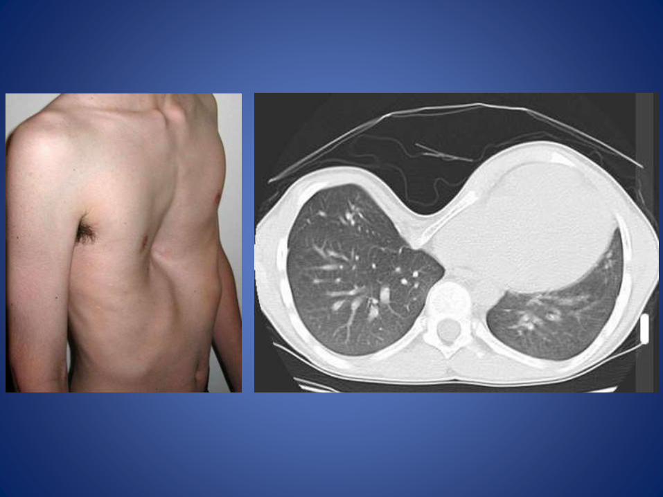

Radiographic featuresPlain filmCharacteristically demonstrates : • blurring of right heart border (PA or AP film)• increased density of the inframedial lung zone• horizontal posterior ribs • vertical anterior ribs (heart shaped) • displacement of heart towards the left• obliteration of the descending aortic interface
CTThe diagnosis is obvious on CT with the degree of deformity and mediastinal shift often dramatic. The Haller index (maximal transverse diameter/ narrowest AP length of chest) is used assess severity. Normal Haller index is 2.5. Significant pectus excavatum has an index greater than 3.25.
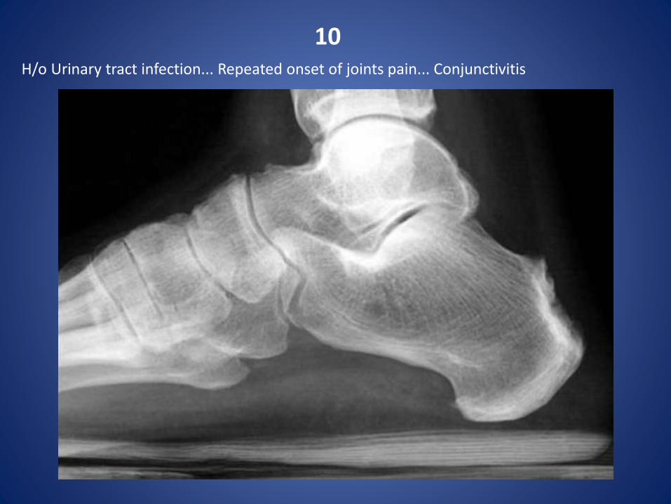
10H/o Urinary tract infection... Repeated onset of joints pain... Conjunctivitis
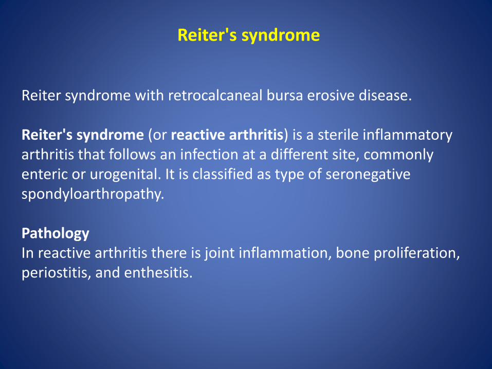
Reiter's syndrome
Reiter syndrome with retrocalcaneal bursa erosive disease.
Reiter's syndrome (or reactive arthritis) is a sterile inflammatory arthritis that follows an infection at a different site, commonly enteric or urogenital. It is classified as type of seronegativespondyloarthropathy.
PathologyIn reactive arthritis there is joint inflammation, bone proliferation, periostitis, and enthesitis.
Associationsurethritisconjunctivitisseropositivity for the HLA-B27 antigen
DistributionIt typically affects hands, wrists, and feet with a distribution that is unilateral or bilateral and symmetric or asymmetric; lower-extremity involvement is more common than upper-extremity involvement

11
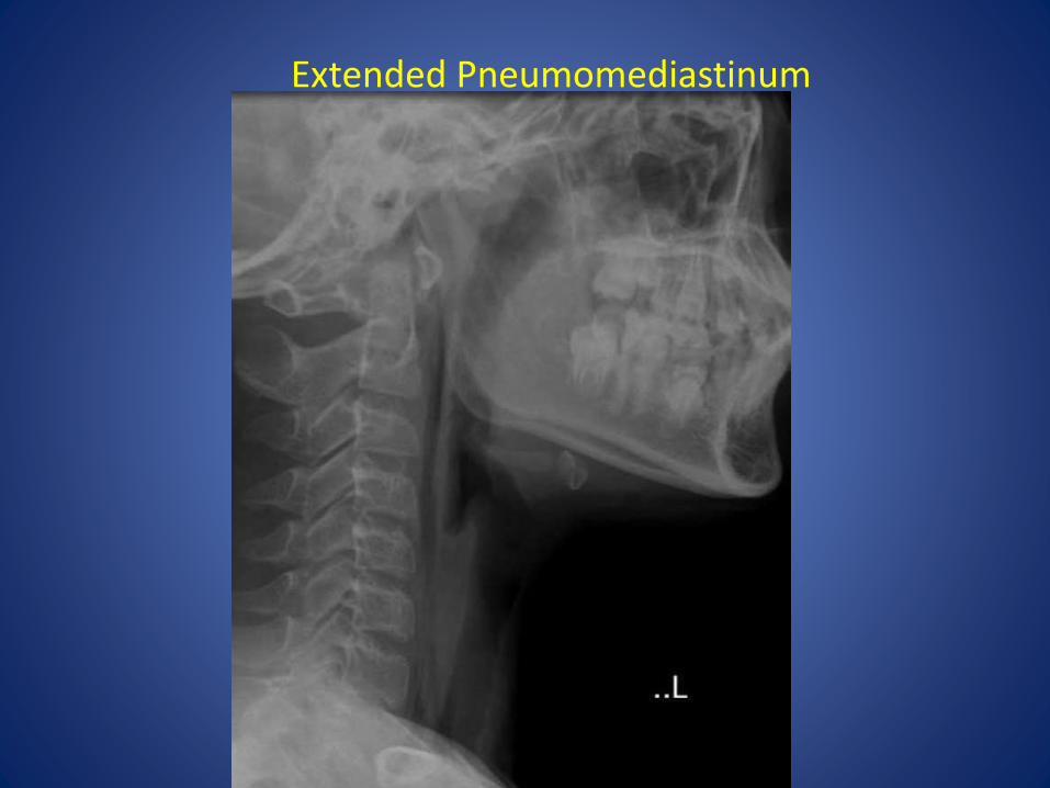
Extended Pneumomediastinum
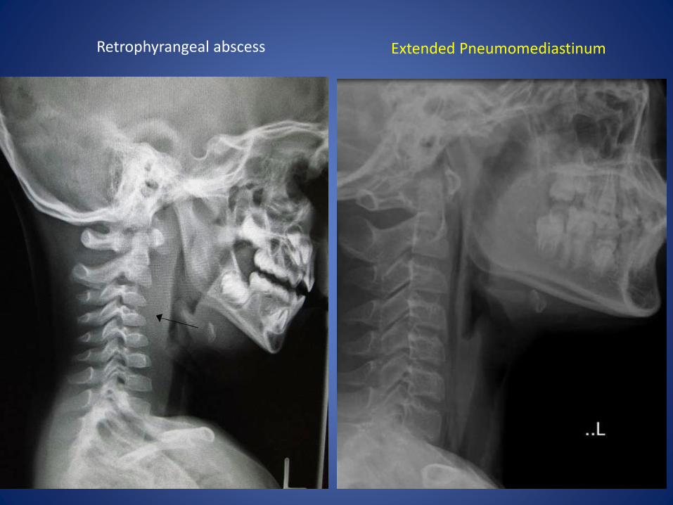
Retrophyrangeal abscess Extended Pneumomediastinum
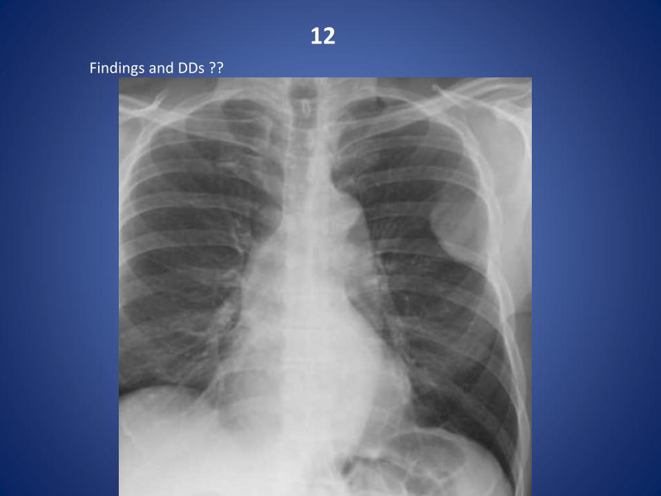
12Findings and DDs ??

A pleural based malignant mass with overlying rib destruction - Plasmacytoma
DDs:pleural fibromaMesotheliomaPleural mets
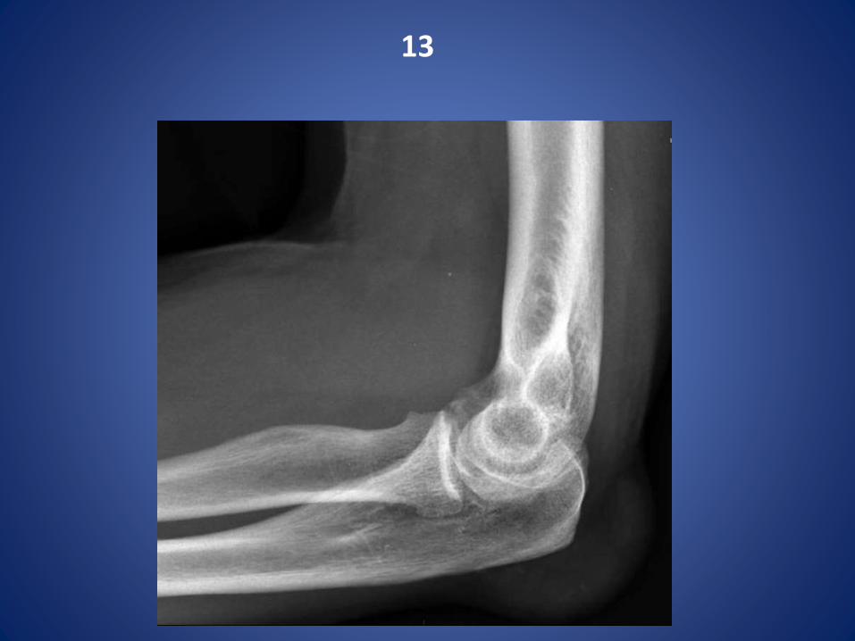
13

Olecranon bursitis- GOUT
X-rays of the left elbow demonstrate a soft tissue swelling overlying the olecranon. It is somewhat hyperdense.
Deposition of sodium urate monohydrate crystals in synovial membranes, articular cartilage, ligaments, bursae leading to destruction of cartilage
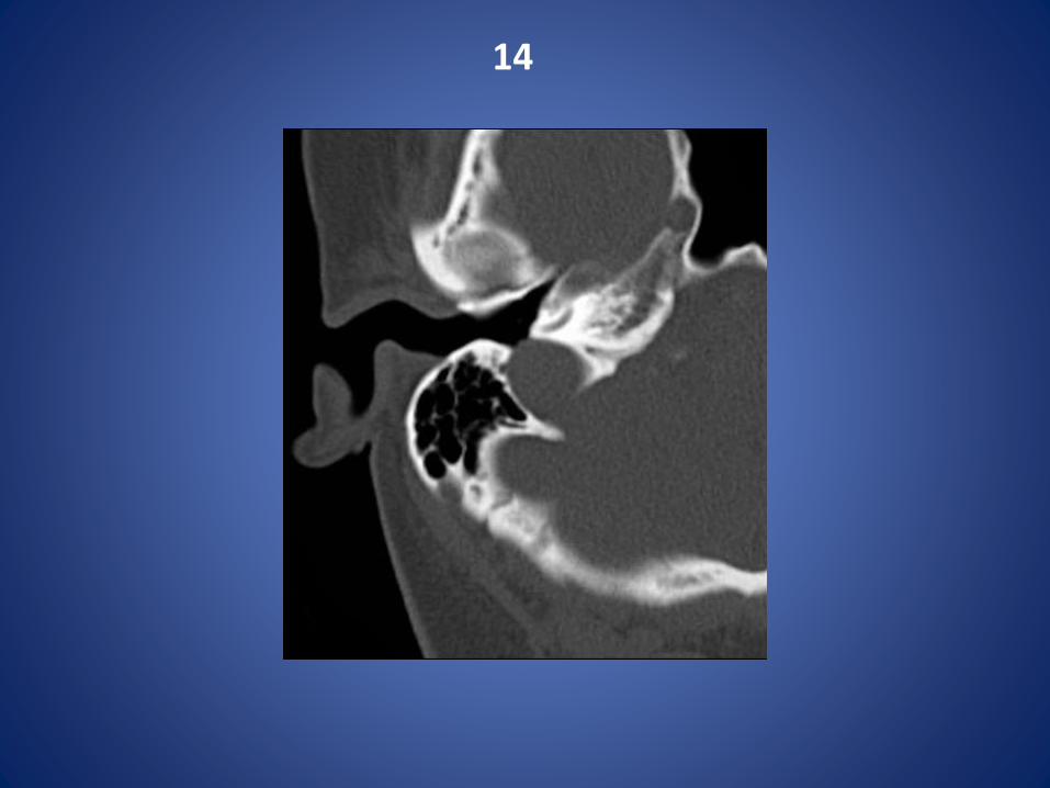
14

Dehiscent jugular bulb


15
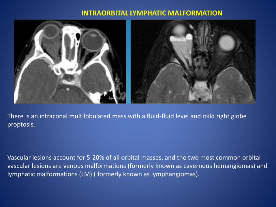
INTRAORBITAL LYMPHATIC MALFORMATION
There is an intraconal multilobulated mass with a fluid-fluid level and mild right globe proptosis.
Vascular lesions account for 5-20% of all orbital masses, and the two most common orbital vascular lesions are venous malformations (formerly known as cavernous hemangiomas) and lymphatic malformations (LM) ( formerly known as lymphangiomas).

Intraorbital venous-lymphatic malformations are present at birth, but tend not to be discovered clinically until early childhood when they enlarge as a result of either intralesional hemorrhage or lymphoid hyperplasia and result in acute proptosis.
Radiologic imaging of intraorbital LMs demonstrates unencapsulated, irregular, lobulated, and multicompartmental masses.
These lesions can have cystic as well as more solid components. The cystic elements of these masses commonly exhibit fluid-fluid levels as a result of intralesionalhemorrhage
Ultrasound images of LMs demonstrate heterogeneous, ill-defined lesions with anechoic cystic portions and extraconal extension.
On CT, these masses exhibit ill-defined borders, irregular attenuations, and variable enhancement with peripheral rim enhancement in cystic regions. Additionally, calcified phleboliths can be seen on CT in venous portions of these lesions.
MR imaging -LMs demonstrate iso- to slightly high signal intensities on T1-weighted images and very high signal intensities on T2-weighted images.
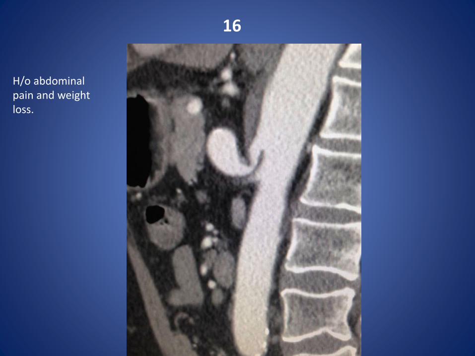
16
H/o abdominal pain and weight loss.

Coeliac artery compression syndromeOr Median arcuate ligament syndrome
Compression of celiac axis with post-stenotic dilation. This case demonstrates classic fish hook sign.

The median arcuate ligament is normally several millimeters to centimeters superior to the origin of the celiac artery.
In MALS, the ligament is anterior, rather than superior, to the celiac artery, resulting in compression of the vessel and a characteristic hook-shaped contour.
17
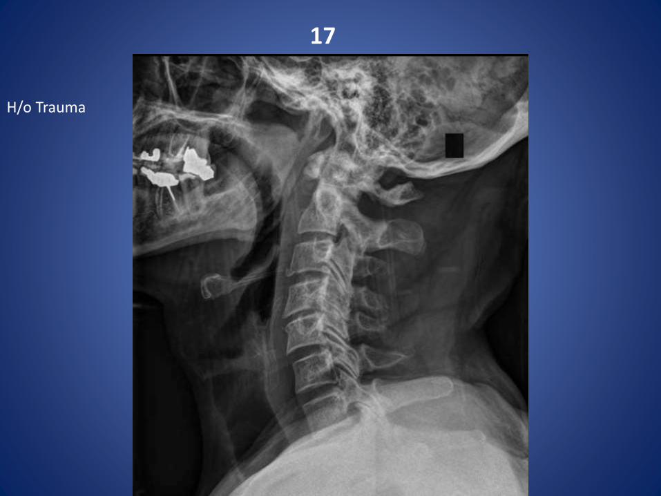
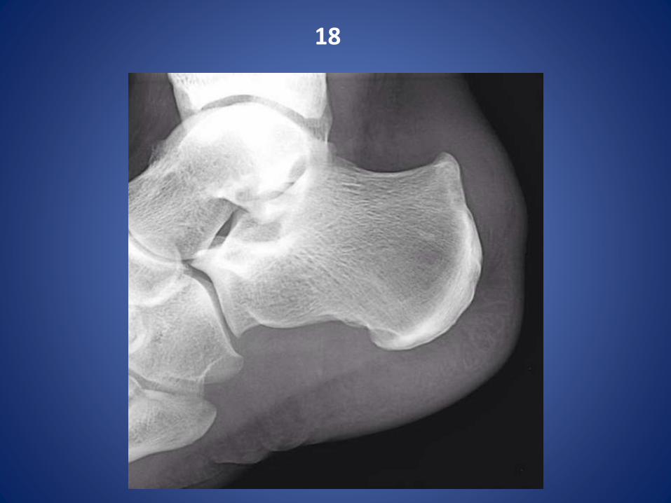
H/o Trauma

Extension teardrop fracture
18

Haglund syndrome - bony prominence of the posterosuperior aspect of the calcaneum (Haglunddeformity) with marked surrounding soft-tissue oedema due to insertional Achilles tendonosisand retrocalcaneal bursitis.
Haglund syndrome
19
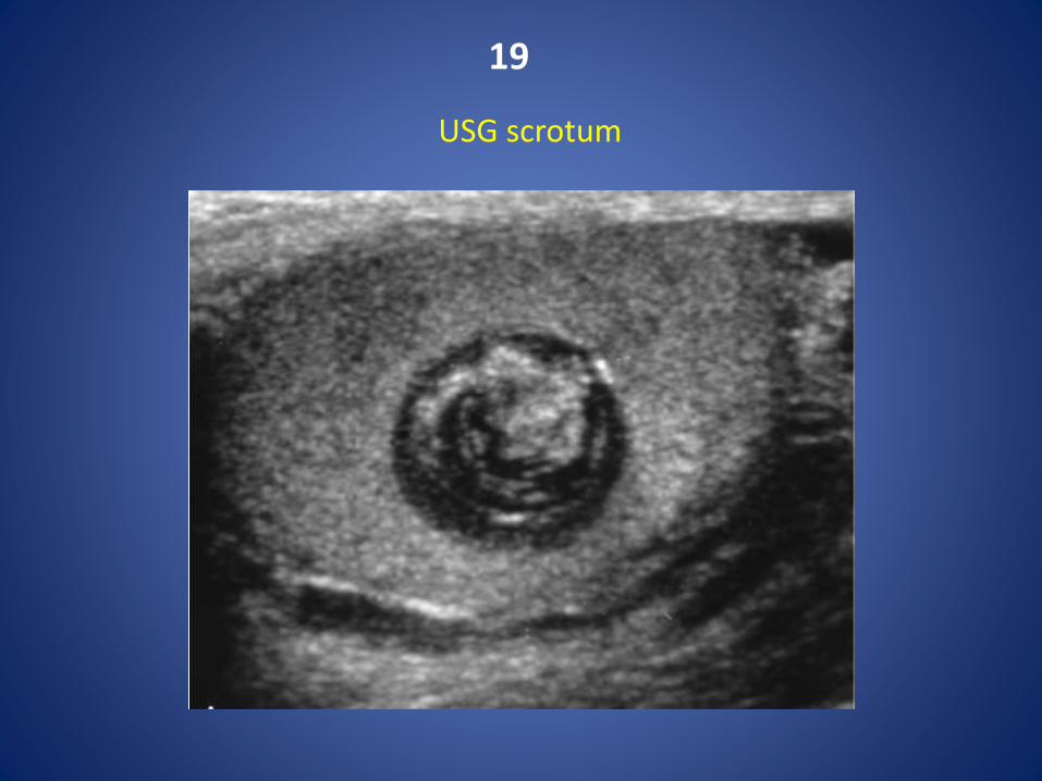
USG scrotum
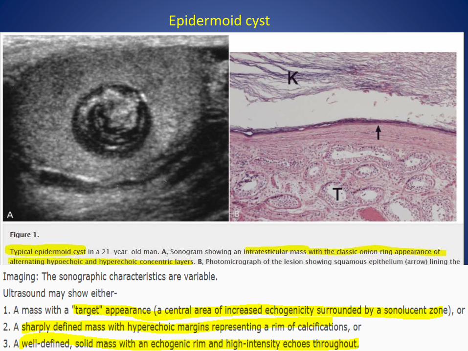
Epidermoid cyst

20
Name the views and the indication for taking this view…..
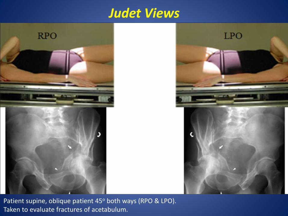
Judet Views
Patient supine, oblique patient 45o both ways (RPO & LPO).Taken to evaluate fractures of acetabulum.
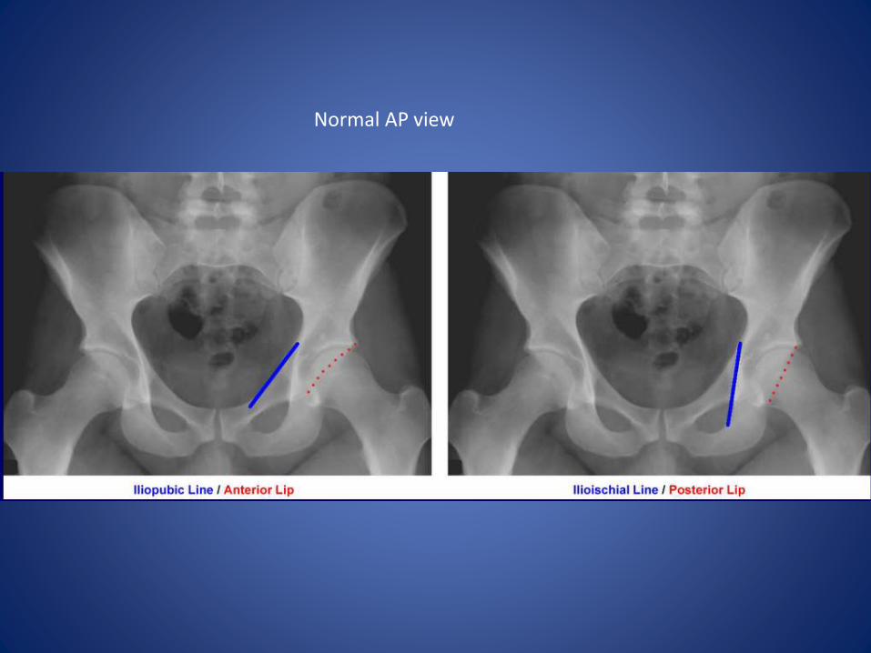
Normal AP view

LPO
AP
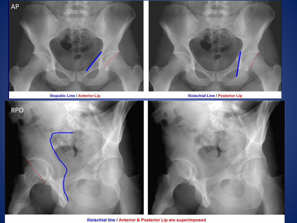
RPO
AP

Thank you