Embed Size (px)
Citation preview
CLINICAL CASE PRESENTATION
BY
DR.IMTIAZ AHMAD
PGR.WMW

HISTORY
Patient hina D/O M.abid, unmarried female 21,resident of BILAL GANJ LAHORE, working as beautician in parlor presented to us in OPD with the complaints of:-
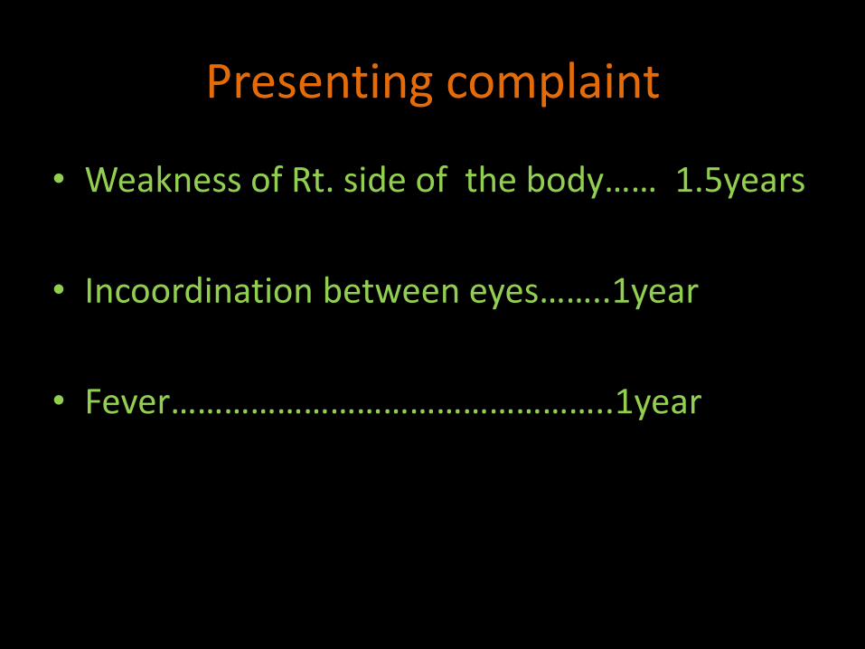
Presenting complaint
• Weakness of Rt. side of the body…… 1.5years
• Incoordination between eyes……..1year
• Fever…………………………………………..1year
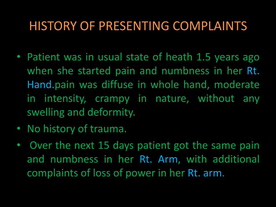
HISTORY OF PRESENTING COMPLAINTS
• Patient was in usual state of heath 1.5 years agowhen she started pain and numbness in her Rt.Hand.pain was diffuse in whole hand, moderatein intensity, crampy in nature, without anyswelling and deformity.
• No history of trauma.
• Over the next 15 days patient got the same painand numbness in her Rt. Arm, with additionalcomplaints of loss of power in her Rt. arm.

• Patient took medication form local GP. And hekeem but symptoms persisted.
• Over the next 3 months these symptoms worsed, pain at all the times. She had to take pain killers multiple time per day.
• Pain and weakness developed in Rt. lower limb over the period of 6 months. She was having mild difficulty in carrying out her routine activities,
• There is history of repeated fall due reduced power in Rt. side of the body.
• Weakness was gradually progressive.• There is history of speech abnormality.
• About one year ago she started having fever, that was low grade, continuous, at all the time, not associated with rigor and chills. Relieved only by medication, it was associated with headache, that patient could not localize.
• Fever was not associated with vomiting, diarrhea, burning micturition, ear and nasal discharge.
• There is nonspecific history of cough and sputum to which patient did not give any attention.
• History is suggestive of loss of apatite and significant weight loss ( 5-8 kg in 6months)

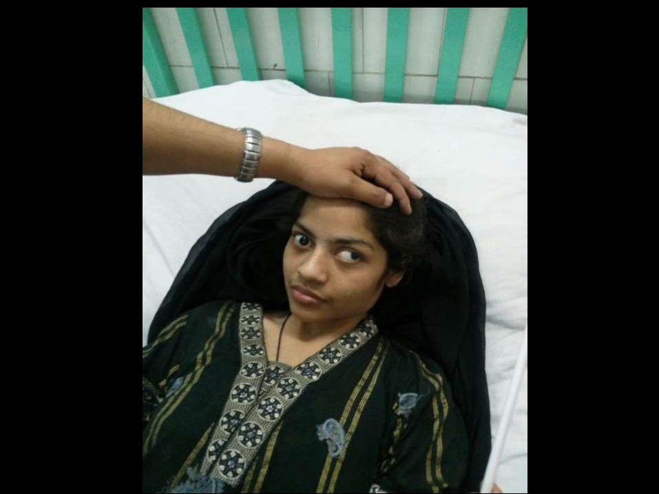
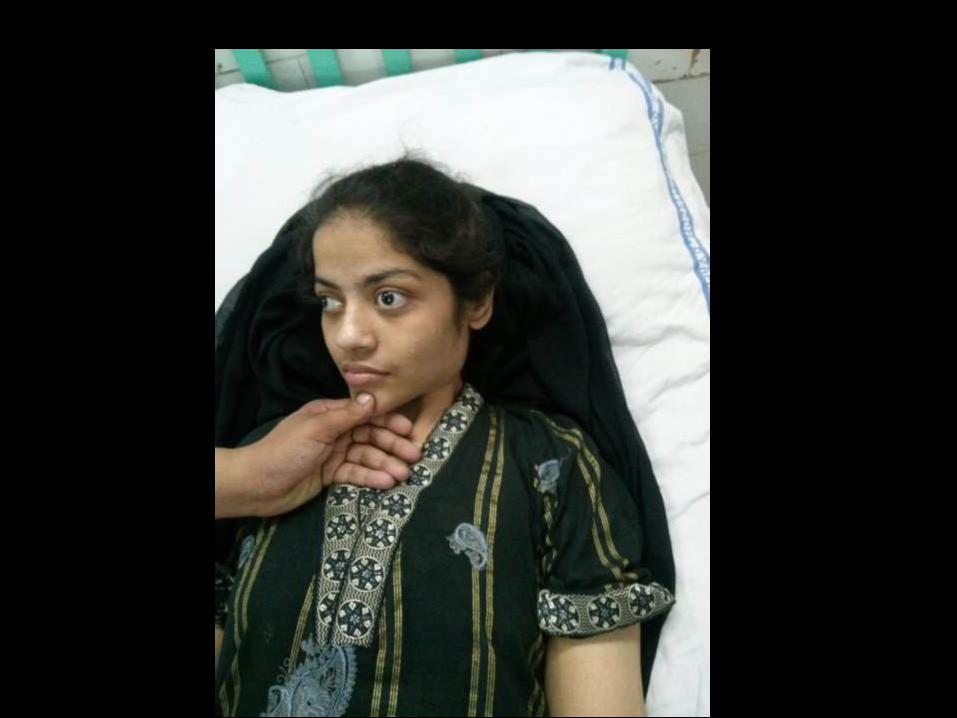
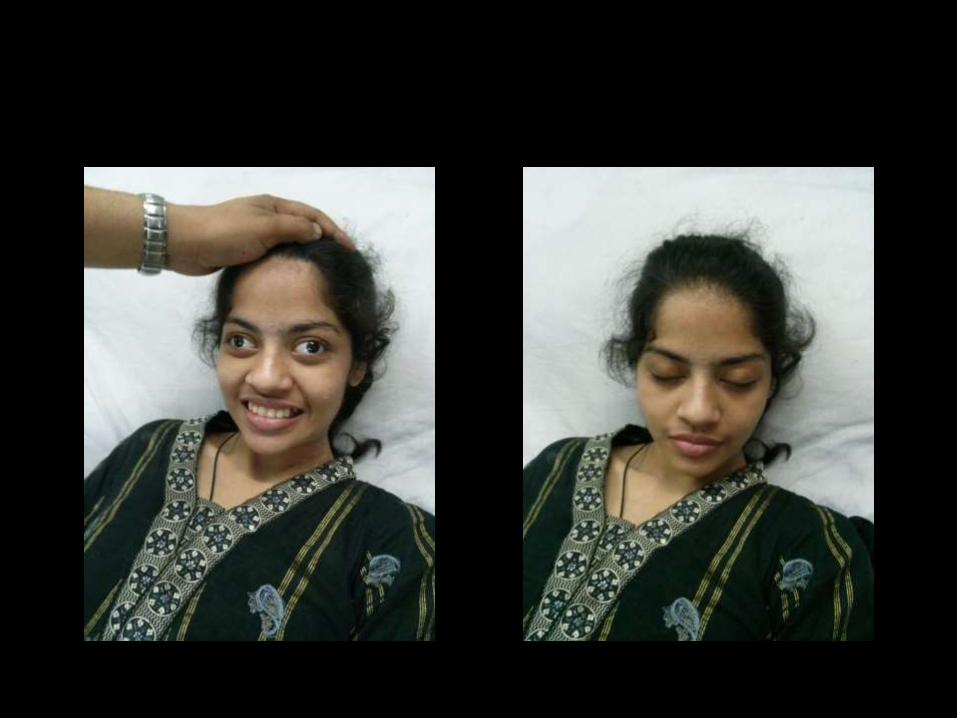
• 1 year ago she noticed double vision and dropping of Rt. Eyelid, it was progressive over 20 days after which it got static. It was not associated with impairment in vision. Mother noticed that her Rt. Eye was deviated outward.
• She was not a known case of having squint.
• She was an obese girl before this whole ailment , no history of excessive body hair, menstrual irregularities and oral contraceptive intake.
• 1 month after fever and diplopia she got 2 episodes of complex partial tonic clonic fits on Rt. side of the body In 15 days. Each time fits were about 5 mints duration, without tongue bite , fecal and urine incontinence.
• Few days after the seizures she got complete loss of power on Rt. side of body, with fecal and urine incontinence and impaired conscious level.
• All the neurological deficit was gradually progressive without any remission.

PAST HISTORY
• No history of weakness in past.• No past history of epilepsy.• No history of tuberculosis.• No history of bleeding diathesis• No history of head trauma.• No past history of any acute visual loss• No past history of admission and any intervention
in the hospital.• No history of arthritis and rash.• No history of pain or swelling in the spine.
PERSONAL HISTORY
• Unmarried sexually inactive female with regular menstrual cycle.
• Non addict non smoker
• Working as beautician in her congested house in a densely populated area.
• No history of foreign travel.
• Normal sleep and bowel habits.

DRUG HISTORY
• NO history of drug abuse
• She received proper immunization according to EPI. Program
• No history drug allergy.

SOCIO-ECONOMIC HISTORY
• Poor family.
• Living in small apartment with six siblings and parents.
• Tuberculosis is common disease in the area she lives
• Good relation with kith and kin,

FAMILY HISTORY
• No family history of infectious diseases.
• history of hypertension and diabetes mellitus & IHD positive in family.
• No family history of disabling disease.
• No family history of epilepsy.

SYSTEMIC REVIEW.
• No history of palpitation, SOB, pedal swelling
• No history of hemoptysis and hematemesis
• No history of joint pain and rash
• No history of vomiting, abdominal pain, distention, diarrhea, burning micturition and pus in urine
• No history of oral and genital ulcers
summray
• A young girl got progressive weakness of rt. side of body, two episodes of fits, diplopia ,scanned speech and incoordination in a setting of low grade fever and headache over the period of 1.5 years.
• Progressive neurological deficit that she attained never remitted except for improvement in diplopia, eyes coordination & autonomic improvement.
• She is not known epileptic, no history of head trauma, ear and nasal discharge, bleeding diatheses, oral ulcers, skin rash, joint pain,
• Signs of meningeal irritation remained absent through out the course of disease

DIFFERENTIALS ????
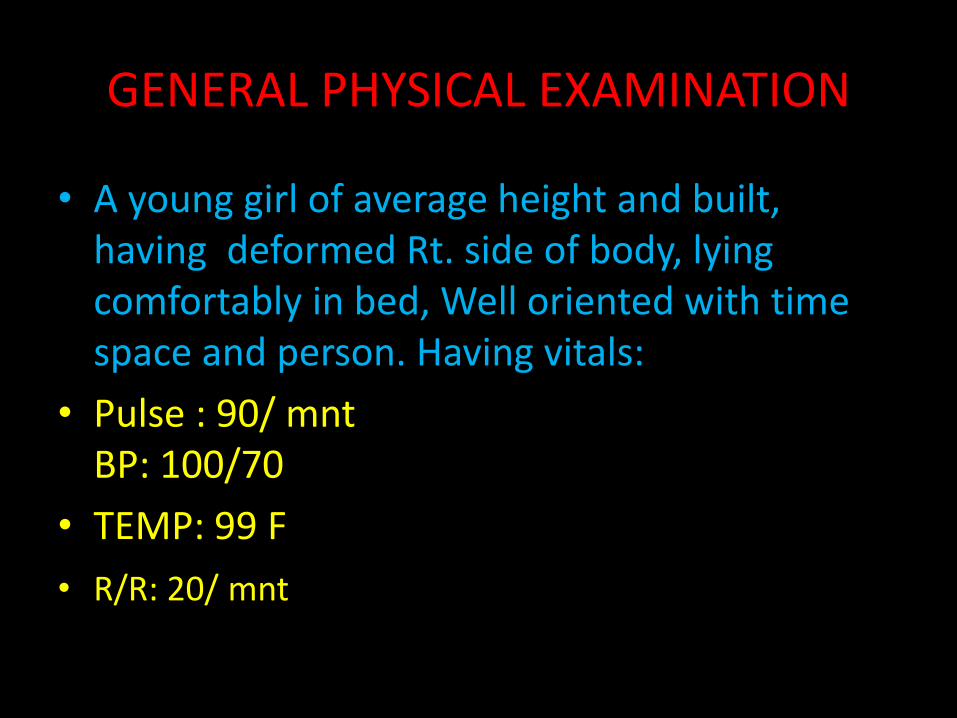
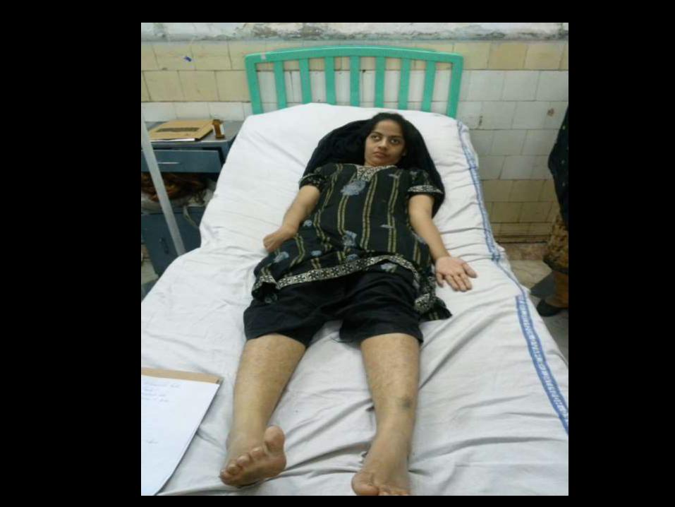
GENERAL PHYSICAL EXAMINATION
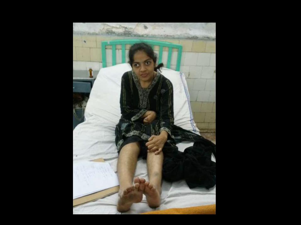
• A young girl of average height and built, having deformed Rt. side of body, lying comfortably in bed, Well oriented with time space and person. Having vitals:
• Pulse : 90/ mntBP: 100/70
• TEMP: 99 F
• R/R: 20/ mnt

Pallor, cynosis, clubbing, jaundice, edema, lymphadenpathy ………………………. Absent
JVP….. Not raised
Thyroid not enlarged.
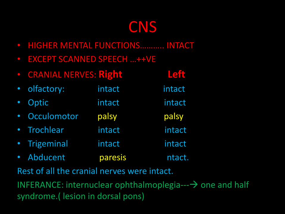
CNS• HIGHER MENTAL FUNCTIONS……….. INTACT
• EXCEPT SCANNED SPEECH …++VE
• CRANIAL NERVES: Right Left
• olfactory: intact intact
• Optic intact intact
• Occulomotor palsy palsy
• Trochlear intact intact
• Trigeminal intact intact
• Abducent paresis ntact.
Rest of all the cranial nerves were intact.
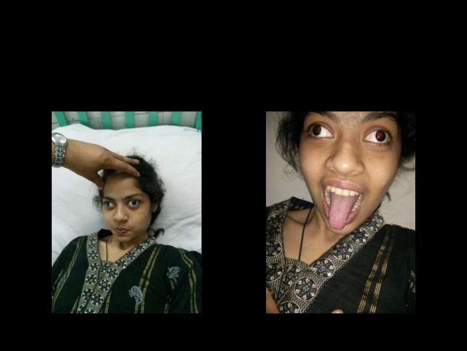
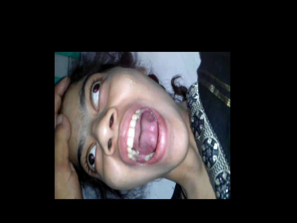
INFERANCE: internuclear ophthalmoplegia--- one and half syndrome.( lesion in dorsal pons)
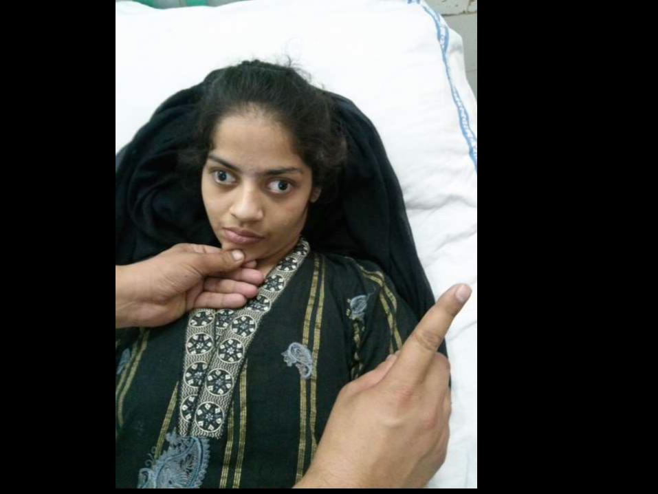

D/D of incoordinant eyes
Deviated in primary gaze
1. False localizing sign.
2. Paralytic squint
Deviated on movement
1. internuclear ophthalmoplegia
2. Latent squint.

CNS
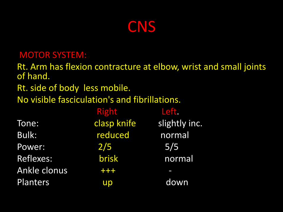
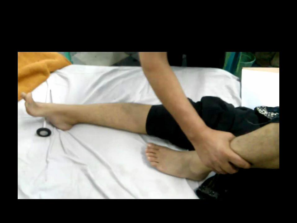
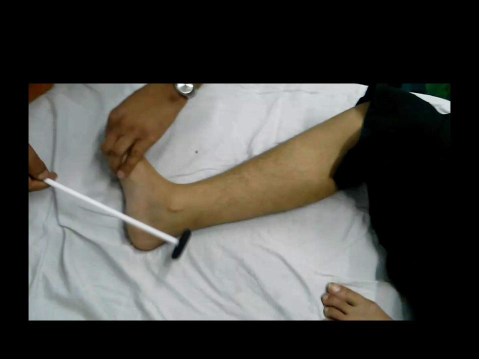
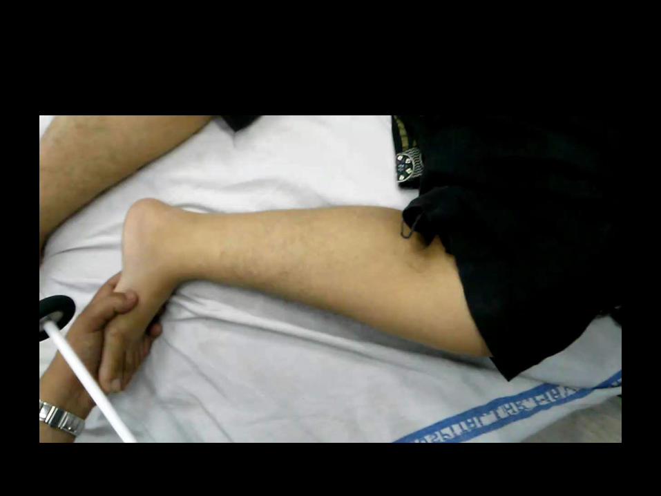
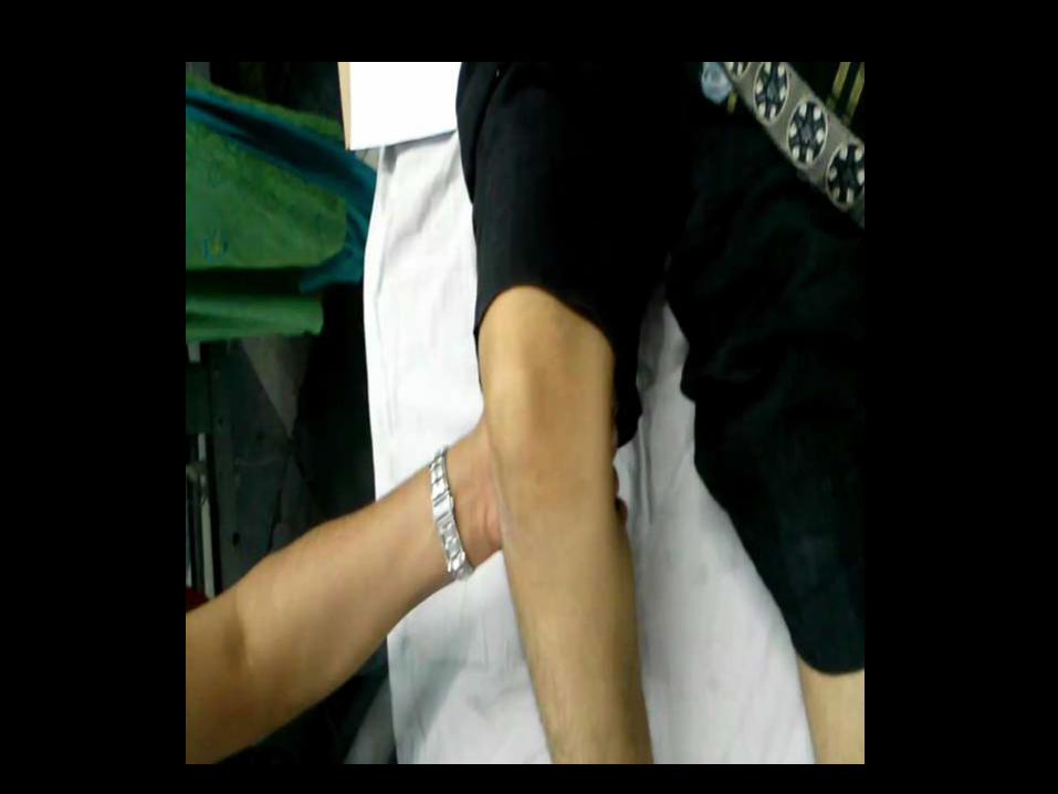
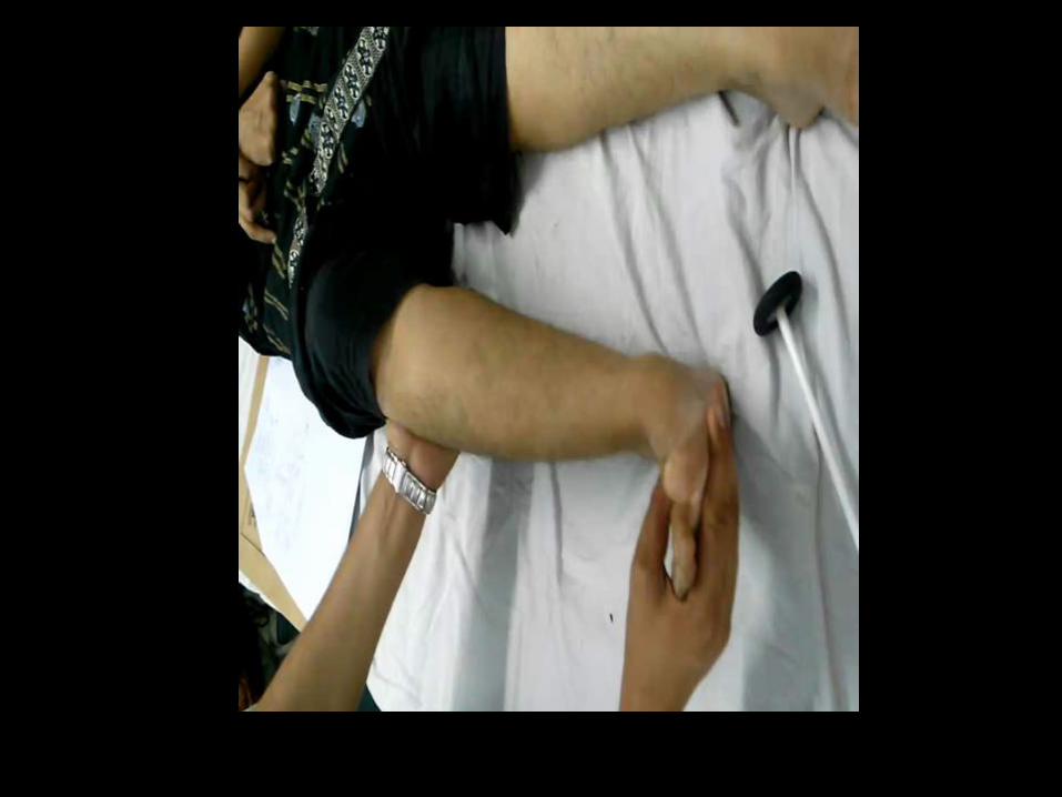
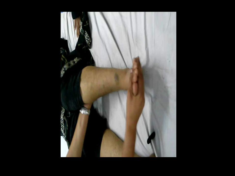
MOTOR SYSTEM:Rt. Arm has flexion contracture at elbow, wrist and small joints of hand.Rt. side of body less mobile.No visible fasciculation's and fibrillations.
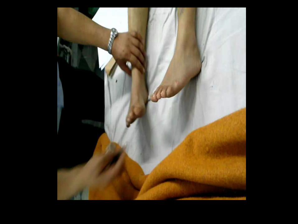
Right Left.Tone: clasp knife slightly inc.Bulk: reduced normalPower: 2/5 5/5Reflexes: brisk normalAnkle clonus +++ -Planters up down
CNS
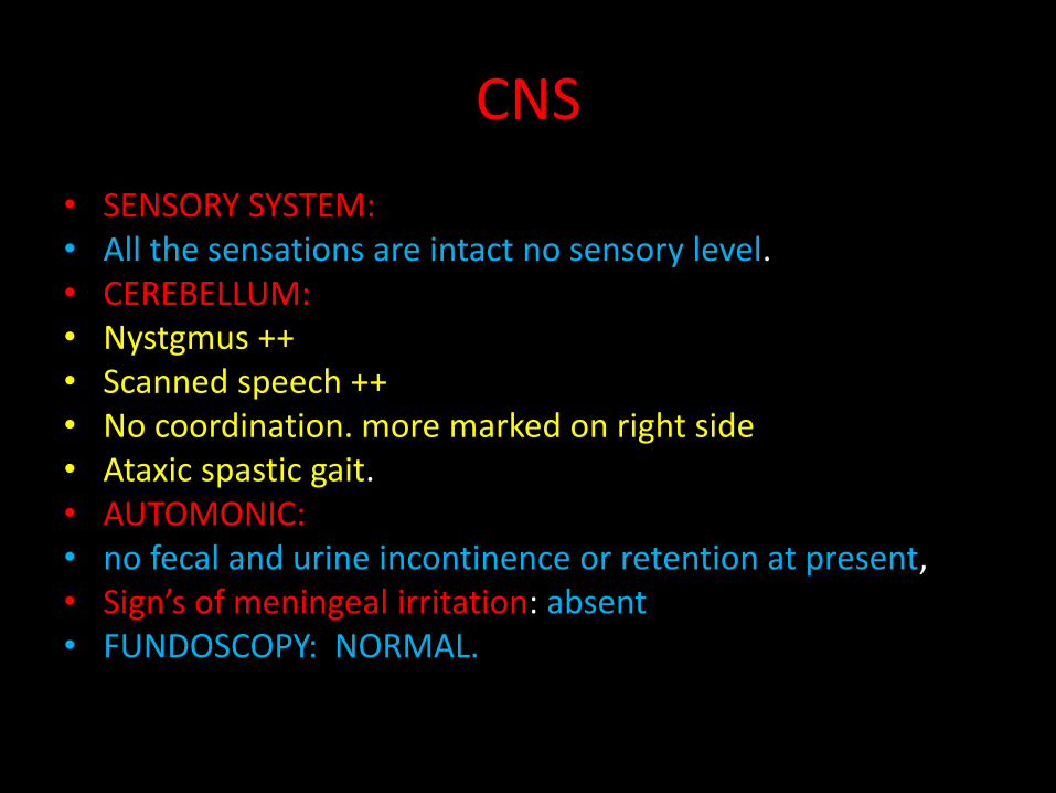
• SENSORY SYSTEM:• All the sensations are intact no sensory level.• CEREBELLUM:• Nystgmus ++• Scanned speech ++• No coordination. more marked on right side • Ataxic spastic gait.• AUTOMONIC: • no fecal and urine incontinence or retention at present,• Sign’s of meningeal irritation: absent• FUNDOSCOPY: NORMAL.


CVS; NORMAL
RESPIRATION: NORMAL
GIT: NORMAL
BREAST EXAMINTIION NORMAL
SKIN AND JOINTS EXAMINATION NORMAL
CASE SUMMARY
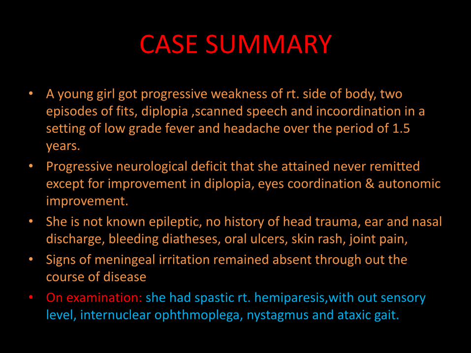
• A young girl got progressive weakness of rt. side of body, two episodes of fits, diplopia ,scanned speech and incoordination in a setting of low grade fever and headache over the period of 1.5 years.
• Progressive neurological deficit that she attained never remitted except for improvement in diplopia, eyes coordination & autonomic improvement.
• She is not known epileptic, no history of head trauma, ear and nasal discharge, bleeding diatheses, oral ulcers, skin rash, joint pain,
• Signs of meningeal irritation remained absent through out the course of disease
• On examination: she had spastic rt. hemiparesis,with out sensory level, internuclear ophthmoplega, nystagmus and ataxic gait.
Differential diagnosis
1. Multiple sclerosis
2. Tuberculous encephalitis with multifocal tuberculomas
3. Primary or Metastatic brain tumor
4. Vasculitis with multifocal infarcts
5. Multisystem atrophy

INVESTIGATIONS
• CBC: normal• TLC: normal• DLC: normal• ESR: 100-7040/mnt.• C-Reactive protiens: negative• LFT’s : normal• RFT’s : normal• S/E : normal• Sr. CALCIUM: 8.1mg/dl ( 8.5-10.5)• Phosphorous: 5.1mg/dl (2.5-5.0)• ALK.po4: 132 (90-295)• BSL: normal

• ANA : -VE
• Anti DS DNS: -ve
• RA factor: -ve
• Blood cultures –ve
• sr.Homocystein level normal
• LUMBAR PUNCTURE: they refused at all the occasions. So not done
• CHEST X-RAYS : NORMAL
• ECG: NORMAL
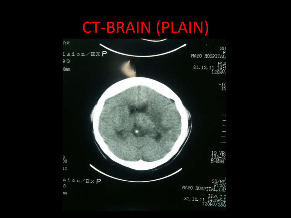
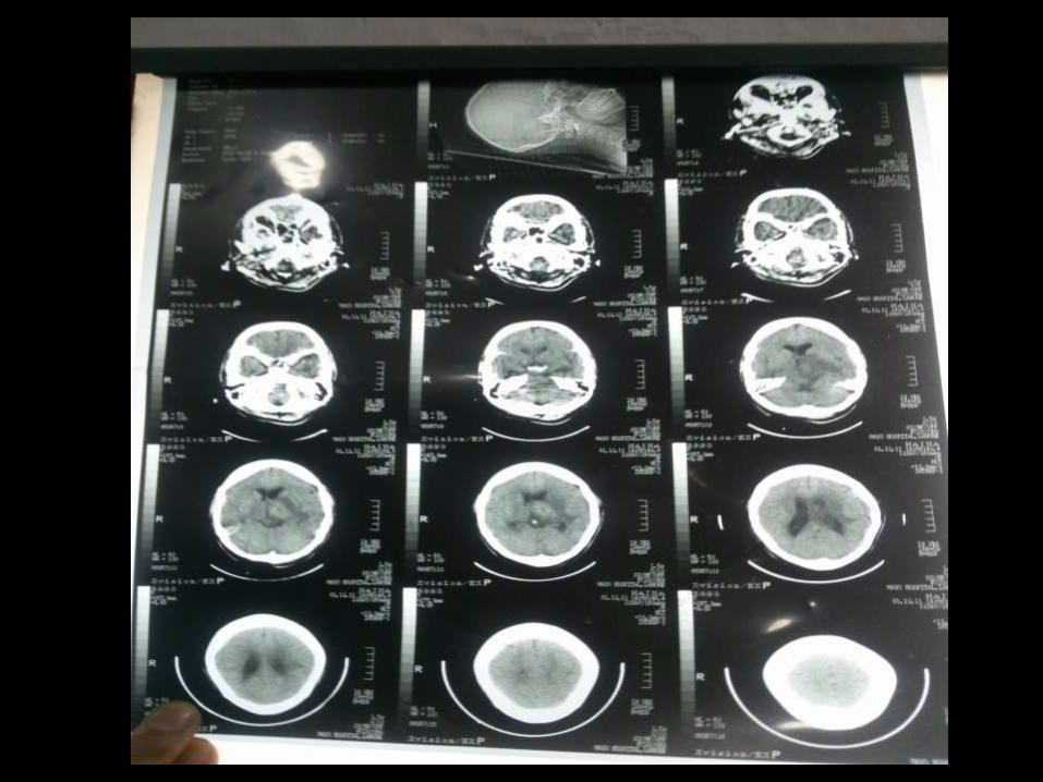
CT-BRAIN (PLAIN)
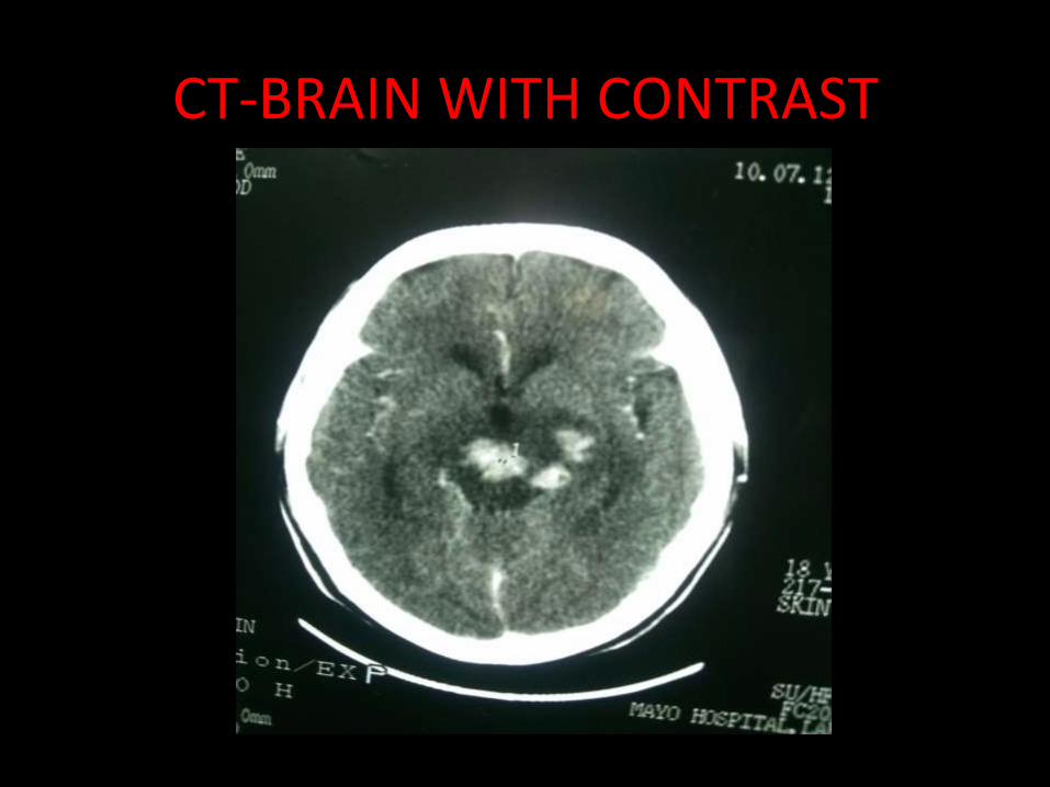
CT-BRAIN WITH CONTRAST

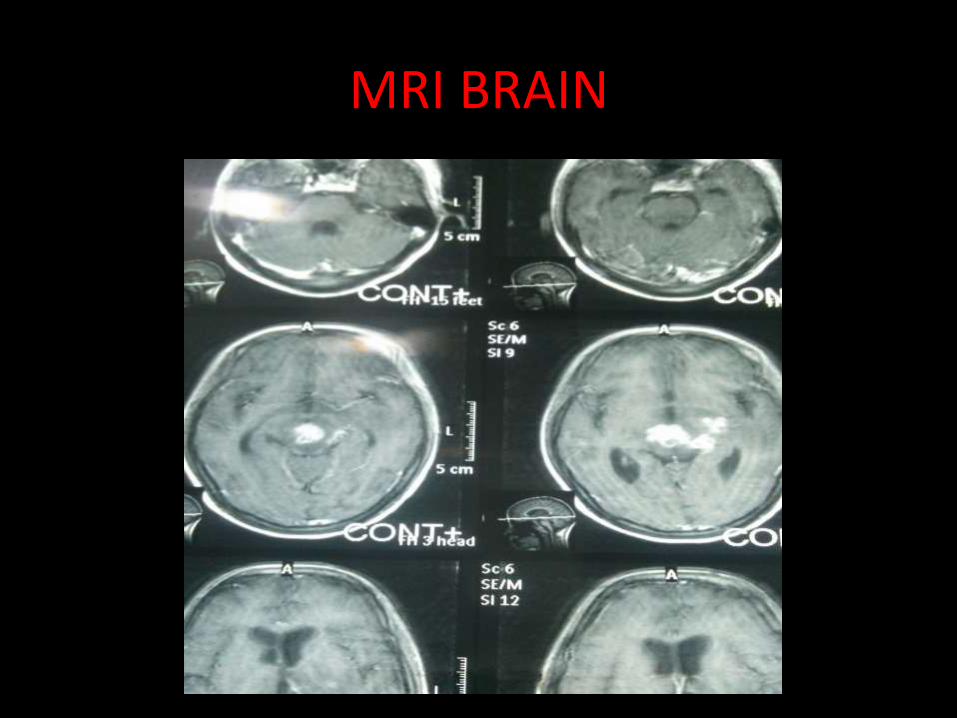
MRI BRAIN
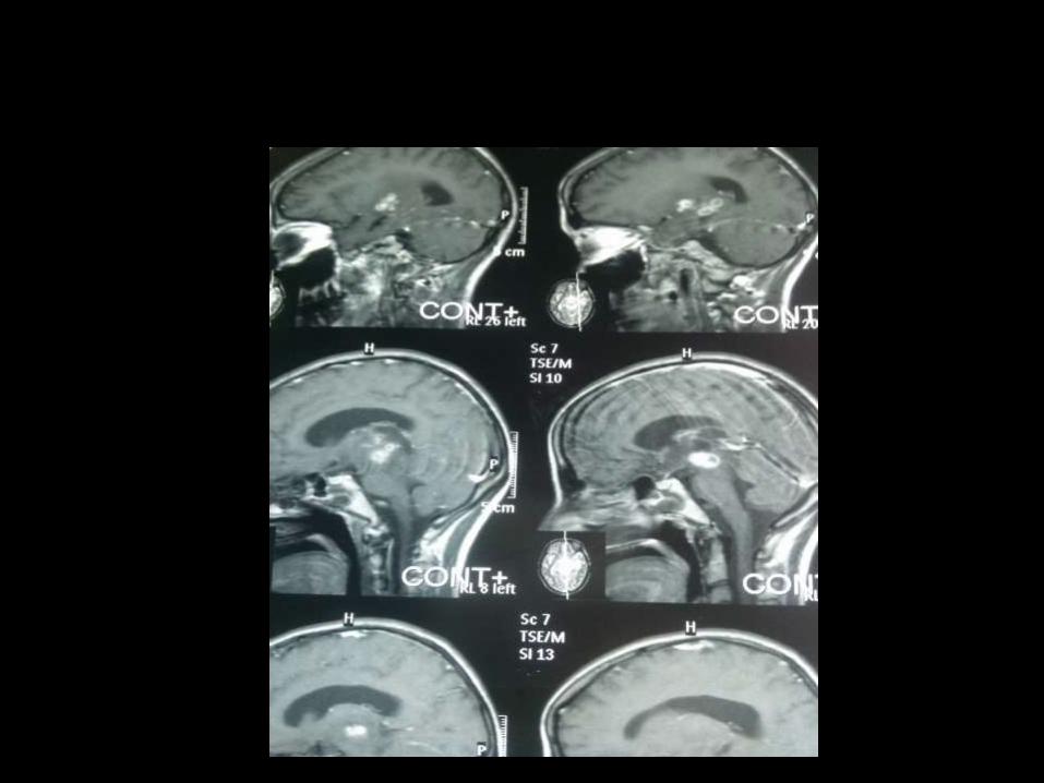
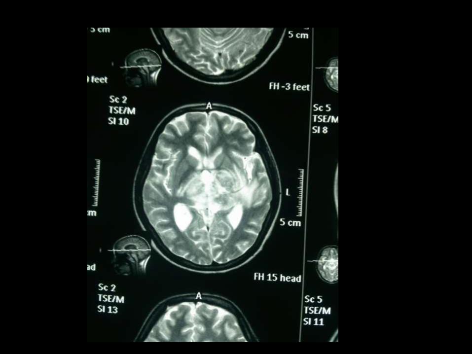
MRI report
• Multiple enhancing leisions are noted in midbrain, left periventricular area and left basal ganglia with disproportional perileisional edema, compressing the third ventricle. Mild non-communicating hydrocephalus.
D/D of midbrain and basal ganglia enhancing lesions
1. Tuberculomas2. Willson’ s disease3. Tuberous sclerosis( sub-ependymal
calcifications)4. Hypoparathyroidism5. Toxoplasmosis6. Mitochondrial diseases.7. CNS lymphoma after radiotherapy
• NERVE CONDUCTION STUDY: NORMAL
• EMG: NORMAL
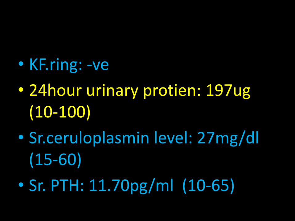
• KF.ring: -ve
• 24hour urinary protien: 197ug (10-100)
• Sr.ceruloplasmin level: 27mg/dl (15-60)
• Sr. PTH: 11.70pg/ml (10-65)
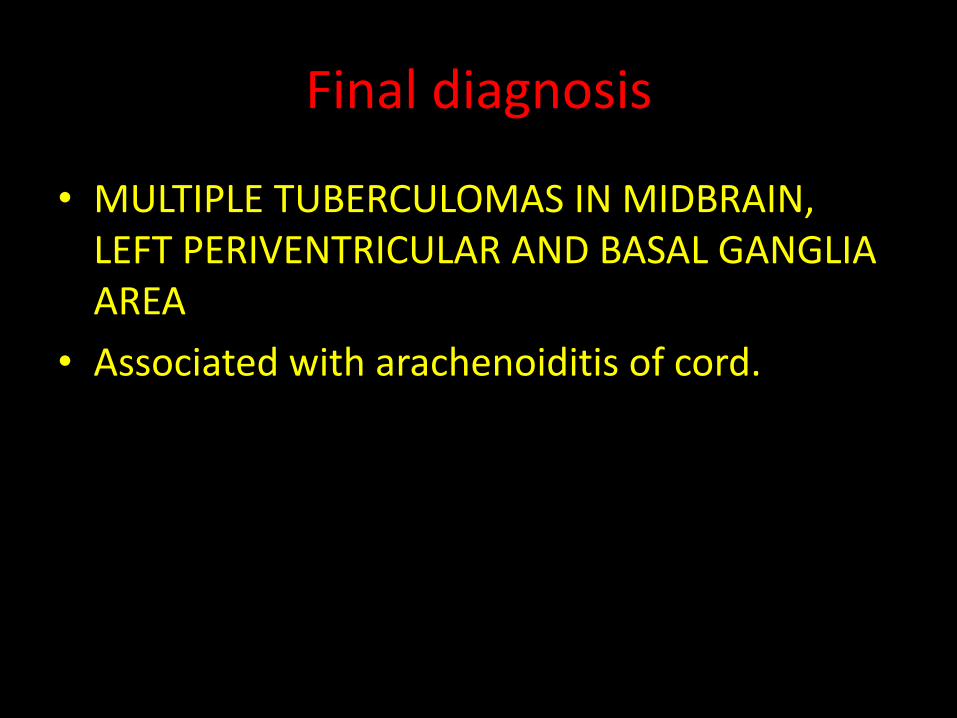
Final diagnosis
• MULTIPLE TUBERCULOMAS IN MIDBRAIN, LEFT PERIVENTRICULAR AND BASAL GANGLIA AREA
• Associated with arachenoiditis of cord.

TREATMENT
• ANTI- TUBERCULOUS THERAPY
• STEROIDS: for 4 to 6 months.
• MANITOL.
• DURATION OF THERAPY:
• ATT. SHOULD BE CONTINUED TILL THE TUBERCULOMA RESOLVE ON REPEATED MRI.