Embed Size (px)
Citation preview

THE ROLE OF FRUCTOSE IN PANCREATIC CANCER CELLS
BY
ADJEKUKOR, UFUOMA CYNTHIA13CP015803
BIOCHEMISTRY PROGRAMMEDEPARTMENT OF BIOLOGICAL SCIENCES,
COVENANT UNIVERSITY, OTASUPERVISED BY
DR T.M. DOKUNMU14th SEPTEMBER 2016 1

OUTLINE Introduction Fructose Metabolism Roles of Fructose in Pancreatic Cancer
Cell proliferation Uric Acid Production DNA damage
Fructose Mechanisms in Carcinogenesis and Cancer Growth Proliferation of cancer cells through the following mechanisms Induction of Oxidative Stress and insulin resistance Oxidative Stress, Inflammation and DNA damage
Recent Advances in the Role of Fructose on Pancreatic Cancer Cells Role of Fructose in Other Cancers Importance and Implications Conclusion References
2

INTRODUCTION
The word ‘cancer’ is used to describe any disease in which cells are abnormal, grow out of control, and can spread. Thus, pancreatic cancer occurs when abnormal cells grow out of control in tissue of the pancreas and form tumour.
Pancreatic cancer is the fourth leading cause of cancer related death in the United States (Siegel et al., 2013). By 2030, it is expected to be the second leading cause of cancer death.
95% of pancreatic cancer type is the pancreatic adenocarcinoma. In 2012, it was the twelfth most common cancer worldwide (World Cancer Research Fund International, 2015).
Just like other living cells, tumour cells possess the potential for proliferation, differentiation, cell cycle arrest and apoptosis.
3

Cancer is not a single disease with a single aetiology, but it is rather a general disease category that induces many distinct diseases with distinct aetiologies (Terry et al., 2001).
A.
B.
Fig.1 Structure of pancreatic tumour and normal pancreas4

Introduction The survival rates of tumour cells is determined by macromolecule synthesis
pathway (Boros et al., 2002).
Fructose promotes flux through the pentose phosphate, which enhances protein synthesis and may indirectly increase tumour growth (Port et al., 2012).
Fructose consumption has increased over the past years especially in its principal form; High-Fructose Corn Syrup (HFCS, 10-53% glucose and 42-90% fructose).
HFCS is used as a sweetener in processed foods, baked goods, condiments, soft drinks, candy, dairy products and concentrated fruit juices.
HFCS has been linked to the epidemiology of different human diseases such as obesity, diabetes and cancer (Charrez et al., 2015).
Therefore, understanding the metabolism of fructose will give an insight on its effects and roles in tumour progression.
5

Glucose and fructose have identical formulas (C6H12O6),
Differences in their chemical structure result in completely distinct absorptive and metabolic properties, which have fundamental implications for cellular functions and disease (Varman, 2011).
Fig 2. Structure of glucose and fructose (Charrez et al., 2015)
FRUCTOSE METABOLISM
6

Fructose is transported using the fructose specific transporter, glucose transporter 5 (GLUT5).
While glucose is transported to the hepatocytes and other cell types using the glucose specific insulin-dependent transporter, GLUT4.
GLUT5 does not respond to insulin, thus leaving fructose uptake uninhibited.
Fig 3. Fructose and glucose metabolism in the liver (Charrez et al., 2015) 7
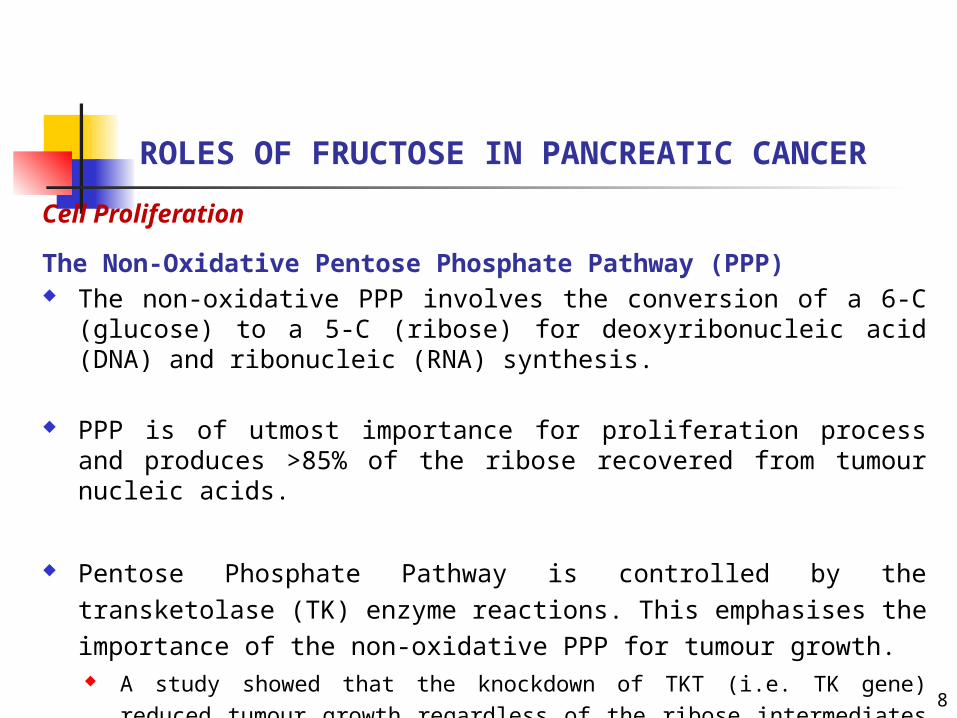
ROLES OF FRUCTOSE IN PANCREATIC CANCERCell Proliferation
The Non-Oxidative Pentose Phosphate Pathway (PPP) The non-oxidative PPP involves the conversion of a 6-C (glucose)
to a 5-C (ribose) for deoxyribonucleic acid (DNA) and ribonucleic (RNA) synthesis.
PPP is of utmost importance for proliferation process and produces >85% of the ribose recovered from tumour nucleic acids.
Pentose Phosphate Pathway is controlled by the transketolase (TK) enzyme reactions. This emphasises the importance of the non-oxidative PPP for tumour growth. A study showed that the knockdown of TKT (i.e. TK gene) reduced
tumour growth regardless of the ribose intermediates suggesting that the 5-carbon sugar was not a limiting factor in cancer growth (Xu et al., 2016).
8

Fig 4. Non-oxidative Pentose Phosphate Pathway
Fructose is preferentially used to glucose in non-oxidative PPP (Port et al., 2012).
The body uses PPP to generate reduced nicotinamide adenine dinucleotide phosphate (NADPH) and pentoses as well as ribose-5-phosphate, a precursor for nucleotide synthesis.
9

Roles of fructose in pancreatic cancer
Uric Acid Production Dietary fructose when absorbed by the body is the only sugar
that raises the uric acid concentration in the blood (Nakagawa et al., 2006).
Uric acid is highly implicated in the pathogenesis of pancreatic cancer.
Ketohexokinase (KHK) also known as fructokinase, is insulin and citrate independent.
KHK permits the phosphorylation of fructose to fructose-1-phosphate in the liver and this reaction occurs without any feed back inhibition (Vos and Lavine, 2013).
The phosphate molecule required for this reaction comes from the depletion of adenosine triphosphate (ATP).
10

Uric Acid Production in Pancreatic Cancer
Fructose promotes the production of uric acid.
Uric acid is a by
product of nucleotide metabolism through the non-oxidative PPP (Charrez et al., 2015).
Fig 5. Fructose metabolism resulting in production of uric acid 11

DNA Damage For protein synthesis in pancreatic cancer cells, fructose is
preferentially used to glucose through the TK-mediated metabolism to synthesise additional nucleic acid that facilitate proliferation capacity (Liu et al., 2010).
Liu and Heaney (2011) reported that after the incubation of Escherichia coli plasmid PBR322 for 15 days with fructose and glucose phosphates metabolites, it resulted in variety of DNA modifications and damage.
The intensity of the damage done is in the following order: glucose-1-phosphate < glucose < glucose-6-phosphate < fructose-1-phosphate < fructose < fructose-6-phosphate.
Roles of fructose in pancreatic cancer
12

1. Proliferation of cancer cells occur through the following mechanisms:
Large amounts of fructose expressed in the liver leads to de novo lipogenesis (DNL, which is the synthesis of fatty acids from the condensation of two carbon units in the form of acetyl CoA), triglyceride accumulation, reduced insulin sensitivity and hepatic insulin resistance.
Fructose increase production of tumour necrosis factor-alpha (TNF-α), reactive oxygen species (ROS), insulin-like growth factor (IGF), sex hormone-binding globulin (SHBG), interleukin 6 (IL-6), and vascular endothelial growth factor (VEGF).
These are mediators of inflammatory response which induce oxidative stress in cancer cells.
These factors leads to hyperglycaemia, obesity, metabolic syndrome, chronic inflammation of cells, DNA damage and ultimately promoting carcinogenesis.
FRUCTOSE MECHANISMS IN CARCINOGENESIS AND CANCER GROWTH
13

Fig 6. Fructose mechanism in carcinogenesis and cancer growth (Liu and Heaney, 2011). 14
Fructose mechanism in carcinogenesis and cancer growth
2. Induction of Oxidative Stress and insulin resistance: Fructose supplies substrates like glycerol-3-phosphate and acyl-CoA for
the up regulation of DNL. This contributes to cancer risk by increasing oxidative stress, and
compromising the cellular antioxidant defense mechanisms.
Fructose increases reduced nicotinamide adenine dinucleotide phosphate (NADPH),
This is required in large amounts for fatty acid synthesis, Acts as a co-factor in maintaining the reduced level of glutathione and
thioreductase.
Miatello et al. (2001) reported that rats treated with fructose led to hyperinsulinaemia, insulin resistance, impaired glucose tolerance and decreased endothelium Nitric Oxide Synthase (eNOS) activity.
15

Fig 7. Mechanisms for fructose-induced insulin resistance (Tappy and Lê, 2010).16

3. Oxidative Stress, inflammation and DNA damage Fructose leads to elevated production of uric acid, a potent inhibitor of
nitric oxide synthase which is an anti-inflammatory factor.
Uric acid induces oxidative stress in vascular smooth muscle cells, endothelial cells, adipocytes, islet cells and hepatocytes (Johnson et al., 2013).
Oxidative stress can cause oxidative damage to large biomolecules such as proteins, DNA, lipids, resulting in increased risk of cancer and cardiovascular diseases (Liu, 2013).
Inflammation is associated with high level of reactive oxygen species that can damage most of the body tissues and the genetic material (DNA), which leads ultimately to cancer formation (Banzzan et al., 2013).
Fructose mechanism in carcinogenesis and cancer growth
17

18
RECENT ADVANCES IN THE ROLE OF FRUCTOSE IN PANCREATIC CANCER CELLS
Fructose is the only sugar that raises uric acid concentration in the blood, which can block the ability of insulin to regulate how body cells use and store sugar and other nutrients for energy.
Jiao et al. (2009) reported that serum uric acid concentration has been shown to have a strong positive association with the risk of pancreatic cancer mortality in men.
Pericleous et al. (2014) reported that hyperinsulinaemia, metabolic syndrome and type II diabetes mellitus have all been associated with pancreatic cancer.
Interestingly, the drug metformin that is used to treat diabetes appears to reduce pancreatic cancer.

ROLE OF FRUCTOSE IN OTHER CANCERS
The glucose transporter-1 (GLUT1) and glucose transporter-2 (GLUT2) were found to be present in both normal and neo-plastic breast cancer. But GLUT5, which is the fructose transporter, was found to be over expressed in the human breast cancer tissue and absent from normal breast tissue (Zamora-Léon et al., 1996).
In a study, males with high intakes of dietary glycaemic load: fructose and sucrose resulted in a significant 27% - 37% increase in colorectal cancer. However, for women, these factors did not lead to an increase in colorectal cancer (Michaud et al., 2005).
Banzzan et al. (2013) reported that fructose intake that is associated with obesity has be shown to be used preferentially in the proliferation of cancer cells of the small intestine.
Also, the TkT enzyme in the non-oxidative pentose phosphate pathway was found to be the most predominant enzyme in hepatocellular carcinoma (HCC). This enzyme was over expressed, which lead to venous invasion, increased tumour size, and absence of tumour encapsulation. (Xu et al., 2016).
19

IMPORTANCE AND IMPLICATIONS The consumption of beverages sweetened with sugar or HFCS has been
linked to the risk of developing obesity, diabetes, heart disease, metabolic syndrome, nonalcoholic fatty liver disease (NAFLD) and gout (Bray, 2013).
Clinically, patients with cancer burden have the tendency to develop thiamine depletion, which is a co-factor for TK-mediated reactions.
Thiamine can be reduced in diets to reduce cancer proliferation.
Fasting fructose levels in pancreatic cancer patients was shown to be 2.5 folds higher than in healthy patients (Liu et al., 2010).
Also, increased serum uric acids were present in patients with high consumption of fructose.
Fructose intake should be reduced, especially in cancer patients to reduce the progression of cancer and uric acid production which induces oxidative stress. 20

CONCLUSION Although fructose is implicated in pancreatic cancer cells, the natural form of
fructose from fruits is believed to have the potency to fight against different human diseases.
Natural fruits containing fructose are thought to be safe because they contain numerous substances that can block the side effects of fructose such as:
potassium, vitamin C and antioxidants –e.g. quercetin and other flavonols (Johnson et al., 2013).
It is fructose from added sugars that is linked with hypertension, DNA damage, cancers etc.
Dietary supplement containing fructose does not have the same effect as natural fruit because phytochemicals in fruits and vegetables posses potent antioxidant property.
A study reported that apple extract with skin reduced tumour growth compared with apple extract without skin (Liu, 2013).
Together, glucose and fructose may act synergistically to support malignant growth (Port et al., 2012).
This is possible because excess glucose can be converted to fructose which will have the same effect as fructose in carcinogenesis and cancer growth (Charrez et al., 2015).
21

REFERENCES
Bazzan, A.J., Newberg, A.B., Cho, W.C. and Monti, D.A. (2013). Diet and Nutrition in Cancer Survivorship and Palliative Care. Evidence-Based Complementary and Alternative Medicine 2013: 1-12.
Boros, L.G., Lee, W.P. and Go, V.L. (2002). A Metabolic Hypothesis of Cell Growth and Death in Pancreatic Cancer. Pancreas 24: 26-33.
Bray, G.A. (2013). Energy and Fructose From Beverages Sweetened With Sugar or High-Fructose Corn Syrup Pose a Health Risk for Some People. Advances in Nutrition 4: 220-225.
Charrez, B., Qiao, L. and Hebbard, L. (2015). The role of fructose in metabolism and cancer. Hormone Molecular Biology and Clinical Investigation 22: 79-89.
Liu, H. and Heaney, A.P. (2011). Refined fructose and cancer. Expert Opinion on Therapeutic Targets 15: 1049-1059.
Liu, H., Huang, D., McArthur, D.L., Boros, L.G., Nissen, N. and Heaney, A.P. (2010). Fructose Induces Transketolase Flux to Promote Pancreatic Cancer Growth. Cancer Research 70:
22

References
6368-6376.
Liu, R.H. (2003). Health benefits of fruit and vegetables are from additive and synergistic combinations of phytochemicals. The American Journal of Clinical Nutrition 78: 517-520.
Jiao, L., Flood, A., Subar, A.F., Hollenbeck, A.R., Schatzkin, A. and Stolzenberg-Solomon, R. (2009). Glycemic Index, Carbohydrates, Glycemic Load, and the Risk of Pancreatic Cancer in a Prospective Cohort Study. Cancer Epidemiology, Biomarkers & Prevention 18: 1144-1151.
Johnson, R.J., Nakagawa, T., Sanchez-Lozada, G., Shafiu, M., Sundaram, S., Le, M., Ishimoto, T., Sautin, Y.Y. and Lanaspa, M.A. (2013). Sugar, Uric Acid, and the Etiology of Diabetes and Obesity. Diabetes 62: 3307-3315.
Miatello, R., Risler, N., Castro, C., Gronzález, S., Rüttler, M. and Cruzado, M. (2001). Aortic smooth muscle cell proliferation and endothelial nitric oxide synthase activity in fructose-fed rats. American Journal of Hypertension 14: 1135-1141. 23

24
References
Michaud, D.S., Fuchs, C.S., Liu, S., Willet, W.C., Colditz, G.A. and Giovannucci, E. (2005). Dietary Glycemic Load, Carbohydrate, Sugar, and Colorectal Cancer Risk in Men and Women. Cancer Epidemiology, Biomarkers & Prevention 14: 138-143.
Nakagawa, T., Hu, H., Zharikov, S., Tuttle, K.R., Short, R.A., Glushakova, O., Ouyang, X., Feig, D.I., Block, E.R., Herrera-Acosta, J., Patel, J.M. and Johnson, R.J. (2006). A casual role for uric acid in fructose-induced metabolic syndrome. American Journal of Physiology - Renal 290: 625-631.
Port, A.M., Ruth, M.R. and Istfan, N.W. (2012). Fructose consumption and cancer: Is there a connection? Current Opinion in Endocrinology, Diabetes, and Obesity 19: 367-374.
Siegel, R., Naishadham, D. and Jemal, A. (2013). Cancer Statistics, 2013. CA: A Cancer Journal for Clinicians 63: 11-30.
Terry, P., Terry, J.B. and Wolk, A. (2001). Fruit and vegetable consumption in the prevention of cancer: An update. Journal of Internal Medicine 250: 280-290.

References
World Cancer Research Fund International (2015). Pancreatic cancer statistics. http://www.wcrf.org/int/cancer-facts-figures/data-specific-cancers/pancreatic-cancer-statistics. Sourced on Monday, August 01, 2016 at 03:00pm.
Varman, T.S. (2011). Fructose induced lipogenesis: From sugar to fat to insulin resistance. Trends in Endocrinology & Metabolism 22: 60-65.
Vos, M.B. and Lavine, J.E. (2013). Dietary Fructose in Nonalcoholic Fatty Liver Disease. Hepatology 57: 2525-2531.
Xu, I.M., Lai, R.K., Lin, S., Tse, A.P., Chiu, D.K., Koh, H., Law, C., Wong, C., Cai, Z., Wong, C.C. and Ng, I.O. (2016). Transketolase counteracts oxidative stress to drive cancer development. Proceedings of the National Academy of Sciences of the United States of America 113: 725-734.
Zamora-León, S.P., Golde, D.W., Concha, I.I., Rivas, C.I., Delgado-López, F., Baselga, J., Nualart, F. and Vera, J.C. (1996). Expression of the fructose transporter GLUT5 in human breast cancer. Proceedings of the National Academy of Sciences of the United States of America 93: 1847-1852. 25

THANK YOU FOR
LISTENING 26