Embed Size (px)
Citation preview

Ottawa 2015
THE USE OF LASERS
IN PODIATRIC
MEDICINE
© 2015 BritaMed Inc. All rights reserved. Any use of the material, including reproduction in whole or in part, requires permission in writing from BritaMed Inc.

© 2015 BritaMed Inc. All rights reserved. Any use of the material, including reproduction in whole or in part, requires permission in writing from BritaMed Inc.
Achilles
tendinitis
Plantar
fasciitis
Neuroma
Wound
Healing
Callus
& Corns
Foot
Telangiectasia
Warts Mosaic &
Solitary
Fungal
Nail Infection

© 2015 BritaMed Inc. All rights reserved. Any use of the material, including reproduction in whole or in part, requires permission in writing from BritaMed Inc.
Fungal nail infection Onychomycosis caused by Dermatophytes and Yeasts
Soft tissue inflammation - Plantar fasciitis - Achilles tendinitis - Morton's neuroma
Warts Plantar warts (solitary and mosaic); ablative and non-ablative programs
Callus & Corns
Small vessels coagulation Foot telangiectasia, Venous lakes, Spider naevi
Wound healing Diabetic foot ulcers; Burns; Post surgery recovery
© 2015 BritaMed Inc. All rights reserved. Any use of the material, including reproduction in whole or in part, requires permission in writing from BritaMed Inc.

© 2015 BritaMed Inc. All rights reserved. Any use of the material, including reproduction in whole or in part, requires permission in writing from BritaMed Inc.
Nail plate
Cuticle
made of translucent keratin protein
("small moon") the visible part of the matrix
A transparent thin fold of dead skin protecting the new grown nail
Lunula
Eponychium “seals” the proximal
nail plate & bed

© 2015 BritaMed Inc. All rights reserved. Any use of the material, including reproduction in whole or in part, requires permission in writing from BritaMed Inc.
Nail bed
Hyponychium distally “seals” the nail bed
Nail matrix Nail plate
Lunula Eponychium proximally “seals” the nail bed

© 2015 BritaMed Inc. All rights reserved. Any use of the material, including reproduction in whole or in part, requires permission in writing from BritaMed Inc.
The matrix, the “Mother of the Nail”, is the part of the nail that lies underneath the proximal nail fold just in front of the nail root. The leading edge of the matrix is seen as the lunula. The matrix cannot be seen on all nails, but is generally seen on the thumbs, index and middle fingers. The soft, plump cells that comprise the nail plate are developed in the matrix. As they grow out, they loose their inner material and become flat, hard and translucent.
The oldest cells are the most compact, making the nail plate harder and more dense closest to the free edge. The longer the matrix, the more cells it can produce, resulting in a thicker nail plate. Any damage to the matrix can be seen on the emerging nail plate.
© 2015 BritaMed Inc. All rights reserved. Any use of the material, including reproduction in whole or in part, requires permission in writing from BritaMed Inc.
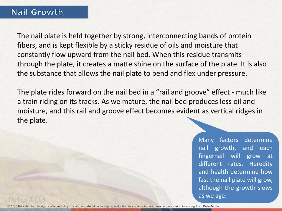
The nail plate is held together by strong, interconnecting bands of protein fibers, and is kept flexible by a sticky residue of oils and moisture that constantly flow upward from the nail bed. When this residue transmits through the plate, it creates a matte shine on the surface of the plate. It is also the substance that allows the nail plate to bend and flex under pressure. The plate rides forward on the nail bed in a “rail and groove” effect - much like a train riding on its tracks. As we mature, the nail bed produces less oil and moisture, and this rail and groove effect becomes evident as vertical ridges in the plate.
Many factors determine nail growth, and each fingernail will grow at different rates. Heredity and health determine how fast the nail plate will grow, although the growth slows as we age.
© 2015 BritaMed Inc. All rights reserved. Any use of the material, including reproduction in whole or in part, requires permission in writing from BritaMed Inc.
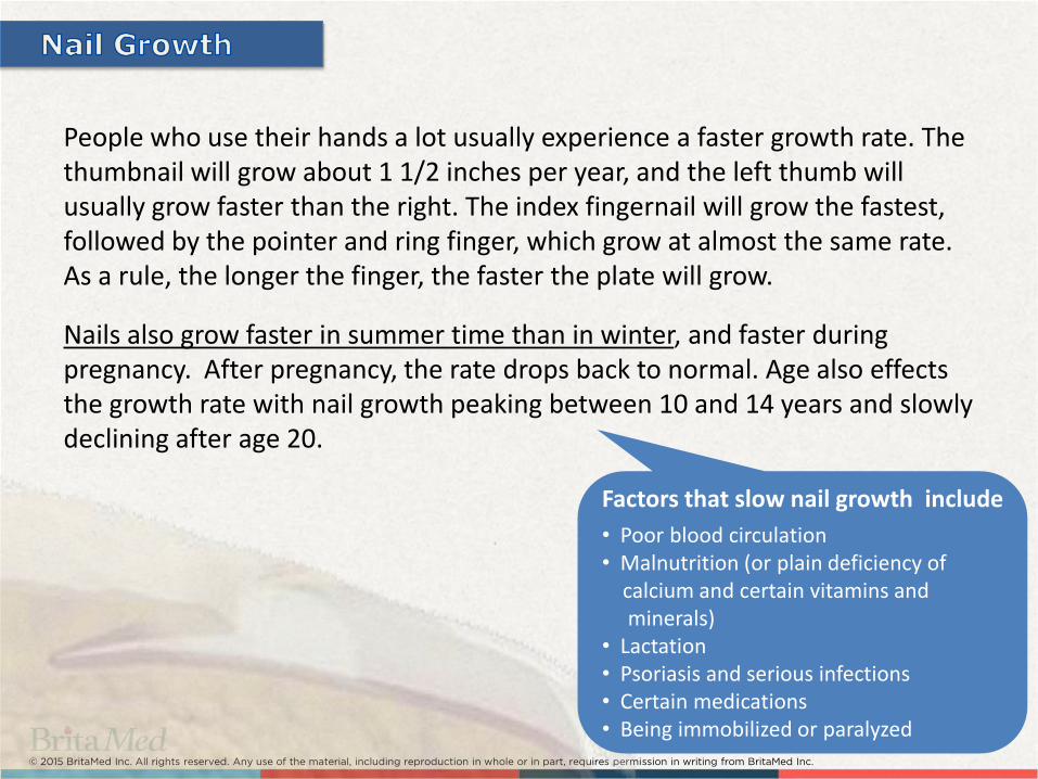
People who use their hands a lot usually experience a faster growth rate. The thumbnail will grow about 1 1/2 inches per year, and the left thumb will usually grow faster than the right. The index fingernail will grow the fastest, followed by the pointer and ring finger, which grow at almost the same rate. As a rule, the longer the finger, the faster the plate will grow.
Nails also grow faster in summer time than in winter, and faster during pregnancy. After pregnancy, the rate drops back to normal. Age also effects the growth rate with nail growth peaking between 10 and 14 years and slowly declining after age 20.
Factors that slow nail growth include
• Poor blood circulation • Malnutrition (or plain deficiency of calcium and certain vitamins and minerals) • Lactation • Psoriasis and serious infections • Certain medications • Being immobilized or paralyzed

© 2015 BritaMed Inc. All rights reserved. Any use of the material, including reproduction in whole or in part, requires permission in writing from BritaMed Inc.
Some people erroneously believe that eating certain foods or using special creams, oils or lotions will increase the growth rate. Although the nail plate requires certain nutrients for proper growth, there is very little evidence that eating any particular food will cause them to grow faster.
Creams, oils and lotions are sometimes sold as 'growth accelerators', although these claims are false, misleading and illegal.
No cosmetic product may claim that it can alter or change any body function. These products and others are only for beautifying the nail plate, and only medical drugs can make such claims

© 2015 BritaMed Inc. All rights reserved. Any use of the material, including reproduction in whole or in part, requires permission in writing from BritaMed Inc.
Onychomycosis is not easy to treat because nails develop slowly and receive very little blood supply.
Fungal infection that causes the fingernails or toenails to thicken, discolor, disfigure and split is called Onychomycosis (also known as Tinea Unguium) . Without treatment, the nails will turn so thick that they press against the side of the shoes, causing strain, irritation and pain.
Areas affected by Onychomycosis
Diabetic patients are usually more sensitive to Onychomycosis infections due to reduced blood circulation at the extremities, as well as the body's capabilities to fight infections. Small cuts, infections and other foot injuries can have serious consequences with diabetic patients.

© 2015 BritaMed Inc. All rights reserved. Any use of the material, including reproduction in whole or in part, requires permission in writing from BritaMed Inc.
Distal and Lateral Subungual (DSO)
Proximal Subungual (PSO)
White Superficial (WSO)
Total dystrophic (all 3 sites affected)

© 2015 BritaMed Inc. All rights reserved. Any use of the material, including reproduction in whole or in part, requires permission in writing from BritaMed Inc.

© 2015 BritaMed Inc. All rights reserved. Any use of the material, including reproduction in whole or in part, requires permission in writing from BritaMed Inc.
Trichophyton rubrum (T. Rubrum) responsible also for athlete's foot, jock itch and ringworm.
Trichophyton mentagrophytes
(T. Mentagrophytes) has at least five different variants.
Candida Albicans or Candida Parapsilosis
these infections are less frequent though produce similar symptoms.
White superficial
Surrounding tissue effects
Dermatophytes (Primary fungi family causing Onychomycosis)
Yeasts
Oily, yellowish hue
© 2015 BritaMed Inc. All rights reserved. Any use of the material, including reproduction in whole or in part, requires permission in writing from BritaMed Inc.
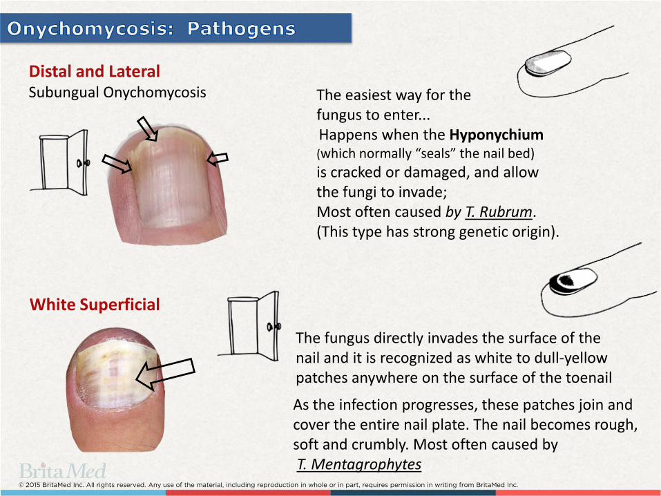
Distal and Lateral Subungual Onychomycosis The easiest way for the
fungus to enter... Happens when the Hyponychium (which normally “seals” the nail bed)
is cracked or damaged, and allow the fungi to invade; Most often caused by T. Rubrum. (This type has strong genetic origin).
White Superficial
As the infection progresses, these patches join and cover the entire nail plate. The nail becomes rough, soft and crumbly. Most often caused by T. Mentagrophytes
The fungus directly invades the surface of the nail and it is recognized as white to dull-yellow patches anywhere on the surface of the toenail
© 2015 BritaMed Inc. All rights reserved. Any use of the material, including reproduction in whole or in part, requires permission in writing from BritaMed Inc.
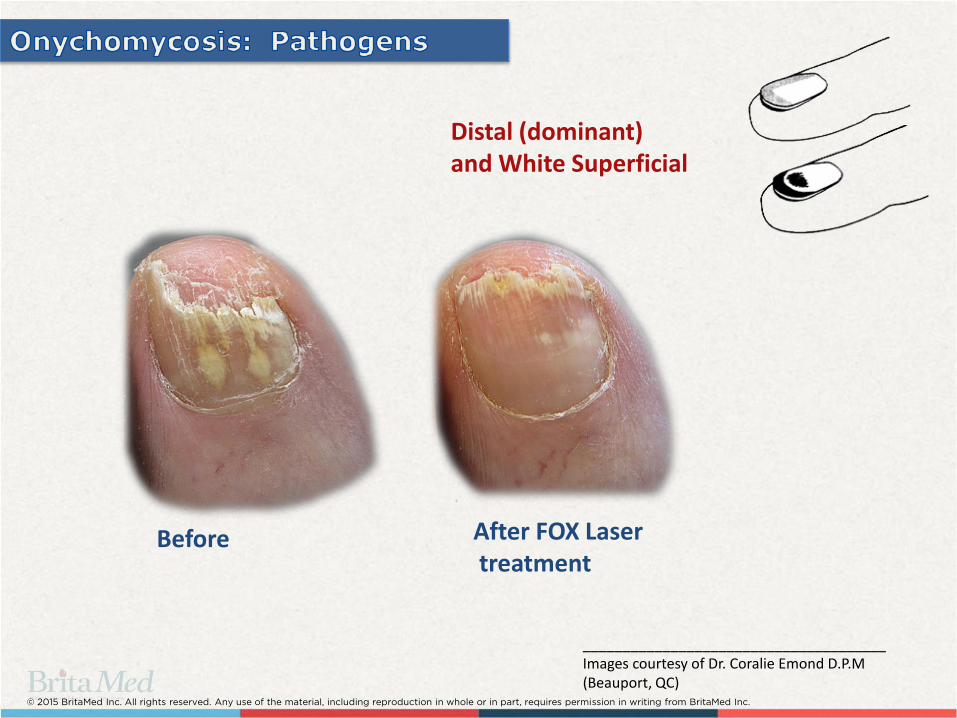
After FOX Laser treatment
Before
Distal (dominant) and White Superficial
______________________________________ Images courtesy of Dr. Coralie Emond D.P.M (Beauport, QC)
© 2015 BritaMed Inc. All rights reserved. Any use of the material, including reproduction in whole or in part, requires permission in writing from BritaMed Inc.
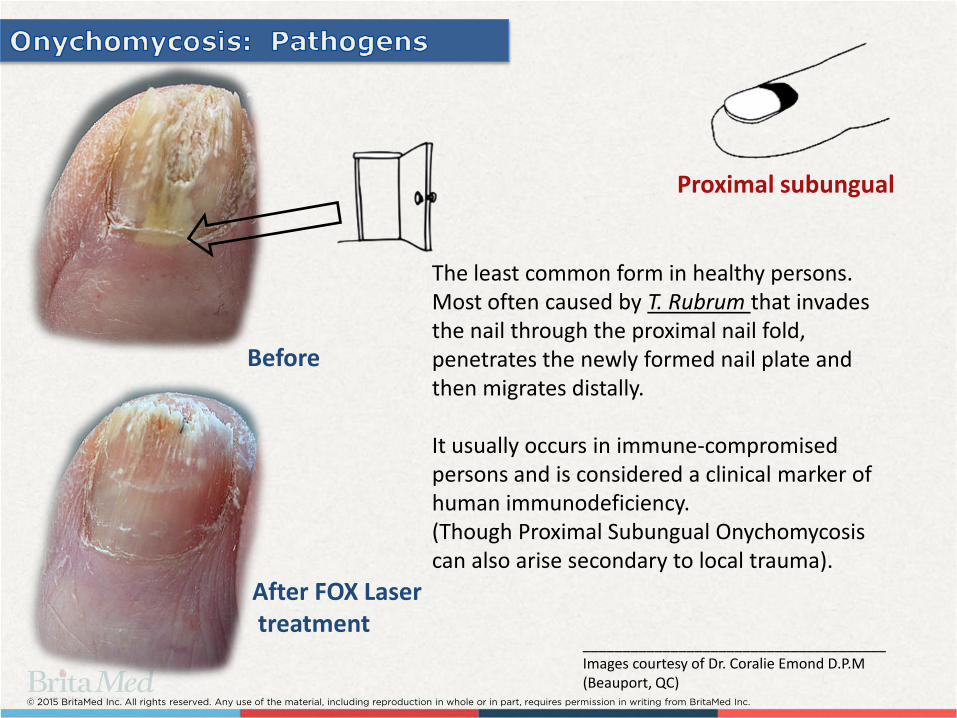
Proximal subungual
The least common form in healthy persons. Most often caused by T. Rubrum that invades the nail through the proximal nail fold, penetrates the newly formed nail plate and then migrates distally. It usually occurs in immune-compromised persons and is considered a clinical marker of human immunodeficiency. (Though Proximal Subungual Onychomycosis can also arise secondary to local trauma).
After FOX Laser treatment
Before
______________________________________ Images courtesy of Dr. Coralie Emond D.P.M (Beauport, QC)

© 2015 BritaMed Inc. All rights reserved. Any use of the material, including reproduction in whole or in part, requires permission in writing from BritaMed Inc.
Photoinactivation: (Cytoplasmatic membrane damage)
Local white blood cells increment
2. Generate instant localized temperature rise
1. High intensity infrared radiation
3. Accelerate healing
Enzymes denaturing
© 2015 BritaMed Inc. All rights reserved. Any use of the material, including reproduction in whole or in part, requires permission in writing from BritaMed Inc.
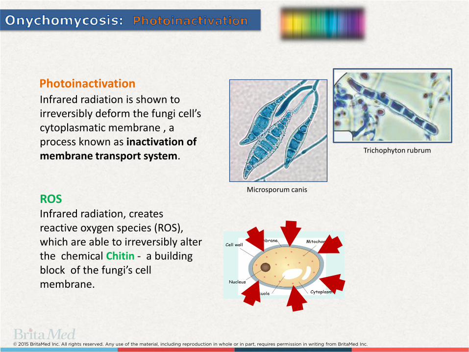
Photoinactivation Infrared radiation is shown to irreversibly deform the fungi cell’s cytoplasmatic membrane , a process known as inactivation of membrane transport system.
ROS Infrared radiation, creates reactive oxygen species (ROS), which are able to irreversibly alter the chemical Chitin - a building block of the fungi’s cell membrane.
Trichophyton rubrum
Microsporum canis

© 2015 BritaMed Inc. All rights reserved. Any use of the material, including reproduction in whole or in part, requires permission in writing from BritaMed Inc.
High levels of heat effect (or thermal damage) the Mycelium by denaturing the enzymes the fungus uses to digest the Keratin - nail’s protein. Heat basically solidify these enzymes just like cooking an egg and... Solid enzymes cannot flow freely through the membrane, causing the fungi to starve. Practically eliminating further spreading.
Mycelium is the vegetative part of a fungus, it is through this part that a fungus absorbs its nutrients from the environment.
Fungi enzymes - Thermal damage
Temperature rise at the nail plate area within 1 minute
(FOX-1064).
© 2015 BritaMed Inc. All rights reserved. Any use of the material, including reproduction in whole or in part, requires permission in writing from BritaMed Inc.
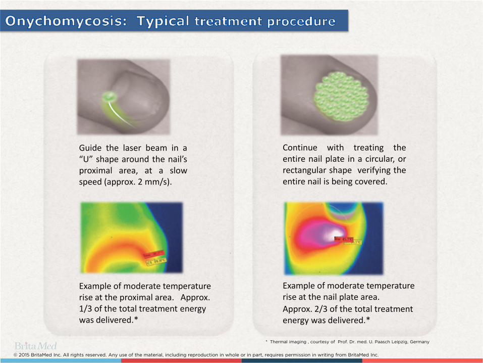
Continue with treating the entire nail plate in a circular, or rectangular shape verifying the entire nail is being covered.
Guide the laser beam in a “U” shape around the nail’s proximal area, at a slow speed (approx. 2 mm/s).
* Thermal imaging , courtesy of Prof. Dr. med. U. Paasch Leipzig, Germany
Example of moderate temperature rise at the proximal area. Approx. 1/3 of the total treatment energy was delivered.*
Example of moderate temperature rise at the nail plate area.
Approx. 2/3 of the total treatment energy was delivered.*

© 2015 BritaMed Inc. All rights reserved. Any use of the material, including reproduction in whole or in part, requires permission in writing from BritaMed Inc.
The treatment is NOT painful. Frequent cooling prevent discomfort to the patient, without affecting the laser effectiveness.
Cool the nail for
a few seconds
Apply laser

© 2015 BritaMed Inc. All rights reserved. Any use of the material, including reproduction in whole or in part, requires permission in writing from BritaMed Inc.
Why doesn't the laser eradicate the fungus pathogens completely in only one session?
Because fungi leaves behind Spores that are incredibly resistant to heat and chemicals.
Spores are like eggs and will hatch when the time is right. After hatching, it becomes possible to eradicate them with the laser.
© 2015 BritaMed Inc. All rights reserved. Any use of the material, including reproduction in whole or in part, requires permission in writing from BritaMed Inc.
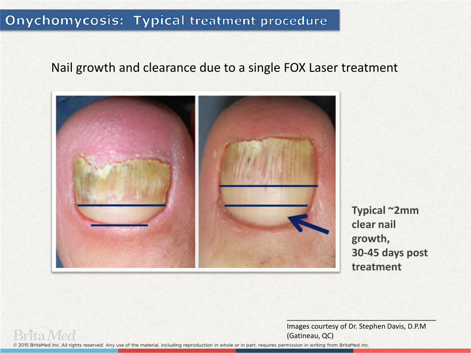
Nail growth and clearance due to a single FOX Laser treatment
Typical ~2mm clear nail growth, 30-45 days post treatment
______________________________________ Images courtesy of Dr. Stephen Davis, D.P.M (Gatineau, QC)

© 2015 BritaMed Inc. All rights reserved. Any use of the material, including reproduction in whole or in part, requires permission in writing from BritaMed Inc.
Day 0 Day 78
Day 180
Typical nail clearance during 3-4 sessions with the FOX Laser
______________________________________ Images courtesy of Dr. Stephen Davis, D.P.M (Gatineau, QC)

© 2015 BritaMed Inc. All rights reserved. Any use of the material, including reproduction in whole or in part, requires permission in writing from BritaMed Inc.
LOW LEVEL
LASER
THERAPY
© 2015 BritaMed Inc. All rights reserved. Any use of the material, including reproduction in whole or in part, requires permission in writing from BritaMed Inc.
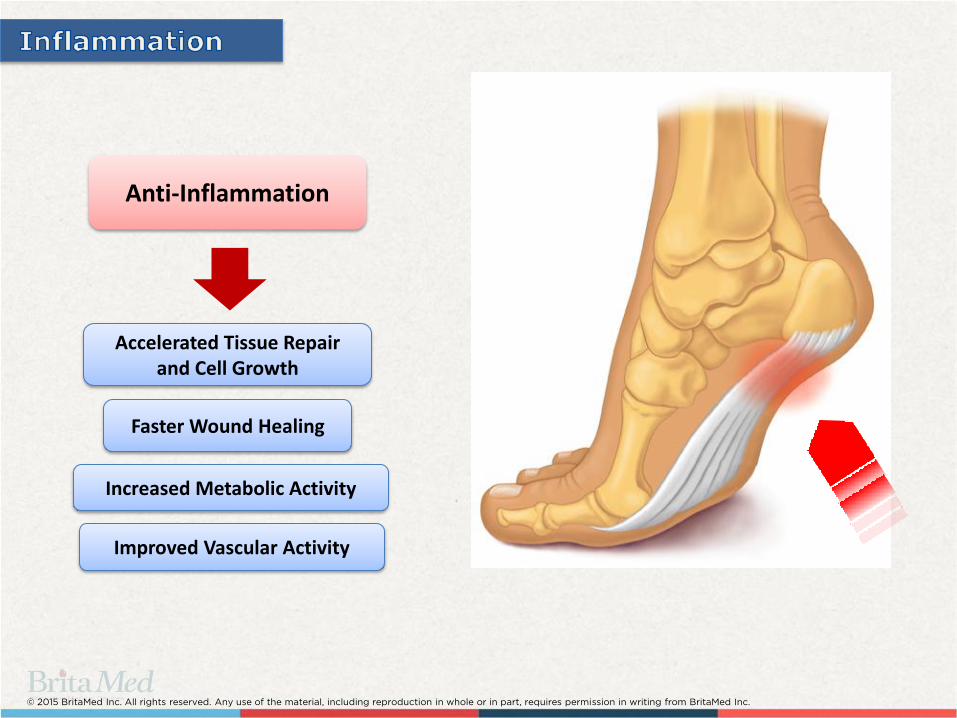
Anti-Inflammation
Accelerated Tissue Repair and Cell Growth
Increased Metabolic Activity
Faster Wound Healing
Improved Vascular Activity
© 2015 BritaMed Inc. All rights reserved. Any use of the material, including reproduction in whole or in part, requires permission in writing from BritaMed Inc.
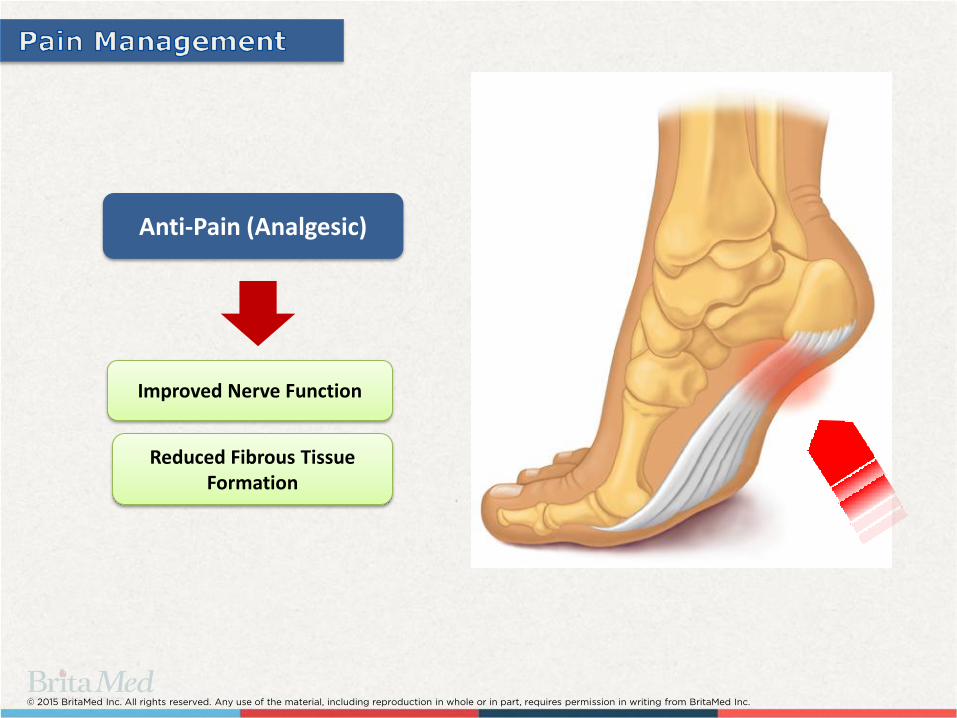
Anti-Pain (Analgesic)
Reduced Fibrous Tissue Formation
Improved Nerve Function

© 2015 BritaMed Inc. All rights reserved. Any use of the material, including reproduction in whole or in part, requires permission in writing from BritaMed Inc.
Laser radiation between in wavelengths of
800nm - 1200nm absorbs very little in
water, meaning the beam can penetrate
through tissue without loosing much of its
energy.
© 2015 BritaMed Inc. All rights reserved. Any use of the material, including reproduction in whole or in part, requires permission in writing from BritaMed Inc.
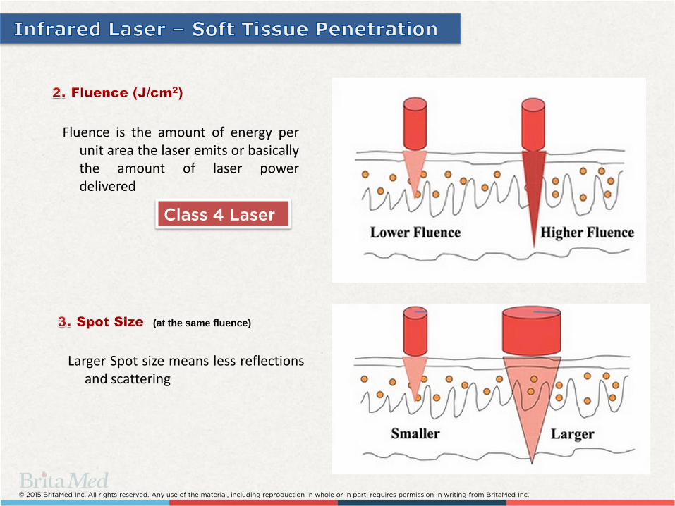
Class 4 Laser
Fluence is the amount of energy per unit area the laser emits or basically the amount of laser power delivered
(at the same fluence)
Larger Spot size means less reflections and scattering

© 2015 BritaMed Inc. All rights reserved. Any use of the material, including reproduction in whole or in part, requires permission in writing from BritaMed Inc.
What is the optimal
Laser power ?
Why not choose 100W Laser?
There is a direct correlation between a
laser’s beam spot size and its effective
penetration depth.
For example: a 980-1064nm Laser with 10 watt creating 2.5cm beam spot, will have penetration depth of approx. 2-3cm 2-3cm
980-1064nm
10-12 Watt
A 2.5-3cm beam spot is an ideal size for focused localized treatment, and therefore a 10-12W laser would be an ideal power for such treatment. This rule applies to most LLLT (Class 4) lasers on the market.
Ø 2.5cm
_______________________________________________ Image courtesy of Dr. Joseph Stern, D.P.M. (Vancouver, BC)

© 2015 BritaMed Inc. All rights reserved. Any use of the material, including reproduction in whole or in part, requires permission in writing from BritaMed Inc.
Acute Inflammation Reduction
How do lasers reduce inflammation?
Stabilization of the cellular membrane
Enhancement of ATP production and synthesis
Stimulation of vasodilation
Acceleration of leukocytic activity
Increased prostaglandin synthesis
Reduction in interleukin
Enhanced lymphocyte response
Increased angiogenesis
Temperature modulation
Enhanced superoxide dismutase (SOD) levels
Decreased C-reactive protein and neopterin levels
© 2015 BritaMed Inc. All rights reserved. Any use of the material, including reproduction in whole or in part, requires permission in writing from BritaMed Inc.
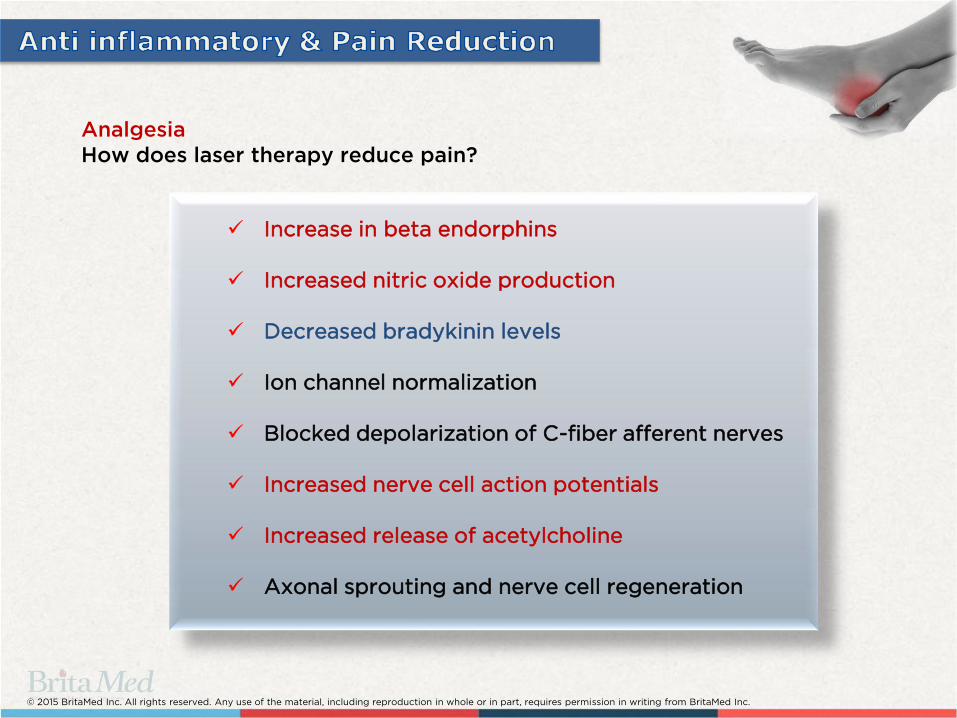
Analgesia
How does laser therapy reduce pain?
Increase in beta endorphins
Increased nitric oxide production
Decreased bradykinin levels
Ion channel normalization
Blocked depolarization of C-fiber afferent nerves
Increased nerve cell action potentials
Increased release of acetylcholine
Axonal sprouting and nerve cell regeneration

© 2015 BritaMed Inc. All rights reserved. Any use of the material, including reproduction in whole or in part, requires permission in writing from BritaMed Inc.
PHOTONS
Absorbed in Mitochondria and Cell Membrane within
Cytochromes and Porphyry’s
Singlet Oxygen is Produced
Changes in Membrane Permeability
ATP Synthesized and DNA Produced
Increase in Cell Metabolism from a Depressed Rate to a
Normal Level
Selective Bio-Stimulatory Effect on Impaired Cells
(note cells and tissues functioning normally are not affected

© 2015 BritaMed Inc. All rights reserved. Any use of the material, including reproduction in whole or in part, requires permission in writing from BritaMed Inc.
_______________________________________________ Image courtesy of Dr. Joseph Stern, D.P.M. (Vancouver, BC)

© 2015 BritaMed Inc. All rights reserved. Any use of the material, including reproduction in whole or in part, requires permission in writing from BritaMed Inc.
Repeat laser irradiation through the side of the foot as well.
Applying massage in between laser irradiation helps dissipate the heat and increase blood flow.
Energy 4,000J-5,000J
Class 4 Laser
_______________________________________________ Images courtesy of Dr. Joseph Stern, D.P.M. (Vancouver, BC)

© 2015 BritaMed Inc. All rights reserved. Any use of the material, including reproduction in whole or in part, requires permission in writing from BritaMed Inc.
Defocus beam to approx. 2cm spot size, maintain perpendicular to the surface.
Energy 2,500J-3,000J
Class 4 Laser
* Thermal imaging, using FLIR-ONE technology done by
BritaMed Inc.
_______________________________________________ Images courtesy of Dr. Joseph Stern, D.P.M. (Vancouver, BC)

© 2015 BritaMed Inc. All rights reserved. Any use of the material, including reproduction in whole or in part, requires permission in writing from BritaMed Inc.
____________________________________________________ Image courtesy of Dr. Anthony Yung, D.P.M. MD (Vancouver, BC)

© 2015 BritaMed Inc. All rights reserved. Any use of the material, including reproduction in whole or in part, requires permission in writing from BritaMed Inc.
Increased carbonization Increased coagulation
Energy = Power x Time
Power
Time Power
Time
Two clinical approaches:
1. Coagulation Non-ablative
2. Carbonization Ablative!

© 2015 BritaMed Inc. All rights reserved. Any use of the material, including reproduction in whole or in part, requires permission in writing from BritaMed Inc.

© 2015 BritaMed Inc. All rights reserved. Any use of the material, including reproduction in whole or in part, requires permission in writing from BritaMed Inc.
Coagulation is a non-ablative treatment, achieved by applying focused laser radiation, that dry out the wart, by coagulating its capillaries; This method works extremely well on mosaic warts, especially superficial cases at the dorsal areas but can be used also on plantar areas after initial prep.

© 2015 BritaMed Inc. All rights reserved. Any use of the material, including reproduction in whole or in part, requires permission in writing from BritaMed Inc.
Laser radiation eradicate the Papilloma virus, since IR laser radiation causes intense heat at a sub dermis level.
The dried tissue will separate from the healthy tissue underneath and fall off during the following days. In some cases blistering may appear, which promote the healing process.
© 2015 BritaMed Inc. All rights reserved. Any use of the material, including reproduction in whole or in part, requires permission in writing from BritaMed Inc.
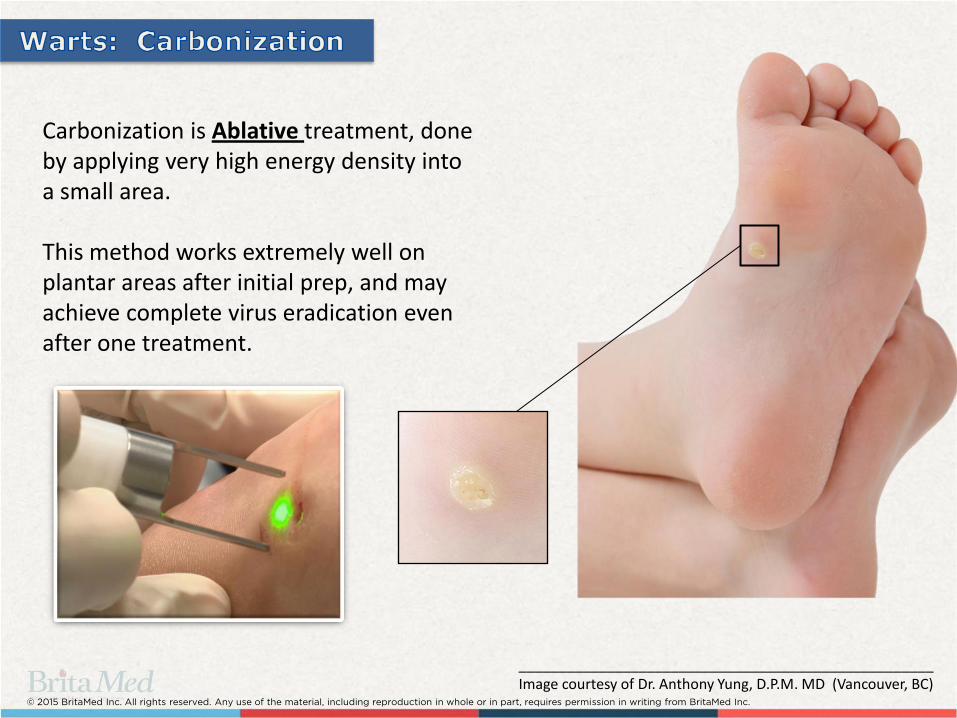
Carbonization is Ablative treatment, done by applying very high energy density into a small area. This method works extremely well on plantar areas after initial prep, and may achieve complete virus eradication even after one treatment.
____________________________________________________ Image courtesy of Dr. Anthony Yung, D.P.M. MD (Vancouver, BC)

© 2015 BritaMed Inc. All rights reserved. Any use of the material, including reproduction in whole or in part, requires permission in writing from BritaMed Inc.
1. Prepare the patient
3. Irradiate at the wart’s root 4. Scrape away loose tissue
2. Remove excess skin
____________________________________________________ Images courtesy of Dr. Anthony Yung, D.P.M. MD (Vancouver, BC)
© 2015 BritaMed Inc. All rights reserved. Any use of the material, including reproduction in whole or in part, requires permission in writing from BritaMed Inc.
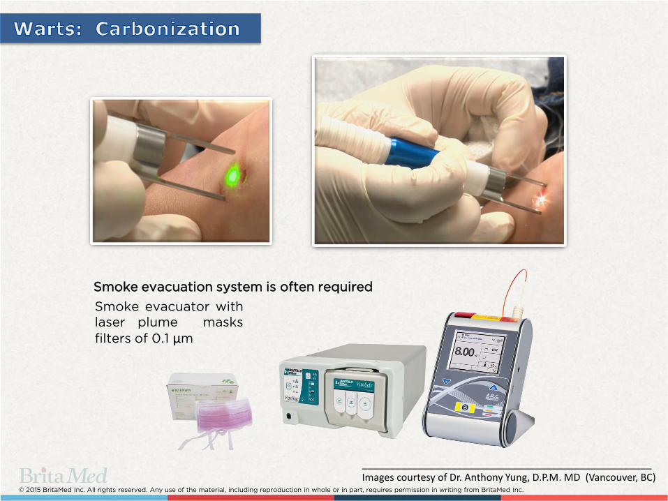
Smoke evacuator with
laser plume masks
filters of 0.1 µm
Smoke evacuation system is often required
____________________________________________________ Images courtesy of Dr. Anthony Yung, D.P.M. MD (Vancouver, BC)

© 2015 BritaMed Inc. All rights reserved. Any use of the material, including reproduction in whole or in part, requires permission in writing from BritaMed Inc.
BACKGROUND
Laser treatment is based on controlled carbonization: for the non deep callus and corns, usually surface carbonization will be sufficient to dry out the tissue, and allow safe transformation to eschar neoplasm.
Deeper IPK conditions will require more focused energy and possible more invasive type of procedures.
Plantar Callus (“Corn seed”, Clavus durus), is a very unpleasant and painful condition caused by an accumulation of dead skin cells that harden and thicken over an area of the foot. Calluses are normally found on the ball-of-the-foot, the heel, and/or the inside of the big toe. Some calluses have a deep seated core known as a nucleation. Currently there are various treatment options such as electrosurgery, cryosurgery, chemical agents (salicylic acid), and scalpel removal. Among the available therapeutic options, none are uniformly effective and most are associated with different limitations and side effects, like necrotic debris, inflammation, extensive bleeding, long recovery times, post-operative pain, scaring and relatively high recurrence rates due to difficulties in removing deeper corns.
© 2015 BritaMed Inc. All rights reserved. Any use of the material, including reproduction in whole or in part, requires permission in writing from BritaMed Inc.
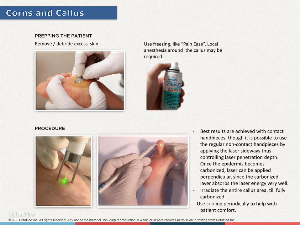
Remove / debride excess skin Use freezing, like “Pain Ease”. Local anesthesia around the callus may be required.
PREPPING THE PATIENT
PROCEDURE - Best results are achieved with contact
handpieces, though it is possible to use the regular non-contact handpieces by applying the laser sideways thus controlling laser penetration depth. Once the epidermis becomes carbonized, laser can be applied perpendicular, since the carbonized layer absorbs the laser energy very well.
- Irradiate the entire callus area, till fully carbonized.
- Use cooling periodically to help with patient comfort.
© 2015 BritaMed Inc. All rights reserved. Any use of the material, including reproduction in whole or in part, requires permission in writing from BritaMed Inc.

© 2015 BritaMed Inc. All rights reserved. Any use of the material, including reproduction in whole or in part, requires permission in writing from BritaMed Inc.
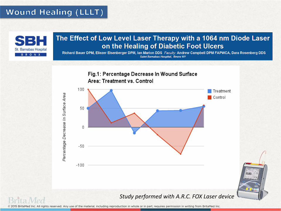
• Lower extremity ulcerations are a common complication seen in diabetic patients; numerous treatment modalities exist to manage lower extremity diabetic ulcers. Frequently, multiple adjunctive treatments are required to successfully manage an ulceration. • Treatments include surgical debridement and revascularization (when necessary), ointments, hydrogels, antibiotic therapies, offloading and hyperbaric oxygen treatment (HBO). • Healing of diabetic ulcers is often slow even when treated appropriately and occasionally full healing cannot be achieved. • Low Level Laser Therapy (LLLT) is believed to function through multiple Mechanisms, stimulates damaged or injured cells to increase cellular metabolism and ATP production. LLLT is also believed to increase oxygenation and blood flow to the irradiated area. • There are a wide variety of methods to provide laser irradiation to wound surfaces and as such there are a wide variety of treatment parameters that have shown successful results. All studies agree however, repeated treatment sessions are required for successful healing of wounds.
© 2015 BritaMed Inc. All rights reserved. Any use of the material, including reproduction in whole or in part, requires permission in writing from BritaMed Inc.
Study performed with A.R.C. FOX Laser device
© 2015 BritaMed Inc. All rights reserved. Any use of the material, including reproduction in whole or in part, requires permission in writing from BritaMed Inc.
_______________________________________________ Images courtesy of Dr. Joseph Stern, D.P.M. (Vancouver, BC)
© 2015 BritaMed Inc. All rights reserved. Any use of the material, including reproduction in whole or in part, requires permission in writing from BritaMed Inc.
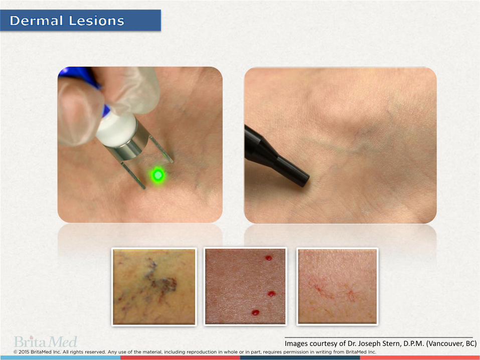
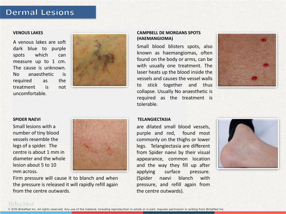
SPIDER NAEVI
VENOUS LAKES
A venous lakes are soft dark blue to purple spots which can measure up to 1 cm. The cause is unknown. No anaesthetic is required as the treatment is not uncomfortable.
CAMPBELL DE MORGANS SPOTS (HAEMANGIOMA)
Small blood blisters spots, also known as haemangiomas, often found on the body or arms, can be with usually one treatment. The laser heats up the blood inside the vessels and causes the vessel walls to stick together and thus collapse. Usually No anaesthetic is required as the treatment is tolerable.
Small lesions with a number of tiny blood vessels resemble the legs of a spider. The centre is about 1 mm in diameter and the whole lesion about 5 to 10 mm across. Firm pressure will cause it to blanch and when the pressure is released it will rapidly refill again from the centre outwards.
are dilated small blood vessels, purple and red, found most commonly on the thighs or lower legs. Telangiectasia are different from Spider naevi by their visual appearance, common location and the way they fill up after applying surface pressure. (Spider naevi blanch with pressure, and refill again from the centre outwards).
TELANGIECTASIA

© 2015 BritaMed Inc. All rights reserved. Any use of the material, including reproduction in whole or in part, requires permission in writing from BritaMed Inc.
FITZPATRICK CLASSIFICATION OF SKIN TYPES
Type I Always burns, never tans
Type II Usually burns, difficult in tanning
Type III Sometimes burns, average tan
Type IV Rarely burns, tans with ease
Type V Very rarely burns, tans very easily
Type VI Never burns, tans very easily
Fitzpatrick classification of skin types:
_______________________________________________ Images courtesy of Dr. Joseph Stern, D.P.M. (Vancouver, BC)

© 2015 BritaMed Inc. All rights reserved. Any use of the material, including reproduction in whole or in part, requires permission in writing from BritaMed Inc.
Blood vessel
Laser’s
beam spot
HS11009
HS11008
FOOT TELANGIECTASIA - FOX 980
Handpiece Skin Type Power (W) Pulse ON (ms) Pulse OFF (ms)
Red (HS11009)
I-IV 8-10 140-160 500
V-VI 7-8 120-140 500
Blue (HS11008)
I-IV 7-9 100-120 500
V-VI 5-7 80-100 500
• Always start with lowest laser settings and gradually increase
when necessary.
_______________________________________________ Image courtesy of Dr. Joseph Stern, D.P.M. (Vancouver, BC)

© 2015 BritaMed Inc. All rights reserved. Any use of the material, including reproduction in whole or in part, requires permission in writing from BritaMed Inc.


![podiatric medicine - · PDF filePodiatric Medicine [d p m] Doctors of ... Doctors of podiatric medicine placed 15th in Forbes magazine’s survey titled “America’s 25 Best-Paying](https://img.pdfslide.net/doc/110x75/5aa2811f7f8b9ada698cfecd/podiatric-medicine-medicine-d-p-m-doctors-of-doctors-of-podiatric-medicine.jpg)