Embed Size (px)
Citation preview
THE VASCULAR COAT OF THE EYE
ANIS SUZANNA BINTI MOHAMAD
OPTOMETRIST

WALL OF THE EYE• Fibrous (outer): sclera, cornea
• Vascular (middle): choroid, ciliary body, iris
• Nervous (inner): retina
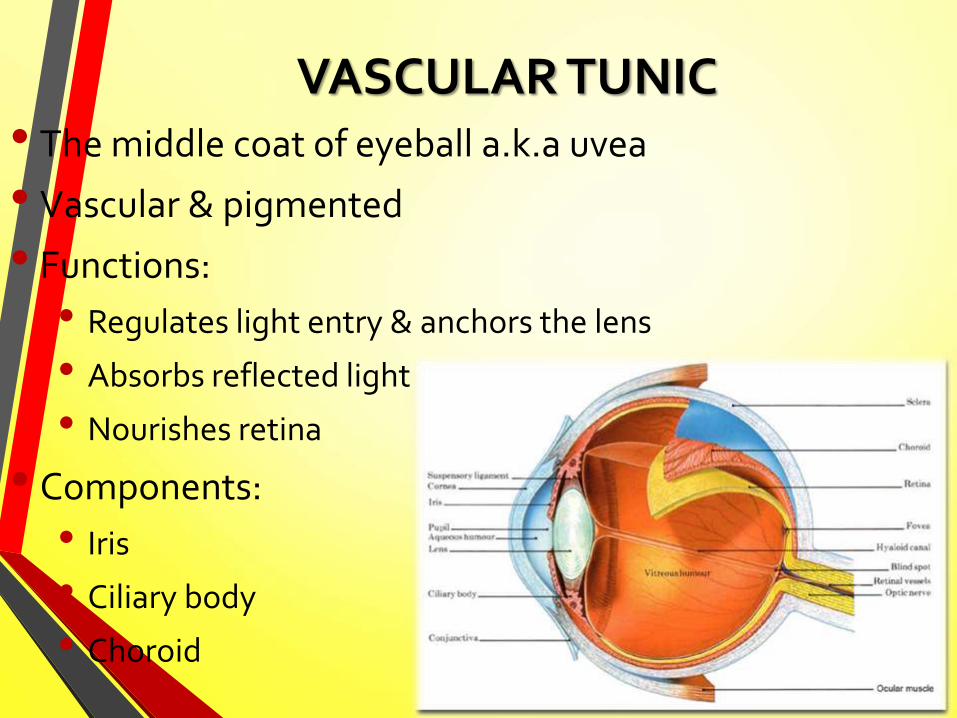
VASCULAR TUNIC• The middle coat of eyeball a.k.a uvea
• Vascular & pigmented
• Functions:
• Regulates light entry & anchors the lens
• Absorbs reflected light
• Nourishes retina
• Components:
• Iris
• Ciliary body
• Choroid

1. IRIS

1. IRIS
• Thin, pigmented contractile circular structure
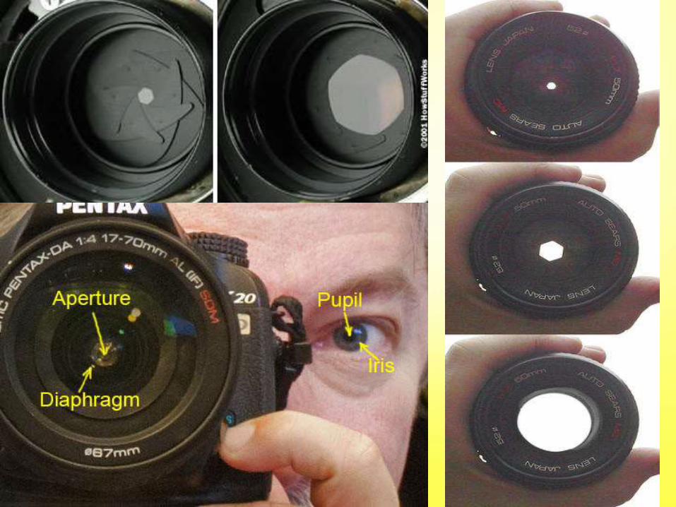
• Analogous to the diaphragm of a camera
• Lies on anterior lens surface, surrounded by aquos humor
• Extends from iris root to iris margin that forms the pupil
• The free edge is known as pupillary margin
• Separates anterior segment ant & post chambers which are continuous through pupil
• Stroma of iris is continuous with stroma of ciliary body
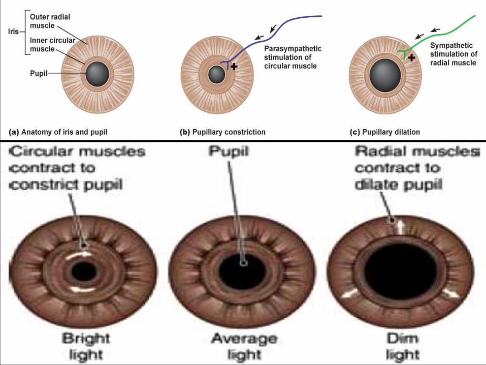
• Muscles: pupillary sphincter m. & pupillary dilator m.(circular) (radial)

1. IRIS
• Color depends on density of pigment & collagen in anterior limiting layer & stroma
• Heavily pigmented: brown eyes
• Lightly pigmented: lighter iris ranges from gray-blue-green
• Albino iris contains no pigment either in connective tissue cells (stroma) or in pigment layer
• Iris appears delicate pink with associated problems due to oversensitivity to light

1. IRIS
Dimensions:
• Average diameter: 12mm – varies with lighting
• Thickness: 0.5mm
• Thickest at collarette
• Collarette divides ant iris into pupillary & ciliary zones
• Thinnest at the root
• Thin regions eg iris root & margin are more susceptible to tearing in injuries
• Rough anterior surface & smooth posterior surface


1. IRIS – 4 layers
1. Anterior limiting layer
2. Stroma & sphincter muscle
3. Anterior epithelium & dilator muscle
4. Posterior epithelium
1. IRIS – 4 layers
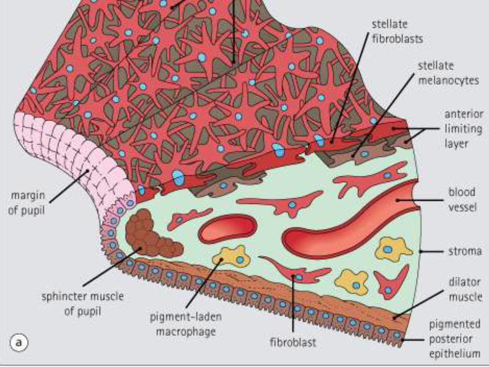
1. Anterior border layer:
• Thin, discontinuous (cryptic)
• Composed of collagen fibrils & fibroblasts
• Underlying melanocytes

1. IRIS – 4 layers
2. Stroma & sphincter muscle
• Loose, pigmented, highly vascular connective tissue
• Pigmented epithelium lining the posterior surface
• Composed of pigmented & non-pigmented cells, collagen fibrils & extensive ground substance
• Pigmented: melanocytes & clump cells
• Non-pigmented: fibroblast, lymphocyte, macrophage, mast cells
• Amount of melanocytes in stroma determines eye color
• Sphincter muscle: lies in stroma, near pupil, concentrically arranged, contraction causes pupil constriction (miosis)
• Innervated by oculomotor, parasympathetic nervous system

1. IRIS – 4 layers
3. Anterior epithelium & dilator muscle
• Composed of unique myoepithelial cells
• Dilator muscle:
• Smooth m. which extends into stroma forming 4-5 layers
• From iris root to a point in stroma below sphincter midpoint
• Radially arranged
• Contraction causes pupil dilatation (mydriasis) –innervated by sympathetic nervous system


1. IRIS – 4 layers
4. Posterior epithelium
• Single layer of heavily pigmented simple columnar cells
• At posterior, continuous with inner non-pigmented epithelial layer of ciliary body
• Curled to anterior surface at pupil margin –pupillary ruff

• Pupillary ruff
• Pigmented epithelium
• Dilator muscle
• Circular muscle
• Blood vessels
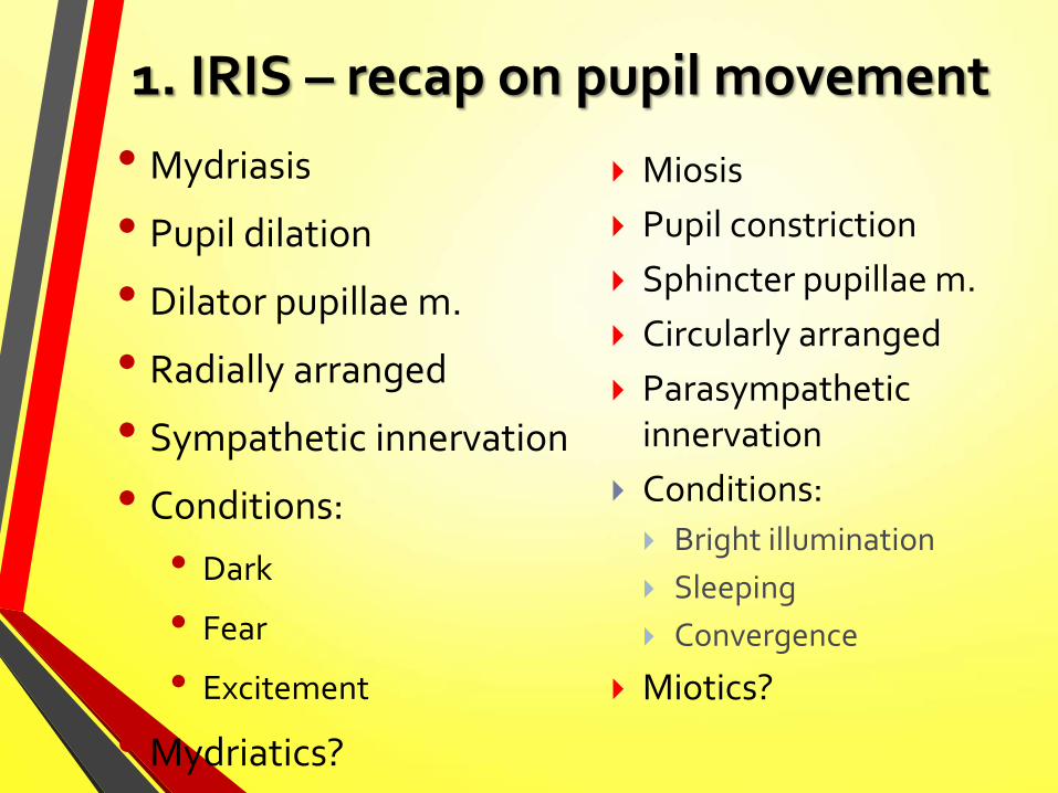
1. IRIS – recap on pupil movement
• Mydriasis
• Pupil dilation
• Dilator pupillae m.
• Radially arranged
• Sympathetic innervation
• Conditions:
• Dark
• Fear
• Excitement
• Mydriatics?
Miosis
Pupil constriction
Sphincter pupillae m.
Circularly arranged
Parasympathetic innervation
Conditions:
Bright illumination
Sleeping
Convergence
Miotics?
• e

EYE vs CAMERA

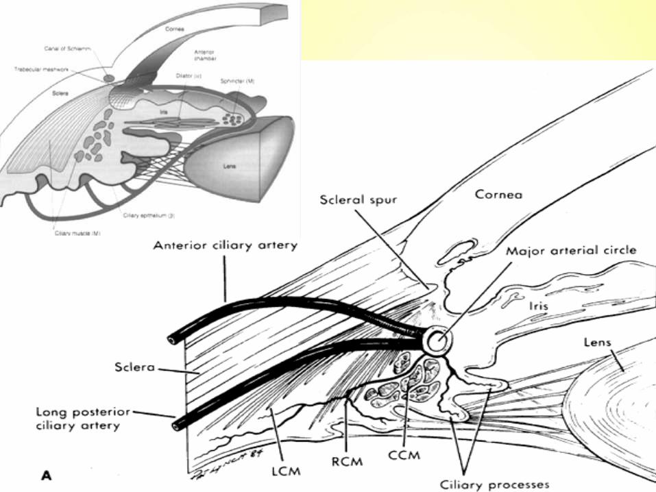
2. CILIARY BODY
• End of choroid, connects choroid to iris circumference
• Muscular & vascular
• Functions:
• Aquous humor production (via non-pigmented epithelium)
• Control of lens accommodation (via ciliary muscles)

2. CILIARY BODY
• Anterior view: ring-shaped structure
• Sagittal view: triangular shape
• Base faces anterior chamber
• Apex at ora serrata
• 2 surfaces:
• Pars plicata: Wider anterior portion containing 70-80 ciliary processes extending into posterior chamber
• Pars plana: Flatter region extending from posterior of pars plicata to ora serrata

• k

2. CILIARY BODY
• Ora Serrata
• Transition between c.body & choroid
• Serrated pattern
• Non pigmented ciliary epithelium undergoes sharp transition to become the neural retina
• Ciliary processes
• 70-80 finger-like projections radiating from pars plicata
• Occupy peripheral part of posterior chamber
• Grooves in between serve as attachment for lens zonules
• Each process is about 2mm long and 0.5mm in diameter

2. CILIARY BODY
• 3 structures: c. muscle, c. stroma, c. epithelium
1. Ciliary muscles – 3 groups of smooth muscle fibers
• Longitudinal (outermost)
• Radial
• Circular (innermost)
2. Ciliary stroma
• Highly vascularised loose connective tissue
• Anteriorly continuous with iris stroma
• Posteriorly continuous with choroidal stroma
• Thin in pars plana

2. CILIARY BODY
3. Ciliary epithelium (innermost part of ciliary body)
• 2 layers of cells: pigmented & non-pigmented epithelial cells
1. Pigmented epithelium (outer – next to stroma)
- Anterior part continuous with anterior iris epithelium
2. Non-pigmented epithelium (inner – faces post chamber)
- Columnar cells in pars plana, cuboidal cells in pars plicata
- Anterior part continuous with posterior iris epithelium
- Produces aquous humor & glycoprotein of vitreous
- Diffusion barrier between blood & aquous

3. CHOROID
• Thin, highly pigmented, vascular loose connective tissue
• Rich in melanocytes gives characteristic dark color
• Situated between sclera & retina
• Extends from optic nerve to ciliary body (at ora serrata)
• Thickness decreases from post (0.22mm) to ant (0.1mm)
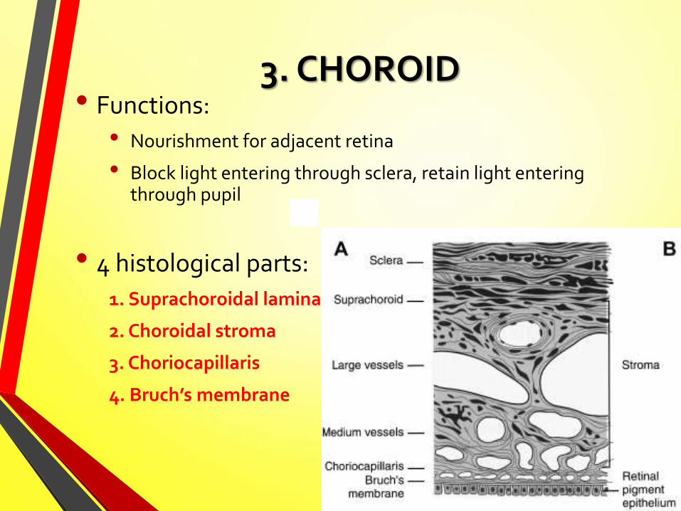
3. CHOROID• Functions:
• Nourishment for adjacent retina
• Block light entering through sclera, retain light entering through pupil
• 4 histological parts:
1. Suprachoroidal lamina
2. Choroidal stroma
3. Choriocapillaris
4. Bruch’s membrane

3. CHOROID
1. Suprachoroidal lamina
• Transition zone of choroid & sclera
• Consists of melanocytes, fibroblasts & conn. tissue fibers
• Blends with choroid & lamina fusca of sclera
• Carries long posterior ciliary arteries & nerves

3. CHOROID
2. Choroidal stroma
• Layer of pigmented, vascularised loose connective tissue
• Contains melanocytes, fibroblasts, macrophages, mast cells
• Arteries are branches of short posterior ciliaryarteries
• Veins drain via vortex veins into ophthalmic veins
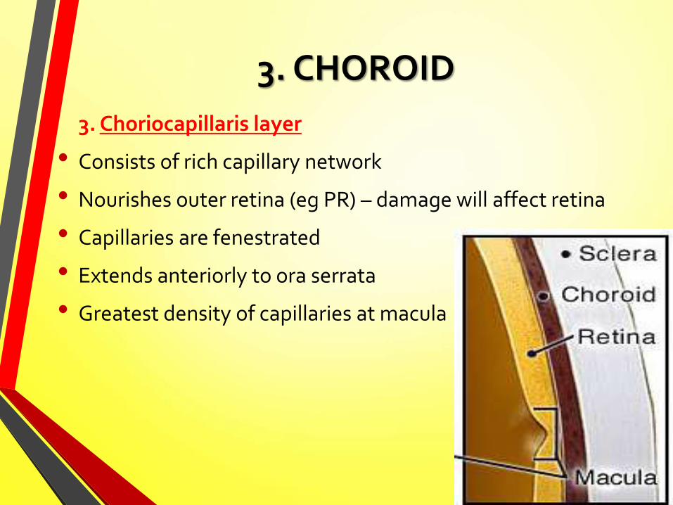
3. CHOROID
3. Choriocapillaris layer
• Consists of rich capillary network
• Nourishes outer retina (eg PR) – damage will affect retina
• Capillaries are fenestrated
• Extends anteriorly to ora serrata
• Greatest density of capillaries at macula

3. CHOROID
4. Bruch’s membrane
• Extend from optic nerve head to ora serrata
• Thin refractile connective tissue membrane between choriocapillaris (choroid) and RPE (retina)
• Constitutes outer limit of retina
• Prevents choroid vessels from penetrating the retina but allows nutrients, proteins etc


RELATED DISORDERS
• Uveitis: inflammation of uveal tract eg. Iritis, choroiditis
• Age-related macular degeneration (AMD)
• Drusen (yellow deposits) in Bruch membrane at macula
• Horner’s syndrome: loss of symp innervation to the head
• Causes ptosis, anhydrosis, miosis
• Malignant choroidal melanoma


* tHE END *