Embed Size (px)
Citation preview

Trisomy
Prepared by :
Dr.Maher Shoblak Dr. Zuhair Dajani
Dr. Mary Baraka
Trisomy
• A trisomy is a type of polysomy in which
there are three instances of a particular
chromosome, instead of the normal two.
A trisomy is a type of aneuploidy
(an abnormal number of chromosomes)
Trisomy
• Description and causes
• Most organisms that reproduce sexually have pairs of chromosomes in each cell, with one chromosome inherited from each parent. In such organisms, a process called meiosis creates cells called gametes (eggs or sperm) that have only one set of chromosomes. The number of chromosomes is different for different species. Humans have 46 chromosomes (i.e. 23 pairs of chromosomes). Human gametes have only 23 chromosomes.
Trisomy
• If the chromosome pairs fail to separate
properly during cell division, the egg or sperm
may end up with a second copy of one of the
chromosomes. (See non-disjunction) If such a
gamete results in fertilization and an embryo,
the resulting embryo may also have an entire
copy of the extra chromosome.
Trisomy
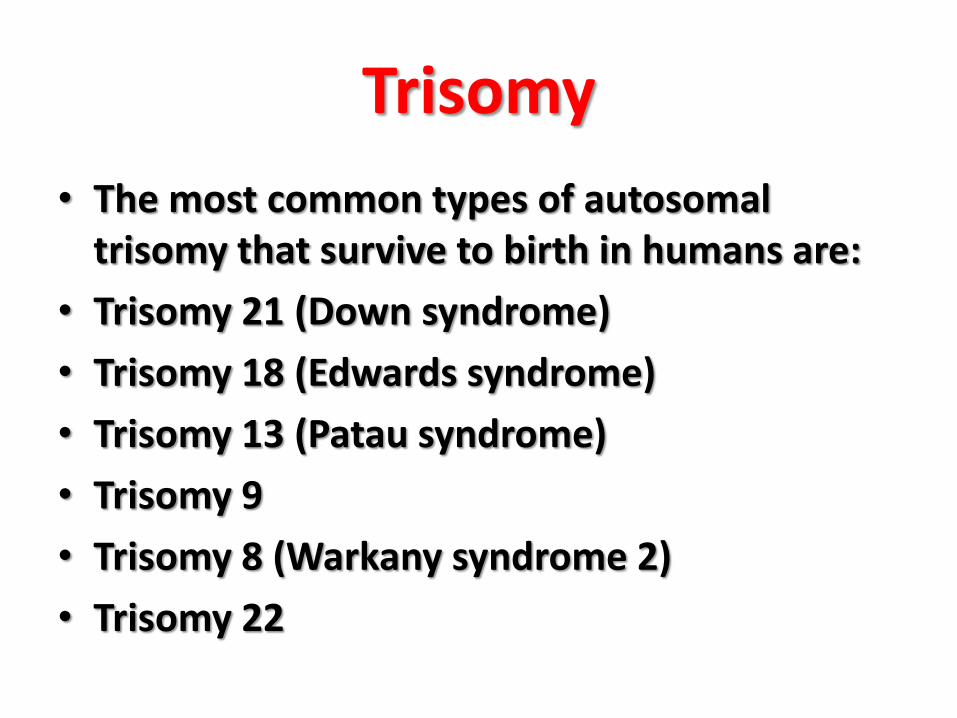
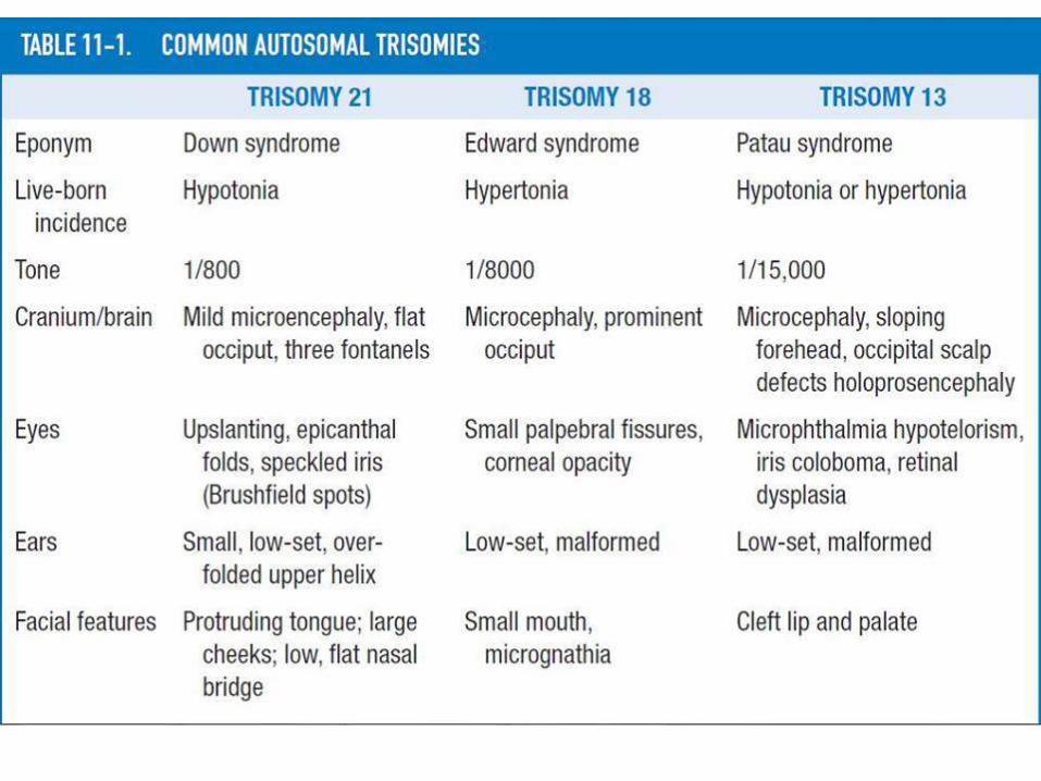
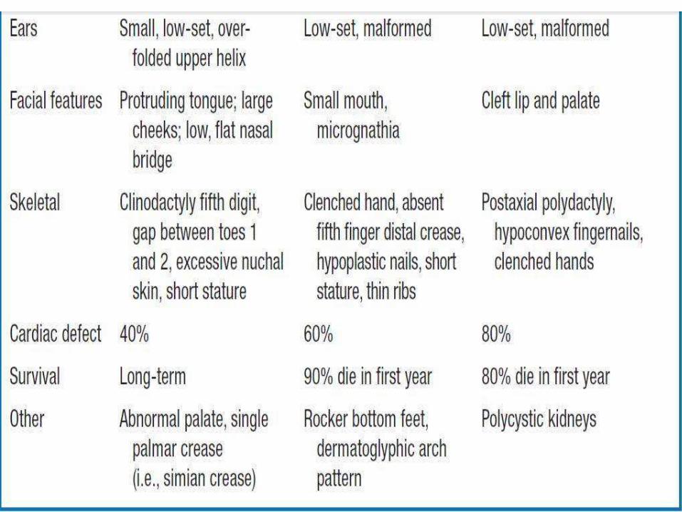
• The most common types of autosomaltrisomy that survive to birth in humans are:
• Trisomy 21 (Down syndrome)
• Trisomy 18 (Edwards syndrome)
• Trisomy 13 (Patau syndrome)
• Trisomy 9
• Trisomy 8 (Warkany syndrome 2)
• Trisomy 22
Trisomy
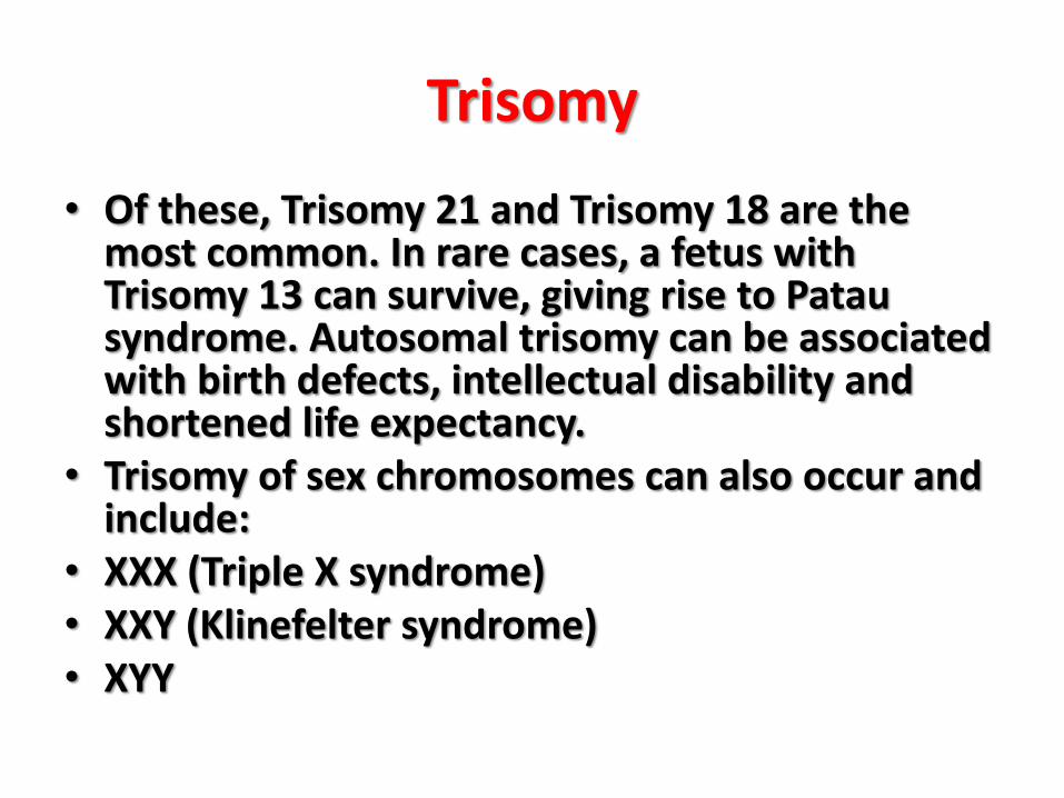
• Of these, Trisomy 21 and Trisomy 18 are the most common. In rare cases, a fetus with Trisomy 13 can survive, giving rise to Patausyndrome. Autosomal trisomy can be associated with birth defects, intellectual disability and shortened life expectancy.
• Trisomy of sex chromosomes can also occur and include:
• XXX (Triple X syndrome)• XXY (Klinefelter syndrome)• XYY
Trisomy
• Compared to trisomy of the autosomal
chromosomes, trisomy of the sex chromosomes
normally has less severe consequences.
Individuals may show few or no symptoms and
have a normal life expectancy
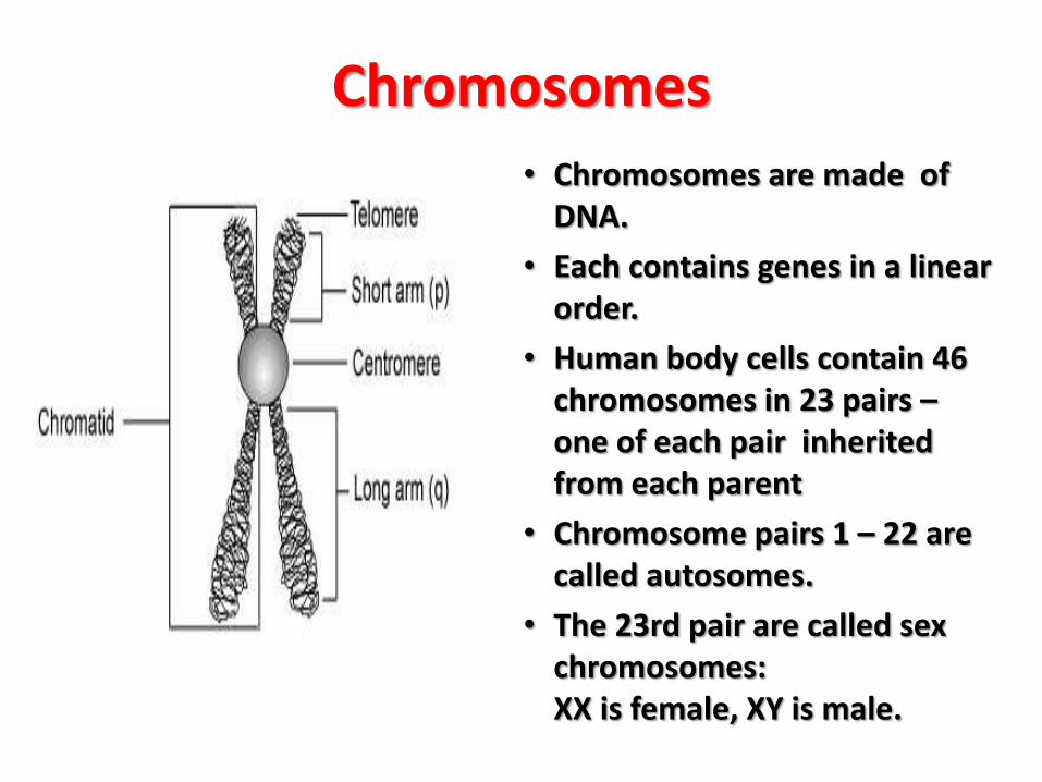
Chromosomes• Chromosomes are made of
DNA.
• Each contains genes in a linear order.
• Human body cells contain 46 chromosomes in 23 pairs –one of each pair inherited from each parent
• Chromosome pairs 1 – 22 are called autosomes.
• The 23rd pair are called sex chromosomes: XX is female, XY is male.
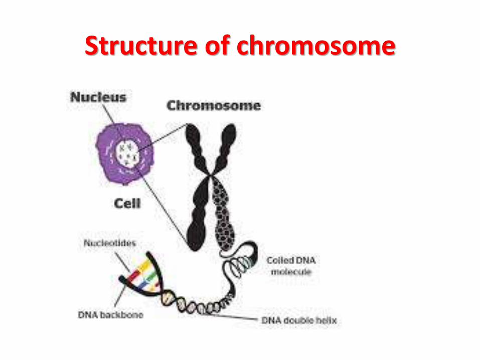
Structure of chromosome
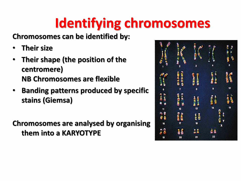
Identifying chromosomesChromosomes can be identified by:
• Their size
• Their shape (the position of the centromere) NB Chromosomes are flexible
• Banding patterns produced by specific stains (Giemsa)
Chromosomes are analysed by organising them into a KARYOTYPE
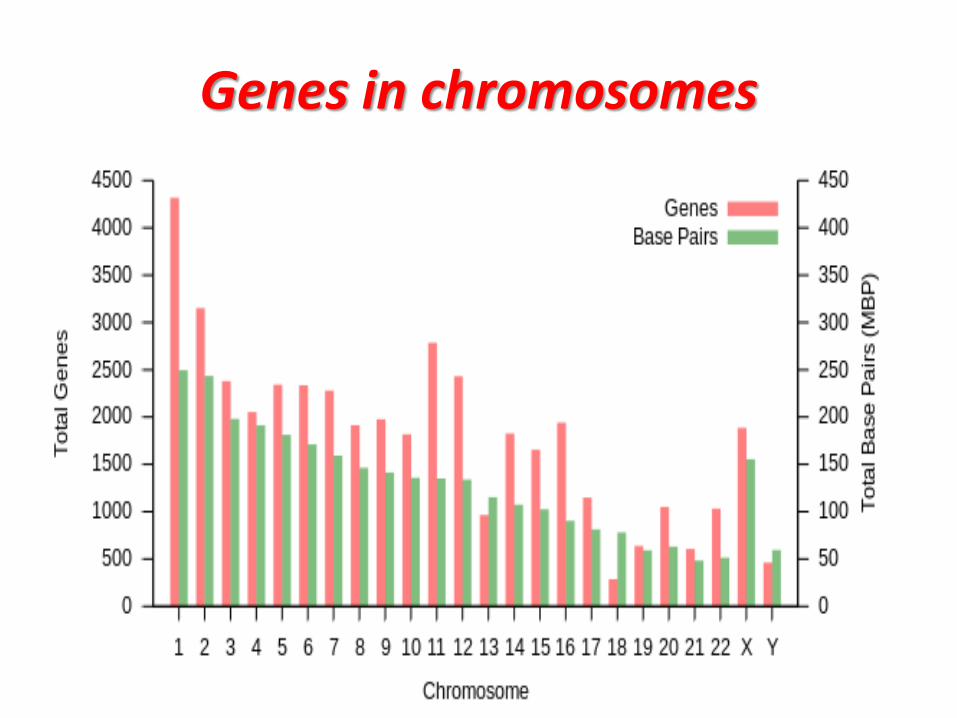
Genes in chromosomes
•Definitions
• Gene : segment of DNA Which controls the production of a particular polypeptide chain located in chromosome .
• It is Greek word .. Genos means origin .
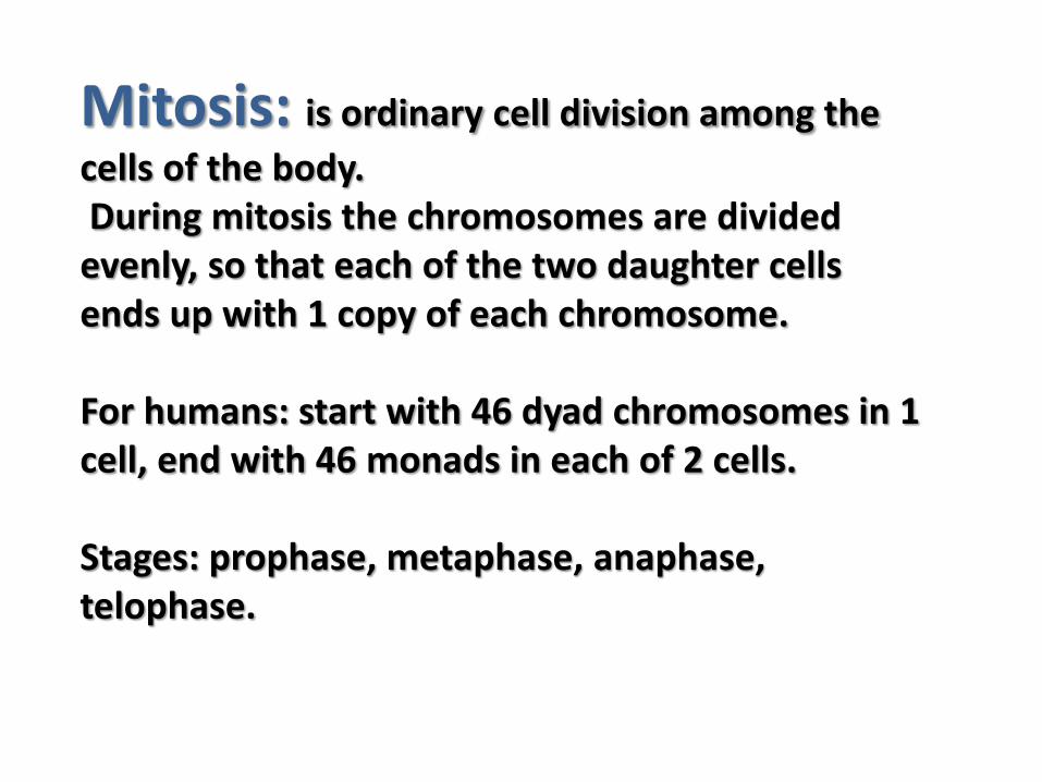
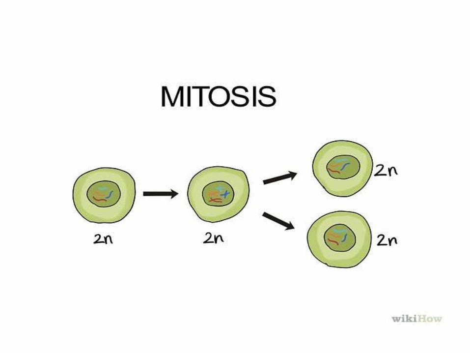
Mitosis: is ordinary cell division among the
cells of the body. During mitosis the chromosomes are divided
evenly, so that each of the two daughter cells ends up with 1 copy of each chromosome.
For humans: start with 46 dyad chromosomes in 1 cell, end with 46 monads in each of 2 cells.
Stages: prophase, metaphase, anaphase, telophase.
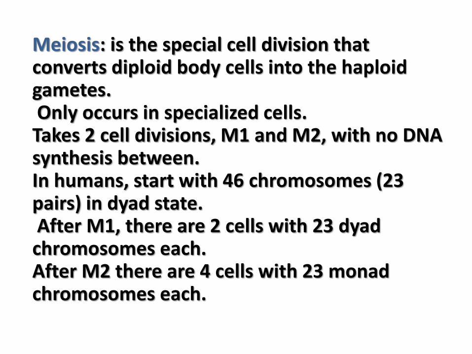
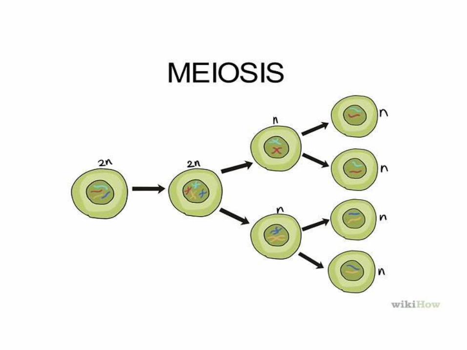
Meiosis: is the special cell division that converts diploid body cells into the haploid gametes. Only occurs in specialized cells.Takes 2 cell divisions, M1 and M2, with no DNA synthesis between.In humans, start with 46 chromosomes (23 pairs) in dyad state. After M1, there are 2 cells with 23 dyad chromosomes each. After M2 there are 4 cells with 23 monad chromosomes each.
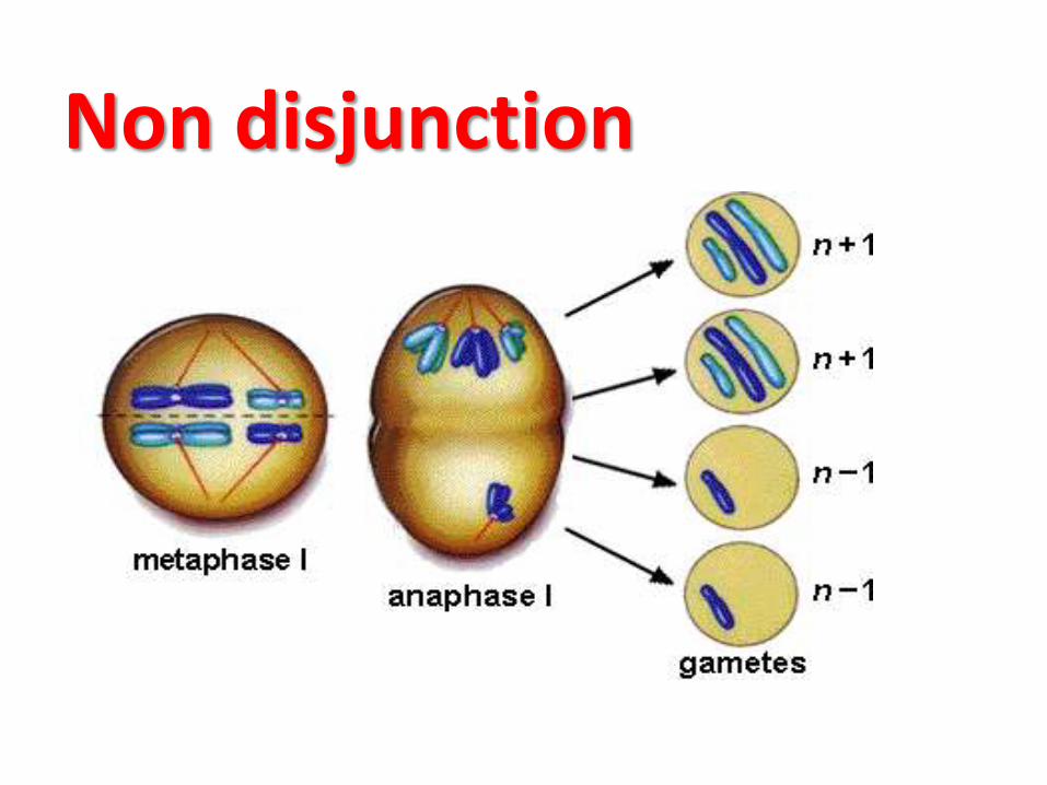
Non disjunction
Obtaining a Sample
Fetal samples for karyotypes are commonly obtained in two ways
1.Amniocentesis – sample taken from thefluid of the amniotic sac2.Chorionic Villus Sampling – sample taken
from the fetal tissue that forms part of the placenta
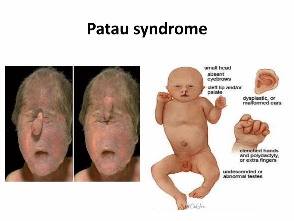
Trisomy 13 (Patau Syndrome)
• 1st described by Bartholin (1657) & redefined byPatau (1960).
• Chromosomal complement: 47,XX,+13 (female) or 47,XY,+13 (male)
• Phenotype: Male or female
• Incidence: 1:12,000 (increases with the age of mother)
Features of Patau Syndrome• Mental deficiency• Low birth weight• Abnormal development
of frontal lobe • Absence of corpus
callosum• Hypoplasia of cerebellum• Sloping forehead• Scalp defects
• Malformed ears
• Congenital heart defects
• Renal tract anomalies
• Microphthalmia
• Bilateral cleft lip/palate
• Polydactyly with
rudimentary digits
• Rocker-bottom heel
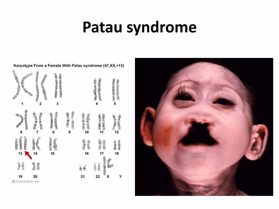
Patau syndrome
Patau syndrome
Trisomy 18 (Edward Syndrome)
• 1st described by Johin Edward (1960)
• Chromosomal complement: 47,XX,+18 (female) or 47,XY,+18 (male)
• Phenotype: Male or female
• Incidence: 1:8000
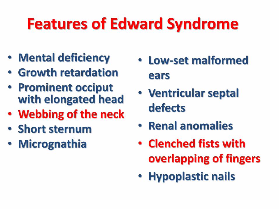
Features of Edward Syndrome
• Mental deficiency• Growth retardation• Prominent occiput
with elongated head• Webbing of the neck • Short sternum• Micrognathia
• Low-set malformed ears
• Ventricular septaldefects
• Renal anomalies
• Clenched fists with overlapping of fingers
• Hypoplastic nails
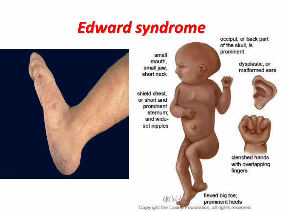
Edward syndrome
Trisomy 21 (Down Syndrome)
• 1st described by Johen Down (1866)
• Chromosomal complement: 47,XX,+21 (female) or47,XY,+21 (male)
• Phenotype: Male or female
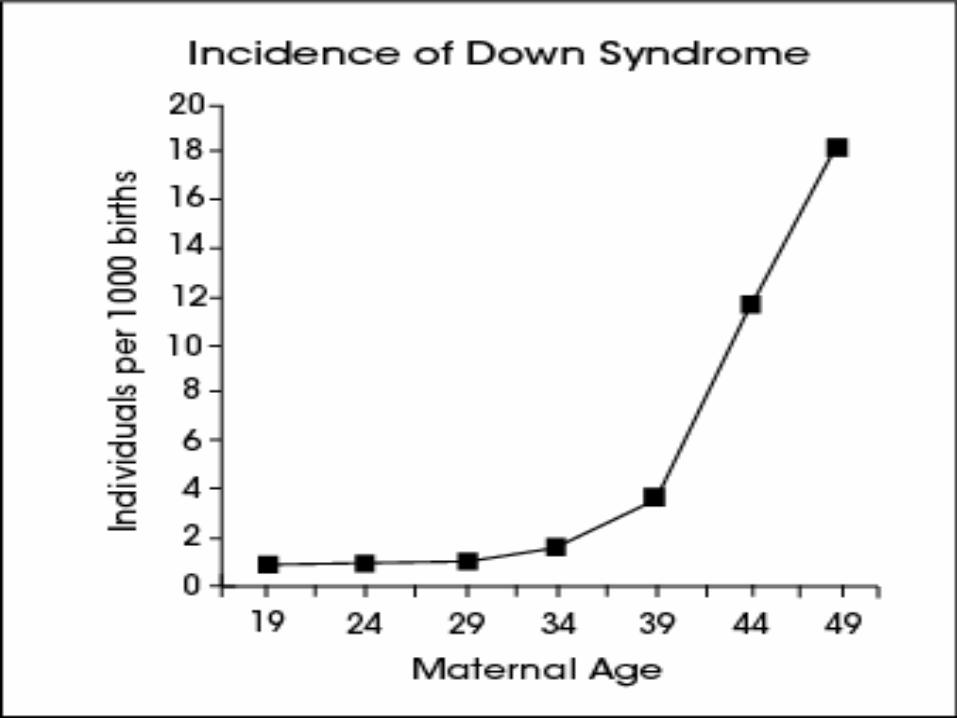
• Incidence: 1:800 (increases with the age of mother)
• |
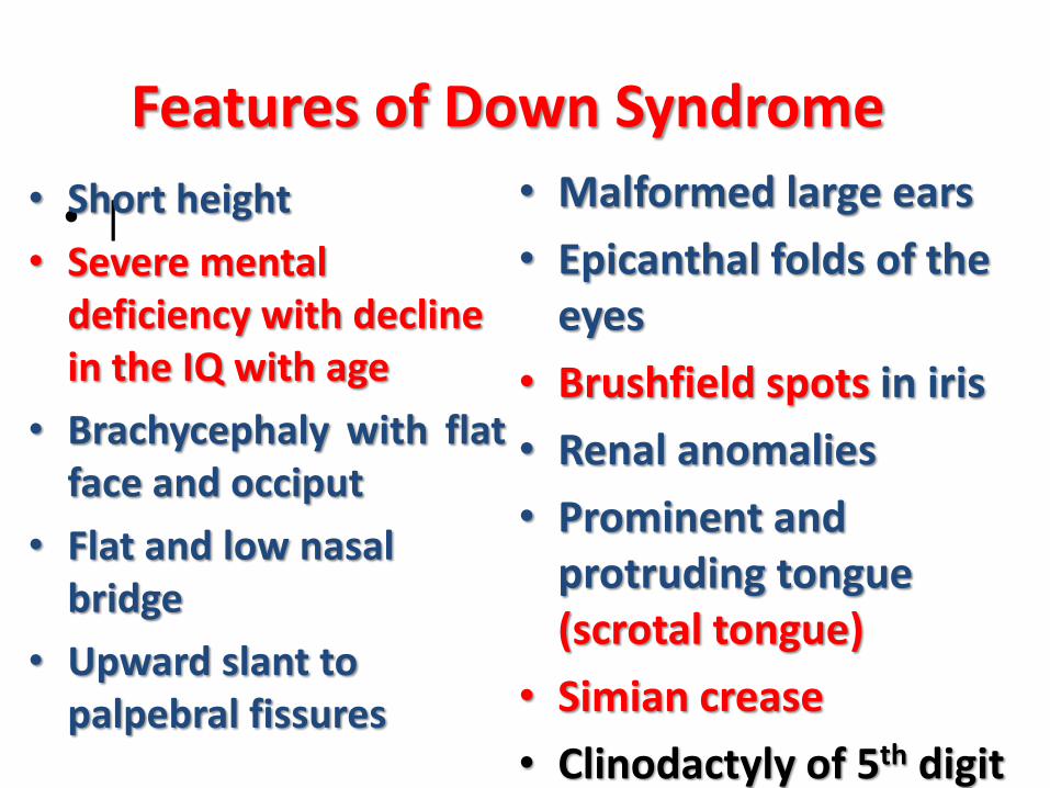
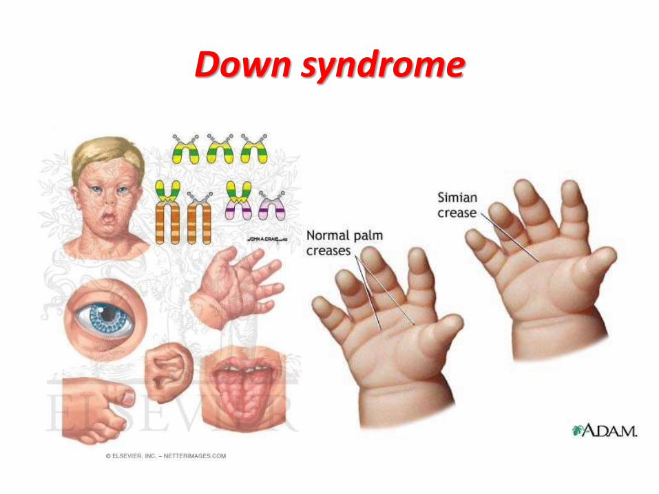
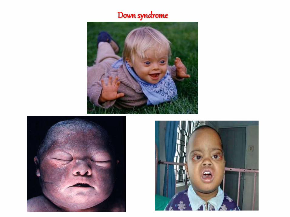
Features of Down Syndrome
• Short height
• Severe mental deficiency with decline in the IQ with age
• Brachycephaly with flatface and occiput
• Flat and low nasal bridge
• Upward slant to palpebral fissures
• Malformed large ears
• Epicanthal folds of the eyes
• Brushfield spots in iris
• Renal anomalies
• Prominent and protruding tongue (scrotal tongue)
• Simian crease
• Clinodactyly of 5th digit
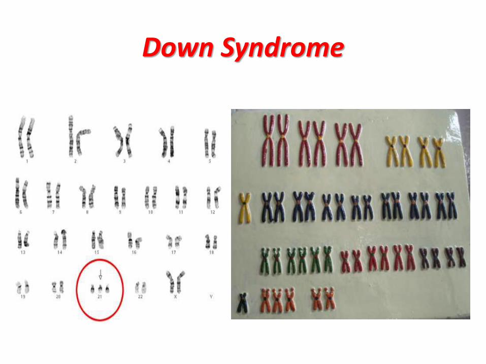
Down Syndrome
• |
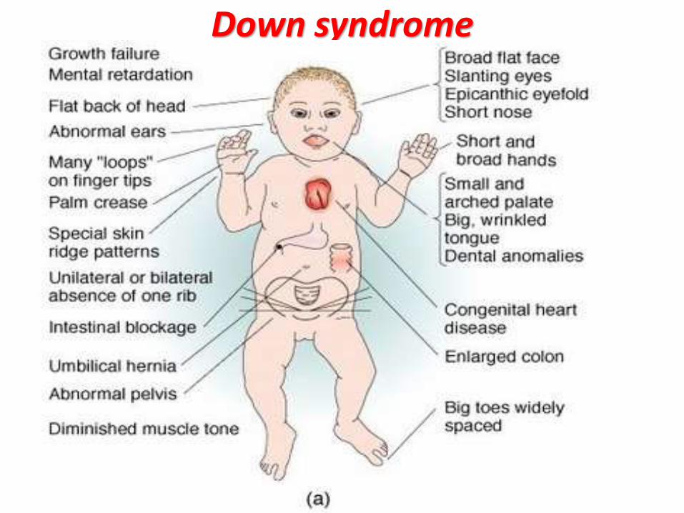
Down syndrome
Down syndrome
Down syndrome
1. Growth – Measurements should be plotted on the appropriate growth chart for children with DS.This will help in prevention of obesity and early diagnosis of celiac disease and hypothyroidism.
2. Cardiac disease – All newborns should be evaluated by cardiac ECHO for CHD in consultation with pediatric cardiologist.
3. Hearing – Screening to be done in the newborn period, every 6 months until 3 yrs of age and then annually.
Management
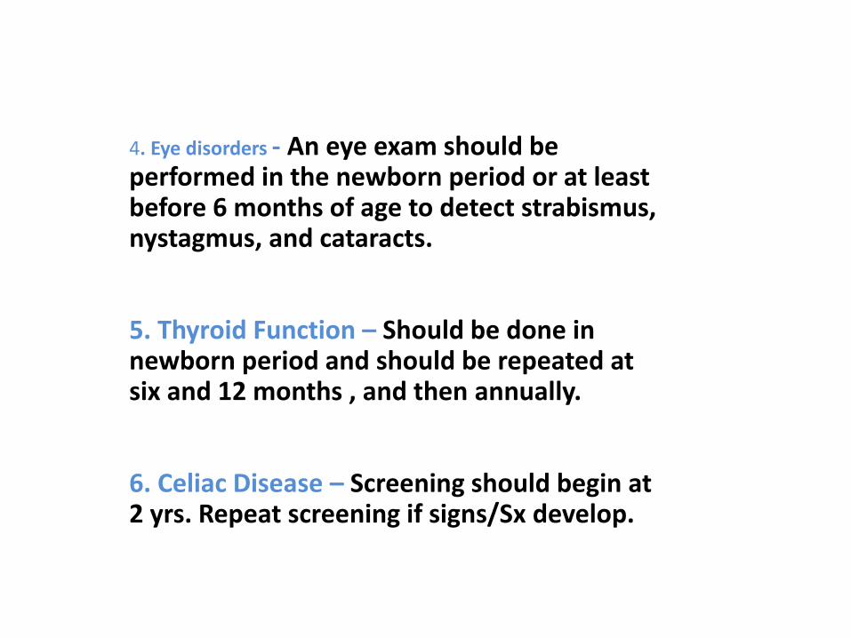
4. Eye disorders - An eye exam should be performed in the newborn period or at least before 6 months of age to detect strabismus, nystagmus, and cataracts.
5. Thyroid Function – Should be done in newborn period and should be repeated at six and 12 months , and then annually.
6. Celiac Disease – Screening should begin at 2 yrs. Repeat screening if signs/Sx develop.
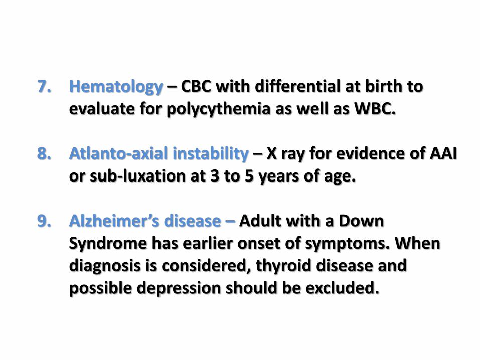
7. Hematology – CBC with differential at birth to evaluate for polycythemia as well as WBC.
8. Atlanto-axial instability – X ray for evidence of AAI or sub-luxation at 3 to 5 years of age.
9. Alzheimer’s disease – Adult with a Down Syndrome has earlier onset of symptoms. When diagnosis is considered, thyroid disease and possible depression should be excluded.
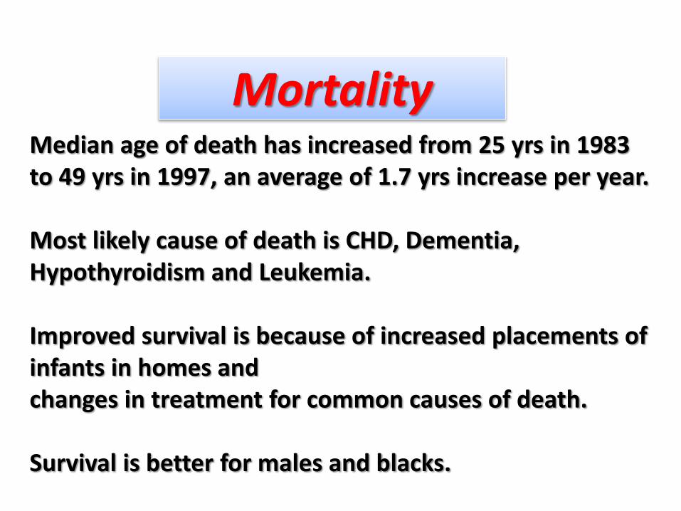
Median age of death has increased from 25 yrs in 1983 to 49 yrs in 1997, an average of 1.7 yrs increase per year.
Most likely cause of death is CHD, Dementia, Hypothyroidism and Leukemia.
Improved survival is because of increased placements of infants in homes andchanges in treatment for common causes of death.
Survival is better for males and blacks.
Mortality
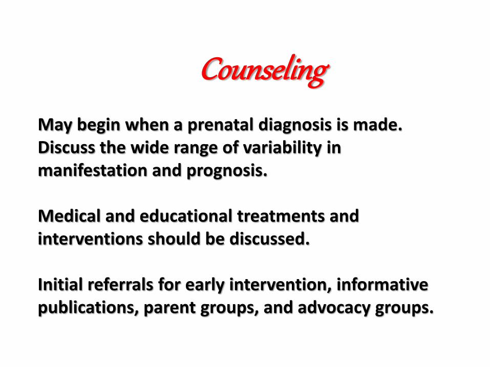
May begin when a prenatal diagnosis is made.Discuss the wide range of variability in manifestation and prognosis.
Medical and educational treatments and interventions should be discussed.
Initial referrals for early intervention, informative publications, parent groups, and advocacy groups.
Counseling


Rare Trisomytrisomy 14
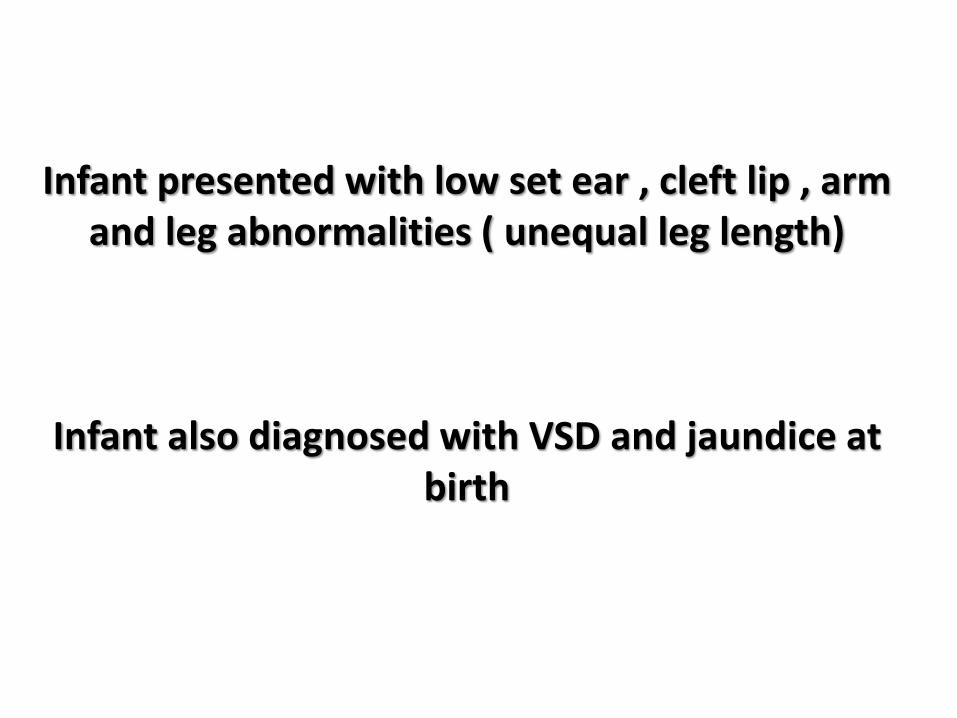
Infant presented with low set ear , cleft lip , arm and leg abnormalities ( unequal leg length)
Infant also diagnosed with VSD and jaundice at birth
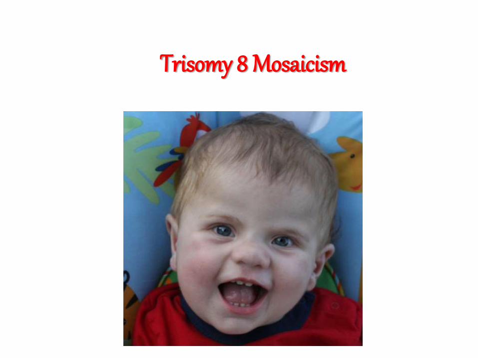
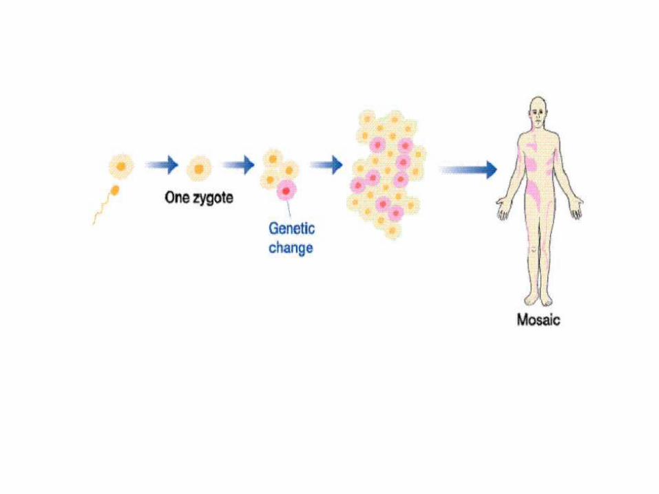
Trisomy 8 Mosaicism
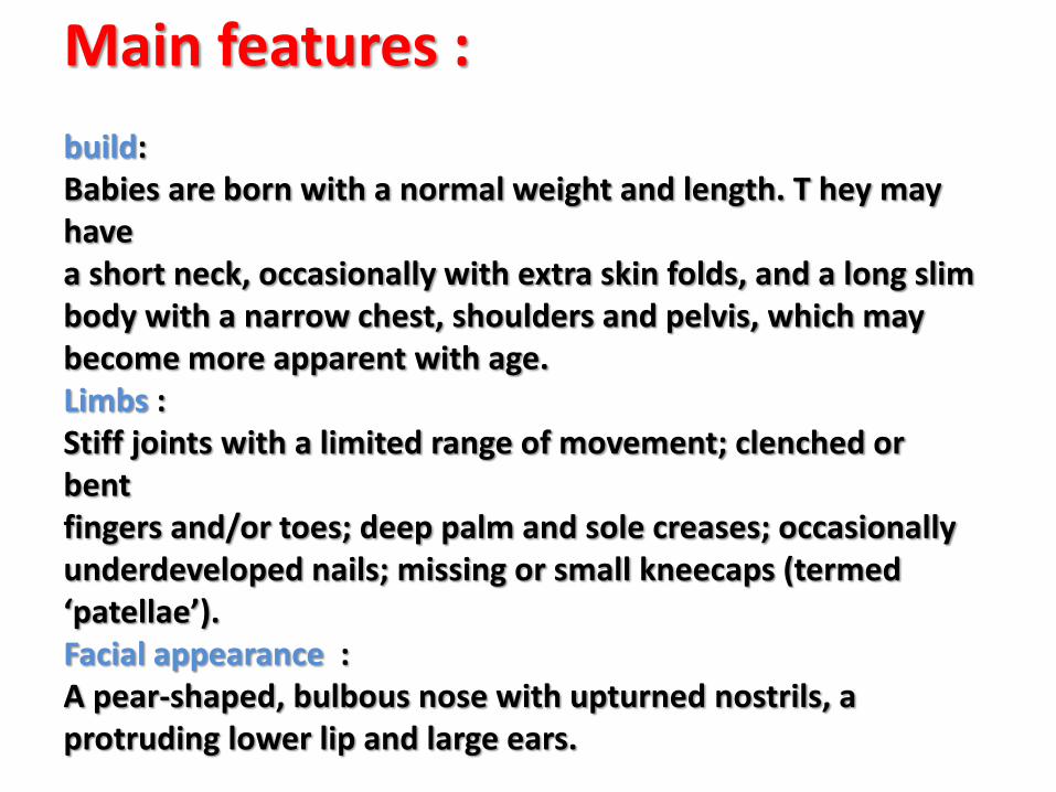
Main features :
build:Babies are born with a normal weight and length. T hey may have a short neck, occasionally with extra skin folds, and a long slim body with a narrow chest, shoulders and pelvis, which may become more apparent with age. Limbs :Stiff joints with a limited range of movement; clenched or bent fingers and/or toes; deep palm and sole creases; occasionally underdeveloped nails; missing or small kneecaps (termed ‘patellae’). Facial appearance :A pear-shaped, bulbous nose with upturned nostrils, a protruding lower lip and large ears.
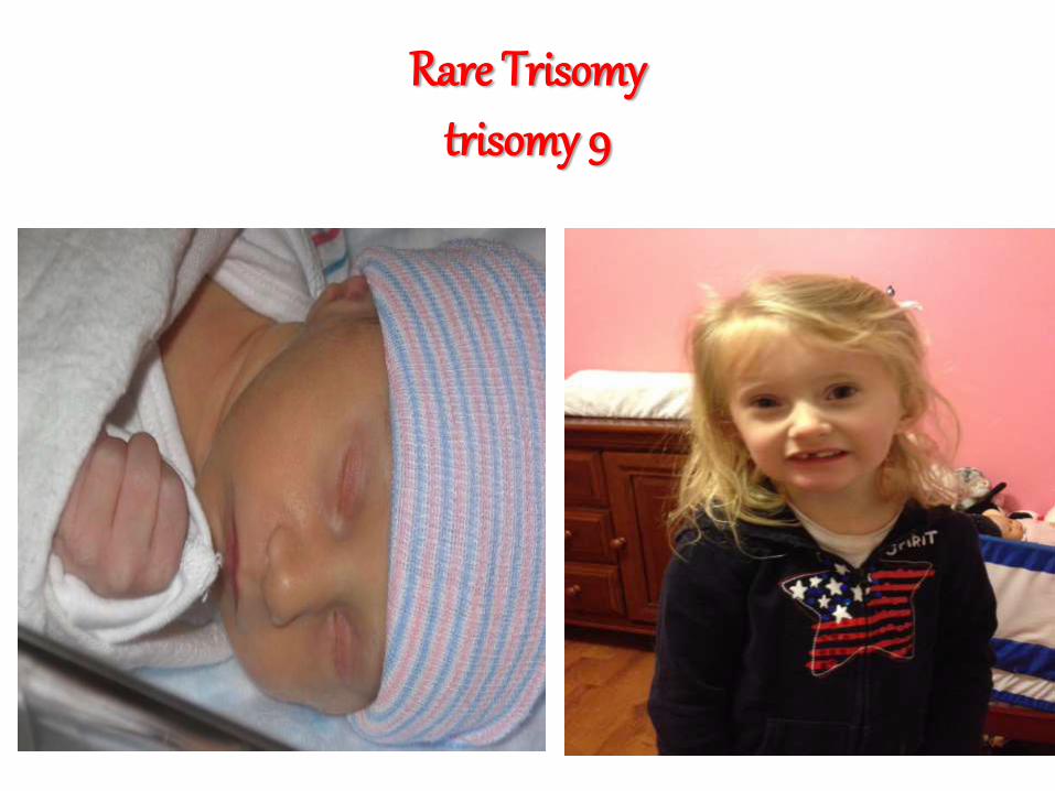
Rare Trisomytrisomy 9
Trisomy 9 is a chromosomal disorder caused by having three copies (trisomy) of chromosome number 9.
It can appear with or without mosaicism. CharacteristicsSymptoms vary, but usually result in dysmorphisms in the skull, nervous system, and developmental delay. Dysmorphisms in the heart, kidneys, and musculoskeletal system may also occur.
An infant with complete trisomy 9 surviving 20 days after birth showed clinical features including a small face, wide fontanelle, prominent occiput, micrognathia, low set ears, upslantingpalpebral fissures, high arched palate, short sternum, overlapping fingers, limited hip abduction, rocker bottom feet, heart murmurs and also a webbed neck.
Detection
Trisomy 9 can be detected prenatally with chorionic villus sampling and cordocentesis, and can be suggested by obstetric ultrasonography
Because trisomy 9 may appear with mosaicism, it is suggested that doctors take samples from multiple tissues when karyotyping for diagnosis
Trisomy 16
Trisomy 16 is a chromosomal abnormality in which there are 3 copies of chromosome 16 rather than two.
It is the most common trisomy leading to miscarriage and the second most common chromosomal cause of it, closely following X-chromosome monosomy.
Like most chromosomal abnormalities, trisomy 16 usually causes miscarriage in the first trimester of pregnancy.
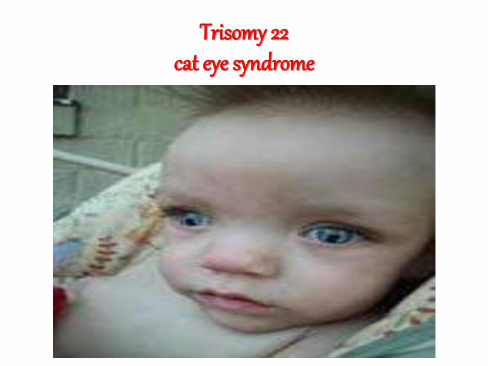
Trisomy 22 cat eye syndrome