Embed Size (px)
Citation preview

Emergency OB USDr. Wananee Meennuch
6-July-2016

Objectives
• Refresh your (& my) knowledge• Experience share• Inspiration in one of your learning paths
Outlines
• Normal anatomy and US findings• Common 1st trimester emergency conditions: Vaginal bleeding & pelvic pain
• Common 2nd-3rd trimester emergency conditions: Vaginal bleeding +/- contraction

Ant
Post
Ant
Ant Ant
Post
Post Post
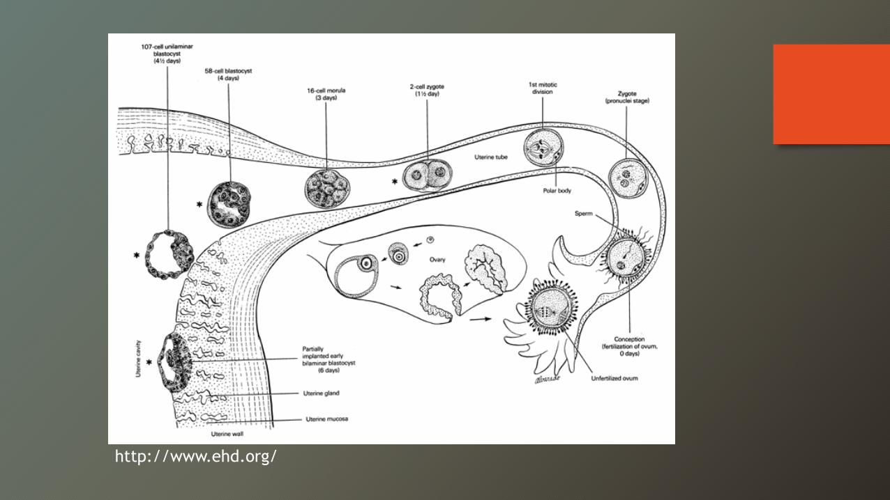
http://www.ehd.org/
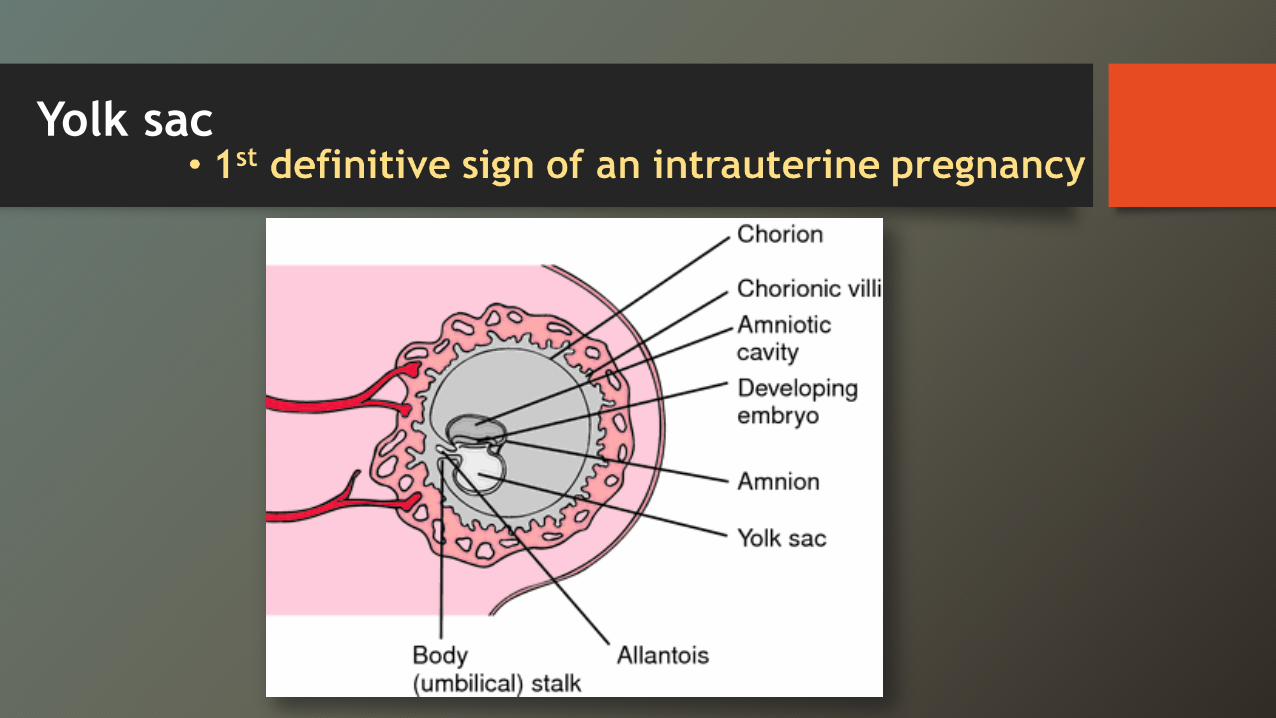
Yolk sac
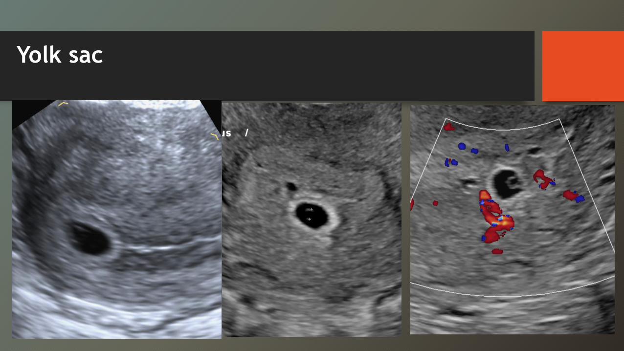
Yolk sac
The yolk sac
• No YS in GS >8 mm Æ abnormal • No embryo in a GS >16 mm Æ abnormal • No embryo in GS of 25 mm Æ Dx failed pregnancy • No yolk sac or embryo on 2 scans / 7-10 days apart = definitive evidence of a failed intrauterine pregnancy
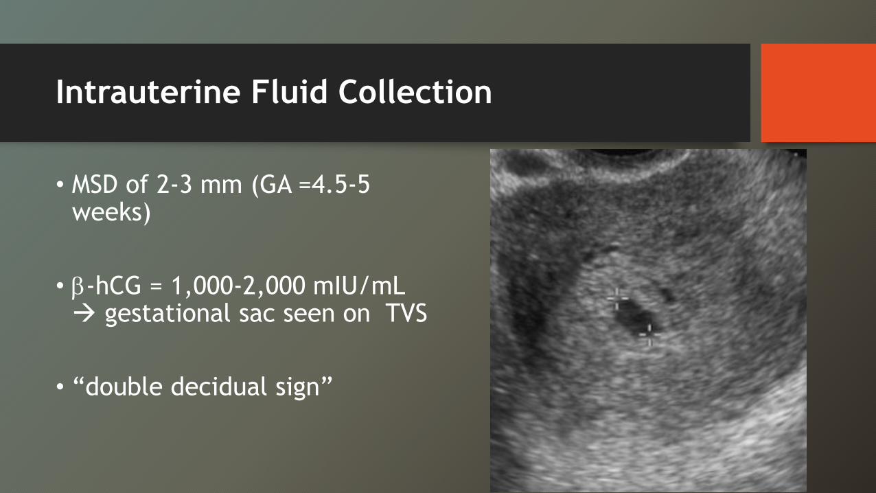
Intrauterine Fluid Collection
• MSD of 2-3 mm (GA =4.5-5 weeks)
• E-hCG = 1,000-2,000 mIU/mL Æ gestational sac seen on TVS
• “double decidual sign”
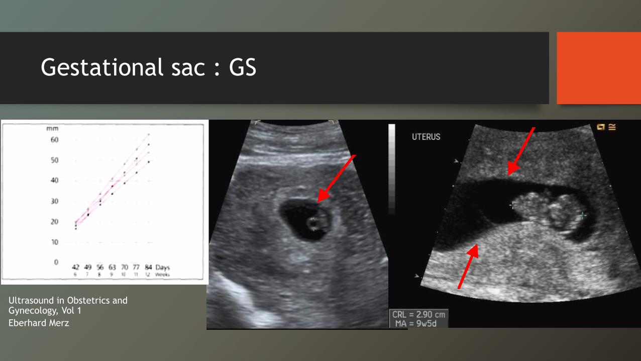
Gestational sac : GS
Ultrasound in Obstetrics and Gynecology, Vol 1Eberhard Merz
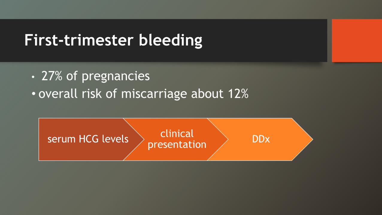
First-trimester bleeding
• 27% of pregnancies• overall risk of miscarriage about 12%
serum HCG levels clinical presentation DDx
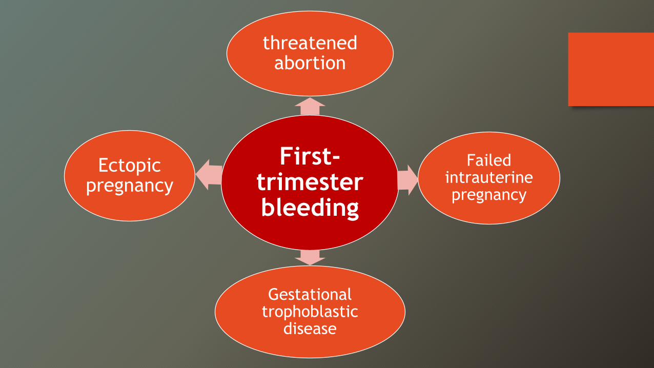
First-trimester bleeding
threatened abortion
Failed intrauterine pregnancy
Gestational trophoblastic
disease
Ectopic pregnancy
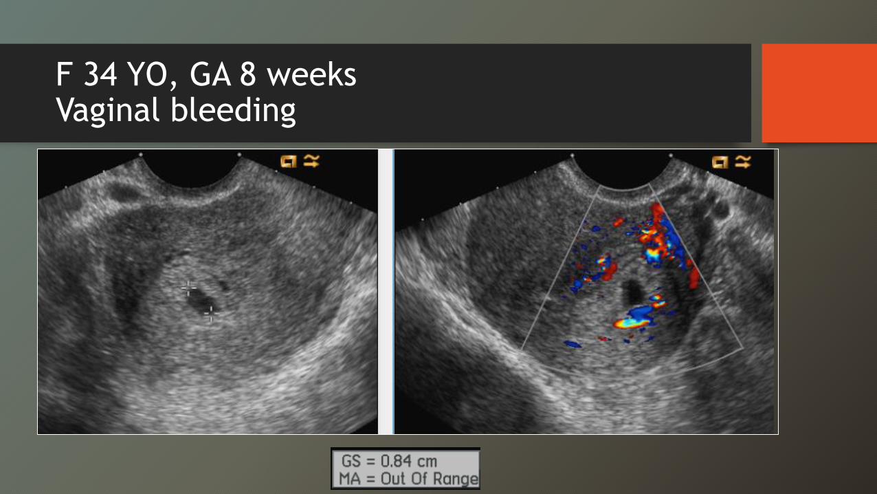
F 34 YO, GA 8 weeksVaginal bleeding
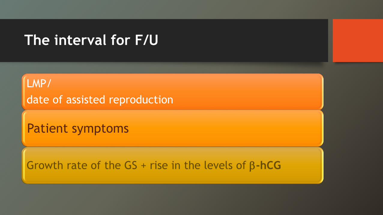
The interval for F/U
LMP/ date of assisted reproduction
Patient symptoms
Growth rate of the GS + rise in the levels of E-hCG
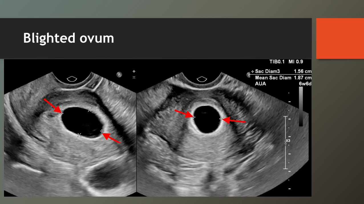
Blighted ovum
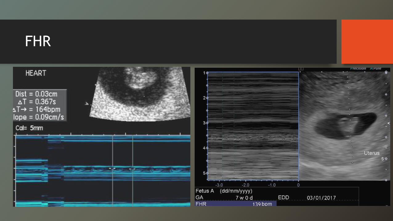
FHR
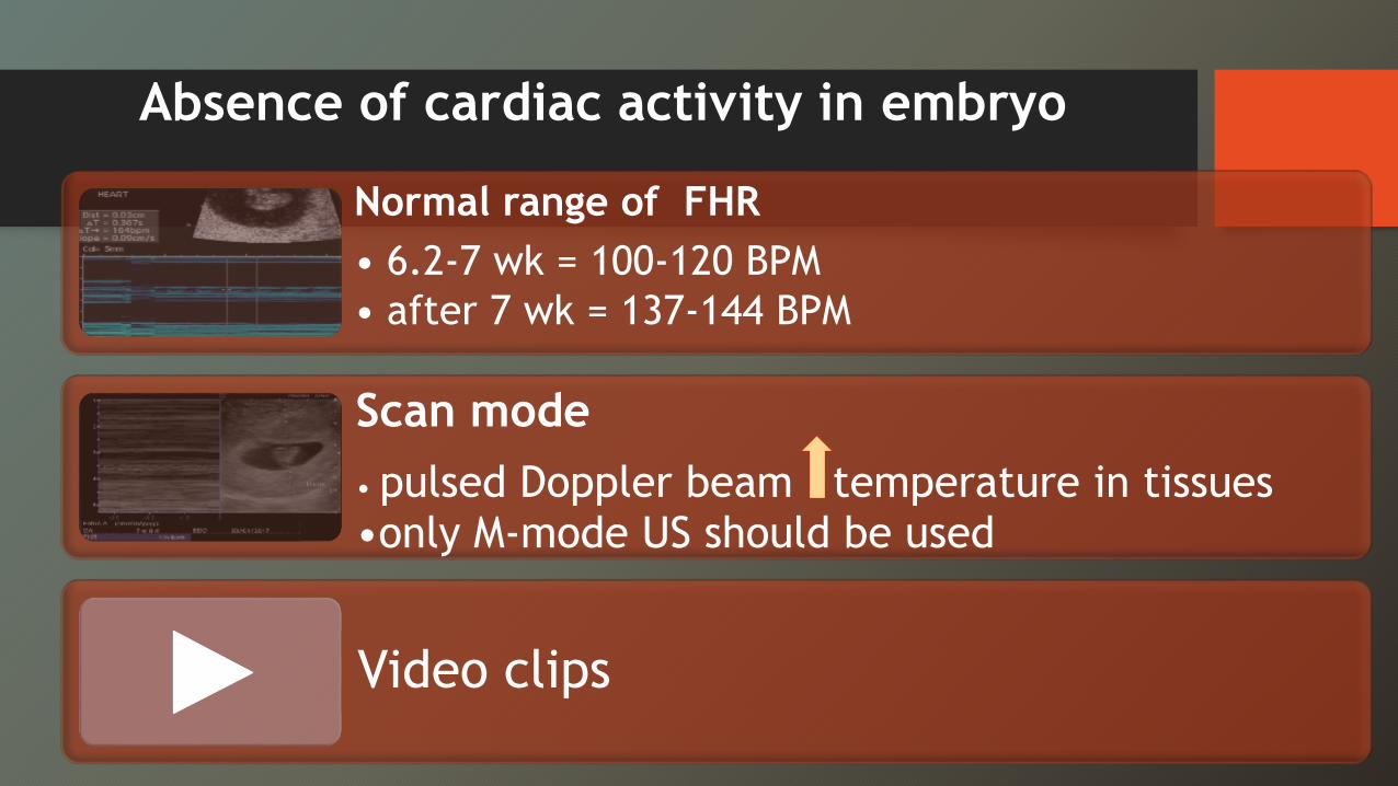
Absence of cardiac activity in embryo
Normal range of FHR • 6.2-7 wk = 100-120 BPM • after 7 wk = 137-144 BPM
Scan mode• pulsed Doppler beam temperature in tissues•only M-mode US should be used
Video clips
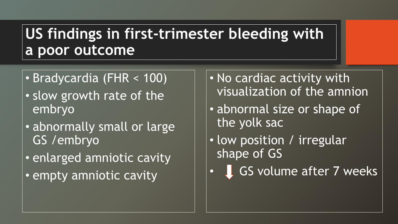
US findings in first-trimester bleeding with a poor outcome
• Bradycardia (FHR < 100)• slow growth rate of the embryo
• abnormally small or large GS /embryo
• enlarged amniotic cavity • empty amniotic cavity
• No cardiac activity with visualization of the amnion
• abnormal size or shape of the yolk sac
• low position / irregular shape of GS
• GS volume after 7 weeks
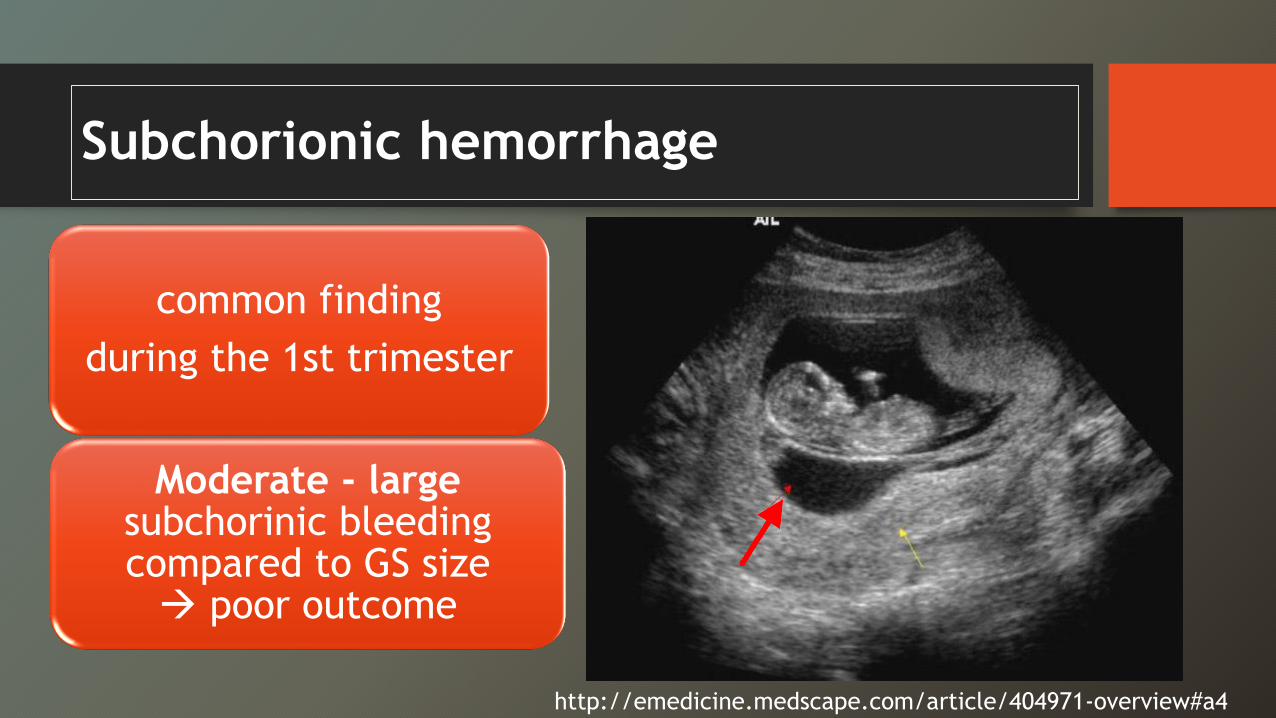
Subchorionic hemorrhage
common finding during the 1st trimester
Moderate - large subchorinic bleeding compared to GS size Æ poor outcome
http://emedicine.medscape.com/article/404971-overview#a4
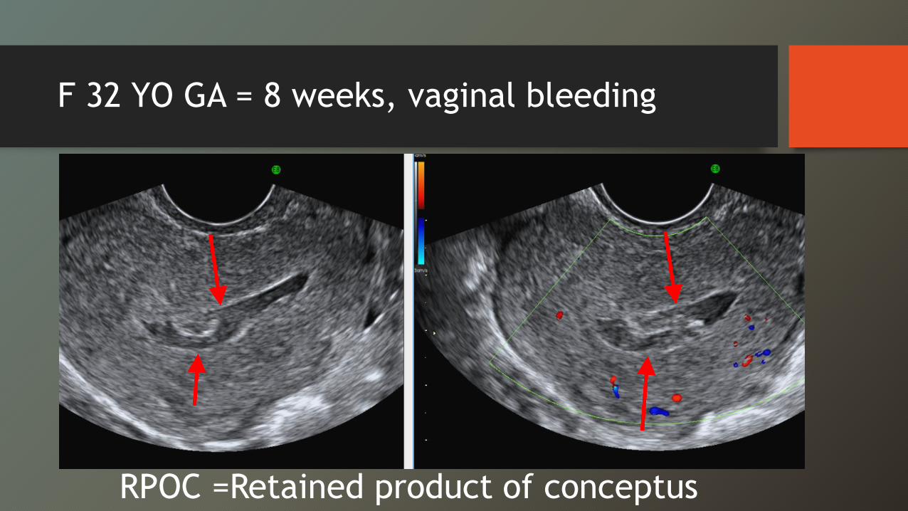
F 32 YO GA = 8 weeks, vaginal bleeding
RPOC =Retained product of conceptus
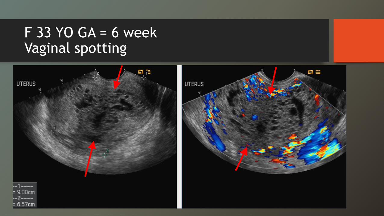
F 33 YO GA = 6 weekVaginal spotting
Gestational Trophoblastic Disease -GTD
• First trimester : variable appearance- a small, echogenic endometrial mass without cystic spaces- mixed echogenic and cystic material - DDx hydropic degeneration and retained products of
conception.
• Second trimester : distended endometrial cavity filled with innumerable small cystic spaces
Summary 1st trimester bleeding
• TVS is the study of choice for early pregnancies. • TAS : useful to assess the amount of free fluid and for
abnormalities beyond the FOV of TVS• correlate with the quantitative E-hCG level & with the
clinical presentation• The lack of an intrauterine GS does not necessarily
indicate ectopic pregnancy
Summary 1st trimester bleeding
• A failed pregnancy : - GS >25 mm & no yolk sac/ embryo- an embryo measuring ≥7 mm & no cardiac activity.• Use M-mode to document embryonic viability and measure
heart rate
• Doppler US should not be used to evaluate a normal early embryo.
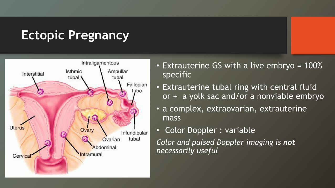
Ectopic Pregnancy
• Extrauterine GS with a live embryo = 100% specific
• Extrauterine tubal ring with central fluid or + a yolk sac and/or a nonviable embryo
• a complex, extraovarian, extrauterinemass
• Color Doppler : variableColor and pulsed Doppler imaging is not
necessarily useful
• MRI : unusual ectopic pregnancies, GTD, or vascular abnormalities, but should not delay urgent or emergent care in an unstable patient.
• CT may be useful in trauma or acute non-gynecologic pain, for staging of malignancy, or if MRI is not possible.
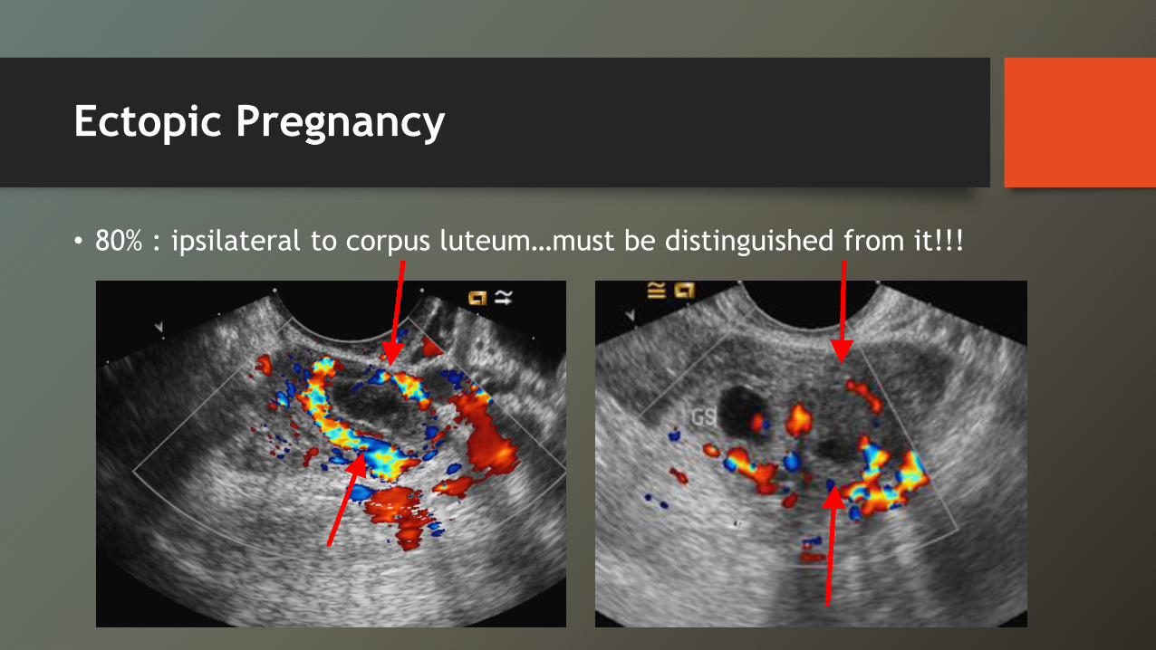
Ectopic Pregnancy
• 80% : ipsilateral to corpus luteum…must be distinguished from it!!!
Ectopic pregnancy
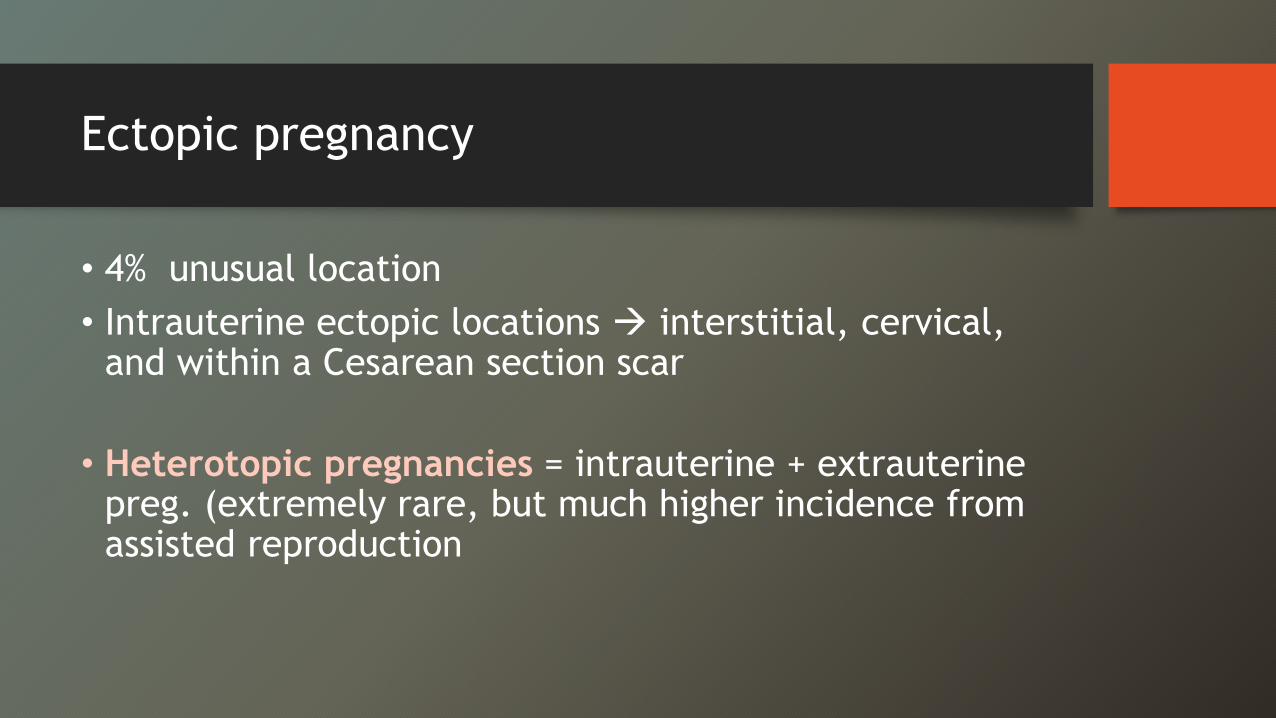
• 4% unusual location• Intrauterine ectopic locations Æ interstitial, cervical,
and within a Cesarean section scar
• Heterotopic pregnancies = intrauterine + extrauterinepreg. (extremely rare, but much higher incidence from assisted reproduction
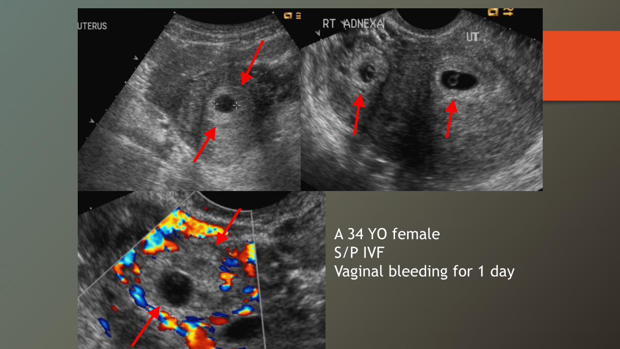
A 34 YO femaleS/P IVFVaginal bleeding for 1 day
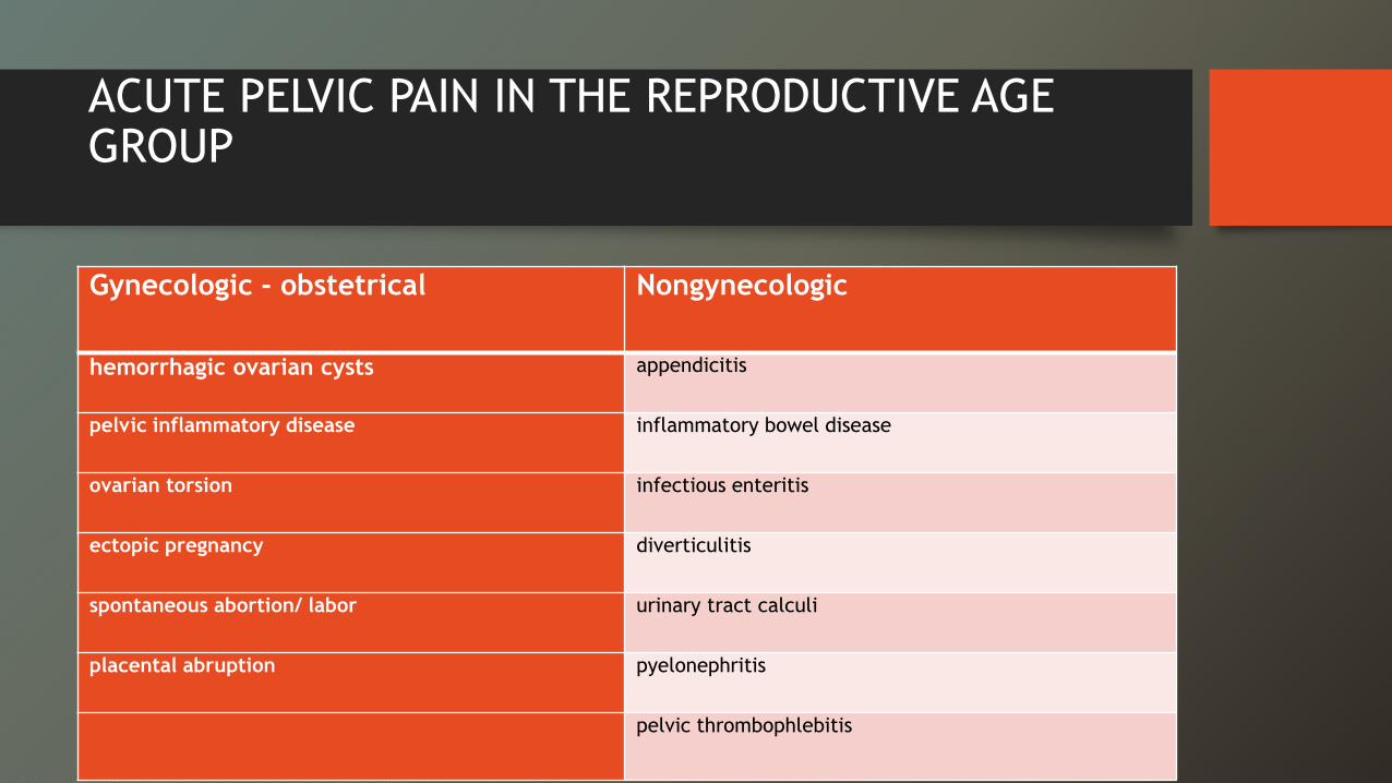
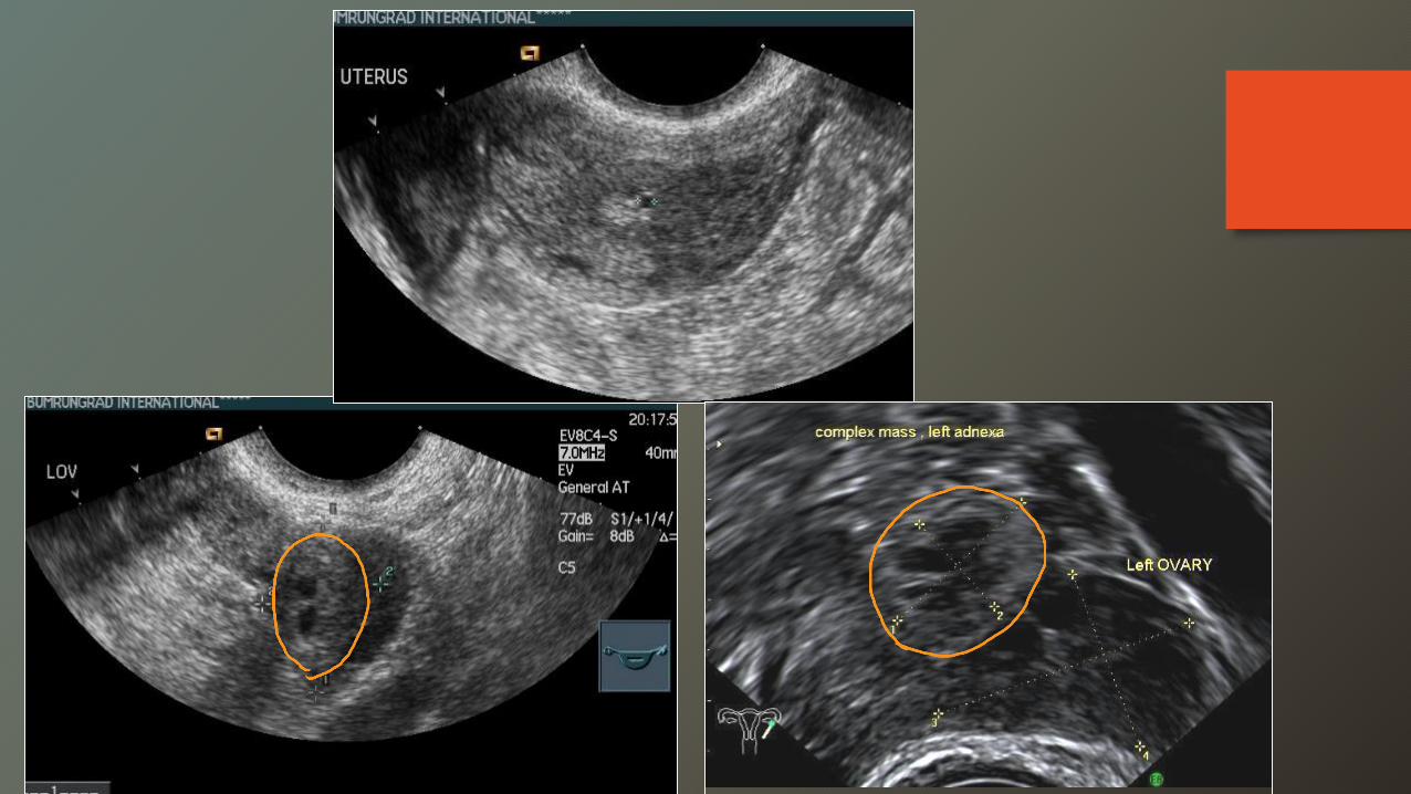
ACUTE PELVIC PAIN IN THE REPRODUCTIVE AGE GROUP
Gynecologic - obstetrical Nongynecologic
hemorrhagic ovarian cysts appendicitis
pelvic inflammatory disease inflammatory bowel disease
ovarian torsion infectious enteritis
ectopic pregnancy diverticulitis
spontaneous abortion/ labor urinary tract calculi
placental abruption pyelonephritis
pelvic thrombophlebitis
ACUTE PELVIC PAIN IN THE REPRODUCTIVE AGE GROUP
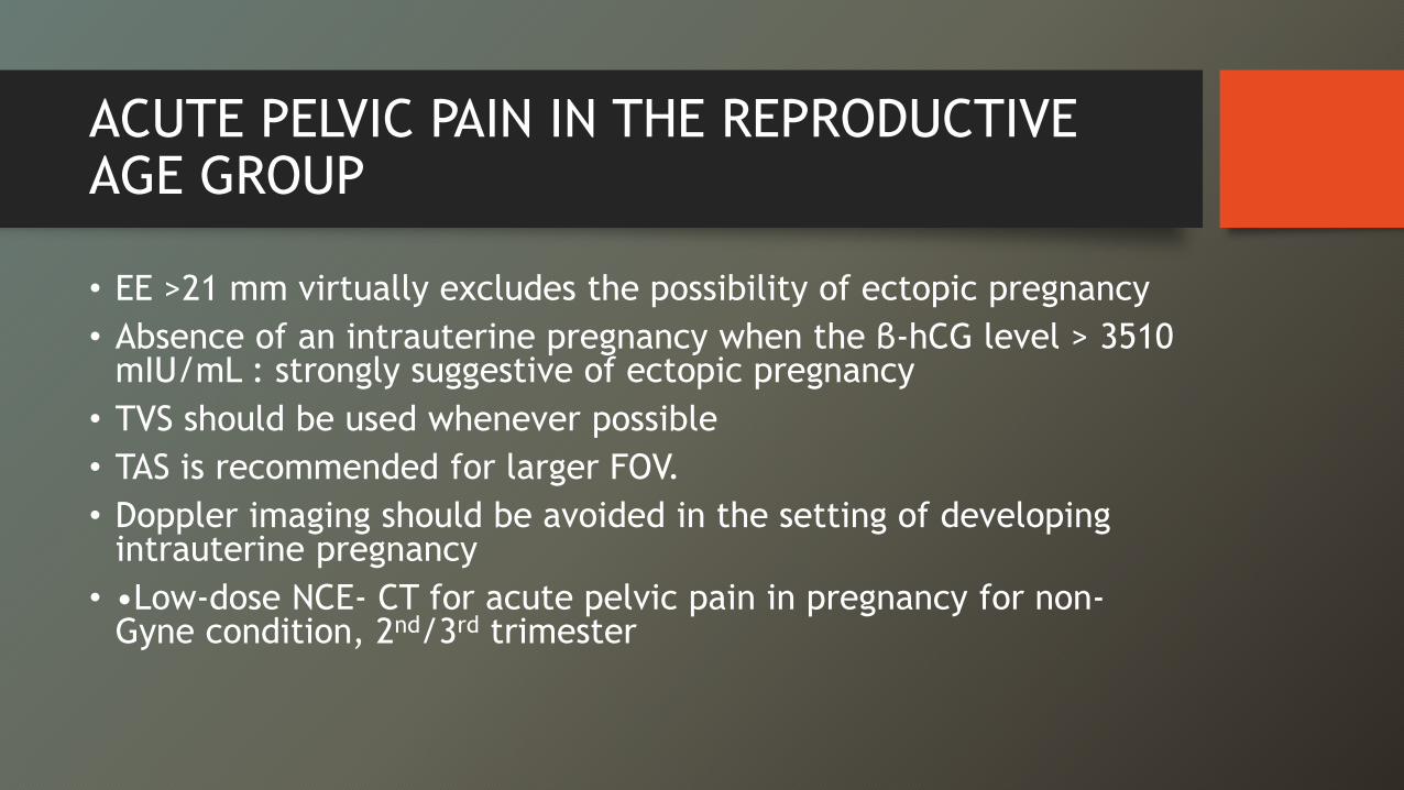
• EE >21 mm virtually excludes the possibility of ectopic pregnancy• Absence of an intrauterine pregnancy when the β-hCG level > 3510
mIU/mL : strongly suggestive of ectopic pregnancy• TVS should be used whenever possible• TAS is recommended for larger FOV. • Doppler imaging should be avoided in the setting of developing
intrauterine pregnancy• •Low-dose NCE- CT for acute pelvic pain in pregnancy for non-
Gyne condition, 2nd/3rd trimester
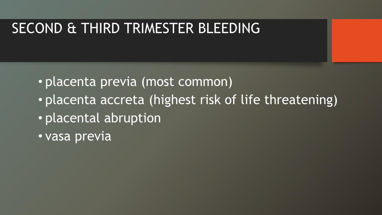
SECOND & THIRD TRIMESTER BLEEDING
• placenta previa (most common) • placenta accreta (highest risk of life threatening)• placental abruption• vasa previa
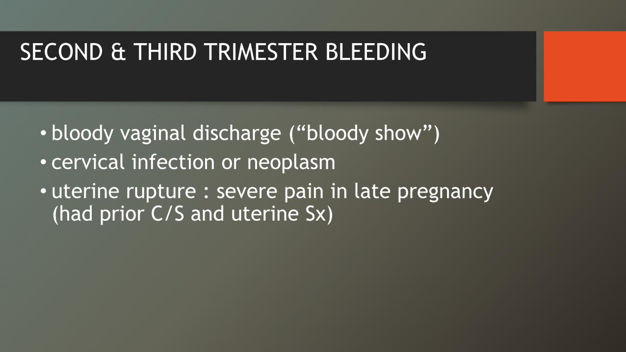
SECOND & THIRD TRIMESTER BLEEDING
• bloody vaginal discharge (“bloody show”) • cervical infection or neoplasm• uterine rupture : severe pain in late pregnancy (had prior C/S and uterine Sx)

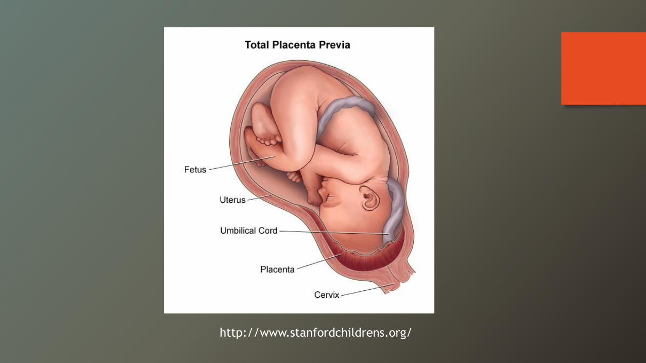
http://www.stanfordchildrens.org/
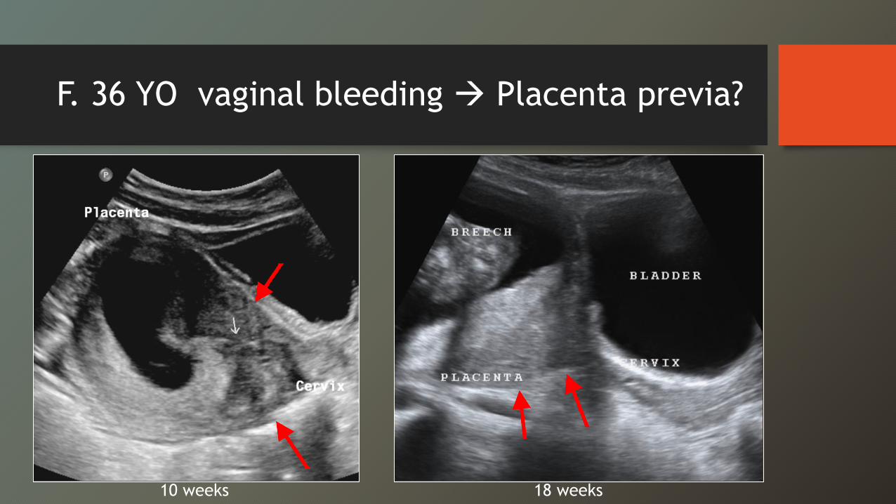
F. 36 YO vaginal bleeding Æ Placenta previa?
18 weeks10 weeks
Placenta previa
• painless bleeding• near the end of 2nd- 3rd trimester• 2.8/1,000 in singleton pregnancies• 3.9/1,000 in twin pregnancies• risk factors: age over 30, multiparity, prior C/S, and
prior abortions
Placenta previa
4 types:
(1) complete previa—covers the internal os (central or asymmetric)(2) partial previa—partially covering(3) marginal—placental edge going to the internal os(4) low-lying—to within 2 cm of the internal os
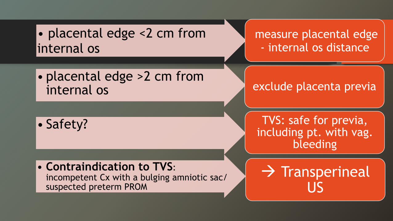
• placental edge <2 cm from internal os
measure placental edge - internal os distance
• placental edge >2 cm from internal os exclude placenta previa
• Safety? TVS: safe for previa, including pt. with vag.
bleeding
• Contraindication to TVS: incompetent Cx with a bulging amniotic sac/ suspected preterm PROM
Æ TransperinealUS
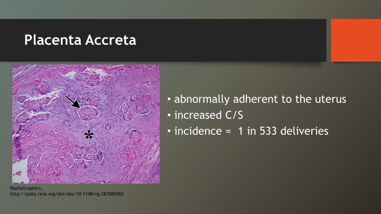
Placenta Accreta
• abnormally adherent to the uterus• increased C/S• incidence = 1 in 533 deliveries
RadioGraphics, http://pubs.rsna.org/doi/abs/10.1148/rg.287085060
Placenta Accreta
Risks: • placenta previa & multiple C/S • Advanced maternal age• Multiparity• Asherman’s syndrome (Uterine synechiae)• Fibroids
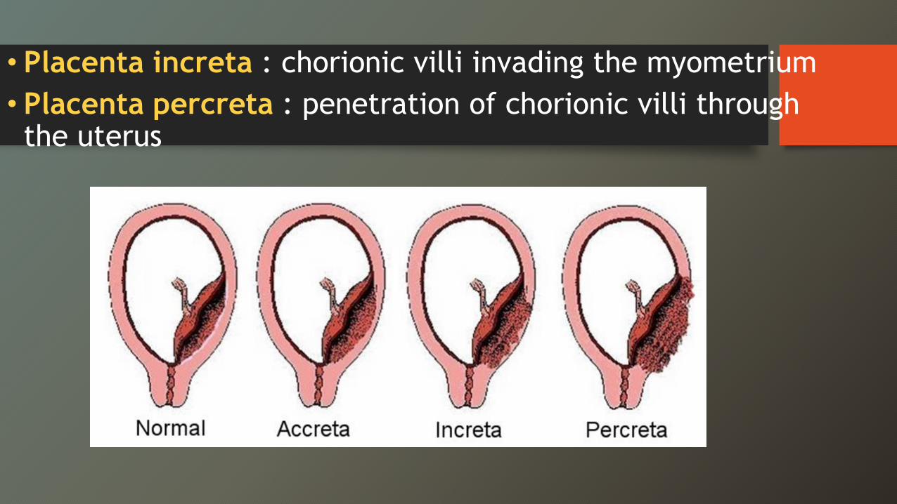
• Placenta increta : chorionic villi invading the myometrium• Placenta percreta : penetration of chorionic villi through the uterus
Placenta Accreta: US findings
• loss of the normal retroplacental hypoechoic zone• localized thinning of the myometrium• increased vascularity at placental-myometrial interface
on CDUS
Placenta Accreta: US findings
• “Numerous coherent vessels” at the placental base: • inseparable cotyledon (fetal villous) and intervillous
(maternal) circulations with extreme hypervascularity
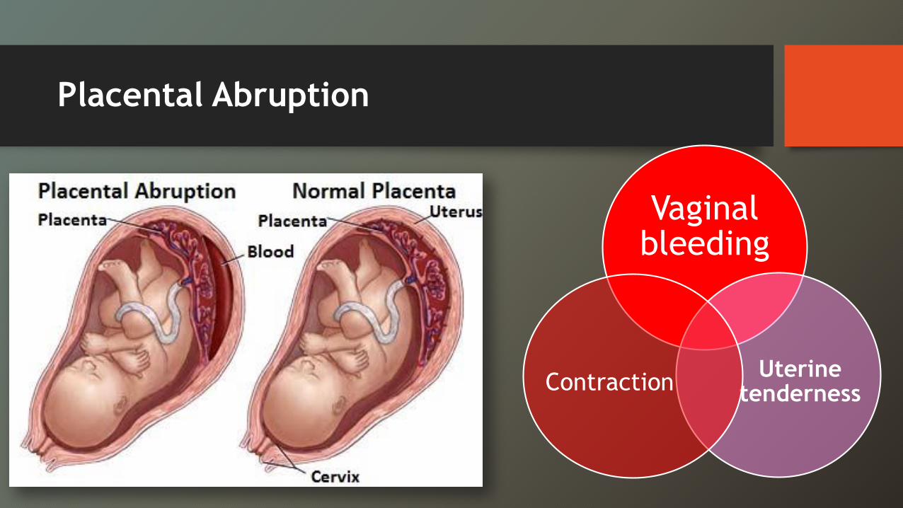
Placental Abruption
Vaginal bleeding
Uterine tenderness Contraction
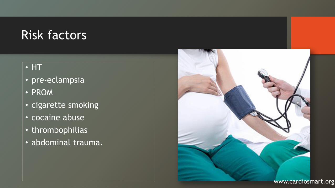
Risk factors
• HT• pre-eclampsia• PROM• cigarette smoking• cocaine abuse• thrombophilias• abdominal trauma.
www.cardiosmart.org
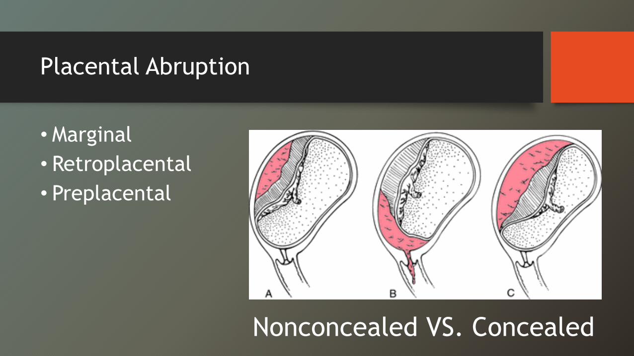
Placental Abruption
• Marginal• Retroplacental• Preplacental
Nonconcealed VS. Concealed
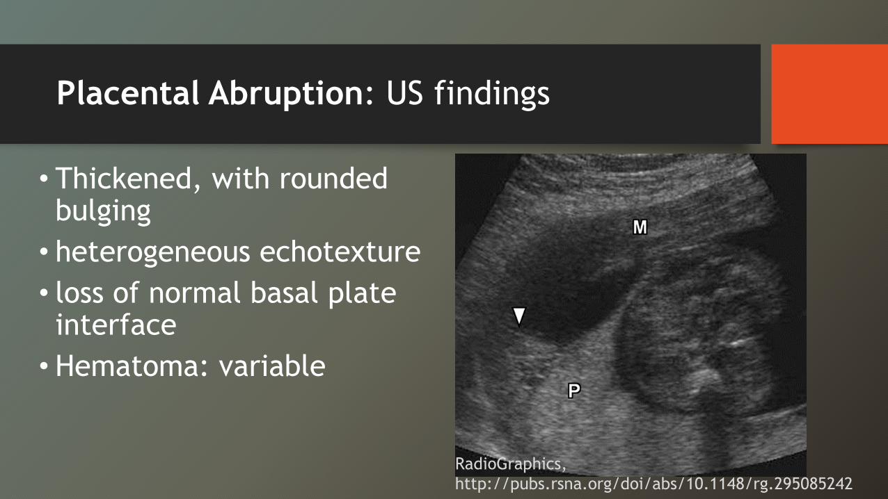
Placental Abruption: US findings
• Thickened, with rounded bulging
• heterogeneous echotexture• loss of normal basal plate interface
• Hematoma: variable
RadioGraphics, http://pubs.rsna.org/doi/abs/10.1148/rg.295085242
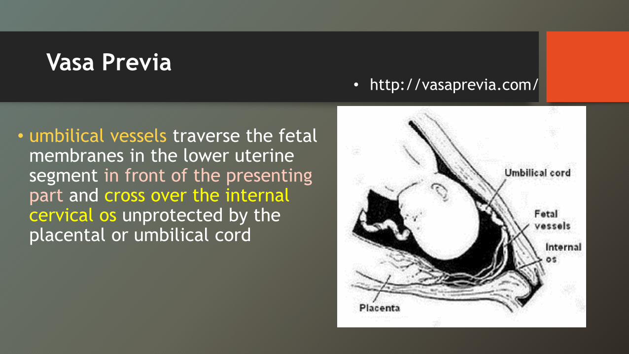
Vasa Previa
• umbilical vessels traverse the fetal membranes in the lower uterine segment in front of the presenting part and cross over the internal cervical os unprotected by the placental or umbilical cord
• http://vasaprevia.com/
Vasa Previa
• high risk of fetal death• Neurologic deficit due to fetal exsanguination• Incidence is 1/2,500 deliveries
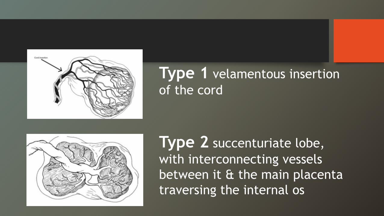
Type 1 velamentous insertion of the cord
Type 2 succenturiate lobe, with interconnecting vessels between it & the main placenta traversing the internal os
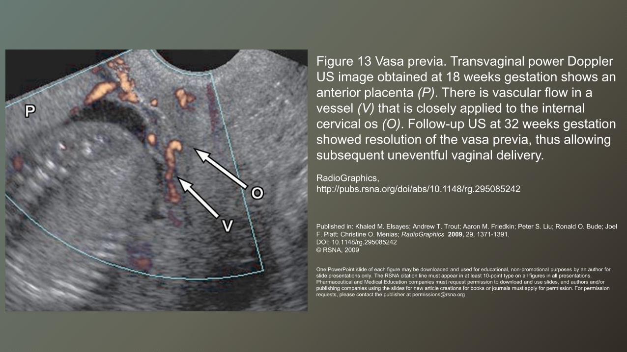
Figure 13 Vasa previa. Transvaginal power Doppler US image obtained at 18 weeks gestation shows an anterior placenta (P). There is vascular flow in a vessel (V) that is closely applied to the internal cervical os (O). Follow-up US at 32 weeks gestation showed resolution of the vasa previa, thus allowing subsequent uneventful vaginal delivery.RadioGraphics, http://pubs.rsna.org/doi/abs/10.1148/rg.295085242
Published in: Khaled M. Elsayes; Andrew T. Trout; Aaron M. Friedkin; Peter S. Liu; Ronald O. Bude; Joel F. Platt; Christine O. Menias; RadioGraphics 2009, 29, 1371-1391.DOI: 10.1148/rg.295085242© RSNA, 2009
One PowerPoint slide of each figure may be downloaded and used for educational, non-promotional purposes by an author for slide presentations only. The RSNA citation line must appear in at least 10-point type on all figures in all presentations. Pharmaceutical and Medical Education companies must request permission to download and use slides, and authors and/or publishing companies using the slides for new article creations for books or journals must apply for permission. For permission requests, please contact the publisher at [email protected]
Summary
xVaginal bleeding in the 2nd or 3rd trimester ass. with increased risks Mother & Fetus.
xTAS = primary imagingxTVS needed for visualization of Cx & internal os(+/- transperineal US)

Summary : Placenta previa
xDecribe distance of the placental edge to the internal os
x Reevaluated during pregnancy for a potential resolution depending on the degree of attachment to the opposing wall.
Summary :Placental abruption
• clinical diagnosis• emergency US : placental thickening, heterogeneity, and
a periplacental hematoma
Summary: Placental accreta
• Prior C/S Æ increase the risk of placental accreta• US findings: intraplacental lacunes, increased vascularity,
myometrial thinning, and focal placental bulge • MRI improves Dx confidence
Summary: Vasa previa
• Serious risk that needs to be recognized and requires a planned C/S.
• Velamentous cord insertion VS. interconnecting vessels of accessory placental lobe (succenturiate lobe), over internal Cx os


![15. Obstetric Emergencies-Bates[1]](https://img.pdfslide.net/doc/110x75/577c780d1a28abe0548e8727/15-obstetric-emergencies-bates1.jpg)