Embed Size (px)
Citation preview

Urinary Tract
Pathology Lab
By the Name of Allah,
The Most Gracious, The Most Merciful

2
Kidney & Urinary tract
3
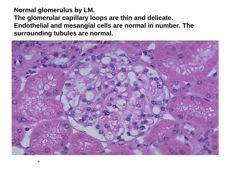
Normal glomerulus by LM.
The glomerular capillary loops are thin and delicate.
Endothelial and mesangial cells are normal in number. The
surrounding tubules are normal.

5
IF-Granula deposition of immune complexes .
characteristic of circulating and in situ immune complex
deposition

6
immunofluorescence linear deposition of
immune complexes
7
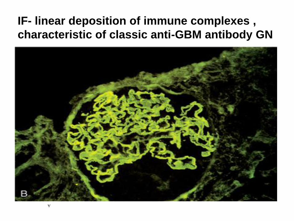
IF- linear deposition of immune complexes ,
characteristic of classic anti-GBM antibody GN

8
Minimal change
disease.
A
Under the light
microscope the
PAS-stained
glomerulus
appears normal,
with a delicate
basement
membrane
B
Schematic diagram
illustrating diffuse
effacement of foot
processes of
podocytes with no
immune deposits.

9
MCD-EM
the capillary loop in the lower half contains two electron dense RBC's.
Fenestrated endothelium is present and the BM is normal.
The overlying epithelial cell foot processes are fused (arrows).
10
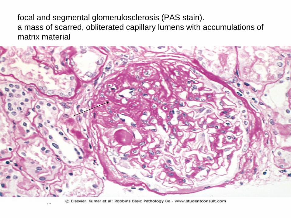
focal and segmental glomerulosclerosis (PAS stain).
a mass of scarred, obliterated capillary lumens with accumulations of
matrix material

11
FSGS
blue collagen deposition (MT stain).
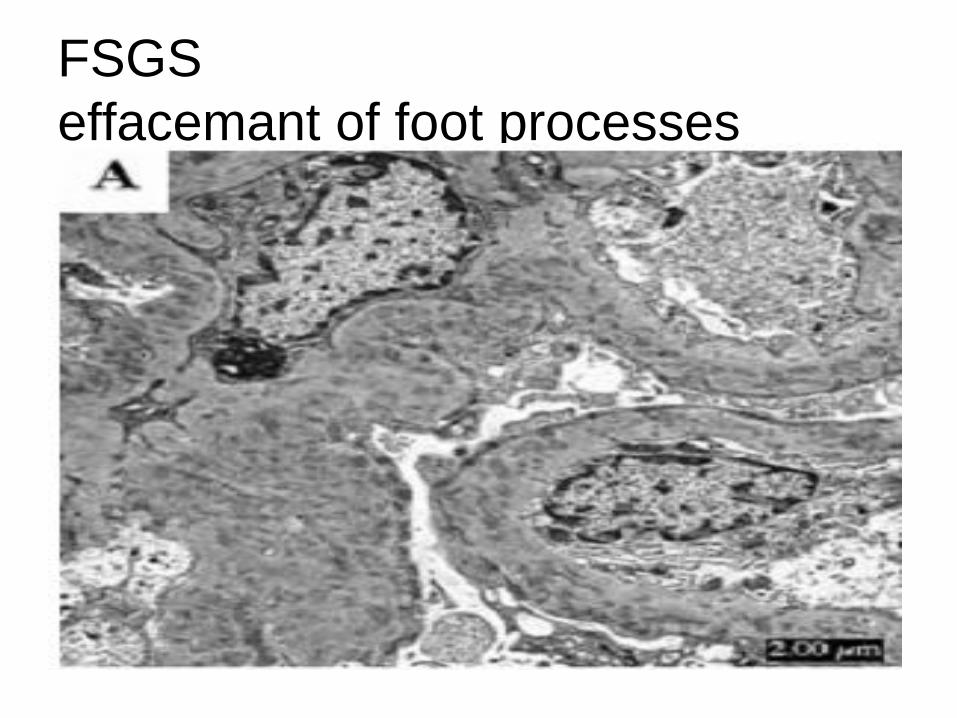
FSGS
effacemant of foot processes

13
LM- membranous glomerulonephritis in which the
capillary loops are thickened and prominent, but the
cellularity is not increased

14
Membranous nephropathy.
A ,Diffuse thickening of the glomerular
basement membrane .
B ,Schematic diagram illustrating
subepithelial deposits, effacement of
foot processes, and the presence of
"spikes" of basement membrane
material between the immune deposits .

15
A silver stain of the glomerulus highlights the proteinaceous basement
membranes in black. There are characteristic "spikes" seen with
membranous glomerulonephritis seen here in which the black basement
membrane material appears as projections around the capillary loops.

16
MGN
IF-deposits of mainly IgG and complement collect in the basement
membrane and appear in a diffuse granular pattern

17
EM-the darker electron dense immune deposits
are seen scattered within the thickened
basement membrane .

18
Membranoproliferative GN, showing mesangial cell proliferation,
basement membrane thickening, leukocyte infiltration, and accentuation
of lobular architecture.
19
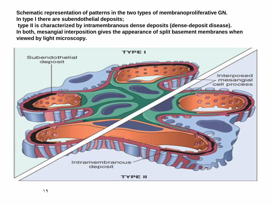
Schematic representation of patterns in the two types of membranoproliferative GN.
In type I there are subendothelial deposits;
type II is characterized by intramembranous dense deposits (dense-deposit disease).
In both, mesangial interposition gives the appearance of split basement membranes when
viewed by light microscopy.

20
membranoproliferative
glomerulonephritis (MPGN(

21
This silver stain demonstrates a double contour of the basement
membranes("tram-tracking" )that is characteristic of
(MPGN)(arrows).
22
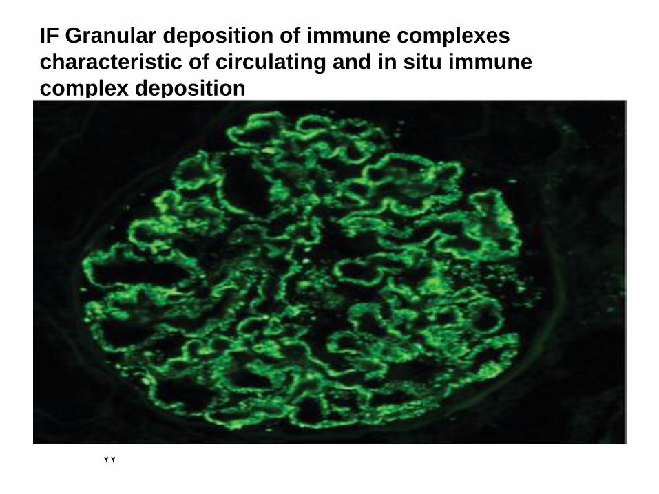
IF Granular deposition of immune complexes
characteristic of circulating and in situ immune
complex deposition

23
EM-MPGN type I a mesangial cell at the lower left that is
interposing its cytoplasm at the arrow into the basement
membrane leading to splitting" of the GBM (tram track).

24
EM-dense deposits in the basement membrane of MPGN type II.
There are dark electron dense deposits within the basement
membrane that often coalesce to form a ribbon-like mass of
deposits ) arrows)

25
Post-streptococcal glomerulonephritis.
This glomerulus is hypercellular and capillary loops
are poorly defined.

26
Post-streptococcal glomerulonephritis is due to increased
numbers of epithelial, endothelial, and mesangial cells as well as
neutrophils in and around the capillary loops (arrows)
27
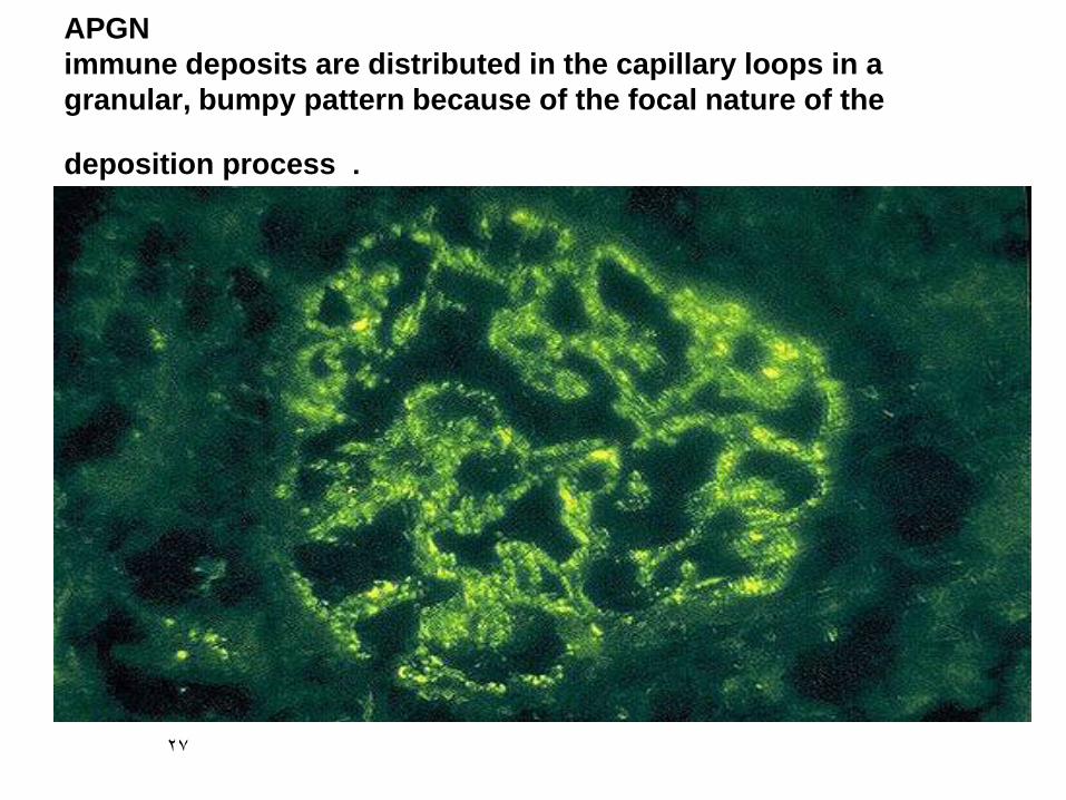
APGN
immune deposits are distributed in the capillary loops in a
granular, bumpy pattern because of the focal nature of the
deposition process .

28
EM -immune deposits of PSGN are predominantly subepithelial,
a large subepithelial "hump" at the right of the BM (arrows).
The capillary lumen is filled with a PMN whose nuclear lobes (arrows)and
cytoplasmic granules are visibl(arrows).
29
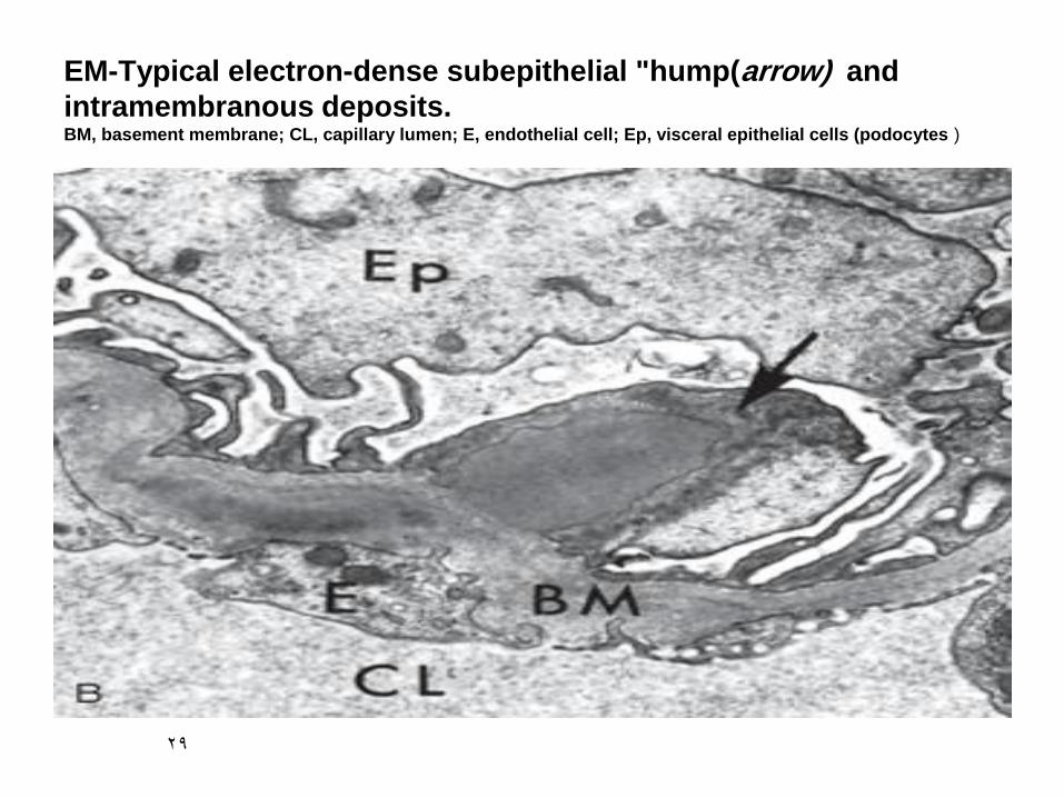
EM-Typical electron-dense subepithelial "hump(arrow) and
intramembranous deposits. BM, basement membrane; CL, capillary lumen; E, endothelial cell; Ep, visceral epithelial cells (podocytes )

30
The IgA is deposited mainly in mesangium, which then
increases mesangial cellularity (arrow) .

31
mesangial matrix enlargement is conspicuous and predominates over a
relatively mild mesangial cell proliferation.

32
Mesangial proliferation can be much more intense, global
and diffuse

33
IF demonstrates positivity with antibody to IgA.
the pattern is that of mesangial staining.
34
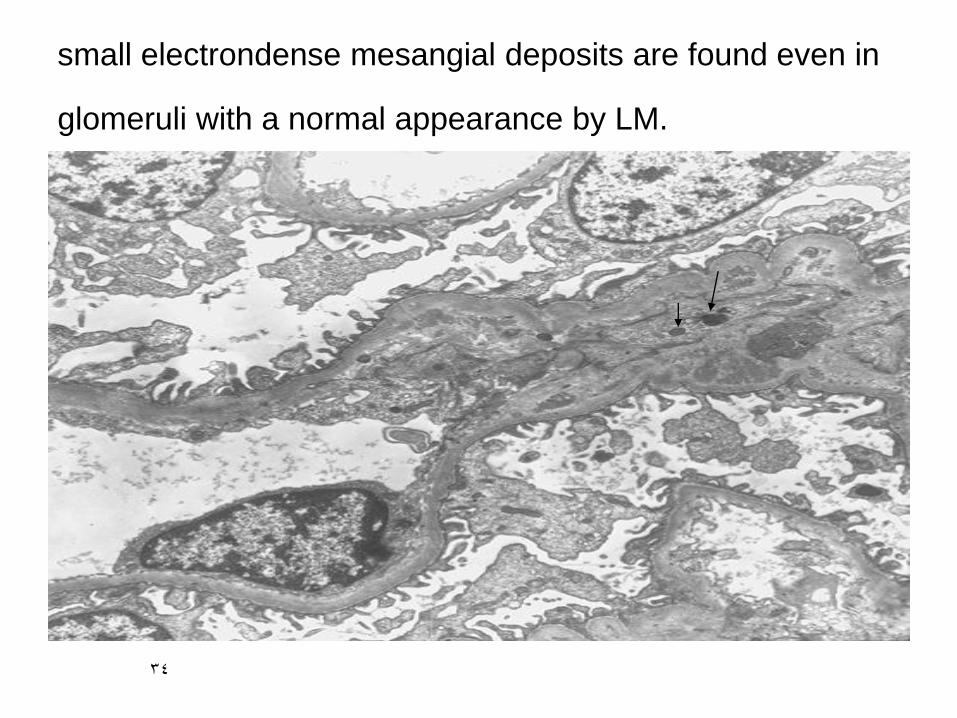
small electrondense mesangial deposits are found even in
glomeruli with a normal appearance by LM.
35
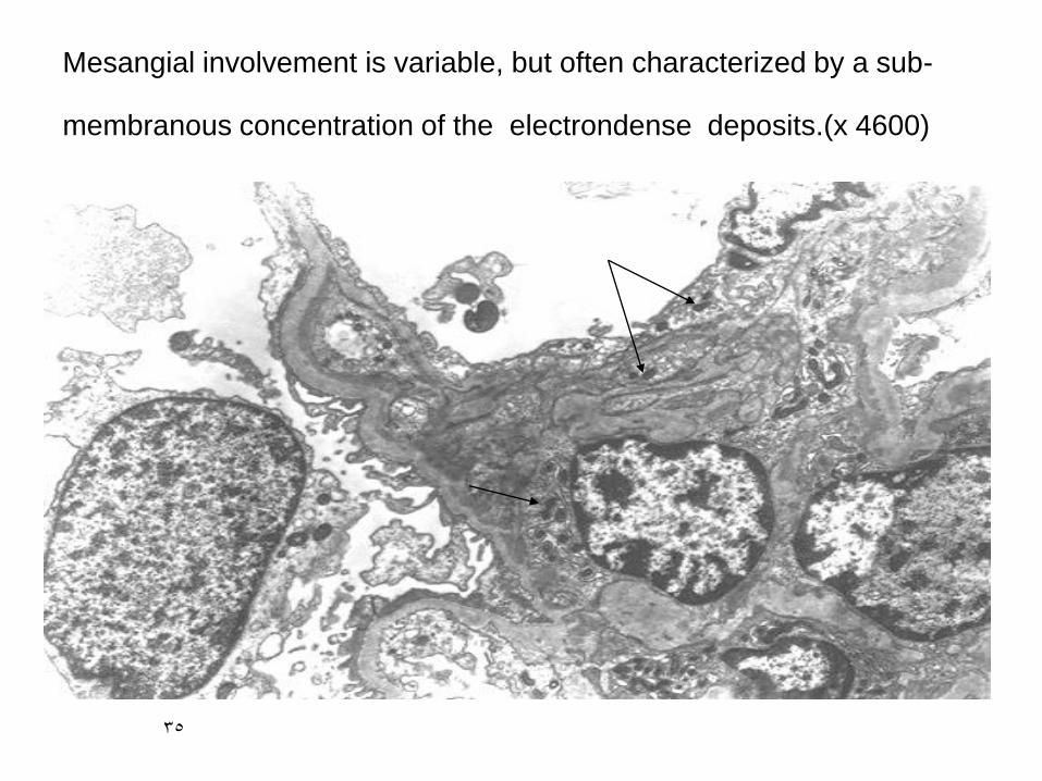
Mesangial involvement is variable, but often characterized by a sub-
membranous concentration of the electrondense deposits.(x 4600)

36
LM-The renal tubular cells appear foamy (arrows)because of the
accumulation of neutral fats and mucopolysaccharides. The
glomeruli show irregular thickening and splitting of basement
membranes.

37
thin and attenuated BM in Alport
syndrome

38
The diagrams below illustrate normal BM(LT) vs the
thickened and 'falling apart' of Alport GBM(RT)
BM

39
The final

40
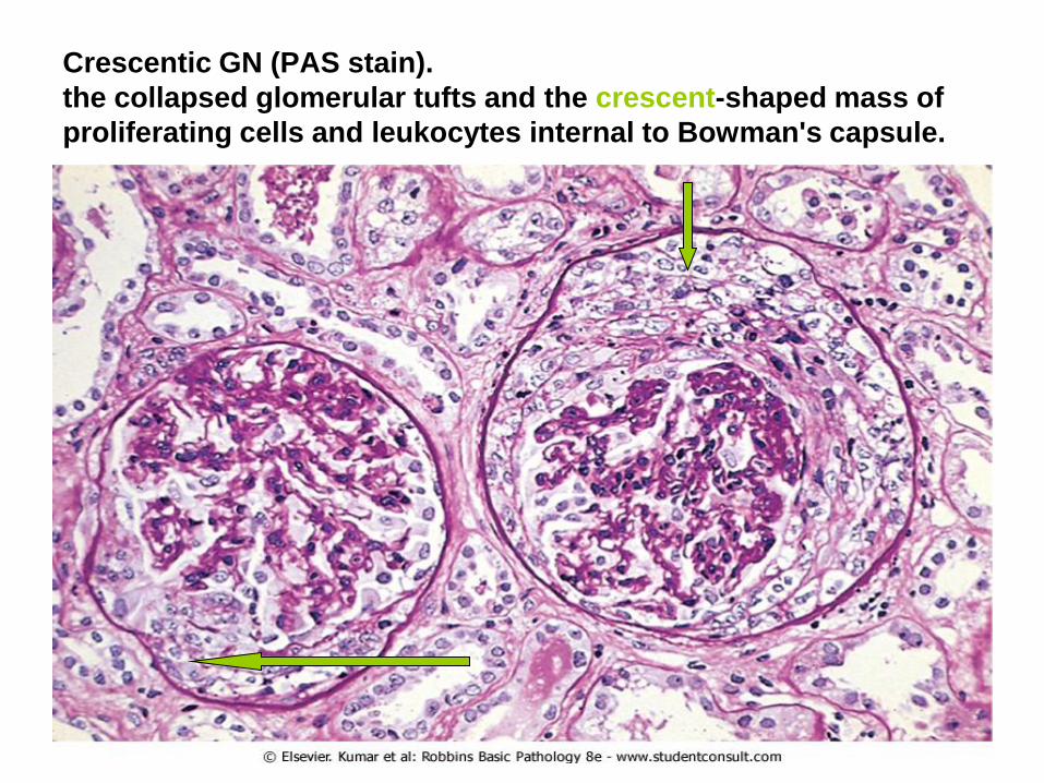
Crescentic GN (PAS stain).
the collapsed glomerular tufts and the crescent-shaped mass of
proliferating cells and leukocytes internal to Bowman's capsule.

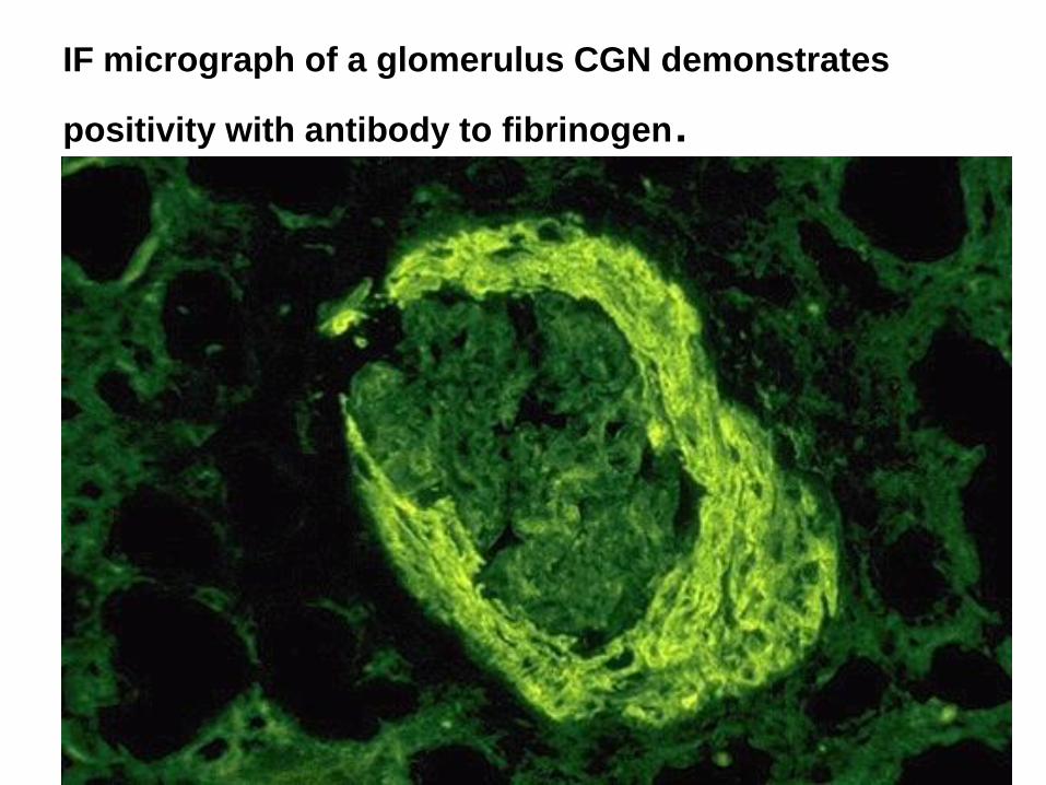
IF micrograph of a glomerulus CGN demonstrates
positivity with antibody to fibrinogen.

42
Chronic GN.
A MT stain shows complete replacement of virtually all
glomeruli by blue-staining collagen.
43
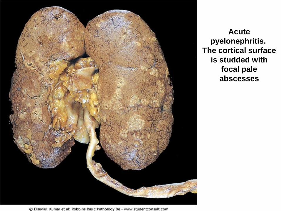
Acute
pyelonephritis.
The cortical surface
is studded with
focal pale
abscesses
44
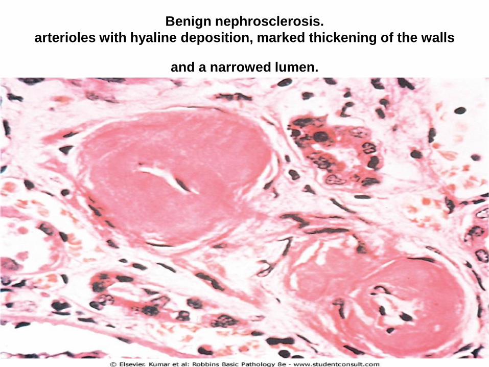
Benign nephrosclerosis.
arterioles with hyaline deposition, marked thickening of the walls
and a narrowed lumen.

45
Malignant hypertension.
Fibrinoid necrosis of afferent arteriole (PAS stain).

46
Malignant hypertension
Hyperplastic arteriolosclerosis (onion-skin lesion).

47
CYSTIC DISEASES OF THE
KIDNEY

Simple renal Cysts
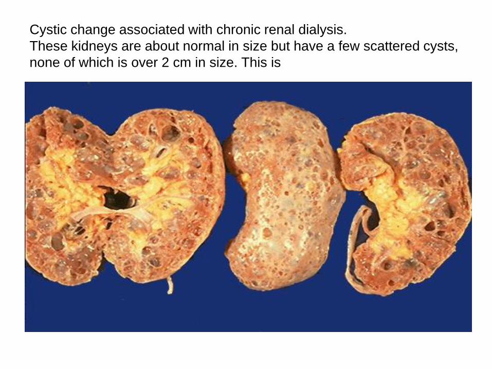
Cystic change associated with chronic renal dialysis.
These kidneys are about normal in size but have a few scattered cysts,
none of which is over 2 cm in size. This is

50

Normal term infant kidneys

52
Cysts are fairly small but uniformly distributed throughout the
parenchyma so that the disease is usually symmetrical in appearance
with both kidneys markedly enlarged.

53
Hydronephrosis of the kidney,
with marked dilation of the pelvis
and calyces and thinning of renal
parenchyma.

Renal cell carcinoma:
typical cross-section of
yellowish, spherical
neoplasm in one pole of
the kidney.
Note the tumor in the
dilated, thrombosed renal
vein.

Renal cell carcinoma High-power detail of the clear cell pattern
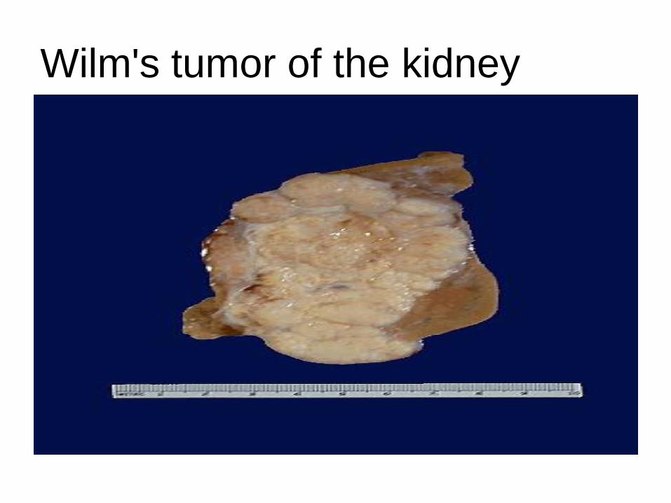
Wilm's tumor of the kidney
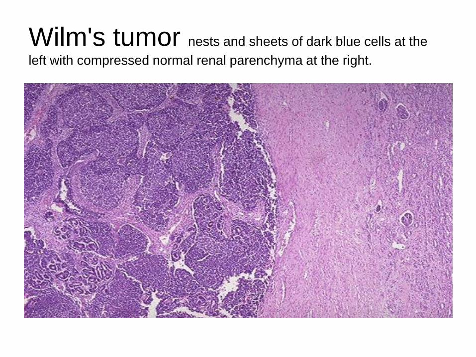
Wilm's tumor nests and sheets of dark blue cells at the
left with compressed normal renal parenchyma at the right.

The tumor shows attempts to form primitive glomerular and
tubular structures

Transitional cell carcinoma of UB

Papillary Urothelial (transitional)
carcinoma-low grade

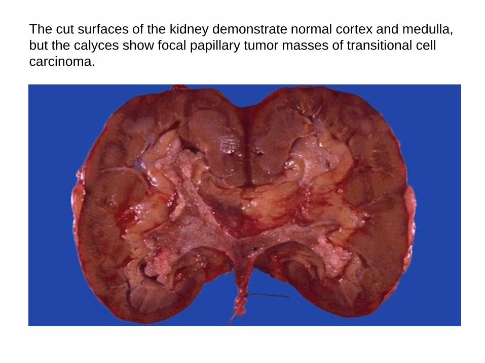
The cut surfaces of the kidney demonstrate normal cortex and medulla,
but the calyces show focal papillary tumor masses of transitional cell
carcinoma.

62
Final material
63
Crescentic GN (PAS stain).
the collapsed glomerular tufts and the crescent-shaped mass of
proliferating cells and leukocytes internal to Bowman's capsule.
IF micrograph of a glomerulus CGN demonstrates
positivity with antibody to fibrinogen.

65
Chronic GN.
A MT stain shows complete replacement of virtually all
glomeruli by blue-staining collagen.

66
Acute
pyelonephritis.
The cortical surface
is studded with
focal pale
abscesses

67
Benign nephrosclerosis.
arterioles with hyaline deposition, marked thickening of the walls
and a narrowed lumen.

68
Malignant hypertension.
Fibrinoid necrosis of afferent arteriole (PAS stain).

69
Malignant hypertension
Hyperplastic arteriolosclerosis (onion-skin lesion).

70
CYSTIC DISEASES OF THE
KIDNEY

Simple renal Cysts

Cystic change associated with chronic renal dialysis.
These kidneys are about normal in size but have a few scattered cysts,
none of which is over 2 cm in size. This is

73

Normal term infant kidneys

75
Cysts are fairly small but uniformly distributed throughout the
parenchyma so that the disease is usually symmetrical in appearance
with both kidneys markedly enlarged.

76
Hydronephrosis of the kidney,
with marked dilation of the pelvis
and calyces and thinning of renal
parenchyma.

Renal cell carcinoma:
typical cross-section of
yellowish, spherical
neoplasm in one pole of
the kidney.
Note the tumor in the
dilated, thrombosed renal
vein.

Renal cell carcinoma High-power detail of the clear cell pattern

Wilm's tumor of the kidney

Wilm's tumor nests and sheets of dark blue cells at the
left with compressed normal renal parenchyma at the right.

The tumor shows attempts to form primitive glomerular and
tubular structures

Transitional cell carcinoma of UB

Papillary Urothelial (transitional)
carcinoma-low grade
The cut surfaces of the kidney demonstrate normal cortex and medulla,
but the calyces show focal papillary tumor masses of transitional cell
carcinoma.


![7 Catheter-associated Urinary Tract Infection (CAUTI) · UTI Urinary Tract Infection (Catheter-Associated Urinary Tract Infection [CAUTI] and Non-Catheter-Associated Urinary Tract](https://img.pdfslide.net/doc/110x75/5c40b88393f3c338af353b7f/7-catheter-associated-urinary-tract-infection-cauti-uti-urinary-tract-infection.jpg)