Embed Size (px)
Citation preview
WHO monographs on
selectedmedicinalplantsVolume 4


WHOmonographson selected
medicinal plantsVOLUME 4
WHO Library Cataloguing-in-Publication Data
WHO monographs on selected medicinal plants. Vol. 4.
1. Plants, Medicinal. 2. Angiosperms. 3. Medicine, Traditional. I. WHO Consultation on Selected Medicinal Plants (4th: 2005: Salerno-Paestum, Italy) II. World Health Organization.
ISBN 978 92 4 154705 5 (NLM classification: QV 766)
© World Health Organization 2009
All rights reserved. Publications of the World Health Organization can be obtained from WHO Press, World Health Organization, 20 Avenue Appia, 1211 Geneva 27, Switzerland (tel.: +41 22 791 3264; fax: +41 22 791 4857; e-mail: [email protected]). Requests for permission to reproduce or translate WHO publications – whether for sale or for noncommercial distribution – should be addressed to WHO Press, at the above address (fax: +41 22 791 4806; e-mail: [email protected]).
The designations employed and the presentation of the material in this publication do not imply the expression of any opinion whatsoever on the part of the World Health Organization concerning the legal status of any country, territory, city or area or of its authorities, or concerning the delimitation of its frontiers or boundaries. Dotted lines on maps represent approximate border lines for which there may not yet be full agreement.
The mention of specific companies or of certain manufacturers’ products does not imply that they are endorsed or recommended by the World Health Organization in preference to others of a similar nature that are not mentioned. Errors and omissions excepted, the names of proprietary products are distinguished by initial capital letters.
All reasonable precautions have been taken by the World Health Organization to verify the information contained in this publication. However, the published material is being distributed without warranty of any kind, either expressed or implied. The responsibility for the interpretation and use of the material lies with the reader. In no event shall the World Health Organization be liable for damages arising from its use.
Printed in Spain
iii
Contents
Acknowledgements vIntroduction 1General technical notices 5
Monographs (in alphabetical order of plant name)
Fructus Agni Casti 9Cortex Berberidis 30Gummi Boswellii 48Semen Cardamomi 61Fructus Chebulae 71Semen Cucurbitae 83Folium Cynarae 92Cortex Granati 108Pericarpium Granati 117Folium Guavae 127Lichen Islandicus 140Fructus Macrocarponii 149Cortex Magnoliae 167Herba Millefolii 179Fructus Momordicae 192Fructus Myrtilli 210Radix Panacis Quinquefolii 226Cortex Phellodendron 244Rhizoma Picrorhizae 258Oleum Ricini 271Aetheroleum Rosmarini 284Folium Rosmarini 294Cortex Salicis 309Fructus Tribuli 323Flos Trifolii 335
iv
Ramulus cum Uncis Uncariae 353Cortex Viburni Prunifolii 364Radix Withaniae 373
Annex 1Participants of the Fourth WHO Consultationon Selected Medicinal Plants Salerno-Paestum,Italy, 3–6 October 2005 392
Annex 2Cumulative index (in alphabetical order of plant name) 395
Annex 3Cumulative index (in alphabetical order of plant material of interest) 397
Annex 4Cumulative index of medicinal plants (in alphabetical order of Latin binomial plant name) 400
Annex 5Cumulative index of major chemical constituents (by compound name in alphabetical order) 406
Annex 6Cumulative index of major chemical constituents (ordered by CAS number) 430
Annex 7Cumulative index of major chemical constituents (ordered by molecular formula) 437
Contents

v
Acknowledgements
Special acknowledgement is due to Professor Norman R. Farnsworth, Professor Harry H.S. Fong, and Professor Gail B. Mahady of the WHO Collaborating Centre for Traditional Medicine, College of Pharmacy, University of Illinois at Chicago, Chicago, IL, USA, for drafting and re-vising the monographs. Special acknowledgement is also due to Dr Ray-mond Boudet-Dalbin of the Laboratoire de Chimie Thérapeutique, Uni-versity of René Descartes, Paris, France, for drawing the chemical structures and for compiling the index of major chemical constituents in-cluding information on their molecular formula and CAS numbers. The photograph for the front cover was kindly provided by the Research Center for Medicinal Plant Resources, National Institute of Biomedical Innovation, Tsukuba City, Japan.
WHO also acknowledges with thanks the valuable work of the ap-proximately 200 experts including 81 national health authorities, who provided comments and advice on the draft texts; those who submitted comments through the World Self-Medication Industry (a nongovern-mental organization in official relations with WHO) and the Internation-al Federation of Pharmacists (a nongovernmental organization in official relations with WHO); and those who participated in the Fourth WHO Consultation on Selected Medicinal Plants held in Salerno-Paestum, Italy, in October 2005 to review the monographs (see Annex 1).
Sincere appreciation is extended to the Ministry of Health of Italy, the Government of the Province of Campagna, Italy, the Municipal Govern-ment of Salerno, Italy, and the State University of Salerno, who hosted the above-mentioned Fourth WHO Consultation and supported it financial-ly. Finally, WHO wishes to express thanks to Mr Raymond Tsai, Boston, USA, Dr Hermann Garden, Basel, Switzerland, Ms Lynn Morra, Abu Dhabi, United Arab Emirates, and Ms Tina Lu, Rochester, USA, for their indispensable assistance in finalizing and editing the manuscripts.


Introduction
Increasing role of the WHO monographs on selected medicinal plantsOver the past two decades, there has been a tremendous increase in the use of herbal medicine; however, there is still a significant lack of research data in this field. Therefore since 1999, WHO has published three vol-umes of the WHO monographs on selected medicinal plants: volume 1 includes 28 monographs; volume 2 contains an additional 30 monographs; and volume 3 provides 31 monographs. Including the 28 new monographs published in this volume, a total of 118 monographs in four volumes are now available on the WHO web site (http://www.who.int/medicinedocs/en/m/abstract/Js14213e/).
Due to the diversity of medicinal plants and herbal medicines, it is dif-ficult for WHO to continue to develop more monographs on commonly used medicinal plants. One of the objectives of WHO monographs is to provide a model that will support countries in developing their own na-tional or regional monographs on medicinal plants or national formular-ies on herbal medicines. Experts can be trained through the process of developing country-specific or regional monographs, and national capac-ity in this field can thus be built up.
For example, at WHO’s regional training workshop on regulation of herbal medicines held for the WHO European Region, in September 2003, the participating national drug regulatory authorities of many of the Newly Independent States (NIS) submitted their request to WHO di-rectly, for assistance in the development of monographs on medicinal plants commonly used in NIS.
In order to respond to their urgent need, WHO initiated a new project to develop a set of regional (NIS) monographs on commonly used me-dicinal plants, based on available scientific information relating to their safety, efficacy and quality, which will facilitate the creation of effective and practical regulatory and quality assurance measures on herbal medi-cines. WHO has been working with 15 national drug regulatory authori-ties interested in this publication in NIS, Countries of Central and Eastern
1

2
Introduction
Europe (CCEE) and their neighbouring countries, in close collaboration with the WHO Regional Office for Europe. The 13 new monographs on commonly used medicinal plants in NIS have been drafted based on the format of the WHO monographs by the experts in NIS and CCEE coun-tries with the support of experts, national health authorities and NGOs within and also outside the NIS and CCEE countries. The WHO mono-graphs on medicinal plants commonly used in NIS have been completed and will be published soon. Based on the NIS countries model, in the fu-ture, WHO would like to cooperate with more countries or regions to develop their monographs on commonly used medicinal plants.
Preparation of monographs for volume 4Selection of medicinal plantsThe selection of medicinal plants for inclusion in the WHO monographs is based on worldwide use. The medicinal plants selected must meet two major criteria: (1) they must be in common use in at least two WHO Re-gions; and (2) there must be sufficient scientific data available to satisfy the requirements of the various sections in the monograph format.
The recommended selection criteria discussed at the Third WHO Consultation on Selected Medicinal Plants (Ottawa, Canada, July 2001) were applied to the preparation of volume 4 of the WHO monographs.
PreparationDuring the preparation of volume 4, more than 200 experts were involved in addition to members of WHO’s Expert Advisory Panel on Traditional Medicine, a significant expansion compared to the numbers involved in the previous three volumes. National drug regulatory authorities in 81 countries participated in the process, again a greater number than for the previous volumes. This global network of active players facilitated wider access to the available scientific references and information, in terms of both quality and quantity. This considerable level of support contrib-uted greatly to the efficiency of the preparation process.
The Fourth WHO Consultation on Selected Medicinal Plants was held in Salerno-Paestum, Italy, in October 2005 to review and finalize the draft monographs. Thirty-four experts and drug regulatory authorities from WHO Member States participated (Annex 1). Following extensive dis-cussion, 28 of the 32 draft monographs were adopted for inclusion.
Changes in format in volume 4A description of selected sections of the monographs is given in the General technical notices, which reflect the above-mentioned format

3
Introduction
changes. For easy reference, two cumulative indexes are provided as an-nexes. Annex 2 lists the monographs in alphabetical order of the plant name, while Annex 3 is arranged according to the plant materials of in-terest. For the convenience of readers, an additional cumulative list of plant names (scientific name followed by family name) which appeared under the heading “definition” in each monograph, has been added (An-nex 4). In response to the recommendation of the Ottawa Consultation in 2001, a cumulative index of major chemical constituents arranged in alphabetical order (Annex 5), a cumulative index ordered by Chemical Abstracts Service (CAS) number (Annex 6) and a cumulative index or-dered by molecular formula (Annex 7) have also been included in this volume.
Under the heading “Geographical distribution”, an attempt has been made to describe the geographical distribution of the plant, i.e. its natural distribution, where it is cultivated, and conditions of cultivation, harvest-ing and storage. This has been a challenge, owing to the lack of data based on established national good agricultural practices and/or good collection practices (GACP) for medicinal plants. In 2007, WHO published the WHO guidelines on good manufacturing practices (GMP) for herbal med-icines, which provide general technical guidance on obtaining medicinal plant materials of good quality for the production of herbal medicines in the overall context of quality assurance and control of herbal medicines. WHO also compiled new guidelines on assessing quality for safety of herbal medicines with reference to contaminants and residues which were published in 2007. It is hoped that these guidelines will facilitate the im-plementation of GMP for herbal medicines at the national level, and the development of national quality standards/specifications for herbal medi-cines, which in turn should bridge the current information gap in this area.
At the consultation, the following technical issues were pointed out for consideration during the preparation of the monographs:
Specify extract according to original literature, whenever possible.Posology: as a general rule make clear that a given posology has been found either in the report of clinical trials or referring to tra-ditional indications. These guidelines have been followed in the preparation of this volume wherever possible.Provide posologies as they relate to the specific indications – the dosage forms have to be consistent with their respective indications.Specific posologies should be indicated for specific uses and dos-age forms whenever available.
4
Introduction
Purpose and content of monographsAlthough the monographs include one section on “medicinal uses” with three categories of information, the purpose of the monographs was clearly explained in the introduction to volume 1, and it is unnecessary to repeat it here. But it should be noted that the word “monograph” is used as a technical term only. It does not have the same meaning as “mono-graph” in any type of pharmacopoeia. In addition, it is reiterated here that this publication is not intended to replace any official compendia such as pharmacopoeias, formularies or legislative documents.
It should also be emphasized that the descriptions included in the sec-tion on medicinal uses should not be taken as implying WHO’s official endorsement or approval. They merely represent the systematic collec-tion of scientific information available at the time of preparation, for the purpose of information exchange.
Finally, the Fourth WHO Consultation on selected medicinal plants recommended WHO to update existing monographs in view of the avail-ability of new data and of the format change that has been employed for volumes 3 and 4, and that this process should be given priority over the development of new monographs, if resources are limited. This volume might therefore be the last volume of WHO monographs on commonly used medicinal plants. We should like to express our appreciation to all the experts, national health authorities, WHO collaborating centres and NGOs for their efforts, technical contributions and support in the prepa-ration of the four volumes of WHO monographs on selected medicinal plants.
In the future, WHO will consider updates of existing volumes of monographs and will focus on providing technical assistance for national capacity building through development of sets of regional and/or sub-regional monographs by transferring the know-how and by mobilizing the established global network.
Dr Xiaorui ZhangCoordinatorTraditional MedicineDepartment of Essential Medicines and Pharmaceutical PolicyWorld Health OrganizationGeneva, Switzerland

5
General technical notices
These WHO monographs are not pharmacopoeial monographs. Their purpose is to provide scientific information on the safety, efficacy and quality control/quality assurance of widely used medicinal plants, in or-der to facilitate their appropriate use in WHO’s Member States; to pro-vide models to assist WHO’s Member States in developing their own monographs or formularies for these and other herbal medicines; and to facilitate information exchange among WHO’s Member States.
The format used for volume 4 follows that of volume 3, which was es-sentially that of volume 2, with the following modification made: to keep relevant sections together, Adverse reactions appears immediately after the section on Pharmacology. The titles of three categories under the Me-dicinal uses have been changed to the following:
Uses supported by clinical dataUses described in pharmacopoeias and well established documentsUses described in traditional medicine
The Definition provides the Latin binomial name, the most impor-tant criterion in quality assurance. Latin binomial synonyms and ver-nacular names, listed in Synonyms and Selected vernacular names respec-tively, are names used in commerce or by local consumers. The monographs place outdated botanical nomenclature in the synonyms category, based on the International Code of Botanical Nomenclature. The vernacular names comprise an alphabetical list of selected names from individual countries worldwide, in particular from areas where the medicinal plant is in common use. They refer to the medicinal plant itself not the medicinal plant part, which is identical to the monograph name. The lists are not complete, but reflect the names of the concerned me-dicinal plant appearing in the official monographs and reference books consulted and those in the Natural Products Alert (NAPRALERT) data-base (a database of literature from around the world on ethnomedical, biological and chemical information on medicinal plants, fungi and ma-rine organisms, located at the WHO Collaborating Centre for Tradition-al Medicine at the University of Illinois at Chicago, Chicago, IL, USA). While every effort has been made to delete names referring to the
6
General technical notices
medicinal plant part, the relevant section of each monograph may still include these.
Geographical distribution is not normally found in official compendia, but is included here to provide additional quality assurance information. The detailed botanical description under Description is intended for qual-ity assurance at the stages of production and collection; the description of the crude drug material under Plant material of interest is for the same purpose at the manufacturing and commerce stages.
General identity tests, Purity tests and Chemical assays are all normal compendial components included under those headings in these mono-graphs. Where purity tests do not specify accepted limits, those limits should be set in accordance with national requirements by the appropri-ate authorities of Member States.
Each medicinal plant and the specific plant part used as crude drug material contains active or major chemical constituents with a characteris-tic profile that can be used for chemical quality control and quality assur-ance. These constituents are described in the Major chemical constituents.
Descriptions included in Medicinal uses should not be taken as imply-ing WHO’s official endorsement or approval for such uses. They merely represent the systematic collection of scientific information available at the time of preparation, for information exchange.
The first category, Uses supported by clinical data, includes medical in-dications that are well established in some countries and have been vali-dated by clinical studies documented in the scientific literature. Clinical trials may be controlled, randomized, double-blind studies, open trials, co-hort studies or well documented observations on therapeutic applications.
The second category, Uses described in pharmacopoeias and well estab-lished documents, includes medicinal uses that are well established in many countries and are included in official pharmacopoeias or govern-mental monographs. Uses having a pharmacologically plausible basis are also included, as well as information resulting from clinical studies that clearly need to be repeated because of conflicting results.
The third category, Uses described in traditional medicine, refers to indications described in unofficial pharmacopoeias and other literature, and to traditional uses. Their appropriateness could not be assessed, be-cause sufficient data to support the claims could not be found in the lit-erature. Traditional uses that address severe pathologies, such as cancer, AIDS, hepatitis, etc., as they relate to these modern biomedical terms, should only be included under the third heading if pharmacological data or robust ethnopharmacological/ethnobotanical reports are available to support the claims.
7
General technical notices
The Experimental pharmacology section includes only the results of investigations that prove or disprove the cited medicinal uses. Abbrevi-ated details of the best-performed studies have been included in this sec-tion. Other published experimental data that are not associated with the medicinal uses have not been included, to avoid confusion.
The details included in the References have been checked against the original sources wherever possible. For references in languages other than English, except for those in Chinese, Japanese and Korean, the title is given in the original language, except in cases where an English summary is available.

8

9
Fructus Agni Casti
DefinitionFructus Agni Casti consists of the dried, ripe fruits of Vitex agnus-castusL. (Lamiaceae) (1, 2).
SynonymsAgnus-castus vulgaris Carr., Vitex verticillata Lam. (3).
Selected vernacular namesAbraham’s balm, Abrahamsstrauch, agneau-chaste, agnocasto, agnos-cas-to cumune, agnus-castus, angarf, ârbol casto, ârbolde la castidad, arbre au poivre, athlak, banjankusht, barátcserje, bish barmagh aghaji, chaste tree, chasteberry, common chaste tree, daribrahim, felfele barry, fanfangosht, gatileira comum, gattilier, gattilier commun, hab an nasl, hab el fakd, hab a khouraf, hayit, hemp tree, jurema, kaff maryam, kef-meriem, kerwa, Keuschbaum, Keuschlamm, kyskhedstrae, lilac chastetree, lygos, Mönchs-pfeffer, Mönchspfeller, monk’s pepper, monk’s pepper tree, Müllen, non’s peppertree, panj angosht, panjangusht, pape falso, peperella, petite poivre, pimiento menor, poivre de moine, poivre sauvage, ranukabija mah, saget-ree, sauzgatillo, seiyo-ninzin-boku, shajerat Ebrahim, shagareh Ibrahim, sinduvara, tree of chastity, true chaste tree, vitex, vitiu, wild lavender, Yemen safrani (1–7).
Geographical distributionNative to the Mediterranean region and Asia (2, 4, 8). Cultivated in warm temperate regions of the world, and obtained primarily from Mediterra-nean countries, especially Albania and Morocco (3, 9).
DescriptionA small tree or deciduous shrub, approximately 1–6 m in height, with aro-matic odour. Leaves: opposite, long-petiolate, palmately-compound with 3–9 stipulate leaflets; leaflet blade linear-lanceolate, apex and base acumi-nate, 1.5–10.0 cm long, 0.5–2.0 cm wide; the central leaflet is the longest,

10
WHO monographs on selected medicinal plants
dark green and glabrous above, velvety white-tomentose below; margin entire to sparsely toothed. Inflorescence: terminal panicle, 12.0–17.5 cm long, and composed of many sessile-subsessile cymes. Flower: perfect, campanulate symmetric, white-tomentose; calyx 5-toothed, campanulate, 2.0–2.5 cm long; corolla blue, pink, yellowish or white, salverform, tube 6–7 mm long, limb 2-lipped, upper lip 2-lobed, lower lip 3-lobed; stamens 4, exerted, 2 long, 2 short, inserted near top of corolla tube, alter-nate with corolla lobes; ovary superior, style exerted, stigma bifid. Fruit: drupe, globose to subglobose, 2–4 mm in diameter, reddish (3, 4).
Plant material of interest: dried ripe fruitsGeneral appearanceMature fruit is round to ovoid, 2–4 mm in diameter, glandular hairy, ex-tremely hard, reddish-brown to black, slightly rough, and usually accom-panied by a short pedicel and some smaller, immature fruits in close groups of up to six. The apex has a slight depression with 4 faint grooves at right angles to one another. A tubular persistent calyx with 5 short, often indistinct, teeth covers half to three quarters of the surface. The ca-lyx is grey-green and tomentose (1).
Organoleptic propertiesOdour: faintly aromatic; taste: slightly aromatic and bitter (4, 9, 10).
Microscopic characteristicsFruit: The exocarp is brown and narrow, consisting of parenchymatous cells with thin walls and partially lignified cells with many pitted thicken-ings on the inside. In surface view, the exocarp shows an epidermis of polygonal cells with thickened walls and some with large, conspicuous, simple pits; among the cells are short-stalked glandular trichomes with unicellular or multicellular heads and some short covering trichomes. In cross-section, the fruit shows small epicarp cells covered with a thick cu-ticle. The mesocarp consists of several layers of isodiametric parenchyma cells with slightly thickened and pitted cell walls; occasionally these cells have brownish granular contents. The walls of the outer mesocarp cells are brown whereas those of the inner cells lack colour. The inner meso-carp consists of finely pitted sclerenchymatous cells, some with moder-ately thickened walls, others consisting of isodiametric stone cells with a small lumen. In the outer part, very small brown-coloured vascular bun-dles are arranged in a circle. Towards the endocarp the cells become small-er and their cell walls thicker; the innermost cell layers consist of small sclereids with a small branched lumen. The seeds are small, having large
11
Fructus Agni Casti
cotyledons surrounded by thin-walled, large parenchymatous cells that have ribbed thickenings. The nutritive tissue and the cells of the germ contain aleurone grains and oil globules. Calyx: composed of outer epi-dermis of small, isodiametric polygonal cells, densely covered by short, bent or undulate, unicellular or bicellular covering trichomes of fairly uniform length; inner epidermal cells a little larger, walls slightly wavy, some thickened; trichomes absent (1).
Powdered plant materialGreyish to dark brown, with a musty, slightly aromatic odour and un-pleasant, bitter taste, reminiscent of sage; abundant, more or less isodia-metric stone cells with walls of varying thickness and degree of pitting; ovoid lignified cells with thin bands of reticulate thickening; fragments of calyx with closely-spaced, short covering and glandular trichomes on the outer side and birefractive elongated sclereids on the inner side; epicarp cells with large pits in the outer wall; thin-walled parenchymatous cells and globules of fixed oil; small glandular trichomes (1).
General identity testsMacroscopic and microscopic examinations (4, 9, 11), thin-layer chromatog-raphy for the presence of agnuside and aucubin (1), and high-performance liquid chromatography for the presence of the marker compounds, casticin and agnuside (1, 12) and for the biologically active diterpenes vitexilactone, rotundifuran and 6 ,7 -diacetoxy-13-hydroxy-labda-8,14-diene (12).
Purity testsMicrobiologicalTests for specific microorganisms and microbial contamination limits are as described in the WHO guidelines on assessing quality of herbal medi-cines with reference to contaminants and residues (13).
Foreign organic matterNot more than 2.0% (1).
Total ashNot more than 8.0% (1).
Acid-insoluble ashNot more than 2.0% (1).
Water-soluble extractiveNot less than 8.0% (9).

12
WHO monographs on selected medicinal plants
Loss on dryingNot more than 10.0% (1).
Pesticide residuesThe recommended maximum limit of aldrin and dieldrin is not more than 0.05 mg/kg (13). For other pesticides, see the European pharmacopoeia (13)and the WHO guidelines on assessing quality of herbal medicines with ref-erence to contaminants and residues (14) and pesticide residues (15).
Heavy metalsFor maximum limits and analysis of heavy metals, consult the WHO guidelines on assessing quality of herbal medicines with reference to con-taminants and residues (14).
Radioactive residuesWhere applicable, consult the WHO guidelines on assessing quality of herbal medicines with reference to contaminants and residues (14).
Chemical assaysContains not less than 0.05% agnuside and 0.08% casticin calculated on the basis of dried drug by high-performance liquid chromatography (1).
Major chemical constituentsUp to 2.0% essential oil with bornyl acetate, 1,8-cineol, limonene, -pineneand -pinene being primary constituents. Flavonoids, iridoids and diter-penes represent major groups of secondary constituents found in the fruit (4, 5). Casticin, in concentrations up to 0.2% (12) is considered the major flavonoid, with chrysosplenetin, chrysosplenol D, cynaroside, 5-hydroxy-3,4´,6,7-tetramethoxyflavone, 6-hydroxykaempferol, isorhamnetin, luteo-lin and luteolin 6-C-glycoside (isoorientin) derivatives being other com-pounds of this class. Diterpene constituents include vitexilactone (0.001–0.004%), 6 ,7 -diacetoxy-13-hydroxylabda-8,14-diene, rotundi-furan, and vitexlactam A (3, 5, 16–18). The structures of representative flavonoids, iridoids and diterpenes are presented below.
Medicinal usesUses supported by clinical dataOrally for the symptomatic treatment of gynaecological disorders includ-ing corpus luteum insufficiency and hyperprolactinaemia (19), premen-strual syndrome (20–25), menstrual irregularities (26, 27), cyclic mastalgia (28, 29) and also to treat hormonally-induced acne (30, 31).

13
Fructus Agni Casti
Uses described in pharmacopoeias and well established documentsOrally for the treatment of endometrial hyperplasia and secondary amen-orrhoea (32); endocrine-dependent dermatoses (dermatitis symmetrica dysmenorrhoica (Matzenauer-Polland syndrome)) acne vulgaris, eczema, acne rosacea), hypermenorrhoea (33), infertility due to hyperprolactin-aemia and luteal phase defect (34). Used to treat fibroid cysts and infertil-ity, to stop miscarriages due to progesterone insufficiency, to help expel the placenta after birth (35) and also as a digestive aid, sedative, anti-infec-tive and for the treatment of hot flushes (36).
Uses described in traditional medicineUsed as an anaphrodisiac, calefacient, contraceptive, emmenagogue, seda-tive and as a tonic (5).
PharmacologyExperimental pharmacologyReceptor bindingNumerous mechanisms have been proposed for the many activities of the crude drug. Extracts of the fruit have been shown to act as dopamine ago-nists in vitro and in vivo. The binding of an 80% ethanol extract of the fruit and various fractions of the extract to the dopamine D2 and other re-ceptors was evaluated both by radioligand binding studies and by super-
CH3CasticinOO
R2
OH O
OH
O
R2
R1
R3
R1 R2 R3
Glc
OCH3CH3
H
Chrysosplenol D
Cynaroside H
H OCH3 CH3
H3C CH3
CH3
H
OH
H
CH3
OH
OO
Vitexilactone
CH3
O
H3C CH3
CH3
H
OH
H
CH3
OH
HN
CH3
O
Vitexlactam A
O
O
OH
HO
HO
OH
-D-glucopyranosyl
Glc =
O
H
O
HOH
HHO
Glc
R Aucubin
Agnuside
R = H
R = p-HB
p-HB = C
O
HO
p-hydroxybenzoyl

14
WHO monographs on selected medicinal plants
fusion experiments (35). The extract bound to the dopamine D2 and opioid (μ and subtype) receptors with a range of median inhibitory concentra-tions between 40 and 70 μg/ml. Binding was not observed for the hista-mine H1 and benzodiazepine receptor or the serotonin transporter. Two diterpenes isolated from the hexane fraction of the extract, rotundifuran and 6 ,7 -diacetoxy-13-hydroxy-labda-8,14-diene, exhibited inhibitory actions on dopamine D2 receptor binding with a median inhibitory con-centration of 45 and 79 μg/ml, respectively (16, 37). While lipophilic frac-tions of the extract bound to the μ- and -opioid receptors, binding to -opioid receptors was inhibited mainly by an aqueous fraction of the ex-
tract. In superfusion experiments, the aqueous fraction of a methanol ex-tract inhibited the release of acetylcholine in a concentration-dependent manner. In addition, the D2 receptor antagonist, piperone, antagonized the effect of the extract suggesting a dopaminergic action mediated by D2 re-ceptor activation. A labdane diterpene, -acetoxy-13-hydroxylabdadiene,isolated from a fruit extract, was found to displace 125I-sulpiride from re-combinant human D2 receptor binding sites in a dose-dependent manner (38). This group also demonstrated that rotundifuran, at a concentration of 100 M, significantly inhibited the secretion of prolactin from cultured rat pituitary cells (p < 0.05). In addition, rotundifuran inhibited forskolin-induced prolactin and cyclic adenosine monophosphate secretion in rat pituitary cells, when added to the medium at a concentration range of 10–100 M (38). Bicyclic clerodane diterpenes have also been isolated from extracts of the fruit and were found to have a 10-fold higher activity than rotundifuran for inhibiting synthesis of cyclic adenosine monophosphate and release of prolactin in prolactin secreting cells of the rat pituitary by binding directly to the D2 receptors (39).
In membrane preparations from rat corpus striatum, a lyophilized 60% ethanol extract of the fruit at a concentration of 0.5 mg/ml displaced 125I-sulpiride from dopamine D2 receptor binding sites in a dose-depen-dent manner (40). An extract of the fruit as well as the synthetic dopamine agonist (lisuride) significantly inhibited basal and thyroid releasing hor-mone-stimulated secretion of prolactin by rat pituitary cells in vitro (41).
A reduction in the concentrations of endogenous opioids during the late luteal phase has also been proposed as one of the mechanisms which may induce the symptoms of premenstrual syndrome, such as mood swings, headaches and water retention (39). A number of fruit extracts and chromatographic fractions have been tested in vitro for their ability to displace receptor binding ligands to the -, -, and -opioid receptors (37,42). The extract and butanol, chloroform and hexane fractions bound to

15
Fructus Agni Casti
the - and -receptors, while the aqueous extract was more active in the -opioid receptor. No binding in the orphan opioid receptor was noted.
Rat brain striatal tissue was preincubated with 3H-choline. Treatment of the preincubated tissue with a fruit extract inhibited electrically stimu-lated release of 3H-acetylcholine with a median inhibitory concentration of 30 g/ml (37). The inhibitory effect was reduced by co-incubation of the tissues with spiroperidol. Atropine partially reduced the inhibitory effects of the fruit extract suggesting that the extract may also work on the cholinergic receptors (37).
Several extracts of chaste berry have been shown to bind to the estro-gen receptor and have weak estrogenic effects, suggesting that chaste ber-ry may also affect the estrogen/progesterone balance (43–45). A fruit ex-tract dose-dependently bound to both estrogen receptor isotypes, but binding appeared to be more selective for estrogen receptor than estro-gen receptor (45). The extract also dose-dependently inhibited the se-cretion of progesterone from human granuloma cells (44), an effect that is mediated by estrogen receptor , as it can be blocked by tamoxifen. Fur-thermore one in vivo study has shown that treatment of ovariectomized rats with an undefined extract of the fruit (dose not stated) increased uter-ine growth, and the expression of uterine c-myc mRNA levels and liver ceruloplasm mRNA levels, indicating an estrogenic effect (43).
A methanol extract of the crude drug bound to both estrogen recep-tor and estrogen receptor , and induced the expression of estrogen-dependent genes, progesterone receptor, and pS2 (presenelin-2) in Ishikawa cells (an estrogen-dependent endometrial adenocarcinoma cell line) (45). Significant binding affinity for both estrogen receptor and estrogen receptor , with a median inhibitory concentration of 46.3 μg/ml and 64.0 μg/ml, respectively, and the affinity for estrogen receptor and estrogen receptor was not significantly different (45). In Ishikawa cells, the extract exhibited weak estrogenic activity, as indicated by up-regulation of the progesterone receptor mRNA; however alkaline phos-phatase activity was not changed. In S30 breast cancer cells, the presen-elin-2 gene was up-regulated in the presence of 20.0 μg/ml of the same extract. Based on bioassay-guided isolation, the “estrogenic” compo-nent from the fruit extract was identified as linoleic acid, which also bound to estrogen receptor and estrogen receptor (46). Like the ex-tract, linoleic acid also induced expression of the progesterone receptor mRNA in Ishikawa cells, at a concentration of 1 μg/ml, indicating that binding produced a biological estrogenic effect in vitro. In addition, low concentrations of the extract or linoleic acid (10 g/ml) up-regulate the expression of estrogen receptor mRNA in the estrogen receptor+

16
WHO monographs on selected medicinal plants
hormone-dependent T47D:A18 cell line, a further indication of estrogen-ic activity (46).
Effect on prolactin secretionAn ethanol extract of the fruit (1:10 with ethanol, 62%), in a range of con-centrations from 0.41 to 3.3 mg/ml, significantly inhibited basal and thy-roid stimulating hormone-stimulated prolactin secretion from rat primary pituitary cell cultures in vitro (p < 0.05) (41, 47). At a concentration of 3.3 mg/ml the inhibition was 80% for basal secretion and 65% for stimu-lated secretion. These results were confirmed in another study demonstrat-ing significant inhibition of prolactin release from rat pituitary cells by the extract at concentrations of 0.5 mg/ml for basal secretion and 0.125 mg/ml for stimulated secretions (41). Furthermore, inhibition of prolactin secre-tion from rat pituitary cells was also observed after treatment with an ex-tract of the fruit at concentrations of 460 μg/ml (p < 0.0003) for basal secre-tion and 115 μg/ml for stimulated secretion (p < 0.01) (41). The inhibitory effect of a fruit extract on prolactin secretion was investigated in male rats (48). Intravenous administration of a 53% ethanol fruit extract containing 20 mg/ml of water-soluble constituents significantly inhibited stress-induced prolactin secretion as compared with the baseline (p < 0.05) (48).
ToxicologyThe median lethal dose of an ethanol extract of the fruit after a single in-tragastric or intraperitoneal injection was greater than 2.0 g/kg body weight (bw) in rats and mice, and no deaths were reported (4).
In a 28-day subacute toxicity study the no-observed-effect level was 50.0 mg/kg bw; chronic administration over 26 weeks resulted in a no-observed-effect level of 40.0 mg/kg bw (4). No genotoxic effects were observed when the same extract was tested in the thymidine kinase muta-tion assay in mammalian cell lines, the unscheduled DNA repair assay in rat hepatocytes or in the micronucleus assay of murine bone marrow cells (4).
Clinical pharmacologyApproximately 32 clinical trials have assessed the safety and efficacy of various fruit extracts or tinctures (53–70% ethanol) for the treatment of acne, corpus luteum insufficiency, cyclic breast pain, hyperprolactinae-mia, menopausal symptoms, increasing lactation, premenstrual syndrome, uterine bleeding disorders and miscellaneous menstrual irregularities (47).A review of all of the clinical data is beyond the scope of this monograph; for the complete details of all trials please refer to the cited references (4,47). Most of the studies were open, uncontrolled studies investigating the effects of the extracts on menstrual cycle abnormalities or premenstrual

17
Fructus Agni Casti
syndrome. One double-blind placebo-controlled study investigated a fruit extract in treatment of luteal phase defects due to hyperprolactinae-mia (19). Two other double-blind placebo-controlled studies investigated fruit extracts in treatment of premenstrual syndrome (24, 49).
Abnormal menstrual cycles and infertilitySince 1954 at least 17 studies have assessed the effects of extracts of the fruit on a variety of menstrual cycle disorders including amenorrhoea, oligomenorrhoea, polymenorrhoea, corpus luteum insufficiency and in-fertility (4). Two double-blind placebo-controlled clinical trials and sev-eral observational studies have investigated the effect of various extracts of the fruit on corpus luteal phase dysfunction and infertility (19, 34). The products tested were all ethanol extracts (53–70% ethanol), and the doses used in these investigations were: 20 drops twice daily; 15 drops three times daily; 30 drops twice daily; or one to two tablets or capsules daily.
A randomized, double-blind, placebo-controlled trial involving 52 women with luteal phase defects due to latent hyperprolactinaemia as-sessed the efficacy of a dried fruit extract (19). The aim of the study was to find out whether elevated pituitary prolactin levels could be reduced and if deficits in luteal phase length and luteal phase progesterone synthesis could be normalized. Blood for hormone analysis was taken on days 5–8 and day 20 of the menstrual cycle, before and after 3 months of therapy. Latent hyperprolactinaemia was analysed by monitoring the prolactin release 15 and 30 min after intravenous administration of 200 g of thyroid hormone. Thirty-seven cases (placebo: n = 20; treatment: n = 17) were included in the final statistical analysis. After 3 months of treatment with the extract at a dose of 20 mg per day, prolactin release was reduced; a significant increase in the length of the luteal phase (10.5 days; p < 0.05) was observed. Deficits in luteal progesterone synthesis were eliminated. These changes only oc-curred in women in the treatment group, no change was observed in the placebo group. All other hormonal parameters remained unchanged, ex-cept for 17- -estradiol, which increased during the luteal phase in women in the treatment group. The overall length of the menstrual cycle did not change, suggesting that there was a corresponding shortening of the folli-cular phase. Two women in the group given the extract had become preg-nant by the end of the study. No side-effects were reported.
The second randomized, double-blind, placebo-controlled study as-sessed the efficacy of a 53% ethanol extract of the crude drug in 96 infer-tile women (34). The outcome criteria included pregnancy or menstrual bleeding in women with secondary amenorrhoea or improved luteal hor-mone concentrations. The women were administered 30 drops twice dai-ly for 3 months. Sixty-six women completed the study, but no statisti-

18
WHO monographs on selected medicinal plants
cally significant results were found (p = 0.069). In the women with amenorrhoea or luteal phase dysfunction, pregnancy resulted twice as of-ten in women in the treatment group (15%) as in those in the placebo group (7%); however no statistical analysis was reported.
In open (uncontrolled) trials involving 48 women who were infertile due to luteal-phase dysfunction, the efficacy of a fruit extract for the nor-malization of progesterone concentrations was determined (50). The in-clusion criteria were normal prolactin levels (below 20 ng/ml), normal results in the prolactin and thyroid-stimulating hormone stimulation tests and an abnormally low serum progesterone level (below 12.0 ng/ml) on the 20th day of the cycle. Treatment consisted of a fruit extract, 40 drops daily, without any other medication for 3 months. Forty-five women completed the studies (3 were excluded because of concurrent hormone use). The outcome of therapy was assessed by the normalization of the mid-luteal progesterone level and by correction (lengthening) of any pre-existing shortening of the phases of the cycle. Treatment was deemed suc-cessful in 39 of the 45 patients. Seven women became pregnant; serum progesterone was restored to normal in 25 patients (> 12 ng/ml) and in seven women there was a trend towards normalization of progesterone levels. However, no statistical analysis was performed.
Two larger post-marketing trials, involving 479 women, assessed the safety and efficacy of fruit extracts for the treatment of oligomenorrhoea or polymenorrhoea (50). The women were treated with 30 drops of the extract twice daily and the outcome measured was the bleeding-free inter-val. An increase in the bleeding-free interval was observed after 35 days in 187/287 women receiving treatment for oligomenorrhoea and after 26 days in 139/192 women receiving treatment for polymenorrhoea.
Acne treatmentTwo uncontrolled clinical studies and one observational report have as-sessed the effects of extracts of the fruit on acne due to hormone imbalance (30, 31, 33). In one open study, 118 people with acne were treated with a fruit extract (20 drops twice daily for 4–6 weeks, then 15 drops twice daily for 1–2 years) and the results were compared with those of conventional treatments for acne (31). Patients treated with the fruit extract reported a quicker healing rate after 6 weeks and after 3 months of therapy, 70% of patients treated with the fruit extract had complete healing.
Cyclic breast pain (mastalgia)Breast pain (mastalgia) is a common complaint usually classified as cycli-cal (associated with the menstrual cycle) or non-cyclical (not associated with the menstrual cycle). Mild premenstrual breast discomfort, lasting

19
Fructus Agni Casti
for 1–4 days prior to menstruation that resolves upon the initiation of menstruation, is considered to be within normal physiology. Non-cyclic breast pain lasting for five or more days should be brought to the atten-tion of a health care provider. Several open (uncontrolled) trials (28, 51–56) and three randomized controlled clinical trials (28, 29, 56–58) have assessed the safety and efficacy of extracts of the fruit for the treatment of cyclic mastalgia.
A randomized, double-blind, placebo-controlled clinical trial involving 104 women with cyclic breast pain (at least 3 cycles) assessed the effects of a preparation of the fruit (tincture 1:5 equivalent to 2 g of the fruit in 53% ethanol) for the treatment of cyclic breast pain (58). The patients were treated with either placebo, tincture (30 drops twice daily), or tablets (one tablet twice daily) for three cycles. Patients assessed the intensity of breast pain once per cycle using a visual analogue scale and also recorded the presence of menstrual bleeding and the intensity of pain in a diary. Prolac-tin levels were also measured during the premenstrual week of cycles one and three. At the end of the third cycle of treatment, a significant reduc-tion in breast pain was observed in the treated patients as compared with those who received placebo (tincture, p = 0.006; tablets, p = 0.0076). Nei-ther the tablets nor the tincture of crude drug had any effect on concentra-tions of progesterone, follicle stimulating hormone or luteinizing hor-mone. While the basal prolactin levels decreased in both treatment groups, this was not statistically significant when compared with placebo (58).
A second randomized, placebo-controlled, double-blind study with a similar design compared the tincture (30 drops = 1.8 ml, twice daily for 3 cycles) with placebo for the treatment of 97 women (n = 48 in the treat-ment group; 49 in the placebo group) who had had breast pain at least 5 days prior to menses in the last cycle before the study (57). A visual analogue scale was used for assessment of the efficacy. Intensity of breast pain diminished more quickly in the group that received the tincture. The study design and duration were similar to that of Wuttke et al. (57, 58).The results of this study showed a decrease in the visual analogue scale scores of women in both the treatment and the placebo groups. However, compared with women in the placebo group, those in the treatment group had significantly lower visual analogue scale values at the end of each cy-cle (p = 0.018, 0.006 and 0.064 for cycles 1, 2 and 3, respectively).
In a randomized, placebo-controlled trial the effects of a Vitex agnus-castus solution and a placebo (double-blind) were compared with that of gestagen (lynestrenol) in 160 women with mastalgia (59). A complete re-mission, or improvement of symptoms, was reported in 82.1%, 74.5%, and 36.8% of the patients in the gestagen, chaste tree, and placebo groups,
20
WHO monographs on selected medicinal plants
respectively. The difference in effect between treatment and placebo was significant (p < 0.01). No significant differences were found between the two treatments (59).
Numerous open studies have assessed the effect of a solution of Vitex agnus-castus (VAC solution) for the treatment of over 1700 women with mastalgia (28, 29, 51, 52, 54–56). All of these studies assessed the efficacy of one product, VAC solution, at a dose of 45–75 drops per day for 1–6 menstrual cycles. Two studies compared VAC treatment with ly-nestrenol (5 mg daily on days 12–24 of each cycle). Elimination of symp-toms was observed in 46–81.5% of treated women; improvement of symptoms in 12–39.6% and no effect in 6.5–29%. Reported side-effects included circulatory disturbances, acne and weight gain.
Premenstrual syndromePremenstrual syndrome refers to the regular occurrence of affective symptoms such as depressive moods, irritability, anxiety, confusion and social withdrawal, as well as somatic symptoms including breast tender-ness or heaviness and breast pain (mastalgia), abdominal bloating, crav-ings, fatigue and headache.
Twelve clinical trials have assessed the efficacy of extracts of the fruit for the symptomatic treatment of premenstrual syndrome (22–24, 26, 2749, 58–63). Of these studies, only three were randomized controlled trials and two were double-blind (22, 49, 63). A positive placebo effect was ruled out by one randomized placebo-controlled study carried out in compliance with good clinical practice (63). In this study, patients (n = 86) with premenstrual syndrome were given either a chaste tree fruit extract (60% ethanol), in the form of a product called “Z 440”, one 20-mg tablet daily or a placebo (n = 84) during three consecutive menstrual cycles. Diagnosis was made according to the Diagnostic and Statistical Manual for Mental Disorders. The main efficacy variable was change from base-line to end-point (end of the third cycle) in the patient’s self-assessment of six premenstrual syndrome symptoms (irritability, mood alteration, an-ger, headache, breast fullness, and other symptoms including bloating). A secondary efficacy variable was change in Clinical Global Impressions score for the factors: severity of condition, global improvement, and risk–benefit. Mean improvement in patient’s self-assessment was significantly greater in the women in the treatment group than in women who received the placebo (p < 0.001). Clinical Global Impressions scores for each of the three factors also revealed significant superiority of the treatment relative to placebo (p < 0.001). Responder rates (> 50% reduction in symptoms) were 52% and 24% for treatment and placebo, respectively. Adverse events reported in the active treatment arm (n = 4) included acne, multiple

21
Fructus Agni Casti
abscesses, inter-menstrual bleeding and urticaria; in the placebo arm (n = 3) the adverse events were acne, early menstrual period and gastric upset.
A randomized, double-blind, placebo-controlled trial involving 217 women with self-diagnosed premenstrual syndrome according to a modified version of the Menstrual Distress Questionnaire, a rating scale covering most of the important symptoms, assessed the efficacy of the fruit for the management of symptoms of premenstrual syndrome (49).Subjects were treated with either a powder of the dried fruit (300-mg tab-lets; two tablets three times daily; n = 105) or a soy-based placebo (n = 112) for a period of 3 months, after which they all completed the Men-strual Distress Questionnaire again. Other than a statistically significant difference in effect between the active powder and the soy-based placebo for the symptom “feel jittery and restless” (p = 0.05), no other statistically significant results were reported. Unfortunately, soy was a poor choice for use as a placebo, as it is not considered to be biologically inert.
A multi-centre, randomized, double-blind, controlled clinical trial compared the activity of a dried ethanol extract of the fruit with that of pyridoxine (vitamin B6) in the treatment of women with premenstrual syndrome (22). The intent-to-treat population included 127 women: 61 of whom were given one capsule of extract plus one placebo capsule daily for three cycles, while 66 were given one capsule of placebo twice daily on days 1–15 of their cycle, followed by one capsule (100 mg) of pyridoxine twice daily on days 16–35. Therapeutic response was assessed using the Premenstrual Tension Syndrome scale, the Clinical Global Impressions scale, and by recording six characteristic symptoms of premenstrual syn-drome (breast tenderness, oedema, inner tension, headache, constipation and depression). Therapeutic efficacy was assessed by both patients and physicians at the end of the trial. Initial mean scores on the Premenstrual Tension Syndrome scale were higher in the group treated with the chaste tree extract (15.2) than in those treated with pyridoxine (11.9). By the end of therapy, the mean absolute change in Premenstrual Tension Syndrome score in each group was 5.1, representing a reduction of 10.1 and 6.8, re-spectively, for the chaste tree and pyridoxine-treated groups (p < 0.038, both groups, 95% confidence interval –6.4261 to –0.1670). Therefore no difference was evident between the two treatment groups. The Clinical Global Impressions scale showed that 77.1% of the women who received chaste berry and 60.6% of those treated with pyridoxine showed im-provement. Adverse events were rare, but included gastrointestinal com-plaints, skin reactions and transient headache.
Six post-marketing studies assessed the safety and efficacy of various extracts of the fruit in 8391 female patients with menstrual abnormalities

22
WHO monographs on selected medicinal plants
or symptoms of premenstrual syndrome (23, 26, 27, 58, 60, 62). Three open (uncontrolled) trials (24, 26, 59) also investigated the effect of vari-ous fruit extracts on menstrual abnormalities. The dose ranged from 40–42 drops or 1 capsule daily, for 1 day to 9 years and the outcomes mea-sured included the physician’s and patient’s self-assessments. Elimination of symptoms was observed in 29–42% of patients; improvements in symptoms were observed in 51–59% of patients and symptoms were un-changed in 1–10% of patients. Adverse events were reported in 1–5% of patients and were generally not reported to be serious. The limitations of these studies include the lack of a control group and the fact that most did not distinguish between premenstrual syndrome and the other menstrual disorders.
An open (uncontrolled) clinical trial involving 50 women (43 of whom completed the study) with late luteal phase dysphoric disorder (Diagnos-tic and Statistical Manual for Mental Disorders) assessed the effect of an ethanol fruit extract on the management of premenstrual syndrome (59).Thirteen of the subjects were concurrently taking oral contraceptives. Af-ter 2 months of baseline observation, one tablet of the extract was admin-istered daily for three cycles, followed by a post-treatment phase lasting three cycles. Treatment effectiveness was evaluated using both the Men-strual Distress Questionnaire and the visual analogue scale. The Menstru-al Distress Questionnaire was filled out by patients at the end of the first cycle and again during cycles 3 and 6. The visual analogue scale was com-pleted twice per cycle, once in the late luteal phase when symptoms peaked and once after menstruation during the follicular phase. By the end of the third cycle, the Menstrual Distress Questionnaire scores were reduced by 42.5% (p < 0.001), with a 50% reduction in the score in 20/43 patients. By the end of the post-treatment period, the scores remained approximately 20% below baseline (p < 0.001). The main improvements following treat-ment were reported for symptoms of breast tenderness, behavioural changes, negative feelings and oedema. The average late-luteal phase vi-sual analogue scale score was reduced by 47.2% during the 3-month treat-ment phase (p < 0.01), and remained at 21.7% below baseline (p < 0.001) during the post-treatment phase. By contrast, the follicular phase score did not significantly change. The number of days with premenstrual syn-drome symptoms was slightly reduced from 7.5 to 6 days, and the con-comitant use of oral contraceptives had no significant effect on any of the parameters investigated. Twenty patients (47%) reported 37 adverse events during the treatment and post-treatment periods (59).
An open (uncontrolled) study involving 36 women with premenstrual syndrome assessed the effect of a 58% ethanol extract of the fruit for the

23
Fructus Agni Casti
management of premenstrual syndrome symptoms (24). The women were treated with 40 drops of the extract daily over three cycles and the out-comes measured were a reduction in physical and psychological symp-toms such as headache, swollen breasts, breast tenderness, bloating, fa-tigue and psychological changes such as increased appetite, sugar craving, nervousness and restlessness, anxiety, irritability, lack of concentration, depression, crying spells, mood changes and aggressiveness. The duration of the luteal phase was also determined. After 3 months of treatment, 69% of women had a reduction in physical symptoms and 80% showed a reduction in psychological symptoms (p < 0.05). The duration of the luteal phase was lengthened from 5.4 to 11.4 days. A randomized open (uncon-trolled) trial assessed a tincture of the fruit (10 g tincture containing 2 g fruit in 53% ethanol) for the treatment of premenstrual syndrome. Women were treated with 30 drops twice daily in combination with vita-min E (400 mg daily). Treatment significantly reduced the symptoms of irritability or anxiety (p = 0.028), breast tenderness (p = 0.0001) and mas-talgia (p = 0.015) (61).
A randomized single-blind study compared the efficacy of fluoxetine, a selective serotonin reuptake inhibitor with that of the crude drug (64).Forty-one patients with premenstrual dysphoric disorder according to the Diagnostic and Statistical Manual of Mental Disorders were randomly allocated to the group receiving fluoxetine or that receiving the extract for 2 months. The outcomes measured included the Penn daily symptom re-port, the Hamilton depression rating scale, and the clinical global impres-sion severity of illness and improvement scales. After 2 months, 68.4% of patients had responded to fluoxetine and 57.9% to the crude drug extract. There was no statistically significant difference between the groups in the rate of responders. However, fluoxetine was more effective in alleviating the psychological symptoms, while the extract reduced the physical symptoms (64).
Effects on lactationOnly one randomized, double-blind controlled trial examined the effect of the fruit in lactating women (65). Women were treated with the fruit extract (15 drops three times daily) or vitamin B1 (no dose stated) or as-signed to the control group (details not stated). Lactation in all groups increased up to day 10 postpartum; from days 10–20 a decrease in lacta-tion was observed in women in the control and vitamin B1-treated groups. Lactation in women in the group treated with the fruit extract increased or was maintained up to day 20. Lactating women with poor milk pro-duction treated with a fruit extract were able to effectively increase pro-duction. No statistical analyses were performed.
24
WHO monographs on selected medicinal plants
Adverse reactionsAdverse reactions have been reported in some clinical trials. A review of 30 studies involving 11 506 subjects reported a total of 246 adverse events, thus representing an adverse reaction rate of approximately 2% (4). The major reactions reported included acne, changes to the menstrual cycle, dizziness, gastrointestinal distress, increased menstrual flow, nausea, skin reactions, urticaria and weight gain (4). Minor adverse events include fa-tigue, hair loss, increased intraocular pressure, palpitations, polyurea, sweating and vaginitis (4, 57).
ContraindicationsFructus Agni Casti should not be used during pregnancy (35).
WarningsNo information available.
PrecautionsGeneralPatients reporting a feeling of tension and swelling of the breasts or men-strual disturbances should consult a health care provider for a medical diagnosis (66).
Drug interactionsAlthough no interactions have been documented, the reported dopamin-ergic effect may reduce the efficacy of dopamine-receptor antagonists (3).Furthermore, due to its potential hormonal effects, Fructus Agni Casti may interfere with the effectiveness of oral contraceptives and hormone replacement therapy (67).
Carcinogenesis, mutagenesis, impairment of fertilityIntragastric administration of an ethanol fruit extract to male and female rats at doses up to 80 times the recommended human daily dose had no effect on fertility, mating behaviour, pregnancy or lactation (3). No patho-logical changes were observed in any of the offspring of treated animals when compared with those animals treated with vehicle control (3).
Pregnancy: non-teratogenic effectsSee Contraindications.
Pregnancy: teratogenic effectsIntragastric administration of an ethanol fruit extract to rats and rabbits at doses up to 100 and 74 times higher than the human daily dose, respec-

25
Fructus Agni Casti
tively, was not teratogenic and did not affect maternal health as compared with controls (3).
Nursing mothersOne study in rats assessed the effect of a fruit extract administered orally to lactating dams on their offspring (68). A decrease in milk consumption in the offspring was observed and a high rate of mortality resulted com-pared with untreated animals. Normal milk consumption patterns were resumed in the offspring when the dams were no longer given the extract (68). No further data are available; therefore the use of the crude drug by nursing mothers is not recommended.
Paediatric useNo safety data are available, therefore the use of the crude drug in chil-dren under the age of 12 years is not recommended.
Other precautionsEstrogen-dependent breast cancer patients should use Fructus Agni Cas-ti preparations with caution, as weak estrogenic effects have been reported in vitro (45, 46).
Dosage formsCrude drug, extracts, fluidextracts, tinctures and infusions. The dried ber-ries should be stored in airtight non-plastic containers and protected from light, heat, moisture and insect infestation (4).
Posology(Unless otherwise indicated)Dry native extract: 8.3–12.5:1 (w/w), approximately 1.0% casticin: 1 tablet containing 2.6–4.2 mg native extract, swallowed whole with some liquid each morning (4).
Dry native extract: 9.58–11.5:1 (w/w): 1 tablet containing 3.5–4.2 mg na-tive extract each morning with some liquid (22).
Dry native extract: 6.0–12.0:1 (w/w), approximately 0.6% casticin. For premenstrual syndrome: 1 tablet containing 20 mg native extract daily with water (63).
Fluidextract: 1:1 (g/ml), 70% alcohol (v/v): 0.5–1.0 ml (9).
Tincture: ethanol 58% (100 g of aqueous-alcoholic solution contains 9 gof 1:5 tincture): 40 drops, once daily with some liquid each morning (4).
26
WHO monographs on selected medicinal plants
Tincture: ethanol 53% (10 g of the solution contains 2 g crude drug moth-er tincture): 30 drops twice daily (25, 28, 34).
Tablet: containing 162 mg of crude drug mother tincture (1:10 with 62% ethanol), twice daily (57).
Hydroalcoholic extracts (50–70% v/v): corresponding to 30–40 mg dried fruit (4, 69).
ReferencesThe United States Pharmacopeia. 29. 1. Rockville, MD, United States Pharma-copeia Convention, 2005.Farmacopea homeopática de los estados unidos mexicanos2. . Mexico City, Sec-retaría de salud, Comisión permanente de la farmacopea de los Estados Uni-dos Mexicanos, 1998 [in Spanish].Abel G. 3. Vitex. In: Hänsel R, et al., eds. Hagers Handbuch der pharmazeutisch-en Praxis. Vol. 6 (P–Z). Berlin, Springer, 1994:1183–1196.Upton R, ed. 4. Chaste tree fruit, American herbal pharmacopoeia and thera-peutic compendium. Santa Cruz, CA, American Herbal Pharmacopoeia, 2001.Farnsworth NR, ed. NAPRALERT database. Chicago, University of Illinois 5.at Chicago, IL (an online database available directly through the University of Illinois at Chicago or through the Scientific and Technical Network [STN] of Chemical Abstracts Services), 30 June 2005.Parsa A. 6. Flore de l’Iran, Vol. VIII. Tehran, University of Tehran, 1960 (Pub-lication No. 613).Bedevian AK. 7. Illustrated polyglottic dictionary of plant names. Cairo, Med-bouly Library, 1994.Tyler VE. 8. Herbs of choice. Binghampton, NY, Pharmaceutical Products Press, Haworth Press, 1994:137.British Herbal Pharmacopoeia9. , 4th ed. Exeter, British Herbal Medicine As-sociation, 1996.Hänsel R, Sticher O, Steinegger E. 10. Pharmakognosie-Phytopharmazie. Ber-lin, Springer-Verlag, 1999 [in German].Anon. Chase tree fruit. Agni casti fructus. 11. Pharmeuropa, 2003, 15:661–663.Hoberg E, Meier B, Sticher O. Quantitative high performance liquid chro-12.matography analysis of casticin in the fruits of Vitex agnus-castus. Pharma-ceutical Biology, 2001, 39:57–61.European Pharmacopoeia13. , 5th ed, Strasbourg, Directorate for the Quality of Medicines of the Council of Europe (EDQM), 2005.WHO guidelines on assessing quality of herbal medicines with reference to 14.contaminants and residues. Geneva, World Health Organization, 2007.Guidelines for predicting dietary intake of pesticide residues15. , 2nd rev. ed. Ge-neva, World Health Organization, 1997 (WHO/FSF/FOS/97.7).

27
Fructus Agni Casti
Hoberg E et al. Diterpenoids from the fruits of 16. Vitex agnus-castus. Phyto-chemistry, 1999, 52:1555–1558.Li SH, et al. Vitexlactam A, a novel labdane diterpene lactam from the fruits 17.of Vitex agnus castus. Tetrahedron Letters, 2002, 43:5131–5134.Bruneton J. 18. Pharmacognosy, phytochemistry, medicinal plants. Paris, Lavoisi-er, 1995.Milewicz A et al. 19. Vitex agnus-castus Extrakt zur Behandlung von Regeltem-poanomalien infolge latenter Hyperprolaktinämie: Ergebnisse einer ran-domisierten Plazebo-kontrollierten Doppelblindstudie. Arzneimittel-For-schung, 1993, 43:752–756 [in German].Loch EG, Selle H, Boblitz N. Treatment of premenstrual syndrome with a 20.phytopharmaceutical formulation containing Vitex agnus-castus. Journal of Women’s Health and Gender Based Medicine, 2000, 9:315–320.Meier B, Hoberg E. Agni-casti fructus. New findings on quality and effec-21.tiveness. Zeitschrift für Phytotherapie, 1999, 20:140–158 [in German].Lauritzen C et al. Treatment of premenstrual tension syndrome with 22. Vitexagnus-castus; controlled double-blind study versus pyridoxine. Phytomedi-cine, 1997, 4:183–189.Dittmar FW et al. Prämenstruelles Syndrom: Behandlung mit einem Phyto- 23.pharmakon. TW Gynäkologie, 1992, 5:60–68.Coeugniet E, Elek E, Kühnast R. Das prämenstruelle Syndrom (PMS) und 24.seine Behandlung [Premenstrual syndrome (PMS) and its treatment]. Ärztezeitchrift für Naturaheilverf, 1986, 27:619–622.Wuttke W et al. Dopaminergic compounds in 25. Vitex agnus castus. In: Lowe D, Rietbrock N eds. Phytopharmaka in Forschung und klinischer Anwend-ung. Darmstadt, Steinkopff, 1995:S81–S91.Loch EG et al. Die Behandlung von Blutungsstörungen mit 26. Vitex-agnus-castus-Tinktur. Der Frauenarzt, 1991, 32:867–870.Loch EG, Kaiser E. Diagnostik und Therapie dyshormonaler Blutungen in 27.der Praxis. Gynäkologie Praxis, 1990, 14:489–495.Halaska M et al. Treatment of cyclical mastalgia with a solution containing an 28.extract of Vitex agnus-castus: recent results of a placebo-controlled double-blind study. Breast, 1999, 8:175–181.Kubista E, Müller G, Spona J. Behandlung der Mastopathie mit zyklischer 29.Mastodynie: Klinische Ergebnisse und Hormonprofile. GynäkologischeRundschau, 1986, 26:65–79.Amann W. Akne vulgaris and 30. Agnus castus (Agnolyt®). Zeitschrift für Allge-meinmedizin, 1975, 51:1645–1648 [in German].Giss G, Rothenberg W. Phytotherapeutische Behandlung der Akne. 31.Zeitschrift für Haut- und Geschlechtskrankheiten, 1968, 43:645–647.Probst V, Roth O. A vegetable extract with a hormone-like action. 32. DeutscheMedizin Wochenschrift, 1954, 79:1271–1274.Bleier W. Phytotherapy in irregular menstrual cycles or bleeding periods 33.and other gynaecological disorders of endocrine origin. Zentralblatt für Gynäkologie, 1959, 81:701–709 [in German].

28
WHO monographs on selected medicinal plants
Gerhard I et al. Mastodynon bei weiblicher Sterilität: Randomisierte plaze-34.bokontrollierte klinische Doppelblindstudie. Forschende Komplementärme-dizin, 1998, 20:272–278 [in German].Roemheld-Hamm B. Chasteberry. 35. American Family Physician, 2005, 72:821–824.Christie S, Walker AF. 36. Vitex agnus-castus L.: (1) A review of its traditional and modern therapeutic use: (2) Current use from a survey of practitioners. European Journal of Herbal Medicine, 1997, 3:29–45.Meier B et al. Pharmacological activities of 37. Vitex agnus-castus extracts in vitro. Phytomedicine, 2000, 7:373–381.Christoffel V et al. Prolactin inhibiting dopaminergic activity of diterpenes from 38.Vitex agnus-castus. In: Loew D, Blume H, Dingermann TH, eds. Phytophar-maka V, Forschung und klinische Anwendung. Darmstadt, Steinkopff, 1999.Jarry H et al. Diterpenes isolated from 39. Vitex agnus castus BNO 1095 inhibit prolactin secretion via specific interaction with dopamine D2 receptors in the pituitary [abstract]. 10. Jahrestagung der Gesellschaft für Phytotherapie, 11–13 November 1999. Münster, Köln, Science Data Supply, 1999(suppl):3–4.Jarry H et al. 40. In vitro prolactin but not LH and FSH release is inhibited by compounds in extracts of Agnus castus: direct evidence for a dopaminergic principle by the dopamine receptor assay. Experimental and Clinical Endo-crinology, 1994, 102: 448–454.Sliutz G et al. 41. Agnus castus extracts inhibit prolactin secretion of rat pituitary cells. Hormone Metabolism Research, 1993, 25:253–255.Brugisser R et al. Untersuchungen an Opioid-Rezeptoren mit 42. Vitex agnus-castus L. Zeitschrift für Phytotherapie, 1999, 20:154 [in German].Eagon CL et al. Medicinal botanicals: estrogenicity in rat uterus and liver. 43.Proceedings of the American Association of Cancer Research, 1997, 38:193.Jarry H et al. Erste Hinweise für Estrogen-wirkende Inhaltstoffe im 44. Vitexagnus-castus: Effekte auf die in-vitro Steroidsekretion von humanen Granu-losa-und porcinen Luteal-Zellen. Menopause, 2000, 4:12–13.Liu J et al. Evaluation of estrogenic activity of plant extracts for the potential 45.treatment of menopausal symptoms. Journal of Agricultural and Food Chem-istry, 2001, 49:2472–2479.Liu J et al. Isolation of linoleic acid as an estrogenic compound from the fruits 46.of Vitex agnus-castus L. (chaste-berry). Phytomedicine, 2004, 11:18–23.Mahady GB et al. Vitex agnus castus. In: Coates P et al. eds. 47. Encyclopedia of dietary supplements. London, Informa Healthcare, 2005.Jarry H et al. 48. Agnus castus als dopaminerges Wirkprinzip in Mastodynon. Zeitschrift für Phytotherapie, 1991, 12:77–82.Turner S, Mills S. A double-blind clinical trial on a herbal remedy for premenstrual 49.syndrome: a case study. Complementary Therapy in Medicine, 1993, 1:73–77.Mergner R. Zyklusstörungen: Therapie mit einem 50. Vitex–agnus-castus-halti-gen Kombinationsarzneimittel. Der Kassenarzt, 1992, 7:51–60.Fournier D, Grumbrecht C. Behandlung der Mastopathie, Mastodynie und 51.des prämenstruellen Syndroms. Verleich medikamentöser Behandlung zu unbehandelten Kontrollen. Therapiewoche, 1987, 37:430–434.

29
Fructus Agni Casti
Gregl A. Klinik und Therapie der Mastodynie. 52. Die Medizinische Welt, 1985, 36:242–246.Kress D, Thanner E. Behandlung der Mastopathie: möglichst risikoarm. 53. Me-dizinische Klinik, 1981, 76:566–567 [in German].Roeder D. Zur Therapie der Mastodynie und Mastopathie mit Mastodynon. 54.[On the treatment of mastodynia and mastopathy with Mastodynon]. DieMedizinische Welt, 1976, 27:591–592.Opitz G, Liebl A. Zur konservativen Behandlung der Mastopathie mit Mas-55.todynon. Therapie der Gegenwart, 1980, 119:804–809.Schwalbe E. Ein Beitrag zur Behandlung der Mastodynie. 56. Zeitschrift für Allgemeinmedizin, 1979, 55:1239–1242 [in German].Wuttke W, et al. Behandlung zyklusabhängiger Brustschmerzen mit einem 57.Agnus-castus-haltigen Arzneimittel. Ergebnisse einer randomisierten plaze-bokontrollierten Doppelblindstudie. Geburtshilfe und Frauenheilkunde,1997, 57:569–574 [in German].Feldman HU et al. Therapie bei Gelbkörperschwäche bzw. Prämenstruellem 58.Syndrom mit Vitex–agnus-castus-Tinktur. Gyne, 1990, 11:421–425 [in German].Berger D et al. Efficacy of 59. Vitex agnus-castus L. extract Ze 440 in patients with premenstrual syndrome (PMS). Archive of Gynecology and Obstetrics,2000, 264:150–153.Liebl A. Behandlung des prämenstruellen Syndroms: 60. Agnus-castus-haltigesKombinationsarzneimittel im Test. TW Gynäkologie, 1992, 5:147–154.Meyl C. Therapie des prämenstruellen Syndroms. Vergleich einer kom-61.binierten Behandlung von Mastodynon und Vitamin E mit der Vitamin E-Monotherapie. Therapeutikon, 1991, 5:518–525.Peters-Welte C, Albrecht M. Regeltempostörungen und PMS: 62. Vitex agnus-castusin einer Anwendungsbeobachtung. TW Gynäkologie, 1994, 7:49 [in German].Schellenberg R et al. Treatment for the premenstrual syndrome with 63. agnuscastus extract: prospective, randomized, placebo-controlled study. BritishMedical Journal, 2001, 322:134–137.Atmaca M et al. Fluoxetine versus Vitex agnus castus extract in the treatment 64.of premenstrual dysphoric disorder. Human Psychopharmacology, 2003, 18:191–195.Mohr H. Clinical investigations of means to increase lactation. 65. Deutsche Me-dizin Wochenschrift, 1954, 79:1513–1516 [in German].Blumenthal M, Goldberg A, Brinckmann J. 66. Herbal medicine. Expanded Commission E monographs. Austin, TX, American Botanical Council, 2000.Boon H, Smith M. 67. The complete natural medicine guide to the 50 most com-mon medicinal herbs, 2nd ed. Toronto, Robert Rose, 2004.Winterhoff H, Münster C, Gorkow C. Die Hemmung der Laktation bei Rat-68.ten als indirekter Beweis für die Senkung von Prolaktin durch Agnus castus.Zeitschrift für Phytotherapie, 1991, 12:175–179 [in German].Blumenthal M et al. 69. The complete German Commission E Monographs: Therapeutic guide to herbal medicines. Austin, TX, American Botanical Council, 1998

30
Cortex Berberidis
DefinitionCortex Berberidis consists of the dried stem bark of Berberis vulgaris L.(Berberidaceae) (1, 2).
SynonymsNo information was found.
Selected vernacular namesAgracejo, aigret, aigribouet, almindelig berberis, ameer Paris, anbarba-ris, barberry, berberi, Berberitze, berbero, berberry, berbis, cigret, cigre-tier, common barberry, crespino, crespino commune, epine-vinette, es-pino cambrón, European barberry, Gemeiner Sauerdorn, harilik kukerpuu, jaundice-berry, khichirma, kirsing, libdane, oad alreeah, pip-rage, pipperidge, Rocky Mountain grape, salq, Sauerdorn, skerpa, snow-berry, surtorn, tarab, trailing mahonia, uqdah, vincllier, venittier, zerk, zereshk (1, 3–5).
Geographical distributionFound in Europe and North Asia, and naturalized in North America (2, 5).
DescriptionAn erect, deciduous, heavily branched thorny shrub, up to 2 metres high. The thorny branches are angular, deeply grooved, initially brownish-yellow, later becoming grey-white. The thorns are 1–2 cm long and pro-trude horizontally. The leaves are compound, obovate to elliptoid 2–4 cm long, and bear leaflets with dentate margins and spine. The flowers are 5–7 cm long in yellow, dense, hanging clusters. Sepals (6) are yellow and petals (6) are orange at the base. Ovary is superior with a flat stigma. The edible fruit is a bright, scarlet, oblong-cylindrical berry, 10–13 mm long and 6 mm thick, containing 2 seeds. The exocarp is membranous-coria-ceous (5, 6).

31
Cortex Berberidis
Plant material of interest: dried stem barkGeneral appearanceCrude drug may be small, flat and irregular shaped, up to 2.0 mm thick, or curved pieces up to 0.5 mm thick; outer surface dark yellowish-grey with shallow, longitudinal furrows, or thicker pieces with deeper cracks and fissures. Occasionally, black apothecia of lichens are present. Inner surface dark yellow to brown, distinctly longitudinally striated and glis-tening, frequently with patches of paler yellow wood attached. Fracture in the outer part is short, and readily separates, but fracture in the inner part is fibrous (1, 5).
Organoleptic propertiesOdour: faint aromatic; taste: bitter and imparts a yellow colour to the saliva (1).
Microscopic characteristicsOuter rhytidome consists of successive areas of thin-walled, lignified cork cells alternating with dark yellowish-brown areas of dead cortex and sec-ondary phloem. The secondary phloem contains tangential bands of fi-bres, usually one or two cells wide, alternating with wider bands of sieve tissue and separated by medullary rays, 2–4 cells wide. The phloem fibres are small, yellow, thick walled and lignified with very numerous, con-spicuous pits. Phloem consists of narrow sieve tubes and small-celled pa-renchyma cells. Many of the medullary ray cells contain one or occasion-ally two large prism crystals of calcium oxalate per cell. Other medullary ray cells contain starch granules and some medullary ray cells, especially those adjacent to the phloem fibres, will develop into stone cells with moderately thickened walls (1).
Powdered plant materialYellowish-brown, containing fragments of thin-walled, lignified, poly-gonal cork cells; abundant short, yellow fibres occurring singly or in small groups with thick, lignified walls and very numerous pits; thin-walled sieve tubes and associated parenchyma; prism crystals of calcium oxalate in medullary ray cells and scattered, individual crystals, occa-sionally twinned; small, simple starch granules, rounded to ovoid; groups of rectangular stone cells with moderately thickened walls and numerous pits; abundant groups of yellowish-brown crushed paren-chyma; occasional lignified fibres and vessels from the adherent xylem (1).

32
WHO monographs on selected medicinal plants
General identity testsMacroscopic and microscopic examinations as well as thin-layer chroma-tography (1).
Purity testsMicrobiologyTests for specific microorganisms and microbial contamination limits are as described in the WHO guidelines on assessing quality of herbal medi-cines with reference to contaminants and residues (7).
ChemicalTo be established according to national requirements.
Foreign organic matterNot more than 2% (1).
Total ashNot more than 10% (1).
Acid-insoluble ashNot more than 2.5% (1).
Water-soluble extractiveNot less than 12% (1).
Alcohol-soluble extractiveTo be established in accordance with national requirements.
Loss on dryingTo be established in accordance with national requirements.
Pesticide residuesThe recommended maximum sum limit of aldrin and dieldrin is not more than 0.05 mg/kg (8). For other pesticides, see the European pharmaco-poeia (8) and the WHO guidelines on assessing quality of herbal medi-cines with reference to contaminants and residues (7) and pesticide resi-dues (9).
Heavy metalsFor maximum limits and analysis of heavy metals, consult the WHO guidelines on assessing quality of herbal medicines with reference to con-taminants and residues (7).

33
Cortex Berberidis
Radioactive residuesWhere applicable, consult the WHO guidelines on assessing quality of herbal medicines with reference to contaminants and residues (7).
Chemical assaysTo be established according to national requirements. Since the major constituents are berberine and related alkaloids, the high-performance liquid chromatography method of analysis established for berberine in Rhizoma Hydrastis (10) may be employed for standardization.
Major chemical constituentsThe major constituents are isoquinoline alkaloids, principally berberine, occurring at a concentration up to 4.5%. Other major alkaloids include oxyacanthine (0.04%), berbamine (0.6–1.2%), columbamine (0.3%), ja-trorrhizine (0.4%) and magnoflorine (2.1%) (11–13). Structures of ber-bamine, berberine, columbamine, jatrorrhizine and magnoflorine are pre-sented below.
Medicinal usesUses supported by clinical dataNone.
Berberine
N+
H3CO
OCH3
OO
N+
H3CO
OCH3
OH
OCH3ColumbamineN
H3CO
OH
CH3N
OCH3
OCH3
CH3
OH
H
O
Berbamine
N+
H3CO
OCH3
OCH3
OHJatrorrhizine
N+
H3CO
HO
HO
H3CO
CH3
CH3
Magnoflorine
H

34
WHO monographs on selected medicinal plants
Uses described in pharmacopoeias and well established documentsUsed orally for the treatment of digestive complaints, such as dyspepsia, diarrhoea, gastritis, feelings of distention and flatulence (1).
Although no clinical trials evaluating Cortex Berberidis have been conducted, the most relevant active principle is known to be berberine. Berberine has been shown to be effective for the treatment of bacterially-induced diarrhoea (14–20), ocular trachoma (21) and cutaneous leishma-niasis (22–25).
Uses described in traditional medicineUsed orally for the treatment of cystitis, dysmenorrhoea, eczema, fever, haemorrhoids, inflammation, menorrhagia, nasal congestion, rheuma-tism, tinnitus and vaginitis (4, 26, 27). Also used as a cholagogue, diuretic, emmenagogue, haemostat, laxative and a tonic (4).
PharmacologySince no clinical studies have directly evaluated Cortex Berberidis for any therapeutic effect and there have been very few pharmacological studies on extracts of the bark, this section focuses on the pharmacology and clinical studies of the major alkaloid constituents, particularly berberine. The correlation of these data with the activity of the crude drug or ex-tracts thereof requires further investigation.
Experimental pharmacologyAntidiarrhoeal activityIn vitro, Vibrio cholerae grows in a medium containing berberine, but fails to produce diarrhoea-inducing toxins (28). It has been hypothesized that the antidiarrhoeal activity of berberine is due to localized effects on the intestinal tract and not to bactericidal activity. The suggested mecha-nism by which berberine exerts this action is through the activation of
2-adrenoceptors and by reducing cyclic adenosine monophosphate pro-duction through the inhibition of the activity of adenylate cyclase (29),which in turn decreases intestinal motility (30). Berberine inhibits in vivo and in vitro intestinal secretions induced by cholera toxin (31–34).
Berberine, at a concentration of 10.0 mg/loop, reduced intestinal se-cretion stimulated by the heat-labile toxin of Escherichia coli in the ligated rabbit intestine loop model by 70% in situ, and inhibited the secretory response of the heat stable toxin of E. coli in mice (35, 36). Berberine, at concentrations up to 500 μM, stimulated ion transport responses in hu-man colonic mucosa that were non-specific for calcium ions or cyclic ad-

35
Cortex Berberidis
enosine monophosphate-mediated signals. In cultured intestinal epithelial monolayers, berberine inhibits calcium and cyclic adenosine monophos-phate-mediated responses, indicating that the drug exerts a direct anti-secretory effect on the epithelial cells through the inhibition of mucosal chloride secretion (37).
Anti-inflammatory activityTreatment of mice with berberine, at a dose of 10.0 mg/kg body weight (bw) for 3–12 days, inhibited the development of adjuvant-induced ar-thritis (38). Berberine, administered to mice at a dose of 10.0 mg/kg bw, inhibited serotonin-induced hind paw oedema (26). Berberine also pos-sessed dose-dependent antinociceptive activity, which was assessed by the inhibition of 1,4-benzoquinone-induced writhing movements as well as antipyretic activity in animals with increased rectal temperature (26). In-tragastric administration of berberine at a dose of 7.5 or 15.0 mg/kg bw to rats with trinitrobenzene sulfonic acid-induced colitis reduced histologi-cal lesions, morphological damage and myeloperoxidase activity after 1 week of treatment (39).
In in vitro studies, berberine also inhibited the production of the in-flammatory cytokine interleukin-8 in rectal mucosal cells at a concentra-tion of 10-5 M (39). Treatment of a human oesophageal squamous cell car-cinoma cell line, YES-2, with berberine (8.0–32.0 μM) for 24 hours reduced the expression of messenger ribonucleic acid for the inflammatory cy-tokine interleukin-6 (40).
The anti-inflammatory properties of an ethanol extract of the bark, three alkaloid fractions, the major alkaloid berberine, and oxyacanthine isolated from the crude drug were assessed after intragastric administra-tion to mice at various doses (41). The outcome assessed was a reduction in the acute inflammatory response in the carrageenan- and zymosan-in-duced hind paw oedema model. The results indicated that ethanol extract had the greatest anti-inflammatory effects. The ability of the ethanol ex-tract to alter in vivo and in vitro complement activity was also determined in mice. The ethanol extract also reduced chronic inflammation in this model of adjuvant arthritis. The protoberberine fractions Bv2, Bv3 and berberine suppressed a delayed-type hypersensitivity reaction. Fraction Bv1 and berberine diminished antibody response in mice (41).
Antiplatelet effectsBerberine significantly inhibited rabbit platelet aggregation induced by adenosine diphosphate, arachidonic acid, collagen or calcium ionophore A23187 (p < 0.01) (42). In vitro, berberine significantly inhibited synthe-sis of thromboxane A2 in rabbit platelets induced by adenosine diphos-

36
WHO monographs on selected medicinal plants
phate, arachidonic acid or collagen, in which collagen-induced throm-boxane A2 synthesis was also inhibited (42). In vivo, the plasma prostacyclin level was reduced by 34.6% during a 30-min period after intravenous administration of 50.0 mg/kg bw of berberine to rabbits. These results suggest that berberine inhibits arachidonic acid metabolism in rabbit platelets and endothelial cells at two or more sites: cyclooxyge-nase in the arachidonic acid cascade and possibly the enzyme(s) for arachi-donic acid liberation from membrane phospholipids (42).
Antimicrobial activityA methanol extract of the bark and berberine inhibited the growth of Helicobacter pylori in vitro. Berberine inhibited the growth of 15 strains of H. pylori, with a minimum inhibitory concentration of 12.5 μg/ml (range 0.78–25.0 μg/ml) (43). The antibacterial activity of berberine against Staphylococcus aureus at a sub-inhibitory dose was potentiated by the fla-vones chrysosplenol-D and chrysoplenetin, from Artemisia annua. The potentiation is due to the inhibition of an S. aureus multidrug resistance pump (44). Berberine had a minimum inhibitory concentration of 25–50 μg/ml against S. aureus and Mycobacterium smegmatis (45, 46). The antimicrobial activity of berberine was evaluated against 17 microorgan-isms including two Gram-negative bacteria, Pseudomonas aeruginosa and Escherichia coli (both resistant and sensitive), two Gram-positive bacte-ria, Bacillus subtilis and S. aureus, Zoogloea ramigera, six filamentous fungi, Penicillium chrysogenum, Aspergillus niger, Aureobasidium pullu-lans (black and white strain), Trichoderma viride (original green strain and brown mutant), Fusarium nivale, Microsporum gypseum and two yeasts, Candida albicans and Saccharomyces cerevisiae (47).
The results demonstrated that the minimum inhibitory concentration, minimum microbicidal concentration and minimum microbistatic con-centrations varied considerably depending on the microorganism tested. In general, the sensitivity of the microorganisms to berberine in decreas-ing order was as follows: S. aureus > P. aeruginosa S (sensitive) > E. coli(sensitive) > P. aeruginosa R (resistant) > E. coli (resistant) > B. subtilis> Z. ramigera > C. albicans > S. cerevisiae > A. pullulans > T. viride(brown) > M. gypseum > A. niger > F. nivale > P. chrysogenum > T. viride (green).
Berberine weakly inhibited the growth of Bacillus subtilis and Salmo-nella enteritidis in vitro at a concentration of 1 mg/ml and 0.5 mg/ml, re-spectively (48). Berberine also weakly inhibited the growth of Clostridi-um perfringens in vitro (150 μg/ml), and, at a concentration of 1.0 mg/ml,weakly inhibited the growth of Entamoeba histolytica, Giardia lamblia

37
Cortex Berberidis
and Trichomonas vaginalis and induced morphological changes in the parasites (49).
Cardiovascular effectsBerberine has been reported to have protective effects against cardiac ar-rhythmias and severe congestive heart failure (50–52). This physiological effect is due to the ability of berberine to prolong action potential dura-tion and inhibit the inward rectifier potassium current and outward de-layed rectifier potassium current (50–100 μM). Berberine has also been shown to inhibit the human ether-a-go-go-related channel expression in Xenopus oocytes (median effective dose was 95 μM). In rats, Langendorff perfusion of an isolated heart was performed using verapamil to bring about acute heart failure. Treatment of the heart with berberine (10 μmol/l) before the use of verapamil significantly reduced the degree of heart fail-ure as compared with the control group (p < 0.001) (52).
Effects on smooth muscleBerberine, at a concentration of 1 μM, relaxed norepinephrine-precon-tracted isolated rat aorta strips (53). Berberine induced relaxation in iso-lated precontracted rat mesenteric arteries at a concentration of 10-5 M (54, 55). At a concentration of 0.1–100 μM, berberine suppressed basal tone and induced a concentration-dependent relaxation of phenyleph-rine-precontracted rabbit corpus cavernosum in vitro (56). Intracavern-ous injection of berberine to anaesthetized rabbits at a dose of 5.0 mg/kgincreased intracavernosal pressure from 12.7 to 63.4 mmHg, and the du-ration of tumescence ranged from 11.5 to 43.7 minutes (56). Berberine inhibited norepinephrine and phenylephrine-induced contractions in prostate strips isolated from rabbit (57).
The effects and mechanism of berberine on the intracellular free calci-um concentration in the smooth muscle cells of guinea-pig colon were assessed in vitro using bi-wavelength spectrophotometry in suspensions of these cells. In the resting state, berberine had no significant effects on free calcium concentration, but inhibited the increase in free calcium lev-els induced by 60 mm potassium chloride in a concentration-dependent manner. The median inhibitory concentration was 34.09 μM. At concen-trations of 30 and 100 μM, berberine inhibited the elevation of free calci-um evoked by 10 μM of acetylcholine in the presence or absence of extra-cellular calcium. These data suggest that berberine inhibits the influx of extracellular calcium and calcium release from intracellular stores in the smooth muscle cells of colon, indicating that berberine may be a calcium channel blocker (58).

38
WHO monographs on selected medicinal plants
Immunological effectsBerberine inhibited the proliferation of mouse spleen cells induced by T-dependent mitogens (concanavalin A) and phytohaemagglutinin. Spleen cells were obtained from mice treated with berberine at a dose of 10.0 mg/kg bw for 3 days (38).
Intragastric administration of an extract of the cortex to rats for 6 weeksincreased production of antigen-specific immunoglobulin M (59). Intra-peritoneal administration of berberine to mice, at a daily dose of 10.0 mg/kg bw for 3 days before the induction of tubulointerstitial nephritis, sig-nificantly (p = 0.001) reduced pathological injury and improved renal function, as well as decreasing the number of CD3+, CD4+ and CD8+ T-lymphocytes as compared with controls (60).
Miscellaneous activityIntraperitoneal administration of 150.0 mg/kg bw of cyclophosphamide caused serious haemorrhagic cystitis in rats after 12 hr, including bladder oedema, haemorrhage and dramatic elevation of nitric oxide metabolites in urine and in plasma. Rats were pretreated with one or two doses of berberine at 50.0, 100.0 or 200.0 mg/kg bw intraperitoneally then chal-lenged with cyclophosphamide (150 mg/kg, intraperitoneally). The re-sults indicated that pretreatment of rats with berberine reduced cyclo-phosphamide-induced cystitis in a dose-dependent manner. Furthermore, two doses of berberine showed greater protection against cyclophosph-amide urotoxicity than a single dose. In addition, a single dose of 200.0 mg/kg bw berberine, or two doses of 100.0 and two doses of 200.0 mg/kg bw berberine could completely block cyclophosphamide-induced bladder oedema and haemorrhage, as well as the increase in nitric oxide metabo-lites in rat urine and plasma (61).
ToxicologyThe oral median lethal dose of berberine in mice was 329 mg/kg bw (62). Oral administration of berberine (2.75 g) to dogs produced severe gastro-intestinal irritation, profuse watery diarrhoea, salivation, muscular tremors and paralysis. Respiration was not affected. Postmortem analysis of the intestines found them to be contracted, inflamed, and empty or containing mucous and watery fluid. An oral dose of 25.0 mg/kg bw of berberine sul-fate induced depression lasting for 6 to 8 hours; 50 mg/kg bw caused sali-vation and sporadic emesis. A dose of 100.0 mg/kg bw induced persistent emesis and death of all animals 8–10 days later (62). Pretreatment of ro-dents with a single oral dose of berberine (4.0 mg/kg bw) induced pro-longation of the pentobarbital (60 mg/kg bw, intraperitoneally)-induced sleeping time as well as increased strychnine (0.3 mg/kg bw, intraperiton-

39
Cortex Berberidis
eally)-induced toxicity, suggestive of an inhibitory effect on microsomal drug metabolizing enzymes, cytochrome P450 isozymes (63).
Uterine stimulant effectsA hot aqueous extract of the bark (alkaloid free) stimulated contractions in isolated guinea-pig uteri at a concentration of 1:200 (1 ml extract in 200 ml bath medium) (64, 65). A 70% ethanol extract of the crude drug inhibited spontaneous, oxytocin- or serotonin-induced contractions in isolated rat uteri, with a median inhibitory concentration range of 10.0–19.9 μg/ml (66).
Clinical pharmacologyBerberine has demonstrated efficacy for the treatment of secretory diar-rhoea (14–20). However, the quality of the trials was limited by a lack of positive controls. Few studies have compared the efficacy of berberine with that of tetracycline for the treatment of fluid loss caused by diarrhoea in patients with cholera or in non-cholera diarrhoea (14, 15, 17, 62).
In a randomized, double-blind, placebo-controlled clinical trial in-volving 400 patients with acute watery diarrhoea, the antisecretory and vibriostatic effects of berberine and tetracycline were evaluated (14). In this study of 185 patients with cholera, oral administration of tetracycline or tetracycline and berberine, at a dose of 100.0 mg orally four times daily, reduced the stool volume and frequency, as well as the duration of diar-rhoea. In the berberine-treated group, together with a reduction in stool volume, a 77% reduction in the concentration of cyclic adenosine mono-phosphate in the stool was observed. Neither berberine nor tetracycline exhibited any benefit over placebo in patients with non-cholera diarrhoea of unspecified etiologies (14).
A randomized comparison-controlled trial involving 165 patients as-sessed the antisecretory activity of berberine sulfate for enterotoxigenic Escherichia coli-induced diarrhoea. Patients received either 400.0 mg of berberine as a single oral dose or 1200 mg of berberine sulfate (400.0 mgevery 8 hours) for the treatment of cholera (17). In patients with Escheri-chia coli-induced diarrhoea who received a single oral dose of berberine, the mean stool volumes were significantly (p < 0.05) reduced over those of controls during three consecutive 8-hour periods after treatment. At 24 hours after treatment, more of the patients who were treated with ber-berine and had Escherichia coli-induced diarrhoea, had less diarrhoea than controls (42% versus 20%, p < 0.05). Patients with diarrhoea due to Vi-brio chlolerae who received 400.0 mg of berberine had a reduction in stool volume, but those treated with 1200 mg of berberine plus tetracycline did not. No adverse effects were observed in the patients receiving berberine. The results of this study indicated that berberine was an effective and safe

40
WHO monographs on selected medicinal plants
antisecretory drug for treatment of Escherichia coli-induced diarrhoea, but had only a modest antisecretory effect in cholera patients, in whom the activity of tetracycline alone was superior (17).
A clinical study was conducted in primary-school children with ocular trachoma. Ninety-six children with this disease were selected for a blind-ed study. Patients were advised to instil the drops containing 0.2% ber-berine into the eye three times daily and to apply an ointment containing 0.2% berberine at bedtime to both eyes for a period of 3 months. The comparison drugs tested were berberine drops plus 0.5% neomycin oint-ment at bedtime; 20% sodium sulfacetamide drops with 6% sodium sulf-acetamide ointment at bedtime, or placebo (normal saline) drops. The children treated with berberine had a response rate of 87.5%, whereas those treated with berberine and neomycin had a response rate of 58.83%. The study showed that 83.88% were clinically cured but only 50% be-came microbiologically negative when treated with berberine alone (21).
Berberine has been used therapeutically for the treatment of cutaneous leishmaniasis, commonly referred to as “oriental sore”, by subcutaneous injection of berberine near the site of the lesion (22–24). In patients with cutaneous leishmaniasis caused by Leishmania tropica, injection of a preparation containing 2% berberine into lesions was an effective treat-ment (22, 23).
Pharmacokinetics and pharmacodynamicsIn animal models, berberine has poor bioavailability because it is a qua-ternary ammonium compound. It has been suggested that the poor bio-availability of berberine may be due to the activation of P-glycoprotein (67). In an investigation on enhancing its bioavailability, the effect of P-glycoprotein inhibitors cyclosporin A, verapamil and the monoclonal antibody C219 were investigated using in vivo and in vitro models of intestinal absorption to determine the role of P-glycoprotein in berberine absorption (51). In the rat re-circulating perfusion model, berberine ab-sorption was improved sixfold by co-administration of P-glycoprotein inhibitors. In the rat intestinal sac model, berberine serosal-to-mucosal transport was significantly decreased by cyclosporin. In Ussing-type chambers, the rate of serosal-to-mucosal transport across rat ileum was threefold higher than in the reverse direction and was significantly de-creased by cyclosporin. In Caco-2 cells, berberine absorption was signifi-cantly increased by P-glycoprotein inhibitors and by monoclonal anti-body C219. P-glycoprotein appears to contribute to the poor intestinal absorption of berberine, indicating that P-glycoprotein inhibitors could be of therapeutic value by improving its bioavailability (51).

41
Cortex Berberidis
The pharmacokinetics of berberine were assessed by high-performance liquid chromatography coupled to microdialysis to determine the con-centration of unbound berberine in rat blood, liver and bile (68). Micro-dialysis probes were simultaneously inserted into the jugular vein towards the right atrium, the median lobe of the liver and the bile duct of rats for biological fluid sampling after administration of berberine, at a dose of 10 mg/kg bw, through the femoral vein. A linear concentration–response relationship was observed over the concentration range 0.05–50.0 μg/ml. The results demonstrated that the disposition of berberine occurred in the blood, liver and bile fluid. These data indicate that berberine is metabo-lized in the liver and undergoes hepatobiliary excretion (68).
The structures of the unknown metabolites of berberine were deter-mined after isolation of the compounds from the urine of five volunteers. Each of the volunteers was given an oral dose of berberine chloride, 0.9 gper day, for 3 days. Metabolites in urine samples were isolated and puri-fied by polyporous resin column chromatography and identified by elec-trospray ionization mass spectroscopy and proton nuclear magnetic reso-nance spectroscopy. The three major unknown metabolites (M1, M2 and M3) were isolated and identified as jatrorrhizine-3-sulfate, demethylene-berberine-2-sulfate and thalifendine-10-sulfate (50).
Toxicology and overdoseFollowing accidental ingestion by humans of more than 500 mg of ber-berine, lethargy, nosebleeds, dyspnoea, and skin and eye irritation may occur. In addition, kidney irritation, nephritis and lethal poisoning have been reported. Disturbances of the gastrointestinal tract with nausea, vomiting and diarrhoea have also been observed (69).
Adverse reactionsNo information found.
ContraindicationsCortex Berberidis is contraindicated in patients with hypersensitivity or allergy to the plant material.
WarningsExposure of patients to sunlight or artificial light sources emitting UVA should be avoided when topical preparations derived from the crude drug or berberine are applied (70).

42
WHO monographs on selected medicinal plants
PrecautionsGeneralUse with caution in patients with high blood pressure, diabetes, glaucoma or a history of cardiovascular disease.
Drug interactionsBerberine is reported to upregulate the expression of the human multi-drug resistance gene coding for multidrug resistance transporter (PGP-170); thus the treatment of tumours with berberine may result in reduced retention of chemotherapeutic agents such as paclitaxel (71, 72). Berberine has been reported to interact with cyclosporin in renal transplant patients (73). Blood concentrations of cyclosporin were enhanced by 75% after co-administration of berberine hydrochloride in renal transplant patients, but this did not increase the toxicity of cyclosporin (73).
Drug and laboratory test interactionsNone reported.
Carcinogenesis, mutagenesis, impairment of fertilityThe genotoxic effects of berberine in prokaryotic cells were assessed in the SOS chromotest in Saccharomyces cerevisiae (74). No genotoxic activ-ity with or without metabolic activation was observed, and no cytotoxic or mutagenic effects were seen under nongrowth conditions. However, in dividing cells, the alkaloid induced cytotoxic and cytostatic effects in pro-ficient and repair-deficient Saccharomyces cerevisiae. In dividing cells, the induction of frameshift and mitochondrial mutations, as well as crossing over, showed that the compound is not a potent mutagen (74).
Pregnancy: teratogenic effectsNo information was found.
Pregnancy: non-teratogenic effectsDue to a lack of safety data, the use of the crude drug during pregnancy is not recommended.
Nursing mothersDue to a lack of safety data, the use of the crude drug during breastfeed-ing is not recommended.
Paediatric useDue to a lack of safety data, the use of the crude drug in children under the age of 12 years is not recommended.

43
Cortex Berberidis
Dosage formsCrude drug and dried extracts, fluidextracts, and tinctures (1, 75). Store in a tightly sealed container away from heat and light.
Posology(Unless otherwise indicated)Daily dose: crude drug 0.5–1.0 g three times daily, or by decoction; liquid extract 1:1 in 60% ethanol, 0.3–1.0 ml three times daily; tincture 1:10 60% ethanol, 2–4 ml three times daily (1).
ReferencesBritish herbal pharmacopoeia1. , Vol. 1. Exeter, British Herbal Medicine Asso-ciation, 1996.Farmacopea homeopática de los estados unidos mexicanos2. . Mexico City, Sec-retaría de salud, Comisión permanente de la farmacopea de los Estados Uni-dos Mexicanos, 1998 [in Spanish].Bedevian AK. 3. Illustrated polyglottic dictionary of plant names. Cairo, Med-bouly Library, 1994.Farnsworth NR, ed. 4. NAPRALERT database. Chicago, University of Illinois at Chicago, IL (an online database available directly through the University of Illinois at Chicago or through the Scientific and Technical Network [STN] of Chemical Abstracts Services), 30 June 2005.Youngken HW. 5. Textbook of pharmacognosy. Philadelphia, Blakiston, 1950.PDR for herbal medicine6. . Montvale, New Jersey, Medical Economics Com-pany, 1998.WHO guidelines on assessing quality of herbal medicines with reference to 7.contaminants and residues. Geneva, World Health Organization, 2007.European Pharmacopoeia8. , 5th ed. Strasbourg, Directorate for the Quality of Medicines of the Council of Europe (EDQM), 2005.Guidelines for predicting dietary intake of pesticide residues9. , 2nd rev. ed. Ge-neva, World Health Organization, 1997 (unpublished document WHO/FSF/FOS/97.7).The United States Pharmacopoeia, 29. 10. Rockville, MD, United States Phar-macopeia Convention, 2005.Drost-Karbowska K, Kowalewski Z, Szaufer M. Determination of protober-11.berine alkaloid complex in various organs of Berberis vulgaris. Acta Poloniae Pharmaceutica, 1974, 31:683–687.Slavik J, Slavikova L. Quaternary isoquinoline alkaloids and some diterpe-12.noid alkaloids in plants of the Czech Republic. Collection Czechoslovakian Chemical Communication, 1995, 60:1034–1041 [in English].Velluda CC et al. Effect of 13. Berberis vulgaris extract, and of berberine, ber-bamine and oxyacanthine alkaloids on liver and bile function. Lucrarileprezentate. Conference Nationale Farmacia Bucharest, 1958:351–354.

44
WHO monographs on selected medicinal plants
Khin-Maung U et al. Clinical trial of berberine in acute watery diarrhoea. 14.British Medical Journal, 1986, 291:1601–1605.Lahiri SC, Dutta NK. Berberine and chloramphenicol in the treatment of 15.cholera and severe diarrhoea. Journal of the Indian Medical Association, 1967, 48:1–11.Chauhan RK, Jain AM, Bhandari B. Berberine in the treatment of childhood 16.diarrhoea. Indian Journal of Pediatrics, 1970, 37:577–579.Rabbani GH et al. Randomized controlled trial of berberine sulfate therapy 17.for diarrhea due to enterotoxigenic Escherichia coli and Vibrio cholerae. Jour-nal of Infectious Diseases, 1987, 155:979–984.Sharda DC. Berberine in the treatment of diarrhoea in infancy and child-18.hood. Journal of the Indian Medical Association, 1970, 54:22–24.Sharma R, Joshi CK, Goyal RK. Berberine tannate in acute diarrhoea. 19. IndianJournal of Pediatrics, 1970, 7:496–501.Tang W, Eisenbrand G. 20. Chinese drugs of plant origin. London, Springer-Ver-lag, 1992.Mohan M et al. Berberine in trachoma. 21. Indian Journal of Ophthalmology,1982, 30:69–75.Das Gupta BM. The treatment of oriental sore with berberine acid sulfate. 22.Indian Medical Gazette, 1930, 65:683–685.Das Gupta BM, Dikshit BB. Berberine in the treatment of Oriental boil. 23. In-dian Medical Gazette, 1929, 67:70.Devi AL. Berberine sulfate in oriental sore. 24. Indian Medical Gazette, 1929, 64:139.Peirce A. 25. The APhA practical guide to natural medicines. New York, NY, Stonesong Press, Wm. Morrow & Co., 1999.Kupeli E et al. A comparative study on the anti-inflammatory, antinocicep-26.tive and antipyretic effects of isoquinoline alkaloids from the roots of Turk-ish Berberis species. Life Sciences, 2002, 72:645–657.Zolotnitskaya SY, ed. 27. Pharmaceutical resources of Armenian Flora, Vol. 2.Yerevan, Armenia, SSR AN Publishers, 1965.Hahn FE, Ciak J. Berberine. 28. Antibiotics, 1975, 3:577–588.Uebaba K et al29. . Adenylate cyclase inhibitory activity of berberine. JapaneseJournal of Pharmacology, 1984, 36 (Suppl.1):352.Hui KK et al. Interaction of berberine with human platelet alpha-2 adreno-30.ceptors. Life Sciences, 1989, 49:315–324.Gaitonde BB, Marker PH, Rao NR. Effect of drugs on cholera toxin induced 31.fluid in adult rabbit ileal loop. Progress in Drug Research, 1975, 19:519–526.Sabir M, Akhter MH, Bhide NK. Antagonism of cholera toxin by berberine 32.in the gastrointestinal tract of adult rats. Indian Journal of Medical Research,1977, 65:305–313.Sack RB, Froehlich JL. Berberine inhibits intestinal secretory response of 33.Vibrio cholerae and Escherichia coli enterotoxins. Infection and Immunity,1982, 35:471–475.
45
Cortex Berberidis
Swabb EA, Tai YH, Jordan L. Reversal of cholera toxin-induced secretion in 34.rat ileum by luminal berberine. American Journal of Physiology, 1981, 241:G248–G252.Guandalini S et al. Berberine effects on ion transport in rabbit ileum. 35. Pediat-ric Research, 1983, 17:423.Tai YH et al. Antisecretory effects of berberine in rat ileum. 36. American Jour-nal of Physiology, 1981, 241:G253–G258.Taylor CT et al. Berberine inhibits ion transport in human colonic epithelia. 37.European Journal of Pharmacology, 1999, 368:111–118.Ivanovska N, Philipov S, Hristova M. Influence of berberine on T-cell medi-38.ated immunity. Immunopharmacology and Immunotoxicology, 1999, 21:771–786.Zhou H, Mineshita S. The effect of berberine chloride on experimental colitis 39.in rats in vivo and in vitro. Journal of Pharmacology and Experimental Ther-apeutics, 2000, 294:822–829.Iizuka N et al. Inhibitory effect of Coptidis Rhizoma and berberine on the 40.proliferation of human esophageal cancer cell lines. Cancer Letters, 2000, 148:19–25.Ivanovska N, Philipov S. Study on the anti-inflammatory action of 41. Berberisvulgaris root extract, alkaloid fractions and pure alkaloids. InternationalJournal of Immunopharmacology, 1996, 18:553–561.Huang CG et al. Effect of berberine on arachidonic acid metabolism in rab-42.bit platelets and endothelial cells. Thrombosis Research, 2002, 106:223–227.Mahady GB et al. 43. In vitro susceptibility of Helicobacter pylori to isoquino-line alkaloids from Sanguinaria canadensis and Hydrastis canadensis. Phyto-therapy Research, 2003, 17:217–221.Stermitz FR et al. Two flavonols from 44. Artemisia annua which potentiate the activity of berberine and norfloxacin against a resistant strain of Staphylococ-cus aureus. Planta Medica, 2002, 68:1140–1141.Gentry EJ et al. Antitubercular natural products: berberine from the roots of 45.commercial Hydrastis canadensis powder. Isolation of inactive 8-oxotetrahy-drothalifendine, canadine, -hydrastine, and two new quinic acid esters, hy-candinic acid esters-1 and -2. Journal of Natural Products, 1998, 61:1187–1193.Chi HJ, Woo YS, Lee YJ. Effect of berberine and some antibiotics on the 46.growth of microorganisms. Korean Journal of Pharmacognosy, 1991, 22:45–50.Cernakova M, Kostalova D. Antimicrobial activity of berberine – a constitu-47.ent of Mahonia aquifolium. Folia Microbiologia (Praha), 2002, 47:375–378.Iwasa K et al. Structure-activity relationships of protoberberines having an-48.timicrobial activity. Planta Medica, 1998, 64:748–751.Kaneda Y et al. 49. In vitro effects of berberine sulphate on the growth and structure of Entamoeba histolytica, Giardia lamblia, and Trichomonas vagi-nalis. Annals of Tropical Medicine and Parasitology, 1991, 85:417–425.

46
WHO monographs on selected medicinal plants
Pan JF et al. Identification of three sulfate-conjugated metabolites of ber-50.berine chloride in healthy volunteers’ urine after oral administration. ActaPharmacologia Sinica, 2002, 23:77–82.Pan GY et al. The involvement of P-glycoprotein in berberine absorption. 51.Pharmacology and Toxicology, 2002, 91:193–197.Zhou Z, Xu J, Lan T. [Protective effect of berberine on isolated perfused 52.heart in heart failure.] Huaxi Yike Daxue Xuebao, 2001, 32:417–418.Wong KK. Mechanism of the aorta relaxation induced by low concentrations 53.of berberine. Planta Medica, 1998, 64:756–757.Chiou WF, Yen MH, Chen CF. Mechanism of vasodilatory effect of ber-54.berine in rat mesenteric artery. European Journal of Pharmacology, 1991, 204:35–40.Ko WH et al. Vasorelaxant and antiproliferative effects of berberine. 55. Euro-pean Journal of Pharmacology, 2000, 399:187–196.Chiou WF, Chen J, Chen CF. Relaxation of corpus cavernosum and raised 56.intracavernous pressure by berberine in rabbit. British Journal of Pharmacol-ogy, 1998, 125:1677–1684.Baldazzi C et al. Effects of the major alkaloid of 57. Hydrastis canadensis L., berberine, on rabbit prostate strips. Phytotherapy Research, 1998, 12:589–591.Cao JW et al. Effects of berberine on intracellular free calcium in smooth 58.muscle cells of guinea pig colon. Digestion, 2001, 64:179–183.Rehman J et al. Increased production of antigen-specific immunoglobulins G59.and M following in vivo treatment with the medicinal plants Echinacea an-gustifolia and Hydrastis canadensis. Immunology Letters, 1999, 68:391–395.Marinova EK et al. Suppression of experimental autoimmune tubulointersti-60.tial nephritis in BALB/c mice by berberine. Immunopharmacology, 2000, 48:9–16.Xu X, Malave A. Protective effect of berberine on cyclophosphamide-in-61.duced haemorrhagic cystitis in rats. Pharmacological Toxicology, 2001, 88:232–237.Lampe KF. Berberine. In: De Smet PA, et al., eds. 62. Adverse effects of herbal drugs, Vol. I. Berlin, Springer-Verlag, 1992:97–104.Janbaz KH, Gilani AH. Studies on preventive and curative effects of ber-63.berine on chemical-induced hepatotoxicity in rodents. Fitoterapia, 2000, 71:25–33.Supek Z, Tomic D. Pharmacological and chemical investigations of barberry 64.(Berberis vulgaris). Lijec vjesnic, 1946, 68:16.Haginiwa J, Harada M. Pharmacological studies on crude drugs. V. Com-65.parison of the pharmacological actions of berberine type alkaloid containing plants and their components. Yakugaku Zasshi, 1962, 82:726.Cometa MF, Abdel-Haq H, Palmery M. Spasmolytic activities of 66. Hydrastiscanadensis L. on rat uterus and guinea pig trachea. Phytotherapy Research,1998, 12(Suppl 1):S83–S85.

47
Cortex Berberidis
Maeng HJ et al. P-glycoprotein-mediated transport of berberine across 67.Caco-2 cell monolayers. Journal of Pharmaceutical Sciences, 2002, 91:2614–2621.Tsai PL, Tsai TH. Simultaneous determination of berberine in rat blood, liv-68.er and bile using microdialysis coupled to high-performance liquid chroma-tography. Journal of Chromatography A, 2002, 961:125–130.Blumenthal M et al., eds. 69. The complete German Commission E monographs.Austin, TX, American Botanical Council, 1998.Inbaraj JJ et al. Photochemistry and photocytotoxicity of alkaloids from 70.Goldenseal (Hydrastis canadensis L.) 1. Berberine. Chemical Research Toxi-cology, 2001, 14:1529–1534.Lin HL et al. Up-regulation of multidrug resistance transporter expression 71.by berberine in human and murine hepatoma cells. Cancer, 1999, 85:1937–1942.Lin HL et al. Berberine modulates expression of mdr1 gene products and the 72.responses of digestive tract cancer cells to Paclitaxel. British Journal of Can-cer, 1999, 81:416–422.Li Q et al. Clinical study on coadministration of cyclosporin A and ber-73.berine hydrochloride in renal transplant recipients. China Journal of Clinical Pharmacology, 2001, 17:114–117.Pasqual MS et al. Genotoxicity of the isoquinoline alkaloid berberine 74.in prokaryotic and eukaryotic organisms. Mutation Research, 1993, 286:243–252.Bradley PR ed. 75. British herbal compendium, Vol. 1. London, British Herbal Medicine Association, 1992.

48
Gummi Boswellii
DefinitionGummi Boswellii consists of the dried gum resin of Boswellia serrataRoxb. ex Colebr. (Burseraceae) (1).
SynonymsBoswellia glabra Roxb., B. thurifera (Colebr.) Roxb. (2, 3).
Selected vernacular namesAlberodell’incenso, anduga, arbore à encerns, boswellia, boswellie-dent-elee, chilakdupa, dhupelio, dhup-gugali, dhupdo, fan hun hsiang, fan hun shu, gajabhakshya, gandhabiroz, gobahr shalla, gugal, guggul, guggula, husn-e-lubban, Indian frankincense tree, Indian olibanum, Indischer-weihrauch, kapitthaprani, kondagugi tamu, kondor, koonkanadhoopam, kundre, kundrikam, kundur, kundur luban, kunduru, kunthreekan, kun-turukkam, labana, loban, loban zakar, lobhan, luban, luban-dacar-hindi, luban dhakar, maddi, madi, madimar, pahadi, parangisambrani, parang-isampirani, saladi, salai, Salaibaum, salai cha dink, salai gonad, salai gug-gal, salakhi, saleda, saledhi, saledo, salgai, sallaki, sambrani, samprani, sanlaki, shaledum, shallaki, susrava, tallaki, vishesha dhoop, visheshdhup, zarw (1, 2, 4–7).
Geographical distributionNative to India (3, 6, 7).
DescriptionA medium to large deciduous tree, up to 18 m in height and 2.4 m in girth. Leaves imparipinnate, leaflets ovate or ovate-lanceolate, variable. Flowers small, white, in axillary racemes or panicles. Drupes 12 mm long, trigo-nous, scarlet when young, turn white at maturity. Bark thick and aro-matic. When cut, a secretion exudes and becomes gum-like after exposure to air (1, 2, 7).
49
Gummi Boswellii
Plant material of interest: dried gum-resinGeneral appearanceThe gum solidifies slowly with time. It is reddish brown, greenish yellow, or dull yellow to orange in colour. It occurs in small, ovoid, fragrant tears. Some-times the tears form agglomerated masses up to 5 cm long and 2 cm thick. Fracture is brittle, fractured surface is waxy and translucent. Burns readily and emanates an agreeable characteristic balsamic resinous odour (1, 2).
Organoleptic propertiesOdour: aromatic, characteristically balsamiferous; taste: agreeable (1, 2).
Microscopic characteristicsDebris of fibres, rectangular cork cells, very few yellowish oil globules and numerous, small or large, oval to round or rhomboidal crystalline fragments present (2).
Powdered plant materialNot applicable.
General identity testsMacroscopic examinations (1, 2), microchemical and fluorescence tests and thin-layer chromatography (1), high-performance liquid chromatog-raphy (8, 9) and gas chromatography–mass spectrometry for the presence of boswellic acids (10), as well as gas chromatography–mass spectrometry analysis for volatile and semi-volatile terpenes (11).
Purity testsMicrobiologicalTests for specific microorganisms and microbial contamination limits are as described in the WHO guidelines on assessing quality of herbal medi-cines with reference to contaminants and residues (12).
Foreign organic matterNot more than 5% (1).
Total ashNot more than 10% (1).
Acid-insoluble ashNot more than 8% (1).
Water-soluble extractiveNot less than 28% (1).
50
WHO monographs on selected medicinal plants
Alcohol-soluble extractiveNot less than 45% (1).
Loss on dryingTo be established in accordance with national requirements.
Pesticide residuesThe recommended maximum limit of aldrin and dieldrin is not more than 0.05 mg/kg (13). For other pesticides see the European Pharmaco-poeia (13) and the WHO guidelines on assessing quality of herbal medi-cines with reference to contaminants and residues (12) and pesticide residues (14).
Heavy metalsFor maximum limits and analysis of heavy metals, consult the WHO guidelines on assessing quality of herbal medicines with reference to con-taminants and residues (12).
Radioactive residuesWhere applicable, consult the WHO guidelines on assessing quality of herbal medicines with reference to contaminants and residues (12).
Chemical assaysTo be established in accordance with national requirements.
Major chemical constituentsContains 5–9% essential oil with major constituents being -thujene (50–61%), sabinene (5%), -pinene (8%) and -phellandrene (2%). Major triterpene constituents of biological interest are members of the boswellic acids (more than 12) including 11-oxo- -boswellic acid, 3-O-acetyl-11-oxo- -boswellic acid, -boswellic acid, -boswellic acid, 3-O-acetyl- -boswellic acid, and 3-O-acetyl -boswellic acid (3, 15–17). The structures of representative boswellic acids are presented below.
Medicinal usesUses supported by clinical dataOrally for the management of arthritis, bronchial asthma, Crohn’s disease and ulcerative colitis (18–22).
Uses described in pharmacopoeias and well established documentsOrally for the treatment of rheumatism and arthritis (1, 2, 23).
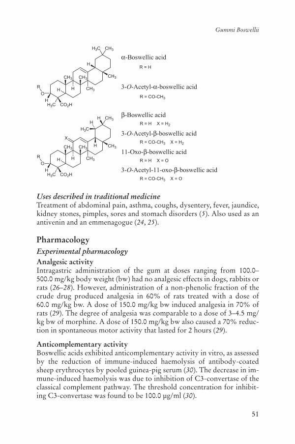
51
Gummi Boswellii
Uses described in traditional medicineTreatment of abdominal pain, asthma, coughs, dysentery, fever, jaundice, kidney stones, pimples, sores and stomach disorders (5). Also used as an antivenin and an emmenagogue (24, 25).
PharmacologyExperimental pharmacologyAnalgesic activityIntragastric administration of the gum at doses ranging from 100.0–500.0 mg/kg body weight (bw) had no analgesic effects in dogs, rabbits or rats (26–28). However, administration of a non-phenolic fraction of the crude drug produced analgesia in 60% of rats treated with a dose of 60.0 mg/kg bw. A dose of 150.0 mg/kg bw induced analgesia in 70% of rats (29). The degree of analgesia was comparable to a dose of 3–4.5 mg/kg bw of morphine. A dose of 150.0 mg/kg bw also caused a 70% reduc-tion in spontaneous motor activity that lasted for 2 hours (29).
Anticomplementary activityBoswellic acids exhibited anticomplementary activity in vitro, as assessed by the reduction of immune-induced haemolysis of antibody-coated sheep erythrocytes by pooled guinea-pig serum (30). The decrease in im-mune-induced haemolysis was due to inhibition of C3-convertase of the classical complement pathway. The threshold concentration for inhibit-ing C3-convertase was found to be 100.0 μg/ml (30).
CH3
H3C CO2H
CH3H3C
O
H
H CH3
CH3
H
CH3
H
-Boswellic acid
3-O-Acetyl- -boswellic acidR
R = H
R = CO-CH3
-Boswellic acid
3-O-Acetyl- -boswellic acid
11-Oxo- -boswellic acid
3-O-Acetyl-11-oxo- -boswellic acid
CH3
H3C CO2H
CH3H
O
H
H CH3
CH3CH3
H
H
H
H3C
X
R
R = CO-CH3 X = H2
R = CO-CH3 X = O
R = H X = H2
R = H X = O

52
WHO monographs on selected medicinal plants
Anti-inflammatory activityThe anti-inflammatory activity of an aqueous extract of the crude drug was assessed in vivo. The extract significantly inhibited both the maximal oedema response and the total oedema response during 6 hours of carra-geenan-induced rat paw oedema (31). Intragastric administration of the gum or an aqueous methanol extract of the gum (9:1) to rats, at a dose of 50.0–200.0 mg/kg bw reduced carrageenan- or adjuvant-induced pedal oedema by 34–73% (26, 32). An ethanol extract of the crude drug, admin-istered at a dose of 50.0–200.0 mg/kg bw, exhibited anti-inflammatory activity in carrageenan-induced oedema in rats and mice and dextran-in-duced oedema in rats (28). The extract also had considerable anti-arthritic activity but no significant effect was observed in the cotton pellet-induced granuloma test. Treatment with the extract inhibited inflammation-in-duced increase in serum transaminase levels and leukocyte counts, but lacked any analgesic or antipyretic effects in rats (28).
The anti-inflammatory effects of the crude drug and boswellic acids were assessed in rats with adjuvant-induced arthritis. The animals were treated with 100.0 mg/kg bw of the crude drug or 200.0 mg/kg bw of bos-wellic acid administered by gastric lavage for 2 weeks (33). The activity of
-glucuronidase was used to assess lysosomal stability, which is an im-portant factor in the arthritic syndrome. Induction of arthritis reduced lysosome stability, but treatment with either the extract or boswellic acid increased stability and had a protective effect on lysosomal integrity (33).Specific boswellic acids inhibit elastase in leukocytes, inhibit prolifera-tion, induce apoptosis and inhibit topoisomerases of leukaemia and glio-ma cell lines (23).
A methanol extract of the crude drug, containing boswellic acids and their structural derivatives, was applied topically to the backs of mice to determine its anti-inflammatory effects (23). The treatment markedly in-hibited 12-O-tetradecanoylphorbol-13-acetate-induced skin inflamma-tion, epidermal proliferation, the number of epidermal cell layers and tu-mour promotion in 7,12-dimethylbenz[a]anthracene-initiated mice. Feeding 0.2% of crude drug in the diet to CF-1 mice for 10–24 weeks reduced the accumulation of parametrial fat pad weight under the abdo-men, and inhibited azoxymethane-induced formation of aberrant crypt foci by 46% (22).
Boswellic acids (15.0 μg/ml) inhibit the biosynthesis of the pro-inflam-matory leukotrienes in neutrophilic granulocytes by a non-redox, non-competitive inhibition of 5-lipoxygenase, an enzyme in the pro-inflamma-tory arachidonic acid cascade (34–36). The extract and its derivatives caused a concentration-dependent decrease in the formation of leukotriene

53
Gummi Boswellii
B4 from endogenous arachidonic acid in rat peritoneal neutrophils in vitro (37). Acetyl-11-keto- -boswellic acid induced the most pronounced inhi-bition of 5-lipoxygenase product formation, with a mean inhibitory con-centration (IC50) of 1.5 μM. However, boswellic acid in concentrations of up to 400.0 μM did not inhibit cyclooxygenase or 12-lipoxygenase in iso-lated human platelets, or peroxidation of arachidonic acid by Fe-ascorbate. These data suggest that boswellic acids are specific, nonreducing-type in-hibitors of the 5-lipoxygenase product formation either interacting direct-ly with the 5-lipoxygenase or blocking its translocation (37). 3-O-acetyl-11-keto- -boswellic acid is the most potent inhibitor of 5-lipoxygenase. In vitro, 3-oxo-tirucallic acid inhibited 5-lipoxygenase product synthesis in cell-free assays, with an IC50 of 3 μM, but in intact cells enhanced 5-li-poxygenase product formation in ionophore-challenged polymorphonu-clear leukocytes and initiated calcium mobilization, MEK-1/2 phosphory-lation and 5-lipoxygenase translocation at a concentration of 2.5 to 15 μM. These data indicate that the pentacyclic triterpenes that inhibit 5-lipoxy-genase in cell-free systems activate 5-lipoxygenase product formation in intact cells (38).
Extracts of the crude drug and its constituents, the boswellic acids, activate the mitogen-activated protein kinases p42 and p38 in isolated hu-man primed polymorphonuclear leukocytes in vitro (39). Activation of mitogen-activated protein kinases was rapid and transient with maximal activation after 1–2.5 min of exposure and occurred in a dose-dependent manner. The keto-boswellic acids (11-keto- -boswellic acid and 3-O-acetyl-11- -keto-boswellic acid) activated kinase at 30.0 μM, whereas other boswellic acids lacking the 11-keto group were less effective. More-over, 11-keto-boswellic acids induced rapid and prominent mobilization of free Ca2+ in polymorphonuclear leukocytes. Inhibitor studies revealed that phosphatidylinositol 3-kinase is involved in boswellic acid-induced mitogen-activated protein kinase activation, whereas a minor role was ap-parent for protein kinase C. Mitogen-activated protein kinase activation by 3-O-acetyl-11- -keto-boswellic acid was partially inhibited when Ca2+ was removed by chelation (39).
Mixed acetyl-boswellic acids significantly inhibited ionophore-stimu-lated release of the leukotrienes B4 and C4 from intact human polymor-phonuclear leukocytes, with 50% inhibitory concentration (IC50) values of 8.48 μg/ml and 8.43 μg/ml, respectively. Purified acetyl-11-keto- -boswellic acid was about three times more potent as an inhibitor of the formation of both leukotriene B4 (IC50, 2.53 μg/ml) and leukotriene C4 (IC50, 2.26 μg/ml) from human polymorphonuclear leukocytes in the same assay. Daily intraperitoneal administration of an extract of mixed

54
WHO monographs on selected medicinal plants
acetyl-boswellic acids (20.0 mg/kg bw) significantly reduced the clinical symptoms in guinea-pigs with experimental autoimmune encephalomy-elitis between days 11 and 21. However, the inflammatory infiltrates in the brain and the spinal cord were not significantly less extensive in the treated animals than in the respective control group. The multiple intra-peritoneal administrations of boswellic acids did not inhibit the ionophore-challenged ex vivo release of leukotrienes B4 and C4 from polymorpho-nuclear leukocytes separated from the blood of guinea-pigs with experimental autoimmune encephalomyelitis or the biosynthesis of leu-kotrienes in vitro (40). Suspensions of rat peritoneal polymorphonuclear leukocytes elicited with glycogen, stimulated by calcium and ionophore to produce leukotrienes and 5-15-hydroxyeicosateraenoic acid (HETE) from endogenous arachidonic acid, were treated with various concentra-tions of an ethanol extract of the crude drug. A concentration-dependent inhibition of leukotriene B4 and 5-HETE production was observed. All products of 5-lipoxygenase from endogenous arachidonic acid in poly-morphonuclear leukocytes were reduced (41).
Miscellaneous activitiesIntragastric administration of a 50% ethanol extract of the crude drug, at a dose of 250.0 mg/kg bw, to rats reduced blood glucose levels (42). Intravenous administration to dogs of a 50% ethanol extract, at a dose of 50.0 mg/kg bw, reduced blood pressure (42). Intragastric adminis-tration of the crude drug at a dose of 100.0 mg/kg bw to cockerels, rabbits and rats fed a high cholesterol diet reduced serum cholesterol by 25–45% (26). Intragastric administration of the crude drug, at a dose of 100.0–500.0 mg/kg bw did not reduce fever in rats, rabbits or dogs (26). Acetyl-11-keto-beta-boswellic acid, a constituent of a herbal medicine from Boswellia serrata resin, attenuated experimental ileitis in animals (43).
ToxicologyThe gestation period or parturition time in pregnant rats and the onset time of castor oil-induced diarrhoea were unaffected by the extract and no significant effect was seen on cardiovascular, respiratory and central nervous system functions. Intragastric administration of the crude drug to dogs, rabbits or rats, at a dose of 500.0 or 1000.0 mg/kg bw, did not have ulcerogenic effects. The oral and intraperitoneal median lethal doses were greater than 2.0 g/kg bw in mice and rats (28). Intragastric adminis-tration of the crude drug to monkeys (500.0 mg/kg bw), mice (2.0 g/kg bw) or rats (1.0 g/kg bw) for 6 months produced no observable behav-ioural, biochemical or histological abnormalities (44).
55
Gummi Boswellii
Clinical pharmacologyA double-blind pilot study, involving 37 patients with rheumatoid arthri-tis, assessed the effects of the crude drug on the symptoms of swelling and pain, and amount of self-medication with nonsteroidal anti-inflammatory drugs (41). Patients were treated with 3.6 g of the crude drug or placebo for 12 weeks. Outcome measures included Ritchie’s index for swelling and pain, and the dose of nonsteroidal anti-inflammatory drugs patients felt they needed. There were no subjective, clinical or laboratory parameters showing significant changes from baseline to 12 weeks of treatment (45).
A randomized double-blind, placebo-controlled cross-over study was conducted to assess the efficacy, safety and tolerability of a crude Boswellia serrata extract in 30 patients with osteoarthritis of the knee. Fifteen subjects received the active Boswellia serrata extract or placebo for 8 weeks. After the first treatment, a washout period was permitted and the groups were then crossed over to receive the opposite intervention for 8 weeks. All patients receiving Boswellia serrata extract reported a de-crease in knee pain, increased knee flexion and increased walking distance. The frequency of swelling in the knee joint was decreased, but radiology detected no change. The observed differences between drug treatment and placebo were statistically significant (p < 0.05). Boswellia serrata ex-tract was well tolerated by the subjects except for minor gastrointestinal adverse events (22).
A double-blind, placebo-controlled study involving 40 patients with bronchial asthma assessed the effects of an extract of the crude drug for treatment of symptoms. The patients were treated with a preparation of gum resin of 300 mg three times daily for a period of 6 weeks. After treat-ment, 70% of patients showed improvement as evident from the disap-pearance of physical symptoms and signs such as dyspnoea, bronchial asthma and decreased number of attacks, as well as a decrease in eosino-phil count and electron spin resonance. In the control group (treated with lactose, 300.0 mg three times daily, for 6 weeks), only 27% of patients in the control group showed improvement. The data show a role for the crude drug in the treatment of bronchial asthma (20).
A randomized, double-blind, controlled, parallel group comparison clinical trial involving 102 patients assessed the effects of the crude drug for the treatment of Crohn’s disease (18). The positive control arm was treated with mesalazine. The primary outcome measure was the change in the Crohn’s Disease Activity Index between the time of enrolment and the end of therapy. The Crohn’s Disease Activity Index was reduced by 90 after treatment with the crude drug and by 53 after treatment with mesalazine; however, the difference between the two treatments was not

56
WHO monographs on selected medicinal plants
statistically significant. Thus, the study concluded that an extract of the crude drug was as effective as mesalazine for the treatment of Crohn’s disease.
The effect of an extract of the crude drug was assessed in patients with ulcerative colitis, grade II or III (19). Patients were treated with the gum resin preparation (350 mg three times daily for 6 weeks), and the effects on stool properties, histolopathology and scan microscopy of rectal biop-sies, blood parameters including haemogloblin, serum iron, calcium, phosphorus, proteins, total leukocytes and eosinophils were measured. Patients receiving sulfasalazine (1 g three times daily) served as controls. At the end of the treatment period, all parameters tested had improved after treatment with Boswellia serrata gum resin and the results were sim-ilar to those in the control group: 82% of treated patients went into re-mission following treatment with the gum resin as did 75% of those treat-ed with sulfasalazine (19).
A controlled clinical trial assessed the efficacy of the crude drug in patients with chronic colitis characterized by vague lower abdominal pain, bleeding per rectum with diarrhoea and palpable tender descend-ing and sigmoid colon. Thirty patients with chronic colitis, 17 men and 13 women aged between 18 and 48 years, were included in the study. Twenty of the patients were given a preparation of the gum resin of Bos-wellia serrata (300 mg three times daily for 6 weeks) and 10 were given sulfasalazine (1 g three times daily for 6 weeks) and served as controls. Of 20 patients treated with Boswellia gum resin, 18 showed an improve-ment in one or more of the parameters: stool properties; histopathology; scanning electron microscopy; and measures of haemoglobin, serum iron, calcium, phosphorus, proteins, total leukocytes and eosinophils. Of 20 patients treated with Boswellia gum resin, 14 went into remission whereas in the patients treated with sulfasalazine, the remission rate was 4 out of 10 (20).
Adverse reactionsMinor gastrointestinal side-effects have been reported in the clinical trials (19–22).
ContraindicationsHypersensitivity or allergy to the crude drug.
WarningsNo information was found.
57
Gummi Boswellii
PrecautionsGeneralNo information was found.
Drug interactionsNone reported.
Drug and laboratory test interactionsNone reported.
Carcinogenesis, mutagenesis, impairment of fertilityNo information was found.
Pregnancy: teratogenic effectsNo information was found.
Pregnancy: non-teratogenic effectsDue to a lack of safety data, the use of the crude drug during pregnancy is not recommended.
Nursing mothersDue to a lack of safety data, the use of the crude drug during breastfeed-ing is not recommended.
Paediatric useDue to a lack of safety data, the use of the crude drug in children under the age of 12 years is not recommended.
Dosage formsCrude drug, extracts.
StorageStore in a cool dry place away from heat or light.
Posology(Unless otherwise indicated)Crude drug: 1–3 g daily (frequency not specified) (1).Extracts: 300–350 mg three times daily (19–21).

58
WHO monographs on selected medicinal plants
ReferencesThe Ayurvedic pharmacopoeia of India1. , Part I, Vol. IV. New Delhi, Ministry of Health and Family Welfare, Department of Indian System of Medicine and Homeopathy, 1999.Database on medicinal plants used in Ayurveda2. . New Delhi, Central Council for Research in Ayurveda and Siddha, Department of ISM and H, Ministry of Health and Family Welfare, Government of India, 2000.Horhammer L, eds. 3. Hagers Handbuch der pharmazeutischen Praxis, 4th ed. Berlin, Springer Verlag, 1975 [in German].Bedevian AK. 4. Illustrated polyglottic dictionary of plant names. Cairo, Med-bouly Library, 1994.Farnsworth NR, ed. 5. NAPRALERT database. Chicago, University of Illinois at Chicago, IL (an online database available directly through the University of Illinois at Chicago or through the Scientific and Technical Network [STN] of Chemical Abstracts Services), 30 June 2005.Nadkarni KM. 6. Indian materia medica. Bombay, Popular Prakashan, 1954.Kapoor LD.7. Handbook of Ayurvedic medicinal plants. Boca Raton, CRC Press, 1990.Hahn-Deinstrop E, Koch A, Müller M. Guidelines for the assessment of the tradi-8.tional herbal medicine olibanum by application of HPTLC and DESAGA ProVi-Doc video documentation. Journal of Planar Chromatography, 1998, 11:404–410.Ganzera M, Khan IA. A reversed phase high performance liquid chromatog-9.raphy method for the analysis of boswellic acids in Boswellia serrata. Planta Medica, 2001, 67:778–780.Hairfield EM, Hairfield HH Jr. McNair HM. GC, GC/MS, and TLC of 10.
-boswellic acid and O-acetyl- -boswellic acid from B. serrata, B. carteii and B. papyrifera. Journal of Chromatographic Science, 1989, 27:127–133.Hamm S et al. A chemical investigation by headspace SPME and GC-MS of 11.volatile and semi-volatile terpenes in various olibanum samples. Phytochem-istry, 2005, 66:1499–1514.WHO guidelines on assessing quality of herbal medicines with reference to 12.contaminants and residues. Geneva, World Health Organization, 2007.European Pharmacopoeia13. , 5th ed. Strasbourg, Directorate for the Quality of Medicines of the Council of Europe (EDQM), 2005.Guidelines for predicting dietary intake of pesticide residues14. , 2nd rev. ed. Ge-neva, World Health Organization, 1997 (WHO/FSF/FOS/97.7).Girgune JB, Garg BD. Chemical investigation of the essential oil from 15. Bos-wellia serrata Roxb. Journal of Scientific Research, 1979, 1:119–122.Verghese J et al. A fresh look at the constituents of Indian Olibanum oil. 16.Flavor and Fragrance Journal, 1987, 2:99–102.Wichtl M. 17. Teedrogen und phytopharmaka. Stuttgart, Wissenschaftliche Ver-lagsgesellschaft, 2002 [in German].Gerhardt H et al. [Therapy of active Crohn disease with 18. Boswellia serrataextract H 15]. Zeitschrift für Gastroenterologie, 2001, 39:11–17 [in German].
59
Gummi Boswellii
Gupta, I et al. Effects of 19. Boswellia serrata gum resin in patients with ulcer-ative colitis. European Journal of Medical Research, 1997, 21:37–43.Gupta I et al. Effects of 20. Boswellia serrata gum resin in patients with bron-chial asthma: results of a double-blind, placebo-controlled, 6-week clinical study. European Journal of Medical Research, 1998, 3:511–514.Gupta I et al. Effects of gum resin of 21. Boswellia serrata in patients with chron-ic colitis. Planta Medica, 2001, 67:391–395.Kimmatkar N et al. Efficacy and tolerability of 22. Boswellia serrata extract in treatment of osteoarthritis of knee – a randomized double blind placebo con-trolled trial. Phytomedicine, 2003, 10:3–7.Ammon HP. [Boswellic acids (components of frankincense) as the active 23.principle in treatment of chronic inflammatory diseases]. Wiener Medizinis-che Wochenschrift, 2002, 152:373–378 [in German].Chopra RN, ed. 24. Indigenous drugs of India. Their medical and economic as-pects. Calcutta, India, Art Press, 1933.Saha JC, Savini EC, Kasinathan S. 25. Ecbolic properties of Indian Medicinal Plants. Part 1. Indian Journal of Medical Research, 1961, 49:130–151.Atal CK, Gupta OP, Singh GB. Salai guggal: A promising anti-arthritic and 26.anti-hyperlipidemic agent. British Journal of Pharmacology, 1981, 74:203–204.Menon MK, Kar A. Analgesic and psychopharmacological effects of the gum 27.resin of Boswellia serrata. Planta Medica, 1971, 19:333–341.Singh GB, Atal CK. Pharmacology of an extract of salai guggal ex-28. Boswellia serrata,a new nonsteroidal anti-inflammatory agent. Agents and Actions, 1986, 18:407–412.Kar A, Menon MK. Analgesic effect of the gum resin of 29. Boswellia serrata. Life Sciences, 1969, 8:1023–1028.Kapil A, Moza N. Anticomplementary activity of boswellic acids, an inhibi-30.tor of C3-convertase of the classical complement pathway. InternationalJournal of Immunopharmacology, 1992, 14:1139–1143.Duwiejua M et al. Anti-inflammatory activity of resins from some species of 31.the plant family Burseraceae. Planta Medica, 1993, 59:12–16.Etzel R. Special extract of 32. Boswellia serrata (H15)* in the treatment of rheu-matoid arthritis. Phytomedicine, 1996, 3:91–94.Reddy GK, Dhar SC. Effect of a new nonsteroidal anti-inflammatory agent 33.on lysosomal stability in adjuvant induced arthritis. Italian Journal of Bio-chemistry, 1987, 36:205–217.Ammon HP et al. Mechanism of anti-inflammatory actions of curcurmine 34.and boswellic acids. Journal of Ethnopharmacology, 1993, 38:113–119.Safayhi H et al. Concentration-dependent potentiating and inhibitory effects 35.of Boswellia extracts on 5-lipoxygenase product formation in stimulated PMNL. Planta Medica, 2000, 66:110–113.Schweizer S et al. Workup-dependent formation of 5-lipoxygenase inhibitory 36.boswellic acid analogues. Journal of Natural Products, 2000, 63:1058–1061.Safayhi H et al. Boswellic acids: novel, specific, nonredox inhibitors of 5-li-37.poxygenase. Journal of Pharmacology and Experimental Therapeutics, 1992, 261:1143–1146.
60
WHO monographs on selected medicinal plants
Boden SE et al. Stimulation of leukotriene synthesis in intact polymorpho-38.nuclear cells by the 5-lipoxygenase inhibitor 3-oxo-tirucallic acid. MolecularPharmacology, 2001, 60:267–273.Altmann A et al. Boswellic acids activate p4239. MAPK and p38 MAPK and stimu-late Ca 2+ mobilization. Biochemical and Biophysical Research Communica-tions, 2002, 290:185–190.Wildfeuer A et al. Effects of boswellic acids extracted from a herbal medicine 40.on the biosynthesis of leukotrienes and the course of experimental autoim-mune encephalomyelitis. Arzneimittelforschung, 1998, 48:668–674.Ammon HP et al. Inhibition of leukotriene B4 formation in rat peritoneal 41.neutrophils by an ethanolic extract of the gum resin exudate of Boswellia ser-rata. Planta Medica, 1991, 57:203–207.Dhar ML et al. Screening of Indian plants for biological activity: part I. 42. In-dian Journal of Experimental Biology, 1968, 6:232–247.Krieglstein CF et al. Acetyl-11-keto-beta-boswellic acid, a constituent of a 43.herbal medicine from Boswellia serrata resin, attenuates experimental ileitis. International Journal of Colorectal Diseases, 2001, 16:88–95.Singh GB, Bani S, Singh S. Toxicity and safety evaluation of Boswellic acids. 44.Phytomedicine, 1996, 3:87–90.Sander O, Herborn G, Rau R. Is H15 (resin extract of 45. Boswellia serrata, “in-cense”) a useful supplement to establish drug therapy of chronic polyarthri-tis? Results of a double-blind pilot study. Zeitschrift für Rheumatologie,1998, 57:11–16.

61
Semen Cardamomi
DefinitionSemen Cardamomi consists of the dried ripe seed of Elettaria cardamo-mum (L.) Maton (Zingiberaceae) recently removed from the capsule (1–9).
Note: In a number of formularies and standards, the drug is official as Fructus Cardamomi, but the part employed is the seed, with the fruit capsule removed prior to use (5–10).
SynonymsAmomum repens Sonn., A. cardamomum Lour. (11), Alpinia cardamo-mum Roxb. (12).
Selected vernacular namesAlaicha, bach dau khau, bastard cardamom, cardamomier, Cardamom-pflanze, cardamomo minore, cardamomo, cardamon, cardamone, carda-mone petite, Cardamonen, chinne elakulu, chittelam, chhoti lachi, chota elaich, chotaa leicaha, choti ilayachi, Echte Kardamon, elã, elachi, elakay, elakkki, elam, elayachi, elchi, Elettarie, ensal, ga qolia, garidimong, guju-rati, haal boa, habbahan, habbahal, hab el haal, heel khurd, hel, hhabb el hal, hiliki, illlachi, kakilahekurd, kakula saghira, kapoelaga, kardoem, Kleine Kardamomen, krako, küçük kakule, lahanveldoda, lesser carda-mom, luuk en, malabar cardamon, Malabar Cardamonen, pelaga, qaqui-lah saghirah, ronde kardemon, sa nhon, sanna elakulu, sanna yalakii, sa-rooplaachi, shozuku, siruelam, so du gu, sodugo, syouzuku, truti, velchi, velloda (2–6, 12, 13).
Geographical distributionNative to India and introduced to Sri Lanka (14, 15).
DescriptionA perennial zingiberaceous herb attaining a height of 2–4 m, with lanceo-late leaves borne on long sheathing stems. Flowers numerous, borne on

62
WHO monographs on selected medicinal plants
horizontal racemes that arise from the rhizome and run horizontally along the ground. The fruit is inferior, ovoid or oblong, nearly ellipsoidal, capsule plump or slightly shrunken, the seeds of which are covered by an aril (14).
Plant material of interest: dried seed (in dried or nearly dried fruit)General appearanceUsually in agglutinated groups of 2–7 seeds. Each irregularly angular, 3- to 4-sided, oblong, ovoid; 2–4 mm long, up to 3 mm broad; pale orange to dark reddish-brown; usually enveloped by a thin colourless membranous aril externally, transversely wrinkled but not minutely pitted; hilum de-pressed; raphe indicated by a channel extending on one side from base to apex; hard; internally, whitish, showing a thin dark testa, a whitish starchy perisperm grooved on one side, and in the centre a small yellowish trans-lucent endosperm, surrounding a paler minute embryo (1–3, 5–7).
Fruit: Inferior, ovoid or oblong, nearly ellipsoidal, capsule plump or slightly shrunken; 8–20 mm, but most fruits are 10–15 mm long, 5–10 mmin diameter; green to pale buff, sometimes yellowish-grey, mostly 3-sided; externally, smooth or longitudinally striated; base, rounded and may bear the remains of the stalk; apex, more or less blunt and sometimes crowned by a short beak formed of the remains of the floral parts; interior longitu-dinally divided into 3 loculi, each loculus is an adherent mass of two rows of 3–7 small seeds attached to the axile placenta (2, 5–7). According to Evans (15), four varieties of fruits are available: “Mysore”, “Malabar”, “Mangalore” and “Alleppy”.
Organoleptic propertiesOdour: aromatic; taste: aromatic, pungent and slightly bitter (1–3, 5–8).
Microscopic characteristicsAril of several layers of elongated, more or less collapsed, flattened, thin-walled parenchyma cells containing small rounded or oval droplets of oil. Testa with an outer epidermis of long, narrow fusiform cells, 20–30 μm wide, having slightly thickened undulating walls, followed by a layer of collapsed parenchymatous cells, with brownish contents, becoming 2–3 layers in the region of the raphe, composed of large thin-walled, rect-angular cells containing volatile oil; a band of 2–3 layers of parenchyma-tous cells and an inner epidermis of thin-walled flattened cells. Inner in-tegument consists of 2 layers of cells, an outer layer of yellowish to reddish-brown, rectangular, strongly lignified sclereids, about 40 μm long and 20 μm wide, nearly filled with a small warty nodule of silica and an in-
63
Semen Cardamomi
ner epidermis of flattened cells. Perisperm of thin-walled cells, packed with minute starch grains, 1–6 μm in diameter, and containing 1–7 small prisms of calcium oxalate, about 10–20 μm long. Endosperm, of small thin-walled parenchymatous cells, each filled with a hyaline or granular mass of protein but no starch; embryo, of small thin-walled cells containing aleurone grains; fibrous sclerenchyma and large vessels present in pericarp (2, 3).
Powdered plant materialReddish to greyish-brown, characterized by numerous fragments of perisperm cells, each filled with starch granules and containing one or more prisms of calcium oxalate, 10–25 μm long; polyhedral masses of ad-herent starch granules from perisperm, individual granules, up to 4 μm in diameter; numerous fragments of yellowish to reddish-brown sclereids; occasional particles of epidermal cells, often crossed at right angles by the cells of the collapsed layer (2).
General identity testsMacroscopic and microscopic examinations (1–3, 5–8, 14).
Purity testsMicrobiologyTests for specific microorganisms and microbial contamination limits are as described in the WHO guidelines on assessing quality of herbal medi-cines with reference to contaminants and residues (16).
ChemicalTo be established in accordance with national requirements.
Foreign organic matterNot more than 3% (2, 17).
Total ashNot more than 6% (3, 6–8, 17).
Acid-insoluble ashNot more than 4% (1, 3, 5–8).
Water-soluble extractiveNot less than 10% (3).
Alcohol-soluble extractiveNot less than 2% (3).
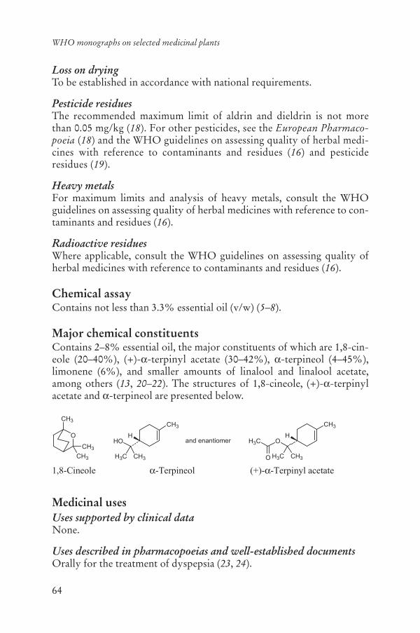
64
WHO monographs on selected medicinal plants
Loss on dryingTo be established in accordance with national requirements.
Pesticide residuesThe recommended maximum limit of aldrin and dieldrin is not more than 0.05 mg/kg (18). For other pesticides, see the European Pharmaco-poeia (18) and the WHO guidelines on assessing quality of herbal medi-cines with reference to contaminants and residues (16) and pesticide residues (19).
Heavy metalsFor maximum limits and analysis of heavy metals, consult the WHO guidelines on assessing quality of herbal medicines with reference to con-taminants and residues (16).
Radioactive residuesWhere applicable, consult the WHO guidelines on assessing quality of herbal medicines with reference to contaminants and residues (16).
Chemical assayContains not less than 3.3% essential oil (v/w) (5–8).
Major chemical constituentsContains 2–8% essential oil, the major constituents of which are 1,8-cin-eole (20–40%), (+)- -terpinyl acetate (30–42%), -terpineol (4–45%), limonene (6%), and smaller amounts of linalool and linalool acetate, among others (13, 20–22). The structures of 1,8-cineole, (+)- -terpinylacetate and -terpineol are presented below.
Medicinal usesUses supported by clinical dataNone.
Uses described in pharmacopoeias and well-established documentsOrally for the treatment of dyspepsia (23, 24).
O
CH3
CH3
CH3
1,8-Cineole
CH3
O
H3C CH3
H3C
O
H
CH3
HO
H3C CH3
Hand enantiomer
-Terpineol (+)- -Terpinyl acetate

65
Semen Cardamomi
Uses described in traditional medicineTreatment of asthma, bronchitis, colic, coughs, fainting, fever, rheuma-tism, stomach cramps and urinary stones. Also used as an aphrodisiac, appetizer, diuretic and emmenagogue (13, 24).
PharmacologyExperimental pharmacologyAnti-inflammatory and analgesic activitiesAn in vivo study was performed to compare the anti-inflammatory activ-ity of the essential oil of the seed, at doses of 175 and 280 μl/kg body weight (bw) with that of indometacin at a dose of 30.0 mg/kg bw against acute carrageenan-induced plantar oedema in rats. Intraperitoneal admin-istration of 280 μl/kg bw of the essential oil to rats or 233 μl/kg bw to mice suppressed carrageenan-induced pedal oedema (25).
One study assessed the analgesic activity of the essential oil from the seed using 1,4-benzoquinone as a chemical stimulus for pain in mice. In-tragastric administration of a dose of 233 μl/kg bw of the essential oil produced a 50% reduction in writhing (stretching syndrome) induced by intraperitoneal administration of a 0.02% solution of 1,4-benzoquinone (25). Butanol and ether extracts of the seeds had anti-inflammatory activ-ity in vitro, as assessed in the albumin stabilizing assay (concentration not stated). The aqueous extract of the seeds, however, was not active (26).
Antimicrobial effectsAt a concentration of 500 ppm, the essential oil from the seed weakly in-hibited the growth of the fungi Arthroderma simii, Chaetomium indicum,Microsporum canis and Trichophyton mentagrophytes in vitro (27).
Antispasmodic activityA 95% ethanol extract of the crude drug at a concentration of 200 μg/ml reduced histamine-induced and barium chloride-induced contractions in guinea-pig ileum in vitro (28). Administration of essential oil from the seed to rabbits, at a dose of 0.4 ml/kg bw, inhibited acetylcholine-induced intestinal spasms (25). However, an aqueous extract of the crude drug was reported to stimulate the rectus abdominus muscle of the frog and rat je-junum in vitro at a concentration of 10% of the bath media (29).
Smooth-muscle relaxant activityThe essential oil from the seed relaxed isolated guinea-pig ileum and tra-chea in vitro with a median effective dose of 15 mg and 27 mg/l, respec-tively. An aqueous ethanol seed extract (10 mg/ml in bath medium) re-laxed guinea-pig ileum in vitro (30).
66
WHO monographs on selected medicinal plants
Antiulcer activityIntragastric administration of dried aqueous or methanol extracts of the seeds to mice, at a dose of 53.0–126.9 mg/kg bw, reduced the secretion of gastric juice (31). A methanol extract of the dried seeds (5:1) at a concentra-tion of 100 μg/ml inhibited the growth of Helicobacter pylori in vitro (32).
Choleretic activityIntraduodenal administration of a dried acetone extract of the dried seed to rats at a dose of 50.0–500.0 mg/kg bw, had significant choleretic effects (p < 0.01–0.05) (33).
Central nervous system depressant activityIntraperitoneal administration of the essential oil from the seed, at a dose of 1.6 ml/kg bw, induced drowsiness and a staggering gait in mice (25).
Dermatological effectsApplication of the essential oil from the seed at a concentration of 1.0% to rabbit skin enhanced the dermal penetration of piroxicam, indometacin and diclofenac sodium (34). Application of the essential oil from the seed, at a dose of 1.0 ml/per square inch enhanced the dermal penetration of indometacin in rats, rabbits and humans (34, 35). External pre-application of the essential oil from the seed, at a concentration of 5%, to rabbit skin increased the bioavailability of piroxicam gel (34).
ToxicityIntragastric or subcutaneous administration of an aqueous ethanol (1:1) extract of the seed, at a dose of 10 g/kg bw had no observed toxicity in mice (36).
Miscellaneous activitiesAn aqueous ethanol (1:1) extract of the seed, at a concentration of 10.0 mg/ml had histaminergic activity in guinea-pig ileum in vitro (30). Aqueous extracts (10% w/v) of the crude drug were used for an ex vivo study of their effect on gastric secretion. The stomach of pentobarbitone-anaes-thetized rats was perfused at 0.15 ml/min with aqueous extracts of carda-mom or acetylcholine (1.0 μg/ml or 10.0 μg/ml solutions, in 40-minute time periods, twice in each experiment) followed by saline perfusions. The acid content in the samples was estimated by titration with 0.1 N so-dium hydroxide with phenolphthalein as indicator. Acute gastric mucosal injury was induced by leaving aspirin 125.0 mg/kg bw in the stomach for 2 h before perfusion. The aqueous extract significantly increased gastric secretion from 0.10 to 0.28 (p < 0.005) (37).
67
Semen Cardamomi
Adverse reactionsContact dermatitis has been reported (38).
ContraindicationsHypersensitivity or allergy to the crude drug.
WarningsNo information was found.
PrecautionsGeneralPatients with gallstones should consult their health care provider before using preparations of Semen Cardamomi (23).
Drug interactionsNone reported.
Drug and laboratory test interactionsNone reported.
Carcinogenesis, mutagenesis, impairment of fertilityA hot aqueous or methanol extract of the crude drug at concentrations of 50 mg/disc was not mutagenic in the Ames test in Salmonella typhimuri-um strains TA98 and TA100 (39). The essential oil inhibited formation of DNA adducts with aflatoxin B1 by inhibiting activation in rat liver mi-crosomes, thus demonstrating antimutagenic activity. The median inhibi-tory concentration was 10 μl/disc (40).
Drug interactionsNo information was found.
Drug and laboratory test interactionsNone reported.
Pregnancy: teratogenic effectsNo information was found.
Pregnancy: non-teratogenic effectsNo information was found.

68
WHO monographs on selected medicinal plants
Nursing mothersNo information was found.
Paediatric useNo information was found.
Dosage forms and storageCrude drug, extracts and tinctures (23).
Posology(Unless otherwise indicated)Average daily dosage: 1.5 g of drug; equivalent preparations.Tincture: daily dosage equivalent to 12 g (23).
ReferencesThe National Formulary 1. 20th ed, 1st Suppl. Rockville, MD, The United States Pharmacopeia Convention, 2002.African pharmacopoeia2. , Vol. 1. Lagos, Nigeria, Organization of African Uni-ty, Scientific Technical & Research Commission, 1985.The API Pharmacopoeia of India, Part I. Vol. I, 3. 1st ed. New Delhi, Govern-ment of India Ministry of Health and Family Welfare, Department of Indian Systems of Medicine and Homeopathy, 1990 (reprinted 2001).Nadkarni AK, ed. 4. Dr. K.M. Nadkarni’s Indian materia medica, Vol. 1. Bom-bay, Popular Prakshan, 1976.The Japanese pharmacopeia5. , 14th ed. (English ed.). Tokyo, Ministry of Health, Labour and Welfare, 2001 (http://jpdb.nihs.go.jp/jp14e/).Pharmacopoeia of the Republic of Korea6. , 7th ed. (English ed.). Seoul, Korea Food and Drug Administration, and Korean Association of Official Com-pendium for Public Health, 1998.Asian crude drugs, their preparations and specifications. Asian Pharmacopeia7. .Manila, Federation of Asian Pharmaceutical Associations, 1978.The Japanese standards for herbal medicines8. . Tokyo, Yakuji Nippon, 1993.British herbal pharmacopoeia9. . Exeter, British Herbal Medicine Association, 1996.Reynolds JEF, ed. 10. Martindale, the extra pharmacopoeia. London, Pharma-ceutical Press, 1996.Perry LM, Metzger J. 11. Medicinal plants of east and southeast Asia, attributed properties and uses. Cambridge, MA, MIT Press, 1980.Bedevian AK. 12. Illustrated polyglottic dictionary of plant names. Cairo, Med-bouly Library, 1994.Farnsworth NR, ed. 13. NAPRALERT database. Chicago, University of Illinois at Chicago, IL (an online database available directly through the University

69
Semen Cardamomi
of Illinois at Chicago or through the Scientific and Technical Network [STN] of Chemical Abstracts Services), 30 June 2005.Youngken HW. 14. Textbook of pharmacognosy. Philadelphia, Blakiston, 1950.Evans WC. 15. Trease and Evans pharmacognosy, 15th ed. Edinburgh, WB Saunders, 2002.WHO guidelines on assessing quality of herbal medicines with reference to 16.contaminants and residues. Geneva, World Health Organization, 2007.British pharmacopoeia17. . London, Her Majesty’s Stationery Office, 1993.European pharmacopoeia18. , 5th ed. Strasbourg, Directorate for the Quality of Medicines of the Council of Europe (EDQM), 2005.Guidelines for predicting dietary intake of pesticide residues19. , 2nd rev. ed. Ge-neva, World Health Organization, 1997 (WHO/FSF/FOS/97.7).Hänsel R, Sticher O, Steinegger E. 20. Pharmakognosie-phytopharmazie. Berlin, Springer-Verlag, 1999 [in German].Shaban MAE et al. The chemical composition of the volatile oil of 21. Elettariacardamomum seeds. Pharmazie, 1987, 42:207–208.Marongiu B, Piras A, Porcedda S. Comparative analysis of the oil and super-22.critical CO2 extract of Elettaria cardomomum (L) Maton. Journal of Agricul-tural Chemistry, 2004, 52:6278–6282.Blumenthal M et al. 23. The complete German Commission E monographs: Therapeutic guide to herbal medicines. Austin, TX, American Botanical Council, 1998.Govindarajan VS et al. Cardamom – production, technology, chemistry, and 24.quality. Critical Reviews in Food Science Nutrition, 1982, 16:229–326.Al-Zuhair H et al. Pharmacological studies of cardamon oil in animals. 25. Phar-macological Research, 1996, 34:79–82.Han BH et al. Screening of the anti-inflammatory activity of crude drugs. 26.Korean Journal of Pharmacognosy, 1972, 4:205–209.El-Kady IA, El-Maraghy SSM, Mostafa E. Antibacterial and antidermato-27.phyte activities of some essential oils from spices. Qatar University Science Journal, 1993, 13:63–69.Itokawa H et al. Studies on the constituents of crude drugs having inhibitory 28.activity against contraction of the ileum caused by histamine or barium chlo-ride. Shoyakugaku Zasshi, 1983, 37:223–228.Haranath PSRK, Akther MH, Sharif SI. Acetylcholine and choline in com-29.mon spices. Phytotherapy Research, 1987, 1:91–92.Mokkhasmit M, Swatdimongkol K, Satrawaha P. Study on toxicity of Thai 30.medicinal plants. Bulletin of the Department of Medical Sciences, 1971, 12:36–65.Sakai K et al. Effect of extracts of Zingiberaceae herbs on gastric secretion in 31.rabbits. Chemical and Pharmaceutical Bulletin, 1989, 37:215–217.Mahady GB et al. 32. In vitro susceptibility of Helicobacter pylori to botanicals used traditionally for the treatment of gastrointestinal disorders. Phytother-apy Research, 2005, 19:988–991.

70
WHO monographs on selected medicinal plants
Yamahara J et al. Biologically active principles of crude drugs. Cholagogic 33.substances in cardamon seeds and its properties. Yakugaku Zasshi, 1983,103:979–985.Huang YB et al. Cardomom oil as a skin permeation enhancer for indo-34.methacin, piroxicam and diclofenac. International Journal of Pharmaceutics,1995, 126:111–117.Huang YB et al. Crude drug (Zingiberaceae) enhancement of percutaneous 35.absorption of indomethacin: in vitro and in vivo permeation. Gaoxiong Yi Xue Ke Xue Za Zhi, 1993, 9:392–400.Mokkhasmit M et al. Pharmacological evaluation of Thai medicinal plants. 36.Journal of the Medical Association of Thailand, 1971, 54:490–504.Vasudevan K et al. Influence of intragastric perfusion of aqueous spice ex-37.tracts on acid secretion in anesthetized albino rats. Indian Journal of Gastro-enterology, 2000, 19:53–56.Seetharam KA, Pasricha JS. Condiments and contact dermatitis of the finger 38.tips. Indian Journal of Dermatology, Venerology, and Leprology, 1987, 53:325–328.Yamamoto H, Mizutani T, Nomura H. Studies on the mutagenicity of crude 39.drug extracts. I. Yakugaku Zasshi, 1982, 102:596–601.Hashim S et al. Modulatory effects of essential oils from spices on the forma-40.tion of DNA adducts by aflatoxin B1 in vitro. Nutrition in Cancer, 1994, 21:169–175.

71
Fructus Chebulae
DefinitionFructus Chebulae consists of the dried fruits of Terminalia chebula Retz. or T. chebula Retz. var. tomentella Kurt. (Combretaceae) (1–3).
Selected vernacular namesAbhaya, ahlilaj kâbuli, alalekai, alayla, amagola, arabi, aralu, areyra, ari-dadi, badamier chebule, bal har, black myrobalan, bush kaduka, chebulic myrobalan, Chebulische Myrobalane, divya, Ga ja, Habra, hacha, halela, halela kabuli, halela zard, halileh, halileh kaboli, halilehsiyâh, halileh zard, hallilaj, harad, harar, harda, hardo, harir, haritaki, harra, harro, harroh, haser, helikha, hezi, himaja, hirda, hirdo, hireda, hlilej khel, hlijej sfer, hokikha, ihlilaj kabuli, inknut tree, jivathi, kabuli-harda, hora, kadukka, kadukkai, kale har, karaka, karakkaya, kashi, katukka, kãyasthã, kot-pung-pla, kurka, medicine terminalia, mirobalan de caboul, mirobalano, myrobalan, myrobalano nero, myrobalans, myrobaran, pathyã, pile har, pilo-harde, post-e-haleela kabli, post-e-haleela siyah, post-e-haleela zard, pulo-harda, rispiger Myrobalanenbaum, rong mao he zi, silikha, sa-mo-thai, samo-thai, shajar shiir hindi, sirri hindi, silikha, sivã, sringitiga, sud-dha, terminaalia, vayastha, vijayã, yellow myrobalan, yellow myrobalan plum, zama, zangli har (2–11).
Geographical distributionNative to Cambodia, China, India, Lao People’s Democratic Republic, Malaysia, Myanmar, Philippines, Thailand and Viet Nam and cultivated elsewhere (4, 9, 10, 12).
DescriptionA tropical shade tree, usually 15–20 m high, but can be up to 30 m in height, and up to 1.3 m in girth; bark rough, scaly; shoots and young leaves usually rusty villous. Leaves simple, opposite, coriaceous, broadly ovate to ovate-elliptic, 7–15 cm in width by 8–25 cm in length, glabrescent; veins obscure above, slightly raised and usually brownish pubescent beneath;
72
WHO monographs on selected medicinal plants
apex acute or abruptly acuminate; base cuneate, slightly cordate or round-ed; petiole 1–3 cm long, glabrous or sparsely pubescent with a pair of nod-ular glands near leaf base. Inflorescences axillary or terminal panicles, usu-ally with 3–6 spikes (each 3–6 cm long); rachis pubescent; flowers 2 mm long, 3–4 mm in diameter; bracts nearly glabrous, 1.5–2.0 mm long; calyx outside glabrous, inside densely villous, calyx-segments triangular; sta-mens 3–4 mm long; ovary glabrous, ovoid, 1 mm long; style glabrous, 2.5–3.0 mm long; disc lobed, densely villous. Fruit a drupe, glabrous, sub-globose to ellipsoid, 2.5–5.0 cm by 1.5–2.5 cm, usually smooth or fre-quently 5-angulate, ridged, wrinkled, turning blackish when dry. Seed: one, rough, ellipsoid, 1.0–2.0 cm by 0.2–0.7 cm, and without ridges (9).
Plant material of interest: dried fruitsGeneral appearanceOblong or ovoid, 2.5–5.0 cm in length, 1.5–2.5 cm in diameter. Externally yellowish-brown or dark brown, somewhat lustrous, marked with 5 or 6 longitudinal ribs and irregular wrinkles, base with a rounded fruit stalk scar. Texture compact. Sarcocarp 2–5 mm thick, yellowish-brown or dark yellowish-brown; kernels 1.5–2.5 cm long, 1.0–1.5 cm in diameter, pale yellow, rough and hard. One seed, narrowly fusiform, 1.0–2.0 cm by 0.2–0.7 cm; testa yellowish-brown, cotyledons 2, white, overlapping and con-volute (1, 2, 8).
Organoleptic propertiesOdour: slight and characteristic (8); taste: bitter, sour, astringent, then sweet (1, 8).
Microscopic characteristicsTransverse section of the fruit shows epicarp composed of a layer of epi-dermal cells, the outer tangential wall and upper portion of the thick ra-dial walls. Mesocarp, 2 or 3 layers of collenchyma followed by a broad zone of parenchyma with fibres and sclereids in groups, and vascular bundles, scattered; fibres, simple-pitted walls; porous parenchyma; scle-reids, various shapes and sizes, mostly elongated; tannins and aggregate crystals of calcium oxalate in parenchyma; starch grains simple rounded or oval in shape, measuring 2–7 μm in diameter. Endocarp consists of thick-walled sclereids of various shapes and sizes, mostly elongated. Fi-bres, sclereids and vessels, lignified. Testa, one layer of large cubical cells, followed by a zone of reticulate parenchyma and vessels; tegmen consists of collapsed parenchyma. Cotyledon folded and containing aleurone grains, oil globules and some rosette aggregate crystals (2).
73
Fructus Chebulae
Powdered plant materialBrownish in colour and shows the diagnostic characteristics of the un-ground drug (2).
General identity testsMacroscopic (1, 2, 8) and microscopic examinations (2), microchemical test (8), and thin-layer chromatography (1) and high-performance capil-lary electrophoresis for the presence of the marker tannins chebulinic and chebulagic acids (13).
Purity testsMicrobiologicalTests for specific microorganisms and microbial contamination limits are as described in the WHO guidelines on assessing quality of herbal medi-cines with reference to contaminants and residues (14).
Foreign organic matterNot more than 1% (2).
Total ashNot more than 5% (1).
Acid-insoluble ashNot more than 1% (1).
Water-soluble extractiveNot less than 30% (1).
Alcohol-soluble extractiveNot less than 30–40% (2, 8).
Loss on dryingNot more than 14% (8).
Pesticide residuesThe recommended maximum limit of aldrin and dieldrin is not more than 0.05 mg/kg (15). For other pesticides, see the European Pharmacopoeia(15) and the WHO guidelines on assessing quality of herbal medicines with reference to contaminants and residues (14) and pesticide residues (16).
Heavy metalsFor maximum limits and analysis of heavy metals, consult the WHO guidelines on assessing quality of herbal medicines with reference to con-taminants and residues (14).

74
WHO monographs on selected medicinal plants
Radioactive residuesWhere applicable, consult the WHO guidelines on assessing quality of herbal medicines with reference to contaminants and residues (14).
Chemical assaysTo be established in accordance with national requirements.
Major chemical constituentsMajor constituents of the fruit are hydrolysable tannins and components thereof, including chebulagic acid, chebulinic acid, chebulanin, corilagin, gallic acid, gallic acid methyl ester, punicalagin, terchebulin and termi-nalic acid. Flavonols of interest include quercetin, isoquercitrin and rutin (6). Structures of chebulagic acid, chebulinic acid, chebulanin and gallic acid are presented below.
Gallic acid
CO2H
OH
HO
HO
Chebulanin
O
O
HO
O OH
OH
H
HO2CH
O
H
OH
O O
O
O
HO
OH
OH
OH
O
Chebulagic acid Chebulinic acid
O
O
HO
O OH
OH
H
HO2CH
O
H
O
O O O
O
O
OO
HO
HO
OH
HO
HO
OH
OHO
OH
OH
O
O
HO
O OH
OH
H
HO2CH
O
H
O
O O O
O
O
HO
OH
OH
O
OHO
HO
OH
O
OH
HO
HO

75
Fructus Chebulae
Medicinal usesUses supported by clinical dataNone.
Uses described in pharmacopoeias and well established documentsUsed orally to treat cough with sore throat, as well as diarrhoea (1).
Uses described in traditional medicineUsed orally as an anthelminthic, astringent, cardiotonic, dentifrice, di-uretic and laxative. Also used to treat bleeding gums, diabetes, gastroin-testinal disorders, ulcers and urinary disorders (6).
PharmacologyExperimental pharmacologyAntiallergic activityAn aqueous ethanol (1:1) extract of the fruit exhibited antihistamine and antispasmodic activities at a concentration of 10 mg/ml in guinea-pig ile-um (17). The effect of an aqueous soluble fraction of a fruit extract (AF) was investigated in models of systemic and local anaphylaxis (18, 19).Oral administration of AF 1 hour before injection of compound 48/80, in-hibited compound 48/80-induced anaphylactic shock by 100% when AF was administered at doses of 0.01–1.0 g/kg body weight (bw). When the extract was administered 5 or 10 min after injection of compound 48/80, the mortality also decreased in a dose-dependent manner. In addition, passive cutaneous anaphylaxis was inhibited by 63.5 ± 7.8% after oral administration of the aqueous extract at a dose of 1.0 g/kg bw. In vitro, AF, in a concentration range of 0.01–1.0 mg/ml also significantly sup-pressed compound 48/80-induced histamine release from rat peritoneal mast cells (p < 0.01), and significantly increased production of tumour necrosis factor-alpha induced by anti-dinitrophenyl IgE (19).
Antimicrobial activityAn aqueous extract of the fruit (concentration not stated) was active against six dermatophytes, namely Trichophyton mentagrophytes, T. ru-brum, T. soudanense, Candida albicans, Torulopsis glabrata and C. krusei in vitro (20). The in vitro antibacterial activity of an extract of the crude drug was assessed in the disc diffusion assay. The extract was active (con-centration range 30–500 μg/disc) against human pathogenic Gram-posi-tive and Gram-negative bacteria, including Shigella dysenteriae, S. flex-neri, S. boydii, Proteus mirabilis, P. vulgaris, Klebsiella pneumoniae,Pseudomonas aeruginosa and Salmonella species (21). A 50% ethanol ex-

76
WHO monographs on selected medicinal plants
tract of the fruit inhibited the growth of methicillin-resistant Staphylococ-cus aureus (MRSA), with a minimum inhibitory concentration of 31.3 μg/ml (22).
The effect of ether, alcohol and aqueous extracts of the fruit on Helico-bacter pylori was assessed using the agar diffusion method. An aqueous extract of the fruit inhibited the growth of the bacterium with a minimum inhibitory concentration of 125 mg/l and a minimum bactericidal concen-tration of 150 mg/l. Aqueous extracts, at a concentration of 1–2.5 mg/ml,also weakly inhibited urease activity of H. pylori (23).
Treatment with the powdered fruit significantly suppressed murine cytomegalovirus load in the lungs of treated mice compared with mice that received water treatment. Intragastric administration of 750 mg/kgbw per day of the dried fruit increased the body weight of infected mice and reduced the virus yield in the lungs (24).
The fruit showed stronger anti-herpes simplex virus type 1 activity when used in combination with acyclovir (25). When acyclovir and/or the extract were administered to mice by gavage at doses corresponding to those used in humans, the combinations significantly limited the develop-ment of skin lesions and/or prolonged the mean survival times of infected animals as compared with that of animals that received either acyclovir or the extract alone (p < 0.01 or 0.05). The combinations were not toxic to mice. The extract reduced virus yields in the brain and skin more than acyclovir alone and exhibited stronger anti-herpes simplex virus type 1 activity in the brain than in the skin, in contrast to treatment with acy-clovir alone (25).
A hot aqueous extract of the fruit was active against anti-herpes sim-plex virus and was also examined for anti-cytomegalovirus activity in vitro and in vivo. The extract inhibited the replication of human cyto-megalovirus and murine cytomegalovirus in vitro and inhibited plaque formation of human cytomegalovirus at a median effective concentration of 2.3 μg/ml. The anti-cytomegalovirus activities of the extract were also examined, in immunosuppressed mice. Mice were treated with various doses of cyclosporin, and immunosuppression and murine cytomegalovi-rus infection were monitored by measuring suppression of antibody pro-duction and virus load in the lung. The extract (15 mg/day) was adminis-tered intragastrically to mice which had been treated with 50 mg/kg bw of cyclosporin from one day before intraperitoneal infection. Concomitant administration of the extract reduced the viral load in the lung (26).
The effect of a methanol extract of the fruit on HIV-1 reverse tran-scriptase was assessed. The extracts showed significant inhibitory activity with a median inhibitory concentration (IC50) 6 μg/ml (27).
77
Fructus Chebulae
The inhibitory activity of a hot aqueous extract of the fruit against HIV-1 protease was assessed in vitro (28). The extract inhibited the activ-ity of HIV protease at a concentration of 25 μg/ml (28).
Antihyperlipidaemic activityThe effect of intragastric administration of an extract of the fruit (500.0 mg/kg bw for 45 days) was investigated in a model of experimen-tal atherosclerosis in rabbits fed a cholesterol-rich diet (29). Atheroscle-rotic lesions of the aorta were examined histologically and hyperlipidae-mia was assessed. Treatment of the rabbits with the extract significantly reduced cholesterol, phospholipids and triglyceride levels as compared with those in control animals (p < 0.05), and reduced atherosclerotic lesions.
The effect of an extract of the fruit on cholesterol-induced hypercho-lesterolaemia and atherosclerosis was investigated in rabbits (30). The control group was fed a high-cholesterol diet alone whereas the treatment group was fed both the extract and a high-cholesterol diet. Hypercholes-terolaemia was significantly less (p < 0.001) in the treated group (166 mg/dl) than in the control group (630 mg/dl). Aortic sudanophilia was sig-nificantly less after treatment (6%), than in the control group (38%) (p < 0.001). The cholesterol contents of the liver and aorta were signifi-cantly less in the treatment group (46 mg/100 g and 28 mg/100 g, respec-tively), than in the control group (604 mg/100 g and 116 mg/100 g) (30).
Antioxidant activityVarious extracts (butanol, chloroform, ethyl acetate and methanol) and the isolated pure compounds: casuarinin, chebulanin, chebulinic acid and 1,6-di-O-galloyl- -D-glucose from the crude drug were investigated for anti-lipid peroxidation, anti-superoxide radical formation and free radical scavenging activities in vitro. The results showed that all tested extracts and isolated pure compounds of the crude drug exhibited active to weak-ly active antioxidant activity at different potencies. Median inhibitory concentrations ranged from 0.005–5.39 mg/ml for the extracts and 0.031–7.27 mg/ml for the pure compounds (31).
The antioxidant activity of an aqueous extract of the crude drug, as estimated by thiobarbituric acid reactive substances, was tested by study-ing the inhibition of radiation-induced lipid peroxidation in rat liver mi-crosomes at different doses in the range of 100–600 μg/ml. The IC50 in this assay was 14.5 μg/ml. The extract was also found to restore the antioxi-dant enzyme superoxide dismutase following radiation-induced damage. The median inhibitory activity of the extract was 11.5 μg/ml in the 1,1-diphenyl-2-picrylhydrazyl radical scavenging assay (32).

78
WHO monographs on selected medicinal plants
Cardiovascular effectsA 90% ethanol extract of the fruit increased cardiac output and had posi-tive inotropic effects in isolated frog hearts, when added to the bath media at concentrations of 0.3 to 3.0 mg/ml (33).
Gastrointestinal activityThe effect of the dried powdered fruits on gastrointestinal motility in rats was assessed. The animals were divided into four groups as follows: group 1 (n = 15), normal animals; group 2 (n = 6), rats administered metoclopr-amide (1.35 mg/kg bw); group 3 (n = 8), rats given atropine (0.45 mg/kgbw). These agents were injected intramuscularly, 30 minutes before the experiment. Rats in group 4 (n = 8) were administered the dried fruits by gavage at a dose of 100 mg/kg/day for 15 days before the experiment. All rats were then given a test meal of methyl cellulose (1.5%) mixed with phenol red (50 mg/100 ml) orally, and gastric emptying was measured 20 minutes later. Gastric emptying of normal rats (group 1) was found to be 51.6 ± 7.79%. Treatment with metoclopramide (group 2) significantly increased the gastric emptying (76.33 ± 12.37%; p < 0.01) and treatment with atropine (group 3) inhibited the motility (gastric emptying 7.26 ± 19.76%; p < 0.01). Administration of the powdered crude drug (group 4) increased the gastric emptying (86.57 ± 6.65%; p < 0.01) (34).
Intragastric administration of the crude drug to rats, at a dose of 1.5 g/l for 15 days, reduced the number of gastric ulcerations induced by penta-gastrin and carbachol (35).
Immunosuppressive effectsGallic acid and chebulagic acid were isolated from a fruit extract as active chemical constituents that block cytotoxic T lymphocyte (CTL)-mediat-ed cytotoxicity. Gallic acid and chebulagic acid weakly inhibited the kill-ing activity of a CD8+ CTL clone with an IC50 of 30 μM and 50 μM, re-spectively. Granule exocytosis in response to anti-CD3 stimulation was also blocked by gallic acid and chebulagic acid at equivalent concentra-tions (36).
ToxicologyDietary administration of the fruit to rats, as 25% of the diet, produced hepatic lesions which included centrilobular vein abnormalities and centri-lobular sinusoidal congestion. Marked renal lesions were also observed, and included marked tubular degeneration, tubular casts and intertubular congestion. A brown pigmentation of the tail and limbs was also observed after 10 days (37). The median lethal dose of a 50% ethanol extract of the fruit was 175.0 mg/kg bw after intraperitoneal administration (38).

79
Fructus Chebulae
Clinical pharmacologyNo information was found.
Adverse reactionsNo information was found.
ContraindicationsHypersensitivity or allergy to the plant material.
WarningsNo information was found.
PrecautionsGeneralNo information was found.
Carcinogenesis, mutagenesis, impairment of fertilityExtracts of the fruit are not mutagenic, but have antimutagenic activities in various experimental systems. Aqueous, chloroform and acetone ex-tracts were tested in the Ames histidine reversion assay using TA98 and TA100 tester strains of Salmonella typhimurium against the direct-acting mutagens, 4-nitro-o-phenylenediamine and sodium azide, and the in- direct-acting promutagen, 2-aminofluorene, in the presence of phenobarb-itone-induced rat hepatic S9 enzymes. The chloroform and acetone ex-tracts inhibited mutagenicity induced by both direct-acting mutagens and by S9-dependent mutagens. A significant inhibition of 98.7% was ob-served with the acetone extract against the revertants induced by the S9-dependent mutagen, 2-aminofluorene, in a co-incubation mode of treat-ment (39).
The antimutagenic activity of a tannin fraction (TC-E) from the dried fruit pulp of the crude drug was evaluated against two direct-acting mu-tagens, 4-nitro-o-phenylenediamine and 4-nitroquinoline-N-oxide, and S9-dependent mutagen, 2-aminofluorene, in TA98 and TA100 strains of Salmonella typhimurium. The results showed that the extract (TC-E) and its fractions were antimutagenic against the S9-dependent mutagen, 2-aminofluorene. The effective concentrations ranged from 8.9–320 μg/ml (40).
The antimutagenicity of aqueous and chloroform extracts of the fruit were determined against two direct-acting mutagens: sodium azide in strains TA100 and TA1535, and 4-nitro-o-phenylenediamine in TA97a

80
WHO monographs on selected medicinal plants
and TA98 strains of Salmonella typhimurium, and the S9-dependent mu-tagen 2-aminofluorene in the TA97a, TA98 and TA100 strains. The aque-ous extract reduced 4-nitro-o-phenylenediamine- as well as 2-aminofluo-rene-induced his+ revertants, but had no perceptible effect against sodium azide-induced his+ revertants in TA100 and TA1535 strains of S. typhimu-rium (41).
Pregnancy: non-teratogenic effectsDue to a lack of safety data, the use of the crude drug during pregnancy is not recommended.
Nursing mothersDue to a lack of safety data, the use of the crude drug during breastfeed-ing is not recommended.
Paediatric useDue to a lack of safety data, the use of the crude drug in children under the age of 12 years is not recommended.
Dosage formsCrude drug and extracts.
Posology(Unless otherwise indicated)Daily dosage: 3–9 g of crude drug for decoction in divided doses (1). Store in an airtight container in a dry place (1).
ReferencesPharmacopoeia of the People’s Republic of China. 1. Beijing, Chemical Indus-try Press, 2005.The Ayurvedic pharmacopoeia of India2. , Part I, Vol. I, 1st ed. New Delhi, Government of India Ministry of Health and Family Welfare, Department of Indian Systems of Medicine and Homeopathy, 1990 (reprinted 2001).Unani pharmacopoeia of India3. , Part I, Vol. I. New Delhi, Ministry of Health and Family Welfare, Department of Indian System of Medicine and Hom-eopathy, 1999.Bensky D, Gamble A. 4. Chinese herbal medicine. Materia medica, revised ed. Seattle, Washington, Eastland Press, 1993.Bedevian AK. 5. Illustrated polyglottic dictionary of plant names. Cairo, Med-bouly Library, 1994.Farnsworth NR, ed. 6. NAPRALERT database. Chicago, University of Illinois at Chicago, IL (an online database available directly through the University

81
Fructus Chebulae
of Illinois at Chicago or through the Scientific and Technical Network [STN] of Chemical Abstracts Services), 30 June 2005.Hooper D, Field H. Useful plants and drugs of Iran and Iraq. 7. Field Museum of Natural History, Botanical Series, 1937, 9:177.The Japanese standards for herbal medicines8. . Tokyo, Yakuji Nippo, 1993.Medicinal plants in China9. . Manila, World Health Organization Regional Of-fice for the Western Pacific, 1989 (WHO Regional Publications, Western Pa-cific Series, No. 2).Nadkarni AK, ed. 10. Dr. K.M. Nadkarni’s Indian materia medica. Vol. 1. Bom-bay, Popular Prakshan, 1976.Schlimmer JL. 11. Terminologie medico-pharmaceutic et Francaise – Persane.Tehran, University of Tehran, 1970 [in French].Perry LM, Metzger J. 12. Medicinal plants of east and southeast Asia: Attributed properties and uses. Cambridge, MA, MIT Press, 1980.Ding G, et al. Analysis of tannins in Fructus Chebulae and its confusion va-13.rieties by HPCE. Acta Pharmaceutica Sinica, 2001, 36:292–295.WHO guidelines on assessing quality of herbal medicines with reference to 14.contaminants and residues. Geneva, World Health Organization, 2007.European Pharmacopoeia15. , 5th ed. Strasbourg, Directorate for the Quality of Medicines of the Council of Europe (EDQM), 2005.Guidelines for predicting dietary intake of pesticide residues16. , 2nd rev. ed. Ge-neva, World Health Organization, 1997 (WHO/FSF/FOS/97.7).Mokkhasmit M et al. Pharmacological evaluation of Thai medicinal plants. 17.Journal of the Medical Association of Thailand, 1971, 54:490–504.Aeom YD et al. Anaphylactic reaction inhibitory effect of Fructus Chebula. 18.Korean Journal of Herbology, 2000, 15:123–128.Shin TY et al. Inhibitory action of water soluble fraction of 19. Terminalia cheb-ula on systemic and local anaphylaxis. Journal of Ethnopharmacology, 2001, 74:133–140.Vonshak A et al. Screening South Indian medicinal plants for antifungal activ-20.ity against cutaneous pathogens. Phytotherapy Research, 2003, 17:1123–1125.Phadke SA, Kulkarni SD. Screening of in vitro antibacterial activity of 21. Ter-minalia chebula, Eclapta alba and Ocimum sanctum. Indian Journal of Med-ical Science, 1989, 43:113–117.Sato Y et al. Extraction and purification of effective antimicrobial constitu-22.ents of Terminalia chebula Rets. against methicillin-resistant Staphylococcusaureus. Biological and Pharmaceutical Bulletin, 1997, 20:401–404.Malekzadeh F et al. Antibacterial activity of black myrobalan (23. Terminalia chebula Retz) against Helicobacter pylori. International Journal of Antimi-crobial Agents, 2001, 18:85–88.Shiraki K et al. [Cytomegalovirus infection and its possible treatment with 24.herbal medicines]. Nippon Rinsho, 1998, 56:156–160 [in Japanese].Kurokawa M et al. Efficacy of traditional herbal medicines in combination 25.with acyclovir against herpes simplex virus type 1 infection in vitro and in vivo. Antiviral Research, 1995, 27:19–37.

82
WHO monographs on selected medicinal plants
Yukawa TA et al. Prophylactic treatment of cytomegalovirus infection with 26.traditional herbs. Antiviral Research, 1996, 32:63–70.El-Mekkawy S et al. Inhibitory effects of Egyptian folk medicines on human 27.immunodeficiency virus (HIV) reverse transcriptase. Chemical and Pharma-ceutical Bulletin, 1995, 43:641–648.Xu HX et al. Screening of traditional medicines for their inhibitory activity 28.against HIV-1 protease. Phytotherapy Research, 1996, 10:207–210.Shaila HP, Udupa SL, Udupa AL. Hypolipidemic activity of three indi- 29.genous drugs in experimentally induced atherosclerosis. International Journal of Cardiology, 1998, 67:119–124.Thakur CP et al. The Ayurvedic medicines Haritaki, Amla and Bahira reduce 30.cholesterol-induced atherosclerosis in rabbits. International Journal of Car-diology, 1988, 21:167–175.Cheng HY et al. Antioxidant and free radical scavenging activities of 31. Termi-nalia chebula. Biological and Pharmaceutical Bulletin, 2003, 26:1331–1335.Naik GH et al. Comparative antioxidant activity of individual herbal com-32.ponents used in Ayurvedic medicine. Phytochemistry, 2003, 63:97–104.Reddy VRC et al. Cardiotonic activity of the fruits of 33. Terminalia chebula. Fitoterapia, 1990, 61:517–525.Tamhane MD et al. Effect of oral administration of 34. Terminalia chebula on gastric emptying: an experimental study. Journal of Postgraduate Medicine,1997, 43:12–13.Dahanukar SA, Date SG, Karandikar SM. Cytoprotective effect of 35. Termina-lia chebula and Asparagus racemosus on gastric mucosa. Indian Drugs, 1983, 20:442–445.Hamada S et al. Immunosuppressive effects of gallic acid and chebulagic acid 36.on CTL-mediated cytotoxicity. Biological and Pharmaceutical Bulletin, 1997, 20:1017–1019.Arseculeratne SN, Gunatilaka AAL, Panabokke RG. Studies on medicinal 37.plants of Sri Lanka. Part 14: Toxicity of some traditional medicinal herbs. Journal of Ethnopharmacology, 1985, 13:323–335.Abraham Z et al. Screening of Indian plants for biological activity. Part XII. 38.Indian Journal of Experimental Biology, 1986, 24:48–68.Kaur S et al. The in vitro antimutagenic activity of Triphala – an Indian herb-39.al drug. Food and Chemical Toxicology, 2002, 40:527–534.Kaur S et al. Antimutagenicity of hydrolyzable tannins from 40. Terminalia chebula in Salmonella typhimurium. Mutation Research, 1998, 419:169–179.Grover IS, Bala S. Antimutagenic activity of 41. Terminalia chebula (myroblan) in Salmonella typhimurium. Indian Journal of Experimental Biology, 1992, 30:339–341.

83
Semen Cucurbitae
DefinitionSemen Cucurbitae consists of the dried seeds of Cucurbita pepo L. (Cu-curbitaceae) or its cultivars (1–3).
SynonymsCucurbita aurantia Willd., C. courgero Ser., C. esculenta Gray, C. fastuo-sa Salisb., C. melopepo L., C. ovifera L., C subverrucosus Willd., C. ver-rucosus L., Pepo melopepo Moench., P. verrucosus Moench., P. vulgarisMoench. (4, 5).
Selected vernacular namesAbobora, bitter bottle gourd, bucka, calabaza, cubini, duraffere, dubba, dynia, étkezési tök, Garten-Kürbis, geonwomu, ghia kaddu, giramonte, giraumon, gourd, græskar, guicoy, harilik kõrvits, herkules-keule, jeri-mum, kadu, kadu I maghrebi, kadu I rumikao montini, kaula, kurlaru, kumra, lob-abyad, lob-kar-e-asal, malange, mandelgræskar, marrow, na-vadna buca, ntite, ntsuudya, pepokabocha, pompion, pompoen, pottai-gummadi, pumpkin, qar, qar maghrebi, qar rumi, qara’a, safed kaddu, Schmuckkürbis, shada kumra, summer pumpkin, uritök, zapallo, zapayo, zerri at l-ger-a, zucca indi, zucchette, zucchini (6–8).
Geographical distributionNative to North America and cultivated worldwide (4, 9, 10).
DescriptionAnnual, running, monoecious herbs with dark green, non-glossy, 3–5 lobed leaves; prostrate or climbing; branched, prickly stems, up to 10 m long. The solitary flowers are large and yellow being arranged singly in the axils of leaves; the male flowers have a peduncle of 10–17 cm, a calyx with very small sepals, a campanulate deep yellow corolla (7–10 cm in diameter) gradually widening towards the top. Calyx lobes are narrow. Female flow-ers are similar to the male ones, but with a shorter peduncle, small stamin-
84
WHO monographs on selected medicinal plants
odes, and inferior ovary of various shapes. The gourd-fruit varies in size (15–40 cm in diameter) and shape in the many cultivated varieties, and the toughened, furrowed peduncle does not enlarge near it (4, 10).
Plant material of interest: dried seedsGeneral appearanceThe seeds are ovate, constricted at one end forming a short, blunt exten-sion; flat or weakly biconvex; up to 25 mm long and 8–14 mm wide, 3–4 mm thick; on both faces, close to the edge, is an encircling ridge and groove, 1–2 mm wide, absent from projection; testa creamy-white to pale beige with a satiny sheen, smooth or with irregular wrinkles; texture brit-tle, somewhat papery; inner surface of seed coat fawnish-white, dull, rough or scurfy. The seed is non-endospermic. Embryo easily separated from testa, more or less entirely covered in a dark olive-green pellicle, with metallic lustre; light patches of inner seed coat may be adherent. Embryo pale greenish-yellow, oily; large, almost flat cotyledons, small conical radical at constricted end of seed; inner surfaces of cotyledons with three or five rudimentary veins, palmately arranged (11).
Organoleptic propertiesOdour: indistinct; taste: bland, oily and slightly nut-like (2, 10).
Microscopic characteristicsEpidermal cells of testa erect, prismatic, up to 200 μm long; walls thin, bearing slender vertical strips of thickening, usually sinuous in upper por-tion; in surface view polygonal, large with conspicuous beads; starch grains abundant, up to 5 μm, simple but frequently clumped; a band, about six cells deep, of small, thin-walled, isodiametric or small, elongated parenchymatous cells, finely reticulately thickened and strongly lignified; a few larger, irregular simple pits; a single layer of large, sub-rectangular sclereids, lumen narrow, ovoid, walls very thick and conspicuously lay-ered, pits few and not well-defined, only middle lamella and primary wall strongly lignified; in surface view the sclereids are somewhat elongated and the anticlinal walls deeply sinuous. Internal to the sclereid band sev-eral layers of progressively larger lignified parenchymatous cells with very fine reticulate thickening; the cells, having short arm-like projec-tions, form a spongy, lacunose tissue; areas of contact between branches of cells have quite large simple perforations. Innermost layers less well-defined, parenchymatous, largest cells internally; greenish chromoplasts present. Cotyledon cells variable, very thin-walled, containing oily glob-ules and aleurone grains up to 4 μm in diameter (2).

85
Semen Cucurbitae
Powdered plant materialTo be established in accordance with national requirements.
General identity testsMacroscopic and microscopic examinations (2, 4, 10), and thin-layer chromatography (2).
Purity testsMicrobiologicalTests for specific microorganisms and microbial contamination limits are as described in the WHO guidelines for assessing quality of herbal medi-cines with reference to contaminants and residues (11).
Foreign organic matterNot more than 1% (1).
Total ashNot more than 7% (2).
Acid-insoluble ashTo be established in accordance with national requirements.
Water-soluble extractiveTo be established in accordance with national requirements.
Loss on dryingNot more than 12.0% (1).
Pesticide residuesThe recommended maximum limit of aldrin and dieldrin is not more than 0.05 mg/kg (12). For other pesticides, see the European pharmacopoeia (12)and the WHO guidelines for assessing quality of herbal medicines with reference to contaminants and residues (11) and pesticide residues (13).
Heavy metalsFor maximum limits and analysis of heavy metals, consult the WHO guidelines for assessing quality of herbal medicines with reference to con-taminants and residues (11).
Radioactive residuesWhere applicable, consult the WHO guidelines for assessing quality of herbal medicines with reference to contaminants and residues (11).

86
WHO monographs on selected medicinal plants
Chemical assaysTo be established in accordance with national requirements.
Major chemical constituentsMajor constituents of the seed include a fixed oil (30–53%), phytosterols (1%) and terpenes. The main fatty acids found in the fixed oil are linoleic acid (35–68%) and oleic acid (15–48%). Squalene is a characteristic con-stituent and accumulates in the non-saponifiable fraction of the oil to a concentration of 39–46%. The primary phytosterols are 7-sterols includ-ing spinasterol, -spinasterol, 7-avenasterol, 7-ergostenol and 7-stig-mastenol, together with smaller amounts of 5-sterols (e.g. campesterol, stigmasterol, clerosterol and isofucosterol). Rare amino acids, including -aminobutyric acid, ethylasparagine, citrulline and cucurbitine (3-amin-
ocarboxypyrrolidine) are also found (4, 6, 14). Structures of the potential markers, squalene and cucurbitine are presented below.
Medicinal usesUses supported by clinical dataFor symptomatic treatment of difficulties with micturition associated with stage I–II prostatic adenoma and irritable bladder (15–17).
Uses described in pharmacopoeias and well established documentsNo information was found.
Uses described in traditional medicineUsed for the treatment of asthma, burns, constipation, eczema, fever, tapeworms and toothache (4, 6).
PharmacologyExperimental pharmacologyAnti-androgenic activityA supercritical carbon dioxide extract of the seeds (dose not stated) an-tagonized the development of the prostate gland when administered with
H3C
CH3 CH3 CH3
CH3
CH3 CH3 CH3
Squalene
NH
H2N CO2H
Cucurbitine

87
Semen Cucurbitae
testosterone in castrated rats (4). A sterol mixture isolated from the seeds dose-dependently inhibited the binding of labelled dihydroxytestoster-one to cultured human prostate fibroblasts. Following preincubation of one sample of the fibroblasts with 120 ng of sterol mixture and another with 240 ng of the mixture, dihydroxytestosterone binding was reduced from 68.3% to approximately 47% in the first sample and from 68.3% to 38% in the second (4).
Anthelminthic activityA dried methanol extract of the seeds administered orally to mice, at a dose of 20.0 mg/kg body weight (bw), for 3–4 days had weak activity against Hymenolepis diminuta, inducing a 37% clearance of worms in 6 days (18). Cucurbitine (3-aminocarboxypyrrolidine) is reported to be one of the most actively anthelminthic constituents of the crude drug (4).
Anti-inflammatory activityThe anti-inflammatory effects of intragastric administration of the seed oil to rats with arthritis induced by Freund’s complete adjuvant, at a dose of 100 mg/kg bw (19), were compared with the effects of indometacin, a classical anti-inflammatory agent. Two models of inflammation were in-vestigated. In the acute inflammatory phase model only seed oil was used, and in the chronic inflammatory phase model both seed oil and indo-metacin were used. Treatment with the seed oil normalized blood gluta-thione and serum N-acetyl- -D-glucosaminidase levels, which were ele-vated in the acute phase of inflammation. Plasma total proteins and albumin, which were reduced during the chronic phase of inflammation, were increased after treatment. Liver glucose-6-phosphate dehydroge-nase activity, which was markedly increased during the induction of in-flammation, was also reduced after treatment. Also, a remarkable inhibi-tion of paw oedema was observed. A similar pattern was noted following treatment with indometacin (19).
Intragastric administration of a supercritical carbon dioxide extract of the crude drug (dose not stated) reduced carrageenan- or dextran-induced oedema in castrated rats (4).
Antischistosomal activityOral administration of 3.0 g of the seeds daily to mice for 28 days reduced the number of Schistosoma japonicum parasites (20).
Inhibition of 5 -reductaseThe effect of an extract of the seeds on the activity of 5 -reductase, the enzyme responsible for the conversion of testosterone to dihydrotestos-terone, was assessed in vitro in cultured human prostate fibroblasts (4).
88
WHO monographs on selected medicinal plants
The extract inhibited the activity of the enzyme with a median inhibitory concentration of 128 μg/ml. The affinity of the extract for the dihydrotes-tosterone receptors was very weak.
Clinical pharmacologyA 12-month randomized, placebo-controlled, multicentre study assessed the efficacy of a 92% ethanol extract of the seeds (15–25:1) in 476 men with benign prostatic hyperplasia stages I and II (mean age 63 years) (15).The men were treated with two capsules (500 mg extract per capsule) of the extract (n = 233) or placebo (n = 243) for 12 months. One capsule of the active treatment contained 500 mg pumpkin seed extract (15–25:1, 92% ethanol w/w). The outcome measured was a change in the Interna-tional Prostate Symptom Score (IPSS). The median baseline IPSS for the placebo group was 17.7 and that for the treatment group was 17.6. A mean reduction of the IPSS of 6.8 in the treatment group and 5.6 in the placebo group (p = 0.014) was reported. A decrease in the IPSS by > 5 points was reported in more patients in the treatment group than in the placebo group, 65% versus 54% (p = 0.021). Urological flow parameters, quality of life, residual urine, prostate volume and prostate-specific antigen re-mained unchanged in both groups (15).
In a 3-month open multicentre study involving 2245 patients with be-nign prostatic hyperplasia stages I and II according to Alken, the effects of a seed extract (15–25:1, 92% ethanol w/w) were assessed (22). Patients were treated with 1–2 capsules of the extract (500 mg per capsule). The results of this study demonstrated an improvement in the IPSS by 41.4% and in quality of life by 46.1%. Micturition (average number of times patients urinated) decreased during the day from 6.7 to 4.8, and at night (nocturia) from 2.7 to 1.1.
In an open study involving 79 male patients with benign prostatic hy-perplasia, treatment with the seed fixed oil (dose not specified) reduced the amount of residual urine and led to better bladder emptying and greater volume of emptying after 12 weeks of treatment (22).
In a 3-month multicentre open study involving 39 women and 19 men with irritable bladder, patients were treated with 6 g of the crude drug three times daily for 8 weeks. Subjective symptoms such as polyuria and nocturia improved in more than 80% of patients (17).
In a study in men, a mixture of 7-sterols, isolated from the crude drug, was administered to patients with benign prostatic hyperplasia. Patients received 90 mg of the mixture by the oral route on days 3 to 4 prior to prostatectomy. A significant decrease in the level of dihydrotestosterone in the prostate tissue was observed (p < 0.05), as well as a significant de-crease in serum acid phosphatase (23). Oral administration of 30.0 g of the

89
Semen Cucurbitae
seed to a single human volunteer decreased urine output, but increased urea and uric acid output over a 3-day period (24).
ToxicologyIntraperitoneal administration of an aqueous or aqueous alcoholic extract of the seeds to mice had a median lethal dose of > 5000 mg/kg bw (25).
Adverse reactionsGastrointestinal disorders (heartburn, nausea, stomach ache and diar-rhoea), allergic skin reactions and tinnitus.
ContraindicationsHypersensitivity or allergy to the crude drug.Pregnancy: in traditional medicine, the crude drug has been used as an emmenagogue (6), thus extracts of the crude drug should not be ingested during pregnancy.
WarningsNo information was found.
PrecautionsNo information was found.
Pregnancy: non-teratogenicSee contraindications.
Nursing mothersDue to the lack of safety data, extracts of the crude drug should not be used during breastfeeding.
Paediatric useDue to the lack of safety data, extracts of the crude drug should not be used in children aged under 12 years.
Other precautionsNo information was found.
Dosage formsCrude drug and extracts.
90
WHO monographs on selected medicinal plants
Posology(Unless otherwise indicated)Oral daily dose: 10 g of seed; equivalent preparations (16).
ReferencesDeutsches Arzneibuch1. . Stuttgart, Deutscher Apotheker Verlag, 1999.British herbal pharmacopoeia2. . Exeter, British Herbal Medicine Association, 1996.PharmaMed: Aufbereitungsmonographien (Komm. E) 3. [CD-ROM]. Stutt-gart, Deutscher Apotheker Verlag, 2002 [in German].Bombardelli E, Morazzoni P. 4. Cucurbita pepo L. Fitoterapia, 1997, 68:291–302.Hagers Handbuch der Drogen5. [CD ROM]. Heidelberg, Springer Verlag, 2003 [in German].Farnsworth NR, ed. 6. NAPRALERT database. Chicago, University of Illinois at Chicago, IL (an online database available directly through the University of Illinois at Chicago or through the Scientific and Technical Network [STN] of Chemical Abstracts Services), 30 June 2005.Nadkarni AK. 7. Dr. K.M. Nadkarni’s Indian materia medica. Bombay, Popu-lar Prakashan, 1976.Bedevian AK. 8. Illustrated polyglottic dictionary of plant names. Cairo, Med-bouly Library, 1994.Wichtl M. 9. Herbal drugs and phytopharmaceuticals, English ed. [Bisset NG, translated and edited]. Boca Raton, FL, CRC Press, 1994.Youngken HW. 10. Textbook of pharmacognosy, 6th ed. Philadelphia, PA, Blak-iston Company, 1950.WHO guidelines for assessing quality of herbal medicines with reference to 11.contaminants and residues. Geneva, World Health Organization, 2007.European Pharmacopoeia12. , 5th ed. Strasbourg, Directorate for the Quality of Medicines of the Council of Europe (EDQM), 2005.Guidelines for predicting dietary intake of pesticide residues13. , 2nd rev. ed. Ge-neva, World Health Organization, 1997 (WHO/FSF/FOS/97.7).Bruneton J. 14. Pharmacognosy, phytochemistry, medicinal plants. Paris, Lavoisi-er Publishing, 1995.Bach D. Placebokontrollierte Langzeittherapiestudie mit Kürbissamen-ex-15.trakt bei BPH-bedingten Miktionsbeschwerden [Placebo-controlled long-term study with pumpkin seeds in BPH-induced problems with micturition]. Urologe [B], 2000, 5:437–443.Blumenthal M et al., eds. 16. The complete German Commission E monographs: Therapeutic guide to herbal medicines. Austin, TX, American Botanical Council, 1998.Nitsch-Fitz R et al. Ergebnisse einer Praxisstudie über das Kürbiskern-17.Diätetikum “Kürbis-Granufink” bei Patienten mit Miktionsbeschwerden
91
Semen Cucurbitae
verschiedener Genese. Zeitschrift für die Ärztliche Praxis, 1979, 3:38–40 [in German].Sharma LD, Bahga HS, Srivastava PS. In vitro anthelmintic screening of in-18.digenous medicinal plants against haemonchus contortus (Rudolphi, 1803) Cobbold, 1898 of sheep and goats. Indian Journal of Animal Research, 1971, 5:33–38.Fahim AT et al. Effect of pumpkin-seed oil on the level of free radical scav-19.engers induced during adjuvant-arthritis in rats. Pharmacological Research,1995, 31:73–79.Chou HJ, Hu HK, Ch’iu TW. Prophylactic and therapeutic effect of pump-20.kin seed on Schistosoma japonicum. Chinese Medical Journal, 1958, 77:565.Friederich M et al. Forte capsules in the treatment of benign prostatic hyper-21.plasia. Forschende Komplementarmedizin Klassische Naturheilkunde, 2000, 7:200–204.Sabo E et al. Pharmacodynamic effect of pumpkin seed oil (Oleum cucurbi-22.tae pepo) in patients with adenoma prostate. Fundamentals in Clinical Phar-macology, 1999, 13(Suppl 1):360.Koch E. Pharmakologie und Wirkmechanismen von Extrakten aus Sa-23.balfrüchten (Sabal fructus), Brennesselwurzeln (Urticae radix) und Kürbis-samen (Cucurbitae peponis semen) bei der Behandlung der benignen Prosta-tahyperplasie. In: Loew D, Rietbrock N, eds. Phytopharmaka in Forschung und klinischer Anwendung. Steinkopff Verlag, Darmstadt, 1995.Masurovsky B. Study of the effects of 24. Cucurbita pepo seeds on kidney excre-tion. Proceedings of the National Academy of Sciences, 1922, 8:39–43.Desta B. Ethiopian traditional herbal drugs. Part I: Studies on the toxicity 25.and therapeutic activity of local taenicidal medications. Journal of Ethno-pharmacology, 1995, 45:27–33.

92
Folium Cynarae
DefinitionFolium Cynarae consists of the dried basal leaves of Cynara cardunculus L. (Asteraceae) (1–4).
Note: The fresh lower part of the flower head is official in the Africanpharmacopoeia (5).
SynonymsCynara scolymus L. was the name of the plant cited in the above-men-tioned pharmacopoeias and monographs. However, the correct name of the plant is Cynara cardunculus L. (Asteraceae) according to the current-ly accepted nomenclature (6).
Selected vernacular namesAlcachofa, alcachofra, alcaucil, alcaucoe, artichaut, artichaut commun, artichiocco, artichoke, artichoke thistle, Artischocke, artiskok, carcioffa, carciofo, carciuffolo, cardo alcachofero, cardo de comer, cardo sen-zaspine, cardoon, dofital ‘roza, edible thistle, enginar, garden artichoke, Gemüseartischocke, globe artichoke, hathi choka, hatichuk, kangar, kan-gar I dahri, kharshoul, kharsuf, kunjor, Scotch thistle, som-eonggeongqui (2, 3, 5, 7–13).
Geographical distributionNative to the Mediterranean, northern Africa and southern Europe, and the Canary Islands; cultivated in subtropical regions (8, 14, 15).
DescriptionA large herbaceous perennial, thorny plant, approximately 1.5 m in height. The leaves are large, alternate, deeply dentate. The tall purple flowers are grouped in large capitulums, 10–15 cm in diameter borne by hardy ramified grooved stems, with sessile and almost entire leaves (5, 14, 16).

93
Folium Cynarae
Plant material of interest: dried leavesGeneral appearanceLeaves are very large, up to approximately 50 cm long by 25 cm wide with a long petiole approximately 1 cm thick; lamina deeply pinnatifid, forming flat, lanceolate segments with coarsely-toothed margins; upper surface brownish-green, lower surface greyish-white and densely covered with trichomes; segments with pinnate venation, the side veins terminating in a short point on each marginal tooth; midrib and petiole deeply grooved on the upper surface, the lower surface prominently raised, with several lon-gitudinal ridges and covered with long, whitish trichomes (1, 2, 17).
Organoleptic propertiesOdour: faint, slightly sour; taste: salty at first, then bitter (1, 2, 17).
Microscopic characteristicsLamina: The dorsiventral view reveals a fairly large and loosely packed palisade layer of cells. Cells of the upper epidermis have straight to slightly sinuous anticlinal walls, whereas the cells of the lower epidermis are more wavy-walled. Anomocytic stomata on both surfaces, more numerous on the lower surface, with covering trichomes scattered on the upper epider-mis, especially over the veins, very abundant on the lower epidermis; indi-vidual trichomes mostly of the whiplash type with several small cells form-ing the uniseriate bases and very long, narrow and sinuous terminal cells intertwining to form a felted mass covering the surface; other less numer-ous, uniseriate covering trichomes composed of 4–6 cells, tapering to a blunt apex with the cells sometimes more or less globular to ovoid; fairly large glandular trichomes also abundant on both surfaces, with short, 1- or 2-celled stalk and a spherical head filled with a brownish secretion.
Midrib and petiole: Epidermal cells rectangular and longitudinally elon-gated, with scattered glandular and covering trichomes similar to those in the lamina. Transverse section shows bands of collenchyma below both the upper and lower epidermises; a large vascular bundle in each ridge on the lower surface, and a number of smaller bundles arranged in an arc sur-rounding the groove on the upper surface; vascular bundles composed of a dense group of pericyclic fibres with thick, lignified walls, a wide area of thin-walled sieve tissue and a lignified xylem containing small vessels, tra-cheids and xylem parenchyma; below each xylem group a mass of ligni-fied fibres, which, in the larger bundles, extends as a narrow layer on either side of the vascular tissue to join with the fibres of the pericycle; ground tissue composed of large-celled, rounded parenchyma, some with lignified walls (1, 2).

94
WHO monographs on selected medicinal plants
Powdered plant materialGreyish-green to brown powder with faint odour; fragments of the lam-ina with more or less sinuous walls and anomocytic stomata; covering trichomes, scattered or in felted masses and large, glandular trichomes with brown contents; groups of lignified fibres and vessels from the mid-rib and petiole, the larger vessels with reticulate thickening (1, 2).
General identity testsMacroscopic and microscopic examinations (1, 2, 17), and thin-layer chromatography (1, 2).
Purity testsMicrobiologicalTests for specific microorganisms and microbial contamination limits are as described in the WHO guidelines on assessing quality of herbal medi-cines with reference to contaminants and residues (18).
Foreign organic matterNot more than 2.0% (1, 2).
Total ashNot more than 15.0% (1, 2).
Acid-insoluble ashNot more than 4% (2).
Water-soluble extractiveNot less than 25.0% (2).
Loss on dryingNot more than 8% (1).
Pesticide residuesThe recommended maximum limit of aldrin and dieldrin is not more than 0.05 mg/kg (19). For other pesticides, see the European pharmacopoeia (19)and the WHO guidelines on assessing quality of herbal medicines with reference to contaminants and residues (18) and pesticide residues (20).
Heavy metalsFor maximum limits and analysis of heavy metals, consult the WHO guidelines on assessing quality of herbal medicines with reference to con-taminants and residues (18).
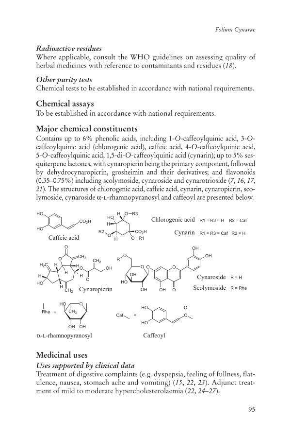
95
Folium Cynarae
Radioactive residuesWhere applicable, consult the WHO guidelines on assessing quality of herbal medicines with reference to contaminants and residues (18).
Other purity testsChemical tests to be established in accordance with national requirements.
Chemical assaysTo be established in accordance with national requirements.
Major chemical constituentsContains up to 6% phenolic acids, including 1-O-caffeoylquinic acid, 3-O-caffeoylquinic acid (chlorogenic acid), caffeic acid, 4-O-caffeoylquinic acid, 5-O-caffeoylquinic acid, 1,5-di-O-caffeoylquinic acid (cynarin); up to 5% ses-quiterpene lactones, with cynaropicrin being the primary component, followed by dehydrocynaropicrin, grosheimin and their derivatives; and flavonoids (0.35–0.75%) including scolymoside, cynaroside and cynarotrioside (7, 16, 17,21). The structures of chlorogenic acid, caffeic acid, cynarin, cynaropicrin, sco-lymoside, cynaroside -L-rhamnopyranosyl and caffeoyl are presented below.
Medicinal usesUses supported by clinical dataTreatment of digestive complaints (e.g. dyspepsia, feeling of fullness, flat-ulence, nausea, stomach ache and vomiting) (15, 22, 23). Adjunct treat-ment of mild to moderate hypercholesterolaemia (22, 24–27).
HO
HO
Caffeic acid O
CO2H
OHHO
H
OH
R2
Chlorogenic acid
CynarinR1
R1 = R3 = H R2 = Caf
R1 = R3 = Caf R2 = H
CO2H
R3
HO
HO
Caffeoyl
O
OH
CH3
OH
HO
-L-rhamnopyranosyl
Rha =Caf = C
O
O
O
CH2
CH2
HH
HH
O
H
HO
OH
O
CH2H2C
H Cynaropicrin
O
O
OH
OH
OH
O
OH
O
HO
OH
O
Cynaroside
Scolymoside
R = H
R
R = Rha
96
WHO monographs on selected medicinal plants
Uses described in pharmacopoeias and well established documentsOrally for the treatment of atherosclerosis and kidney dysfunctions (di-uretic) (5).
One study has indicated that the crude drug may be of benefit for the treatment of irritable bowel syndrome (28), but further randomized con-trolled clinical trials are needed before any therapeutic recommendations can be made.
Uses described in traditional medicineOral treatment of anaemia, diabetes, fever, gout, rheumatism and urinary stones (7, 9, 29).
PharmacologyExperimental pharmacologyAntiatherosclerotic and antihypercholesterolaemic activitiesA dried aqueous extract of the leaves (4.5:1) inhibited cholesterol biosyn-thesis from 14C-acetate in primary cultured rat hepatocytes in a concentra-tion-dependent biphasic manner with moderate inhibition (approximately 20%) being noted between 0.007 and 0.1 mg/ml and stronger inhibition at 1 mg/ml (80%). Replacement of 14C-acetate by 14C-mevalonate largely prevented the inhibitory effects of the extracts, indicating inhibition of the activity of hydroxyl-methyl-glutaryl-CoA-reductase. Stimulation of hy-droxyl-methyl-glutaryl-CoA-reductase activity by insulin was efficiently blocked by the extract. Cynaroside and its aglycone luteolin, constituents of the extract, were mainly responsible for enzyme inhibition (30). The effect of an extract of the leaves in vivo was investigated in four groups of 10 rats each fed an atherosclerogenic diet. Group one was administered 110 mg/kg body weight (bw) powdered leaves; group two, 80.0 mg/kg bw powdered Cynara cardunculus; group three, 10.0 mg/kg bw heparaxal; and group four served as the control. Examination of tissue after 120 days showed that the leaf extract prevented formation of atherosclerotic chang-es, prevented serum cholesterol increase, caused a decrease in lipid phos-phate, slightly increased the level of glycoproteins in the blood, prevented an increase in serum -globulin, decreased albumin, glycoproteins and liver cholesterol, and increased -globulin and -globulin fractions. Cy-nara cardunculus showed a similar but weaker activity (31). A methanol extract of the leaves was shown to reduce serum triglyceride levels in olive oil-loaded mice. Oral administration of the extract, at doses between 125 and 500.0 mg/kg bw, significantly suppressed serum triglyceride elevation 2 h after administration of olive oil. In contrast, 6 h after administration of olive oil, increases in triglyceride level were observed in the groups that
97
Folium Cynarae
received the extract at doses of 125.0 and 250.0 mg/kg bw. Orlistat, a li-pase inhibitor, completely suppressed the serum triglyceride elevation at 250.0 mg/kg bw. Clofibrate, a hypolipidaemic medicine, also suppressed the triglyceride level at doses of 250.0 and 500.0 mg/kg bw. Three sesqui-terpenes (cynaropicrin, aguerin B and grosheimin) from the extract were isolated as the active components (32).
Antihepatotoxic activityThe effects of an aqueous extract of the leaves on taurolithocholate-in-duced cholestatic bile canalicular membrane distortions were studied in primary cultured rat hepatocytes using electron microscopy. Artichoke extracts at concentrations between 0.08 and 0.5 mg/ml were able to pre-vent the formation of canalicular membrane transformations in a dose-dependent manner when added simultaneously with the bile acid. How-ever, prevention also occurred when the hepatocytes were preincubated with the extracts, indicating that absorption of the bile acid to compo-nents of the extracts was not involved (33).
The hepatoprotective activity of cynarin against carbon tetrachloride (CCl4)-induced toxicity in isolated rat hepatocytes was compared with other phenolic compounds. Only cynarin and, to a lesser extent, caffeic acid showed a cytoprotective effect (34). Treatment of rats with three con-secutive doses of 500.0 mg/kg bw of an extract of the crude drug, admin-istered by gavage 48, 24 and 1 h before CCl4 intoxication, produced a significant decrease in glutamic-oxaloacetic transaminase, glutamic-pyru-vic transaminase (also known as alanine aminotransferase or ALT), direct bilirubin and glutathione levels, thus indicating a reduction in the poten-tial for hepatotoxicity (35).
Primary cultures of rat hepatocytes exposed to tert-butyl hydroperox-ide were used for characterizing the antioxidative and hepatoprotective potential of an aqueous extract of the crude drug and some selected con-stituents. Addition of tert-butyl hydroperoxide to the culture media re-sulted in enhanced lipid peroxidation as measured by the production of malondialdehyde and enhanced cytotoxicity detected by leakage of lac-tate dehydrogenase. The extract added prior to or simultaneously with tert-butyl hydroperoxide reduced both phenomena with a median effec-tive concentration (EC50) of 95.0 and 12.0 μg leaf powder/ml, respectively. Furthermore, the aqueous extract prevented the loss of intracellular glu-tathione caused by tert-butyl hydroperoxide. Several polyphenolic and flavonoid constituents of the extract were found to reduce malondialde-hyde production. The median effective concentration values were 8.1, 12.5, 15.2 and 28 μg/ml for caffeic acid, chlorogenic acid, cynarin and cynaroside, respectively (36).
98
WHO monographs on selected medicinal plants
Primary rat hepatocyte cultures exposed to tert-butyl hydroperoxide or cumene hydroperoxide were used to assess the antioxidative and pro-tective potential of aqueous extracts of the leaves. Both hydroperoxides stimulated the production of malondialdehyde, particularly when the cells were pretreated with diethylmaleate in order to diminish the level of cellular glutathione. Addition of the extract did not affect basal malondi-aldehyde production, but prevented the hydroperoxide-induced increase of malondialdehyde formation in a concentration-dependent manner when presented simultaneously with or prior to the peroxides. The effec-tive concentrations were as low as 0.001 mg/ml (37).
Antioxidant activityA study measured the effects of aqueous and ethanol extracts of the leaves on intracellular oxidative stress stimulated by inflammatory mediators, tu-mour necrosis factor alpha and oxidized low-density lipoprotein (ox-LDL) in endothelial cells and monocytes. Both extracts inhibited basal and stim-ulated reactive oxygen species production in endothelial cells and mono-cytes, in a dose-dependent manner. In endothelial cells, the ethanol extract (50.0 μg/ml) significantly reduced ox-LDL-induced intracellular reactive oxygen species production by 60% (p < 0.001) and the aqueous extract (50 μg/ml) reduced ox-LDL-induced intracellular reactive oxygen species production by 43% (p < 0.01). The ethanol extract (50 μg/ml) reduced ox-LDL-induced intracellular reactive oxygen species production in mono-cytes by 76% (p < 0.01). Effective concentrations of 25–100 μg/ml were well below the cytotoxic levels of the extracts which started at 1.0 mg/ml as assessed by lactate dehydrogenase leakage and trypan blue exclusion (38).
An aqueous dried extract (9:2) of the leaves was studied in human leukocytes to assess activity against oxidative stress. The extract (medi-an effective concentration 0.23 μg/ml) produced a concentration-dependent inhibition of oxidative stress when cells were stimulated with agents that generate reactive oxygen species: hydrogen peroxide, phor-bol-12-myristate-13-acetate and N-formyl-methionyl-leucyl-phenyl-alanine. Cynarin, caffeic acid, chlorogenic acid and luteolin, constituents of artichoke leaf extracts, also showed a concentration-dependent in-hibitory activity in the above models, contributing to the antioxidant activity of the extract in human neutrophils (39).
Choleretic effectsTwo aqueous alcoholic extracts of the fresh leaves (total extract contain-ing 19% caffeoylquinic acids, at a dose of 200.0 mg/kg bw and a semi-purified extract containing 46% caffeoylquinic acids, at a dose of 25.0 mg/kg bw) were assessed in rats. Intraperitoneal administration stimulated
99
Folium Cynarae
choleresis, and significantly increased bile dry residue and total cholate secretion (p < 0.05). Intragastric administration of the same extracts (400.0 mg/kg bw, total extract and 200.0 mg/kg bw of the semipurified extract) also increased gastrointestinal motility by 11% and 14%, respec-tively (p < 0.05) (40).
The effects of an extract of the crude drug on bile flow and the forma-tion of bile compounds in anaesthetized rats after acute administration and repeated oral administration (twice a day for 7 consecutive days) were studied. A significant increase in bile flow was observed after acute treat-ment with the extract as well as after repeated administration. The choler-etic effects of the extract were similar to those of the reference compound dehydrocholic acid. Total bile acids, cholesterol and phospholipid were determined by enzymatic assays. At the highest dose (400.0 mg/kg bw), a significant increase was observed after single and repeated administration (p < 0.01) (41).
The choleretic effects of four extracts of the leaves (not described) were assessed in vivo in a study in rats. Extracts 1, 2 and 4 did not show sig-nificant choleretic activity at a dose of 1.0 and 2.0 g/kg bw. Extract 3, however, was found to induce an increase of bile flow, which was gradual and sustained. Cynarin and chlorogenic acid, administered as pure com-pounds, did not show choleretic activity at any of the doses tested and neither of them decreased the malondialdehyde content in liver (42).
ToxicologyThe oral and intraperitoneal median lethal doses of a hydroalcoholic ex-tract of the leaves in rats were 2.0 g/kg and 1.0 g/kg bw, respectively (40).The oral median lethal dose of cynarin in mice was 1.9 g/kg bw. Intraperi-toneal administration of cynarin to rats for 15 days, at doses between 50.0 and 400.0 mg/kg bw per day produced no macroscopic, haematological or histological abnormalities. Intraperitoneal administration of cynarin to rats for 40 days at a dose between 100.0 and 400.0 mg/kg bw per day in-creased body and kidney weight, as well as producing some degenerative changes in the liver (43). External application of a leaf extract to the skin of white rats, at doses of 1.0–3.0 g/kg bw for 21 days, did not produce any toxic effects or have any cumulative effects on haematological parameters or the biochemistry of rats. No skin-irritating or eye-irritating effects were observed in guinea-pigs (44).
Clinical pharmacologyAntidyspeptic effectsA multicentre open study assessed the effects of a dried aqueous leaf ex-tract (3.8–5.5:1, 320 mg per capsule) in 553 patients with dyspeptic com-

100
WHO monographs on selected medicinal plants
plaints. The daily dose was 4–6 capsules (containing 320.0 mg of extract per capsule) per day, for an average of 43.5 days. Digestive complaints declined significantly, by 71%, over the treatment period (p < 0.001). Compared with the baseline data on subjective symptoms, a reduction in abdominal pain (76%), emesis (88%), meteorism (66%) and nausea (82%) was observed. In a subgroup of 302 patients, total cholesterol decreased by 11.5% and triglycerides by 12.5% (25). In a similar study, the same extract was assessed in a 6-month open trial involving 203 patients with dyspepsia. The daily dose administered was 3–6 capsules (each capsule containing 320.0 mg of the extract). After 21 weeks of treatment, symp-toms such as vomiting, abdominal pain, nausea and flatulence decreased by 84%, 78%, 77% and 70%, respectively. Total blood cholesterol and triglycerides were reduced by 10.9% and 11%, respectively. Data from 159 patients indicated that low-density lipoprotein-cholesterol decreased by 15.8% and high-density lipoprotein-cholesterol increased by 6.3%. Global efficacy as assessed by physicians was good to excellent in 85.7% of patients. No adverse reactions were reported (26).
In a double-blind, randomized controlled trial, 247 patients with func-tional dyspepsia were treated with either a commercial extract of the crude drug (2 × 320.0 mg of plant extract three times daily) or a placebo. The primary outcome measured was the sum score of the patient’s weekly rat-ing of the overall change in dyspeptic symptoms (four-point scale). Sec-ondary variables were the scores for each dyspeptic symptom and the quality of life as assessed by the Nepean Dyspepsia Index. Of the 247 pa-tients enrolled, data from 244 patients (129 given active treatment, 115 giv-en placebo) were suitable for inclusion in the statistical analysis (intention-to-treat). The overall improvement in symptoms over the 6 weeks of treatment was significantly greater in patients treated with the commercial extract than in those treated with the placebo (8.3 ± 4.6 versus 6.7 ± 4.8, p < 0.01). Similarly, patients treated with the commercial extract showed significantly greater improvement in the global quality of life scores (Ne-pean Dyspepsia Index) than the placebo-treated patients (-41.1 ± 47.6 ver-sus -24.8 ± 35.6, p < 0.01). The preparation tested was significantly better than the placebo at alleviating symptoms and improving the disease-specific quality of life in patients with functional dyspepsia (45).
Antihypercholesterolaemic and lipid-lowering effectsTwo randomized controlled clinical trials assessed the effects of a dried aqueous extract of the leaves on cholesterol levels in 187 patients (24, 27).The first, a randomized, double-blind, placebo-controlled pilot study in-volving 44 healthy volunteers, assessed the effect of an extract of the crude drug on cholesterol levels. Patients were randomly assigned to receive

101
Folium Cynarae
either 640.0 mg of the extract or a placebo three times daily for 12 weeks. No significant effects on serum cholesterol were found. However in sub-group analysis, significant cholesterol-lowering effects were observed in subjects with a total cholesterol level of > 210 mg/dl (p < 0.022) (27).
The second placebo-controlled study assessed the safety and efficacy of a dried aqueous extract of fresh artichoke (25–35:1). Patients received either 1800 mg of artichoke extract as coated tablets, each containing 450.0 mg extract, or a placebo. Patients (n = 143) with hyperlipoprotein-aemia – initial total cholesterol of > 7.3 mmol/l (> 280 mg/dl) received 1.8 g of a dried leaf extract per day or the placebo for 6 weeks. Changes in total cholesterol and low-density lipoprotein-cholesterol from baseline to the end of treatment showed a statistically significant superiority of the dry artichoke extract over the placebo (p = 0.0001). Observed reductions in total cholesterol levels were 18.5% in those who received the extract and 8.6% in those who received the placebo after 6 weeks of treatment (24). The decrease in low-density lipoprotein-cholesterol in the group treated with the extract was 22.9% and was 63% in those treated with the placebo. The ratio of low-density lipoprotein to high-density lipoprotein showed a decrease of 20.2% in the group that received the extract and 7.2% in the group that received the placebo. No drug-related adverse events were reported (24).
In a randomized, placebo-controlled clinical trial, two groups of 30 pa-tients presenting various dislipidaemic profiles were treated for 50 days with either cynarin, 2 × 250 mg tablets per day, or a placebo. Cynarin was able to induce a significant reduction of hypercholesterolaemia (p < 0.001), the level of pre- -lipoproteins (p < 0.01), the / -lipoprotein ratio (p < 0.01) and patient’s body weight (46).
Several uncontrolled studies have found that cynarin reduced total se-rum cholesterol in patients after treatment with oral doses of 750–1500 mgper day. Oral administration of cynarin to 17 patients, at a dose of 1000 mg/day, for 4 weeks resulted in a significant decrease in total cholesterol (15%, p < 0.005) (9).
Choleretic effectA randomized, double-blind, placebo-controlled trial assessed the chol-eretic effects of a dry aqueous extract of the leaves (4.5–5:1) in 20 male volunteers with acute or chronic metabolic disorders. The treatment group (n = 10) received a single intraduodenal dose of the extract at a dose of 1.92 g/day in 50 ml of water on an empty stomach, while the control group received a placebo of similar appearance. Crossover to the alterna-tive treatment followed an 8-day washout period. The outcomes included intraduodenal bile secretion measured using multi-channel probes. Com-
102
WHO monographs on selected medicinal plants
pared with baseline values, 60 minutes after administration there was a significant increase in bile secretion in the treatment group (151%) as compared with the placebo group (p < 0.01) (23).
Irritable bowel syndromeIrritable bowel syndrome, characterized by abdominal pain and altered bowel habit, has symptoms that overlap with those of dyspepsia. Since the crude drug is used for the treatment of dyspepsia, a postmarketing surveillance study was performed to assess its effects on irritable bowel syndrome. A subgroup of patients (n = 279) with symptoms of irritable bowel syndrome was identified from a sample of individuals (n = 553) with dyspeptic syndrome who were being monitored in a postmarketing surveillance study of the extract for 6 weeks. Analysis of the data from the subgroup with irritable bowel syndrome revealed significant reductions in the severity of symptoms including abdominal pain, bloating, flatu-lence and constipation, and favourable evaluations of overall effectiveness by both physicians and patients (28).
PharmacokineticsA study to investigate the absorption, metabolism and disposition of arti-choke leaf extract was performed using two different extracts (47). The extracts were administered to 14 healthy volunteers in a crossover study. Each subject received doses of both extracts. The administered dose of extract A contained caffeoylquinic acids equivalent to 107.0 mg caffeic acid and luteolin glycosides equivalent to 14.4 mg luteolin. The adminis-tered dose of extract B contained caffeoylquinic acids equivalent to 153.8 mg caffeic acid and luteolin glycosides equivalent to 35.2 mg luteo-lin. Urine and plasma analysis were performed by a validated high-per-formance liquid chromatography method using 12-channel coulometric array detection. None of the genuine target extract constituents could be detected in the plasma or urine of the subjects. However, caffeic acid, its methylated derivates ferulic acid and isoferulic acid and the hydrogena-tion products dihydrocaffeic acid and dihydroferulic acid were identified as metabolites derived from caffeoylquinic acids. Except for dihydrofer-ulic acid, all of these compounds were present as sulfates or glucuronides. Peak plasma concentrations of total caffeic acid, ferulic acid and isoferulic acid were reached within 1 h and declined over 24 h showing almost bi-phasic profiles. By contrast, maximum concentrations for total dihydro-caffeic acid and dihydroferulic acid were observed only after 6–7 h, indi-cating two different metabolic pathways for caffeoylquinic acids. Luteolin administered as glucoside was recovered from plasma and urine only as sulfate or glucuronide, but neither in the form of genuine glucosides nor

103
Folium Cynarae
as free luteolin. Peak plasma concentrations were reached rapidly within 0.5 h. The elimination showed a biphasic profile (47).
Adverse reactionsGastrointestinal complaints included mild diarrhoea, accompanied by ab-dominal cramps, upper abdominal pain, nausea and heartburn. Allergic reactions may occur in sensitized patients (22, 25).
No significant adverse events other than gastrointestinal discomfort have been reported from open or controlled clinical trials (24–27, 48).
ContraindicationsHypersensitivity or allergies to artichokes and other plants from the Compositae/Asteraceae, and obstruction of the bile ducts (15).
WarningsPossible interaction with coumarin-type anticoagulants.
PrecautionsGeneralPatients with gallstones should seek the advice of a health care provider prior to use.
Carcinogenesis, mutagenesis, impairment of fertilityThe genotoxic effects of flavonoid constituents present in the crude drug (quercetin and luteolin) were assessed in two short-term bacterial assays (49). In Salmonella typhimurium (strains TA1538 uvrB- and TA1978 uvrB+) the flavonoids did not induce damage in the DNA as recognized by UvrABC nuclease. Results of the SOS-chromotest in Escherichia coliK-12 strains PQ37 and PQ243 indicated that the flavonoids only weakly induced the SOS system (49).
Drug interactionsNo information was found.1
Pregnancy: teratogenic effectsDue to the lack of safety and efficacy studies, the use of the crude drug during pregnancy is not recommended.
1 A report of a potential drug interaction with Folium Cynarae or its preparations and with couma-rin-type anticoagulants such as phenprocoumone and warfarin has been recorded by a national regulatory authority.

104
WHO monographs on selected medicinal plants
Pregnancy: non-teratogenic effectsDue to the lack of safety data, the use of the crude drug during pregnancy is not recommended.
Nursing mothersDue to the lack of safety data, the use of the crude drug during breastfeed-ing is not recommended.
Paediatric useDue to the lack of safety data, the use of the crude drug for the treatment of children under the age of 12 years is not recommended.
Other precautionsNo information was found.
Dosage formsCrude drug, extracts and other Galenical preparations for internal use.
Posology(Unless otherwise indicated)Average oral daily dose: for hypercholesterolaemia and dyspepsia, 1–2 gof a dried aqueous extract (24, 27, 45). Adult daily dose: 5–10 g of crude drug; or equivalent preparations (15, 17).
ReferencesPharmacopée Francaise1. [French pharmacopoeia]. Paris, Adrapharm, 1987.British herbal pharmacopoeia2. . Exeter, British Herbal Medicine Association, 1996.Reynolds JEF, ed. 3. Martindale: The extra pharmacopoeia, 13th ed. London, Pharmaceutical Press, 1993.PharmaMed: Aufbereitungsmonographien (Kommission E CD-ROM)4. . Stut-tgart, Deutscher Apotheker Verlag, 2004.African pharmacopoeia5. , Vol. 1, 1st ed. Lagos, Nigeria, Organization of Afri-can Unity, Scientific Technical & Research Commission, 1985.National Genetic Resources Program. 6. Germplasm Resources Information Network (GRIN) [Online Database]. Beltsville, Maryland, National Germ-plasm Resources Laboratory (available at: http://www.ars-grin.gov2/cgi-bin/npgs/html/tax_search.pl?cynara+scolymus).Farnsworth NR, ed. 7. NAPRALERT database. Chicago, University of Illinois at Chicago, IL (an online database available directly through the University of Illinois at Chicago or through the Scientific and Technical Network [STN] of Chemical Abstracts Services), 30 June 2005.
105
Folium Cynarae
Nadkarni KM, Nadkarni AK, eds. 8. Indian materia medica, reprint of 3rd re-vised and enlarged ed. Bombay, Popular Prakashan, 1976.Mills S, Bone K. 9. Principles and practice of phytotherapy. Edinburgh, Churchill Livingstone, 2000.Farmacopea homeopática de los estados unidos mexicanos 10. [HomeopathicPharmacopoeia of the United States of Mexico.] Mexico City, Secretaría de Salud, Comisión Permanente de la Farmacopea de los Estados Unidos Mexi-canos, 1998 [in Spanish].Bedevian AK. 11. Illustrated polyglottic dictionary of plant names. Cairo, Med-bouly Library, 1994.Hooper D, Field H. Useful plants and drugs of Iran and Iraq. 12. Field Museum of Natural History, Botanical series, 1937, 9:111.Han DR, et al. 13. Modern pharmacognosy. Seoul, Hakchang, 1989 [in Korean].Iwu MM. 14. Handbook of African medicinal plants. Boca Raton, FL, CRC Press, 1993.Blumenthal M, Goldberg A, Brinckmann J, eds. 15. Herbal medicine: expanded Commission E monographs. Austin, TX, American Botanical Council, 2000.Bruneton J. 16. Pharmacognosy, phytochemistry, medicinal plants. Paris, Lavoisi-er, 1996.Hagers Handbuch der Drogen17. (CD ROM). Heidelberg, Springer Verlag, 2003 [in German].WHO guidelines on assessing quality of herbal medicines with reference to 18.contaminants and residues. Geneva, World Health Organization, 2007.European Pharmacopoeia19. , 5th ed. Strasbourg, Directorate for the Quality of Medicines of the Council of Europe (EDQM), 2005.Guidelines for predicting dietary intake of pesticide residues20. , 2nd rev. ed. Ge-neva, World Health Organization, 1997 (WHO/FSF/FOS/97.7).Brand N. Monographie Cynara. In Hänsel R et al eds. 21. Hager's Handbuch der pharmazeutischen Praxis. Band 4 Drogen A-D. Berlin, Springer Verlag, 1992.Kraft K. Artichoke leaf extract – Recent findings reflecting effects on lipid 22.metabolism, liver and gastrointestinal effects. Phytomedicine, 1997, 4:369–378.Kirchhoff R et al. Increase in choleresis by means of artichoke extract. 23. Phy-tomedicine, 1994, 1:107–115.Englisch W et al. Efficacy of artichoke dry extract in patients with hyperlipo-24.proteinemia. Arzneimittel-Forschung, 2000, 50:260–265.Fintelmann V. Antidyspeptische und lipidsenkende Wirkungen von Ar-25.tischockenblätterextrakt. Ergebnisse klinischer Untersuchungen zur Wirk-samkeit und Verträglichkeit von Hepar-SL® forte an 553 Patienten. Zeitschriftfür Allgemeinmedizin, 1996, 72(Suppl 2):3–19 [in German].Fintelmann V, Petrowicz O. Langzeitanwendung eines Artischocken-Ex-26.tracktes bei dyspeptischem Symptomkomplex. Naturamed, 1998, 13:17–26 [in German].Petrowicz O et al. Effects of artichoke leaf extract (ALE) on lipoprotein me-27.tabolism in vitro and in vivo. Atherosclerosis, 1997, 129:147.

106
WHO monographs on selected medicinal plants
Walker AF, Middleton RW, Petrowicz O. Artichoke leaf extract reduces 28.symptoms of irritable bowel syndrome in a post-marketing surveillance study. Phytotherapy Research, 2001, 15:58–61.Wegener T, Fintelmann V. Pharmakologische Eigenschaften und therapeu-29.tisches Profil der Artischocke (Cynara scolymus L.). Wiener medizinische Wochenschrift, 1999, 149:241–247 [in German].Gebhardt R. Inhibition of cholesterol biosynthesis by artichoke extracts is 30.mainly due to luteolin. Cell Biology and Toxicology, 1997, 13:58.Samochowiec L. Cz XV. Działanie karczochów (31. Cynara scolymus L.) i kardów (Cynara cardunculus L.) na rozwój mia’d’ycy do’wiadczalnej u białych szczurów. [Experimental atherosclerosis. XV. The effect of Cynarascolymus and Cynara cardunculus on the development of experimental ath-erosclerosis in white rats.] Polish Dissertationes Pharmaceuticae, 1959, 11:99–113 [in Polish].Shimoda H et al. Anti-hyperlipidemic sesquiterpenes and new sesquiterpene 32.glycosides from the leaves of artichoke (Cynara scolymus L.): structure re-quirement and mode of action. Bioorganic and Medicinal Chemistry Letters,2003, 13:223–228.Gebhardt R. Prevention of taurolithocholate-induced hepatic bile canalicu-33.lar distortions by HPLC-characterized extracts of artichoke (Cynara scoly-mus) leaves. Planta Medica, 2002, 68:776–779.Adzet T et al. Action of an artichoke extract against carbon CCl34. 4-inducedhepatotoxicity in rats. Acta Pharmaceutica Jugoslavica, 1987, 37:183–187.Adzet T et al. Hepatoprotective activity of polyphenolic compounds from 35.Cynara scolymus against carbon tetrachloride toxicity in isolated rat hepato-cytes. Journal of Natural Products, 1987, 50:612–617.Gebhardt R, Fausel M. Antioxidant and hepatoprotective effects of artichoke 36.extracts and constituents in cultured rat hepatocytes. Toxicology in Vitro,1997, 11:669–672.Gebhardt R. Antioxidative and protective properties of extracts from leaves 37.of the artichoke (Cynara scolymus L.) against hydroperoxide-induced oxida-tive stress in cultured rat hepatocytes. Toxicology and Applied Pharmacology,1997, 144:279–286.Zapolska-Downar D et al. Protective properties of artichoke (38. Cynara scoly-mus) against oxidative stress induced in cultured endothelial cells and mono-cytes. Life Sciences, 2002, 71:2897–2908.Pérez-García F, Adzet T, Cañigueral S. Activity of artichoke leaf extract on 39.reactive oxygen species in human leukocytes. Free Radical Research, 2000, 33:661–665.Lietti A. Choleretic and cholesterol lowering properties of two artichoke 40.extracts. Fitoterapia, 1977, 48:153–158.Saénz Rodriguez T, García Giménez D, de la Puerta Vázquez R. Choleretic 41.activity and biliary elimination of lipids and bile acids induced by an arti-choke leaf extract in rats. Phytomedicine, 2002, 9:687–693.

107
Folium Cynarae
Speroni E et al. Efficacy of different 42. Cynara scolymus preparations on liver complaints. Journal of Ethnopharmacology, 2003, 86:203–211.Preziosi P, Loscalzo B. Pharmacological properties of 1,4 dicaffeylquinic 43.acid, the active principle of Cynara scolymus. Archives of International Phar-macodynamics, 1958, 117:63–75.Halkova J. An experimental study of skin and eye-irritating effects of the 44.preparate “Artishok”. Problemi na Khigienata, 1996, 21:74–80.Holtmann G et al. Efficacy of artichoke leaf extract in the treatment of pa-45.tients with functional dyspepsia: a six-week placebo-controlled, double-blind, multicentre trial. Alimentary Pharmacology and Therapeutics, 2003, 18:1099–1105.Montini M et al. Kontrollierte Anwendung von Cynarin in der Behandlung 46.hyperlipämischer Syndrome [Controlled application of cynarin in the treat-ment of hyperlipemic syndrome. Observations in 60 cases]. Arzneimittel-Forschung, 1975, 25:1311–1314 [in German].Wittemer SM et al. Bioavailability and pharmacokinetics of caffeoylquinic 47.acids and flavonoids after oral administration of artichoke leaf extracts in humans. Phytomedicine, 2005, 12:28–38.Pittler MH, Ernst E. Artichoke leaf extract for serum cholesterol reduction. 48.Perfusion, 1998, 11:338–340.Czeczot H, Kusztelak J. A study of the genotoxic potential of flavonoids us-49.ing short-term bacterial assays. Acta Biochimica Polonica, 1993, 40:549–554.

108
Cortex Granati
DefinitionCortex Granati consists of the dried root or trunk bark of Punica grana-tum L. (Lythraceae) (1–3).
SynonymsPunica nana L. (Punicaceae) (4).
Note: According to current botanical authorities, Punica granatum be-longs to the Lythraceae family (5).
Selected vernacular namesAnar, anara, anar-ke-per, Carthaginian apple, dadam, dadima-phalam, dalima, dalimb, dalimbay, dalimbuhannu, dalimo, darakte-naiar, darimba, darinko bokra, daru, delima, delum, delun, dhalima, dila dae lok, dlima, ende limau, gangsalan, glima glineu mekah, granada, granade, granado, Granatbaum, granatum, grenadier, grenadillo, gul armini, gulnar, jaman, jeliman, kupchaphala, lelo kase, madalai, madalam, madalangkai, mata-lam, mathalanarkom, melograno, mkoma manga, nar, pomegranate, pos-nar, qsur roman, qsur rommam, quishr-al-romman, quishr-romman, ranato, romã, roman, romeira, rommam, roman amruj, romanzeira, rou-mammam-goulnar, ruman, rumau, sekiryuu-karpi, seog-ryu, seokryupi, shajratur-rummam, sham-al-rumman, shih liu pi, shiliupi, shukadana, talima, thab thim, thap thim, zakuro-juhi (1, 3, 4, 6–12).
Geographical distributionNative to the Middle East (eastern Mediterranean to northern India), and now widely cultivated in warm regions throughout the world (6, 8, 9).
DescriptionA deciduous shrub or small tree, erect, up to 7 m high, much branched from the base; branches slender; branchlets often ending in spines, the young ones quadrangular or almost tetrapterous. Leaves: simple, oppo-

109
Cortex Granati
site, verticillate, oblong-lanceolate, glabrous, 1–9 cm long and 0.5–2.5 cmwide; apex acute, obtuse or emarginate; base cuneate. Flowers: 1–5 at the highest leaf axil of branchlets, 1 terminal, sessile or subsessile. Calyx: 2–3 cm long, red or pale yellow; lobes erectopatent to recurved; petals round or obtuse, red or white. Fruit, a berry, globose, 5–13 cm in diam-eter, with a leathery rib enclosing numerous seeds, variously coloured, yellowish-green, white, reddish brown or rarely blackish-purple. Seeds: numerous, red, pink or yellowish white (6, 7, 9).
Plant material of interest: root and stem barkGeneral appearanceStem bark: curved pieces or quills; up to 15 cm long, 0.5–3.0 mm thick: outer surface, yellowish to blackish-brown, with occasional greyish patches of lichens, longitudinally wrinkled and marked; small, broadly elliptical lenticels; inner surface, light yellow to light brown, finely stri-ated; fracture, very short and granular.
Root bark: flat, irregular, curved, or recurved small pieces; outer surface, brownish yellow, rough, with darker patches and conchoidal depressions due to exfoliation of the outer portion, but no lenticels; inner surface, yel-low, smooth, with irregular darker brown patches. Other characteristics similar to those of stem bark (1–3).
Organoleptic propertiesOdour: slight; taste: astringent and slightly bitter (1, 2).
Microscopic characteristicsCork, formed of several alternating layers of suberized thin-walled cells and of lignified cells with greatly thickened inner tangential walls. Cortex, consisting of parenchyma containing small starch granules; crystals of calcium oxalate from scattered prisms to rosette clusters; and large sc1e-reids, which are isolated, rarely in small groups, with very thick and strongly stratified walls, up to 400 μm long and 200 μm broad. Phloem shows numerous cells containing cluster crystals of calcium oxalate in more or less tangential rows, and parenchyma cells with numerous small starch granules or amorphous tannin masses. Medullary rays, 1–2 cell rows with occasional cells containing numerous small prisms (1–3).
Powdered plant materialYellowish brown to dark brown, characterized by fragments of paren-chyma containing numerous starch granules and crystals of calcium ox-alate; sclereids with very thick and pitted walls; fragments of cork with

110
WHO monographs on selected medicinal plants
prominent, thickened and lignified walls; numerous calcium oxalate crys-tals, prisms, 6–10 μm long and rosette cluster crystals up to 15 μm in di-ameter; starch granules, abundant, simple, 2–10 μm in diameter, or rarely compound; occasional long wood fibres, 15–20 μm in diameter, associated with pitted vessels; bast fibres absent (1, 3).
General identity testsMacroscopic and microscopic examinations, microchemical tests (1–3,13) and thin-layer chromatography (3, 13).
Purity testsMicrobiologicalTests for specific microorganisms and microbial contamination limits are as described in the WHO guidelines on assessing quality of herbal medi-cines with reference to contaminants and residues (14).
Foreign organic matterNot more than 2% (1, 2).
Total ashNot more than 15% (2).
Acid-insoluble ashTo be established in accordance with national requirements.
Water-soluble extractiveTo be established in accordance with national requirements.
Alcohol-soluble extractiveTo be established in accordance with national requirements.
Loss on dryingTo be established in accordance with national requirements.
Pesticide residuesThe recommended maximum limit of aldrin and dieldrin is not more than 0.05 mg/kg (15). For other pesticides, see the European Pharmacopoeia(15) and the WHO guidelines on assessing quality of herbal medicines with reference to contaminants and residues (14) and pesticide residues (16).
Heavy metalsFor maximum limits and analysis of heavy metals, consult the WHO guidelines on assessing quality of herbal medicines with reference to con-taminants and residues (14).
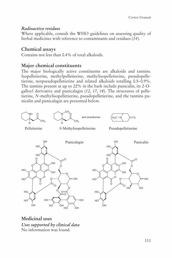
111
Cortex Granati
Radioactive residuesWhere applicable, consult the WHO guidelines on assessing quality of herbal medicines with reference to contaminants and residues (14).
Chemical assaysContains not less than 0.4% of total alkaloids.
Major chemical constituentsThe major biologically active constituents are alkaloids and tannins. Isopelletierine, methylpelletierine, methylisopelletierine, pseudopelle-tierine, norpseudopelletierine and related alkaloids totalling 0.5–0.9%. The tannins present at up to 22% in the bark include punicalin, its 2-O-galloyl derivative and punicalagin (12, 17, 18). The structures of pelle-tierine, N-methylisopelletierine, pseudopelletierine, and the tannins pu-nicalin and punicalagin are presented below.
Medicinal usesUses supported by clinical dataNo information was found.
NH3C O
Pseudopelletierine
N
CH3
O
H
CH3
and enantiomer
N-Methylisopelletierine
NH
CH3
O
H
Pelletierine
Punicalagin
OO
OHHO
HO
HO OH
OH
O
OH
O
O
O
OO
O
O
O
HO
HO
OH
OH
HO
HO
OH
HO
HO
OH
O
O
O
OH
OH
O
OO
O
O
O
HO
HO
OH
OH
HO
HO
OH
HO
HO
OH
O
OHO
Punicalin
112
WHO monographs on selected medicinal plants
Uses described in pharmacopoeias and well established documentsUsed orally for the treatment of diarrhoea and intestinal parasites (7, 19).
Uses described in traditional medicineUsed orally to treat dyspepsia, sore throat, menorrhagia, leukorrhoea and ulcers (12).
PharmacologyExperimental pharmacologyAntimicrobial activityAn aqueous and a 95% ethanol extract of the bark had weak activity in vitro against Bacillus cereus, Enterococcus faecalis, Escherichia coli, Kleb-siella pneumoniae, Pseudomonas aeruginosa and Staphylococcus aureus ata concentration of 25.0 mg/well (20). A decoction of the crude drug in-hibited the growth of Trichophyton tonsurans, T. rubrum, T. simii, Tricho-sporon beigelii, Microsporum fulvum, M. gypseum and Candida albicanswhen added to the nutrient medium at a concentration of 5% (21).
Anthelminthic and molluscicidal activitiesPelletierine, an alkaloid constituent of the bark, was active against tape-worms (Taenia solium), but was not active against other intestinal para-sites (4). At a concentration of 1:10 000, pelletierine hydrochloride kills tapeworms within 5–10 minutes (22). This alkaloid acts by causing the tapeworm to relax its grip on the intestinal walls and thereby making it possible to be expelled by cathartics. The molluscicidal activity of the crude drug against the snail Lymnaea acuminata was found to be both time- and dose-dependent. An ethanol extract of the bark was effective in killing the test animals, with a 24 h median lethal concentration of 22.42 mg/l (23). The extract was not toxic to the fish, Colisa fasciatus,which shares the same habitat with the snail (23).
Antiuraemic activityAdministration of a decoction of the bark in the drinking-water, at a dose of approximately 150.0 mg/kg body weight (bw), prevented casein/ade-nine-induced kidney failure in rats (24).
PharmacokineticsThe metabolism of punicalagin, a water-soluble ellagitannin isolated from the crude drug, was assessed in rats (25). The animals were treated with repeated oral administration of a 6% punicalagin-containing diet for 37 days. Punicalagin and related metabolites were identified by high per-formance liquid chromatography–diode array detector–mass spectrome-
113
Cortex Granati
try–mass spectrometry (HPLC-DAD-MS-MS) in plasma, liver and kid-neys. Five punicalagin-related metabolites were detected in liver and kidney, that is, two ellagic acid derivatives, gallagic acid, 3,8-dihydroxy-6H-dibenzo[b,d]pyran-6-one glucuronide, and 3,8,10-trihydroxy-6H-dibenzo[b,d]pyran-6-one (25).
ToxicologyIn animal experiments, intragastric administration of very large doses (not stated) of the alkaloids isolated from the bark caused respiratory arrest and death (26). Punicalagin has been reported to be toxic to cattle and rats (25). The chronic toxicity of punicalagin was assessed in rats. Treatment consisted of repeated oral administration of a 6% punicalagin-containing diet for 37 days. Feedstuff intake, food utility index and growth rate were lower in treated rats during the first 15 days, but no significant adverse effects were observed. No significant differences were found in treated rats in any blood parameter analysed (including the antioxidant enzymes glutathione peroxidase and superoxide dismutase) with the exception of urea and triglycerides, which remained low throughout the experiment (25).
Clinical pharmacologyToxicologyIngestion by humans of more than 80.0 g of drug may cause severe vomit-ing with blood, dizziness, fever, tremor and collapse. After 10 hours to 3 days temporary blindness may occur, which usually resolves after sev-eral weeks. Ingestion of pelletierine may cause visual disturbances with mydriasis (dilated pupils), dizziness and headache, as well as long-lasting anaesthesia or somnolence. Further symptoms of overdose include colic, cold sweat, dizziness, headache, muscle cramps, weakness or paralysis of the lower extremities, nausea, cardiac and respiratory collapse (27).
Adverse reactionsCommon adverse events observed in humans include dizziness, visual disturbances, weakness, calf spasms and tremors. Large overdoses (> 80.0 g) of the crude drug may lead to dizziness, mydriasis, severe head-ache, vertigo, vomiting, lethargy, collapse and possible death due to the alkaloid content (26).
ContraindicationsHypersensitivity or allergy to the bark.

114
WHO monographs on selected medicinal plants
WarningsFor diarrhoea lasting for longer than 3 days, contact a health care provider. For diarrhoea associated with fever, nausea and vomiting, or bloody stools, contact a health care provider. Do not exceed recommended dosage.
PrecautionsGeneralNo information was found.
Drug interactionsNo information was found.
Drug and laboratory test interactionsNo information was found.
Carcinogenesis, mutagenesis, impairment of fertilityThe genotoxic effects of the crude drug were examined in established hu-man cell lines, Raji and P3HR-1. Cells were treated with a decoction of the bark at various concentrations for 24 and 48 hours in vitro. Cell growth and viability were dose-dependently reduced. No apparent chro-mosomal aberrations were induced by the treatment. Administration of a bark extract induced apoptotic DNA fragmentation (28).
Pregnancy: teratogenic effectsNo information was found.
Pregnancy: non-teratogenic effectsDue to the lack of safety data, the use of Cortex Granati during preg-nancy is not recommended.
Nursing mothersDue to the lack of safety data, the use of Cortex Granati during breast-feeding is not recommended.
Paediatric useDue to the lack of safety data, the use of Cortex Granati in children under the age of 12 years is not recommended.
Other precautionsNo information was found.
Dosage formsCrude drug, extracts and tablets. Store in a cool dry place (19).

115
Cortex Granati
PosologyOral daily dose: 3–9 g for decoction (19).Daily dose: 20 g root bark fluidextract (1:1) in 59% ethanol for the treat-ment of Taenia infestation (27).
ReferencesThe Ayurvedic pharmacopoeia of India1. , Part I, Vol. II, 1st ed. New Delhi, Ministry of Health & Family Welfare, Department of Indian System of Med-icine and Homoeopathy, 1999.Asian crude drugs, their preparations and specifications. Asian Pharmacopoe-2.ia, 1st ed. Manila, Federation of Asian Pharmaceutical Associations, 1978.Materia medika Indonesia, Jilid V. 3. Jakarta, Departemen Kesehatan, Republik Indonesia, 1989 [in Indonesian].Iwu MM. 4. Handbook of African medicinal plants. Boca Raton, FL, CRC Press, 1993.National Genetic Resources Program. 5. Germplasm Resources Information Network (GRIN) [Online Database]. National Germplasm Resources Labo-ratory, Beltsville, MD (available at http://www.ars-grin.gov2/cgi-bin/npgs/html/tax_search.pl?punica+granatum).Standard of ASEAN herbal medicine,6. Vol. I. Jakarta, ASEAN Countries, 1993.Medicinal plants in China7. . Manila, World Health Organization Regional Of-fice for the Western Pacific, 1989 (WHO Regional Publications, Western Pa-cific Series, No. 2).Medicinal plants in the South Pacific.8. Manila, World Health Organization Regional Office for the Western Pacific, 1998 (WHO Regional Publications, Western Pacific Series, No. 19).Medicinal plants of India,9. Vol. II. New Delhi, Indian Council of Medical Research, 1987.Nadkarni AK. 10. Dr. K.M. Nadkarni’s Indian materia medica. Bombay, Popu-lar Prakashan, 1976.Ross IA. 11. Medicinal plants of the world. Totowa, New Jersey, Humana Press, 1999.Farnsworth NR, ed. 12. NAPRALERT database. Chicago, University of Illinois at Chicago, IL (an online database available directly through the University of Illinois at Chicago or through the Scientific and Technical Network [STN] of Chemical Abstracts Services), 30 June 2005.Deutsches Arzneibuch13. , 6th ed. Stuttgart, Deutscher Apotheker Verlag, 1951.WHO guidelines on assessing quality of herbal medicines with reference to 14.contaminants and residues. Geneva, World Health Organization, 2007.European Pharmacopoeia15. , 4th ed. Strasbourg, Directorate for the Quality of Medicines of the Council of Europe (EDQM), 2005.Guidelines for predicting dietary intake of pesticide residues16. , 2nd rev. ed. Ge-neva, World Health Organization, 1997 (WHO/FSF/FOS/97.7).

116
WHO monographs on selected medicinal plants
Evans WC. 17. Trease and Evans pharmacognosy, 15th ed. Edinburgh, WB Saunders, 2002.Tanaka T, Nonaka G-I, Nishioka I. Tannins and related compounds. XL. 18.Revision of the structures of punicalin and punicalagin, and isolation and characterization of 2-O-galloylpunicalin from the bark of Punica granatumL. Chemical and Pharmaceutical Bulletin, 1986, 34:650–655.Pharmacopoeia of the People’s Republic of China19. . Vol. I (English ed.). Bei-jing, Chemical Industry Press, 2005.Nimri LF, Meqdam MM, Alkofahi A. Antibacterial activity of Jordanian me-20.dicinal plants. Pharmaceutical Biology, 1999, 37:196–201.Dutta BK, Rahman I, Das TK. Antifungal activity of Indian plant extracts. 21.Mycoses, 1998, 41:535–536.Kee CH. 22. The pharmacology of Chinese herbs. Boca Raton, FL, CRC Press, 1993.Tripathi SM, Singh DK. Molluscicidal activity of 23. Punica granatum bark and Canna indica root. Brazilian Journal of Medical Biology Research, 2000, 33:1351–1355.Yokozawa T et al. Confirmation that tannin-containing crude drugs have a 24.uraemic toxin-decreasing action. Phytotherapy Research, 1995, 9:1–5.Cerdá B et al. Repeated oral administration of high doses of the pomegranate 25.ellagitannin punicalagin to rats for 37 days is not toxic. Journal of Agriculture and Food Chemistry, 2003, 51:3493–3501.Bensky D, Gamble A. 26. Chinese herbal medicine. Materia medica, revised ed. Seattle, Washington, Eastland Press, 1993.Hager ROM 2003, 27. Granati cortex (Granatrinde). Heidelberg, Springer-Ver-lag.Settheetham W, Ishida T. Study of genotoxic effects of antidiarrheal medici-28.nal herbs on human cells in vitro. Southeast Asian Journal of Tropical Medi-cine and Public Health, 1995, 26 (Suppl 1):306–310.

117
Pericarpium Granati
DefinitionPericarpium Granati consists of the dried pericarp of Punica granatum L. (Lythraceae) (1–3).
SynonymsPunica nana L. (Punicaceae) (4).
Note: According to current botanical authorities, Punica granatum be-longs to the Lythraceae family (5).
Selected vernacular namesAnar, anara, anar-ke-per, Carthaginian apple, dadam, dadima-phalam, dalima, dalimb, dalimbay, dalimbuhannu, dalimo, darakte-naiar, darimba, darinko bokra, daru, delima, delum, delun, dhalima, dila dae lok, dlima, ende limau, gangsalan, glima glineu mekah, granada, granade, granado, Granatbaum, granatum, grenadier, grenadillo, gul armini, gulnar, jaman, jeliman, kupchaphala, lelo kase, madalai, madalam, madalangkai, mata-lam, mathalanarkom, melograno, mkoma manga, nar, pomegranate, pos-nar, qsur roman, qsur rommam, quishr-al-romman, quishr-romman, ranato, romã, roman, romeira, rommam, roman amruj, romanzeira, rou-mammam-goulnar, ruman, rumau, sekiryuu-karpi, seog-ryu, seokryupi, sham-al-rumman, shajratur-rummam, shih liu pi, shiliupi, shukadana, talima, thab thim, thap thim, zakuro-hi (2–4, 6–12).
Geographical distributionNative to the Middle East (eastern Mediterranean to northern India), and now widely cultivated in warm regions throughout the world (6, 8, 9).
DescriptionA deciduous shrub or small tree, erect, up to 7 m high, much branched from the base; branches slender; branchlets often ending in spines, the young ones quadrangular or almost tetrapterous. Leaves: simple, oppo-

118
WHO monographs on selected medicinal plants
site, verticillate, oblong-lanceolate, glabrous, 1–9 cm long and 0.5–2.5 cmwide; apex acute, obtuse or emarginate; base cuneate. Flowers: 1–5 at the highest leaf axil of branchlets, 1 terminal, sessile or subsessile. Calyx 2–3 cm long, red or pale yellow; lobes erectopatent to recurved; petals round or obtuse, red or white. Fruit, a berry, globose, 5–13 cm in diame-ter, with a leathery rind enclosing numerous seeds, variously coloured, yellowish green, white, reddish brown or rarely blackish purple. Seeds: numerous, red, pink or yellowish white (6, 7, 9).
Plant material of interest: pericarpGeneral appearanceIrregular slices or gourd-shaped, brittle, varying in size, 1.5–3.0 mm thick. The outer surface reddish brown, brownish yellow or dark brown; some-what lustrous, rough, with numerous warty protuberances. Some with a raised tubular persistent calyx and a stout and short peduncle or its scar. Inner surface yellow or reddish brown, with raised reticulated remains of the pe-duncle. Texture hard and fragile, fracture yellow, somewhat granular (1, 6).
Organoleptic propertiesOdourless; taste: bitter and astringent (1, 6).
Microscopic characteristicsTransverse section of the pericarp shows the following: exocarp consists of irregular polygonal cells with slightly thickened but not lignified outer wall and a thick smooth cuticle. Mesocarp generally consists of thin-walled parenchyma cells containing starch granules, clusters or prisms of calcium oxalate. Scattered among the parenchyma cells are sclereids oc-curring singly or in small groups with strongly thickened lignified walls and rather small lumen or with slightly thickened somewhat lignified walls and wider lumen. Small bicollateral vascular bundles course through the parenchyma; rather thick-walled, non-lignified fibres occasionally ac-companying the bundles. Larger bicollateral vascular bundles course through the central part of the mesocarp: phloem fibres are few and pos-sess clear non-lignified walls and a narrow lumen; some phloem paren-chyma contains prisms of calcium oxalate. Endocarp cells relatively small, containing starch granules and crystals of calcium oxalate, and stone cells also relatively small (1, 6).
Powdered plant materialReddish brown. Stone cells sub-rounded, rectangular or irregular, rarely branched, 27–102 m in diameter, with relatively thick walls and a large lumen, some containing brown matter. Epidermal cells sub-square or sub-
119
Pericarpium Granati
rectangular, with slightly thickened walls. Clusters of calcium oxalate 10–25 m in diameter, prisms infrequent. Spiral and reticulate vessels 12–18 min diameter. Starch granules sub-rounded, 2–10 m in diameter (1).
General identity testsMacroscopic and microscopic examinations (1, 2, 6), microchemical tests (1, 6), and thin-layer chromatography (2, 6).
Purity testsMicrobiologicalTests for specific microorganisms and microbial contamination limits are as described in the WHO guidelines on assessing quality of herbal medi-cines with reference to contaminants and residues (13).
Foreign organic matterNot more than 6% (1).
Total ashNot more than 4% (2, 6).
Acid-insoluble ashNot more than 1% (6).
Water-soluble extractivePericarp: not less than 30% (6).
Alcohol-soluble extractivePericarp: not less than 15% (6).
Loss on dryingNot more than 10% (6).
Pesticide residuesThe recommended maximum limit of aldrin and dieldrin is not more than 0.05 mg/kg (14). For other pesticides see the European pharmacopoeia (14)and the WHO guidelines on assessing quality of herbal medicines with reference to contaminants and residues (13) and pesticide residues (15).
Heavy metalsFor maximum limits and analysis of heavy metals, consult the WHO guidelines on assessing quality of herbal medicines with reference to con-taminants and residues (13).

120
WHO monographs on selected medicinal plants
Radioactive residuesWhere applicable, consult the WHO guidelines on assessing quality of herbal medicines with reference to contaminants and residues (13).
Chemical assaysTotal tannins not less than 10% (1).
Major chemical constituentsThe major constituents are hydrolysable ellagitannins (up to 28%) and other polyphenols (3, 12, 16, 17). Structures of ellagic acid and punicalin are presented below.
Medicinal usesUses supported by clinical dataNone.
Uses described in pharmacopoeias and well established documentsOrally for the treatment of chronic diarrhoea, dysentery, gingivitis and intestinal parasites (1, 7, 18).
Uses described in traditional medicineTreatment of bronchitis, fever, gastrointestinal ailments, menorrhagia, respiratory tract infections, skin rashes, vaginal infections and worms (12, 19).
O
O
O
O
OH
OHHO
HO
Ellagic acid
O
OH
OH
O
OO
O
O
O
HO
HO
OH
OH
HO
HO
OH
HO
HO
OH
O
OHO
Punicalin

121
Pericarpium Granati
PharmacologyExperimental pharmacologyAntidiarrhoeal activityIntragastric administration of a decoction or a 95% ethanol extract of the crude drug at a dose of 200.0 mg/kg or 50.0 mg/kg body weight (bw), respectively, reduced faecal output in rats with castor-oil induced diar-rhoea (21). The same extracts, at a dose of 500.0 mg/kg bw, exhibited in-testinal antisecretory activity in rats with magnesium sulfate-induced en-teropooling (20).
Antimicrobial, antiparasitic and antiviral activityAn aqueous, butanol, or 95% ethanol extract of the crude drug had in vitro activity against Proteus mirabilis, Haemophilus influenzae, Kleb-siella pneumoniae, Pseudomonas aeruginosa, Streptococcus aureus, Staph-ylococcus aureus and Candida albicans at a concentration of 60.0 μg/ml (21, 22). An aqueous extract and a 95% ethanol extract of the fruit rind had weak in vitro activity against Bacillus cereus, Enterococcus faecalis,Escherichia coli, Klebsiella pneumoniae, Pseudomonas aeruginosa andStaphylococcus aureus at a concentration of 25 mg/well (23, 24).
A decoction of the crude drug weakly inhibited the growth of Trichophyton tonsurans, T. rubrum, T. simii, Trichosporon beigelii, Mi-crosporum fulvum, M. gypseum and Candida albicans when added to the nutrient medium at a concentration of 5% (25).
Tannins separated from the crude drug were effective against genital herpes virus (herpes simplex virus type 2). The tannin not only inhibited replication of the virus, but also blocked herpes type 2 viral adsorption in cultured cells (26).
An aqueous extract of the crude drug had antiviral activity against hepatitis B virus in PLC/PRF/5 cells in vitro (27). An aqueous extract inhibited hepatitis C virus in vitro at a concentration of 100 μg/ml. An acetone extract of the crude drug had larvicidal activity against Chry-somya albiceps at a concentration of 25 μg/ml (28). At a concentration of 10.0 ml/plate, an aqueous extract of the crude drug weakly inhibited the growth of Ascaris galli, Pheritima posthuma and Taenia solium (29), as well as Ascaris lumbricoides (30).
Antioxidant activityAqueous, alcohol and ethyl acetate extracts of the crude drug have shown significant antioxidant activity in various in vitro and in vivo models. A methanol extract of the crude drug had antioxidant activity in vitro, in the
-carotene-linoleate and 1,1-diphenyl-2-picrylhydrazyl radical model

122
WHO monographs on selected medicinal plants
systems. The methanol extract exhibited 83% and 81% antioxidant activ-ity at a concentration of 50.0 μg/ml using the -carotene-linoleate and 1,1-diphenyl-2-picrylhydrazyl radical model systems, respectively (31).A dried methanol extract fed to rats (50.0 mg/kg bw) following treatment with carbon tetrachloride (CCl4) (2.0 g/kg bw) decreased the levels of catalase, superoxide dismutase and peroxidase by 81, 49 and 89%, respec-tively. Pretreatment of the rats with the methanol extract maintained the levels of catalase, peroxidase and superoxide dismutase at values compa-rable with control values, whereas lipid peroxidation was reduced by 54% compared to the control group (32).
Antiulcer activityIntragastric administration of an aqueous extract (5 ml/kg bw) or an un-specified protein-containing fraction (50 mg/kg bw) of the crude drug to rats prevented hydrochloric acid and ethanol-induced gastric ulceration (33). The gastroprotective effects of tannic acid and the aqueous extract of the crude drug against ethanol-induced damage were investigated in rats. Oral administration of the extract or tannic acid induced a significant de-crease in gastric lesions (48–76%). The protection observed was more pronounced when the test solution was given at the same time as the eth-anol. The acid content of the stomach was increased by 368% after ad-ministration of the ethanol extract (34).
Immune effectsOral administration of a powder made from the fruit rind, at a dose of 100.0 mg/kg bw, as an aqueous suspension, stimulated cell-mediated and humoral components of the immune system in rabbits. The suspension elicited an increase in antibody titre to typhoid-H antigen. It also en-hanced the inhibition of leukocyte migration in the leukocyte migration inhibition test and induration of skin in the delayed hypersensitivity test with purified protein derivative, confirming its stimulatory effect on the cell-mediated immune response (35).
ToxicologyAn aqueous extract of the crude drug was reported to stimulate uterine contractions in non-pregnant rats but the dose and the route of adminis-tration were not stated (36).
Clinical pharmacologyA water-soluble component derived from an ethanol extract of the crude drug possessing antimicrobial activity was used in an antibacterial mouth-wash at a concentration of 0.8% w/w (18). The mouthwash was com-posed of a combination of the water-soluble component (0.8 g/100 ml),
123
Pericarpium Granati
menthol (0.03 g/100 ml), and absolute alcohol (5 g/100 ml). Eighty volun-teers (30 healthy and 50 with dental caries) were required to rinse their mouths with 20 ml of the mouthwash for 30 seconds, they then closed their mouths for 90 seconds and then spat out their saliva for analysis. The salivary microbes were cultured and identified. Oral pathogens such as Staphylococcus aureus, S. mutans and Lactobacillus spp. were killed whereas Candida albicans was not (18).
Adverse reactionsNo information was found.
ContraindicationsHypersensitivity or allergy to the plant material.
WarningsIn cases of diarrhoea lasting for longer than 3 days, or associated with fever, nausea and vomiting, or bloody stools, contact a health care pro-vider. Do not exceed recommended dose.
PrecautionsCarcinogenesis, mutagenesis, impairment of fertilityThe genotoxic effects of the crude drug were examined in established hu-man cell lines, Raji and P3HR-1. Cells were treated with a decoction of the crude drug at various concentrations for 24 and 48 hours in vitro. Cell growth and viability were dose-dependently reduced. No apparent chro-mosomal aberrations were induced by the treatment. Administration of the extract induced apoptotic DNA fragmentation (37).
Pregnancy: teratogenic effectsNo information was found.
Pregnancy: non-teratogenic effectsDue to the lack of safety data, the use of the crude drug during pregnancy is not recommended.
Nursing mothersDue to the lack of safety and efficacy studies, the use of the crude drug by breastfeeding mothers is not recommended.
Paediatric useDue to the lack of safety data, the use of the crude drug for the treatment of children under the age of 12 years is not recommended.

124
WHO monographs on selected medicinal plants
Other precautionsNo information was found.
Dosage formsCrude drug, extracts and tablets. Store in a cool dry place (1).
Posology(Unless otherwise indicated)Oral daily dose: 2.5–4.5 g (1, 7).
ReferencesPharmacopoeia of the People’s Republic of China. 1. Beijing, Chemical Indus-try Press, 2005.The Ayurvedic pharmacopoeia of India2. , Part I, Vol. II, 1st ed. New Delhi, Ministry of Health & Family Welfare, Department of Indian System of Med-icine and Homoeopathy, 1999.Materia medika Indonesia, Jilid V. 3. Jakarta, Departemen Kesehatan, Republik Indonesia, 1989 [in Indonesian].Iwu MM. 4. Handbook of African medicinal plants. Boca Raton, FL, CRC Press, 1993.National Genetic Resources Program. 5. Germplasm Resources Information Network (GRIN) [Online Database]. National Germplasm Resources Labo-ratory, Beltsville, MD (available at: http://www.ars-grin.gov2/cgi-bin/npgs/html/tax_search.pl?punica+granatum).Standard of ASEAN herbal medicine6. . Vol. I. Jakarta, ASEAN Countries, 1993.Medicinal plants in China7. . Manila, World Health Organization Regional Of-fice for the Western Pacific, 1989 (WHO Regional Publications, Western Pa-cific Series, No. 2).Medicinal plants in the South Pacific.8. Manila, World Health Organization Regional Office for the Western Pacific, 1998 (WHO Regional Publications, Western Pacific Series, No. 19).Medicinal plants of India,9. Vol. II. New Delhi, Indian Council of Medical Research, 1987.Nadkarni AK. 10. Dr. K.M. Nadkarni’s Indian materia medica. Bombay, Popu-lar Prakashan, 1976.Ross IA. 11. Medicinal plants of the world. Totowa, NJ, Humana Press, 1999.Farnsworth NR, ed. 12. NAPRALERT database. Chicago, University of Illinois at Chicago, IL (an online database available directly through the University of Illinois at Chicago or through the Scientific and Technical Network [STN] of Chemical Abstracts Services), 30 June 2005.WHO guidelines on assessing quality of herbal medicines with reference to 13.contaminants and residues. Geneva, World Health Organization, 2007.

125
Pericarpium Granati
European Pharmacopoeia14. , 5th ed. Strasbourg, Directorate for the Quality of Medicines of the Council of Europe (EDQM), 2005.Guidelines for predicting dietary intake of pesticide residues15. , 2nd rev. ed. Ge-neva, World Health Organization, 1997 (WHO/FSF/FOS/97.7).Evans WC. 16. Trease and Evans pharmacognosy, 15th ed. London, WB Saun-ders, 2002.Tanaka T, Nonaka G-I, Nishioka I. Tannins and related compounds. XL.17.Revision of the structures of punicalin and punicalagin, and isolation and characterization of 2-O-galloylpunicalin from the bark of Punica granatumL. Chemical and Pharmaceutical Bulletin, 1986, 34:650–655.Chulasiri M et al. A water-soluble component with antimicrobial activity 18.from pomegranate rind: an ingredient in an antiseptic mouthrinse. MahidolJournal of Pharmaceutical Sciences, 1995, 22:150–159.De Feo V, Senatore F. Medicinal plants and phytotherapy in the Amalfitan 19.coast, Salerno Province, Campania, Southern Italy. Journal of Ethnopharma-cology, 1993, 39:39–51.Pillai NR. Anti-diarrhoeal activity of 20. Punica granatum in experimental ani-mals. International Journal of Pharmacognosy, 1992, 30:201–204.Alkofahi A, Masaadeh H, Al-Khalil S. Antimicrobial evaluation of some 21.plant extracts of traditional medicine of Jordan. Alexandria Journal of Phar-maceutical Sciences, 1996, 10:123–126.Prashanth D, Asha MK, Amit A. Antibacterial activity of 22. Punica granatum.Fitoterapia, 2001, 72:171–173.Chulasiri M, Thakerngpol K, Temsiririrkkul R. A water-soluble component 23.with antimicrobial activity from pomegranate rind: electron microscopic and preliminary chemical studies. Mahidol Journal of Pharmaceutical Sciences,1995, 22:107–112.Chulasiri M et al. A water-soluble component with antimicrobial activity 24.from pomegranate rind: antimicrobial potency and stability study. MahidolJournal of Pharmaceutical Sciences, 1995, 22:1–7.Dutta BK, Rahman I, Das TK. Antifungal activity of Indian plant extracts. 25.Mycoses, 1998, 41:535–536.Zhang J et al. [Antiviral activity of tannin from pericarp of 26. Punica granatumL. against genital herpes virus in vitro] [in Chinese]. Zhongguo Zhongyao Zazhi, 1995, 20:556–558.Goto W et al. Suppression of hepatitis B virus surface antigen secretion by 27.traditional plant medicines. Phytotherapy Research, 1996, 10:504–507.Morsy TA, Mazyad SAM, El-Sharkawy IM. The larvicidal activity of solvent 28.extracts of three medicinal plants against third instar larvae of Chrysomyiaalbiceps. Journal of the Egyptian Society of Parasitology, 1998, 28:699–709.Hukkeri VI et al. 29. In vitro anthelmintic activity of aqueous extract of fruit rind of Punica granatum. Fitoterapia, 1993, 64:69–70.Raj RK. Screening of indigenous plants for anthelmintic action against hu-30.man Ascaris lumbricoides: Part II. Indian Journal of Physiology and Pharma-cology, 1975, 19:47–49.
126
WHO monographs on selected medicinal plants
Singh RP, Chidambara Murthy KN, Jayaprakasha GK. Studies on the anti-31.oxidant activity of pomegranate (Punica granatum) peel and seed extracts using in vitro models. Journal of Agriculture and Food Chemistry, 2002, 50:81–86.Chidambara Murthy KN, Jayaprakasha GK, Singh RP. Studies on antioxi-32.dant activity of pomegranate (Punica granatum) peel extract using in vivo models. Journal of Agriculture and Food Chemistry, 2002, 50:4791–4795.Khennouf S et al. Effects of 33. Quercus ilex L. and Punica granatum L. poly-phenols against ethanol-induced gastric damage in rats. Pharmazie, 1999, 54:75–76.Gharzouli K et al. Effects of aqueous extracts from 34. Quercus ilex L. root bark, Punica granatum L. fruit peel and Artemisia herba-alba Asso leaves on eth-anol-induced gastric damage in rats. Phytotherapy Research, 1999, 13:42–45.Gracious Ross R, Selvasubramanian S, Jayasundar S. Immunomodulatory 35.activity of Punica granatum in rabbits – a preliminary study. Journal of Eth-nopharmacology, 2001, 78:85–87.Dhawan BN, Saxena PN. Evaluation of some indigenous drugs for stimulant 36.effect on the rat uterus: A preliminary report. Indian Journal of Medical Re-search, 1958, 46:808–811.Settheetham W, Ishida T. Study of genotoxic effects of antidiarrheal medici-37.nal herbs on human cells in vitro. Southeast Asian Journal of Tropical Medi-cine and Public Health, 1995, 26 (Suppl 1):306–310.
127
Folium Guavae
DefinitionFolium Guavae consists of the dried and/or young leaves of Psidium gua-java L. (Myrtaceae) (1).
SynonymsPsidium aromaticum L. P. cujavillus Burm. f, P. pomiferum L., P. pyrifer-um L., P. pumilum Vahl (2).
Selected vernacular namesAbas, aduoba, aguoba, amba, amrood, amrud, amrut, amruta-phalam, araca-goiba, arasa, banjiro, banziro, bidji, bihi, bilauti, borimak, bugoyab, buyaki, dijamboé, coloc, djambu bidji, djambu klutuk, eguabe, fa-rang, goavy, goejaba, goiaba, goiabeira, goiabeira-vermelha, goiabeiro, gouyav, gouyavier, goyav, goyavier, goyya, grosse gelbe, gua, guafa, guajave, gua-va, guava tree, Guave, guayaba, guayaba cak, guayaba colorada, guayaba cotorrera, guayaba de gusano, guayaba de venado, guayaba del Peru, guayaba peruana, guayabe, guayabero, guayabo, guayabo agrio, guayabo blanco, guayava, Guayave, guega, guyaba, gwaabaa, gwawa, hind armudu, ipera, jaama, jamba, jambu biji, kautonga, Kiswahili, koyya, krue, ku’ava, kuabas, kuava, kuawa, kuiaba, kuliabas, mabera, maduriam, manssla, motiram, mpera, mugwavha, ngoaba, nulu, oguawa, pat’a, perala, pero delle Indie, peyara, posh, psidio, psidium, punjo, quwawa, sari guafa, sig-ra, sikra, tuava, warakel-guafa, wariafa, woba, xalxoctl (1, 3–10).
Geographical distributionNative to tropical America, but now pantropical (7, 8, 11).
DescriptionA large shrub or small tree up to 10 m high. Stem slender, usually not exceeding 30 cm in width; bark brownish, thin, smooth, and often flaking off in scaly patches. Leaves opposite, oblong, slightly oval shaped, 5–15 cmin length, light green on the upper surface, and downy and pale green on

128
WHO monographs on selected medicinal plants
the underside. They display prominent veins on the underside of the leaf. Flowers white, occur either singly or a few in a cluster in axils of the leaves. About 2.5 cm in diameter, with numerous stamens arranged in groups. Fruits are globose or pyriform, yellow, usually 2.5–10 cm long and 2.5–5 cm in diameter, horticulture varieties with larger fruits with an edible pink mesocarp are common. They contain many seeds, and the calyx persists in fruit (7, 8).
Plant material of interest: dried or young leavesGeneral appearanceOblong, slightly oval, apex acute, round or acuminate; base symmetrical, cordate; margin smooth or dentate; venation reticulate; green (1).
Organoleptic propertiesOdour: slight; taste: astringent (1).
Microscopic characteristicsThe surface view shows nearly straight anticlinal epidermal cell walls on both surfaces, those on upper surface thickened; abundant stomata and cov-ering trichomes on lower surface; oil glands on both surfaces, but more frequent on lower surface. Transverse section shows small epidermal cells with straight anticlinal walls, upper epidermal layer 2–3 cells thick with a few oil glands (schizogeneous); palisade tissue multi-layered; numerous oil glands in the mesophyll region; midrib region shows collenchymatous cells; bicollateral vascular bundle is distinctly horse-shoe shaped and surrounded by lignified pericyclic fibres; xylem elements are generally lignified; uniseri-ate trichomes smooth-walled and abundant on lower epidermis (1).
Powdered plant materialLight green; taste astringent; lamina fragments show abundant stomata, punctate, numerous covering trichomes; rosette crystals; lignified vascu-lar elements (1).
General identity testsMacroscopic and microscopic examinations (1).
Purity testsMicrobiologicalTests for specific microorganisms and microbial contamination limits are as described in the WHO guidelines on assessing quality of herbal medi-cines with reference to contaminants and residues (12).

129
Folium Guavae
Foreign organic matterTo be established in accordance with national requirements.
Total ashTo be established in accordance with national requirements.
Acid-insoluble ashTo be established in accordance with national requirements.
Water-soluble extractiveTo be established in accordance with national requirements.
Loss on dryingTo be established in accordance with national requirements.
Pesticide residuesThe recommended maximum limit of aldrin and dieldrin is not more than 0.05 mg/kg (13). For other pesticides, see the European pharmacopoeia (13)and the WHO guidelines on assessing quality of herbal medicines with reference to contaminants and residues (12) and pesticide residues (14).
Heavy metalsFor maximum limits and analysis of heavy metals, consult the WHO guidelines on assessing quality of herbal medicines with reference to con-taminants and residues (12).
Radioactive residuesWhere applicable, consult the WHO guidelines on assessing quality of herbal medicines with reference to contaminants and residues (12).
Chemical assaysNot less than 14% (w/w) tannins and not less than 0.6% (v/w) essential oil (1).
Major chemical constituentsContains hydrolysable tannins, essential oils, flavonoids, and terpenes (1,6, 15). New leaves contain the flavonols quercetin, guajaverin (= quer-cetin-3-O-arabinoside) and other quercetin glycosides; gallocatechin and the tannins ellagic acid and guavins A, C and D. Several triterpene acids are present, including ursolic and oleanolic acids and their 20-hydroxy-derivatives, crataegoic and guaijavolic acids. The leaf oil contains several mono- and sesquiterpenes, among which 1,8-cineol and -pinene are the

130
WHO monographs on selected medicinal plants
principal monoterpenes, and caryophyllene and -bisbolene are represen-tative of the sesquiterpenes (16–24).
Structures of quercetin, guajaverin (= quercetin-3-O-arabinoside), ur-solic oleanolic acids, ellagic acid, -pinene and caryophyllene are present-ed below.
Medicinal usesUses supported by clinical dataOral treatment of acute diarrhoea, gingivitis and rotaviral enteritis (25–28).
Uses described in pharmacopoeias and well established documentsNo information was found.
Uses described in traditional medicineTreatment of abdominal pain, bleeding gums, cough, gastritis, headache, ringworm, vaginitis, wounds and worms. Also used as an astringent, an antiemetic and an emmenagogue (4, 15).
O
OH
HO
O
O
OH
OH
X
Quercetin
Guajaverin
O
OH
OHArap =
HO
-L-arabinopyranosyl
X = H
X = Arap
H3C CH3
CH3H3C
CH3 CH3 CO2HH
H
H CH3
HO
H
Oleanolic acid Ursolic acidH3C CH3
CH3 CH3 CO2HH
H
H CH3
HO
H
H
H3C
CH3H
H3C
H3C
CH3
-Pinene
and enantiomerH
H
Caryophyllene
H3CCH3
CH2
CH3
H
HO
O
O
O
OH
OHHO
HO
Ellagic acid

131
Folium Guavae
PharmacologyExperimental pharmacologyAnalgesic activityIntragastric administration of 100.0, 200.0 or 400.0 mg/kg body weight (bw) of the essential oil of the leaves produced an antinociceptive effect in mice as assessed in the formalin test (29). A dose of 200.0 mg/kg bw of the essential oil to mice reduced pain as measured in the acetic acid-induced writhing test. One of the major constituents, -pinene, also showed an antinociceptive effect in the formalin test, and when administered intra-gastrically at doses of 100 mg/kg and 200 mg/kg, a reduction of paw lick-ing of 72% and 76%, respectively was observed. At a dose of 400.0 mg/kgbw, paw licking was reduced by 37% in the first (acute) phase and 81% in the second (chronic) phase (29).
Antidiarrhoeal activityA decoction of the leaves inhibited Microlax-induced diarrhoea when ad-ministered by gastric lavage to rats at a dose of 10.0 ml/kg bw (30). Intra-gastric administration of a methanol extract of the leaves to mice at a dose of 200.0 mg/kg bw prevented castor oil-induced diarrhoea (31). Intragastric administration of an aqueous or methanol extract of the leaves to rats at a dose of 400.0 mg/kg bw prevented castor oil-induced diarrhoea, reduced gastric motility and prostaglandin E2-induced enteropooling (32). A dried 70% methanol extract of the leaves inhibited the electrically-induced peri-staltic reflex of isolated guinea-pig ileum at a concentration of 100 g/ml (22). The main active constituent of the extract was quercetin 1.4 × 10⁻⁵
(72.1%), with isoquercetin and hyperin being weakly active (22).
Antihyperglycaemic activityIntragastric administration of a 50% ethanol extract of the leaves to rats, at a dose of 200.0 mg/kg bw, prevented alloxan-induced hyperglycae-mia. A butanol fraction of the 50% ethanol extract reduced alloxan-in-duced hyperglycaemia in rats when administered at a dose of 25.0 mg/kg bw by gastric lavage (33). Intragastric administration of a 50% etha-nol extract to rats at a dose of 200.0 mg/kg bw did not stimulate insulin biosynthesis (33).
Antiinflammatory and antipyretic activityIntragastric administration of 0.8 ml/kg bw of the leaf essential oil to rats reduced inflammation in the carrageenan-induced oedema of the hind paw test and the cotton pellet granuloma test (34). Intragastric adminis-tration of a dried methanol extract of the leaves to mice at a dose of 200.0 mg/kg bw reduced carrageenan-induced pedal oedema (31). Intra-
132
WHO monographs on selected medicinal plants
gastric administration of a dried methanol extract of the leaves to mice at a dose of 200.0 mg/kg bw reduced yeast-induced pyrexia (31).
Antimalarial activityThe following extracts of the crude drug had activity against Plasmodiumfalciparum in vitro with a median inhibitory concentration as follows: ethyl acetate,10.0 μg/ml; 95% ethanol, 36.0 μg/ml; aqueous, 80.0 μg/ml; and petroleum ether, 13.0 μg/ml (35, 36).
Antimicrobial activityA decoction of the crude drug inhibited the growth of Carnobacteriumgallirarum, Escherichia coli, Salmonella enteritidis and Staphylococcus au-reus with a median inhibitory concentration of 31.25 μg/ml and Klebsiellapneumoniae, Pseudomonas aeruginosa, and Shigella flexneri at a concen-tration of 62.5 μg/ml (37). A tannin fraction separated from an extract of the crude drug had antibacterial activity against Escherichia coli, Cit-robacter diversus, Klebsiella pneumoniae, Shigella flexneri, Salmonella enteritidis and Staphylococcus aureus at concentrations of 60.0–95.0 μg/ml in vitro (38). An aqueous extract of the leaves had anti-adhesion activity in Streptomyces mitis (70%); these bacteria are involved in the develop-ment of dental plaque (39).
Antioxidant activityThe antioxidant activity of an aqueous and a 50% ethanol extract of the leaves was assessed using 1,1-diphenyl-2-picrylhydrazyl radical colorim-etry. Compared to ascorbic acid, the extracts of the crude drug had a sig-nificantly lower antioxidant effect (40).
Antispasmodic activityA dried methanol extract of the leaves reduced spontaneous contractions of isolated rat and guinea-pig ileum when added to the bath media at con-centrations of 20.0 ml/l and 80.0 μg/ml, respectively (30, 37, 41). An aque-ous, methanol, or hexane extract of the leaves had smooth muscle relaxant activity in guinea-pig ileum in vitro, at a concentration of 250.0–1000.0 μg/ml (41, 42). A butanol extract of the leaves at a concentration of 0.2 mg/ml reduced acetylcholine-induced contractions in guinea-pig ileum by 95–100% (43).
Antitussive activityIntragastric administration of an aqueous extract of the leaves to rats and guinea-pigs at a dose of 5.0 g/kg bw reduced capsaicin-induced coughing (44).

133
Folium Guavae
Effects on the central nervous systemIntragastric administration of a dried methanol extract of the leaves to mice at a dose of 200 mg/kg bw reduced acetic acid-induced writhing, thereby demonstrating analgesic activity (31). Intragastric administration of a dried methanol extract of the leaves to mice at a dose of 200 mg/kgbw also potentiated phenobarbitone-induced sleeping time (31). Intra-peritoneal administration of a dried hexane extract of the leaves to mice at a dose of 100 mg/kg bw potentiated sodium pentobarbital-induced hyp-nosis and increased the latency of leptazol-induced convulsions (42). In-tragastric or intraperitoneal administration of a dried methanol extract of the leaves at a dose of 3.3 mg/kg bw reduced spontaneous motor activity in mice (45). Intragastric administration of dried hexane, ethyl acetate or methanol extracts of the crude drug, at doses of 100.0–1250.0 mg/kg bw produced dose-dependent antinociceptive effects in mice, and prolonged pentobarbitone-induced sleep (46).
Haemostatic effectsThe effects of an aqueous leaf extract on the bleeding time and the three main mechanisms of haemostasis: vasoconstriction, platelet aggregation and blood coagulation, were investigated. Topical application of the aque-ous extract at a concentration of 0.05 μg/ml did not reduce bleeding times in wounded rats. However, the extract (2–6 μg/ml) potentiated the vascu-lar muscle contraction induced by phenylephrine (4.0 μg/ml) in isolated aortic strips from rabbits. The extract also significantly prolonged blood coagulation time in normal plasma treated with 6.0 mg/ml extract in the activated partial thromboplastin time test (p < 0.05) (47).
Inotropic effectsIn guinea-pig atria, an ethanol extract of the leaves reduced atrial contrac-tions by depressing the myocardial force in a concentration-dependent manner (median effective concentration (EC50) = 1.4 g/l). Concentrations higher than 2.5 g/l completely abolished the myocardial contractility. Furthermore, an acetic acid fraction (EC50 = 0.07 g/l) of the extract in-creased the relaxation time measured at 20 and 50% of the force curve by 30 and 15%, respectively, but did not change the contraction time. The negative inotropic effect of the extract was abolished by atropine sulfate, suggesting that either the active substance acts as a cholinergic agonist or that it could release acetylcholine from parasympathetic synapses (48).
ToxicityIntragastric administration of an aqueous extract of the leaves to rats ex-hibited a median lethal dose of 50.0 g/kg bw (47). In chronic toxicity tests,

134
WHO monographs on selected medicinal plants
an aqueous extract of the leaves was administered by gavage to 128 rats of both sexes at doses of 0.2, 2.0 and 20.0 g/day (1, 10 and 100 times the nor-mal therapeutic dose for the treatment of diarrhoea) for 6 months.
The results showed that the body weight gains in male rats were lower in all treated animals. Significant increases in white blood cell count, alka-line phosphatase, serum glutamate pyruvate transaminase and serum blood urea nitrogen levels were observed (p < 0.05). Serum sodium and cholesterol levels were significantly reduced (p < 0.05) indicating signs of hepatotoxicity. In female rats, serum sodium, potassium and albumin lev-els increased significantly (p < 0.05), while levels of platelets and serum globulin were significantly decreased (p < 0.05). Histopathological assess-ment showed a mild degree of fatty change and hydronephrosis in male rats and nephrocalcinosis and pyelonephritis in female rats (49).
Clinical pharmacologySeventy subjects with gingivitis were enrolled in a 3-week placebo-con-trolled, double-blind clinical trial to assess the efficacy of a mouthwash containing a decoction of the dried leaves (3 kg in 30 l water boiled for 20 minutes). The placebo mouthwash contained the same ingredients with the exception of the decoction of the leaves. The subjects were stratified into two balanced groups according to their baseline pre-prophylaxis gin-givitis scores calculated using the Loe-Silness Gingival Scoring Index. The subjects rinsed their mouth three times daily for 1 minute with 15 ml of their assigned mouthwash. Patients who used the mouthwash containing the leaf extract had less inflammation of the gingiva (19.8%) and fewer sites of severe gingival disease (40.5%) than those using the placebo mouthwash (26).
A randomized, controlled clinical trial assessed the efficacy of a decoc-tion of the crude drug for the treatment of infantile rotaviral enteritis. Sixty-two patients with rotaviral enteritis were randomly assigned either to the group treated with the decoction or to the control group. The time until cessation of diarrhoea, the content of sodium in blood, the content of sodium and glucose in stools, and the rate of negative conversion of hu-man rotavirus antigen were recorded. After 3 days, 87% of the subjects in the treated group had recovered, significantly more than the number in the control group (58.1%, p < 0.05). The time elapsed until cessation of diarrhoea in the treated group (25.1 ± 9.5 h) was significantly shorter than that for the control group (38.7 ± 15.2 h, p < 0.01). The content of sodium and glucose in stools was reduced in the treated group (p < 0.05), while the reduction in the control group was insignificant. The rate of negative con-version of human rotavirus in the faeces of the treated group was 87.1%, significantly better than that of the control group (58.1%, p < 0.05) (28).

135
Folium Guavae
An aqueous extract of the crude drug was tested in a clinical study in-volving small groups of patients aged 5 years and younger or 20–40 years. Patients with acute diarrhoea received the extract, while a comparison group received a kaolin or pectin suspension. The results were similar in all three groups with an efficacy of treatment above 70% (25).
A randomized, double-blind, clinical study was performed to evaluate the safety and efficacy of an extract of the crude drug with a standardized content of quercetin. The extract was administered orally to a group of adult patients with acute diarrhoeic disease. Adult patients of both sexes between 20 and 59 years of age suffering from non-complicated acute di-arrhoea were included. Acute diarrhoeic disease was defined as a clinical condition characterized by the passing of at least three liquid stools dur-ing the previous 24 h and abdominal pain or cramps. Pregnant women and patients with systemic diseases concomitant to acute diarrhoeic dis-ease (such as immunodeficiency and intestinal syndrome), were excluded. The capsules containing 500 mg of the extract were administered every 8 h for 3 days to the treatment group (n = 50), while the control group (n = 50) received capsules of the same size, taste and colour, containing 500 mg of placebo every 8 h for 3 days. Oral rehydration therapy was administered to all patients according to conventional procedures fol-lowed in the medical institution for treatment of acute diarrhoeic disease. The results showed that the guava product decreased the duration of ab-dominal pain in these patients (27).
A randomized, double-blind, clinical trial involving 122 patients (64 men and 58 women) was conducted to compare the efficacy of the powdered crude drug with that of tetracycline for the treatment of acute diarrhoea (50). The patients were treated with 500 mg of the powdered crude drug (2 capsules of 250 mg each) or matching tetracycline capsules (2 capsules of 250 mg each) every 6 hours for 3 days. The results of the study demonstrated that the powdered drug decreased the stool output, fluid intake and the duration of diarrhoea. The differences between the results from the group treated with tetracycline and the group that re-ceived the powdered crude drug were not statistically significant (50).
Adverse reactionsOne report of allergic dermatitis has been recorded after external applica-tion of a tea prepared from the crude drug (51).
ContraindicationsHypersensitivity or allergy to the plant material.

136
WHO monographs on selected medicinal plants
WarningsDo not exceed the recommended dose or duration of treatment (49).
PrecautionsCarcinogenesis, mutagenesis, impairment of fertilityNo information was found.
Pregnancy: teratogenic effectsNo information was found.
Pregnancy: non-teratogenic effectsDue to the lack of safety data, the use of the crude drug during pregnancy is not recommended.
Nursing mothersDue to the lack of safety data, the use of the crude drug during breastfeed-ing is not recommended.
Paediatric useDue to the lack of safety data, the use of the crude drug in children aged under 12 years is not recommended.
Other precautionsNo information was found.
Dosage formsCrude drug, decoctions, extracts and teas.
Posology(Unless otherwise indicated)As a mouthwash: 15 ml of aqueous extract three times daily for at least 1 minute per session (26).
For diarrhoea: 500 mg of the powdered leaf three or four times daily (50).
ReferencesGhana herbal pharmacopoeia1. . Accra, Policy Research and Strategic Planning Institute, 1992.National Genetic Resources Program. 2. Germplasm Resources Information Network (GRIN) [Online Database]. National Germplasm Resources Labo-ratory, Beltsville, MD (available at: http://www.ars-grin.gov/cgi-bin/npgs/html/tax_search.pl?psidium+guajava).

137
Folium Guavae
Ross IA. 3. Medicinal plants of the world. Totowa, NJ, Humana Press, 1999.Farnsworth NR, ed. 4. NAPRALERT database. Chicago, University of Illinois at Chicago, IL (an online database available directly through the University of Illinois at Chicago or through the Scientific and Technical Network [STN] of Chemical Abstracts Services), 30 June 2005.Nadkarni KM, Nadkarni AK, eds. 5. Indian materia medica, reprint of 3rd re-vised and enlarged ed. Bombay, Popular Prakashan, 1976.Medicinal plants of India6. . Vol. 2. New Delhi, Indian Council of Medical Re-search, 1987.Medicinal plants in the South Pacific.7. Manila, World Health Organization Regional Office for the Western Pacific, 1998 (WHO Regional Publications, Western Pacific Series, No. 19).Iwu MM. 8. Handbook of African medicinal plants. Boca Raton, FL, CRC Press, 1993.Matos FJA. 9. Plantas medicinais Brasileiras, 2nd ed. Lima, Universidade Fed-eral do Ceará Edições, 2004.Mejía K, Rengifo E. 10. Plantas Medicinales de Uso Popular en la Amazonia Peruana. Lima, Agencia Española de Cooperación Internacional, 2000 [in Spanish].Perry LM, Metzger J. 11. Medicinal plants of east and southeast Asia: attributed properties and uses. Cambridge, MA, MIT Press, 1980.WHO guidelines on assessing quality of herbal medicines with reference to 12.contaminants and residues. Geneva, World Health Organization, 2007.European pharmacopoeia13. , 5th ed. Strasbourg, Directorate for the Quality of Medicines of the Council of Europe (EDQM), 2005.Guidelines for predicting dietary intake of pesticide residues14. , 2nd rev. ed. Ge-neva, World Health Organization, 1997 (WHO/FSF/FOS/97.7).Farnsworth NR, Bunyapraphatsara N, eds. 15. Psidium guajava. Thai medicinal plants: recommended for primary health care system. Bangkok, Mahidol Uni-versity, 1992:202–207.Okuda T et al. Guavin B, an ellagitannin of novel type. 16. Chemical and Phar-maceutical Bulletin, 1984, 32:3787–3788.Okuda T et al. Guavin A, C and D, complex tannins from 17. Psidium guajava.Chemical and Pharmaceutical Bulletin, 1987, 35:443–446.Soliman G, Farid MK. Constituents of the leaves of 18. Psidium guajava L. Part I. Psidiolic acid. Journal of the Chemical Society, 1952, 1952:134–136.Osman AM, Younes ME, Sheta AE. Triterpenoids of the leaves of 19. Psidiumguajava. Phytochemistry, 1974, 13:2015–2016.Arthur HR, Hui WH. Triterpene acids from the leaves of 20. Psidium guajavaL. Journal of the Chemical Society, 1954, 1954:1403–1406.Begum S et al. Triterpenoids from the leaves of 21. Psidium guajava. Phytochem-istry, 2002, 61:399–403.Lozoya X et al. Quercetine glycosides in 22. Psidium guajava L. leaves and de-termination of a spasmolytic principle. Archives of Medical Research, 1994, 25:11–15.

138
WHO monographs on selected medicinal plants
Matsuo T et al. Identification of (+)-gallocatechin as a bio-antimutagenic 23.compound in Psidium guajava leaves. Phytochemistry, 1994, 36:1027–1029.Xiao DJ et al. The essential oil of the leaves of 24. Psidium guajava L. Journal of Essential Oil Research, 1991, 3:187–189.Caceres A. 25. Plantas de uso medicinal en Guatemala. Guatemala City, Edito-rial Universitaria, Universidad San Carlos de Guatemala, 1996:194–197.Kraivaphan V et al. The effect of a mouthrinse containing 26. Psidium guajavaleaves extract on gingivitis. Journal of the Dental Association of Thailand,1991, 41:323–328.Lozoya X et al. Intestinal anti-spasmodic effect of a phytodrug of 27. Psidiumguajava folia in the treatment of acute diarrheic disease. Journal of Ethno-pharmacology, 2002, 83:19–24.Wei L et al. Clinical study on treatment of infantile rotaviral enteritis with 28.Psidium guajava L. Zhongguo Zhong Xi Yi Jie He Za Zhi, 2000, 20:893–895 [in Chinese].Santos FA et al. Investigation on the antinociceptive effect of 29. Psidium gua-java leaf essential oil and its major constituents. Phytotherapy Research,1998, 12:24–27.Lutterodt GD. Inhibition of Microlax-induced experimental diarrhoea with 30.narcotic-like extracts of Psidium guajava leaf in rats. Journal of Ethnophar-macology, 1992, 37:151–157.Olajide OA, Awe SO, Makinde JM. Pharmacological studies on the leaf of 31.Psidium guajava. Fitoterapia, 1999, 70:25–31.Lin J, Puckree T, Mvelase TP. Anti-diarrhoeal evaluation of some medicinal 32.plants used by Zulu traditional healers. Journal of Ethnopharmacology, 2002, 79:53–56.Maruyama Y et al. Study on 33. Psidium guajava L. (I). Anti-diabetic effect and effective components of the leaf of Psidium guajava L. Shoyakugaku Zasshi,1985, 39:261–269 [in Japanese].Kavimani S et al. Anti-inflammatory activity of volatile oil of 34. Psidium gua-java. Indian Journal of Pharmaceutical Sciences, 1997, 59:142–144.Gessler MC et al. Screening Tanzanian medicinal plants for antimalarial ac-35.tivity. Acta Tropica, 1994, 56:65–77.Weenen H et al. Antimalarial activity of Tanzanian medicinal plants. 36. PlantaMedica, 1990, 56:368–370.Tona L et al. Biological screening of traditional preparations from some me-37.dicinal plants used as antidiarrhoeal in Kinshasa, Congo. Phytomedicine,1999, 6:59–66.Lutete T et al. Antimicrobial activity of tannins. 38. Fitoterapia, 1994, 65:276–278.Razak FA, Rahim ZH. The anti-adherence effect of 39. Piper betle and Psidiumguajava extracts on the adhesion of early settlers in dental plaque to saliva-coated glass surfaces. Journal of Oral Science, 2003, 45:201–206.Qian H, Nihorimbere V. Antioxidant power of phytochemicals from 40. Psidi-um guajava leaf. Journal of Zhejiang University Science, 2004, 5:676–683.

139
Folium Guavae
Lozoya X, Becerril G, Martinez M. Modelo de perfusión intraluminal del 41.ileon del cobayo in vitro en el estudio de las propiedades antidiarréicas de la guayaba (Psidium guajava) [Intraluminal perfusion model of in vitro guinea pig’s ileum as a model of study of the antidiarrheal properties of the guava (Psidium guajava)]. Archivos de Investigacion Médica (México), 1990, 21:155–162 [in Spanish].Meckes M et al. Terpenoids isolated from 42. Psidium guajava hexane extract with depressant activity on central nervous system. Phytotherapy Research,1996, 10:600–603.Kambu K et al. Activité antispasmodique d’extraits à partir de plantes utili-43.sées en préparations comme antidiarrhéiques à Kinshasa, Zaïre [Antispas-modic activity of extracts used in traditional plant antidiarrheic preparations in Kinshasa, Zaire]. Annales Pharmaceutiques Françaises, 1990, 48:200–208 [in French].Jaiarj P et al. Anticough and antimicrobial activities of 44. Psidium guajava Linn. leaf extract. Journal of Ethnopharmacology, 1999, 67:203–212.Lutterodt GD, Maleque A. Effects on mice locomotor activity of a narcotic-45.like principle from Psidium guajava leaves. Journal of Ethnopharmacology,1988, 24:219–231.Shaheen HM et al. Effect of 46. Psidium guajava leaves on some aspects of the central nervous system in mice. Phytotherapy Research, 2000, 14:107–111.Jaiarj P et al. Guava leaf extract and topical haemostasis. 47. Phytotherapy Re-search, 2000, 14:388–391.Conde Garcia EA, Nascimento VT, Santiago Santos AB. Inotropic effects of 48.extracts of Psidium guajava L. (guava) leaves on the guinea pig atrium. Bra-zilian Journal of Medical and Biological Research, 2003, 36:661–668.Attawish A et al. Toxicity study of 49. Psidium guajava Linn. leaves. Bulletin of the Department of Medical Sciences, 1995, 37:289–305.Thanangkul B, Chaichantipayut C. Double-blind study of 50. Psidium guava L. and tetracycline in acute diarrhea. Siriraj Hospital Gazette, 1987, 39:263–267.Obi M et al. Allergic contact dermatitis due to guava tea. 51. Contact Dermatitis,2001, 44:116–117.
140
Lichen Islandicus
DefinitionLichen Islandicus consists of the whole or cut dried thalli of Cetraria is-landica (L.) Acharius s.l. (Parmeliaceae) (1).
SynonymsPhyscia islandica DC, Lichene islandicus L. (2).
Selected vernacular namesAl kharaza, Blätterflechte, brodmose, cetraria, broedmasa, erba rissa, Fie-berflechte, Fiebermoos, focus, hazaz, Heideflechte, Iceland lichen, Ice-land liver wort, Iceland moss, Icelandic moss, Isländische Flechte, Islän-dische Tartschenflechte, Isländisches Moos, kharaz assoukhour, lichen catharticus, lichen d’Islande, lichène islandico, líquén de islandia, Lun-genmoos, matmasa, muscus, Purgiermoos, svinmasa (2–5).
Geographical distributionGrows in northern, eastern and central Europe, Siberia and North Amer-ica (5, 6).
DescriptionA lichen of approximately 10 cm in height, growing on the ground, the brown shrubby thallus lobed and forked, and with a fringed margin. Up-per surface olive-green to brown, with occasional dark reddish brown copular apothecia, and the lower surface whitish-grey with numerous small, whitish depressed spots (5, 7).
Plant material of interest: dried thalliGeneral appearancePieces of foliaceous lichen up to about 15 cm long, composed of numer-ous erect branches about 6 mm broad or more, in an unevenly developed dichotomy, consists of glabrous, groove-shaped or almost flat, stiff, brittle

141
Lichen Islandicus
bands, 0.3–1.5 cm wide and 0.5 mm thick, sometimes serrated with the margin appearing ciliated (pycnidial) thick; lower surface pale greyish, with scattered small, white, ovoid, depressed spots; occasional, dark red-dish-brown, cup-shaped, fruiting bodies (apothecia), about 6 mm in di-ameter, on the upper surface (greenish to greenish brown) near the mar-gins; texture harsh, springy and brittle; on the apex of the terminal lobes, very rarely, there are brown, discoid apothecia (1).
Organoleptic propertiesOdour: none; taste: mucilaginous and distinctly bitter (1).
Microscopic characteristicsCut transversely, branches show upper and lower colourless cortical re-gions composed of closely-packed hyphae, appearing as small-celled pseu-doparenchyma; below the upper cortex, the algal or gonidial layer contain-ing numerous yellowish-green, subspherical cells of the alga Chlorococcum humicola; a central medulla with more closely-packed, greyish-brown hy-phae filaments. Section through an apothecium shows a hymenium layer in the upper cortex with flask-shaped asci, each containing eight ascospores, separated by numerous, narrow, thread-like paraphyses (3).
Powdered plant materialA greyish brown powder, abundant fragments of pseudoparenchyma consist-ing of narrow-lumened, thick-walled hyphae from the marginal layer and wide-lumened hyphae from the adjacent layer consisting of loosely entwined hyphae, in the medullary zone of which, yellowish green algal cells about 15 m in diameter are embedded; occasionally marginal fragments of the thal-lus with tube-like or cylindrical spermogonia, up to about 160 m wide and 400 m long. Many of the particles stain blue-black with iodine water (1).
General identity testsMacroscopic and microscopic examinations and microchemical tests (1,3), thin-layer chromatography for characteristic lichen acid profile (1),and high-performance liquid chromatographic analysis of protolichester-inic and fumarprotocetraric acids (8).
Purity testsMicrobiologicalTests for specific microorganisms and microbial contamination limits are as described in the WHO guidelines on assessing quality of herbal medi-cines with reference to contaminants and residues (9).
142
WHO monographs on selected medicinal plants
Foreign organic matterNot more than 5% (3).
Total ashNot more than 3% (3).
Acid-insoluble ashNot more than 1.5% (3).
Water-soluble extractiveTo be established in accordance with national requirements.
Loss on dryingNot more than 12% (10).
Swelling valueNot less than 4.5 determined on the powdered drug (1).
Pesticide residuesThe recommended maximum limit of aldrin and dieldrin is not more than 0.05 mg/kg (1). For other pesticides, see the European pharmacopoeia (1)and the WHO guidelines on assessing quality of herbal medicines with reference to contaminants and residues (9) and pesticide residues (11).
Heavy metalsFor maximum limits and analysis of heavy metals, consult the WHO guidelines on assessing quality of herbal medicines with reference to con-taminants and residues (9).
Radioactive residuesWhere applicable, consult the WHO guidelines on assessing quality of herbal medicines with reference to contaminants and residues (9).
Chemical assaysTo be established in accordance with national requirements.
Major chemical constituentsContains water-soluble polysaccharides (50%), primarily lichenin, a lin-ear cellulose-like polymer of -D-glucose units, and isolichenin, an -D-glucose polymer. Two glucans, lichenan and isolichenan, have been iso-lated from the lichen Cetraria islandica (12), as well as galactomannans (5). Major secondary metabolites include the lichen acids (depsidones),

143
Lichen Islandicus
fumarprotocetraric acid, protolichestearic acid and cetraric acids (5, 13).A biologically active aliphatic -lactone, (+)-protolichesterinic acid has also been reported (14). The structures of fumarprotocetraric acid, ce-traric acid and (+)-protolichesterinic acid are presented below.
Medicinal usesUses supported by clinical dataUsed orally as a demulcent to treat inflammation and dryness of the pha-ryngeal mucosa (15, 16).
Uses described in pharmacopoeias and well established documentsUsed orally for treatment of loss of appetite and nausea (3, 17).
Uses described in traditional medicineUsed to treat asthma, cramps, bronchitis, cough, diabetes, exhaustion, gastric disturbances, immune depletion, migraine, nausea in pregnancy and wounds. Also used as an emergency food source, an emollient and a galactagogue (4, 18–20).
PharmacologyExperimental pharmacologyAntimicrobial and antiviral activitiesAn aqueous extract of the thalli and protolichesterinic acid were screened for in vitro activity against Mycobacterium aurum, a nonpathogenic or-ganism with a similar sensitivity profile to M. tuberculosis. Protolichester-inic acid had a minimum inhibitory concentration (MIC) of 125.0 μg/ml (21). An aqueous extract of the thalli was screened for in vitro activity against Helicobacter pylori. (+)-Protolichesterinic acid was identified as
O
O
H3CCO2H
OH
OOH3C
HOCHO
Cetraric acid CH3 Fumarprotocetraric acid
O
O
H3CCO2H
OH
OOH3C
HOCHO
OCO2H
H3C
O
O
CO2HHH
CH2(+)-Protolichesterinic acid

144
WHO monographs on selected medicinal plants
an active component. The MIC range of protolichesterinic acid, in free as well as salt form, was 16.0–64.0 μg/ml (14).
Protolichesterinic acid, isolated from the thalli, inhibited the DNA polymerase activity of human immunodeficiency virus-1 reverse tran-scriptase, with a median inhibitory concentration (IC50) of 24.0 μM (22).
Antioxidant activityThe antioxidant activity, reducing power, superoxide anion radical scav-enging and free radical scavenging activities of an aqueous extract of the thalli were investigated in vitro. The antioxidant activity increased with increasing amount of extract (from 50.0 to 500.0 μg) added to linoleic acid emulsion. About 50.0, 100.0, 250.0 and 500.0 μg of aqueous extract of the thalli exhibited higher antioxidant activity than 500.0 μg of -tocopherol.The samples showed 96, 99, 100 and 100% inhibition of peroxidation of linoleic acid, respectively. On the other hand, 500.0 μg of -tocopherolshowed 77% inhibition of peroxidation of linoleic acid emulsion (23).
Antiproliferative effectsThe effects of protolichesterinic acid, isolated from the thalli, on three ma-lignant cell-lines (T-47D and ZR-75-1 from breast carcinomas and K-562 from erythroleukaemia), as well as normal skin fibroblasts and peripheral blood lymphocytes were assessed in vitro. Protolichesterinic acid caused a significant reduction in DNA synthesis, as measured by thymidine uptake, in all three malignant cell lines. The IC50 was between 1.1 and 24.6 μg/ml for protolichesterinic acid. The breast cancer cell-lines were more sensitive than K-562. The proliferative response of mitogen-stimulated lymphocytes was inhibited with an IC50 of 24.5 μg/ml for protolichesterinic acid. These concentrations are of the same order of magnitude as the MICs in the 5-lipoxygenase assay. Significant cell death (assessed by the 3-(4,5-dimethyl-thiazol-2-yl)-5-(3-carboxymethoxyphenyl)-2-(4-sulfophenyl)-2H-tetra-zolium assay and trypan blue exclusion) occurred in the three malignant cell-lines at concentrations of protolichesterinic acid greater than 30.0 μg/ml. In K-562, morphological changes consistent with apoptosis were de-tected. At a concentration of protolichesterinic acid of 20 μg/ml, up to 38% cell death was observed in mitogen-stimulated lymphocytes, but unstimu-lated lymphocytes were clearly less sensitive. In contrast, the DNA syn-thesis, proliferation and survival of normal skin fibroblasts were unaffected at doses of protolichesterinic acid of up to 20 μg/ml (24).
Immune effectsA polysaccharide, isolated from the lichen, and described as a branched galactomannan, with a backbone composed of two structural elements;
145
Lichen Islandicus
(1 6)-linked -D-mannopyranosyl and -D-(1 6)-galactopyranosylunits, had pronounced immunostimulating activity. The polysaccharide enhanced granulocytic phagocytosis by 68%, when administered at a concentration of 1 mg/ml, in human granulocytes in vitro. Intraperito-neal administration of the polysaccharide to mice, at a dose of 1.0 mg/kgbody weight (bw), increased the rate of carbon clearance, indicating sig-nificant activation of the reticuloendothelial system (13, 25, 26). A poly-saccharide named Ci-3, isolated from an aqueous extract of the thalli, as well as from an alkali extract of the thalli had immune stimulating activity in vitro. At a concentration of 100.0 μg/ml, the polysaccharide Ci-3 stim-ulated granulocytic phagocytosis and reduced the complement-induced haemolysis in sheep erythrocytes by 80% (27).
ToxicityLichens are known to naturally contain bitter and potentially toxic lichen acids, as well as concentrated heavy metals that are taken up from the environment. The toxicity of the thalli was investigated using traditional preparation methods (boiling, ash-soaking). Untreated and briefly boiled thalli were lethal to mice when added to feed at concentrations of 25 and 50% w/w, but when the thalli were prepared by ash-soaking, the mice tolerated the crude drug reasonably well for 3 weeks (28).
In another study, the toxicity of the traditionally prepared thalli was as-sessed in mice and rats. As a 50% w/w mixture in normal feed neither the pretreated nor the untreated thalli additive was well tolerated by mice. However, rats tolerated 25% w/w ash-treated thalli in normal feed over 3 months, although the body weight did not increase as much as in control animals. At the end of the experiment, the rats in the thalli-treated group had proteinuria, and on autopsy some tubular changes were found, proba-bly due to high concentrations of lead in the thalli and in the kidneys (29).
Clinical pharmacologyIn an open clinical trial, 100 patients (children and adults) suffering from inflammatory irritative conditions of the respiratory tract were treated with lozenges containing an aqueous extract of the thalli. The illnesses ranged from pharyngitis and laryngitis to chronic or acute bronchitis. The patients were treated with lozenges containing 160.0 mg of an aqueous extract of the crude drug, for between 4 days and 3 weeks. After evaluation of the symptoms such as cough, hoarseness, secretion, inflammation and pain, a positive outcome was observed in 86% of patients. The lozenges were well tolerated, with few side-effects and good gastric tolerance (16).
Sixty-one patients who had recently undergone surgery to the nasal septum were included in a randomized, placebo-controlled, clinical trial.

146
WHO monographs on selected medicinal plants
These patients suffer especially from dryness and inflammation of the oral cavity due to breathing only through the mouth while the nose is permanently closed by a nasal package. Three groups of patients treated with three different concentrations of an aqueous extract of the thalli (0.048 g drug per pastille/day, 0.3 g drug per pastille/day or 0.5 g drug per pastille/day) were compared. Coating, dryness and inflammation of the mucosa, lymph nodes and tongue, the patients’ tolerance of the drug, and symptoms such as hoarseness and sore throat were documented. The oral application was performed from the first to the fifth day after the opera-tion. The treatment with the lichen caused a direct reduction of the ob-served pathological changes. There were no significant differences be-tween the three test groups. A dose of 0.48 g per day (10 pastilles per day) is a sufficient treatment (15).
Adverse reactionsAllergic reactions, abdominal pain and nausea have been reported. Few adverse events were observed in a study of 3143 children aged 4–12 years who were administered 320–600 mg of an aqueous extract of the thalli in lozenges per day over a 1–2 week period. The adverse events were mild and transient (30).
ContraindicationsHypersensitivity or allergic reactions to the crude drug.
WarningsNo information was found.
PrecautionsCarcinogenesis, mutagenesis, impairment of fertilityNo information was found.
Other precautionsNo information was found.
Dosage formsCrude drug, extracts, fluidextract, infusion and tinctures (3, 17).
StorageStore in tightly sealed containers away from light and moisture (5).
147
Lichen Islandicus
Posology(Unless otherwise indicated) (3, 17, 18)As a demulcent to treat inflammation and dryness of the pharyngeal mu-cosa: comminuted herb for infusions and other Galenical formulations for internal use. For loss of appetite: comminuted herb, cold macerates for internal use.
Average daily dosage: 4–6 g of herb in divided doses, or equivalent preparations (17). Infusion: 1.5 g of crude drug in 150 ml water. Fluid-extract 1:1 (g/ml): 4–6 ml. Tincture 1:5 (g/ml): 20–30 ml (18).
ReferencesEuropean Pharmacopoeia1. , 5th ed. Strasbourg, Directorate for the Quality of Medicines of the Council of Europe (EDQM), 2005.Bedevian AK. 2. Illustrated polyglottic dictionary of plant names. Cairo, Med-bouly Library, 1994.British herbal pharmacopoeia3. . Exeter, British Herbal Medicine Association, 1996.Farnsworth NR, ed. 4. NAPRALERT database. Chicago, University of Illinois at Chicago, IL (an online database available directly through the University of Illinois at Chicago or through the Scientific and Technical Network [STN] of Chemical Abstracts Services) 30 June 2005.Wichtl M. (Bisset NG, ed and trans.) 5. Herbal drugs and phytopharmaceuti-cals, English ed. Boca Raton, FL, CRC Press, 1994.Evans WC. 6. Trease and Evans Pharmacognosy, 15th ed. Edinburgh, WB Saunders, 2002.Gathercoal EN, Wirth EH. 7. Pharmacognosy, 2nd ed. Philadelphia, Lea & Fe-biger, 1948.Gudjónsdottir GA, Ingólfsdottir K. Quantitative determination of protoliches-8.terinic- and fumarprotocetraric acids in Cetraria islandica by high-performance liquid chromatography. Journal of Chromatography A, 1997, 757:303–306.WHO guidelines on assessing quality of herbal medicines with reference to 9.contaminants and residues. Geneva, World Health Organization, 2007.Deutsches Arzneibuch 10. 10th ed. (DAB 10). Stuttgart, Deutscher Apotheker Verlag, 1991.Guidelines for predicting dietary intake of pesticide residues11. , 2nd rev. ed. Ge-neva, World Health Organization, 1997 (WHO/FSF/FOS/97.7).Kramer P et al. Rational approach to fractionation, isolation, and character-12.ization of polysaccharides from the lichen Cetraria islandica. Arzneimittel-forschung, 1995, 45:726–731.Ingolfsdottir K. Bioactive compounds from Iceland moss. 13. Proceedings of the Phytochemistry Society of Europe, 2000, 44:25–36.Ingolfsdottir K et al. In vitro susceptibility of 14. Helicobacter pylori to proto-lichesterinic acid from the lichen Cetraria islandica. Antimicrobial Agents Chemotherapy, 1997, 41:215–217.

148
WHO monographs on selected medicinal plants
Kempe C et al. Isländisch-Moos-Pastillen zur Prophylaxe bzw. Heilung von 15.und ausgetrockneter Rachenschleimhaut [Icelandic moss lozenges in the pre-vention or treatment of oral mucosa irritation and dried out throat mucosa].Laryngorhinootologie, 1997, 76:186–188 [in German].Vorberg G. Erfahrungsbericht: Flechtenwirkstoffe lindern Reizzustände der 16.Atemwege: Noben den entzündungshemmenden Eigenschaften wirkt sich der Schleimhautschutz besonders günstig aus. Ärztliche Praxis, 1981, 85:3068.Blumenthal M et al. 17. The complete German Commission E monographs: ther-apeutic guide to herbal medicines. Austin, TX, American Botanical Council, 1998.Blumenthal M et al, eds. 18. Herbal medicine: expanded Commission E mono-graphs. Newton, MA, Integrative Medicine Communications, 2000.Lesiovskaya EE et al, eds. 19. Pharmacotherapy with the principles of phytother-apy, Moscow, Geotar-Med Publishers, 2003 [in Russian].Mommsen H. [Icelandic moss as a drug.] 20. Zeitschrift für Allgemeinmedizin,1970, 46:1566–1567 [in German].Ingolfsdottir K, Jurcic K, Wagner H. Immunomodulating polysaccharides 21.from aqueous extracts of Cetraria islandica (Iceland moss). Phytomedicine,1998, 5:333–339.Pengsuparp T et al. Mechanistic evaluation of new plant-derived compounds 22.that inhibit HIV-1 reverse transcriptase. Journal of Natural Products, 1995, 58:1024–1031.Gulcin I et al. Determination of antioxidant activity of lichen 23. Cetraria is-landica (L) Ach. Journal of Ethnopharmacology, 2002, 79:325–329.Ogmundsdottir HM et al. Anti-proliferative effects of lichen-derived inhibi-24.tors of 5-lipoxygenase on malignant cell-lines and mitogen-stimulated lym-phocytes. Journal of Pharmacy and Pharmacology, 1998, 50:107–115.Ingolfsdottir K et al. Immunologically active polysaccharide from 25. Cetrariaislandica. Planta Medica, 1994, 60:527–531.Ingolfsdottir K et al. Antimycobacterial activity of lichen metabolites 26. in vit-ro. European Journal of Pharmaceutical Sciences, 1998, 6:141–144.Olafsdottir ES et al. Immunologically active (1 27. 3)-(1 4)-alpha-D-glucan from Cetraria islandica. Phytomedicine, 1999, 6:33–39.Airaksinen MM, Peura P, Antere S. Toxicity of Iceland lichen and reindeer 28.lichen. Archives of Toxicology (Suppl) 1986, 9:406–409.Airaksinen MM et al. Toxicity of plant material used as emergency food dur-29.ing famines in Finland. Journal of Ethnopharmacology, 1986, 18:273–296.Hecker M, Volp A. Tolerability of Icelandic moss lozenges in upper respira-30.tory tract diseases – multicentric drug monitoring study with 3,143 children. Forschende Komplementarmedizin und klassische Naturheilkunde, 2004, 11:76–82.

149
Fructus Macrocarponii
DefinitionFructus Macrocarponii consists of the fresh or dried ripe fruit of Vaccini-um macrocarpon Ait. (Ericaceae) (1).
Note: Cranberry liquid preparation is official in the United States Phar-macopeia (2).
SynonymsOxycoccus macrocarpus (Alt.) Pursh., O. painstris var. macrocarpus (1).
Selected vernacular namesAfonya, American cranberry, bear berry, black cranberry, cranberry, Kronsbeere, large cranberry, low cranberry, marsh apple, Moosbeere, Preisselbeere, suureviljaline jõhvikas, trailing swamp cranberry (1, 3, 4).
Geographical distributionNative to eastern North America (1).
DescriptionTrailing evergreen shrub up to 15 cm in height; rhizomatous. Stem: slender, glabrous to hairy, rooting at nodes. Leaf: simple, alternate, sub-sessile; blade narrowly elliptic, rarely oblong, (5–) 7–10 (–18) mm long, (2–) 3–4 (–5) mm wide, apex rounded; leathery, upper surface green, lower surface glaucous; margin entire, rarely revolute. Inflorescence: flowers solitary in leaf axils of new shoots; bracts 2, green, 1–2 mm wide; pedicels 2–3 cm, recurved, joint-ed to the flower. Flower: perfect, radially symmetrical with a nectariferous disc surrounding the style; calyx 4-lobed; corolla white to pink, cup-shaped, 4-lobed with lobes much longer than cup and strongly reflexed at anthesis; stamens 8, filaments with stiff hairs along margins, anthers dehiscent by apical pores; ovary inferior, style 1, stigma capitate. Fruit: berry, globose, 9–20 mm in diameter, glabrous, red to crimson, dark burgundy or almost black, many-seeded. Chromosome number: n = 12 (1).

150
WHO monographs on selected medicinal plants
Plant material of interest: dried ripe fruitGeneral appearanceThe dried berry is dark red to almost black, 7.0–11.5 mm in width and 10–15 mm in length, with a smooth but deeply wrinkled, slightly lustrous surface. At the fruit apex is the dried, slightly raised nectariferous disc, 1.5–2 mm across, with a shallow depression in its centre inside which is a small protuberance from the remains of the style. At the fruit base is a small scar where the berry was attached to the pedicel. The texture is spongy. In cross-section, the mesocarp and endocarp are a dull red. The mesocarp has large air pockets, and varies in thickness when dried from 0.1–2 mm. In cross-section, the fruit is divided into 4 chambers (carpels), each running the entire length of the fruit. The chambers are separated by dull red, thin, translucent septa and each chamber contains 1–5 seeds. Each seed is narrowly ovoid with an acute apex, 1–2.7 mm long and ap-proximately 1 mm wide, with a lustrous rose to red testa that is longitudi-nally wrinkled. The endosperm is an opaque white (1).
Organoleptic propertiesOdour: slightly fruity and sweet; taste: very acidic and tart (1).
Microscopic characteristicsFruit: the exocarp consists of anthocyanin-coloured polygonal cells cov-ered by a thick cuticle. Groups of cells are separated by fairly thick, colourless walls, whereas the walls within the groups are very thin. The mesocarp consists of large, spherical, thin-walled cells in which small bundles of spirally thickened vessels are embedded.
Seed (cross-section): the epidermis of the testa is composed of radially elongated rectangular cells filled with mucilage. The walls are thickened in a U-shape, the thickest wall being the exterior one; the mucilage is radially striated, and in the centre of the cell is a small lumen. Below the epidermis are several layers of polygonal cells with thick, brown, occasionally reticu-lately thickened walls. These cells are 250–350 m long and approximately 80 m broad. The innermost layer of the testa consists of compressed, rec-tangular cells with sinuous longitudinal walls. The voluminous endosperm is made up of small polygonal and oil-containing cells (1).
Powdered plant materialNumerous fragments of the exocarp with colourless cell walls and violet contents; thin-walled parenchyma of the mesocarp; thickened and pitted cells of the testa; oil droplets (1).

151
Fructus Macrocarponii
General identity testsMacroscopic and microscopic examinations and high-performance thin-layer chromatography (1).
Purity testsMicrobiologicalTests for specific microorganisms and microbial contamination limits are as described in the WHO guidelines on assessing quality of herbal medi-cines with reference to contaminants and residues (5).
Foreign organic matterTo be established in accordance with national requirements.
Total ashTo be established in accordance with national requirements.
Acid-insoluble ashTo be established in accordance with national requirements.
Water-soluble extractiveTo be established in accordance with national requirements.
Alcohol-soluble extractiveTo be established in accordance with national requirements.
Loss on dryingTo be established in accordance with national requirements.
Pesticide residuesThe recommended maximum limit of aldrin and dieldrin is not more than 0.05 mg/kg (6). For other pesticides, see the European pharmacopoeia (6)and the WHO guidelines on assessing quality of herbal medicines with reference to contaminants and residues (5) and pesticide residues (7).
Heavy metalsFor maximum limits and analysis of heavy metals, consult the WHO guidelines on assessing quality of herbal medicines with reference to con-taminants and residues (5).
Radioactive residuesWhere applicable, consult the WHO guidelines on assessing quality of herbal medicines with reference to contaminants and residues (5).

152
WHO monographs on selected medicinal plants
Other purity testsChemical tests to be established in accordance with national requirements.
Chemical assaysTo be established in accordance with national requirements.
Major chemical constituentsContains organic acids (e.g. citric and quinic acids predominate), phenolic acids (e.g. chlorogenic and p-coumaric acids). The fruits contain up to 10% tannins (mostly catechol tannins). The major flavonoids are myricetin and quercitin glycoside. The primary constituents of interest in cranberry are the anthocyanins and related compounds based on the anthocyanidins, cyanidin and peonidin, to which are attached a glucose, arabinose or galac-tose sugar at the number 3 carbon, giving a total of 6 anthocyanins. The galactosides of peonidin and cyanidin predominate, with moderate amounts of the arabinosides. Also of interest are proanthocyanidins (condensed tan-nins) consisting of oligomers of two flavan-3-ols, primarily epicatechin plus small amounts of epigallocatechin. Cranberry proanthocyanidins con-tain a variety of complex linkages, including A-type, B-type and branching chains. A-type proanthocyanidins have doubly-linked flavan-3-ol units with both a carbon–oxygen–carbon (C2–O–C7) bond and a carbon–car-bon (C4–C8 or C4–C6) bond (1, 3). Structures of cyanidin, peonidin, myricetin and proanthocyanidin trimer are presented below.
Proanthocyanidin (trimer)O
OH
HO
HO OH
OHH
O
OH
HO
HO OH
OHH
O
OH
HO
HO OH
OHH
O+HO
OH
OH
OR
OH Cyanidin
Peonidin3
R = H
R = CH3
OHO
OH O
OH
OH
OH
OH
Myricetin
3

153
Fructus Macrocarponii
Medicinal usesUses supported by clinical dataOrally as adjunct therapy for the prevention and symptomatic treatment of urinary tract infections in adults (8–20). Two clinical trials have as-sessed the effect of the fruit juice in paediatric populations (17, 21), but the results were negative. Results from clinical trials involving the use of cranberry for the treatment of children with neurogenic bladder were also negative and do not support the use of cranberry products in paediatric populations (22).
Uses described in pharmacopoeias and well established documentsNo information was found.
Uses described in traditional medicineTreatment of asthma, fever, loss of appetite, scurvy and stomach ailments, as well as gallbladder and liver disease and for treatment of wounds (3, 23).
PharmacologyExperimental pharmacologyAntimicrobial and antiadhesive activityA concentrated extract of the juice of the crude drug was tested for activ-ity against the following pathogens: Alcaligenes faecalis, Clostridium per-fringens, Enterococcus faecalis, Escherichia coli, Mycobacterium phlei,Pseudomonas aeruginosa, Salmonella california, Salmonella enteritidis,Salmonella typhimurium, Shigella sonnei, Staphylococcus aureus and me-thicillin-resistant S. aureus, as well as Candida albicans. The crude drug concentrate did not inhibit the growth of C. albicans, but had variable activity against the bacteria at dilutions of 1:5 and 1:10 (24).
Early investigations of the mechanism of cranberry’s bacteriostatic ac-tivity suggested that acidification of the urine may be responsible for its activity. However, more recent studies have demonstrated that urinary acidification is not the mechanism by which cranberry exerts its effects, but it does so through a mechanism that involves the inhibition of bacte-rial adherence (25–27). In vitro studies have shown that, in the 77 clinical isolates tested, cranberry juice decreased bacterial adherence of E. coli to uroepithelial cells by 60% when compared with saline solution (28). Fur-thermore, cranberry juice inhibited the adherence of E. coli to human uri-nary epithelial cells three times more stongly than E. coli isolated from other clinical sources (27, 28). One study demonstrated that cranberry juice dose-dependently inhibited the haemagglutination activity of E. coliurinary isolates expressing type I and P adhesion (27).

154
WHO monographs on selected medicinal plants
The effect of the fruit on the expression of P-fimbriae of E. coli was as-sessed by growing P-fimbriated E. coli in solid media containing cranberry juice. Cranberry concentrate at pH 7.0 was added to the medium to achieve a final concentration of 25%. E. coli strains JR1 and DS17 were plated on to this medium, with a plain medium control, and incubated at 37 °C. Cul-tures were tested for ability to agglutinate P-receptor-specific beads. The results demonstrated that for E. coli strain JR1, P-fimbrial agglutination was inhibited after the third plating and strain DS17 was fully inhibited after the second plating. Fully inhibited bacteria had a 100% reduction in expression of fimbriae (25). Two compounds in cranberry juice inhibited lectin-mediated adherence of E. coli to mucosal cells. One of these com-pounds was fructose and the other was a nondialysable polymeric com-pound (25). Further investigations have found that exposure of pathogen-ic bacteria to the nondialysable polymeric compound in either the gut or bladder produces a bacteriostatic effect by inhibiting the expression of specific adhesins present on the pili on the bacterial surface (26, 28).
The proanthocyanidin fraction, isolated from an ethyl acetate extract of the fruit, was assessed for its ability to prevent adherence of E. coli by measuring the ability to prevent agglutination of both isolated P-receptor resin-coated beads and human erythrocytes. The proanthocyanidin frac-tion, at a concentration as low as 75.0 mg/ml, exhibited potent antiadher-ence activity in uropathogenic isolates of P-fimbriated E. coli bacteria to cellular surfaces containing alpha-Gal(1 4)beta-Gal receptor sequences similar to those on epithelial cells in the urinary tract. The chemical struc-tures of the proanthocyanidins consisted predominantly of epicatechin units containing at least one A-type linkage. The procyanidin A2 was the most common terminating unit, occurring about four times as frequently as the epicatechin monomer (29). Isolated A-type proanthocyanidins iso-lated from cranberry juice cocktail had anti-adhesive activity at 60 g/ml,while the B-type exhibited weak activity at 1.2 mg/ml (30).
A high-molecular-weight constituent of the fruit, named nondialys-able material, at concentrations of 100.0 μg/ml, inhibited the adhesion of Helicobacter pylori strain BZMC-25 to human gastric mucosal cells. The inhibition of adhesion by nondialysable material was dose-dependent, and the 50% inhibitory concentration was strain-dependent, the concen-trations being 37.0, 125.0 and 305.0 μg/ml for H. pylori strains BZMC-25, EHL-65 and 17874, respectively (31).
In one study, uropathogenic P-fimbriated E. coli isolates were obtained from the urine of women with clinically diagnosed, culture-confirmed urinary tract infections and incubated for 20 minutes in urine collected over a 12-hour period from healthy women before and after consumption

155
Fructus Macrocarponii
of 240 ml of commercial cranberry juice cocktail (30% pure juice), and in cranberry proanthocyanidin extract (pH 6.5) (2-fold dilution series). These bacteria were then harvested and screened for ability to adhere to isolated uroepithelial cells and to agglutinate human red blood cells (A1, Rh+), and resin beads coated with isolated P-receptor oligosaccharides. Urine collected from healthy women after consumption of the cranberry juice cocktail (average pH 6.2) prevented adhesion in 31 (80%) of the 39 isolates and 19 (79%) of the 24 antibiotic-resistant isolates in all bioas-says, while urine collected prior to the administration of the juice (average pH 6.2) failed to prevent adhesion in any of the samples. Anti-adhesion activity was evident in the urine within 2 hours and persisted for up to 10 hours following ingestion of the cranberry juice cocktail. The extracted proanthocyanidins inhibited adhesion of all isolates at concentrations ranging from 6 to 375.0 μg/ml, demonstrating potent in vitro anti-adhe-sion activity against these bacterial strains (32).
In another study, urine samples were collected from groups of volunteers following the consumption of water, ascorbic acid or cranberry supplements (unspecified) and tested in an anti-adhesion assay. Only intake of ascorbic acid consistently produced acidic urine. Surface tension measurements of the urine collected showed that both water and cranberry supplementation consistently produced urine with a surface tension higher than that in urine from the control group or urine collected following ascorbic acid intake. Urine obtained after supplementation with ascorbic acid or cranberry re-duced the initial in vitro deposition rates and numbers of adherent E. coliand Enterococcus faecalis, but not Pseudomonas aeruginosa, Staphylococcus epidermidis, or Candida albicans. Conversely, urine obtained from subjects with increased water intake vastly increased the initial deposition rates and numbers of adherent E. coli and E. faecalis (p < 0.05) (33).
Antioxidant activityThe antioxidant activities of the fruit and its phenolic constituents were measured in vitro. The fruit had an anthocyanin concentration of 0.32 mg/gfresh weight and a total phenolic concentration of 3.15 mg/g fresh weight. In vitro the fruit exhibited antioxidant effects at a concentration of 18.5 μmol/g fresh weight. Chlorogenic acid, peonidin 3-galactoside, cya-nidin 3-galactoside and cyanidin 3-galactoside were the most important antioxidants in the fruit (34). Methanol extracts of the fruit were assayed for radical-scavenging activity and cell growth inhibition using seven tu-mour cell lines. A methanol extract of the fruit caused inhibition of the proliferation of K562 and HT-29 cell lines at concentrations in the range of 16.0–125.0 μg/ml. Radical-scavenging activity was greatest in an ex-tract composed primarily of flavonol glycosides. Seven flavonol mono-

156
WHO monographs on selected medicinal plants
glycosides: myricetin 3-alpha-arabinofuranoside, quercetin 3-xyloside, 3-methoxyquercetin 3-beta-galactoside (isorhamnetin), myricetin 3-beta-galactoside, quercetin 3-beta-galactoside, quercetin 3-alpha-arabinofura-noside and quercetin 3-alpha-rhamnopyranoside were evaluated for 1,1-diphenyl-2-picrylhydrazyl radical-scavenging activity and ability to inhibit low-density lipoprotein oxidation in vitro. Most of the flavonol glycosides had antioxidant activity comparable to or superior to that of vitamin E. Cyanidin 3-galactoside showed activity superior to that of the flavonoids as well as vitamin E or of a water-soluble vitamin E analogue in both antioxidant assays (35).
In another in vitro study, the antioxidant and anti-inflammatory ef-fects of cranberry anthocyanins and hydroxycinnamic acids were mea-sured against hydrogen peroxide- and tumour necrosis factor (TNF )-induced damage to human microvascular endothelial cells. Polyphenols from the crude drug were absorbed into endothelial cells and subsequent-ly reduced the vulnerability of endothelial cells to increased oxidative stress in the membrane and cytosol. Furthermore, the polyphenols also reduced TNF -induced up-regulation of various inflammatory media-tors (interleukin-8) involved in the recruitment of leukocytes to sites of damage or inflammation along the endothelium. Maximum inhibition for all treatments was observed at a concentration of 0.1 mg/ml for 2 hours (p < 0.001 in all cases) (36).
Clinical pharmacologyNumerous clinical studies have assessed the effects of the fruit and of fruit juice preparations in humans, but only 18 clinical trials have assessed the effects of cranberry juice on urinary pH and urinary tract infections (8–20,37). Of these 18 trials assessing the safety and efficacy of cranberry for the prevention and treatment of urinary tract infections (UTIs), only four were controlled and of sufficient scientific quality (8, 14, 16, 19, 38).
A double-blind, placebo-controlled trial involving 376 elderly patients in hospital assessed the prophylactic effects of cranberry juice (30% pure juice, 300 ml per day) against the onset of urinary tract infections (37).Although the results suggested that cranberry supplementation may be protective, these results were not statistically significant.
A randomized placebo-controlled clinical trial compared the effective-ness and cost-effectiveness of concentrated cranberry tablets (1:30 pure dried juice), cranberry juice, and a placebo used as prophylaxis against lower urinary tract infections in adult women. One hundred and fifty sexually active women aged between 21 and 72 years were randomly as-signed for one year to one of three groups receiving prophylaxis: placebo juice + placebo tablets; placebo juice + cranberry tablets; or cranberry

157
Fructus Macrocarponii
juice + placebo tablets. Tablets were taken twice daily; juice was adminis-tered at a dose of 250 ml three times daily. Outcome measures were:
— a > 50% decrease in number of symptomatic UTIs per year (symptoms 100 000 single organisms/ml); and
— a > 50% decrease in annual consumption of antibiotics.
Cost-effectiveness was calculated as cost in US dollars per urinary tract infection prevented. Both cranberry juice and cranberry tablets led to a statistically significant decrease in the number of patients experiencing at least one symptomatic UTI per year (to 20% and 18%, respectively) com-pared with placebo (to 32%) (p < 0.05) (19).
An open, randomized controlled 12-month follow-up trial was per-formed to determine whether recurrences of urinary tract infection could be prevented with cranberry–lingonberry juice or with a lactobacillus drink. One hundred and fifty women, with UTIs caused by E. coli, were allocated to one of three groups. The first group received 50.0 ml of cran-berry–lingonberry juice concentrate mixed in 200.0 ml water (7.5 g cran-berry concentrate and 1.7 g lingonberry concentrate in 50.0 ml water, no sugar added) daily for 6 months; the second, 100 ml of lactobacillus drink 5 days per week for one year; and the third group received no interven-tion. Outcomes measured included the first recurrence of symptomatic UTI, defined as bacterial growth of 105 colony-forming units/ml in a clean voided midstream urine specimen. The cumulative rate of the first recurrence of UTI during the 12-month follow-up period differed sig-nificantly between the groups (p = 0.048). After 6 months, eight (16%) women in the group treated with cranberry, 19 (39%) in the lactobacillus-treated group, and 18 (36%) in the control group had had at least one re-currence. A 20% reduction in absolute risk in the group given cranberry was observed as compared with the control group (14).
A randomized, double-blind, placebo-controlled parallel trial in-volving 153 elderly female volunteers (mean age 78.5 years) assessed the effect of a cranberry juice preparation (27% pure juice, saccharin-sweet-ened) on asymptomatic bacteriuria (defined as > 105 colony forming units/ml) and pyuria (38). The subjects were randomly assigned to re-ceive 300 ml/day of cranberry juice or a cranberry-flavoured placebo containing vitamin C for 6 months. Urine samples were collected at monthly intervals. The subjects receiving the cranberry juice had a low-er frequency of bacteriuria with pyuria than the subjects in the control group (odds ratio 42% of the control group, p = 0.004), of nearly 50% after 4–8 weeks of cranberry use. There was no evidence of urinary acid-ification, and the median pH of urine in the cranberry-treated group was 6.0.

158
WHO monographs on selected medicinal plants
A randomized, double-blind, placebo-controlled trial assessed the ef-fects of capsules containing concentrated cranberry (400.0 mg of cran-berry solids) in 19 women with a history of recurring UTI (20). Half of the women were given two capsules of a dried extract of the crude drug daily for 3 months followed by 3 months of treatment with a placebo. The other group received a placebo for the first 3 months then the active treat-ment for a further 3 months. However nine subjects dropped out due to pregnancy or unrelated infections, or were lost to follow-up. Treatment with cranberry capsules significantly reduced the occurrence of UTIs (p < 0.005). On average, 2.4 UTIs per year were recorded in the cranber-ry-treated group and 6.0 UTIs per year occurred in the placebo-treated group. Treatment was well tolerated and no side-effects were reported.
A pilot study of 15 patients who had spinal cord injuries was per-formed to determine whether alteration of fluid intake and use of cran-berry juice altered the bacterial biofilm load in the bladder. Urine samples were collected on day 0 (start of study), on day 7 – after each patient had drunk one glass of water three times during the day in addition to the normal diet, and on day 15 – after each patient had drunk one glass of cranberry juice three times during the day. The results showed that cran-berry juice intake significantly reduced the biofilm load compared to baseline (p = 0.013). This was due to a reduction in adhesion of Gram-negative (p = 0.054) and Gram-positive (p = 0.022) bacteria to cells. Water intake did not significantly reduce the bacterial adhesion or biofilm pres-ence (16).
A randomized, placebo-controlled trial assessed the efficacy of 30 mlof pure cranberry juice for reducing the bacterial concentrations in the urine of elderly subjects with a mean age of 81 years (10). Thirty-eight volunteers were treated with 30 ml of cranberry juice mixed with water or with water alone for 4 weeks, followed by cross-over for a further 4 weeks.Statistically significant results were reported; cranberry treatment de-creased the frequency of bacteriuria (p = 0.004). However, 21 patients dropped out before the end of the trial, leaving only 17 patients in the fi-nal evaluation. No side-effects were reported (10).
In an uncontrolled study involving 60 subjects with symptoms of acute UTI such as frequency, dysuria, urgency and nocturia, the effects of cran-berry on the numbers of bacteria in the urine was assessed (15). Patients were treated with 16 ounces of cranberry juice daily for 21 days. After 3 weeks, a positive clinical response (no urogenital complaints and fewer than 100 000 bacteria per ml of urine) was noted in 32 patients (53%). An-other 12 patients (20%) were “moderately improved” and 16 patients (27%) showed no bacteriological improvement or symptomatic relief (15).
159
Fructus Macrocarponii
In an uncontrolled study, 28 patients in a nursing home were treated with 4–6 ounces of cranberry juice (30% juice) daily for 7 weeks (9).Twice-weekly urine samples were examined for leukocytes and/or ni-trates as a measure of UTI. At the end of 7 weeks, 10 patients had no leukocytes or nitrates in the urine; nine patients had from a trace to 2+ leukocytes and no nitrates; nine had a trace or greater number of leuko-cytes. However, this study did not include a non-exposure cohort (con-trol) (9).
A number of uncontrolled observational studies have assessed the ef-fects of cranberry juice (33% juice) on urinary pH (12, 13). In the study by Kinney & Blount (13), 59 patients (40 of whom completed the study) were treated with 450–720 ml of a preparation containing 80% cranberry juice per day for 6 days, followed by 6 days of no juice, and the pH of their urine was measured. A decrease in urine pH was observed, but it was not dose-related. The second study involved four healthy volunteers who were administered from 1.5 to 4.0 l per day of a 33% cranberry juice product (12). Three of the four subjects showed transient changes in urine pH, from 6.6 to 6.33 (p = 0.01) and titratable acidity (12).
A randomized, double-blind, placebo-controlled study was conducted on 48 patients with neurogenic bladder secondary to spinal cord injury (39). Twenty-six of the patients received 2 g of cranberry juice concen-trate and 22 received placebo. After 6 months of treatment, bacteriuria and pyuria were not reduced (39).
Paediatric populationsAn uncontrolled study assessed the efficacy of cranberry (30% pure juice) in 17 children with spina bifida who were using either an indwelling cath-eter or intermittent self-catheterization (21). The children received one, two or three glasses of cranberry juice over a 2-week period. The results of this study showed a reduction of white and red blood cell counts in nearly all urine samples; however the urine from most of the children remained positive for E. coli (21).
At least two controlled clinical trials have assessed the effects of cran-berry in children with neurogenic bladder. A randomized single-blind, cross-over study assessed the efficacy of 15 ml/kg body weight/day of cranberry cocktail juice (30% concentrate) as prophylaxis for bacterial UTIs in 40 children with neuropathic bladder, managed by intermittent catheterization (8). The subjects were treated for 6 months with either cranberry juice or water as a control. Outcomes measured were a positive or negative urine culture with symptomatic UTI. The results of this study did not support the use of cranberry juice as prophylaxis against UTIs in children with neuropathic bladder. However, 19 subjects dropped out of

160
WHO monographs on selected medicinal plants
the trial, and the diagnostic criteria for the UTI were much lower than those used in any other trial (103 colony-forming units/l of a pathogenic organism).
The results of this trial were confirmed in a double-blind, placebo-controlled, cross-over study involving 15 paediatric patients with neuro-genic bladder receiving clean intermittent catheterization (17). Two ounces of cranberry juice concentrate (equal to 300 ml of cranberry juice cocktail) or a placebo were administered daily for 3 months, followed by a 3-month cross-over (no washout period was described). Weekly home visits were made, during which a sample of bladder urine was obtained by intermittent catheterization. Signs and symptoms of UTI and all medica-tions were recorded, and compliance was assessed. The results of the study demonstrated that during consumption of the cranberry concen-trate, the frequency of bacteriuria remained high. Cultures of 75% (114) of the 151 samples obtained during consumption of placebo were positive for a pathogen ( 104 colony-forming units/ml) compared with 75% (120) of the 160 samples obtained during consumption of cranberry concen-trate. Escherichia coli remained the most common pathogen during peri-ods of consumption of placebo and cranberry. Three symptomatic infec-tions occurred during the period of treatment with the placebo and three during the period of treatment with the cranberry juice. No significant difference was observed between the acidification of urine in the placebo-treated group and that in the cranberry-treated group (median, 5.5 and 6.0, respectively). Thus, cranberry ingestion did not reduce bacteriuria or symptomatic UTIs in this population (17). Unfortunately the only out-comes measured were bacteriuria and presence of symptoms and no blood cell counts were performed.
A review of these clinical data on cranberry juice and supplements in paediatric populations indicates that these products are not effective for the treatment of UTIs in children with neuropathic bladder (22).
In an uncontrolled trial involving 13 urostomy patients, the effects of cranberry juice on skin complications were assessed (40). Patients were treated with 160 to 320 ml of cranberry juice daily for 6 months. The re-sults of this study showed an improvement of the skin conditions in four urostomy patients with peristomal skin disorders. A decrease in erythe-ma, maceration and pseudoepithelial hyperplasia was observed, but no effects on urine acidity were noted (40).
A study was performed to assess the effects of cranberry on the com-plications of long-term indwelling bladder catheterization (e.g. encrusta-tion and blockage by crystalline Proteus mirabilis biofilms). Urine was collected from groups of volunteers who had been treated with up to

161
Fructus Macrocarponii
1000 ml of cranberry juice or water over an 8-hour period. Laboratory models of the catheterized bladder were supplied with urine from these groups and then inoculated with P. mirabilis. After incubation for 24 or 48 hours, the extent of catheter encrustation was determined by chemical analysis for calcium and magnesium. The amount of calcium and magne-sium recovered from catheters incubated in urine pooled from individuals who had drunk 500 ml of cranberry juice did not differ significantly from that on catheters incubated in pooled urine from control subjects who had drunk 500 ml of water. However, there was significantly less encrus-tation (p = 0.007) on catheters from models incubated in urine from vol-unteers who had been given 1000 m1 of water than on catheters incubated in models supplied with urine from volunteers who had been given 1000 ml of cranberry juice. The amounts of encrustation on these two groups of catheters were also significantly less than that on catheters in-cubated in models supplied with urine from volunteers who had not sup-plemented their normal fluid intake (p < 0.001) (41).
UrolithiasisThe potential influence of cranberry juice on urinary biochemical and physicochemical risk factors associated with the formation of calcium ox-alate kidney stones was assessed in a randomized cross-over trial. Urinary variables were assessed in 20 South African men with no previous history of kidney stones. The first group of 10 subjects drank 500 ml of cranberry juice diluted with 1500 ml tap water every day for 2 weeks, while the sec-ond group drank 2000 ml of tap water daily for the same period. This was followed by a 2-week washout period before the two groups were crossed over. During the experimental phase, subjects kept a 3-day food diary to assess their dietary and fluid intakes; 24-hour urine samples were col-lected at baseline and on day 14 of the trial periods, and analysed using modern laboratory techniques. Urine analysis data were used to calculate the relative urinary supersaturations of calcium oxalate, uric acid and cal-cium phosphate. The results demonstrated that the ingestion of cranberry juice decreased oxalate and phosphate excretion and increased citrate ex-cretion. There was also a decrease in the relative supersaturation of calci-um oxalate, which tended to be significantly lower than that induced by water alone (42).
In another study, five healthy volunteers on a normal diet provided 24-hour urine samples for analysis of pH, volume, creatinine, oxalate, cal-cium, phosphate, uric acid, sodium, citrate, magnesium and potassium. Tablets containing the crude drug were administered to these volunteers at the manufacturer’s recommended dosage (not stated) for 7 days. On the seventh day, a second 24-hour urine sample was obtained from each

162
WHO monographs on selected medicinal plants
volunteer. The results demonstrate that urinary oxalate levels in the vol-unteers significantly increased (p = 0.01) by an average of 43.4% during treatment. The excretion of potential lithogenic ions, calcium, phosphate, and sodium also increased. However, inhibitors of stone formation, mag-nesium and potassium, rose as well (43).
The effect of cranberry juice on risk factors for urinary stones was as-sessed in a trial involving 12 healthy male subjects. The subjects provided 24-hour urine samples (three samples from each patient during the three loading phases with water only). In each loading phase a neutral mineral water was substituted for 330 ml of the juice. Cranberry juice decreased the urinary pH, whereas the excretion of oxalic acid and the relative su-persaturation for uric acid were increased, indicating the potential for the development of urinary stones (44).
Adverse reactionsOne rare case of immune-related thrombocytopaenia has been reported in a 68-year-old patient (45).
Nausea and severe vomiting were reported in a 4-month-old infant after administration of 180 ml of cranberry juice (46).
ContraindicationsThe use of cranberry for the treatment of diseases of the urinary tract in paediatric populations is ineffective and is not recommended (22, 43).
WarningsPatients with kidney stones or impaired kidney function should use products containing the fruit only after consulting their health care provider (47).
PrecautionsA health care professional should be consulted prior to treatment to rule out serious conditions such as pyelonephritis.
People with diabetes should be aware of the high content of sugar in the juice and use sugar-free preparations.
Drug interactionsOne case of a fatal internal haemorrhage was reported in a male patient whose international normalized ratio (INR) increased to > 50 after being treated with cefalexin, digoxin, phenytoin and warfarin, as well as cran-berry juice for 2 weeks. The subject was also unable to eat during the 2-week period (48). One case report of a possible interaction with warfa-

163
Fructus Macrocarponii
rin was reported in a 69-year-old male patient with a mitral valve replace-ment (49). The patient was ingesting 2 l of cranberry juice per day in ad-dition to warfarin and his INR ranged between 8 and 11. The patient was advised to stop drinking cranberry juice and his INR returned to 3 (nor-mal) after 3 days.
The Medicines and Healthcare Products Regulatory Agency (MHPRA) in the United Kingdom has reported numerous cases of a possible interac-tion between cranberry juice and warfarin, leading to changes in the INR or to bleeding. In four cases, the increase in the INR or bleeding was not significant. Two cases of an unstable INR and one case of a decreased INR were reported (48).
Carcinogenesis, mutagenesis, impairment of fertilityNo information was found.
Other precautionsNo information was found.
Dosage formsCrude drug, extracts, juice, tablets, capsules. Store in a well-closed con-tainer, in a refrigerator (2).
Posology(Unless otherwise indicated)For the prevention of UTIs in adults the recommended daily dose of cran-berry juice is 30–300 ml of a 30% pure juice product; for the treatment of UTIs in adults the daily dosage range is 360–960 ml or equivalent (1).
Capsules containing a concentrated cranberry extract: 1–6 capsules daily, equivalent to 3 fluid ounces (90 ml) cranberry juice or 400–450 mgcranberry solids (1).
ReferencesUpton R, Graff A, Swisher D, eds. Cranberry fruit. 1. Vaccinium macrocarpon Aiton. In: American herbal pharmacopeia. Santa Cruz, CA, American Herb-al Pharmacopeia, 2002.The United States Pharmacopeia. 29. 2. Rockville, MD, United States Pharma-copeia Convention, 2005.Farnsworth NR, ed. 3. NAPRALERT database. Chicago, University of Illinois at Chicago, IL (an online database available directly through the University of Illinois at Chicago or through the Scientific and Technical Network [STN] of Chemical Abstracts Services), 30 June 2005.

164
WHO monographs on selected medicinal plants
Ernst E et al., eds. 4. The desktop guide to complementary and alternative med-icine. Edinburgh, Mosby, 2001.WHO guidelines on assessing quality of herbal medicines with reference to 5.contaminants and residues. Geneva, World Health Organization, 2007.European Pharmacopoeia6. , 5th ed. Strasbourg, Directorate for the Quality of Medicines of the Council of Europe, 2005.Guidelines for predicting dietary intake of pesticide residues7. , 2nd rev. ed. Ge-neva, World Health Organization, 1997 (WHO/FSF/FOS/97.7).Foda M et al. Efficacy of cranberry in prevention of urinary tract infection in 8.a susceptible pediatric population. Canadian Journal of Urology, 1995, 2:98–102.Gibson L et al. Effectiveness of cranberry juice in preventing urinary tract 9.infections in long term care facility patients. Journal of Naturopathic Medi-cine, 1991, 2:45–47.Haverkorn MJ, Mandigers J. Reduction of bacteriuria and pyuria using cran-10.berry juice [letters]. Journal of the American Medical Association, 1994, 272:590.Jackson B, Hicks LE. Effect of cranberry juice on urinary pH in older adults. 11.Home Healthcare Nurse, 1997, 15:199–202.Kahn HD et al. Effect of cranberry juice on urine. 12. Journal of the American Dietetic Association, 1967, 51:251.Kinney AB, Blount M. Effect of cranberry juice on urinary pH. 13. NursingResearch, 1979, 28:287–290.Kontiokari T et al. Randomised trial of cranberry-lingonberry juice and 14. Lac-tobacillus GG drink for the prevention of urinary tract infections in women. British Medical Journal, 2001, 322:1–5.Papas PN, Brusch CA, Ceresia GC. Cranberry juice in the treatment of uri-15.nary tract infections. Southwestern Medicine, 1966, 47:17–20.Reid G et al. Cranberry juice consumption may reduce biofilms on uroepi-16.thelial cells: pilot study in spinal cord injured patients. Spinal Cord, 2001, 39:26–30.Schlager TA et al. Effect of cranberry juice on bacteriuria in children with 17.neurogenic bladder receiving intermittent catheterization. Journal of Pediat-rics, 1999, 135:698–702.Sobota AE. Inhibition of bacterial adherence by cranberry juice: potential 18.use for the treatment of urinary tract infections. Journal of Urology, 1984, 131:1013–1016.Stothers L. A randomized trial to evaluate effectiveness and cost effectiveness 19.of naturopathic cranberry products as prophylaxis against urinary tract in-fections in women. Canadian Journal of Urology, 2002, 9:1558–1562.Walker EB et al. Cranberry concentrate: UTI prophylaxis. 20. Journal of Family Practice, 1997, 45:167–168.Rogers J. Pass the cranberry juice. 21. Nursing Times, 1991, 87:36–37.Hrastinger A et al. Is there clinical evidence supporting the use of botanical 22.dietary supplements in children? Journal of Pediatrics, 2005, 146:311–317.

165
Fructus Macrocarponii
Mahady GB, Fong HHS, Farnsworth NR. Cranberry. In: 23. Botanical dietary supplements: quality, safety and efficacy. Lisse, Swets & Zeitlinger, 2001.Cavanagh HMA, Hipwell M, Wilkinson JM. Antibacterial activity of berry 24.fruits used for culinary purposes. Journal of Medicinal Food, 2003, 6:57–61.Ahuja S, Kaack B, Roberts J. Loss of fimbrial adhesion with the addition of 25.Vaccinum macrocarpon to the growth medium of P-fimbriated Escherichiacoli. Journal of Urology, 1998, 159:559–562.Howell AB et al. Inhibition of the adherence of P-fimbriated 26. Escherichia colito uroepithelial cell surfaces by proanthocyanidin extracts from cranberries. New England Journal of Medicine, 1998, 339:1085–1086.Ofek I et al. Anti-27. Escherichia adhesin activity of cranberry and blueberry juices. New England Journal of Medicine, 1991, 324:1599.Zafriri D et al. Inhibitory activity of cranberry juice on adherence of type 1 28.and type P fimbriated Escherichia coli to eucaryotic cells. AntimicrobialAgents Chemotherapy, 1989, 33:92–98.Foo LY et al. The structure of cranberry proanthocyanidins which inhibit 29.adherence of uropathogenic P-fimbriated Escherichia coli in vitro. Phy-tochemistry, 2000, 54:173–181.Howell AB et al. A-type cranberry proanthocyanidins and uropathogenic 30.bacterial anti-adhesion activity. Phytochemistry, 2005, 66:2281–2291.Burger O et al. A high molecular mass constituent of cranberry juice inhibits 31.Helicobacter pylori adhesion to human gastric mucus. FEMS Immunology and Medical Microbiology, 2000, 29:295–301.Howell AB, Foxman B. Cranberry juice and adhesion of antibiotic-resistant uro-32.pathogens. Journal of the American Medical Association, 2002, 287:3082–3083.Habash MB et al. The effect of water, ascorbic acid, and cranberry derived 33.supplementation on human urine and uropathogen adhesion to silicone rub-ber. Canadian Journal of Microbiology, 1999, 45:691–694.Zheng W, Wang SY. Oxygen radical absorbing capacity of phenolics in blue-34.berries, cranberries, chokeberries, and lingonberries. Journal of Agricultural and Food Chemistry, 2003, 51:502–509.Yan X et al. Antioxidant activities and antitumor screening of extracts from 35.cranberry fruit (Vaccinium macrocarpon). Journal of Agricultural and Food Chemistry, 2002, 50:5844–5849.Youdim KA et al. Potential role of dietary flavonoids in reducing microvas-36.cular endothelium vulnerability to oxidative and inflammatory insults. Jour-nal of Nutritional Biochemistry, 2002, 13:282–288.McMurdo MET et al. Does ingestion of cranberry reduce symptomatic uri-37.nary tract infections in older people in hospital? A double-blind, placebo-controlled trial. Age and Ageing, 2005, 34:256–261.Avorn J et al. Reduction of bacteriuria and pyuria after ingestion of cranberry 38.juice. Journal of the American Medical Association, 1994, 271:751–754.Waites KB et al. Effect of cranberry extract on bacteriuria and pyuria in per-39.sons with neurogenic bladder secondary to spinal cord injury. Journal of Spi-nal Cord Medicine, 2004, 27:35–40.
166
WHO monographs on selected medicinal plants
Tsukada K et al. Cranberry juice and its impact on peri-stomal skin condi-40.tions for urostomy patients. Ostomy/Wound Management, 1994, 40:60–62, 64, 66–67.Morris NS, Stickler DJ. Does drinking cranberry juice produce urine inhibi-41.tory to the development of crystalline, catheter-blocking Proteus mirabilisbiofilms? British Journal of Urology International, 2001, 88:192–197.McHarg T, Rodgers A, Charlton K. Influence of cranberry juice on the uri-42.nary risk factors for calcium oxalate kidney stone formation. British Journal of Urology International, 2003, 92:765–768.Terris MK, Issa MM, Tacker JR. Dietary supplementation with cranberry 43.concentrate tablets may increase the risk of nephrolithiasis. Urology, 2001, 57:26–29.Kessler T, Jansen B, Hesse A. Effect of blackcurrant-, cranberry- and plum 44.juice consumption on risk factors associated with kidney stone formation. European Journal of Clinical Nutrition, 2002, 56:1020–1023.Davies JK, Ahktar N, Ranasinge E. A juicy problem. 45. The Lancet, 2001, 358:216.Garcia-Calatayud S, Larreina Córdoba JJ, Lozano de la Torre MJ. Intoxi-46.cación grave por zumo de arándanos [Cranberry intoxication in a 4-month-old infant]. Anales Espanoles de Pediatriá, 2002, 56:72–73 [in Spanish].Nowack R. Die amerikanische Cranberry (47. Vaccinum macrocarpon Aiton). Zeitschrift für Phytotherapie, 2003, 24:40–46.Suvarna R, Pirmohamed M, Henderson L. Possible interaction between war-48.farin and cranberry juice. British Medical Journal, 2003, 327:20–27.Grant P. Warfarin and cranberry: an interaction? 49. Journal of Heart Valve Dis-ease, 2004, 13:25–26.

167
Cortex Magnoliae
DefinitionCortex Magnoliae consists of the dried stem, trunk or root bark of Mag-nolia officinalis Rehder and Wilson, M. officinalis Rehder and Wilson var. biloba Rehder and Wilson (1–3), or of M. obovata Thunberg. (1, 3, 4)(Magnoliaceae).12
SynonymsMagnolia hypoleuca Diels, non Sieb. et Zucc. (6, 7).
Selected vernacular namesChung-pu, danghoobak, hou-po, hou-pu, hòupò, hsin-j, koboku, mag-nolia bark, mubak (8–11).
Geographical distributionNative to China (7, 9, 11).
DescriptionDeciduous tree, large, up to 22 m in height. Bark smooth, light rusty-ash grey colour and aromatic. Branchlets light-green or yellowish. Leaves en-tire, very large, elliptic-obovate, up to 35 cm long and 10–20 cm wide. Large creamy-white, fragrant flowers, bisexual, 15–20 cm in diameter; 9–15 petals, 8–10 cm in length by 3 cm in width, with the outer 3 being pale green tinged pink on the edge and the inner 6–12 being creamy white. Fruit oblong-ovoid, 10–12 cm in length, apex truncate and base rounded. Carpels rounded at the base. Seeds single (7, 9).
1 The flowers of Magnolia officinalis Rehder and Wilson and M. officinalis Rehder and Wilson var. biloba Rehder and Wilson are also official in the Chinese Pharmacopoeia and the Japanese Stan-dards for Herbal Medicines (2, 3, 5). However, there is currently insufficient information available to warrant the preparation of a monograph on this material.

168
WHO monographs on selected medicinal plants
Plant material of interest: dried stem, trunk or root barkGeneral appearanceTrunk bark: rough, 2–7 mm thick, plate-like or semi-tubular bark rolled into large, tight cylinders. The outer surface is greyish white to greyish brown, and rough, sometimes cork layer removed, and externally brown. Inner surface smooth, light brown to purplish brown. Cut surface ex-tremely fibrous, light reddish brown to purplish brown (1, 3, 4). Stem bark: quilled or double-quilled, 30–35 cm long, 2–7 mm thick, commonly known as tongpu. Root bark: quilled singly or irregular pieces some curved like chicken intestine, commonly known as jichangpo (2).
Organoleptic propertiesOdour: aromatic; taste: pungent and slightly bitter (1–4).
Microscopic characteristicsTransverse section reveals a thick cork layer or several thin cork layers. The outer surface of the cortex shows a ring of stone cells and scattered on the inner surface are numerous oil cells and groups of stone cells. Phlo-em rays 1–3 cells wide; fibre groups, mostly several in bundles, scattered. Bast fibre groups lined stepwise between the medullary rays in the sec-ondary cortex. Most of the oil cells are scattered in the primary cortex, with small numbers scattered in the secondary cortex, but some can also be observed in the narrow medullary rays (1–4).
Powdered plant materialYellow-brown powder consisting of a yellowish to red-brown cork layer; stone cells of various sizes or groups; numerous fibres, 12–32 μm in diameter, walls strongly thickened, sometimes undulate or serrate on one side, 1igni-fied, pit canals indistinct; oil cells containing a yellowish-brown to red-brown substance; single starch grains of about 10 μm in diameter and 2- to 4-com-pound starch grains, and parenchyma cells containing starch grains (1, 2, 4).
General identity testsMacroscopic and microscopic examinations (1–4), microchemical test (4)and thin-layer chromatography (1–3).
Purity testsMicrobiologicalTests for specific microorganisms and microbial contamination limits are as described in the WHO guidelines on assessing quality of herbal medi-cines with reference to contaminants and residues (12).

169
Cortex Magnoliae
ChemicalTo be established in accordance with national requirements.
Foreign organic matterTo be established in accordance with national requirements.
Total ashNot more than 6% (1, 3–5).
Acid-insoluble ashTo be established in accordance with national requirements.
Water-soluble extractiveTo be established in accordance with national requirements.
Alcohol -soluble extractiveNot less than 12% (1).
Loss on dryingTo be established in accordance with national requirements.
Pesticide residuesThe recommended maximum limit of aldrin and dieldrin is not more than 0.05 mg/kg (13). For other pesticides, see the European pharmacopoeia (13), and the WHO guidelines on assessing quality of herbal medicines with ref-erence to contaminants and residues (12) and pesticide residues (14).
Heavy metalsFor maximum limits and analysis of heavy metals, consult the WHO guidelines on assessing quality of herbal medicines with reference to con-taminants and residues (12).
Radioactive residuesWhere applicable, consult the WHO guidelines on assessing quality of herbal medicines with reference to contaminants and residues (12).
Chemical assayTotal magnolol and honokiol not less than 2.0% by high-performance liquid chromatography (2).
Major chemical constituentsMagnolol (0.01–16%) and honokiol (0.005–9%) are bioactive major con-stituents belonging to the lignan group of compounds. Another major

170
WHO monographs on selected medicinal plants
constituent is the isoquinoline alkaloid, magnocurarine (0.15–0.23%). The presence of liriodendrine has also been reported. The bark also con-tains an essential oil, the major constituents of which are cadinol (14%), 1,4-cineole (6%), p-cymene (8%), and -eudesmol (17%) and geraniol (9%), among others (8, 15–18). The structures of magnolol, honokiol and magnocurarine are presented below.
Medicinal usesUses supported by clinical dataNo information was found.
Uses described in pharmacopoeias and well established documentsUsed orally for the treatment of gastrointestinal disorders such as consti-pation, dyspepsia, gastritis, nausea and vomiting. Also used orally to treat anxiety, coughs and shortness of breath (2).
Uses described in traditional medicineTreatment of allergic rhinitis, headache, lack of appetite, respiratory con-gestion, neurosis and fever, and as a uterine stimulant (6, 8, 19, 20).
PharmacologyExperimental pharmacologySince no clinical studies have directly evaluated Cortex Magnoliae for any therapeutic condition and very few pharmacological studies have been conducted on extracts of the bark, most of this section describes the phar-macology and clinical studies of the major chemical constituents, particu-larly magnolol. The correlation of these data to the crude drug or its ex-tracts requires further investigation.
Anti-allergic activityOral administration of 0.01–1.0 g/kg body weight (bw) of an aqueous extract of the bark dose-dependently inhibited compound 48/80-induced systemic anaphylaxis in rats (21). At the same dose, the aqueous extract
OH
OH
H2CHonokiol Magnolol
OH
HO
CH2
N+ CH3
CH3HO
H3CO
OH
H
Magnocurarine
H2CH2C

171
Cortex Magnoliae
also significantly inhibited local immunoglobulin E (IgE)-mediated pas-sive cutaneous anaphylactic reaction and reduced the levels of plasma his-tamine in a dose-dependent manner (p < 0.05). In vitro, the extract (at concentrations of 0.001–1.0 mg/ml) concentration-dependently inhibited the histamine release from rat peritoneal mast cells activated by com-pound 48/80 or anti-dinitrophenyl IgE (21).
Anti-asthmatic activityMagnolol stimulates calcium channel activity in tracheal smooth muscle cells as assessed by the patch clamp technique (22). In whole-cell current recordings, magnolol reversibly increased the amplitude of potassium outward currents. The increase in outward current caused by magnolol was sensitive to inhibition by iberiotoxin (200 nM) or paxilline (1 μM), but not by glibenclamide (10 μM). In inside-out patches, addition of mag-nolol to the bath did not modify single channel conductance, but effec-tively enhanced the activity of large conductance calcium (Ca) activated potassium BK (Ca) channels. Magnolol increased the probability of these channel openings in a concentration-dependent manner with a median effective concentration (EC50) value of 1.5 μM. The direct stimulation of these BK (Ca) channels by magnolol may explain the mechanism by which it acts as an anti-asthmatic compound (22).
Antibacterial activityAn ethanol extract of the bark inhibited the growth of Actinomyces visco-sus ATCC 19246, Streptococcus mutans Ingbritt and Streptococcus sobri-nus 6715, with a minimum bactericidal concentration of 0.488, 0.488 and 1.250 g/l, respectively (23). The antimicrobial activities of honokiol and magnolol were assayed using the agar dilution method and the minimum inhibitory concentration (MIC) was determined for each compound us-ing a twofold serial dilution assay. The results showed that honokiol and magnolol have a marked antimicrobial effect (MIC, 25.0 μg/ml) against Actinobacillus actinomycetemcomitans, Porphyromonas gingivalis, Pre-votella intermedia, Micrococcus luteus and Bacillus subtilis, but were not active against Shigella flexneii, Staphylococcus epidermidis, Enterobacter aerogenes, Proteus vulgaris, Escherichia coli and Pseudomonas aeruginosa(24). An extract of the bark and magnolol inhibited the growth of Helico-bacter pylori in vitro (25).
Anti-gastric ulcer activityIntragastric administration of aqueous and methanol extract of the crude drug (400 mg/kg bw) reduced gastric juice secretion and increased the pH of gastric secretions in mice pretreated with indometacin (26). Indometa-
172
WHO monographs on selected medicinal plants
cin-induced gastric ulceration was reduced in animals treated with 400 mg/kg bw of the methanol extract (26). Intragastric administration of an etha-nol extract of the crude drug at a dose of 5.0 or 15.0 g/kg bw inhibited hydrochloric acid-induced gastric ulceration in mice (27).
Anti-inflammatory activityMagnolol, isolated and purified from the crude drug, inhibited mouse hind-paw oedema induced by carrageenan, compound 48/80 and poly-myxin B and reversed passive Arthus reaction when administered orally at a dose of 30 mg/kg bw (28).
Antioxidant activityThe accumulation of oxygen-free radicals and activation of neutrophils are implicated in the pathophysiological mechanisms that mediate myo-cardial ischaemia/reperfusion injury. Thus, antioxidants are purported to have cardioprotective activity. The antioxidant effect of magnolol was evaluated in an open-chest anaesthetized rat model of myocardial ischae-mia/reperfusion injury (29). Intravenous pretreatment with magnolol, at a dose of 0.2 and 0.5 μg/kg bw at 10 minutes prior to 45 minutes of left coronary artery occlusion, reduced the incidence and duration of ven-tricular fibrillation and reduced mortality when compared with the con-trol group. After 1 hour of reperfusion, pretreatment with magnolol re-duced infarct size. In addition, magnolol, at a dose of 0.2 μg/kg bw, reduced superoxide anion production and myeloperoxidase activity, an index of neutrophil infiltration in the ischaemic myocardium (29).
Restenosis, a common complication after balloon angioplasty, involves a number of cytokines, chemotactic factors and growth factors. Antioxidants have been shown to inhibit intimal thickening after balloon injury in hyper-lipidaemic animals. The effects of magnolol on the expression of monocyte chemotactic protein-1 and on intimal response in balloon-injured aorta of cholesterol-fed rabbits were investigated. The animals were fed a 2% high-cholesterol diet together with daily intramuscular injection of either 1 μg/kg bw of magnolol or vehicle solvent for a total of 6 weeks, while 10 rabbits fed a regular diet served as a control group. A balloon denudation of abdominal aorta was performed in each group at the end of the third week, and aortas were harvested at the end of 6 weeks. Treatment with magnolol significantly inhibited copper-induced low-density lipoprotein oxidation in cholesterol-fed rabbits and reduced atheroma formation (p < 0.05) in thoracic aortas without lowering serum cholesterol. The intimal response was significantly attenuated in magnolol-treated rabbits receiving high cholesterol when com-pared to those of the control high-cholesterol group (p < 0.05) (30).
173
Cortex Magnoliae
The protective effect of magnolol against hypoxia-induced cell injury in cortical neuron-astrocyte mixed cultures was assessed after exposure of the cells to chemical hypoxia (0.5 mm potassium cyanide). Treatment with magnolol (10 and 100 μM) significantly reduced potassium cyanide- induced lactose dehydrogenase release in a concentration-dependent manner (30).
The effects of magnolol on the course of sepsis, survival rate and bio-chemical parameters were analysed in rats with induced sepsis. Intragas-tric administration of magnolol, at doses ranging from 10-9 g/kg to 10-5 g/kg bw, administered either before or after induction of sepsis by cecal li-gation and puncture did not alter the course of sepsis induced by two ce-cal punctures. However, when one cecal puncture was performed, a mod-erately evolving type of sepsis was induced, and the survival rate of affected rats was significantly improved by pretreatment with 10-7 g/kg magnolol (p < 0.05). The intensity of lipid peroxidation in plasma, liver and lung of septic rats was also attenuated in a treatment-dependent manner (31).
Anxiolytic activityIn rat brain membranes, honokiol and magnolol at a concentration of 5.0 μM, enhanced binding of [3H]muscimol and [3H]flunitrazepam to the -aminobutyric acid receptor, but honokiol was 2.5 to 5.2 times more potent than magnolol. Honokiol and magnolol also enhanced the poten-tiating effect of 200 nM -aminobutyric acid on [3H]flunitrazepam bind-ing with EC50 values of 0.61 μM and 1.6 μM. Honokiol and magnolol in-creased [3H]muscimol binding by approximately 68% with EC50 values of 2.3 and 12.0 μM, respectively (32).
In elevated plus-maze tests, treatment of mice with honokiol pro-longed the time spent in the open arms, showing an anxiolytic effect. Sev-en daily treatments with 0.1–1 mg/kg bw honokiol, demonstrated an anxiolytic effect with the optimal dose at 0.2 mg/kg bw. These results suggest that honokiol is the chemical responsible for the anxiolytic effect of the water extract of the bark in vivo (33).
Cardiovascular effectsThe effect of magnolol on coronary circulation and vascular resistance was assessed in anaesthetized rabbits using a Doppler velocimetry probe. Thirty-nine rabbits received intravenous injection of either vehicle (n = 5), magnolol (10-6–10-4 g/kg bw) or nitroglycerin. Magnolol did not change blood pressure or coronary blood flow velocity, but at a dose of 10-4 g/kg, decreased coronary vascular resistance significantly more than vehicle (p < 0.001). Nitroglycerin increased coronary blood flow velocity and
174
WHO monographs on selected medicinal plants
decreased coronary vascular resistance in a dose-dependent manner (p < 0.01) (34). Magnolol (concentration 10–100 μg/ml) inhibited nor-epinephrine-induced phasic and tonic contractions in rat thoracic aorta in vitro (35).
Miscellaneous activitiesHonokiol and magnolol increase choline acetyl-transferase activity, in-hibit acetylcholinesterase, promote potassium-induced acetylcholine re-lease and exhibit neurotrophic function in vitro. In conscious, freely-moving rats a dose of honokiol or magnolol (10-4–10-6 M) was perfused into the hippocampus via a dialysis probe. The results showed that at a dose of 10-4 M honokiol or magnolol markedly increased extracellular acetylcholine release to 165.5% and 237.83% of the basal level, respec-tively. However, lower concentrations of either compound failed to elicit significant acetylcholine release (36).
The effects of magnolol on thermoregulation and hypothalamic release of 5-hydroxytryptamine were assessed in normothermic rats and in febrile rats treated with interleukin-1 beta. Intraperitoneal administration of mag-nolol (25.0–100.0 mg/kg bw) produced a decrease in colon temperature, an increase in foot skin temperature, a decrease in metabolic rate and a de-crease in the endogenous release of 5-hydroxytryptamine in the rat hypo-thalamus. Depletion of 5-hydroxytryptamine in rat brain, produced by intracerebroventricular pretreatment with 5,7-dihydroxytryptamine, at-tenuated the magnolol-induced hypothermia and cutaneous vasodilation, and decreased metabolism (37).
Muscle relaxation effectsIntraperitoneal administration of an ether extract of the crude drug, at a dose of 1.0 g/kg bw induced a loss of the righting reflex 30 minutes after injection. In mice, intraperitoneal administration of magnolol (50.0–500.0 mg/kg bw) produced sedation, ataxia, muscle relaxation and a loss of righting reflex. Intraperitoneal administration of magnolol and hono-kiol (50.0 mg/kg bw) to young chicks suppressed spinal reflexes (38–40).
ToxicityThe median lethal dose after intraperitoneal injection of a decoction is 6 g/kg bw in mice, and the median lethal dose is 4.25 g/kg bw in cats after intravenous administration (19, 34).
Clinical pharmacologyNo information was found for single-entity preparations of Cortex Magnoliae.

175
Cortex Magnoliae
ContraindicationsHypersensitivity or allergy to the plant material.
WarningsNo information was found.
PrecautionsGeneralNo information was found.
Drug interactionsNone reported.
Drug and laboratory test interactionsNone reported.
Carcinogenesis, mutagenesis, impairment of fertilityAn aqueous extract of the bark was not mutagenic in the Ames test in Salmonella typhimurium strains TA98 and TA100 at concentrations up to 40.0 mg per agar plate (41). Magnolol at concentrations of 10-9 and 10-7 M reduced ferrous sulfate-induced lipid peroxidation and reduction in sperm motility and protected sperm from lipid peroxidation in vitro (42).
Pregnancy: teratogenic effectsNone reported.
Pregnancy: non-teratogenic effectsDue to a lack of safety data the use of the crude drug during pregnancy is not recommended.
Nursing mothersDue to a lack of safety data the use of the crude drug during breastfeeding is not recommended.
Paediatric useDue to a lack of safety data the use of the crude drug in children under the age of 12 years is not recommended.
Adverse reactionsNo information was found.

176
WHO monographs on selected medicinal plants
Dosage formsCrude drug, decoction. Store in a tightly sealed container away from heat and light (2).
Posology(Unless otherwise indicated)For oral administration: 3–9 g of crude drug for decoction daily in divided doses (2, 19).
ReferencesThe Japanese pharmacopoeia1. , 14th ed. (English ed.). Tokyo, Ministry of Health, Labour and Welfare, 2001 (http://jpdb.nihs.go.jp/jp14e/).Pharmacopoeia of the People’s Republic of China. Vol. I2. (English ed.). Bei-jing, Chemical Industry Press, 2005.Pharmacopoeia of the Republic of Korea3. , 8th ed. (English ed.). Seoul, Korea Food and Drug Administration, and Korean Association of Official Com-pendium for Public Health, 2004.Asian crude drugs, their preparations and specifications. Asian Pharmacopoe-4.ia. Manila, Federation of Asian Pharmaceutical Associations, 1978.The Japanese standards for herbal medicines5. . Tokyo, Yakuji Nippon, 1993.Perry LM, Metzger J. 6. Medicinal plants of east and southeast Asia, attributed properties and uses. Cambridge, MA, MIT Press, 1980.Keys JD. 7. Chinese herbs, their botany, chemistry, and pharmacodynamics.Rutland, VT, CE Tuttle, 1976.Farnsworth NR, ed. 8. NAPRALERT database. Chicago, University of Illinois at Chicago, IL (an online database available directly through the University of Illinois at Chicago or through the Scientific and Technical Network [STN] of Chemical Abstracts Services), 30 June 2005.Medicinal plants in China9. . Manila, World Health Organization Regional Of-fice for the Western Pacific, 1989 (WHO Regional Publications, Western Pa-cific Series, No. 2).Han DR et al. 10. Modern Pharmacognosy. Seoul, Hakchang, 1989 [in Korean].Kim IH et al. 11. Medicinal botany. Seoul, Hakchang, 1988.WHO guidelines on assessing quality of herbal medicines with reference to 12.contaminants and residues. Geneva, World Health Organization, 2007.European Pharmacopoeia13. , 5th ed. Strasbourg, Directorate for the Quality of Medicines of the Council of Europe, 2005.Guidelines for predicting dietary intake of pesticide residues14. , 2nd rev. ed. Ge-neva, World Health Organization, 1997 (WHO/FSF/FOS/97.7).Bae EA et al. Quality evaluation on magnoliae cortex. 15. Yakhak Hoeji [Journalof the Pharmaceutical Society of Korea], 1997, 41:78–84.Cui JF, Zhang GD, Song WZ. Reverse phase ion-pair 16. HPLC determina-tion of quaternary ammonium alkaloids in the traditional Chinese drug

177
Cortex Magnoliae
hou-po (Magnolia officinalis). Acta Pharmaceutica Sinica, 1988, 23:383–387 [in Chinese].Zhao ZZ et al. Pharmacognostical studies on the Magnolia bark (3). Determi-17.nation of magnolol and honokiol in “hou po” (Cortex Magnoliae) prepared from the bark of different age. Shoyakugaku Zasshi, 1991, 45:145–147.Pu QL, Pannell LK, Ji X. The essential oil of 18. Magnolia officinalis. Planta Medica, 1990, 56:129–130.Bensky D, Gamble A, eds. 19. Chinese herbal medicine: materia medica. Revised ed. Seattle, WA, Eastland Press, 1993.Lee MM et al. Magnolol protects cortical neuronal cells from chemical hy-20.poxia in rats. Neuroreport, 1998, 9:3451–3456.Shin TY et al. Antiallergic action of 21. Magnolia officinalis on immediate hyper-sensitivity reaction. Archives of Pharmacal Research, 2001, 24:249–255.Wu SN et al. Stimulation of the BK22. Ca channel in cultured smooth muscle cells of human trachea by magnolol. Thorax, 2002, 57:67–74.Wang SH, Fan MW, Bian Z. [Experimental study of bacteriostatic activity of 23.Chinese herbal medicines on primary cariogenic bacteria in vitro]. ZhonghuaKou Qiang Yi Xue Za Zhi, 2001, 36:385–387 [in Chinese].Ho KY et al. Antimicrobial activity of honokiol and magnolol isolated from 24.Magnolia officinalis. Phytotherapy Research, 2001, 15:139–141.Bae EA et al. Anti-25. Helicobacter pylori activity of herbal medicines. Biologicaland Pharmaceutical Bulletin, 1998, 21:990–992.Kim YH, Whang WK, Kim IH. The comparative effects of antigastric ulcer 26.of Magnoliae Cortices. Chung-Ang Journal of Pharmacy Sciences, 1995, 9:97–110.Zhu ZP et al. Pharmacological effect of cortex 27. Magnolia officinalis on diges-tive system. Zhongguo Zhongyao Zazhi, 1997, 22:686–688.Wang JP et al. Anti-inflammatory and analgesic effects of magnolol. 28. Nau-nyn-Schmiedebergs Archives of Pharmacology, 1992, 346:707–712.Lee YM et al. Magnolol reduces myocardial ischemia/reperfusion injury via 29.neutrophil inhibition in rats. European Journal of Pharmacology, 2001, 422:159–167.Chen YL et al. Magnolol, a potent antioxidant from 30. Magnolia officinalis,attenuates intimal thickening and MCP-1 expression after balloon injury of the aorta in cholesterol-fed rabbits. Basic Research in Cardiology, 2001, 96:353–363.Kong CW et al. Magnolol attenuates peroxidative damage and improves sur-31.vival of rats with sepsis. Shock, 2000, 13:24–28.Squires RF et al. Honokiol and magnolol increase the number of [3H] mus-32.cimol binding sites three-fold in rat forebrain membranes in vitro using a filtration assay, by allosterically increasing the affinities of low-affinity sites. Neurochemical Research, 1999, 24:1593–1602.Kuribara H et al. Application of the elevated plus-maze test in mice for eval-33.uation of the content of honokiol in water extracts of magnolia. Phytothera-py Research, 1999, 13:593–596.

178
WHO monographs on selected medicinal plants
Huang CH et al. Effect of magnolol on coronary vascular resistance in rab-34.bits: measurement with pulsed Doppler velocimetry. Journal of the Formo-san Medical Association, 2000, 99:554–558.Teng CM et al. EDRF-release and Ca+(+) channel blockade by magnolol, an 35.antiplatelet agent isolated from Chinese herb Magnolia officinalis, in rat tho-racic aorta. Life Sciences, 1990, 47:1153–1161.Hou YC, Chao PD, Chen SY. Honokiol and magnolol increased hippocam-36.pal acetylcholine release in freely-moving rats. American Journal of Chinese Medicine, 2000, 28:379–384.Hsieh MT, Chueh FY, Lin MT. Magnolol decreases body temperature by 37.reducing 5-hydroxytryptamine release in the rat hypothalamus. Clinical and Experimental Pharmacology and Physiology, 1998, 25:813–817.Watanabe K et al. Studies on the active principles of magnolia bark. Cen-38.trally acting muscle relaxant activity of magnolol and honokiol. JapaneseJournal of Pharmacology, 1975, 25:605–607.Watanabe H, Watanabe K, Hagino K. Chemostructural requirement for cen-39.trally acting muscle relaxing effect of magnolol and honokiol. Journal of Pharmacobio-Dynamics, 1983, 6:184–190.Watanabe K et al. Pharmacological properties of magnolol and honokiol ex-40.tracted from Magnolia officinalis: central depressant effects. Planta Medica,1983, 49:103–108.Yin XJ et al. A study on the mutagenicity of 102 raw pharmaceuticals used in 41.Chinese Traditional Medicine. Mutation Research, 1991, 260:73–82.Lin MH, Chao HT, Hong CY. Magnolol protects human sperm motility 42.against lipid peroxidation: a sperm head fixation method. Archives of An-drology, 1995, 34:151–156.

179
Herba Millefolii
DefinitionHerba Millefolii consists of the whole or cut, dried flowering tops (1, 2)or aerial parts collected during the flowering season (3, 4) of Achilleamillefolium L. (Asteraceae).
SynonymsAchillea borealis Bong., A. lanulosa Nutt., A. magna auct., A. millefolium ssp. borealis (Bong.) Breitung., A. millefolium ssp. lanulosa (Nutt.) Piper, A. millefolium var. occidentale DC (5).
Selected vernacular namesAchillée, achillenkraut, amelotu, artemisia bastarda, Bauchwehkraut, berbe militaris, biranjasif, bloodwort, bumadaran, carpenter’s grass, carpenter’s weed, chipmunk grass, centofoglie, cickafark, ciento en rama, common yarrow, daun seribu, dog daisy, egel tologch ovs, erba da carpentierir, erba da falegnam, erva d’o marchese, flor de la pluma, gandana, gordoloba, green arrow, herbe au charpentier, herbe de millefeuille, hezarbarg, Jungfraukraut, Katzenkraut, knight’s milfoil, mil de tama, mil en rama, mil flores, mil ho-jas, milefolio, milfoil, millefolium, milenrama, nosebleed, old man’s pepper, oum alf ouraka, pharange, saigum, sanguinary, sataraatyoutas, Schafgarbe, Schafgarbenkraut, seiyonokogiriso, seiyounokogirisou, sneezeweed, sol-dier’s milfoil, stratictes, tansy, thou alf ouraka, thousand leaf, thousand leaf grass, thousand seal, thousand weed, trava tysyachelistnik, troneto, umm alf waraqah, western yarrow, wound wort, yarrow, yerba de carpintero, yerba de la muela (2, 6–10).
Geographical distributionNative to Asia, Europe and North America, now widely distributed and cultivated in the temperate regions of the world (2, 7, 8, 11, 12).
DescriptionA perennial herb, 30–90 cm in height, with aromatic odour and greyish-green colour from the numerous small hairs; stem angular. Leaves green
180
WHO monographs on selected medicinal plants
or greyish-green, faintly pubescent on the upper surface and more pubes-cent on the lower surface, 2–3 pinnately divided with linear lobes and a finely pointed whitish tip, alternate, clustered at the base of the stem. Flowering heads (capitula) in a flat-topped corymb (3–5 cm in diameter), small, pedunculate, varying in colour from white to pink, magenta and red; involucral bracts in few rows, the outer somewhat shorter than the inner, with a scarious margin. Outer florets in each capitulum usually 5, female, ligulate with more or less 3-dentate, patent ligules; inner florets hermaphrodite, 5-lobed, with compressed corolla tube and a receptacle scale at the base. Fruit a compressed achene, oblong or obovate, without pappus (1).
Plant material of interest: dried flowering tops and aerial partGeneral appearanceFlowering tops: Leaves green or greyish-green, faintly pubescent on the upper surface and more pubescent on the lower surface, 2–3 pinnately di-vided with linear lobes and a finely pointed whitish tip. The capitula are arranged in a corymb at the end of the stem. Each capitulum (3–5 cm in diameter) consists of the receptacle, usually 4 or 5 ligulate ray-florets and 3–20 tubular disc florets. The involucre consists of 3 rows of imbricate lanceolate, pubescent green bracts arranged with a brownish or whitish, membranous margin. The receptacle is slightly convex, and in the axillae of paleae, bears a ligulate ray floret with a 3-lobed, whitish or reddish ligule and tubular disc florets with a radial, 5-lobed, yellowish or light brownish corolla. The pubescent green, partly brown or violet stems are longitudi-nally furrowed, up to 3 mm thick with a light-coloured medulla (1).
Aerial part: Stems rounded, pubescent, furrowed, usually unbranched, 40 cm or more in length, distinctly woolly, pale green, sometimes pur-plish. Lanceolate leaves, up to 15 cm in length and 3 cm in width, 2 to 3 pinnate with the ultimate segments linear and subulate, pale grey-ish-green and covered with long white hairs; lower leaves with a short petiole, upper leaves sessile, often with two or three small axillary leaves at the base. Flowers numerous, in dense terminal corymbs, each capitu-lum about 3–5 cm in diameter with an ovoid involucre composed of 3 rows of imbricate lanceolate, pubescent green bracts arranged with a brownish or whitish, membranous margin; 4 or 5 white, pink or reddish ligulate ray-florets and 3–20 white or cream tubular disc florets; achenes 2 mm long, shiny, greyish-brown, slightly curved (1, 3, 4).
Organoleptic propertiesOdour: slightly aromatic; taste: bitter, faintly aromatic (3, 4, 7).

181
Herba Millefolii
Microscopic characteristicsAerial part: Stem shows epidermal cells axially elongated with occasional anomocytic stomata and a faintly striated cuticle; abundant covering and scattered glandular trichomes; cortex narrow, parenchymatous with sev-eral layers of collenchyma in the ridges; numerous vascular bundles, ar-ranged in a ring in transverse section, each with a small group of phloem and a wide cap of thick-walled, lignified pericyclic fibres; parenchyma-tous cells of outer pith lignified and pitted, those of the central region unlignified and sometimes collapsed in older stems forming a hollow. Leaf cells isobilateral, with palisades composed of 1–3 layers; upper and lower epidermal cells with sinuous anticlinal walls and numerous anomo-cytic stomata; abundant covering trichomes and scattered glandular trichomes occurring on both epidermises. Flower epidermal cells consist-ing of bracts, longitudinally elongated, thin-walled, filled with dark brown striated pigment, scattered covering trichomes and occasional stomata; the inner central region composed of elongated cells with lignified and finely pitted walls. Corolla of the ray floret with the epidermis of the ligule composed of wavy-walled cells with rounded papillae; corolla of the disc floret composed of rectangular cells with moderately thickened walls; numerous small cluster crystals of calcium oxalate occur in both ray and disc florets. Pollen grains spherical, 30–35 μm in diameter, with a spiny exine and 3 distinct pores (4).
Powdered plant materialFlowering tops: Green or greyish-green. Fragments of stems, leaves, and bracts bearing rare glandular trichomes with a short stalk and a head formed of 2 rows of 3–5 cells enclosed in a bladder-like membrane and uniseriate covering trichomes consisting of 4–6 small, more or less isodia-metric cells at the base and a thick-walled, often somewhat tortuous ter-minal cell, 400–1000 μm in length; fragments of the ligulate corolla with papillary epidermal cells; small-celled parenchyma from the corolla tubes containing cluster crystals of calcium oxalate; groups of lignified and pit-ted cells from the bracts; spherical pollen grains, about 30 μm in diameter, with 3 germinal pores and spiny exine; groups of sclerenchymatous fibres and small vessels with spiral or annular thickening, from the stem (1).
Aerial part: Greyish-green powder with epidermal fragments of stem and leaf with abundant covering trichomes and less numerous glandular trichomes, the covering trichomes frequently broken off and occurring scattered; groups of thick-walled, lignified fibres from the pericycle and xylem, those of the xylem sometimes associated with small vessels with spiral or annular thickening; lignified, pitted parenchyma from the pith;

182
WHO monographs on selected medicinal plants
dark brown fragments of the membranous margins of the bracts and groups of lignified and pitted elongated cells from the central region; oc-casional fragments of the papillose epidermis of the ligulate florets; small-celled parenchyma containing cluster crystals of calcium oxalate; pollen grains with a spiny exine (4).
General identity testsMacroscopic and microscopic examinations (1, 3, 4), and thin-layer chro-matography (1).
Purity testsMicrobiologicalTests for specific microorganisms and microbial contamination limits are as described in the WHO guidelines on assessing quality of herbal medi-cines with reference to contaminants and residues (13).
Foreign organic matterFlowering tops: not more than 5% of stems with a diameter greater than 3 mm and not more than 2% of other foreign matter (1).
Aerial part: not more than 2% (4).
Total ashFlowering tops: not more than 10.0% (1).Aerial part: not more than 10% (4).
Acid-insoluble ashFlowering tops: not more than 2.5% (1).Aerial part: not more than 2.5% (4).
Water-soluble extractiveAerial part: not less than 15.0% (4).
Loss on dryingFlowering tops: not more than 12.0% (1).Aerial part: not more than 13% (3).
Pesticide residuesThe recommended maximum limit of aldrin and dieldrin is not more than 0.05 mg/kg (1). For other pesticides, see the European pharmacopoeia (1)and the WHO guidelines on assessing quality of herbal medicines with reference to contaminants and residues (13) and pesticide residues (14).

183
Herba Millefolii
Heavy metalsFor maximum limits and analysis of heavy metals, consult the WHO guidelines on assessing quality of herbal medicines with reference to con-taminants and residues (13).
Radioactive residuesWhere applicable, consult the WHO guidelines on assessing quality of herbal medicines with reference to contaminants and residues (13).
Chemical assaysFlowering tops: not less than 0.2% (v/w) of essential oil calculated on the basis of dried weight; and not less than 0.02% of proazulenes expressed as chamazulene by a combination of steam distillation and spectroscopic analysis (1).
Aerial part: not less than 0.1% (v/w) of essential oil determined by steam distillation (3).
Major chemical constituentsContains 0.2–1.0% of essential oil. Being a chemically polymorphic aggre-gate plant species, the chemical constitution depends on the number of chromosomes present. Diploid and tetraploid plants contain proazulene sesquiterpenes, which when exposed to heat will be transformed to coloured azulenes, including chamazulene (up to 25%) and achillicin. Other major constituents in tetraploid plants include -pinene (23%), -pinene (5%) and caryophyllene (10–22%). Hexaploid plants are azulene sesquiterpene-free, and contain approximately 50% mono- and sesquiterpenes, many of which are in the oxidized form, as well as camphor (18%), sabinene (12%), 1,8-cineol (10%) and -pinene (9%), among other constituents. Octaploid plants contain approximately 80% oxygen-containing monoterpenes, with linalool being the major constituent. Among the non-essential-oil constitu-ents are flavonoids, coumarins and tannins (6, 7, 9, 11, 15). The structures of representative mono- and sesquiterpenes are presented below.
Medicinal usesUses supported by clinical dataNone.
Uses described in pharmacopoeias and well established documentsOrally for loss of appetite, common cold, dyspeptic ailments such as mild spastic discomfort of the gastrointestinal tract, as a choleretic and for the

184
WHO monographs on selected medicinal plants
treatment of fevers (6, 12, 16). Externally for skin inflammation and wounds (6).
Externally as a sitz bath for treatment of painful, cramp-like condi-tions due to menstrual disorders (12).
Uses described in traditional medicineOrally as an emmenagogue, eyewash, haemostat, laxative, sleep aid, stim-ulant tonic, and to treat baldness, prostatitis and vertigo (8, 9, 15, 17, 18).
Used externally for the treatment of haemorrhoids, haematoma and burn injuries (19).
PharmacologyExperimental pharmacologyNote: While the flowering tops of the plant are official in the Europeanpharmacopoeia 2005 (1), much of the research on the pharmacology of this plant has been performed using the aerial parts of the plant, which include the flowering tops. These data have been included and designated as coming from studies conducted on the aerial parts, but their direct ap-plicability to the flowering tops needs to be further investigated.
H3CCH3
CH2
CH3
H
H
Caryophyllene
CH3
CH3
ChamazuleneAchillicin
O
O
H
O CH3
O
H3C
HO CH3
H
H
H
CH3 H3C
H3C
H3C
CH3
O
H
and enantiomer
Camphor
O
CH3
CH3
CH3
1,8-Cineole Linalool
CH3
CH2H3C
OHH3C
and enantiomer
H3C
H3CCH3
H
H
and enantiomer
-Pinene -Pinene
H3C
H3CCH2
H
H
and enantiomer and enantiomer
Sabinene
H3C
H3C H
CH2

185
Herba Millefolii
Antibacterial activityA 50% ethanol extract of the flowers inhibited the growth of Shigelladysenteriae, but not that of Escherichia coli or Salmonella enteritidis, invitro at a concentration of 50 μl/agar plate (20). A methanol extract of the aerial parts inhibited the growth of 18 clinical strains of Helicobacter py-lori in vitro, with a minimum inhibitory concentration of 50 μg/ml (21).
Anticonvulsant activityIntraperitoneal injection of a 95% ethanol extract of the aerial parts to mice, at a dose of 2.0–4.0 ml/kg body weight (bw), had anticonvulsant activity against supramaximal electroshock- and corazol-induced convul-sions, but was not effective against strychnine-induced convulsions (22).
Anti-inflammatory activityIn a study in mice, intraperitoneal injection of a fraction from an aqueous extract of the flower heads, at a dose of 40.0 mg/kg bw, inhibited yeast-induced pedal oedema (23). Intragastric administration of an 80% etha-nol extract of the aerial parts to rats, at a dose of 100.0 mg/kg bw, inhib-ited carrageenan-induced pedal oedema by 29% (24). External application of a methanol extract of the aerial parts to mice, at a dose of 1.0 mg/ear, had weak anti-inflammatory effects (25). An aqueous extract of the aeri-al parts did not inhibit prostaglandin synthesis in microsomes at a con-centration of 0.2 mg/ml (26). Santamarin, a sesquiterpene lactone from the crude drug, moderately inhibited the transcription of nuclear factor-kappa-beta, a protein that regulates the transcription of inflammatory mediators such as the cytokines and chemokines, at a concentration of 100 μM (27).
Antioxidant activityChamazulene, an artefact constituent of the aerial parts, inhibited cell membrane lipid peroxidation induced by Fe2+/ascorbate as assessed in the 2-thiobarbituric acid reactive assay. Chamazulene inhibited lipid per-oxidation in a concentration- and time-dependent manner, with a median inhibitory concentration of 18 μM. It also inhibited the autoxidation of dimethylsulfoxide (33 mM) by 76% at 25 mM, and had a weak capacity to interact with 2,2-diphenyl-1-picrylhydrazyl (28).
Antipyretic activityOral administration of a hot aqueous extract or the juice of the aerial parts of the plant to rabbits, at a dose of 25 and 55 g/kg bw, respectively, reduced body temperature, while the 95% ethanol extract was not ac-tive (29).

186
WHO monographs on selected medicinal plants
Antispasmodic activityAn aqueous or methanol extract of the aerial parts of the plant (concentration not stated) inhibited contractions of rabbit small intestines in vitro (30).
Antiviral activityA 50% methanol extract of the aerial parts inhibited HIV-1 reverse tran-scriptase in vitro at a concentration of 10% of the nutrient medium (31).Intraperitoneal administration of a hot-water extract of the dried flowers and leaves of the plant to mice (dose not stated) was active against tick-borne viral encephalitis (32).
ToxicologyIntraperitoneal administration of an aqueous extract of the aerial parts to rats had a median lethal dose of 1.5 g/kg bw (33). Intragastric or subcuta-neous administration of an aqueous extract of the flowers to mice had a median lethal dose of > 1 g/kg bw (33).
Clinical pharmacologyOral administration of a 70% ethanol extract of the flowers (dose not stated) increased the secretion of gastric juice in healthy volunteers by 178% (16). No further information on this study was available.
Adverse reactionsNumerous reports of allergic contact dermatitis have been published (33–39). In clinical testing, product formulations containing 2% of extracts of the crude drug were generally not irritating. In provocative testing, pa-tients reacted to a Compositae mix that contained the crude drug, as well as to the crude drug alone. In clinical testing, a formulation containing 0.1% yarrow extract (propylene glycol and water) was not a sensitizer in a maximization test and alcoholic extracts of aerial parts of A. millefoliumdid not produce a phototoxic response (33).
A 5-year follow-up (1985–1990) of patients who were sensitive to Compositae showed that more than 50% reacted when tested with an ether extract of the plant, indicating cross-sensitivity (35). However, exac-erbation of the patch test sites by irradiation with UV light was not ob-served in any of the tested patients. One guaianolide compound, with a peroxide-bridged cyclopentane ring and an -methylene- -butyrolactone structure, named -peroxyachifolide, has been isolated from the flowers and appears to be responsible for the allergic contact dermatitis (35, 37).
Therefore, direct contact with the crude drug or its preparations may cause hypersensitivity reactions of the skin or mucosa, such as rash, for-mation of vesicles and pruritus, in sensitive individuals.

187
Herba Millefolii
ContraindicationsHypersensitivity to the plant and other Asteraceae (Compositae) (12, 40, 41).Gastric and duodenal ulcer, occlusion of the bile duct and gallbladder dis-ease (12).
Due to the traditional use of the drug as an emmenagogue, it is contraindicated during pregnancy (9).
WarningsPatients presenting with hypersensitivity or allergic reactions that include the formation of vesicles should stop treatment with Herba Millefolii im-mediately (40). If signs of hypersensitivity reaction reappear upon further use, the crude drug should not be used again.
PrecautionsCarcinogenesis, mutagenesis, impairment of fertilityA tincture of the crude drug was not mutagenic in the Ames test at a con-centration of 160 μl/disc in Salmonella typhimurium strains TA98 and TA100. Metabolic activation had no effect on the results (33). An infusion of the aerial parts was tested for genotoxicity in the wing somatic mutation and recombination test (SMART) which makes use of the two recessive wing cell markers, multiple wing hairs (mwh) and flare (flr) on the left arm of chromosome 3 of Drosophila melanogaster. Three-day-old larvae, trans-heterozygous for these two markers, were fed the beverage (an infusion of Achillea millefolium (20 g/100 ml water) cooled and used immediately for the larval experiments) at different concentrations and for different feed-ing periods using Drosophila instant medium. Somatic mutations or mi-totic recombinations induced in the cells of the wing imaginal discs gave rise to mutant single or twin spots on the wing blade of the emerging adult flies showing either the mwh phenotype and/or the flr phenotype. An in-fusion of Achillea millefolium was weakly genotoxic (42).
The results of previous investigations assessing the effect of the crude drug on reproduction have been contradictory. In one study, the addition of the plant to the feed of rats, at a concentration of 25–50% w/w, sup-pressed the induction of estrus (43). However, oral administration of an extract of the leaves to rats did not alter the time of first mating, fertility or litter size (44). The effect of a 96% ethanol extract (200 mg/kg bw per day, intraperitoneally, for 20 days) and an 80% ethanol extract (300 mg/kg bw per day, orally, for 30 days) of the flowers on the spermatogenesis of Swiss mice was assessed by examining morphological characteristics with light and electron microscopes. Neither dose caused a significant dif-
188
WHO monographs on selected medicinal plants
ference in body weight gain or in the weight of the testes and seminal vesicles. The alterations observed were exfoliation of immature germ cells, germ cell necrosis, and seminiferous tubule vacuolization. Animals treat-ed with the extracts had an increased number of metaphases in the germ epithelium that might be due to cytotoxic substances or substances stimu-lating cell proliferation (45).
Pregnancy: Non-teratogenic effectsSee Contraindications.
Nursing mothersDue to the lack of safety data the crude drug should not be used by breast-feeding mothers.
Paediatric useDue to the lack of safety data the crude drug should not be used in chil-dren under the age of 12 years.
Other precautionsNo information was found.
Dosage formsCrude drug, extracts, fluidextract, infusions, succus (pressed juice from fresh herb) and tinctures.
Posology(Unless otherwise indicated) (12)Internal: 4.5 g of cut herb (flowering top) per day, or 3.0 g cut flowers for teas (infusions) and other Galenical preparations; pressed juice of freshly harvested herb.Infusion: 1–2 g in 150 ml boiled water for 10–15 minutes, three times dai-ly between meals.Succus (pressed juice from fresh herb): 5 ml (1 teaspoon), three times dai-ly between meals.Fluidextract 1:1 (g/ml): 1–2 ml, three times daily between meals.Tincture (1:5 g/ml): 5 ml, three times daily between meals.External: sitz bath: 100 g per 20 litres of warm or hot water (12).
ReferencesEuropean Pharmacopoeia1. , 5th ed. Strasbourg, Directorate for the Quality of Medicines of the Council of Europe, 2005.

189
Herba Millefolii
Farmacopea homeopática de los estados unidos mexicanos2. . Mexico City, Se-cretaría de Salud, Comisión Permanente de la Farmacopea de los Estados Unidos Mexicanos, 1998 [in Spanish].The USSR State3. Pharmacopoeia, 11th ed. Moscow, Meditsina, 1990.British herbal pharmacopoeia4. . Exeter, British Herbal Medicine Association, 1996.Duke JA. 5. Handbook of medicinal herbs, 2nd ed. Boca Raton, FL, CRC Press, 2002.Bradley PR, ed. 6. British herbal compendium. Vol. 1. Dorset, British Herbal Medicine Association, 1992.Wichtl M. 7. Herbal drugs and phytopharmaceuticals, English ed. [Bisset NG, translated and edited], Boca Raton, FL, CRC Press, 1994.de Padua LS, Bunyapraphatsara N, Lemmens RHMJ, eds. 8. Plant resources of South-East Asia, No. 12(1). Leiden, Backhuys Publishers, 1999.Farnsworth NR, ed. 9. NAPRALERT database. Chicago, University of Illinois at Chicago, IL (an online database available directly through the University of Illinois at Chicago or through the Scientific and Technical Network [STN] of Chemical Abstracts Services), 30 June 2005.Bedevian AK. 10. Illustrated polyglottic dictionary of plant names. Cairo, Med-bouly Library, 1994.Evans WC. 11. Trease and Evans pharmacognosy, 15th ed. Edinburgh, WB Saun-ders, 2002.Blumenthal M, Goldberg A, Brinckmann J, eds. 12. Herbal medicine. Expanded Commission E monographs. Austin, TX, American Botanical Council, 2000.WHO guidelines on assessing quality of herbal medicines with reference to 13.contaminants and residues. Geneva, World Health Organization, 2007.Guidelines for predicting dietary intake of pesticide residues14. , 2nd rev. ed. Ge-neva, World Health Organization, 1997 (WHO/FSF/FOS/97.7).Rauchensteiner F, Nejati S, Saukel J. The 15. Achillea millefolium group (Aster-aceae) in Middle Europe and the Balkans: a diverse source for the crude drug Herba Millefolii. Journal of Traditional Medicine, 2004, 21:113–119.Mahler P. Zur Wirkung der Bittermittel auf die Magensaftssekretion [The 16.action of bitters on the secretion of gastric juice]. Zeitschrift Gesamte Ex-perimentelle Medizin, 1926, 51:267–277 [in German].The Ayurvedic pharmacopoeia of India17. , Part I. Vol. I, 1st ed. New Delhi, Government of India Ministry of Health and Family Welfare, Department of Indian Systems of Medicine and Homeopathy, 1990 (reprinted 2001).Lesiovskaya EE et al, eds. 18. Pharmacotherapy with the principles of phytother-apy. Moscow, Geotar-Med, 2003 [in Russian].Hagers Handbuch der Drogen19. [CD ROM]. Heidelberg, Springer Verlag, 2003 [in German].Caceres A et al. Plants used in Guatemala for the treatment of gastrointesti-20.nal disorders. 1. Screening of 84 plants against enterobacteria. Journal of Eth-nopharmacology, 1990, 30:55–73.
190
WHO monographs on selected medicinal plants
Mahady GB et al. 21. In vitro susceptibility of Helicobacter pylori to botanical extracts used traditionally for the treatment of gastrointestinal disorders. Phytotherapy Research, 2005, 19:988–991.Athanassova SS, Roussinov K. Pharmacological studies of Bulgarian plants 22.with a view to anticonvulsive effect. Critical Reviews of the Academy of Bul-garian Sciences, 1965, 18:691–694.Goldberg AS et al. Isolation of the anti-inflammatory principles from 23. Achil-lea millefolium (Compositae). Journal of Pharmaceutical Sciences, 1969, 58:938–941.Mascolo N et al. Biological screening of Italian medicinal plants for anti-in-24.flammatory activity. Phytotherapy Research, 1987, 1:28–31.Yasukawa K et al. Inhibitory effect of the methanol extracts from Composi-25.tae plants on 12-O-tetradecanoylphorbol-13-acetate-induced ear oedema in mice. Phytotherapy Research, 1998, 12:484–487.Tunon H, Olavsdotter C, Bohlin L. Evaluation of anti-inflammatory activity 26.of some Swedish medicinal plants. Inhibition of prostaglandin biosynthesis and PAF-induced exocytosis. Journal of Ethnopharmacology, 1995, 48:61–76.Lyss G et al. A sesquiterpene and sesquiterpene lactones from the 27. Achilleamillefolium group possess anti-inflammatory properties but do not inhibit the transcription factor NF- B. Pharmaceutical and Pharmacological Let-ters, 2000, 10:13–15.Rekka EA, Kourounakis AP, Kourounakis PN. Investigation of the effect of 28.chamazulene on lipid peroxidation and free radical processes. Research Com-munications in Molecular Pathology and Pharmacology, 1996, 92:361–364.Nikonorow M. Investigation of the antipyretic action of native medicinal 29.plants. Acta Poloniae Pharmaceutica, 1939, 3:23–56.Hoerhammer L. Flavone concentration of medicinal plants with regard to 30.their spasmolytic action. Congress Scientifica Farmicia, Conference Commu-nications, 1962, 21:578–588.Mlinaric A et al. Screening of selected plant extracts for 31. in vitro inhibitory ac-tivity on HIV-1 reverse transcriptase (HIV-1 RT). Pharmazie, 2000, 55:75–77.Fokina GI et al. Experimental phytotherapy of tick-borne encephalitis. 32.Soviet Progress in Virology, 1991, 1:27–31.Anonymous. Final report on the safety assessment of yarrow (33. Achillea mille-folium) extract. International Journal of Toxicology, 2001, 20(Suppl 2):79–84.Guin JD, Skidmore G. Compositae dermatitis in childhood. 34. Archives of Der-matology, 1987, 123:500–502.Hausen BM et al. alpha-Peroxyachifolid and other new sensitizing sesquiter-35.pene lactones from yarrow (Achillea millefolium L., Compositae). ContactDermatitis, 1991, 24:274–280.Paulsen E, Andersen KE, Hausen BM. Compositae dermatitis in a Danish 36.dermatology department in one year (I). Results of routine patch testing with the sesquiterpene lactone mix supplemented with aimed patch testing with extracts and sesquiterpene lactones of Compositae plants. Contact Dermati-tis, 1993, 29:6–10.

191
Herba Millefolii
Rucker G, Manns D, Breuer J. Peroxides as plant constituents. 8. Guaiano-37.lide-peroxides from yarrow, Achillea millefolium L., a soluble component causing yarrow dermatitis. Archives of Pharmacology (Weinheim), 1991, 324:979–981 [in German].Schempp CM, Schopf E, Simon JC. Plant-induced toxic and allergic derma-38.titis (phytodermatitis). Hautarzt, 2002, 53:93–97 [in German].Uter W et al. Occupational contact urticaria and late-phase bronchial asthma 39.caused by compositae pollen in a florist. American Journal of Contact Der-matitis, 2001, 12:182–184.Bisset NG, Wichtl M, eds. 40. Herbal drugs and phytopharmaceuticals, 2nd ed. Stuttgart, Medpharm GmbH Scientific Publishers, 2001.Rodrigues E, Towers GHN, Mitchell JC. Biological activities of sesquiter-41.pene lactones. Phytochemistry, 1976, 15:1573–1580.Graf U et al. Genotoxicity testing of different types of beverages in the Dros-42.ophila wing Somatic Mutation and Recombination Test. Food and Chemical Toxicology, 1994, 32:423–430.de Laszlo H, Henshaw PS. Plant materials used by primitive peoples to affect 43.fertility. Science, 1954, 119:626–631.Barnes CS, Price JR, Hughes RL. An examination of some reputed antifertil-44.ity plants. Lloydia, 1975, 38:135–140.Montanari T, de Carvalho JE, Dolder H. Antispermatogenic effect of 45. Achil-lea millefolium L. in mice. Contraception, 1998, 58:309–313.

192
Fructus Momordicae
DefinitionFructus Momordicae consists of the fresh (1) or the dried fruits of Mo-mordica charantia L. (Cucurbitaceae).
SynonymsMomordica balsamina Blanco, M. chinensis Spreng., M. elegans Salisb., M. indica L. (2, 3).
Selected vernacular namesAfrican cucumber, amargoso, ampalaya, art pumpkin, asorosi, assorossi, balsam apple, balsam pear, Balsambirne, balsamina, balsamino, ban kareli, baramasiya, barbof, bitter cucumber, bitter gourd, bitter melon, bitter pear melon, bobobo, broomweed, calaica, caprika, carailla, carilla, cera-see, concombre, condiamor, coraillie, cun de amor, cundeamor, ejirin, em-busabusu, eyezom, futoreishi, goya, haagalakaayi, hagalakai, ‘ha, ‘hail, kaakara kaaya, kabiral, kaippa, kakara, kakayi, kakiral, kakle, kakral, karala, kãravella, kãravellí, karawila, karela, karla, karlara, karolla, kathila, khoor qua, khyar carilla, kokouli, kugua tea, kuguazi, lenzaa, lumba-lum-ba, lumbuzi, ma ra, machete, maha, maiden apple, maiden’s blush, mana-mat, mange kuli, mara, mara khee nok, margoze, mbosa, mbunbulu, melao de sao caetano, meleni, mexicaine, miniklalasni, momotica, mreah, muw, nagareishi, nania nania, nara cheen, nguene, nigauri, nyanyra, nyinya, okookoo, pagel, paharkai, palia, panaminik, papari, papayilla, papilla, paria, pavakka, pavel, ‘phak, paroka, pavackkai, pavakkachedi, pepino montero, peria, periya laut, phakha, pom kouli, pomme nerveille, pomme z’indiens, pomme-cooli, qara à mor, qisaul-barri, quisaul-barri, serimen-tok, seripupa, saga, sail, salara, sorosi, sorrow see, sushavi, tasplira, tofah al, uchhe, ulhimar, varivallí, wild balsam pear, wild sopro, yesquin, zague zrou (1, 3, 4, 5–9).
Geographical distributionPantropical distribution and cultivated widely (3).

193
Fructus Momordicae
DescriptionA monoecious, annual climbing vine up to 5 m long. Stem 5-ridged, with simple tendrils. Leaves alternate, petiolate, broad blade ovate to subor-bicular or ovate-reniform in outline, 2.5–10.0 × 3.0–12.5 cm with 3–9 deep palmate lobes quite variable in size. Unisexual solitary flowers, yellow to orange, 3.5 cm in diameter; male flowers on a 0.5–3.0 cm long peduncle bearing an apical bract up to 2.2 cm long, and 2.0–5.5 cm long pedicel; female flowers on a 0.2–5.0 cm long peduncle bearing an apical bract of up to 1.2 cm long, and pedicel 1.0–10 cm in length. Fruits, fleshy, oblong-fusiform-cylindrical, irregularly warty, usually 3–11 cm, but can be up to 45 cm in length × 2–8 cm in diameter, orange, dehiscing. Seeds, 8–16 × 4–10 × 2.5–3.5 mm, brown, testa ornamented (3, 7, 10).
Plant material of interest: fresh or dried fruitsGeneral appearanceFleshy, oblong-fusiform-cylindrical, irregularly warty, usually 3–11 cm,but cultivars can be up to 45 cm in length × 2–8 cm in diameter, green (young) to orange (old), dehiscing. Seeds, 8–16 × 4–10 × 2.5 × 3.5 mm, brown, testa ornamented (3, 7, 10).
Organoleptic propertiesOdour: characteristic; taste: bitter (10).
Microscopic characteristicsTo be established in accordance with national requirements.
Powdered plant materialTo be established in accordance with national requirements.
General identity testsMacroscopic examination (3, 7, 10).
Purity testsMicrobiologicalTests for specific microorganisms and microbial contamination limits are as described in the WHO guidelines on assessing quality of herbal medi-cines with reference to contaminants and residues (11).
Foreign organic matterNone (1).
194
WHO monographs on selected medicinal plants
Total ashNot more than 8.5% (1).
Acid-insoluble ashNot more than 0.6% (1).
Water-soluble extractiveNot less than 28% (1).
Alcohol-soluble extractiveNot less than 6% (1).
Loss on dryingTo be established in accordance with national requirements.
Pesticide residuesThe recommended maximum limit of aldrin and dieldrin is not more than 0.05 mg/kg (12). For other pesticides, see the European pharmacopoeia (12)and the WHO guidelines on assessing quality of herbal medicines with reference to contaminants and residues (11) and pesticide residues (13).
Heavy metalsFor maximum limits and analysis of heavy metals, consult the WHO guidelines on assessing quality of herbal medicines with reference to con-taminants and residues (11).
Radioactive residuesWhere applicable, consult the WHO guidelines on assessing quality of herbal medicines with reference to contaminants and residues (11).
Chemical assaysTo be established in accordance with national requirements.
Major chemical constituentsA variety of primary and secondary constituents have been reported in the fruits of M. charantia. Characteristic constituents include sterols, tri- terpenes and bioactive proteins ( - and - momorcharins; sterols (e.g. dau-costerol); triterpenes, goyaglycosides a–h, goyasaponins I–III, cucurbit-acins and their glycosides such as momordicosides E1, F1, F2, F–K. Gallic acid, gentisic acid, catechin, chlorogenic acid and epicatechin are the most frequently found low-molecular-weight phenolics (5, 14–20, 22, 23).
Structures of representative major constituents are presented below.

195
Fructus Momordicae
-D-glucopyranosyl-D-allopyranosyl
O
HO
OH
O
OH
HO
HO
OH
GlcAll ==
HO
OH
-D-xylopyranosyl
O
OH
Xyl =
HO
OH
H3C
H3C CH3
H
CH3
H3CO
CH3CH3
H
H
Goyaglycoside b
Goyaglycoside a
Goyaglycoside c
O H
Goyaglycoside d
HR2
R1 R2 R3
Glc
All
OCH3 H
OCH3 H
Glc
All
OCH3 CH3
OCH3 CH3
Goyaglycoside e All H Glc
Goyaglycoside g All GlcOCH3
O
R1
R3
H
Momordicoside I Glc
All
H H
H H
Glc
All
H CH3
H CH3
Momordicoside F2
Momordicoside F1
Momordicoside G
H3C
H3C CH3
H
CH3
H3CO
CH3CH3
H
H
H
OHCR
HO
H
HO
Glc
Momordicoside K R = CH3
R = HMomordicoside L
Goyaglycoside f
H3C
H3C CH3
H
CH3
H3COH
CH3CH3
H
H
H
O
OH
HO
OH
O
HO
O
H
Glc
O
Goyaglycoside h
H3C
H3C CH3
H
CH3
CH3
H
H
O H
O
Glc
H
CH3
H3CO
AllH3C
H
OHH
CH3
H3C CHO
CH3H3C
H
H CH3
CH3
H
H
O
OH
CH3
OH
O
O
O
CH3
O
HO
O
OH
CO2H
HO
O
O
O
OH
OH
HO
HO
O
O
HO
OH
O
OH
CH3
OH
HO
O
O
R
Goyasaponin I
Goyasaponin II
R = H
R = Xyl
CH3
H3C CH3
CH3H3C
H CH3
CH3
H
H
O
O
CO2H
O
O
Goyasaponin III
CO2H
HO
H3C
Glc O
Xyl

196
WHO monographs on selected medicinal plants
Medicinal usesUses supported by clinical dataNone.
Uses described in pharmacopoeias and well established documentsUsed as an antidiabetic agent, an emetic, a laxative and a tonic (1, 6).
Although a number of case-reports and pilot studies have suggested that various preparations of the fruit may improve glucose tolerance, fast-ing blood glucose levels and glycosuria (24–31), none of the trials were randomized or blinded. Furthermore, the dosage, toxicity and adverse events have not been systematically assessed. Adequately powerful, ran-domized, placebo-controlled trials are needed to properly assess the safe-ty and efficacy of preparations of the fruit before therapeutic recommen-dations can be made.
Uses described in traditional medicineUsed to treat anaemia, arthritis, colds, fever, gout, infertility, kidney stones, peptic ulcers, stomach ache and worms (5, 7).
PharmacologyExperimental pharmacologyEffects on streptozotocin-induced diabetesThe effects of a dried ethanol extract of the fruit pulp (1 kg fruit pulp mi-nus seeds in 1500 ml 95% ethanol) were investigated in a normal glucose-primed rat model and streptozotocin-induced diabetic rats. Oral admin-istration of the extract, at a dose of 500.0 mg/kg body weight (bw), reduced the plasma glucose levels in normal rats by 10–15% after 1 hour. In rats with streptozotocin-induced diabetes, the extract significantly improved the oral glucose tolerance (p < 0.002) and reduced plasma glucose by 26% at 3.5 hours while metformin caused a 40–50% reduction after 1, 2 and 3.5 hours (32).
A diet containing the crude drug (30.0 g/kg bw) was fed to mice with streptozotocin-induced diabetes daily for 21 days. The serum glucose concentration of mice fasting for 12 hours and 2 hours after a meal was determined on the 21st day of the experiment. The results demonstrated that the serum glucose levels of diabetic mice were significantly higher than those of the mice in the normal control group (p < 0.01). After treat-ment for 21 days, the serum glucose concentrations of the treated dia-betic mice were significantly lower than those of the mice in the strepto-zotocin-induced diabetic control group (p < 0.01), but were still higher than the normal control group (33).

197
Fructus Momordicae
Alcohol- and aqueous extracts of the fruit pulp (1:1) (50.0, 100.0 and 200.0 mg/kg bw per day) were evaluated in a pilot study (plasma glucose > 180 mg/dl, 21 days), a chronic study in alloxanized rats (plasma glucose > 280.0 mg/dl, 120 days) and streptozotocin-treated mice (plasma glucose > 400.0 mg/dl, 60 days). The maximum antihyperglycaemic effect oc-curred with an aqueous extract at week 3, at a dose of 200.0 mg/kg bw per day. In chronic alloxanized rats, treatment with the aqueous extract led to a reduction of 64.33%, 66.96%, 69.7% and 70.53% in plasma glucose levels after 1, 2, 3 and 4 months of treatment, respectively. In mice with chronic streptozotocin-induced diabetes, treatment with the aqueous ex-tract led to a mean reduction of 15.37%, 18.68% and 22.86% in plasma glucose levels on days 40, 50 and 60, respectively (34).
Another similar study that examined the hypoglycaemic potency of the crude drug in rats with streptozotocin-induced diabetes found no ef-fect (35). In this investigation the animals were maintained on a semi-synthetic diet containing 0.5% freeze-dried bitter gourd powder for 6 weeks. The excretion of glucose, protein, urea and creatinine was moni-tored throughout the experiment. Plasma glucose, albumin, urea and chol-esterol were analysed at the end of the experiment. No beneficial effects were observed (35).
The anti-hyperglycaemic effects of three extracts of the crude drug were assessed in rats. Three different aqueous extracts of powdered fresh or dried whole fruits were prepared and orally administered to rats with streptozotocin-induced diabetes. An aqueous extract of the powdered fresh unripe whole fruits was the most active, and, at a dose of 20.0 mg/kgbw, reduced fasting blood glucose by 48%, an effect comparable to that of glibenclamide. The aqueous extract was tested for nephrotoxicity, hepa-totoxicity and elevation of liver enzymes and showed no signs of nephro-toxicity or hepatotoxicity (36).
The antidiabetic activity of an aqueous extract of the fruit was assessed in KK-Ay mice with type 2 diabetes with hyperinsulinaemia. The extract reduced the blood glucose of KK-Ay mice 3 weeks after oral administra-tion (p < 0.01) and also significantly lowered the serum insulin of KK-Ay mice under similar conditions (p < 0.01). However, the extract did not af-fect the blood glucose in normal mice. In KK-Ay mice treated with the extract, blood glucose was significantly decreased in an insulin tolerance test. Moreover, the muscle content of facilitative glucose transporter iso-form 4 (GLUT4) protein in the plasma membrane fraction of muscle sig-nificantly increased in the mice that received the extract orally when com-pared with that of the controls (p < 0.01) (37). In a follow-up study, the antidiabetic activity of the extract combined with exercise was investi-

198
WHO monographs on selected medicinal plants
gated in KK-Ay mice. The extract in combination with exercise reduced the blood glucose concentration of KK-Ay mice 5 weeks after oral ad-ministration daily for 5 weeks (p < 0.001), and also significantly lowered the plasma insulin of KK-Ay mice under similar conditions (p < 0.01). The blood glucose concentration following treatment with the extract plus exercise was lower than that following treatment with the extract only or exercise only, 5 weeks after the administration. Administration of the extract combined with exercise decreased blood glucose in a glucose tolerance test (38).
Effect on insulin resistanceIn a study in rats, the effect of the crude drug on insulin resistance, a major contributor to the development of hyperglycaemia, was assessed in vivo. The effects of different doses (100.0, 200.0 and 400.0 mg per day) of alcohol extracts or aqueous extracts of the fruits (1 kg of fruit to 500 ml ethanol) on the metabolic parameters (body weight and serum glucose insulin and triglyceride levels) of rats fed fructose were studied. Feeding fructose for 15 days increased serum glucose and insulin levels markedly and triglyceride levels marginally when compared with the controls (75.46 versus 55.59 mg/dl, 6.26 versus 15.04 mg/dl and 50.93 ver-sus 41.1 mg/dl, respectively). Treatment with 400.0 mg/day of aqueous extract (1 kg in 500 ml water) for 15 days substantially reduced hyper-glycaemia and hyperinsulinaemia induced by a diet high in fructose (39).
The antidiabetic activity of an aqueous extract of the fruit was investi-gated in KK-Ay mice, an animal model having type 2 diabetes with hyperinsulinaemia. Intragastric administration of an aqueous extract of the fruit reduced the blood glucose of KK-Ay mice after 3 weeks of intra-gastric administration (p < 0.01) and also significantly lowered the se- rum insulin (p < 0.01). However, the extract did not affect the blood glu-cose in normal mice. Blood glucose in treated KK-Ay mice was significantly decreased in an insulin tolerance test (p < 0.01) (37).
The activity of the fruit juice was assessed in vitro on streptozotocin-treated RIN cells and isolated islet cells, and in vivo in mice. It was found that feeding juice caused a reduction in streptozotocin-induced hypergly-caemia in mice. The juice reduced the streptozotocin-induced lipid per-oxidation in pancreas of mice, RIN cells and islets. It also reduced the streptozotocin-induced apoptosis in RIN cells indicating the mode of protection of the juice on RIN cells, islet cells and pancreatic beta-cells (40). The activity of the fruit juice on the distribution and number of al-pha, beta and delta cells in the pancreas of rats with streptozotocin-in-duced diabetes was determined using immunohistochemical methods. The results suggested that there was a significant (p < 0.004) increase in the

199
Fructus Momordicae
number of beta cells in treated animals, as compared with untreated dia-betic rats, but their number was still significantly smaller than that ob-tained for normal rats. There was also a significant (p < 0.006) increase in the number of delta cells in rats with streptozotocin-induced diabetes compared to non-diabetic rats, but the number of alpha cells did not change. These data indicate that oral administration of the crude drug may renew the beta cells in rats with streptozotocin-induced diabetes or alter-natively may permit the recovery of partially destroyed beta cells (41).
The effect of the fruit juice on rats fed a hyperinsulinaemic high-fat diet was investigated. In a dose–response study (0.375, 0.75 and 1.5% freeze-dried juice administered in rations), oral glucose tolerance was im-proved in rats fed a high fat (30%) diet supplemented with freeze-dried juice at a dose of 0.75% or higher (p < 0.05). At the highest dose, treated rats had lower energy efficiency (p < 0.05) and tended to have a lower visceral fat mass (p = 0.10). In a subsequent experiment, rats habitually fed a high fat diet either continued to consume the diet or were switched to a high fat + fruit juice, low fat (7%), or low fat + fruit juice (0.75%) diet for 7 weeks. Rats which had been switched to the high fat + fruit juice diet gained less weight and had less visceral fat than those fed the high fat diet (p < 0.05). Supplementation of the high fat diet with the crude drug im-proved insulin resistance, lowered serum insulin and leptin, but raised free fatty acid concentration in the serum (p < 0.05) (42).
Antihypercholesterolaemic activityA long-term (10-week) feeding experiment measured the effect of a fruit extract (10 ml 100% fruit juice/kg bw) on blood plasma and tissue lipid profiles in normal rats and rats with streptozotocin-induced type 1 diabe-tes. The results showed a significant (p < 0.05) increase in plasma non-esterified cholesterol, triglycerides and phospholipids in rats with strep-tozotocin-induced diabetes, accompanied by a decrease in high-density lipoprotein-cholesterol. A moderate increase in the plasma lipid peroxi-dase product, malondialdehyde, and about a twofold increase in kidney plasma lipid peroxidase was also observed. The treatment of diabetic rats with the fruit extract over a 10-week period returned these levels to nor-mal. In addition, the extract exhibited an inhibitory effect on membrane plasma lipid peroxidase under in vitro conditions (43).
The effects of a freeze-dried powder of the fruit on serum glucose lev-els and lipid parameters of the serum and liver were studied in rats fed diets supplemented with cholesterol and without cholesterol. For 14 days, rats were fed the diets either containing the freeze-dried powder at 0.5, 1 or 3% without added dietary cholesterol (experiment 1) or containing 1% powder with or without 0.5% cholesterol and 0.15% bile acid (ex-

200
WHO monographs on selected medicinal plants
periment 2). Dietary intake of powdered crude drug resulted in a consis-tent decrease in serum glucose levels in rats fed cholesterol-free diets, but not in those fed cholesterol-enriched diets, although no dose–response was noted. The crude drug had no effect on serum lipid parameters, ex-cept for high-density lipoprotein-cholesterol levels, which were elevated, indicating an anti-atherogenic activity. In addition, the crude drug caused a marked reduction of hepatic total cholesterol and triglyceride levels both in the presence and absence of dietary cholesterol (44).
Antiviral activityMAP30 (Momordica anti-HIV protein of 30 kDa), isolated from the plant material, is capable of inhibiting infection of HIV type 1 (HIV-1) in T lymphocytes and monocytes as well as replication of the virus in in-fected cells. An investigation of the effect of MAP30 on HIV-1 integrase suggests that the protein exhibits a dose-dependent inhibition of HIV-1 integrase. In the presence of 20 ng of viral substrate, 50 ng of target sub-strate, and 4 μM integrase, total inhibition was achieved at equimolar con-centrations of the integrase and the antiviral proteins, with a median ef-fective concentration of 1 μM (45).
A protein (MRK29), isolated from the fruit and seed, inhibited HIV-1 reverse transcriptase by 50% at a concentration of 18 μg/ml. MRK29, at a concentration of 0.175 μg/ml, reduced viral core protein, p24, expression in HIV-infected cells by 82%. MRK29 also produced a 3-fold increase in tumour necrosis factor activity (46).
Xenobiotic metabolismThe effects of a fruit juice (10 ml of 100% pure juice) on streptozotocin-induced diabetes on tissue-specific cytochrome P450 (CYP) and gluta-thione-dependent xenobiotic metabolism was investigated in rats. Results demonstrated that rats with streptozotocin-induced diabetes exhibited an increase (35–50%) in cytochrome P4504A-dependent lauric acid hydroxy-lation in liver, kidney and brain. About a twofold increase in cytochrome P4502E-dependent hepatic aniline hydroxylation and a 90–100% increase in cytochrome P4501A-dependent ethoxycoumarin-O-deethylase activ-ities in kidney and brain was also observed. A significant increase (80%, p < 0.01) in aminopyrene N-demethylase activity was observed only in the kidney of rats with streptozotocin-induced diabetes, and a decrease was observed in the liver and brain of rats with streptozotocin-induced diabe-tes. A significant increase (77%) in NADPH-dependent lipid peroxida-tion (plasma lipid peroxidase) in kidney of diabetic rats was also observed. A marked decrease (65%) in hepatic glutathione content and glutathione S-transferase activity and an increase (about twofold) in brain glutathione

201
Fructus Momordicae
and glutathione S-transferase activity was observed in diabetic rats treated with the extract. Chronic feeding of rats with the extract reversed the ef-fect of chronic diabetes on the modulation of both P450-dependent mono-oxygenase activities and glutathione-dependent oxidative stress-related lipid peroxidase and glutathione S-transferase activities (47).
The effect of oral feeding of the fruit juice (10 ml of 100% juice) on the hepatic cytochrome P450 and glutathione S-transferase drug-metabolizing enzymes was investigated in rats with streptozotocin-induced diabetes. He-patic cytochrome P450 contents, ethoxycoumarin-O-deethylase, ethoxy-resorufin-O-deethylase, aniline hydroxylase and aminopyrene N-demethylase activities were measured in control, diabetic and juice-fed animals. Diabetic rats exhibited a 50–100% increase in aniline hydroxylase and ethoxyresoru-fin-O-deethylase activities, which were reversed after being fed the juice. In addition, a decrease (17–20%) in the activities of N-demethylase and ethoxy-coumarin-O-deethylase was observed in the liver of diabetic rats. Feeding the juice to the diabetic animals brought the level of N-demethylase close to that of control animals, while ethoxycoumarin-O-deethylase was further reduced to 60% of the levels in the control animals (48).
ToxicologyThe effect of the fruit on certain key hepatic enzymes was investigated in rats. A fruit juice product was administered by gavage at a daily dose of 10 ml of pure juice/kg bw for 30 days under light ether anaesthesia while the control group received equivalent amounts of distilled water under identical conditions (n = 10 in each group). Serum -glutamyl transferase and alkaline phosphatase concentrations were found to be significantly elevated (p < 0.001 and p < 0.01–0.001, respectively), following oral ad-ministration of the fruit juice. No consistent significant histopathological changes in the liver were observed in either treatment group, although the prevalence of dilatation and/or congestion of the central vein sinusoidal system appeared to be twice as high following fruit juice treatment as in the other two groups (49).
Feeding of the fruit to normal adult rats at 0.02, 0.1 and 0.5% (dry weight) in a semi-synthetic diet for a period of 8 weeks had no adverse influence on the food intake, growth or organ weights of normal adult rats. The haematological parameters of the experimental rats were also normal. Serum cholesterol levels of the rats receiving 0.5% of the crude drug were significantly lower than those of the control rats (50).
Clinical pharmacologyNumerous case-reports and clinical studies have found that the juice, fruit, and dried fruit powder have a moderate hypoglycaemic effect. These
202
WHO monographs on selected medicinal plants
studies were small and were not randomized or double-blinded. Most of them assessed the anti-diabetic effects of the crude drug in both non- insulin-dependent diabetes mellitus (type II) and insulin-dependent (type I) diabetes.
In a pilot study, the efficacy of the powdered fruit was assessed in 41 patients with diabetes. The patients were treated with varying doses of the fruit for 6 months and the blood glucose level was reduced by up to 25% from a baseline value of 200 mg/dl and glycosylated haemoglobin was lowered by an average of 0.5% (27).
The effects of the fruit on blood glucose levels were assessed in pa-tients with diabetes (26). Nineteen subjects were enrolled in the study, including 14 patients with type I or type II diabetes mellitus, and five healthy volunteers. The preparation used in this investigation was a pro-tein isolated from the crude drug and termed “vegetable insulin”. The protein extract was suspended in sterile water and administered subcuta-neously at a concentration of 1.8 mg of protein per 40-unit dose. Nine of the diabetic patients received 10 units of the formulation if their fasting blood sugar was less than 180 mg/dl; 20 units when it was 180–250 mg/dl;and 30 units when it was > 250 mg/dl. Five patients with diabetes and five healthy volunteers received a placebo (sterile water). The primary end-point was a decrease in fasting blood glucose, which was measured over 12 hours after administration. The results showed a mean decrease in se-rum glucose levels for the diabetic patients receiving treatment as early as 30 minutes after administration, a 21.5% decrease from mean baseline of 295 mg/dl. After 4 hours, a maximum decrease of 49.2% in serum glucose was observed, and at 12 hours a 28% drop persisted. In contrast, patients administered the placebo had a 5% decrease in serum glucose over the study period. The patients with diabetes who received treatment had sub-stantially reduced mean baseline serum glucose concentrations, 295 mg/dlversus a concentration in the placebo group of 210 mg/dl. No blinding, randomization or statistical analyses were performed (26).
One investigation reports a series of case-studies in Asian patients with type II diabetes (n = 9), of whom eight were taking concomitant sulfonyl-urea drugs (29). After undergoing a baseline glucose tolerance test, the test was repeated after oral administration of the fruit juice (50 ml), and then again after oral ingestion, approximately 0.23 kg/day, of the fried fruit for 8–11 weeks. The glucose tolerance test performed after ingestion of the fruit showed a reduction in glucose levels of approximately 6% after 1 hour, but this finding was not statistically significant. The glucose tolerance test, done after consumption of the juice, showed a significant decrease in glucose levels of 12% after 1 hour (p < 0.05) and in conjunc-

203
Fructus Momordicae
tion with a glucose load resulted in a significant improvement in glucose tolerance (p < 0.05) without increasing insulin levels in the blood. In ad-dition, daily consumption of the fried fruit for 8–11 weeks reduced levels of glycosylated haemoglobin by 8% from baseline (29). However, no ran-domization, blinding or proper controls were used in this study.
In another small case-report study (n = 8) an improvement in blood glucose tolerance and fasting blood glucose levels was observed in male and female patients (38–50 years of age) with uncomplicated type II dia-betes. Patients were treated with 50 mg/kg bw of the dried fruit powder twice daily for 1 week (25). In addition, excretion of glucose in the urine was reduced by day 3 and was completely absent after 7 days of treat-ment. Mean post-treatment blood glucose levels were significantly lower than pre-treatment values. A decrease from 248 mg/dl to 155 mg/dl(p < 0.001) was observed in treated patients and this difference was con-siderably higher after the administration of 60 g of glucose. No adverse effects were reported.
In a similar case-report study, involving 12 patients with newly diag-nosed type II diabetes, the effect of the crude drug on blood glucose levels was assessed. Each patient received one of two crude drug preparations. The first was an aqueous extract prepared by boiling 100 g of the crude drug in 200 ml of water until the volume was reduced to 100 ml. The sec-ond preparation was dried fruit powder, administered at a dose of 5 gthree times daily. After 3 weeks of treatment the group using the powder preparation showed a reduced post-prandial blood glucose level of 25%, but the result was not statistically significant. However, in the group re-ceiving the aqueous extract, a significant reduction (54%, p < 0.01) in blood glucose was observed, as well as a reduction in glycosylated haemo-globin from 8.37% to 6.95% (p < 0.01) (30).
A series of case-reports involving 18 patients with newly diagnosed type II diabetes assessed the effects of the fruit juice on blood sugar levels (53). The results of oral administration of 100.0 ml of the fruit juice 30 minutes prior to glucose loading for a glucose tolerance test were com-pared with the results of a glucose tolerance test done the previous day using water as the control test substance. Seventy-three per cent of the patients showed a moderate or significant improvement in glucose toler-ance test results after taking the fruit juice before the glucose tolerance test (51). Unfortunately, the study was neither randomized nor blinded, and the patients’ baseline characteristics were poorly described.
In a clinical study, 22 subjects (12 healthy volunteers and 10 patients with type II diabetes) were administered a powder of the fruit after which the serum cholesterol and glucose tolerance were assessed (31). Oral
204
WHO monographs on selected medicinal plants
administration of the powdered fruit to 10 patients with type II diabetes, at a dose of 2.0 g/day for 11 days, reduced blood glucose and total choles-terol levels by 10.02%. The reduction in blood glucose levels during the glucose tolerance test was 10.64–15.15% in the patients with diabetes, and was highly significant (p < 0.001).
The effects of the crude drug on fasting and post-prandial serum glucose levels (2 hours after oral administration of 75 g glucose) were studied in 100 subjects with moderate non-insulin dependent diabetes mellitus. Drink-ing the aqueous homogenized suspension of the vegetable pulp led to a sig-nificant reduction (p < 0.001) of both fasting and post-prandial serum glucose levels. This hypoglycaemic action was observed in 86 (86%) of the patients. Five patients (5%) showed lowering of fasting serum glucose only (24).
Immune stimulationCervical cancer patients have a decreased total white blood cell count, including that of natural killer cells. Natural killer cells, one type of lym-phocyte, play a role in eliminating cancer cells by antibody-dependent cell-mediated cytotoxicity. A clinical study assessed the effect of the crude drug in cervical cancer patients undergoing normal treatment (radiother-apy). Subjects were divided into three groups:
— normal control (women aged 35–55 years, n = 35);— patient control (n = 30); and— patient treatment (n = 30).
The women in the patient control and patient treatment groups were cer-vical cancer patients (stage II or III) being treated with radiotherapy (without or with addition of the crude drug). Blood samples from women in the patient control and patient treatment groups were analysed for per-centage of natural killer cells and concentration of P-glycoprotein. The results showed an increased percentage of natural killer cells in the patient control and patient treatment groups. The increase in both groups was significant (p < 0.05) when the percentage of natural killer cells from sec-ond and third blood samples (taken after radiation with or without addi-tion of the crude drug for 45 and 90 days) was compared with that from the first blood sample (taken before treatment). The results for the wom-en in the patient treatment group also showed a significant decrease of P-glycoprotein level (p < 0.05) in the second and third blood samples when compared with the first blood samples. There was no significant difference in the P-glycoprotein (P-gp) level between the first, second and third blood samples from the women in the patient control group. Inges-tion of the crude drug did not affect numbers of natural killer cells, but it did affect the decrease of P-gp level on natural killer cell membrane (52).
205
Fructus Momordicae
Adverse reactionsThe reported adverse effects of the crude drug include hypoglycaemic coma and convulsions in children, increases in -glutamyl transferase and alkaline phosphatase levels, and headaches (53).
ContraindicationsOwing to potential abortifacient effects and possible teratogenicity (54,55), the seeds of the crude drug should not be taken during pregnancy.
Owing to reported adverse events such as severe hypoglycaemia and convulsions in children, the crude drug and its preparations should not be administered to children or taken during breastfeeding (53).
WarningsPatients with liver disorders should seek advice from their health care professional before taking any crude drug preparation.
PrecautionsDrug interactionsThe fruit and preparations thereof may have additive effects when taken with other glucose-lowering agents; however these interactions need to be investigated (53).
Pregnancy: teratogenic effectsMomorcharins, isolated from the seeds of the crude drug have been shown to induce early and midterm abortions in mice and were terato-genic in cultured mouse embryos at the early organogenesis stage. Mor-phological abnormalities were observed in the head, trunk and limbs of the embryos (54, 55).
The genotoxic potential of extracts of the crude drug was assessed in the Salmonella typhimurium microsome activation assay and the alkaline single-cell gel electrophoresis (COMET) assay. The extract did not pro-duce a positive response in strains TA98 and TA100 with or without met-abolic activation, but produced an increase above negative control values in the COMET assay (56).
Pregnancy: non-teratogenic effectsSee Contraindications.
Breastfeeding mothersSee Contraindications.

206
WHO monographs on selected medicinal plants
Paediatric useSee Contraindications.
Other precautionsNo information was found.
Dosage formsCrude drug, extracts, fruit juice and tablets.
Posology(Unless otherwise indicated)Adult daily oral dose: 10–15 ml of fresh juice (1); 2–15 g of dried crude drug (25, 28, 30).
ReferencesThe Ayurvedic pharmacopoeia of India1. , Part I. Vol. II, 1st ed. New Delhi, Ministry of Health & Family Welfare, Department of Indian System of Med-icine and Homeopathy, 1999.Perry LM, Metzger J. 2. Medicinal plants of east and southeast Asia: Attributed properties and uses. Cambridge, MA, MIT Press, 1980.de Padua LS, Bunyapraphatsara N, Lemmens RHMJ, eds. 3. Plant resources of South-East Asia, No 12(1): Medicinal and poisonous plants – 1. Leiden, Back-huys Publishers, 1999.Ross IA. 4. Medicinal plants of the world. Totowa, NJ, Humana Press, 1999.Farnsworth NR, ed. 5. NAPRALERT database. Chicago, University of Illinois at Chicago, IL (an online database available directly through the University of Illinois at Chicago or through the Scientific and Technical Network [STN] of Chemical Abstracts Services), 30 June 2005.Nadkarni AK. 6. Dr. K.M. Nadkarni’s Indian materia medica. Bombay, Popu-lar Prakashan, 1976.Medicinal plants in the South Pacific.7. Manila, World Health Organization Regional Office for the Western Pacific, 1998 (WHO Regional Publications, Western Pacific Series, No. 19).Medicinal plants of India8. . Vol. II. New Delhi, Indian Council of Medical Research, 1987.Germosén-Robineau L. ed. 9. Farmacopea Vegetal Caribeña. 2nd ed. Leon, Nicaragua, Universitaria, UNAN-Leon, 2005.Medicinal plants in Thailand10. . Vol. I. Bangkok, Department of Pharmaceuti-cal Botany, Faculty of Pharmacy, Mahidol University, 1996.WHO guidelines on assessing quality of herbal medicines with reference to 11.contaminants and residues. Geneva, World Health Organization, 2007.European Pharmacopoeia12. , 5th ed. Strasbourg, Directorate for the Quality of Medicines of the Council of Europe, 2005.
207
Fructus Momordicae
Guidelines for predicting dietary intake of pesticide residues13. , 2nd rev. ed. Ge-neva, World Health Organization, 1997 (WHO/FSF/FOS/97.7).Sucrow W. Constituents of 14. Momordica charantia. I. 5, 25-stigmastadien-3-ol and its -D-glucoside. Chemische Berichte, 1966, 99:2765–2777.Sucrow W. Constituents of 15. Momordica charantia. II. Two new 7-sterolsfrom Momordica charantia. Chemische Berichte, 1966, 99:3559–3567.Ng T, Yeung H. Bioactive constituents of Cucurbitaceae plants with special 16.emphasis on Momordica charantia and Tricosanthes kirilowii. Proceedings5th Symposium Medicinal Plants and Spices. Seoul, Korea, 1984.Yeung HW et al. Trichosanthin, 17. -momocharin and -momocharin: identity of abortifacient and ribosome-inactivating proteins. International Journal of Peptide and Protein Research, 1988, 31:265–268.Tse PMF et al. New ribosome-inactivating proteins from seeds and fruits of 18.the bitter gourd Momordica charantia. International Journal of Biochemistry and Cell Biology, 1999, 31:895–901.Murakami T et al. Medicinal food stuffs. XXI. Structures of new cucurbitane-19.type triterpene glycosides-a, -b, -c, -d, -e, -f, -g, and -h, and new oleanane-type triterpene saponins, goyasaponins I, II, and III, from the fresh fruit of Japanese Momordica charantia L. Chemistry and Pharmaceutical Bulletin, 2001, 49:54–63.Xiao ZY, Chen D, Si J. [Chemical constituents of 20. Momordica charantia.]Zhongcaoyao, 2000, 31:571–573 [in Chinese].Okabe H, Miyahara Y, Yamauchi T. Studies on the constituents of 21. Momordi-ca charantia. 3. Characterization of new cucurbitacin glycosides of the im-mature fruits. (1). Structures of momordicosides G, F1, F2 and I. Chemistryand Pharmaceutical Bulletin, 1982, 30:3977–3986.Okabe H, Miyahara Y, Yamauchi T. Studies on the constituents of 22. Momordi-ca charantia L. IV. Characterization of new cucurbitacin glycosides of the immature fruits. (2). Structures of bitter glycosides, momordicosides K and L. Chemistry and Pharmaceutical Bulletin, 1982, 30:4334–4340.Horax R. et al. Total phenolic contents and phenolic acid constituents in 23.4 varieties of bitter melons (Momordica charantia) and antioxidant activities of their extracts. Journal of Food Science, 2005, 70:C275–280.Ahmad N et al. Effect of 24. Momordica charantia (Karolla) extracts on fasting and postprandial serum glucose levels in NIDDM patients. BangladeshMedical Research Council Bulletin, 1999, 25:11–13.Akhtar MS. Trial of 25. Momordica charantia Linn (Karela) powder in patients with maturity-onset diabetes. Journal of the Pakistan Medical Association,1982, 32:106–107.Baldwa VS et al. Clinical trial in patients with diabetes mellitus of an insulin-26.like compound obtained from a plant source. Uppsala Journal of Medical Science, 1977, 82:39.Bielenberg J. Bittermelone-Blutzuckersenkung durch ergänzende Bilanziert 27.diät mit Momordica charantia [Bitter melon-reduction of blood sugar levels by supplementary balanced diet with Momordica charantia]. Arztezeitschriftfür Naturheilverfahren, 2004, 45:96–101.

208
WHO monographs on selected medicinal plants
Grover JK, Gupta SR. Hypogylcemic effect of seeds of 28. Momordica charan-tia. European Journal of Pharmacology, 1990, 183:1026–1027.Leatherdale BA et al. Improvement in glucose tolerance due to 29. Momordica charantia (karela). British Medical Journal (Clinical Research Edition), 1981, 282:1823–1824.Srivastava Y. Antidiabetic and adaptogenic properties of 30. Momordica charan-tia extract: an experimental and clinical evaluation. Phytotherapy Research,1993, 7:285–289.Upadhyaya GL, Ajai K, Pant MC. Effect of karela as hypoglycemic and hypo-31.cholesterolemic agent. Journal of the Diabetic Association of India, 1985, 25:12–15.Sarkar S, Pranava M, Marita R. Demonstration of the hypoglycemic action of 32.Momordica charantia in a validated animal model of diabetes. Pharmacologi-cal Research, 1996, 33:1–4.Lin XM et al. [Effects of cactus, aloe vera, 33. Momordica charantia on reducing the blood glucose of diabetic mice.] Wei Sheng Yan Jiu, 2001, 30:203–205 [in Chinese].Rathi SS, Grover JK, Vats V. The effect of 34. Momordica charantia and Mucuna pruriens in experimental diabetes and their effect on key metabolic enzymes in-volved in carbohydrate metabolism. Phytotherapy Research, 2002, 16:236–243.Patel K, Srinivasan K. Effect of dietary intake of freeze dried bitter gourd 35.(Momordica charantia) in streptozotocin induced diabetic rats. Die Nah-rung, 1995, 39:262–268.Virdi J et al. Antihyperglycemic effects of three extracts from 36. Momordica charantia. Journal of Ethnopharmacology, 2003, 88:107–111.Miura T et al. Hypoglycemic activity of the fruit of the 37. Momordica charantiain type 2 diabetic mice. Journal of Nutrition Science and Vitaminology, 2001, 47:340–344.Miura T et al. Suppressive activity of the fruit of 38. Momordica charantia with exercise on blood glucose in type 2 diabetic mice. Biological and Pharmaceu-tical Bulletin, 2004, 27:248–250.Vikrant V et al. Treatment with extracts of 39. Momordica charantia and Eugeniajambolana prevents hyperglycemia and hyperinsulinemia in fructose fed rats. Journal of Ethnopharmacology, 2001, 76:139–143.Sitasawad SL, Shewade Y, Bhonde R. Role of bittergourd fruit juice in stz-40.induced diabetic state in vivo and in vitro. Journal of Ethnopharmacology,2000, 73:71–79.Ahmed I et al. Effects of 41. Momordica charantia fruit juice on islet morphol-ogy in the pancreas of the streptozotocin-diabetic rat. Diabetes Research in Clinical Practice, 1998, 40:145–151.Chen Q, Chan LL, Li ET. Bitter melon (42. Momordica charantia) reduces adi-posity, lowers serum insulin and normalizes glucose tolerance in rats fed a high fat diet. Journal of Nutrition, 2003, 133:1088–1093.Ahmed I et al. Hypotriglyceridemic and hypocholesterolemic effects of anti-43.diabetic Momordica charantia (karela) fruit extract in streptozotocin-induced diabetic rats. Diabetes Research in Clinical Practice, 2001, 51:155–161.

209
Fructus Momordicae
Jayasooriya AP et al. Effects of 44. Momordica charantia powder on serum glu-cose levels and various lipid parameters in rats fed with cholesterol-free and cholesterol-enriched diets. Journal of Ethnopharmacology, 2000, 72:331–336.Lee-Huang S et al. Inhibition of the integrase of human immunodeficiency 45.virus (HIV) type 1 by anti-HIV plant proteins MAP30 and GAP31. Proceed-ings of the National Academy of Sciences USA, 1995, 92:8818–8822.Jiratchariyakul W et al. HIV inhibitor from Thai bitter gourd. 46. Planta Medi-ca, 2001, 67:350–353.Raza H et al. Modulation of xenobiotic metabolism and oxidative stress in 47.chronic streptozotocin-induced diabetic rats fed with Momordica charantia fruit extract. Journal of Biochemistry and Molecular Toxicology, 2000, 14:131–139.Raza H et al. Effect of bitter melon (48. Momordica charantia) fruit juice on the hepatic cytochrome P450-dependent monooxygenases and glutathione S-transferases in streptozotocin-induced diabetic rats. Biochemical Pharmacol-ogy, 1996, 52:1639–1642.Tennekoon KH et al. Effect of 49. Momordica charantia on key hepatic enzymes. Journal of Ethnopharmacology, 1994, 44:93–97.Patel K, Shurpalekar KS, Srinivasan K. Influence of bitter gourd (50. Momordica charantia) on growth and blood constituents in albino rats. Die Nahrung,1993, 37:156–160.Welihinda J et al. Effect of 51. Momordica charantia on the glucose tolerance in maturity onset diabetes. Journal of Ethnopharmacology, 1986, 17:277–282.Pongnikorn S et al. Effect of bitter melon (52. Momordica charantia Linn) on level and function of natural killer cells in cervical cancer patients with radio-therapy. Journal of the Medical Association of Thailand, 2003, 86:61–68.Basch E, Gabardi S, Ulbricht C. Bitter melon (53. Momordica charantia): A re-view of efficacy and safety. American Journal of Health System Pharmacy,2003, 60:356–359.Chan WY et al. Effects of momorcharins on the mouse embryo at the early 54.organogenesis stage. Contraception, 1986, 34:537–544.Yeung HW et al. Purification and partial characterization of momorcharins, 55.abortifacient proteins from the Chinese drug, kuguazi (Momordica charantiaseeds). In: Chang HM et al., eds. Advances in Chinese medicinal materials research. Singapore, World Scientific, 1985:311–318.Basaran AA et al. An investigation of some Turkish herbal medicines in 56. Sal-monella typhimurium and in the COMET assay in human lymphocytes. Teratogenesis, Carcinogenesis and Mutagenesis, 1996, 16:125–138.

210
Fructus Myrtilli
DefinitionFructus Myrtilli consists of the dried ripe fruits of Vaccinium myrtillus L. (Ericaceae) (1).
SynonymsVaccinium angelosums Dulac, V. montanum Salisb., Myrtilis niger Gilib. (2).
Selected vernacular namesAdara, aiges, airadech, airelle myrtille, aires, airolle myrtille, arándano, baceri mirtillo, baggiole, bagolo, Bickbeere, bilberry, bimbelas, blackber-ry, blaeberry, Blaubeere, Blaubessen, blue berry, blueberry, bog bilberry, brimbelle, burren myrtle, European blueberry, harilik, hei guo yue ju, Heidelbeere, Heidelbeerfruchten, huckleberry, maurettes, mirtillo nero, myrtille, petit myrtle, uva del boschi, uva orsina, waldbeere, whortle-berry, wineberry (2–4).
Geographical distributionFound in Europe and in the North American Rocky Mountains (2, 5).
DescriptionTrailing shrub forming large colonies from creeping rhizomes, 10–60 cmin height; twigs green, glabrous, 3-angled. Leaves: deciduous, alternate, short petiolate; blade broadly elliptic to ovate, 6–18 mm wide, 10–30 mmlong, apex acute to obtuse, base rounded; margin serrulate; bright green, lower surface sparsely glandular with prominent venation. Inflorescence: flowers solitary or paired in leaf axils, bracts 2. Flowers: perfect, radially symmetrical, 5-lobed; calyx lobes very short to almost absent; corolla pale green or white to pink, broadly urceolate to globose, 4–7 mm wide, 3–5 mmlong, lobes very short and revolute; stamens 10, filaments glabrous, an-thers awned, dehiscent by terminal pores; ovary inferior, style usually in-cluded. Fruit: berry, oblate-globose, 5–9 mm diameter, blue to black, rare-ly glaucous, many-seeded. Chromosome number: 2x = 2n = 24 (2).

211
Fructus Myrtilli
Plant material of interest: dried ripe fruitGeneral appearanceThe dried berry is a dark blue, subglobular, shrunken berry, about 5 mmin diameter, with a scar at the lower end and coarsely wrinkled exocarp. The pedicel may be attached or detached, and often the remains of the style, nectar disc, and calyx are persistent at the apex of the berry, with the calyx appearing as a circular fold. The mesocarp is purple. There are 4–5 locules, each containing many seeds; each seed is approximately 1 mmlong with a yellowish brown dimpled surface (1).
Organoleptic propertiesOdour: no characteristic odour; taste: sweet and slightly bitter (1).
Microscopic characteristicsThe exocarp consists of polygonal, rectangular or quadratic cells with slightly pitted tangential walls. Groups of 2–4 cells occur, each group sur-rounded by a thick wall, while within the groups the walls are consider-ably thinner. The mesocarp consists of large parenchymatous cells with scattered solitary sclereids and vascular bundles containing vessels with spiral or helical secondary wall thickenings. The endocarp is composedlargely of groups of sclereids similar to those in the mesocarp and having an elongated or nearly quadratic shape. The outer layer of the testa con-sists of elongated, heavily thickened and pitted sclereids. In cross-section these cells have U-shaped secondary walls with the unthickened side oc-curring on the outer tangential wall. The endosperm cells are thin-walledand contain droplets of fixed oil. Calcium oxalate crystals may occur oc-casionally in all tissues (2).
Powdered plant materialViolet-pink sclereids from the endocarp and the mesocarp and testa, usu-ally aggregated, with thick, channelled walls; reddish brown fragments of the epicarp consisting of polygonal cells with moderately thickened walls; brownish yellow fragments of the outer seed testa made up of elongated cells with U-shaped thickened walls; calcium oxalate crystals of various sizes as clusters and prisms. Also present are parenchyma cells, vascular bundles and oil droplets (1).
General identity testsMacroscopic and microscopic examinations, thin-layer chromatography (1, 2), and high-performance liquid chromatography (6, 7).

212
WHO monographs on selected medicinal plants
Purity testsMicrobiologicalTests for specific microorganisms and microbial contamination limits are as described in the WHO guidelines on assessing quality of herbal medi-cines with reference to contaminants and residues (8).
Foreign organic matterPlant material complies with the test for foreign matter (1).
Total ashNot more than 5% (1).
Acid-insoluble ashTo be established in accordance with national requirements.
Water-soluble extractiveTo be established in accordance with national requirements.
Alcohol-soluble extractiveTo be established in accordance with national requirements.
Loss on dryingNot more than 12% (1).
Pesticide residuesThe recommended maximum limit of aldrin and dieldrin is not more than 0.05 mg/kg (1). For other pesticides, see the European pharmacopoeia (1)and the WHO guidelines on assessing quality of herbal medicines with reference to contaminants and residues (8) and pesticide residues (9).
Heavy metalsFor maximum limits and analysis of heavy metals, consult the WHO guidelines on assessing quality of herbal medicines with reference to con-taminants and residues (8).
Radioactive residuesWhere applicable, consult the WHO guidelines on assessing quality of herbal medicines with reference to contaminants and residues (8).
Chemical assaysNot less than 1.0% of tannins, expressed as pyrogallol (1).Not less than 0.2% of anthocyanins, expressed as cyanidin-3-glucoside chloride (2).

213
Fructus Myrtilli
Major chemical constituentsBesides the normal organic acids (3–7%; e.g. citric acid and malic acid), phenolic acids and other phenols, the fruits contain up to 10% tannins (mostly catechol tannins). The major characteristic and biologically active constituents are flavonoids and anthocyanins. Examples of fla-vonol-O-glycosides in fresh bilberry juice and fruit include quer-cetin-3-rhamnoside (quercitrin), quercetin-3-glucoside (isoquercit-rin), quercetin-3-galactoside (hyperoside), and kaempferol-3-glucoside (astragalin). More than 15 anthocyanins (totalling approximately 0.5%) have been identified as 3-arabinosides, 3-glucosides and 3-ga-lactosides of five anthocyanidins: cyanidin, delphinidin, malvidin, peonidin and petunidin, with cyanidin and delphinidin glycosides accounting for 64% of the total anthocyanins (2, 3, 5, 10). Structures of the 3-glucosides of cyanidin, delphinidin, malvidin, peonidin and petunidin are presented below.
Medicinal usesUses supported by clinical dataOral use for the symptomatic treatment of dysmenorrhoea associated with premenstrual syndrome (11), circulatory disorders in patients with capillary leakage or peripheral vascular insufficiency (12–16) and oph-thalmic disorders (17–19).
Uses described in pharmacopoeias and well established documentsOral use for the treatment of acute diarrhoea and local irritation or in-flammation of the mucous membranes of the mouth and throat (20).
Uses described in traditional medicineTreatment of capillary fragility, diarrhoea, eye disorders, haemorrhoids, intestinal disorders, skin disorders and venous insufficiency. Also used as a diuretic (2, 3).
O+HO
OH
O
R1
OH
R2
Cl-
3
DelphinidinMalvidin
Petunidin
Cyanidin
Peonidin
R1 = OH R2 = H
R1 = R2 = OH
R1 = OH R2 = OCH3
R1 = OCH3 R2 = H
R1 = R2 = OCH3
O
OH
HO
HO
OH
Glc =
-D-glucopyranosylGlc 3-monoglucosides

214
WHO monographs on selected medicinal plants
PharmacologyExperimental pharmacologyAnti-inflammatory activityAn extract of the fruit containing 25% anthocyanidins had vasoprotective and anti-oedema (anti-inflammatory) activities in experimental animals (21). In rabbits, chloroform-induced skin capillary permeability was re-duced after intraperitoneal administration of the extract at a dose of 25.0–100.0 mg/kg body weight (bw) or intragastric administration at a dose of 200.0–400.0 mg/kg bw of anthocyanosides. The anti-inflammatory effect of the extract lasted longer than that of the comparison treatments rutin or mepyramine. Intragastric administration of the anthocyanins to rats, at a dose of 25.0 to 100.0 mg/kg bw was effective treatment both in the skin capillary permeability test and on vascular resistance of animals fed a vi-tamin P-deficient diet. In both the animal models (rats and rabbits), the anthocyanins were twice as effective as the flavonoid, rutin. Furthermore, intragastric administration of the anthocyanins (dose not stated) inhibited carrageenan-induced paw oedema in rats after intravenous injection or topical application (21).
Antioxidant activityAn aqueous extract of the fruit inhibited copper-induced oxidation of hu-man low-density lipoprotein in vitro. Results were obtained by measure-ment of oxidative resistance as determined by the lag-phase preceding conjugated diene formation; quantification of the amount of lipoperox-ides, as well as thiobarbituric acid-reactive substances generated, and measurement of the modification in the net negative electrical charge of the lipoproteins, over a 7-hour time course experiment. Trace amounts of the extract, at concentrations of 15 to 20 μg/ml induced statistically sig-nificant changes in the oxidation behaviour of low-density lipoprotein, which included: prolongation of the lag-phase of conjugated diene pro-duction (p < 0.01); reduction in the formation of lipoperoxides and of thiobarbituric acid-reactive substances up to 7 hours and especially be-tween 1 and 5 hours (p < 0.01); and inhibition of modification in the net negative charge of low-density lipoprotein. These results demonstrate that the extract exerts potent protective action on low-density lipoprotein particles during in vitro copper-mediated oxidation (22). An anthocya-nin-containing extract of the fruit inhibited lipid peroxidation and hy-droxyl radical formation with a median inhibitory concentration of 50.3 μg/ml in rat liver microsomes (p < 0.01). The extract also exhibited superoxide scavenging activity (25.0 μg/ml, p < 0.01) (23). A study to compare the concentration of phenolics and anthocyanins and the anti-

215
Fructus Myrtilli
oxidant activity of the berries of various Vaccinium cultivars (including the crude drug) was performed (24). Total antioxidant capacity, measured as oxygen radical absorbance capacity, ranged from 13.9 to 45.9 μmol/g of fresh berries in the extract. Bilberry and the lowbush blueberries from Nova Scotia had the highest antioxidant activity (44.6 and 45.9 μmol/g, respectively).
Radical scavenging properties of an extract or tea of the fruit contain-ing anthocyanins was tested on the 1,1-diphenyl-2-picrylhydryl radical using electron spin resonance spectroscopy. Both the extract and the tea were effective 1,1-diphenyl-2-picrylhydryl radical scavengers. No direct correlation was found between the scavenging activity and the content of anthocyanosides and flavonoids in the extract, suggesting that the cat-echins and ascorbic acid may also play a role (25).
PharmacokineticsAdministration to rats of a fruit extract containing anthocyanins, at a dose of 20–40 mg/kg bw (intravenous) or 25.0 mg/kg bw (intraperitoneal) un-derwent rapid body distribution. Elimination occurred primarily in the urine and bile, following a three-compartment pharmacokinetic model. After 4 hours, 20% of the dose was eliminated in the urine, regardless of route of administration, while at 24 hours, 15% and 18% of the dose was eliminated in the bile after intravenous and intraperitoneal administra-tion, respectively. The anthocyanins possessed a greater affinity for some tissues, namely kidneys (79.0 μg/g tissue) and skin (27.4 μg/g tissue) than for plasma (19.0 μg/g) (26).
The pharmacokinetics were assessed after intravenous administration of anthocyanosides (20.0–40.0 mg/kg bw) or oral administration to rats (400.0 mg/kg bw). The results of the intravenous dose were the same as those reported by Lietti and Forni (26). After a single oral administration, the plasma concentrations of anthocyanins reached a peak after 15 min-utes and then rapidly declined within 2 hours. The extent of cumulative urinary and biliary elimination, together with the gastrointestinal recov-ery, demonstrated an absorption rate of approximately 5%. No hepatic first-pass effect was observed. Despite the modest gastrointestinal ab-sorption and the low absolute bioavailability (1.2% of the administered dose), the peak levels in plasma (2.0–3.0 μg/ml) measured after the oral treatment are in the range of biological activity reported for these sub-stances (27).
Vascular permeabilityThe anthocyanins contained in the fruit are thought to have “vitamin P” activity in that they increase the levels of intracellular vitamin C and de-

216
WHO monographs on selected medicinal plants
crease capillary permeability and fragility (26). In one study, the initial phase of renal hypertension induced by ligature of the abdominal aorta was accompanied by a transient increase in vascular permeability. This increase in permeability is higher in the skin and in the aorta wall than in the blood vessels of the brain. Treatment of rats with anthocyanins from the crude drug (dose not stated) for 12 days prior to the induction of hy-pertension kept the blood–brain barrier permeability normal and limited the increase in vascular permeability in the skin and the aorta wall (28). In cholesterol-fed rabbits, administration of anthocyanidins from the crude drug (dose not stated) did not modify the serum cholesterol levels, but decreased the proliferation of the intima; the extracellular matrix produc-tion; the deposition of calcium and lipid in the aorta; and decreased the DNA and lipid contents. Alteration in the biochemical composition of the isolated brain microvessels was also diminished. The suggested mech-anism of action may be through an interaction of the anthocyanins with collagen, increasing the cross-linking and thus diminishing the permeabil-ity of small, as well as of large, blood vessels (29).
The effects of anthocyanosides from the fruit on capillary filtration in diabetic rats were assessed. Rats with streptozotocin-induced diabetes were randomly allocated to one of three groups to receive either Ginkgobiloba (group A), the crude drug (group B) or no treatment (group C). The isotopic test of capillary filtration consisted of intravenously inject-ing 99mtechnetium-labelled albumin, inducing venous compression on a hindquarter, and measuring radioactivity externally on the limb before, during and after removal of venous compression. Interstitial albumin re-tention and the ratio of the amplitudes of the low- and high-frequency peaks (LF/HF ratio), an index of lymphatic function obtained by the fast Fourier transform of the last part of the radioactivity curve, were calcu-lated. In streptozotocin-treated animals, the isotopic test was performed at a mean age of 97 days (time 1) and after 6 weeks (time 2) and 12 weeks (time 3) of treatment (6 and 12 weeks after time 1). At time 1, albumin retention was significantly higher in the three groups of diabetic rats than in the control rats, with no significant difference between these groups. In group B, albumin retention decreased significantly at times 2 and 3 (p = 0.015). In group C, albumin retention increased significantly from time 1 to time 3 (p < 0.0005). In group A, albumin retention increased slightly (not statistically significantly) between time 1 and time 3. In groups A and C, the LF/HF ratio significantly increased with time (p < 0.0005) and the levels at time 3 were significantly higher than those in control rats (p < 0.0001). In group B, the LF/HF ratio remained unchanged from time 1 to time 3 and was similar to the values found in the control

217
Fructus Myrtilli
rats. The study demonstrated that anthocyanins from the crude drug are effective in preventing the increase in capillary filtration and the failure of lymphatic uptake of interstitial albumin in diabetic animals (30).
The effects of an extract of the crude drug containing anthocyano-sides on ischaemia reperfusion injury were investigated in the hamster cheek pouch microcirculation. Ischaemia was induced by clamping the cheek pouch for 30 min followed by 30 min of reperfusion. The micro-vasculature was visualized by a fluorescence technique. The extract was administered orally at a dose of 100.0 mg/kg bw for 2 and 4 weeks. The outcomes measured were the number of leukocytes adhering to venular vessel walls, the perfused capillary length, the increase in permeability and the changes in arteriolar diameter. Ischaemia and reperfusion were associated with an increased number of leukocytes sticking to venules, a decreased number of perfused capillaries and increased permeability. Administration of the extract decreased the number of leukocytes stick-ing to the venular wall and preserved the capillary perfusion; the increase in permeability was significantly reduced after reperfusion. Administra-tion of the extract preserved the arteriolar tone and induced the appear-ance of rhythmic changes in the diameter of arterioles, indicating that the extract reduced microvascular impairments due to ischaemia reperfusion injury, with preservation of endothelium, attenuation of leukocyte adhe-sion and improvement of capillary perfusion (31).
The effects of anthocyanins from the crude drug on arteriolar vasomo-tion were assessed in cheek pouch microcirculation of anaesthetized ham-sters and in skeletal muscle microvasculature of unanaesthetized hamster skin fold window preparation. Intravenously injected anthocyanins in-duced vasomotion in cheek pouch arterioles and terminal arterioles with higher frequency in smaller vessels. In the arteriolar networks of skeletal muscle, anthocyanins increased vasomotion frequency and amplitude in all orders of vessel. The results indicate that anthocyanins are effective in promoting and enhancing changes in arteriolar rhythmic diameter that play a role in the redistribution of microvascular blood flow and intersti-tial fluid formation (32).
Clinical pharmacologyCataracts and glaucomaIn one randomized, double-blind, placebo-controlled study involving 50 patients with mild senile cortical cataracts, a standardized extract of the crude drug containing 25% anthocyanosides was administered at a dose of 180.0 mg twice daily together with vitamin E, in the form of dl-toco-pheryl acetate (100 mg twice daily) or placebo for 4 months. The treatment

218
WHO monographs on selected medicinal plants
retarded the progression of cataracts in 97% (p < 0.05) of the subjects (n = 25) compared to 76% in the control group (n = 25) (17). However, several studies have also shown that administration of vitamin E alone reduces the incidence of cataracts (18).
Extracts of the crude drug have also been tested in the treatment of glaucoma. In one small pilot study, eight patients with glaucoma were given a single dose of an extract of the crude drug containing 200.0 mg of anthocyanosides. Electroretinography showed improvements in all pa-tients; however no further details are available (18, 33).
Diabetic retinopathyIn one report, oral administration of bilberry anthocyanins, at a dose of 600.0 mg/day for 6 months, to 32 patients with diabetes reduced the num-ber of capillaries with lesions from 34% before treatment to 14% after treatment (2). In another investigation, 31 patients with various types of retinopathy were treated with an extract of the crude drug to determine the effect of anthocyanins on the retinal vessels. In patients with diabetic retinopathy, a reduction in permeability and tendency to haemorrhage was observed (34). In a randomized, double-blind, placebo-controlled study the effect of the crude drug was investigated in 40 patients with dia-betic and/or hypertensive retinopathy. Patients were divided into two equal groups and either treated with a crude drug extract equivalent to 115 mg anthocyanosides daily, or a placebo, for 1 month. Retinopathy in the 20 patients receiving placebo remained unchanged and these patients were treated for a further 30 days. At the end of treatment (30 or 60 days), detectable retinal abnormalities (seen in 13/20 patients in the treatment group initially) were reduced in 10 patients and, in three, symptoms were unchanged. In the placebo group, retinal abnormalities (seen in 15/20 pa-tients initially) were unchanged after 30 days, but when they were given active treatment for a further 30 days, 79% of patients improved (19).
MyopiaAn extract of the crude drug containing 160 mg anthocyanins was inves-tigated for its effect on myopia in 26 patients. Improvement of scotopic function was observed in all patients, but the effects were only statisti-cally significant in subjects with slight myopia ( 6 diopters) (b2 wave, p < 0.01). In subjects with medium myopia, photopic function was sig-nificantly improved (critical central fusion frequency, p < 0.005; b1 wave, p <0.01) (2). The effect of a preparation containing anthocyanosides and vitamin E on refraction, visual acuity and eye fundus was assessed in 36 patients with progressive myopia. After an observation period of 14.5 months an average increase of myopia by 0.53 diopters per eye was

219
Fructus Myrtilli
demonstrated. The final examination of 29 patients showed a stabilization of the fundus alterations, as well as a stable, or an improved visual acuity. In seven patients, a moderate deterioration of the partial or overall medi-cal condition occurred (35).
Night vision improvementThe effect of the crude drug on the enhancement of night vision was first investigated by researchers in studies of Royal Air Force pilots during the Second World War. In these case-reports, the investigators suggested that there were improvements in night vision less than 24 hours after ingesting an unknown quantity of bilberry jam (36). Administration of the bilberry jam resulted in improved nighttime visual acuity, faster adjustment to darkness and faster restoration of visual acuity after exposure to glare (37,38). Later studies supported these observations (36, 39, 40). Two studies showed that the administration of four tablets of an unspecified extract of the crude drug (100 mg per tablet) increased the light sensitivity threshold (36). However, these results have not been duplicated in controlled clini-cal trials. In one study, the ability of anthocyanosides in a single oral dose to improve night vision in normal individuals was evaluated during three night vision tests: full-field scotopic retinal threshold, dark adaptation rate and mesopic contrast sensitivity (41). The study, a double-blind, pla-cebo-controlled, cross-over study, involved 16 young normal volunteers who were randomly assigned to one of four different regimens of single oral administrations of 12, 24 and 36 mg of anthocyanosides or a placebo, with a 2-week washout period between doses. Scotopic retinal threshold, dark adaptation rate and mesopic contrast sensitivity were measured im-mediately before, and 4, 8 and 24 hours after treatment. No significant effect of the treatment was found on any of the three night vision tests during the 24 hours following administration. The study concluded that single oral administration of 12–36 mg of anthocyanosides lacked any sig-nificant effect on night vision tests relevant to the military (41).
In a controlled study, the ability of multiple oral doses of anthocyano-sides to improve night vision in normal individuals was assessed. The ef-fect was tested in the three night vision tests: scotopic retinal threshold, dark adaptation rate and mesopic contrast sensitivity. This double-blind, placebo-controlled, cross-over study involved 18 young normal volun-teers who were randomly assigned to one of three different regimens of oral administrations of 12.0 and 24.0 mg/day anthocyanosides or a pla-cebo, given twice daily for 4 days. A 2-week washout period was allowed between each 4-day treatment period. Scotopic retinal threshold, dark ad-aptation rate and mesopic contrast sensitivity tests were done 1 day be-fore treatment and on days 1, 2, 3 and 4 during the treatment period.

220
WHO monographs on selected medicinal plants
Again, no significant effect on any of the three above-mentioned night vision tests was found (42).
Another double-blind, placebo-controlled, cross-over study conduct-ed on US Navy SEALs personnel investigated the effect of bilberry on night visual acuity and night contrast sensitivity. The test subjects were young men with good vision; eight received a placebo and seven received active capsules containing 160 mg of bilberry extract (25%) for 3 weeks. After the 3-week treatment period, a 1-month washout period was used to allow any effect of bilberry on night vision to dissipate. In the second 3-week treatment period, the eight subjects who first received the placebo were given active capsules, and the seven who first received active cap-sules were given the placebo. Night visual acuity and night contrast sen-sitivity were again tested throughout the 3-month experiment. The results of this investigation showed no difference in night visual acuity during any of the measurement periods when examining the average night visual acuity or the last night visual acuity measurement during active and pla-cebo treatments. In addition, there was no difference in night contrast sensitivity during any of the measurement periods when examining the average night contrast sensitivity or the last night contrast sensitivity measurement during active and placebo treatments (36).
Premenstrual syndrome and dysmenorrhoeaIn a randomized, double-blind, placebo-controlled clinical trial the effi-cacy of a fruit extract containing anthocyanosides on the symptoms of dysmenorrhoea associated with premenstrual syndrome was assessed. Women with primary dysmenorrhoea received either the extract corre-sponding to 115 mg anthocyanosides per day or a placebo for 5 days be-ginning 3 days before menstruation for two consecutive cycles. A signifi-cant improvement in pelvic/lumbosacral pain (p < 0.01), mammary tension (p < 0.01), nausea (p < 0.01), and heaviness of the lower limbs (p < 0.01) was reported as compared with the baseline (11).
Venous insufficiency and varicose veinsThe ability of an extract of the crude drug containing anthocyanins to improve symptoms associated with varicosities and telangiectases was as-sessed. Twenty-seven patients with varices, varicosities and telangiectases were treated orally with 4–6 tablets, equivalent to 100–150 mg anthocya-nins daily for 10–15 days per month for 2 months. An improvement in a variety of symptoms was observed including a reduction in bruising (12).In one study in humans, oral administration of a crude drug extract con-taining anthocyanins (dose not stated) to patients with varicose veins and ulcerative dermatitis reduced capillary leakage (16). The biochemical and

221
Fructus Myrtilli
histochemical data suggested that the anthocyanins protect the capillary walls by a mechanism that involves increasing the endothelium barrier-effect through stabilization of the membrane phospholipids and by in-creasing the biosynthetic processes of the acid mucopolysaccharides of the connective ground substance, by restoring the altered mucopolysac-charidic pericapillary sheet. A marked increase in newly formed capillar-ies and collagen fibrils induced by the anthocyanins was also observed (16). In another study, 47 patients with varicose veins were treated with a commercial extract of the crude drug at a dose of 480 mg/day for 30 days (14). Significant improvements in microcirculation, oedema, feelings of heaviness, parasthesia, pain and skin dystrophy were observed (p < 0.01). A significant reduction in oedema was observed by day 15 (p < 0.01). In another similar study, 15 patients with polyneuritis due to peripheral vas-cular insufficiency were given 480 mg/day of the extract and a significant improvement in microcirculation was noted (43). Furthermore, a review of uncontrolled trials from 1979 to 1985 involving a total of 568 patients with venous insufficiency of the lower limbs showed that an anthocya-nin-containing extract of the crude drug was effective in rapidly decreas-ing symptoms and improving both venous microcirculation and lymph drainage (13).
Pharmacokinetics and toxicologyA randomized parallel dietary intervention study measured the serum quercetin concentrations of 40 healthy subjects who were consuming fruit (including the crude drug) or normal Finnish diets. Twenty subjects consumed 100 g/day of berries (blackcurrants, lingon berries and bilber-ries) for 8 weeks. Twenty subjects consuming their normal diets served as controls. Fasting blood samples were obtained 2 weeks prior to the study, at baseline, and at 2, 4 and 8 weeks. Intake of quercetin was assessed from 3-day food records collected at baseline and at 8 weeks. The serum quer-cetin concentrations were significantly higher in the subjects consuming berries than in subjects in the control group (p = 0.039; analysis of vari-ance with repeated measures). During the period of berry consumption the mean serum concentrations of quercetin ranged between 21.4 and 25.3 g/l in the group consuming the berries; this was 32–51% higher than in the control group. According to the 3-day food records, there was no difference in quercetin intake at baseline, but at 8 weeks the intake was 12.3 mg/day (mean) in the group consuming the berries and 5.8 ± 0.6 mg/day in the control group (p = 0.001) (44).
Long-term oral administration to humans of doses equivalent to 180 mg/kg anthocyanins per day for 6 months produced no toxic ef-fects (2).

222
WHO monographs on selected medicinal plants
Adverse reactionsNo information was found.
ContraindicationsNo information was found.
WarningsIf diarrhoea persists for more than 3–4 days, or is associated with ab-dominal pain or rectal bleeding, consult a health care professional.
PrecautionsDrug interactionsIn one ex vivo study, oral administration of an extract of the fruit contain-ing anthocyanins inhibited platelet aggregation when given at doses of 480 mg daily for 30–60 days (45).
Therefore, fruit extracts have antiplatelet aggregating properties and very high doses should be used cautiously in patients with haem-orrhagic disorders and those taking anticoagulant or antiplatelet drugs.
Carcinogenesis, mutagenesis, impairment of fertilityNo mutagenicity or carcinogenicity has been reported.
Pregnancy: teratogenic effectsNo teratogenic effects were observed in animals treated with 3–5 times the human dose (2).
Other precautionsNo information was found.
Dosage formsCrude drug, extracts, tablets and capsules.
Posology(Unless otherwise indicated)Internal: daily dosage of crude drug 20–60 g (2). Extracts: 80–160 mg of extract standardized to 25% anthocyanosides (three times daily). The dose of anthocyanosides is 20–40 mg three times daily (20). External: 10% decoction; equivalent preparations (20).

223
Fructus Myrtilli
ReferencesEuropean Pharmacopoeia1. , 5th ed. Strasbourg, Directorate for the Quality of Medicines of the Council of Europe, 2005.Upton R et al., eds. Bilberry fruit. 2. Vaccinium myrtillus L. In: American herb-al pharmacopeia. Santa Cruz, CA, American Herbal Pharmacopeia, 2001.Farnsworth NR, ed. 3. NAPRALERT database. Chicago, University of Illinois at Chicago, IL (an online database available directly through the University of Illinois at Chicago or through the Scientific and Technical Network [STN] of Chemical Abstracts Services), 30 June 2005.Bedevian AK. 4. Illustrated polyglottic dictionary of plant names. Cairo, Med-bouly Library, 1994.Bisset NR, Wichtl M, eds. 5. Herbal drugs and phytopharmaceuticals, English ed. Boca Raton, FL, Medpharm, 1994.Chandra A, Rana J, Li Y. Separation, identification, quantitation, and method 6.validation of anthocyanins in botanical supplement raw materials by HPLC and HPLC-MS. Journal of Agriculture and Food Chemistry, 2001, 49:3515–3521.Ichiyanagi T et al. Structural dependence of HPLC separation pattern of an-7.thocyanins from bilberry (Vaccinium myrtillus L.). Chemical and Pharma-ceutical Bulletin, 2004, 52:628–630.WHO guidelines on assessing quality of herbal medicines with reference to 8.contaminants and residues. Geneva, World Health Organization, 2007.Guidelines for predicting dietary intake of pesticide residues9. , 2nd rev. ed. Ge-neva, World Health Organization, 1997 (WHO/FSF/FOS/97.7).Bruneton J. 10. Pharmacognosy, phytochemistry, medicinal plants. Paris, Lavoisi-er, 1995.Colombo D, Vescovini R. Studio clinico controllato sull’efficacia degli anto-11.cianosidi del mirtillo nel trattamento della dismenorrea essenziale [Con-trolled trial of anthocyanosides from Vaccinium myrtillus in primary dys-menorrhea]. Giornale Italiano di Ostetricia e Ginecologia, 1985, 7:1033–1038 [in Italian].Coget J, Merlen JF. Etude clinique d’un nouvel agent de protection vascu-12.laire le difrarel 20, compose d’anthocyanosides extraits du vaccinum myrtil-lus. Phlébologie, 1968, 21:221–228.Bratman S, Kroll D. Bilberry (13. Vaccinium myrtillus). In: The natural pharma-cist: clinical evaluation of medicinal herbs and other therapeutic natural prod-ucts. Roseville, CA, Prima Publishing, 1999.Ghiringhelli C, Gregoratti L, Marastoni F. Attivitá capillarotropa di antocia-14.nosidi ad alto dosaggio nella stasi da flebopatia [Capillarotropic activity of anthocyanosides in high doses in phlebopathic stasis]. Minerva Cardioangio-logia, 1978, 26:255–276 [in Italian].Jayle GE, Aubert L. Action des glucosides d’anthocyanes sur la vision sco-15.topique et mésopique du sujet normal. Thérapie, 1964:19:171–185 [in French].

224
WHO monographs on selected medicinal plants
Mian E et al. Antocianosidi e parete dei microvasi nuovi aspetti sul modo 16.d’azione dell’effetto protettivo nelle sindromi da absorme fragilitá capillare [Anthocyanosides and the walls of the microvessels: further aspects of the mechanism of action of their protective effect in syndromes due to abnormal capillary fragility]. Minerva Medica, 1997, 68:3565–3581 [in Italian].Bravetti G. Preventive medical treatment of senile cataract with vitamin E 17.and anthocyanosides: clinical evaluation. Annali di Ottalmologia e Clinica Oculistica, 1989, 115:109.Head K. Natural therapies for ocular disorders part two: cataracts and glau-18.coma. Alternative Medicine Review, 2001, 6:141–166.Perossini M et al. Diabetic and hypertensive retinopathy therapy with 19. Vac-cinium myrtillus anthocyanosides (Tegens): Double-blind placebo controlled clinical trial. Annali di Ottalmologia e Clinica Oculistica, 1987, 113:1173 [in Italian].Blumenthal M et al., eds. 20. The complete German Commission E monographs: therapeutic guide to herbal medicines. Austin, TX, American Botanical Council, 1998.Lietti A, Cristoni A, Picci M. Studies on 21. Vaccinium myrtillus anthocyano-sides. I. Vasoprotective and antiinflammatory activity. Arzneimittel-For- schung, 1976, 26:829–832.Laplaud PM, Lelubre A, Chapman MJ. Antioxidant action of 22. Vaccinium myrtillus extract on human low density lipoproteins in vitro: initial observa-tions. Fundamental & Clinical Pharmacology, 1997, 11:35–40.Martín-Aragón S et al. Antioxidant action of 23. Vaccinium myrtillus L. Phyto-therapy Research, 1998, 12:S104–S106.Prior RL et al. Antioxidant capacity as influenced by total phenolic and an-24.thocyanin content, maturity, and variety of Vaccinium species. Journal of Ag-ricultural and Food Chemistry, 1998, 46:2686–2693.Wasek M et al. Electron spin resonance assessment of the antioxidant poten-25.tial of medicinal plants. Part I. Contribution of anthocyanosides and fla-vonoids to the radical scavenging ability of fruit and herbal teas. Acta Pola-niae Pharmaceutica, 2001, 58:283–288.Lietti A, Forni G. Studies on 26. Vaccinium myrtillus anthocyanosides. II. As-pects of anthocyanins pharmacokinetics in the rat. Arzneimittel-Forschung,1976, 26:832–835.Eandi M. Post-marketing investigation on Tegens preparation with respect 27.to side effects. 1987. Cited in Morazzoni P, Bombardelli E. Vaccinium myrtil-lus I. Fitoterapia, 1996, 67:3–29.Detre Z et al. Studies on vascular permeability in hypertension: Action of 28.anthocyanosides. Clinical Physiology and Biochemistry, 1986, 4:143–149.Kadar A et al. Influence of anthocyanoside treatment on the cholesterol- 29.induced atherosclerosis in the rabbit. Paroi Arterielle, 1979, 5:187–205.Cohen-Boulakia F et al. In vivo sequential study of skeletal muscle capillary 30.permeability in diabetic rats: effect of anthocyanosides. Metabolism, 2000, 49:880–885.

225
Fructus Myrtilli
Bertuglia S, Malandrino S, Colantuoni A. Effect of 31. Vaccinium myrtillusanthocyanosides on ischaemia reperfusion injury in hamster cheek pouch microcirculation. Pharmacology Research, 1995, 31:183–187.Colantuoni A et al. Effects of 32. Vaccinium myrtillus anthocyanosides on arte-rial vasomotion. Arzneimittel-Forschung, 1991, 41:905–909.Caselli L. Studio clinico ed elettroretinografico sull’attivitá degli antociano-33.sidi [Clinical and electroretinographic study on activity of anthocyanosides]. Archivio di Medicina Interna, 1985, 37:29–35 [in Italian].Scharrer A, Ober M. [Anthocyanosides in the treatment of retinopathies (au-34.thor’s translation)]. Klinische Monatsblatter für Augenheilkunde, 1981,178:386–389.Politzer M. [Experiences in the medical treatment of progressive myopia (au-35.thor’s translation)]. Klinische Monatsblatter für Augenheilkunde, 1977, 171:616–619.Muth ER, Laurent JM, Jasper P. The effect of bilberry nutritional supple-36.mentation on night visual acuity and contrast sensitivity. Alternative Medi-cine Review, 2000, 5:164–173.Jayle GE et al. Étude concernant l’action sur la vision nocturne [Study con-37.cerning the action of anthocyanoside extracts of Vaccinium myrtillus on night vision]. Annales d’oculistique, 1965, 198:556–562 [in French].Terrasse J, Moinade S. Premiers résultats obtenus avec un nouveau facteur 38.vitaminique P «les anthocyanosides» extraits du Vaccinium myrtillus. Presse Medicale, 1964, 72:397–400 [in French].Gloria E, Peria A. Effetto degli antocianosidi sulla soglia visiva assoluta [Ef-39.fect of anthocyanosides on the absolute visual threshold]. Annali di Ottal-mologia e Clinica Oculistica, 1966, 92:595–607 [in Italian].Sala D et al. Effect of anthocyanosides on visual performances at low illumi-40.nation. Minerva Oftalmologia, 1979, 21:283–285.Levy Y, Glovinsky Y. The effect of anthocyanosides on night vision. 41. Eye,1998, 12:967–969.Zadok D, Levy Y, Glovinsky Y. The effect of anthocyanosides in a multiple 42.oral dose on night vision. Eye, 1999, 13:734–736.Pennarola R et al. The therapeutic action of the anthocyanosides in microcir-43.culatory changes due to adhesive-induced polyneuritis. Gazzette Medicina Italiana, 1980, 139:485–491 [in Italian].Erlund I et al. Consumption of black currants, lingonberries and bilberries 44.increases serum quercetin concentrations. European Journal of Clinical Nu-trition, 2003, 57:37–42.Puilliero G et al. 45. Ex vivo study of the inhibitory effects of Vaccinium myrtillusanthocyanosides on human platelet aggregation. Fitoterapia, 1989, 60:69–75.
226
Radix Panacis Quinquefolii
DefinitionRadix Panacis Quinquefolii consists of the dried roots of Panax quinque-folius L. (Araliaceae) (1–3).
SynonymsAralia quinquefolia Dec. & Planch., Ginseng quinquefolium Wool, Panaxamericanum Raf. (4).
Selected vernacular namesAmeerika zensen, American ginseng, American white ginseng, Amerika ninjin, Amerikanischer Ginseng, Amerikanne Ginseng, Canadian gin-seng, Canadian white ginseng, Chinese seng, five fingers, garantogen, gin-seng americano, ginseng d’Amerique, hsi-yang-shen, hua-ch’i-seng, hua-ch’i-senh, little Indian, man root, matc’etasa, ninsin, North American ginseng, seng, seiyou ninjin, shang, wenane, wild American ginseng, xi yang shen, xi-yang-shen (5, 6).
Geographical distributionNative to Canada and the United States, and cultivated in France and northern China (7).
DescriptionHerb, up to more than 1 m high; rootstock fusiform, up to 2 cm in diam-eter; stem straight, slender, subterete, often striate; leaves 3 or 4, 5 (3–7)-foliolate, petiole slender, up to 10 (rarely to 15) cm long, leaflets thin, el-liptic to obovate, up to 16 cm long and 8.5 cm broad (the basal ones smaller, often ovate), acute to rounded at the base, acuminate at the apex, conspicuously and often doubly serrate, the teeth deltoid, acute, the peti-olules up to 4.5 cm long, the principal veins slightly raised on both sur-faces, lateral veins 5–9, ascending; peduncle slender, straight, up to 10 (rarely to 30) cm long, the bractlets deltoid to lanceolate, acute, 2–5 mmlong; pedicels 15–40 per umbel, up to 12 mm long, swollen distally; calyx

227
Radix Panacis Quinquefolii
carnose, cupuliform, at anthesis about 2 mm long and 2 mm in diameter, the lobes deltoid, acute, about 0.5 mm long; petals greenish-white, mem-branous, slightly granular-papillose distally, oblong, about 1.5 mm long and 1 mm broad, subacute and slightly incurved at the apex; filaments carnose, narrowed distally, 1–1.5 mm long, the anthers oblong, about 1 mm long, obtuse at both ends; summit of the ovary flattened or concave, styles 2, carnose, slightly curved, 1–1.5 mm long; locules 2; fruit laterally flattened, transversely oblong, up to 7 mm long and 10 mm broad, longi-tudinally sulcate, the wall at length dry, seeds 2, oblong, 4–5 mm long, 3–4 mm broad (4).
Plant material of interest: dried rootsGeneral appearanceFusiform, cylindrical or conical, 1–12 cm, sometimes up to 20 cm in length, and up to 2.5 cm in diameter at the crown, with one or more stem scars. Externally pale yellow to golden, exhibiting transverse striations and lin-ear lenticels, and showing fine and dense longitudinal wrinkles, and rootlet scars. If stem base is present, then scales thin and perishing (differs from P. ginseng, in which scales at base of stem are fleshy and persistent). The middle and lower part of the main root with 1 or more lateral roots, most-ly broken off. Sometimes, the upper part with rhizome showing distinct annulations, rounded or semi-rounded stem scars, and bearing adventi-tious roots, or broken off. Texture heavy and hard, not easily broken, frac-ture short, fractured surface is white to ivory, with distinct aromatic odour and rings of secretory canals present in secondary phloem, slightly starchy, bark exhibiting yellowish brown dotted resin canals, cambium ring brown-ish yellow, wood exhibiting less distinct radiate striations (1, 2).
Organoleptic propertiesOdour: slight and characteristic; taste: slightly bitter and sweet (1, 2).
Microscopic characteristicsIn contrast to Panax ginseng, transverse sections of P. quinquefolius show abundant large, thin-walled cork parenchyma cells. Secondary phloem is characterized by conspicuous air lacunae; abundant starch-containing storage parenchyma; few sieve elements, found in small groupings; and rings of schizogenous secretory canal lined with 6–8 epithelial cells that lack starch. Xylem is characterized by abundant starch-containing stor-age parenchyma and a few tracheary elements, composed of non-lignified tracheids and slightly lignified spiral or reticulated vessels lacking secre-tory canals and found in isolation or in small groupings. Druse crystals
228
WHO monographs on selected medicinal plants
are sometimes present within vascular parenchyma cells. Diarch or tri-arch primary xylem is in the centre of root starch grains, simple or 2–5-compound, range from 5–6 μm in diameter (1, 8).
Powdered plant materialPale yellowish brown powder with a slightly aromatic odour. Shows cork cells; yellowish secretory canals and yellowish brown secretory sub-stances in and outside the canal; fragments of parenchyma cells contain starch grains, simple or 2–5-compound, range from 5–6 μm in diameter; tracheids with spiral or reticulate thickening rings; irregular shaped bast fibres, lumen smooth, 200–1200 μm in length; rosettes of calcium oxalate 30–33 μm in diameter (1, 8).
General identity testsMacroscopic (1, 2) and microscopic examinations (1, 8); DNA analysis (9); and thin-layer chromatography (1, 2, 10) and high-performance liq-uid chromatography (1, 2, 11, 12) for the presence of 24(R)-pseudogin-senoside F11 and the absence of ginsenoside Rf.
Purity testsMicrobiologicalTests for specific microorganisms and microbial contamination limits are as described in the WHO guidelines for assessing quality of herbal medi-cines with reference to contaminants and residues (13).
Foreign organic matterNot more than 2.0% (1).
Total ashNot more than 8.0% (1).
Acid-insoluble ashNot more than 1% (2).
Water-soluble extractiveTo be established in accordance with national requirements.
Alcohol-soluble extractiveNot less than 30% (2).
Loss on dryingNot more than 10.0% (1).

229
Radix Panacis Quinquefolii
Pesticide residuesThe recommended maximum limit of aldrin and dieldrin is not more than 0.05 mg/kg (14). For other pesticides, see the European pharmacopoeia (14)and the WHO guidelines for assessing quality of herbal medicines with reference to contaminants and residues (13) and pesticide residues (15).
Heavy metalsFor maximum limits and analysis of heavy metals, consult the WHO guidelines for assessing quality of herbal medicines with reference to con-taminants and residues (13).
Radioactive residuesWhere applicable, consult the WHO guidelines for assessing quality of herbal medicines with reference to contaminants and residues (13).
Chemical assaysNot less than 2% total ginsenosides of Rg1, Re and Rb1 (2).
Major chemical constituentsMajor constituents of the root are the damamrane triterpene saponins collectively known as ginsenosides. As in the case of Panax ginseng, the ginsenosides of P. quinquefolius are derivatives of protopanaxadiol or protopanaxatriol, with the majority of these compounds (e.g. ginseno-sides Rb1, Rb2, Rc, Rd, Re, Rg1 and Ro) being common to both spe-cies. However, there are quantitative and qualitative differences. The total ginsenoside content of Panax quinquefolius is higher than that of Panax ginseng whereas the ginsenosides Rf and Rg2 do not occur in P. quinquefolius. On the other hand, 24(R)-pseudoginsenoside F11 is found in P. quinquefolius, but not in P. ginseng. In cultivated P. quin-quefolius, however, the dominant ginsenosides are malonyl (m)-Rbl, Rb1 and Re with the percentages of m-Rb1 and Rb1 being almost iden-tical. (Rg1 levels and total ginsenosides are much higher in wild than in cultivated P. quinquefolius.) (6). Furthermore, the combined amount of Rbl and m-Rb1 often exceeds half of the total ginsenoside content with the total malonyl ginsenoside (m-Rb1, m-Rb2, m-Rc and m-Rd) con-tent being approximately 40% (5, 6). In a study of wild American gin-seng, total ginsenosides range from 1–16%, with the majority being in the range of 4–5% (16). Polysaccharides of biological significance in-clude quinquefolans A, B and C (17). The structures of ginsenosides Rb1, Rb2, Rc, Rd, Re, Rg1 and pseudoginsenoside F11 are presented below.

230
WHO monographs on selected medicinal plants
Medicinal usesUses supported by clinical dataNone.
Uses described in pharmacopoeias and well established documentsWhile some preliminary studies have indicated that the root may be use-ful as an adjunct for the management of postprandial hyperglycaemia in subjects with type 2 diabetes mellitus (18–22), the numbers of subjects participating in the studies was too small to enable any solid conclusions
O
OH
O
OH
HO
HO
OH
Glc Rha= =
OH
O
OH
= OH
O
OH
OHHO
Araf Arap CH3
HO
=
HO
-L-arabinofuranosyl -L-arabinopyranosyl -D-glucopyranosyl -L-rhamnopyranosyl
H3C CH3
H3C
CH3
H3C
CH3
CH3 CH3H
H
O
H
R
O
O
OH
HO
OH
O
OH
HO
HO
O
O
H
Ginsenoside Rb1HHO
Glc
H
R = Glc
R = ArapGinsenoside Rb2
R = ArafGinsenoside Rc
Ginsenoside Rd R = H
H3C CH3
H3C
CH3
H3C
CH3
CH3 CH3H
H
O
H
O
OH
HO
OH
O
OH
HO
HO
HO
O
H
HHO
R
H
OH
OH
Ginsenoside Rg1 R = H
Ginsenoside Re R = Rha Pseudoginsenoside F11
H3C CH3
H3C
CH3
CH3 CH3H
H
H
H
O
OH
HO
HO
HO
O
H
HHO
Rha
OH
OH CH3
CH3
OH

231
Radix Panacis Quinquefolii
to be drawn. Further randomized controlled clinical trials with larger populations of patients with diabetes and better methodology are needed before therapeutic recommendations can be made.
Uses described in traditional medicineUsed orally as a diuretic, digestive, tonic and a stimulant (5, 6, 23). Used to enhance stress resistance, and to treat cough, loss of appetite, colic, vomiting, insomnia, neuralgia, rheumatism and headaches (5, 24).
Experimental pharmacologyAntihyperglycaemic activityIntraperitoneal administration of an aqueous extract of the root exhibited significant hypoglycaemic activity in mice with alloxan-induced hyper-glycaemia when administered at a dose of 10 g/kg body weight (bw) (p < 0.01). Activity-guided fractionation of the extract led to isolation of three glycans, the quinquefolans A, B and C, which displayed significant hypoglycaemic activity (p < 0.01, p < 0.05, p < 0.01, respectively) in normal mice and in mice with alloxan-induced hyperglycaemia at a dose of 10, 100 and 10 mg/kg bw, of quinquefolans A, B and C, respectively (17).
Antioxidant activityThe antioxidant activity of a root extract containing 8% ginsenosides was assessed in the metal chelation, affinity to scavenge 1,1-diphenyl-2-picrylhydrazyl stable free radical, and peroxyl and hydroxyl free radical assays. Dissociation constants (Kd) for the extract to bind transition metals were of the order of Fe2+ > Cu2+ > Fe3+ and corresponded to the affinity to inhibit metal-induced lipid peroxidation. In a metal-free lino-leic acid emulsion, the extract exhibited a significant concentration-dependent (0.01–10 mg/ml) mitigation of lipid oxidation as assessed by the ammonium thiocyanate method (p 0.05). The extract also showed strong 1,1-diphenyl-2-picrylhydrazyl radical scavenging activity up to a concentration of 1.6 mg/ml (r2 = 0.996). The extract also inhibited the non-site-specific deoxynucleic acid (DNA) strand breakage caused by Fenton agents, and suppressed the Fenton-induced oxidation of a 66 Kd soluble protein obtained from mouse brain over a concentration range of 2.0–40.0 mg/ml (25). The free radical scavenging capacity and anti-oxidant effects of the crude drug appear to be related to the concentra-tion of ginsenosides Rb1 and Rb2 in the plant (26).
The effect of saponins, isolated from the root, on the oxidation of low-density lipoprotein (LDL) was measured. The potential of the saponins for reducing LDL oxidation as well as limiting the ability of oxidized-LDL

232
WHO monographs on selected medicinal plants
(Ox-LDL) to impair endothelium-dependent relaxation in rat aortic rings was specifically assessed. Native LDL (0.2 or 0.3 mg/ml) was incubated with the saponins (0.25–1.0 mg/ml), with vitamin C (50 μM) as a control. Oxidation was initiated with copper sulfate at 37 ºC for 0–24 h. The results demonstrate that the saponins reduced lipid peroxide levels concentration-dependently and also reduced alterations in relative electrophoretic mobil-ity of Ox-LDL in a similar manner. Furthermore, measurement of content of phospholipid fractions indicated that the saponins reduced the conver-sion of phosphatidylcholine to lysophosphatidylcholine in Ox-LDL (27). Further investigations indicated that lower concentrations of saponins (1.0–100.0 μg/ml) in combination with vitamin C (1.0–10.0 μM) signifi-cantly enhanced the protective effects of the saponins. Pretreatment with the saponins (1.0–100.0 μg/ml) resulted in concentration-dependent inhibi-tion of LDL oxidation and prolongation of lag time (28).
Antithrombotic activityThe effects of a root extract on thrombin-induced endothelin release were investigated using cultured human umbilical vein endothelial cells. En-dothelial cells were pretreated with 1, 10 and 100.0 μg/ml of the extract and then incubated for 4 and 24 h with thrombin. The results demon-strated that the concentration of endothelin was significantly decreased in a concentration-dependent, time-related manner (at 4 h, 50% inhibitory concentration (IC50) = 5.1 μg/ml; at 24 h, IC50 = 6.2 μg/ml). Furthermore, pretreatment of cultured endothelial cells with NG-nitro-L-arginine, a nitric oxide synthetase inhibitor, significantly (p < 0.05) inhibited throm-bin-induced endothelin release by the extract indicating that the effect was partly due to release of nitric oxide (29).
Hormonal activityA methanol extract of the root (100 g root in methanol 600 ml) did not significantly bind to either estrogen receptor or at a concentration of 20.0 μg/ml, but did increase expression of pS2 (an estrogen-sensitive gene) in S-20 cells (30).
The expression of the estrogen-regulated gene pS2 in MCF-7 breast cancer cells was assessed after treatment of the cells with a decoction of the roots. The extract and estradiol were equally able to induce RNA ex-pression of pS2, and the extract caused a dose-dependent decrease in cell proliferation (p < 0.005) (31). The estrogenic effects of a decoction of the roots on the expression of pS2, an estrogen-regulated gene, in breast can-cer cell lines MCF-7, T-47D and BT-20 was evaluated by Northern and Western blot analyses. Competitive studies were also performed with gin-seng in combination with tamoxifen. The ginseng extract (600.0 μg) and

233
Radix Panacis Quinquefolii
estradiol (10−9 M) induced the expression of pS2 RNA and protein in MCF-7 cells, whereas tamoxifen suppressed expression. Neither the ex-tract nor estradiol induced increased pS2 expression in T-47D or BT-20 cell lines. Although estradiol exhibited a proliferative effect and ta-moxifen had an inhibitory effect, the extract of the crude drug had no significant effect on cell proliferation. Because the extract does not ex-hibit a proliferative effect, it may play a protective role against breast can-cer rather than serving as a mitogen (32).
Intragastric administration of the powdered root, at a dose of 100.0 mg/kg bw for 28 days, enhanced mounting behaviour of male rats and in-creased sperm counts in rabbit testes (33, 34). The effect of ginsenoside-Rb1 on the secretion of luteinizing hormone was investigated in vivo and in vitro. Male rats were orchidectomized 2 weeks or subjected to swim training for 1 week before catheterization via the right jugular vein. They were intravenously injected with ginsenoside-Rb1 (10.0 μg/kg bw) or sa-line 15 minutes before a challenge with gonadotropin-releasing hormone or a 10 minute-swim. Blood samples were collected at several time inter-vals following intravenous injection of ginsenoside-Rb1. In the in vitro experiment, male rats were decapitated and their anterior pituitary glands were either bisected or enzymatically dispersed. The dispersed anterior pituitary gland cells were primed with dihydrotestosterone for 3 days, and then challenged with ginsenoside-Rbl (10.0 and 100.0 μM, n = 8) for 3 h. The concentrations of luteinizing hormone or testosterone in samples were measured by radioimmunoassay. Administration of ginsenoside-Rb1 did not alter the levels of plasma luteinizing hormone in either intact or orchidectomized rats, but significantly increased concentration of plasma luteinizing hormone at the termination of the 10-minute swim-ming exercise. Administration of ginsenoside-Rb1 resulted in a lower tes-tosterone response to gonadotropin-releasing hormone challenge or swimming exercise than seen in saline-treated rats. Ginsenoside-Rb1 dose-dependently increased the release of luteinizing hormone from both hemi-anterior pituitary gland tissues and the dihydrotestosterone-primed dispersed anterior pituitary gland cells in vitro. These results suggest that ginsenoside-Rb1 increases secretion of luteinizing hormone by acting di-rectly on cells of rat anterior pituitary gland (34).
Immune stimulation effectsThe immunostimulatory activities of an aqueous extract (1 g in 10 ml water) and a methanol extract (1 g in 200 ml methanol) of the root were assessed in rat alveolar macrophages. Tumour necrosis factor alpha (TNF- ) produc-tion was used as a measure of activity. The aqueous extracts (1.0–100.0 μg/ml) significantly stimulated alveolar macrophage TNF- release. By con-

234
WHO monographs on selected medicinal plants
trast, a methanol extract containing ginsenosides (but no polysaccharides), and pure ginsenoside-Rb1 were not active. Significant TNF- stimulating activity was found in the extractable polysaccharide fraction (24).
The effect of an aqueous extract of the powdered root, containing mainly oligosaccharides and polysaccharides was assessed for immune stimulatory activity in murine spleen cells and peritoneal macrophages. The extract (5.0–500.0 μg/ml) stimulated the proliferation of normal mouse spleen cells, primarily B lymphocytes. The extract activated peri-toneal exudate macrophages leading to enhanced production of interleu-kin-1, interleukin-6, TNF- and nitric oxide (35).
Neurological effectsThe neuroprotective effects of a hot aqueous extract of the root (no fur-ther description available) on sodium channels were assessed, as neuronal damage during ischaemic episodes has been associated with abnormal so-dium fluxes. An aqueous extract of the root was evaluated in tsA201 cells transfected with cDNA expressing alpha subunits of the brain (2a) sodi-um channel using the whole-cell patch clamp technique. The extract (3.0 mg/ml) tonically and reversibly blocked the sodium channel in a con-centration- and voltage-dependent manner. It shifted the voltage-depen-dence of inactivation by 14 mV (3.0 mg/ml) in the hyperpolarizing direc-tion and delayed recovery from inactivation, whereas activation of the channel was unaffected. Ginsenoside Rb1, a major constituent of the ex-tract, produced similar effects at a concentration of 150 μM (36).
Brainstem neurons receiving subdiaphragmatic vagal inputs were re-corded in an in vitro neonatal rat brainstem–gastric preparation. Aqueous extracts of the root were added to the gastric compartment or to the brain-stem compartment of the bath chamber to evaluate the peripheral gut and central brain effects of the extracts on brainstem unitary activity. After addition of the extract (30.0 μg/ml) to the gastric or brainstem compart-ment, a concentration-related inhibition of neuronal discharge frequency in the brainstem unitary activity (38.2%, p < 0.01) was observed, suggest-ing that extract may play an important role in regulating the digestive process and modulating brain function (37). In a subsequent study, the pharmacological effects of P. quinquefolius cultivated in Wisconsin and of P. quinquefolius cultivated in Illinois were compared. The results showed that P. quinquefolius cultivated in Illinois had both a stronger peripheral gastric and a stronger central brain modulating effect on brainstem neu-ronal activity. The data indicated that this discrepancy was due to a differ-ent ginsenoside profile (38).
In a formalin-induced nociception test, mice were force-fed an aque-ous extract of the roots for 4 days. Another group of mice were fed with

235
Radix Panacis Quinquefolii
water as a placebo in a similar fashion. A two-phase formalin test was performed in both groups. Although there was no difference between groups in the first phase, mice treated with the extract spent significantly less time in licking and biting of the injured paws in the second phase, indicating analgesic effects (39).
The pseudoginsenoside-F11, an ocotillol-type saponin isolated from the roots has been shown to have antagonistic actions on morphine-induced behavioural changes in mice. Pseudoginsenoside-F11 antagonizes mor-phine-induced intracellular production of cyclic adenosine monophosphate (cAMP) and antagonizes morphine-induced decreases in dopamine levels in the limbic area of rat. The antagonistic effects of pseudoginsenoside-F11 on methamphetamine-induced behavioural and neurochemical toxicities were studied in mice. Methamphetamine was administered intraperitone-ally at a dose of 10.0 mg/kg bw four times at 2-hourly intervals, and pseudo-ginsenoside-F11 was orally administered at doses of 4.0 and 8.0 mg/kg bw twice at 4-hourly intervals, 60 min prior to methamphetamine administra-tion. The results showed that pseudoginsenoside-F11 ameliorated the anxi-ety-like behaviour induced by methamphetamine in the light–dark box task, but the change was not statistically significant. In the forced swim-ming task, pseudoginsenoside-F11 also shortened the prolonged immobil-ity time induced by methamphetamine. In the appetitive-motivated T-maze task, pseudoginsenoside-F11 greatly shortened methamphetamine-induced prolonged latency and decreased the error counts. Similar results were also observed in the Morris water maze task, where administration of pseudo-ginsenoside-F11 shortened the escape latency prolonged by metham-phetamine. There were significant decreases in the contents of dopamine, 3,4-dihydroxyphenacetic acid, homovanillic acid and 5-hydroxyindoacetic acid in the brain of methamphetamine-treated mice (40).
Memory effectsThe effect of an alcohol extract of the roots on memory was assessed in the scopolamine-induced memory and performance deficits in a spatial learning task. Long-term oral administration of the extract protected against scopolamine-induced amnesia and increased choline uptake in synaptosomal preparations. Treatment with the extract did not alter brain concentrations of norepinephrine, dopamine, 5-HT (serotonin), 3,4-hy-droxyphenylacetic acid or 5-hydroxyindoleacetic acid. The extract weak-ly inhibited the activity of monoamine oxidase in vitro (41). Ginsenoside Rb1, a saponin of the crude drug was found to exert beneficial effects on memory and learning, putatively through its actions on the cholinergic system. In situ hybridization studies show that ginsenoside Rb1 increases

236
WHO monographs on selected medicinal plants
the expression of choline acetyltransferase and trkA mRNAs in the basal forebrain and nerve growth factor mRNA in the hippocampus (42).
The effect of chronic treatment with ginsenoside Rb1 (5.0 mg/kg/day, intraperitoneal administration for 4 days) was assessed in scopolamine-induced amnesia. The results indicated that treatment with ginsenoside Rb1 can partially attenuate scopolamine-induced amnesia without seda-tive effects (43). In vitro studies show that ginsenoside Rb1 has no effect on quinuclidinyl benzylate binding or on acetylcholinesterase activity, but facilitates the release of acetylcholine from hippocampal slices. The in-crease in acetylcholine release was associated with an increased uptake of choline into nerve endings; however, calcium influx was unaltered. Thus, the ability of ginsenoside Rb1 to prevent memory deficits may be related to facilitation of acetylcholine metabolism in the central nervous system (43). In situ hybridization studies show that ginsenoside Rb1 increases the expression of choline acetyltransferase and trk mRNAs in the basal fore-brain and nerve growth factor mRNA in the hippocampus (42).
PharmacokineticsThe pharmacokinetics of ginsenosides A1, A2, B2 and C were studied in rabbits in a one-component open model. Ginsenoside C (protopanaxa-diol group ginseng saponin) had a significantly longer half-life, greater plasma protein binding, and lower metabolic and renal clearance than gin-senosides A1, A2 and B2 (protopanaxatriol group ginseng saponins). All ginsenosides except ginsenoside A1 were slowly absorbed after intraperi-toneal administration. The ginsenosides were not found in rabbit plasma or urine samples after oral administration. The observed differences in the pharmacokinetics of the ginsenosides may be attributed to differences in protein binding (44). Analysis of the ginsenosides and their sapogenins in rabbit plasma and urine was performed. Linear relationships of peak height ratio to weight ratio were obtained for ginsenosides (A1, 20–350 μg;A2, 20–400 μg; B2, 20–300 μg; C, 20–500 μg), and sapogenins (panaxadiol or panaxatriol, 10–200.0 μg) in 0.1 ml (44).
ToxicologyThe effect of a standardized extract of the roots (4% ginsenosides w/w) and individual ginsenosides (Rb1, Rb2, Rc, Rd, Re, Rf and Rg1) on CYP1 catalytic activities was assessed in vitro. The extract decreased human re-combinant CYP1A1, CYP1A2 and CYP1B1 activities in a concentration-dependent manner. The extract was 45-fold more potent than Panax gin-seng in inhibiting CYP1A2. Rb1, Rb2, Rc, Rd, Re, Rf and Rg1, either separately or as a mixture and at the levels reflecting those found in an inhibitory concentration of 100.0 μg/ml of the extract, did not influence
237
Radix Panacis Quinquefolii
CYP1 activities. However, at a higher concentration of ginsenoside (50 μg/ml), Rb1, Rb2, Rc, Rd and Rf inhibited these activities. Thus, extracts of the root, which were not treated with calf serum or subjected to acid hy-drolysis, inhibited CYP1 catalytic activity, but the effects were not due to Rb1, Rb2, Rc, Rd, Re, Rf or Rg1 (45).
Ginsenoside C was more toxic than ginsenoside A2 after intraperito-neal administration to mice. Toxicity was not observed after oral admin-istration of any of the ginsenosides. The genins, panaxadiol and panaxa-triol, were more toxic and had larger volumes of distribution than the ginsenosides (46).
Clinical pharmacologyVarious extracts of the roots, with a specific ginsenoside profile, are re-ported to decrease postprandial glycaemia. An extract of the roots (con-taining 1.66% total ginsenosides, 0.9% (20S)-protopanaxadiol ginseno-sides and 0.75% (20S)-protopanaxatriol) was assessed on the glycaemic index following a 75 g oral glucose tolerance test in a randomized, single-blind study involving 12 normal volunteers. Each subject received 6 g of the extract orally, or a placebo, 40 minutes before a 75 g oral glucose toler-ance test. Venous blood samples were drawn at -40, 0, 15, 30, 45, 60, 90 and 120 min. Repeated measures analysis of variance demonstrated that there was no significant effect of the extract on incremental plasma glu-cose or insulin or their areas under the curve; indices of insulin sensitivity and release (PI30-0/PG30-0) calculated from the oral glucose tolerance test were also unaffected (18).
The effect of escalation of the dose and dosage interval of a root prod-uct (500 mg powdered root per capsule) was assessed in individuals who did not have diabetes to determine whether further improvements in glu-cose tolerance (seen previously when 3 g of crude drug was taken 40 min-utes before a 25.0 g glucose challenge) could be attained. Ten healthy vol-unteers were randomly assigned to receive 0 (placebo), 3, 6 or 9 g of crude drug prepared from the ground root at 40, 80 or 120 minutes before a 25.0 g oral glucose challenge. Capillary blood glucose was measured prior to ingestion of the crude drug or placebo capsules and at 0, 15, 30, 45, 60 and 90 minutes from the start of the challenge. As compared with the placebo, 3, 6 and 9 g of crude drug reduced postprandial incremental glu-cose at 30, 45 and 60 minutes from the start of the challenge (p < 0.05); 3 gand 9 g of the crude drug also did so at 90 minutes. All doses of the crude drug reduced the area under the incremental glucose curve (3 g, 26.6%; 6 g, 29.3%; 9 g, 38.5%) (p < 0.05). The crude drug taken at different times did not have any additional influence on postprandial glycaemia (19). In a subsequent investigation, 10 patients with type 2 diabetes were randomly

238
WHO monographs on selected medicinal plants
administered 0 g (placebo) or 3, 6 or 9 g of the ground crude drug in capsules at 120, 80, 40 or 0 minutes before a 25 g oral glucose challenge. Capillary blood glucose was measured before ingestion of the crude drug or placebo and at 0, 15, 30, 45, 60, 90 and 120 minutes from the start of the glucose challenge. Two-way analysis of variance (ANOVA) demonstrated that treatment (0, 3, 6 and 9 g crude drug), but not time of administration (120, 80, 40 or 0 min before the challenge) significantly affected post-prandial glucose (p < 0.05), with significant interaction for area under the curve (p = 0.037). Pair-wise comparisons showed that compared with administration of the placebo (0 g), doses of 3, 6 or 9 g of the crude drug significantly (p < 0.05) reduced the area under the curve (by 19.7, 15.3 and 15.9%, respectively) and incremental glycaemia at 30 minutes (16.3, 18.4 and 18.4%, respectively), 45 minutes (12.5, 14.3 and 14.3%, respectively), and 120 minutes (59.1, 40.9 and 45.5%, respectively) (20).
A preliminary short-term clinical study evaluated the efficacy of the powdered crude drug on postprandial glycaemia in humans. On four sep-arate occasions, 10 subjects who did not have diabetes and nine subjects who had type 2 diabetes mellitus were randomly allocated to receive 3.0 gof the powdered root or placebo capsules, either 40 minutes before or together with a 25.0 g oral glucose challenge. A capillary blood sample was taken during fasting and then at 15, 30, 45, 60, 90 and 120 (only for subjects with type 2 diabetes mellitus) minutes after the glucose challenge. The results of this study demonstrated that in subjects who did not have diabetes, there were no differences in postprandial glycaemia between the subjects who received the placebo and those who received ginseng when administered together with a glucose challenge. When powdered roots were administered 40 minutes prior to the glucose challenge, significant reductions were observed (p < 0.05). In subjects with type 2 diabetes mel-litus, the same was true whether capsules were taken before or together with the glucose challenge (p < 0.05). The reductions in the area under the glycaemic curve were 18% ± 31% for subjects who did not have diabetes and 19 ± 22% and 22 ± 17% for subjects with type 2 diabetes mellitus when capsules were administered before or together with the glucose challenge, respectively (21).
A randomized, cross-over study to assess whether a dose of the pow-dered root of < 3.0 g, administered 40 minutes prior to an oral glucose chal-lenge would reduce postprandial glycaemia in subjects without diabetes was performed. Twelve healthy volunteers received treatment: 0 (placebo), 1, 2 or 3.0 g of the crude drug at 40, 20, 10 or 0 minutes prior to a 25.0 g oral glucose challenge. Capillary blood was collected before administration and at 0, 15, 30, 45, 60 and 90 minutes after the start of the glucose challenge.

239
Radix Panacis Quinquefolii
Two-way ANOVA showed that the main effects of treatment and admin-istration time were significant (p < 0.05). Glycaemia was lower over the last 45 minutes of the test after doses of 1, 2 or 3 g than after placebo (p < 0.05); there were no significant differences between the three doses. The reduc-tions in the areas under the curve for these three doses were 14.4 ± 6.5%, 10.6 ± 4.0% and 9.1 ± 6%, respectively. Glycaemia in the last hour of the test and area under the curve were lower when the crude drug was admin-istered 40 minutes before the challenge than when it was administered 20, 10 or 0 minutes before the challenge (p < 0.05). Thus, doses of the crude drug within the range of 1–3 g were equally effective (22).
Adverse reactionsNo adverse reactions have been reported.
ContraindicationsRadix Panacis Quinquefolii is contraindicated in cases of known allergy or hypersensitivity to the plant material.
WarningsNo information available.
PrecautionsDrug interactionsWhile no drug interactions have been reported, an extract of the root (containing 10% ginsenosides) inhibited the activity of cytochrome P450 isozymes CYP1A1, CYP1A2 and CYP1B1 in vitro in human liver microsomes (45). Thus, there is a potential for interactions with other drugs that are metabolized by these enzymes.
Radix Panacis Quinquefolii and its preparations may lower blood sug-ar levels. Interactions with antidiabetic drugs are possible, but this subject has not been sufficiently investigated.
Carcinogenesis, mutagenesis, impairment of fertilityAt concentrations up to 36.0 mg/ml, neither an aqueous nor a butanol extract of the crude drug was mutagenic in the Salmonella microsome as-say using S. typhimurium strain TM677 in the presence or absence of a metabolic activating mix S9 (47).
Pregnancy: non-teratogenic effectsDue to a lack of safety data, use of the crude drug during pregnancy is not recommended.

240
WHO monographs on selected medicinal plants
Nursing mothersDue to a lack of safety data, use of the crude drug during breastfeeding is not recommended.
Paediatric useDue to a lack of safety data, use of the crude drug in children under the age of 12 years is not recommended.
Other precautionsNo information was found.
Dosage formsCrude drug, extracts, tinctures and capsules.
Posology(Unless otherwise indicated)Oral dose: 3–9 g daily in divided doses (2).
ReferencesThe United States Pharmacopeia. 29. 1. Rockville, MD, United States Pharma-copeia Convention, 2005.Pharmacopoeia of the People’s Republic of China. Vol. I 2. (English ed.). Bei-jing, Chemical Industry Press, 2005.Farmacopea homeopática de los estados unidos mexicanos 3. [Homeopathicpharmacopoeia of the United States of Mexico]. Mexico City, Secretaría de Salud, Comisión Permanente de la Farmacopea de los Estados Unidos Mexi-canos, 1998 [in Spanish].Smith AC. Araliaceae. In: 4. North American flora. Vol. 28B. New York, New York Botanical Garden, 1944:3–41.Farnsworth NR, ed. 5. NAPRALERT database. Chicago, University of Illinois at Chicago, IL (an online database available directly through the University of Illinois at Chicago or through the Scientific and Technical Network [STN] of Chemical Abstracts Services), 30 June 2005.Awang DVC. The neglected ginsenosides of North American ginseng (6. Pa-nax quinquefolius L.). Journal of Herbs, Spices and Medicinal Plants, 2000, 7:103–109.Bensky D, Gamble A. 7. Chinese herbal medicine: materia medica. Rev. ed. Seattle, WA, Eastland Press, 1993.Najera MT, Spegazzini ED. Standardisation des drogues végétales. Microgra-8.phie analytique de Panax ginseng Meyer et Panax quinquefolium L. Plantesmédicinales et phytothérapie, 1987, 21:297–304 [in French].Ngan F et al. Molecular authentication of 9. Panax species. Phytochemistry,1999, 50:787–791.

241
Radix Panacis Quinquefolii
Dou DQ, Hou WB, Chen YJ. Studies on the characteristic constituents of 10.Chinese ginseng and American ginseng. Planta Medica, 1998, 64:585–586.Chan TWD et al. Differentiation and authentication of 11. Panax ginseng, Panax quinquefolius, and ginseng products by using HPLC/MS. Analytical Chem-istry, 2000, 72:1281–1287.Li WK, Fitzloff JF. HPLC analysis of ginsenosides in the roots of Asian gin-12.seng (Panax ginseng) and North American ginseng (Panax quinquefolius)with in-line photodiode array and evaporative light scattering detection. Jour-nal of Liquid Chromatography and Related Technologies, 2002, 25:29–41.WHO guidelines for assessing quality of herbal medicines with reference to 13.contaminants and residues. Geneva, World Health Organization, 2007.European Pharmacopoeia14. , 5th ed. Strasbourg, Directorate for the Quality of Medicines of the Council of Europe (EDQM), 2005.Guidelines for predicting dietary intake of pesticide residues15. , 2nd rev. ed. Ge-neva, World Health Organization, 1997 (WHO/FSF/FOS/97.7).Assinewe VA et al. Phytochemistry of wild populations of 16. Panax quinque-folius L. (North American ginseng). Journal of Agricultural and Food Chemistry, 2003, 57:4549–4553.Oshima Y, Sato K, Hikino H. Isolation and hypoglycemic activity of quin-17.quefolans A, B, and C, glycans of Panax quinquefolium roots. Journal of Natural Products, 1987, 50:188–190.Sievenpiper JL et al. Variable effects of American ginseng: a batch of Ameri-18.can ginseng (Panax quinquefolius L.) with a depressed ginsenoside profile does not affect postprandial glycemia. European Journal of Clinical Nutri-tion, 2003, 57:243–248.Vuksan V et al. American ginseng improves glycemia in individuals with nor-19.mal glucose tolerance: effect of dose and time escalation. Journal of the American College of Nutrition, 2000, 19:738–744.Vuksan V et al. Similar postprandial glycemic reductions with escalation of 20.dose and administration time of American ginseng in type 2 diabetes. Diabe-tes Care, 2000, 23:1221–1226.Vuksan V et al. American ginseng (21. Panax quinquefolius L) reduces postpran-dial glycemia in nondiabetic subjects and subjects with type 2 diabetes mel-litus. Archives of Internal Medicine, 2000, 160:1009–1013.Vuksan V et al. American ginseng (22. Panax quinquefolius L.) attenuates post-prandial glycemia in a time-dependent but not dose-dependent manner in healthy individuals. American Journal of Clinical Nutrition, 2001, 73:753–758.Awang DVC. The anti-stress potential of North American ginseng (23. Panax quinquefolius L.). Journal of Herbs, Spices and Medicinal Plants, 1998, 6:87–91.Assinewe VA et al. Extractable polysaccharides of 24. Panax quinquefolius L. (North American ginseng) root stimulate TNFalpha production by alveolar macrophages. Phytomedicine, 2002, 9:398–404.Kitts DD, Wijewickreme AN, Hu C. Antioxidant properties of a North 25.American ginseng extract. Molecular and Cellular Biochemistry, 2000, 203:1–10.

242
WHO monographs on selected medicinal plants
Hu C, Kitts DD. Free radical scavenging capacity as related to antioxidant 26.activity and ginsenoside composition of Asian and North American ginseng extracts. Journal of the American Oil Chemists’ Society, 2001, 78:249–255.Li J et al. 27. Panax quinquefolium saponins protect low density lipoproteins from oxidation. Life Science, 1999, 64:53–62.Li JP et al. Interactions between 28. Panax quinquefolium saponins and vitamin C are observed in vitro. Molecular and Cellular Biochemistry, 2000, 204:77–82.Yuan CS et al. 29. Panax quinquefolium L. inhibits thrombin-induced endothelin release in vitro. American Journal of Chinese Medicine, 1999, 27:331–338.Liu J et al. Evaluation of estrogenic activity of plant extracts for the potential 30.treatment of menopausal symptoms. Journal of Agricultural and Food Chem-istry, 2001, 49:2472 –2479.Duda RB et al. American ginseng and breast cancer therapeutic agents sy-31.nergistically inhibit MCF-7 breast cancer cell growth. Journal of Surgical Oncology, 1999, 72:230–239.Duda RB et al. pS2 expression induced by American ginseng in MCF-7 breast 32.cancer cells. Annals of Surgical Oncology, 1996, 3:515–520.Murphy LL et al. Effect of American ginseng (33. Panax quinquefolium) on male copulatory behavior in the rat. Physiology and Behavior, 1998, 64:445–450.Tsai SC et al. Stimulation of the secretion of luteinizing hormone by ginseno-34.side-Rb1 in male rats. Chinese Journal of Physiology, 2003, 46:1–7.Wang M et al. Immunomodulating activity of CVT-E002, a proprietary ex-35.tract from North American ginseng (Panax quinquefolium). Journal of Phar-macy and Pharmacology, 2001, 53:1515–1523.Liu D et al. Voltage-dependent inhibition of brain Na36. + channels by American ginseng. European Journal of Pharmacology, 2001, 413:47–54.Yuan CS et al. Gut and brain effects of American ginseng root on brainstem 37.neuronal activities in rats. American Journal of Chinese Medicine, 1998, 26:47–55.Yuan CS et al. Effects of 38. Panax quinquefolius L. on brainstem neuronal ac-tivities: Comparison between Wisconsin-cultivated and Illinois-cultivated roots. Phytomedicine, 2001, 8:178–183.Yang JC et al. Effect of American ginseng extract (39. Panax quinquefolius) on formalin-induced nociception in mice. American Journal of Chinese Medi-cine, 2001, 29:149–154.Wu CF et al. Protective effects of pseudoginsenoside-F40. 11 on methamphet-amine-induced neurotoxicity in mice. Pharmacology and Biochemistry Be-havior, 2003, 76:103–109.Sloley BD et al. American ginseng extract reduces scopolamine-induced am-41.nesia in a spatial learning task. Journal of Psychiatry and Neuroscience, 1999, 24:442–452.Salim KN, McEwen BS, Chao HM. Ginsenoside Rb1 regulates ChAT, NGF 42.and trkA mRNA expression in the rat brain. Brain Research and Molecular Brain Research, 1997, 47:177–182.
243
Radix Panacis Quinquefolii
Benishin CG et al. Effects of ginsenoside Rb43. 1 on central cholinergic metabo-lism. Pharmacology, 1991, 42:223–229.Chen SE, Sawchuk RJ, Staba EJ. American ginseng. III. Pharmacokinetics of 44.ginsenosides in the rabbit. European Journal of Drug Metabolism and Phar-macokinetics, 1980, 5:161–168.Chang TKH, Chen J, Benetton SA. In vitro effect of standardized ginseng 45.extracts and individual ginsenosides on the catalytic activity of human CY-P1A1, CYP1A2, and CYP1B1. Drug Metabolism and Disposition, 2002, 30:378–384.Chen SE, Staba EJ. American ginseng. II. Analysis of ginsenosides and their 46.sapogenins in biological fluids. Journal of Natural Products, 1980, 43:463–466.Chang YS et al. Evaluation of the mutagenic potential of American ginseng 47.(Panax quinquefolius). Planta Medica, 1986, 30:338–339.

244
Cortex Phellodendron
DefinitionCortex Phellodendron consists of the dried bark of Phellodendron amu-rense Rupr. or Phellodendron chinense Schneid. (Rutaceae) (1–4).
SynonymsNo information was found.
Selected vernacular namesAmur cork tree, amur oak, Amurkorkbaum, amurkortrae, amuuri kor-gipuu, asiatisk kortrae, Chinese cork tree, cork tree, guan huang bo, huang-bo, huangbai, huangbo, hwangbaek, kihada, korkowiec amurski, obaku, oubaku, Siberian cork tree, sikerpe, sikerpe-ni, whang-byeuk-na-moo (5–7).
Geographical distributionFound in northern China, Japan, and the Democratic People’s Republic of Korea and the Republic of Korea (6–8).
DescriptionA deciduous tree, 10–15 m high, with grey, deeply fissured, corky bark. Leaves opposite, compound-imparipinnate, reaching a length of 30 cm;leaflets 5–13, ovate to ovate-lanceolate, 5–11 cm long by 2.0–3.8 cm wide, tip acuminate, base broadly cuneate rounded, margin obscurely serrate, the underside grayish green, glabrous. Inflorescence terminal panicles, pu-bescent 5–7 cm across. Flowers, unisexual, dioecious, 4 mm in diameter, yellowish green, sepals, petals and stamens 5–6. Fruit an oval drupe, 1.0 cmin diameter, becoming black when ripe and containing 5 seeds (6–8).
Plant material of interest: dried barkGeneral appearanceFlat or rolled semi-tubular pieces of bark, 2–6 mm thick. Outer surface greyish yellow-brown to greyish brown, even or longitudinally furrowed,

245
Cortex Phellodendron
with numerous scars of lenticel. Smooth inner surface yellow to dark yel-low-brown, with fine vertical lines. Texture light and hard, fracture fi-brous and bright yellow (1−4).
Organoleptic propertiesOdour: slight; taste: very bitter, mucilaginous and imparts a yellow colour to the saliva on chewing (1–4).
Microscopic characteristicsTransverse section reveals a thin yellow outer cortex, scattered with stone cells appearing as yellow-brown dots. The inner cortex is thick, with pri-mary medullary rays expanding its width towards the outer end; the phloem appears as a nearly triangular part between the medullary rays in the secondary cortex, with many medullary rays radiating and gathering at the tip of the triangle. Brown phloem fibre bundles aligned in a tangen-tial direction and crossing over the secondary medullary rays, forming a latticework (2–4).
Powdered plant materialBright yellowish to yellow powder, containing fragments of yellow, thick-walled fibrous bundles or isolated fibres; often accompanied by rows of crystal cells; some groups of stone cells together with idioblasts; fragments of parenchyma cells containing starch grains and oil droplets; fragments of medullary ray and phloem; mucilage cells and mucilage masses; numerous solitary crystals of calcium oxalate, 7–20 m in diameter; starch grains; simple grains (2–6 m in diameter) and 2–4 compound grains (2–4).
General identity testsMacroscopic and microscopic examinations, microchemical tests, and thin-layer chromatography (1–4).
Purity testsMicrobiologicalTests for specific microorganisms and microbial contamination limits are as described in the WHO guidelines for assessing quality of herbal medi-cines with reference to contaminants and residues (9).
ChemicalTo be established in accordance with national requirements.
Foreign organic matterTo be established in accordance with national requirements.

246
WHO monographs on selected medicinal plants
Total ashNot more than 7.5% (2, 3).
Acid-insoluble ashNot more than 0.5% (2).
Water-soluble extractiveNot less than 12% (10).
Alcohol -soluble extractiveNot less than 14% (1).
Loss on dryingNot more than 9% (2, 4).
Pesticide residuesThe recommended maximum limit of aldrin and dieldrin is not more than 0.05 mg/kg (11). For other pesticides, see the European pharmaco-poeia (11) and the WHO guidelines for assessing quality of herbal med-icines with reference to contaminants and residues (9) and pesticide residues (12).
Heavy metalsFor maximum limits and analysis of heavy metals, consult the WHO guidelines for assessing quality of herbal medicines with reference to con-taminants and residues (9).
Radioactive residuesWhere applicable, consult the WHO guidelines for assessing quality of herbal medicines with reference to contaminants and residues (9).
Chemical assayNot less than 1.2% berberine by high-performance liquid chromato- graphy (2, 3).
Major chemical constituentsThe major constituents are isoquinoline alkaloids, principally berberine (up to 4.75%). Other major alkaloids include magnoflorine (up to 1.1%), palmatine (up to 1.2%), jatrorrhizine (0.5%), as well as phellodendrine and candicine (5, 13, 14). Structures of berberine, palmatine, jatrorrhizine, magnoflorine and phellodendrine are presented below.

247
Cortex Phellodendron
Medicinal usesUses supported by clinical dataNone.
Uses described in pharmacopoeias and well established documentsUsed orally to treat abdominal pain, diarrhoea, gastroenteritis and uri-nary tract infections (1, 15). While no clinical studies evaluating the safety and efficacy of the crude drug have been reported, clinical studies have been performed to assess the effects of berberine, the major chemical con-stituent of the crude drug, for the treatment of diarrhoea, ocular trachoma and cutaneous leishmaniasis. Berberine has been shown to be effective for the treatment of bacterially-induced diarrhoea (16–21), ocular trachoma and cutaneous leishmaniasis (22–24).
Uses described in traditional medicineTreatment of burns, cough, fever, jaundice, malaria, skin wounds and sores (5).
PharmacologyExperimental pharmacologyAn overview of the pharmacological studies evaluating the efficacy of the crude drug and berberine, the major chemical constituent of the plant is presented. The direct applicability of the pharmacological data for ber-berine to the crude drug is uncertain and needs to be further investigated.
Antidiarrhoeal activityThe infectivity of rotavirus, a virus associated with the development of diarrhoea, was inhibited in vitro by the addition of a decoction of the
Berberine
N+
H3CO
OCH3
OO
N+
H3CO
OCH3
OCH3
RJatrorrhizinePalmatine
R = OH
R = OCH3
N+
H3CO
HO
HO
H3CO
CH3
CH3
Magnoflorine
H
N+
H3CO
OH
OCH3
Phellodendrine
HO
CH3
H

248
WHO monographs on selected medicinal plants
crude drug to the culture medium at a concentration of 50.0 μg/ml (25).In vitro studies have demonstrated that Vibrio cholerae grows in a medi-um containing berberine, but fails to produce diarrhoea-inducing toxins (26). It has been hypothesized that the antidysenteric activity of berberine is due to localized effects on the intestinal tract and not due to bacteri-cidal activity. The suggested mechanism by which berberine exerts its antidiarrhoeal activity is through the activation of 2-adrenoceptors and by reducing production of cyclic adenosine monophosphate through the inhibition of the activity of adenylate cyclase (27), which in turn decreases intestinal motility (28). Berberine inhibits in vivo and in vitro intestinal secretions induced by cholera toxin (29–32).
Berberine at a concentration of 10.0 mg/loop reduced intestinal secre-tion stimulated by the heat-labile toxin of Escherichia coli in the rabbit ligated intestinal loop model by 70% in situ, and inhibited the secretory response of the heat stable toxin of E. coli in mice (33, 34). Berberine at concentrations up to 500.0 μM stimulated ion transport responses in hu-man colonic mucosa that were non-specific for calcium ions or cyclic ad-enosine monophosphate-mediated signals. In cultured intestinal epithelial monolayers, berberine inhibited calcium and cyclic adenosine monophos-phate-mediated responses, indicating that the drug exerts a direct anti-secretory effect on the epithelial cells through the inhibition of mucosal chloride secretion (35).
Antihypertensive activityAdministration of a lyophilized aqueous extract of the crude drug in drinking-water at a dose of 500.0 mg/kg body weight (bw) to rats for 20 days significantly reduced hypertension induced by heat stress or the administration of metyrapone (p < 0.01) (36, 37). Intragastric administra-tion of a 70% ethanol extract of the crude drug at a dose of 600.0 mg/kgbw to rats for 20 days reduced blood pressure (38).
Anti-inflammatory activityA 50% ethanol extract of the crude drug reduced 12-O-tetradecanoylphor-bol-13-acetate and acetic acid-induced oedema and inhibited oxazolone-induced contact-delayed hypersensitivity ear oedema in mice after appli-cation of 0.5 mg/ear (39). A 50% methanol extract (1:1) significantly decreased granulation tissues in chick embryo chorioallantoic membranes, indicating anti-inflammatory activity (p < 0.05) (40). Intragastric admin-istration of berberine to rats with trinitrobenzene sulfonic acid-induced colitis at a dose of 7.5 or 15.0 mg/kg/day reduced histological lesions, morphological damage and myeloperoxidase activity after 1 week of treatment (41). Berberine also inhibited the production of the potent in-
249
Cortex Phellodendron
flammatory cytokine, interleukin-8, in rectal mucosal cells in vitro at a concentration of 10−5 M (41). Treatment of a human oesophageal squamous cell carcinoma cell line (YES-2) with berberine (8.0–32.0 μM) for 24 hours reduced the expression of messenger ribonucleic acid for the inflamma-tory cytokine, interleukin-6 (42).
Antimicrobial and antitrypanosomal activitiesA hot aqueous extract of the crude drug inhibited the growth of Escheri-chia coli and Staphylococcus aureus (43); Streptococcus mutans (44); Can-dida albicans, C. glabrata and C. tropicalis (45). Berberine inhibited the growth of Helicobacter pylori in vitro. The median inhibitory concentra-tion ranged from 0.625–40.00 μg/ml (46, 47). Berberine also inhibited the growth of Staphylococcus aureus and Mycobacterium smegmatis, with a minimum inhibitory concentration of 25.0–50.0 μg/ml (48, 49). Berberine inhibited the growth of Bacillus subtilis and Salmonella enteritidis in vitro at a concentration of 1.0 mg/ml and 0.5 mg/ml, respectively (50). Ber-berine also inhibited the growth of Clostridium perfringens in vitro(150.0 μg/ml), and at a concentration of 1.0 mg/ml significantly inhibited the growth of Entamoeba histolytica, Giardia lamblia and Trichomonas vaginalis and induced morphological changes in the parasites (p < 0.001) (51). At a concentration of 250.0 μg/ml, a methanol extract of the crude drug inhibited the growth of Trypanosoma cruzi in vitro (52).
Antiulcer activityIntragastric administration of a berberine-free fraction of a bark extract (at a dose of 1.0 g/kg bw) reduced ulcer formation in rodents (15). The fraction significantly inhibited the formation of ulcers induced by ethanol or aspirin, and of pylorus-ligated ulcer in rats, as well as that of stress ul-cer in restrained and water-immersed mice (p < 0.05). In addition, secre-tion of gastric acid in pylorus-ligated rats was significantly reduced by the fraction when administered subcutaneously or intraduodenally, but not when it was administered orally (15).
Subcutaneous administration of the same berberine-free fraction to rats at a dose of 20.0 mg/kg bw prevented ethanol-induced ulcers and those induced by pylorus-ligation (15). In rats, a hot aqueous extract of the crude drug (dose 100.0 mg/kg bw) inhibited gastric secretion induced by administration of pentagastrin (53).
Effects on smooth muscleBerberine, at a concentration of 1.0 μM, induced relaxation of norepineph-rine-precontracted isolated rat aorta (54). At a concentration of 10−5 M, berberine induced relaxation in isolated precontracted rat mesenteric ar-

250
WHO monographs on selected medicinal plants
teries (55, 56). At a concentration of 0.1 to 100.0 μM, berberine suppressed basal tone and induced a concentration-dependent relaxation of phenyl-ephrine-precontracted rabbit corpus cavernosum in vitro (57). Intra- cavernous injection of berberine to anaesthetized rabbits at a dose of 5.0 mg/kg bw increased intracavernosal pressure from 12.7 to 63.4 mmHg, and the duration of tumescence ranged from 11.5 to 43.7 minutes (57).
Immunological effectsThe crude drug is reported to have a potent suppressive effect on the cel-lular immune response. The compounds OB-1 and OB-5 were isolated as the active constituents of a methanol extract of the crude drug, and shown to suppress local graft versus host reactions in mice. OB-1 and OB-5 were identified as the quaternary alkaloids magnoflorine and phellodendrine, respectively. Both compounds suppressed the local graft versus host reac-tion, when given by intraperitoneal injection to the host mice at a dose of 5.0–20.0 mg/kg bw for 8 consecutive days from the day of spleen cell transfer to cause the reaction. Both OB-1 and OB-5 suppressed delayed-type hypersensitivity induced by picryl chloride, when given by intra-peritoneal injection to mice at a dose of 10.0 and 20.0 mg/kg bw for 5 con-secutive days from the day of the sensitization, but did not suppress it when given at the time of the challenge (58). Phellodendrine suppressed local semisyngeneic graft versus host reactions and systemic allogenic graft versus host reactions in X-ray irradiated mice. Phellodendrine also suppressed the induction phase of delayed-type hypersensitivity induced by sheep red blood cells in mice and tuberculin-induced delayed-type hypersensitivity in guinea-pigs (59). Intraperitoneal administration of berberine to mice, at a daily dose of 10.0 mg/kg bw for 3 days before the induction of tubulointerstitial nephritis, significantly reduced pathologi-cal injury and improved renal function (p = 0.001), as well as decreasing the number of CD3+, CD4+ and CD8+ T-lymphocytes as compared with control animals (60).
ToxicologyThe oral median lethal dose of berberine in mice was 329.0 mg/kg bw (61). An oral dose of berberine (2.75 g) given to dogs produced severe gastrointestinal irritation, profuse watery diarrhoea, salivation, muscular tremors and paralysis. Respiration was not affected. Postmortem analysis of the intestines found them to be contracted, inflamed, and empty or containing mucus and watery fluid. An oral dose of 25.0 mg/kg bw of berberine sulfate induced depression lasting for 6–8 hours; 50.0 mg/kg bw caused salivation and sporadic emesis. A dose of 100.0 mg/kg bw induced persistent emesis and death of all animals 8–10 days later (62).
251
Cortex Phellodendron
Clinical pharmacologyWhile no clinical studies evaluating the safety and efficacy of the crude drug could be found, there have been numerous studies evaluating the ef-ficacy of berberine, the major chemical constituent of the plant. There-fore, summaries of these clinical studies have been included in the mono-graph. The direct applicability of these clinical data to the crude drug is unknown and needs to be further investigated.
In several clinical trials, berberine was effective in the treatment of se-cretory diarrhoea (16–21). However, few trials have compared the efficacy of berberine with a positive control, such as tetracycline, in treating fluid loss caused by diarrhoea in patients with cholera or in those with non-cholera diarrhoea (16, 17, 19, 61).
A randomized, placebo-controlled, double-blind clinical trial involving 400 patients with acute watery diarrhoea compared the antisecretory and vibriostatic effects of berberine and tetracycline (16). Of 185 patients with cholera, those given tetracycline or tetracycline and berberine had a re-duced volume and frequency of stools, and duration of diarrhoea. In the group treated with berberine, following oral administration of 100.0 mgfour times a day, stool volume was reduced and the concentration of cyclic adenosine monophosphate in the stools was reduced by 77%. Neither berberine nor tetracycline exhibited any benefit over placebo in patients with non-cholera diarrhoea of unspecified etiologies (16).
A randomized comparison-controlled trial of 165 patients assessed the antisecretory activity of a 400.0-mg single-bolus dose of berberine sulfate for enterotoxigenic Escherichia coli-induced diarrhoea, and either 400.0 mg as a single oral dose or 1200.0 mg of berberine sulfate (400.0 mg every 8 hours) for the treatment of cholera (19). In patients with Es-cherichia coli-induced diarrhoea who received a single oral dose of ber-berine, the mean stool volumes were significantly less than those of con-trols during three consecutive 8-hour periods after treatment (p < 0.05). At 24 hours after treatment, patients with Escherichia coli-induced diar-rhoea, who were treated with berberine, had a lower stool volume and frequency than patients in the control group (42% versus 20%, p < 0.05). Patients with cholera, who received 400.0 mg of berberine, also had a reduction in stool volume, while those treated with 1200.0 mg of ber-berine plus tetracycline did not. No adverse effects were observed in the patients receiving berberine. The results of this study indicated that ber-berine was an effective and safe antisecretory drug for treatment of Es-cherichia coli-induced diarrhoea, but had only a modest antisecretory effect in cholera patients, in whom the activity of tetracycline alone was superior (19).

252
WHO monographs on selected medicinal plants
Berberine has been used therapeutically for the treatment of cutaneous leishmaniasis, commonly referred to as “oriental sore”. It is administered by subcutaneous injection near the site of the lesion (22–24). In patients with leishmaniasis caused by Leishmania tropica, injection of a prepara-tion containing 2% berberine into the lesions was effective in the treat-ment of cutaneous leishmaniasis (22, 23).
Adverse reactionsBerberine is well tolerated in humans in therapeutic daily doses of 0.5 g (61).
ContraindicationsCortex Phellodendron is contraindicated in cases of hypersensitivity or allergy to the plant material.
WarningsNo information was found.
PrecautionsGeneralNo information was found.
Drug interactionsNone reported. However, berberine, the major isoquinoline alkaloid oc-curring in the crude drug, is reported to up-regulate the expression of the human multidrug resistance gene coding for multidrug resistance trans-porter (pgp-170), thus the treatment of tumours with berberine may re-sult in reduced retention of chemotherapeutic agents such as paclitaxel (62, 63). In a randomized controlled clinical trial, 52 renal transplant re-cipients were treated with ciclosporin A and 0.2 g berberine three times daily for 3 months, while another 52 subjects received ciclosporin A alone (64). For a pharmacokinetic study, six renal transplant recipients were given a 3-mg/kg dosage of ciclosporin A twice daily before and af-ter oral co-administration of 0.2 g berberine three times daily (i.e. ber-berine was given every 8 hours and cyclosporine every 12 hours) for 12 days. The results of this study showed that the trough blood concen-trations and the ratios of concentration:dose of ciclosporin A in the ber-berine-treated group increased by 88.9% and 98.4%, respectively, com-pared with those at baseline (p < 0.05). Thus, berberine can markedly elevate the blood concentration of ciclosporin A in renal transplant re-cipients in both clinical and pharmacokinetic studies. This combination may allow a reduction of the dosage of ciclosporin A (64).

253
Cortex Phellodendron
Furthermore, an ethanol extract of the crude drug inhibited the activ-ity of cytochrome P450 in vitro, with an IC50 of < 1% (65). Thus, con-comitant administration of the crude drug with a drug metabolized via cytochrome P450 may affect drug metabolism (65).
Drug and laboratory test interactionsNone reported.
Carcinogenesis, mutagenesis, impairment of fertilityA hot aqueous or methanol extract of the crude drug was not mutagenic at a concentration of 100.0 mg/ml in the Ames test in Salmonella typhi- murium strains TA98 and TA100, or in Bacillus subtilis H-17 (Rec+) (66).
Berberine was not genotoxic in Saccharomyces cerevisiae assessed in the SOS chromotest (67). No genotoxic activity was observed with or without metabolic activation, and no cytotoxic or mutagenic effects were seen under non-growth conditions. However, in dividing cells, the alka-loid induced cytotoxic and cytostatic effects in proficient and repair-defi-cient Saccharomyces cerevisiae. In dividing cells, the induction of frame-shift and mitochondrial mutations, as well as crossing over, showed that the compound is not a potent mutagen (67).
Pregnancy: teratogenic effectsIntragastric administration of a 70% methanol extract of the dried bark to pregnant rats, at a dose of 500.0 mg/kg bw daily from day 13 to the time of delivery did not produce teratogenic effects in the offspring (68).
Pregnancy: non-teratogenic effectsIntragastric administration of a 70% methanol extract of the dried bark to pregnant rats, at a dose of 500.0 mg/kg bw daily from day 13 to the time of delivery, did not induce abortion or miscarriage (68).
Nursing mothersOwing to the lack of safety data, the use of Cortex Phellodendron by breastfeeding mothers is not recommended.
Paediatric useNo information was found.
Dosage formsCrude drug and dried extracts, fluidextracts and tinctures. Store in a tight-ly sealed container away from heat and light (3).
254
WHO monographs on selected medicinal plants
Posology(Unless otherwise indicated)Daily dose: 3–10 g daily (1).
ReferencesPharmacopoeia of the People’s Republic of China. 1. Beijing, Chemical Indus-try Press, 2005.The Japanese pharmacopoeia2. , 14th ed. (English ed.). Tokyo, Ministry of Health and Welfare, 2001 (available at: http://jpdb.nihs.go.jp/jp14e/).The Korean Pharmacopoeia3. , 8th ed. (English ed.). Seoul, Korea Food and Drug Administration, 2004.Asian crude drugs, their preparations and specifications. Asian pharmacopoe-4.ia, 1st ed. Manila, Federation of Asian Pharmaceutical Associations, 1978.Farnsworth NR, ed. 5. NAPRALERT database. Chicago, University of Illinois at Chicago, IL (an online database available directly through the University of Illinois at Chicago or through the Scientific and Technical Network [STN] of Chemical Abstracts Services), 2005.Medicinal plants in the Republic of Korea.6. Manila, World Health Organiza-tion Regional Office for the Western Pacific, 1998 (WHO Regional Publica-tions, Western Pacific Series, No. 21).Medicinal plants in China7. . Manila, World Health Organization RegionalOffice for the Western Pacific, 1989 (WHO Regional Publications, Western Pacific Series, No. 2).Keys JD. 8. Chinese herbs: their botany, chemistry, and pharmacodynamics.Rutland, VT, Charles E. Tuttle, 1976.WHO guidelines for assessing quality of herbal medicines with reference to 9.contaminants and residues. Geneva, World Health Organization, 2007.British herbal pharmacopoeia10. , Vol. 1. Exeter, British Herbal Medicine Asso-ciation, 1996.European Pharmacopoeia11. , 5th ed. Strasbourg, Directorate for the Quality of Medicines of the Council of Europe (EDQM), 2005.Guidelines for predicting dietary intake of pesticide residues12. , 2nd rev. ed. Ge-neva, World Health Organization, 1997 (WHO/FSF/FOS/97.7).Han DR et al. [13. Modern pharmacognosy.] Seoul, Hakchang, 1989 [in Korean].Kim IH et al. [14. Medicinal Botany.] Seoul, Hakchang, 1988 [in Korean].Uchiyama T, Kamikawa H, Ogita Z. [Anti-ulcer effect of extract from phel-15.lodendri cortex.] Yakugaku Zasshi, 1989, 109:672–676 [in Japanese].Khin-Maung-U et al. Clinical trial of berberine in acute watery diarrhoea. 16.British Medical Journal, 1985, 291:1601–1605.Lahiri SC, Dutta NK. Berberine and chloramphenicol in the treatment of 17.cholera and severe diarrhoea. Journal of the Indian Medical Association, 1967, 48:1–11.Chauhan RKS, Jain AM, Bhandari B. Berberine in the treatment of child-18.hood diarrhoea. Indian Journal of Pediatrics, 1970, 37:577–579.

255
Cortex Phellodendron
Rabbani GH et al. Randomized controlled trial of berberine sulfate therapy 19.for diarrhea due to enterotoxigenic Escherichia coli and Vibrio cholerae. Jour-nal of Infectious Diseases, 1987, 155:979–984.Sharda DC. Berberine in the treatment of diarrhoea in infancy and child-20.hood. Journal of the Indian Medical Association, 1970, 54:22–24.Sharma R, Joshi CK, Goyal RK. Berberine tannate in acute diarrhoea. 21. IndianPediatrics, 1970, 7:496–501.Das Gupta BM. The treatment of oriental sore with berberine acid sulfate. 22.Indian Medical Gazette, 1930, 65:683–685.Das Gupta BM, Dikshit BB. Berberine in the treatment of Oriental boil. 23. In-dian Medical Gazette, 1929, 67:70.Devi AL. Berberine sulfate in oriental sore. 24. Indian Medical Gazette, 1929, 64:139–140.Kim DH et al. Inhibitory effect of herbal medicines on rotavirus infectivity. 25.Biological and Pharmaceutical Bulletin, 2000, 23:356–358.Hahn FE, Ciak J. Berberine. 26. Antibiotics, 1975, 3:577–584.Uebaba K et al27. . Adenylate cyclase inhibitory activity of berberine. III. Japa-nese Journal of Pharmacology, 1984, 36 (Suppl 1):352P.Hui KK et al. Interaction of berberine with human platelet adrenoceptors. 28.Life Sciences, 1991, 49:315–324.Gaitondé BB, Marker PH, Rao NR. Effect of drugs on cholera toxin induced 29.fluid in adult rabbit ileal loop. Progress in Drug Research, 1975, 19:519–526.Sabir M, Akhter MH, Bhide NK. Antagonism of cholera toxin by berberine 30.in the gastrointestinal tract of adult rats. Indian Journal of Medical Research,1977, 65:305–313.Sack RB, Froehlich JL. Berberine inhibits intestinal secretory response of 31.Vibrio cholerae and Escherichia coli enterotoxins. Infection and Immunity,1982, 35:471–475.Swabb EA, Tai YH, Jordan L. Reversal of cholera toxin-induced secretion in 32.rat ileum by luminal berberine. American Journal of Physiology, 1981, 241:G248–G252.Guandalini S et al. Berberine effects on ion transport in rabbit ileum. 33. Pediat-ric Research, 1983, 17:423.Tai YH et al. Antisecretory effects of berberine in rat ileum. 34. American Jour-nal of Physiology, 1981, 241:G253–G258.Taylor CT et al. Berberine inhibits ion transport in human colonic epithelia. 35.European Journal of Pharmacology, 1999, 368:111–118.Arakawa K, Otsuka Y, Cyong JC. Inhibition of the metyrapone and heat-36.stress induced hypertension by the Phellodendri cortex on rats. Shoyaku-gaku Zasshi, 1985, 39:162–164.Arakawa K, Cyong JC, Otsuka Y. Extract of Phellodendri cortex antago-37.nizes the hypertensive response induced by heat stress in rats. Japanese Jour-nal of Pharmacology, 1984, 36(Suppl): 102.Chang IM et al. Antihypertensive activity of Korean medicinal plants against 38.okamoto-SHR (I). [Korean Journal of Pharmacognosy], 1981, 12:55–57.

256
WHO monographs on selected medicinal plants
Cuéllar MJ et al. Topical anti-inflammatory activity of some Asian medicinal 39.plants used in dermatological disorders. Fitoterapia, 2001, 72:221–229.Otsuka H et al. Studies on anti-inflammatory agents. II. Anti-inflammatory 40.constituents from rhizome of Coptis japonica Makino. Yakugaku Zasshi,1981, 101:883–890.Zhou H, Mineshita S. The effect of berberine chloride on experimental colitis 41.in rats in vivo and in vitro. Journal of Pharmacology and Experimental Ther-apeutics, 2000, 294:822–829.Iizuka N et al. Inhibitory effect of Coptidis Rhizoma and berberine on the 42.proliferation of human esophageal cancer cell lines. Cancer Letters, 2000, 148:19–25.Yang HC, Chang HH, Weng TC. Influence of several Chinese drugs on the 43.growth of some pathologic organisms: preliminary report. Journal of the Formosan Medical Association, 1953, 52:109–112.Namba T et al. Studies on dental caries prevention by traditional Chinese 44.medicines (Part 1). Screening of crude drugs for antibacterial action against Streptococcus mutans. Shoyakugaku Zasshi, 1981, 35:295–302.Nakamoto K, Sadamori S, Hamada T. Effects of crude drugs and berberine 45.hydrochloride on the activities of fungi. Journal of Prosthetic Dentistry, 1990, 64:691–694.Mahady GB et al. In vitro susceptibility of 46. Helicobacter pylori to isoquino-line alkaloids from Sanguinaria canadensis and Hydrastis canadensis. Phyto-therapy Research, 2003, 17:217–221.Bae EA et al. Anti-47. Helicobacter pylori activity of herbal medicines. Biologicaland Pharmaceutical Bulletin, 1998, 21:990–992.Chi HJ, Woo YS, Lee YJ. [Effect of berberine and some antibiotics on the 48.growth of microorganisms.] [Korean Journal of Pharmacognosy], 1991, 22:45–50 [in Korean].Gentry EJ et al. Antitubercular natural products: berberine from the roots 49.of commercial Hydrastis canadensis powder. Isolation of inactive 8-oxo-tetrahydrothalifendine, canadine, -hydrastine, and two new quinic acid esters, hycandinic acid esters-1 and -2. Journal of Natural Products, 1998, 61:1187–1193.Iwasa K et al. Structure-activity relationships of protoberberines having 50.antimicrobial activity. Planta Medica, 1998, 64:748–751.Kaneda Y et al. 51. In vitro effects of berberine sulfate on the growth and struc-ture of Entamoeba histolytica, Giardia lamblia, and Trichomonas vaginalis.Annals of Tropical Medicine and Parasitology, 1991, 85:417–425.Schinella GR et al. Inhibition of 52. Trypanosoma cruzi growth by medicinal plant extracts. Fitoterapia, 2002, 73:569–575.Takase H et al. Features of the anti-ulcer effects of Oren-Gedoku-to (a Tra-53.ditional Chinese Medicine) and its component herb drugs. Japanese Journal of Pharmacology, 1989, 49:301–308.Wong KK. Mechanism of the aorta relaxation induced by low concentrations 54.of berberine. Planta Medica, 1998, 64:756–757.

257
Cortex Phellodendron
Chiou WF, Yen MH, Chen CF. Mechanism of vasodilatory effect of ber-55.berine in rat mesenteric artery. European Journal of Pharmacology, 1991, 204:35–40.Ko WH et al. Vasorelaxant and antiproliferative effects of berberine. 56. Euro-pean Journal of Pharmacology, 2000, 399:187–196.Chiou WF, Chen J, Chen CF. Relaxation of corpus cavernosum and raised 57.intracavernous pressure by berberine in rabbit. British Journal of Pharmacol-ogy, 1998, 125:1677–1684.Mori H et al. Principle of the bark of 58. Phellodendron amurense to suppress the cellular immune response. Planta Medica, 1994, 60:445–449.Mori H et al. Principle of the bark of 59. Phellodendron amurense to suppress the cellular immune response: effect of phellodendrine on cellular and hu-moral immune responses. Planta Medica, 1995, 61:45–49.Marinova EK et al. Suppression of experimental autoimmune tubulointer-60.stitial nephritis in BALB/c mice by berberine. Immunopharmacology, 2000, 48:9–16.Lampe KF. Berberine. In: De Smet PA et al. eds. 61. Adverse effects of herbal drugs. Vol. 1. Berlin, Springer-Verlag, 1992:97–104.Lin HL et al. Up-regulation of multidrug resistance transporter expression 62.by berberine in human and murine hepatoma cells. Cancer, 1999, 85:1937–1942.Lin HL et al. Berberine modulates expression of mdr1 gene products and the 63.responses of digestive tract cancer cells to Paclitaxel. British Journal of Can-cer, 1999, 81:416–422.Wu XC, et al. Effects of berberine on the blood concentration of cyclosporin 64.A in renal transplant recipients: clinical and pharmacokinetic study. Euro-pean Journal of Clinical Pharmacology, 2005, 61:567–572.Budzinski JW et al. An in vitro evaluation of human cytochrome P450 3A4 65.inhibition by selected commercial herbal extracts and tinctures. Phytomedi-cine, 2000, 7:273–282.Morimoto I et al. Mutagenicity screening of crude drugs with 66. Bacillus subtil-lus rec-assay and Salmonella/microsome reversion assay. Mutation Research,1982, 97:81–102.Pasqual MS et al. Genotoxicity of the isoquinoline alkaloid berberine in 67.prokaryotic and eukaryotic organisms. Mutation Research, 1993, 286:243–252.Lee EB. [Teratogenicity of the extracts of crude drugs.] [68. Korean Journal of Pharmacognosy], 1982, 13:116–121 [in Korean].

258
Rhizoma Picrorhizae
DefinitionRhizoma Picrorhizae consists of the dried rhizome with root of Picrorhi-za kurrooa Royle (1) or of Neopicrorhiza scrophulariiflora Hong [syn. Picrorhiza scrophulariiflora Pennell] (Scrophulariaceae) (2).
SynonymsPicrorhiza kurrooa Royle: Picrorhiza kurroa Benth., P. kurroa Royle ex Benth.
In the formularies and most references, the species is known as Picrorhiza kurroa Benth. or Picrorhiza kurroa Royle ex Benth. (Scrophu-lariaceae), but according to the International Code of Botanical Nomen-clature article 42, the correct name of the species should be Picrorhizakurrooa Royle (3).
Neopicrorhiza scrophulariiflora Hong: Picrorhiza scrophulariiflora Pennell.The rhizomes of Neopicrorhiza scrophulariiflora Hong [syn. Picrorhi-
za scrophulariiflora Pennell] are taxonomically similar and have been used in traditional medicine for the same purposes and traded under the same vernacular names (3).
Selected vernacular namesBalakadu len, hohwangryun, honglen, hunglen, honkadu, hú huáng lián, kadu, kadugurohini, kalikutki, karu picrorhiza, karru, katki, katu, katuka, katukaa, katuká, katuka rohini, katukarogani, katukarohini, katuki, katu-ko, katuku rohini, katurohini, katvi, kaur, khanekhaswael, kharbaqe-hin-di, kot kaan phraao, koouren, kot kaanphraao, kour, kurri, kuru, kutaki, kutki, kutta, rohini, sutiktaka, tiktarohini, xi zanghu huang lian (1, 4–9).
Geographical distributionPicrorhiza kurrooa Royle: native to the north-western Himalayas from Kashmir to Sikkim (1, 3, 4, 6).Neopicrorhiza scrophulariiflora Hong: found in the eastern Himalayas to the mountains of Yunan (3, 9).

259
Rhizoma Picrorhizae
DescriptionA perennial herb with a long rhizome. The leaves are basal and alternate, approximately 5–10 cm long. Spikes terminal. Calyx nearly equally in 5 segments. The corolla has 4 or 5 lobes, bilobiate with lobes more or less spreading or nearly actinomorphic. Stamens 4, inserted on corolla tube, slightly didynamous, as long as corolla or strongly exerted. Stigma capi-tate. Fruit an acute capsule, tapered at top, dehiscing first septicidally and then loculicidally into 4 valves, 12 mm long. Seeds numerous, ellipsoid: seed coat very thick, transparent and alveolate. Pollen grains spheroidal, 3-colpate, with partial or perforate tectum, the partial tectum microretic-ulate, colpus membrane smooth or sparsely granular (3, 10).
The corolla of Picrorhiza kurrooa Royle is 4–5 mm long and 5-lobed, whereas the corolla of Neopicrorhiza scrophulariiflora is 9–10 mm long and 4-lobed (3).
Plant material of interest: dried rhizome with rootGeneral appearanceRhizome: 2.5–12.0 cm long and 0.3–1.0 cm thick, subcylindrical, straight or slightly curved, externally greyish-brown, surface rough due to longi-tudinal wrinkles, circular scars of roots and bud scales and sometimes roots attached, tip ends in a growing bud surrounded by a tufted crown of leaves, in places cork exfoliates exposing dark cortex; fracture, short. Root: thin, cylindrical, 5–10 cm long and 0.5–1.0 mm in diameter, straight or slightly curved with a few longitudinal wrinkles and dotted scars, mostly attached with rhizomes, dusty grey, fracture short, inner surface black with whitish xylem (1, 2, 4).
Organoleptic propertiesOdour: pleasant; taste: bitter (1).
Microscopic characteristicsThe rhizome portion shows 20–25 layers of cork consisting of tangen-tially elongated, suberized cells; cork cambium 1–2-layered; cortex single-layered or absent, primary cortex persists in some cases, 1 or 2 small vas-cular bundles present in the cortex. Vascular bundles surrounded by fibrous bundle sheath. Secondary phloem composed of parenchyma cells and a few scattered fibres. Cambium 2–4-layered. Secondary xylem con-sists of vessels, tracheids, fibres and parenchyma cells. Vessels vary in size and shape, have transverse oblique articulation; tracheids long, thick-walled, lignified, more or less cylindrical with blunt tapering ends. Starch grains abundant, 25–105 μm in diameter. Root portion, when young,

260
WHO monographs on selected medicinal plants
shows 1-layered epidermis, some epidermal cells elongate forming unicel-lular hairs. Hypodermis single-layered. Cortex 8–14 layered, consisting of oval to polygonal, thick-walled parenchymatous cells. Primary stele, tetrarch to heptarch, enclosed by a single-layered pericycle and single-layered thick-walled cells of endodermis. Mature roots show 4–15 layers of cork, 1–2 layers of cork cambium. Vessels vary in size and shape, some cylindrical with tail-like, tapering ends; some drum shaped with perfora-tion on end walls or lateral walls. Tracheids cylindrical with tapering pointed ends (1).
Powdered plant materialDusty grey in colour. Groups of cork cell fragments, thick-walled paren-chyma, pitted vessels and aseptate fibres. Simple round to oval starch grains, 25–105 μm in diameter (1).
General identity testsMacroscopic and microscopic examinations, microchemical tests and thin-layer chromatography (1, 2), and high-performance liquid chroma-tography–mass spectrometry (11).
Purity testsMicrobiologicalTests for specific microorganisms and microbial contamination limits are as described in the WHO guidelines for assessing quality of herbal medi-cines with reference to contaminants and residues (12).
Foreign organic matterNot more than 2% (1).
Total ashNot more than 7% (1, 2).
Acid-insoluble ashNot more than 3% (2).
Water-soluble extractiveNot less than 20% (1).
Alcohol-soluble extractiveNot less than 30% (2).
Loss on dryingContains not more than 13% water (2).
261
Rhizoma Picrorhizae
Pesticide residuesThe recommended maximum limit of aldrin and dieldrin is not more than 0.05 mg/kg (13). For other pesticides, see the European pharmacopoeia (13)and the WHO guidelines for assessing quality of herbal medicines with reference to contaminants and residues (12) and pesticide residues (14).
Heavy metalsFor maximum limits and analysis of heavy metals, consult the WHO guidelines for assessing quality of herbal medicines with reference to con-taminants and residues (12).
Radioactive residuesWhere applicable, consult the WHO guidelines for assessing quality of herbal medicines with reference to contaminants and residues (12).
Other purity testsChemical tests to be established in accordance with national require-ments.
Chemical assaysTotal picrosides I and II not less than 9% by high-performance liquid chromatography (2).
Major chemical constituentsThe major biologically active constituents are iridoid glycosides, cucur-bitacin triterpenes and simple phenols. The iridoid glycosides of interest include kutkoside, picrosides I–III, and aucubin, among others. Cucur-bitacins B, D, E, F, I and R are among the relevant major triterpenes, and apocynin is a bioactive phenolic constituent of this plant material (3, 5, 7, 15, 16). Structures of representative major constituents are presented below.
Medicinal usesUses supported by clinical dataNone.
Uses described in pharmacopoeias and well-established documentsUsed orally to treat fever, immune disorders and skin diseases (1, 2). While two studies have suggested a possible role of the rhizome for the treat-ment of bronchial asthma (17, 18) and viral hepatitis (19), no randomized controlled clinical trials have been performed.
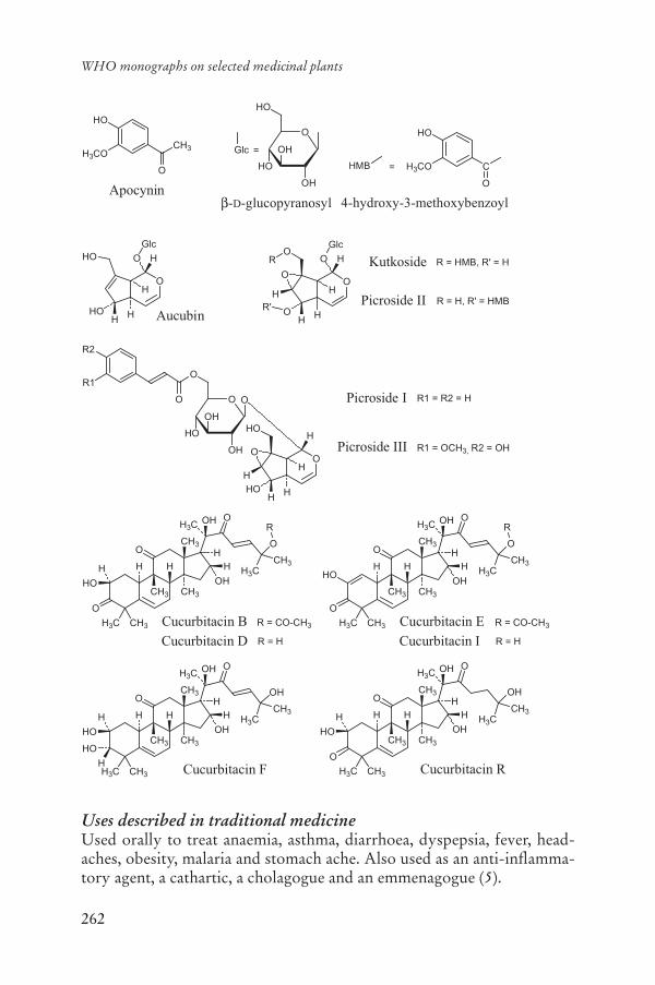
262
WHO monographs on selected medicinal plants
Uses described in traditional medicineUsed orally to treat anaemia, asthma, diarrhoea, dyspepsia, fever, head-aches, obesity, malaria and stomach ache. Also used as an anti-inflamma-tory agent, a cathartic, a cholagogue and an emmenagogue (5).
O
OH
HO
HO
OHApocynin
CH3CO
HO
4-hydroxy-3-methoxybenzoyl-D-glucopyranosyl
HMB =
Glc =
O
CH3H3CO
HO
O
O
H
HO
HOH Aucubin
Kutkoside
Picroside II
HO
H
Glc
R = HMB, R' = H
R = H, R' = HMB
O
HOH
O
O
H
HO
Glc
R'H
R
O
H
H
O
OH
O
HO
OH
O
HOH
HO
O
H
O Picroside I
Picroside IIIH
R2
R1
R1 = R2 = H
R1 = OCH3, R2 = OH
H3CO
H3C CH3
H
CH3
O
O
OH
HH
HOH3C
CH3
OCH3
OH
CH3
H H
R
Cucurbitacin BCucurbitacin D
H3CO
H3C CH3
H
CH3
O
O
OH
H H3CCH3
OCH3
OH
CH3
H H
R
HO
H3CO
H3C CH3
H
CH3
O
OH
HH
HOH3C
CH3
OHCH3
OH
CH3
H H
Cucurbitacin F
HO
H
H3CO
H3C CH3
H
CH3
O
O
OH
HH
HOH3C
CH3
OHCH3
OH
CH3
H H
Cucurbitacin R
R = CO-CH3
R = H
Cucurbitacin ECucurbitacin I
R = CO-CH3
R = H
263
Rhizoma Picrorhizae
PharmacologyExperimental pharmacologyAnti-allergic and anti-asthma activityIntragastric administration of a dose of 25.0 mg/kg body weight (bw) of a standardized iridoid glycoside fraction from an ethanol extract of the rhi-zome, inhibited passive cutaneous anaphylaxis in mice (82%) and rats (50–85%) and protected mast cells from degranulation (60–80%) in a concentration-dependent manner. Its effect was also studied in a sensi-tized guinea-pig ileum preparation in vitro and in normal guinea-pigs in vivo. The Schultz-Dale response was inhibited in sensitized guinea-pig ileum, but histamine-induced bronchospasm was not antagonized or pre-vented by the fraction, indicating the absence of a direct postsynaptic his-tamine receptor-blocking activity (20).
A dried chloroform (0.1 mg/ml) or dried ethyl acetate extract of the rhizome (0.01 mg/ml) inhibited histamine release in human polymorpho-nuclear leukocytes treated with rabbit antihuman IgE antibody, or the cal-cium ionophores A12387 or C5A in vitro (21). Intragastric administration of a dried ethyl acetate extract of the crude drug to guinea-pigs at a dose of 10.0 mg/kg bw 1 hour prior to challenge with platelet-activating factor or administration of ovalbumin reduced bronchial obstruction (21).
Antidiabetic activityIntragastric administration of a dried 75% methanol extract of the rhi-zome (10:1) to rats at a dose of 75.0 mg/kg bw reduced alloxan- or glu-cose-induced hyperglycaemia (22). When administered at a dose of 75.0 mg/kg or 150.0 mg/kg bw, the extract also inhibited the development of alloxan-induced diabetes, and reduced serum peroxide levels and blood urea nitrogen concentration (22).
Antihepatotoxic activityA standardized iridoid glycoside fraction from the rhizome exhibited sig-nificant in vitro activity against toxicity induced by thioacetamide (200 μg/ml), galactosamine (400 μg/ml), and carbon tetrachloride (3 μl/ml) in pri-mary cultured rat hepatocytes (p < 0.01). Incubation of damaged hepato-cytes with the fraction exhibited a concentration-dependent (1–100 μg/ml) curative effect in restoring altered viability parameters (23). Intragastric administration of a 95% ethanol extract of the crude drug to rats at a dose of 0.75–6.0 mg/kg bw for 7 days reduced the levels of glutamate oxalo-acetate transaminase, glutamate pyruvate transaminase, acid phosphatase, glutamate dehydrogenase and bilirubin in animals with paracetamol- or galactosamine-induced hepatotoxicity (24, 25). The level of glycogen in
264
WHO monographs on selected medicinal plants
the liver was also increased in animals treated with the rhizome extract. Intragastric administration of a methanol extract of the crude drug to mice, at a dose of 670.0 mg/kg bw, suppressed carbon tetrachloride-induced hepatotoxicity (26).
Induction of liver injury in 16 mice by injection of carbon tetrachlo-ride (CCl4) for 9 weeks was reduced by the rhizome. Eight animals were fed the crude drug extract (12.0 mg/kg bw) daily, for 10 days before CCl4injection. Control mice (n = 6) were injected with olive oil for the same period. Serum markers of liver injury and histology of liver tissues were studied. Hepatic glutathione, total thiol, glucose-6-phosphate dehydro-genase, catalase, lipid peroxidation and plasma membrane-bound Na+/K+
ATPase were also assessed. Feeding the crude drug extract to CCl4-treat-ed mice reduced serum alanine aminotransferase, aspartate aminotrans-ferase, liver glutathione, catalase and membrane-bound Na+/K+ ATPase. Histological lesions of liver and lipid peroxidation were also significantly fewer in these animals (27).
Intragastric administration of a dried 75% methanol extract of the powdered rhizomes, at a dose of 150.0 mg/kg bw inhibited the induction of N-nitrosodiethylamine-induced hepatocarcinogenesis and prevented lipid peroxide formation as well as reducing the levels of aniline hydrase and -glutamyl transpeptidase (28). Intragastric administration of a 95% ethanol extract of the crude drug at a dose of 12.0–25.0 mg/kg bw for 7 days reduced galactosamine and monocrotaline-induced hepatotoxicity (29, 30). The effect of an iridoid glycoside containing an extract of the rhi-zome on the carcinogenic response and on hepatic and renal antioxidant enzymes of rats treated with 1,2-dimethylhydrazine hydrochloride was investigated (15). 1,2-dimethylhydrazine hydrochloride-induced hepatic carcinogenic response and necrosis were inhibited by oral administration of the extract at doses of 40.0 and 200.0 mg/kg bw. Liver -glutamyl trans-peptidase was reduced to 0.22 ± 0.04 and 0.18 ± 0.03 nmol/mg protein, respectively, by the treatment. Depletion of hepatic and renal antioxidant enzymes such as catalase and superoxide dismutase levels induced by 1,2-dimethylhydrazine hydrochloride was reversed by the treatment, and el-evated lipid peroxidation in liver, kidney and serum was reduced. Renal glutathione S-transferase and hepatic glutathione levels which had been depleted by 1,2-dimethylhydrazine hydrochloride were also increased. Oral administration of a standardized iridoid glycoside fraction of the crude drug (25.0 mg/kg bw daily for 15 days), significantly reduced the increases in the activities of tau-glutamyl transpeptidase, 5´-nucleotidase, acid phosphatase and acid ribonuclease, and decreased the activities of suc-cinate dehydrogenase and glucose-6-phosphatase in liver, and the level of

265
Rhizoma Picrorhizae
transaminases, sorbitol dehydrogenase, glutamate dehydrogenase, lactate dehydrogenase, acid phosphatase, alkaline phosphatase and bilirubin in the serum of aflatoxin B1-treated rats (31).
The mechanism of action of an iridoid glycoside extract of the rhizome on hepatocellular injury and redox status was investigated in a haemor-rhage-resuscitation injury in adult rats (32). Anaesthetized rats were sub-jected to haemorrhagic shock by bleeding 30 ml/kg bw. After 60 minutes of shock, rats were resuscitated with twice the shed blood volume of lac-tated Ringer’s solution and were killed 2 hours after resuscitation. Pre-treatment with the extract (12 mg/kg bw), given orally for 7 days, resulted in a significant decrease in serum aspartate transaminase and -glutamyltranspeptidase levels. The extract also inhibited the lipid peroxidation and nitric oxide release that occurred after haemorrhage-resuscitation and al-tered the activity of glutathione reductase in a favourable manner. The extract down-regulated the stress-sensitive transcription factor activator protein 1 and decreased the level of c-fos mRNA as well as c-jun and c-fos proteins in liver tissue, indicating that its actions could be mediated through AP1 and associated signal transduction pathways (32).
Anti-inflammatory activityIntragastric administration of a 95% ethanol extract of the rhizome to rats, at a dose of 100.0 mg/kg bw for 3 days, reduced carrageenan-induced pedal oedema (32, 33). Co-administration of the anti-inflammatory an-tagonists, propranolol or timolol with the 95% ethanol extract of the rhi-zome reduced its effects.
Choleretic activityIntragastric administration of a glycoside mixture (picroside-1 and kut-koside 1:1.5, isolated from the rhizome) to guinea-pigs, at a dose of 1.5 mg/kg bw, reduced paracetamol-induced cholestasis by 59.2%; at 3 mg/kg bw, treatment reduced ethynylestradiol-induced cholestasis by 77%. At a dose of 6.0 mg/kg bw the same mixture increased bile flow by 120% (34). Intragastric administration of an extract of the crude drug at a dose of 2.0 mg/kg bw increased bile secretion (35). Intragastric adminis-tration of an unspecified extract (no further details available) of the rhi-zome to guinea-pigs at a dose of 6.0 mg/kg bw for 7 days prior to thi- acetamide administration prevented cholestasis and hepatotoxicity (37).
Diuretic activityIntraperitoneal administration of an aqueous ethanol extract (1:1) of the rhi-zome to rats at a dose of 250.0 mg/kg bw increased the amount of urine pro-duced over a 4-hour period, in animals preloaded with saline solution (38).

266
WHO monographs on selected medicinal plants
Immune stimulatory effectsA dried diethyl ether extract of the rhizome inhibited the classical path-way of complement (median inhibitory concentration (IC50) 1.9 μg/ml). Aqueous, petroleum ether, methanol or ethyl acetate extracts of the crude drug had inhibitory concentrations of 13, 17, 38 and 55 μg/ml, respec-tively (3). The diethyl ether extract of the crude drug also had moderate inhibitory activity against the activation of polymorphonuclear cells (IC5053 μg/ml) in vitro, and reduced mitogen-induced proliferation of T lym-phocytes (IC50 13 μg/ml) (3). Two cucurbitacins, picracin and deacetyl-picracin, isolated from the rhizomes of the crude drug were responsible for the inhibition of mitogen-induced human T-lymphocyte proliferation (39). Incubation of picracin and deacetylpicracin with peripheral blood lymphocytes inhibited interleukin-2 release by phytohaemagglutinin- activated T cells. The IC50s were 5 and 16 μM, respectively. Both picracin and cucurbitacin E also inhibited the release of interleukin 1 and tumour necrosis factor from monocytes (IC50s were 15 and 3 μM, respectively). Deacetylpicracin was not active at concentrations up to 100 μM (40).Intraperitoneal injection of picracin or deacetylpicracin, 1 hour prior to induction of delayed-type hypersensitivity significantly (p < 0.001) in-hibited the delayed-type hypersensitivity response at doses of 100.0 mg/kg and 30.0 mg/kg bw in mice (41).
ToxicologyAn aqueous ethanol extract of the rhizome administered intraperitoneally to mice had a median lethal dose of 1.09 g/kg bw, indicating low toxicity (38). A 70% methanol extract of the crude drug administered intragastri-cally to mice exhibited a median lethal dose at > 2 g/kg bw (41). An aque-ous extract of the rhizome did not stimulate cell proliferation in melano-cyte cells in vitro at a concentration of 1 mg/ml (42). The median lethal dose of cucurbitacin B was 0.5 mg/kg bw in rabbits after intravenous ad-ministration and 340 mg/kg bw of cucurbitacin E in mice after oral admin-istration (3). Oral administration of 200 mg/kg bw of an ethanol or diethyl ether extract of the crude drug for 3 or 6 weeks did not increase the weight of the thymus, spleen, liver or mesenterial lymph nodes in mice (3).
Clinical pharmacologyIn one therapeutic observational study, 20 patients with bronchial asthma were treated with a crude extract of the drug (300 mg three times daily) for 1 year. Outcomes measured included clinical improvement, reduction in the use of bronchodilators and pulmonary function tests. Of the 20 pa-tients treated, 10 showed varying degrees of clinical improvement, includ-ing an improvement in pulmonary function tests (17).

267
Rhizoma Picrorhizae
A preliminary clinical trial involving 36 patients with bronchial asthma assessed the efficacy of an extract of the crude drug. Powdered crude drug at a dose of 75 mg twice daily was administered for 2 weeks. The study reported that 53% of patients had no attacks or chest symptoms, 25% had occasional attacks, which were reduced in severity after treatment. Treatment did not improve the condition of patients who smoked or reg-ularly drank alcohol, and was not effective for acute attacks (18).
In a randomized, double-blind placebo-controlled trial involving pa-tients diagnosed with acute viral hepatitis (hepatitis B surface antigen-negative), a powder of the crude drug was administered orally (375 mgthree times a day) for 2 weeks (n = 15) or a matching placebo was given (n = 18). The difference in values of bilirubin, serum glutamic oxaloacetic transaminase and serum glutamic pyruvic transaminase between the group that received the placebo and the group that received the treatment with the crude drug was significant. The time in days required for total serum bilirubin to drop to an average value of 2.5 mg% was 75.9 days in patients who received the placebo versus 27.4 days in the treatment group (19).
Adverse reactionsNo information was found.
ContraindicationsNo information was found.
WarningsNo information was found.
PrecautionsCarcinogenesis, mutagenesis, impairment of fertilityNo information was found.
Pregnancy: teratogenic effectsIntragastric administration of a dried 70% methanol extract of the rhi-zome was not teratogenic in pregnant rats when administered from day 13of pregnancy onwards at doses of 250.0–500.0 mg/kg bw (41).
Pregnancy: non-teratogenic effectsIntragastric administration of a dried 70% methanol extract of the rhi-zome did not induce abortion in pregnant rats when administered from day 13 of pregnancy onwards at doses of 250–500.0 mg/kg bw (41).

268
WHO monographs on selected medicinal plants
Other precautionsNo information was found.
Dosage formsCrude drug, extracts, tablets and tinctures.
Posology(Unless otherwise indicated)Oral daily dose: 1–3 g of the powdered crude drug (1).
ReferencesThe Ayurvedic pharmacopoeia of India1. . Part I, Vol. II, 1st ed. New Delhi, Ministry of Health & Family Welfare, Department of Indian System of Med-icine and Homoeopathy, 1999.Pharmacopoeia of the People’s Republic of China. 2. Beijing, Chemical Indus-try Press, 2005.Smit HF. 3. Picrorhiza scrophulariiflora, from traditional use to immunomodu-latory activity. Utrecht, University of Utrecht, Faculty of Pharmacy, 2000.Medicinal plants of India. Vol. 2.4. New Delhi, Indian Council of Medical Re-search, 1987.Farnsworth NR, ed. 5. NAPRALERT database. Chicago, University of Illinois at Chicago, IL (an online database available directly through the University of Illinois at Chicago or through the Scientific and Technical Network [STN] of Chemical Abstracts Services), 30 June 2005.Nadkarni AK. 6. Dr. K.M. Nadkarni’s Indian materia medica. Bombay, Popu-lar Prakashan, 1976.Rajeshkumar NV, Kuttan R. Modulation of carcinogenic response and anti-7.oxidant enzymes of rats administered with 1,2-dimethylhydrazine by Picro-liv. Cancer Letters, 2003, 191:137–143.Han DR et al. 8. Modern pharmacognosy. Seoul, Hakchang, 1989 [in Korean].Bensky D, Gamble A. 9. Chinese herbal medicine. Materia medica, revised ed. Seattle, WA, Eastland Press, 1993.Keys JD. 10. Chinese herbs: their botany, chemistry, and pharmacodynamics.Tokyo, Charles E. Tuttle, 1976.Sturm S, Stuppner H. Analysis of iridoid glycosides from 11. Picrorhiza kurroaby capillary electrophoresis and high performance liquid chromatography-mass spectrometry. Chromatographia, 2001, 53:612–618.WHO guidelines for assessing quality of herbal medicines with reference to 12.contaminants and residues. Geneva, World Health Organization, 2007.European Pharmacopoeia13. , 5th ed. Strasbourg, Directorate for the Quality of Medicines of the Council of Europe (EDQM), 2005.Guidelines for predicting dietary intake of pesticide residues14. , 2nd rev. ed. Ge-neva, World Health Organization, 1997 (WHO/FSF/FOS/97.7).
269
Rhizoma Picrorhizae
Rajeshkumar NV, Kuttan R. Protective effect of picroliv, the active constitu-15.ent of Picrorhiza kurroa, against chemical carcinogenesis in mice. Teratogen-esis, Carcinogenesis, and Mutagenesis, 2001, 21:303–313.Kim IH et al. 16. Medicinal botany. Seoul, Hakchang, 1988 [in Korean].Yegnenarayan R et al. Study of 17. Picrorrhiza kurrooa (PK 300) in cases of bronchial asthma. Bombay Hospital Journal, 1982, 24:15–18.Rajaram D. A preliminary clinical trial of 18. Picrorhiza kurroa in bronchial asthma. Bombay Hospital Journal, 1976, 18:14–17.Vaidya AB et al. 19. Picrorhiza kurroa (Kutaki) Royle ex Benth as a hepatopro-tective agent – experimental & clinical studies. Journal of Postgraduate Med-icine, 1996, 42:105–108.Baruah CC et al. Anti-allergic and anti-anaphylactic activity of picroliv – a 20.standardised iridoid glycoside fraction of Picrorhiza kurroa. PharmacologyResearch, 1998, 38:487–492.Dorsch W et al. Antiasthmatic effects of 21. Picrorhiza kurroa: Androsin pre-vents allergen- and PAF-induced bronchial obstruction in guinea pigs. Inter-national Archives of Allergy and Applied Immunology, 1991, 95:128–133.Joy KL et al. Effect of 22. Picrorrhiza kurroa extract on transplanted tumours and chemical carcinogenesis in mice. Journal of Ethnopharmacology, 2000, 71:261–266.Visen PK, Saraswat B, Dhawan BN. Curative effect of picroliv on primary 23.cultured rat hepatocytes against different hepatotoxins: an in vitro study. Journal of Pharmacology and Toxicology Methods, 1998, 40:173–179.Ansari RA et al. Antihepatotoxicity properties of Picroliv: an active fraction 24.from rhizomes of Picrorhiza kurroa. Journal of Ethnopharmacology, 1991, 34:61–68.Dwivedi Y et al. Prevention of paracetamol-induced hepatic damage in rats 25.by Picroliv, the standardized active fraction from Picrorhiza kurroa. Phyto-therapy Research, 1991, 5:115–119.Chang IM, Yun HS. Plants with liver-protective activities: pharmacology and 26.toxicology of aucubin. In: Chang HM et al. eds. Advances in Chinese me-dicinal materials research. Singapore, World Scientific Publishing, 1985.Santra A et al. Prevention of carbon tetrachloride-induced hepatic injury in 27.mice by Picrorhiza kurroa. Indian Journal of Gastroenterology, 1998, 17:6–9.Joy KL, Kuttan R. Anti-diabetic activity of 28. Picrorrhiza kurroa extract. Jour-nal of Ethnopharmacology, 1999, 167:143–148.Dwivedi Y et al. Picroliv and its components kutkoside and picroside I pro-29.tect liver against galactosamine-induced damage in rats. Pharmacology & Toxicology, 1992, 71:383–387.Dwivedi Y et al. Picroliv protects against monocrotaline-induced hepatic 30.damage in rats. Pharmacological Research, 1991, 23:399–407.Rastogi R et al. Hepatocurative effect of picroliv and silymarin against afla-31.toxin B1 induced hepatotoxicity in rats. Planta Medica, 2000, 66:709–713.Seth P et al. Picroliv modulates antioxidant status and down-regulates AP1 tran-32.scription factor after hemorrhage and resuscitation. Shock, 2003, 19:169–175.

270
WHO monographs on selected medicinal plants
Pandey BL, Das PK. Immunopharmacological studies on 33. Picrorhiza kurroaRoyle-Ex Benth Part V: Anti-inflammatory action: Relation with cell types involved in inflammation. Indian Journal of Physiology and Pharmacology,1988, 32:289–292.Pandey BL, Das PK. Immunopharmacological studies on 34. Picrorhiza kurroaRoyle-ex Benth Part IV: cellular mechanisms of anti-inflammatory action. Indian Journal of Physiology and Pharmacology, 1989, 33:28–30.Dwivedi Y et al. Picroliv affords protection against thioacetamide-induced 35.hepatic damage in rats. Planta Medica, 1991, 57:25–28.Saraswat B et al. Ex vivo and in vivo investigations of picroliv from 36. Picrorhi-za kurroa in an alcohol intoxication model in rats. Journal of Ethnopharma-cology, 1999, 66:263–269.Shukla B et al. Reversal of thioacetamide induced cholestasis by Picroliv in 37.rodents. Phytotherapy Research, 1992, 6:53–55.Dhar ML et al. Screening of Indian plants for biological activity. Part IV. 38.Indian Journal of Experimental Biology, 1973, 11:43–54.Smit HF et al. Inhibition of T-lymphocyte proliferation by cucurbitacins 39.from Picrorhiza scrophulariaeflora. Journal of Natural Products, 2000, 63:1300–1302.Smit HF et al. Immunomodulatory and anti-inflammatory activity of 40. Picrorhi-za scrophulariaeflora. Journal of Ethnopharmacology, 2000, 73:101–109.Lee EB. Teratogenicity of the extracts of crude drugs. 41. Korean Journal of Pharmacognosy, 1982, 13:116–121.Lin ZX, Hoult JRS, Raman A. Sulphorhodamine B assay for measuring pro-42.liferation of a pigmented melanocyte cell line and its application to the evalu-ation of crude drugs used in the treatment of vitiligo. Journal of Ethnophar-macology, 1999, 66:141–150.

271
Oleum Ricini
DefinitionOleum Ricini is the fixed oil obtained by cold expression from the seeds of Ricinus communis L. (Euphorbiaceae) (1–5).
SynonymsRicinus speciosus Burm., R. viridis Willd. (6).
Vernacular namesAamudamu, aamudamu chettu, aavanak, African coffee tree, agaliva, amanakku, amidamu, amudam, amudamu, andela, ander, andi, angan-tangan, arand, aranda, arash, aril, arundi, audla, avend, awriwra, ayrunkukri, balambaal olyo, balamball, bedanjir, bele ni vavalagi, bherenda, bimázi, bofareira, botareira, carapate, carra-pateira, caster bean plant, caster oil bush, castor, castor-oil plant, castor-oil tree, Cemeiner Wunderbaum, chittmani, ‘dan kwasare, diveli, djarak, djarak djeetoon, djarak kaleeki, endaru, endi, eramudapu, erand, eranda, erendi, eri, erund, fam-pinonoana, franda, gab, gandharva hasthah, harwaa, heran, higuera, higuereta, higuerilla, higuerilla blanca, higuerillo, higuerillo blanco, higuerillo rojo, higuero, himashiyu, himasiyu, ilara, ilarun, jar, kaleeki, kalèkè, karchak, kastalan qajne, kerwa, kesusi, kharwa, kherwa, khirva, khirwa, krapata, kula, kula kula, lahung, lapa-lapa-adete, lara, legezabwende, lepo, lepohina, lepokula, libono, lupono, mahona, masketi, mbono, Mexico weed, mupfure, muriki, noronda, ntoo qaib lab, nyonyo, odagwa, palma Christi, palma de cristo, panchangulam, pei-ma, pomaskwiti, ra’ a kau, red chicken tree, red eagle foot, redh, redhi, ricin, ricini oleum virginale, ricino, ricinus, Rizinus, sadabherenda, tangantangan oil plant, tel-enderu, tobsha, tochem-I-bed-anjir, tomontigi, toto ni vavalagi, ttchakkma, txiv taw dlaav laab, udukaju, unapalan, utouto, vatari, verenda, wetapela’celik, wonder tree, wonderboom, zeitel-kharwaa, zurma (6–16).
Geographical distributionIndigenous to tropical regions of Africa, probably native to Ethiopia, but has been adapted and cultivated in subtropical to temperate areas of the world (6, 8, 14, 17).

272
WHO monographs on selected medicinal plants
DescriptionThe castor-oil plant is either cultivated or is found growing wild in most tropical and warm temperate countries. It varies from an annual, mono-ecious herb in temperate regions to a tree attaining a height of 15 m in some tropical areas. There are hundreds of forms of the plant which vary in size, colour of stem and leaves, leaf markings, branching, size, colour and mark-ings of seeds. Shoots and panicles glaucous. Leaves are large, alternate, petiolate, peltate, palmate, 5–12 lobed, the lobes serrate or dentate, green or reddish. The inflorescence is a raceme of staminate and pistillate flowers, often 30–60 cm high, the pistillate flowers occurring above the staminate ones on the floral axis. The fruit is a 3-celled capsule generally covered with soft spines and dehiscing into 3 cocci, each containing an ovoid albuminous seed. Seeds oblong, smooth, mottled and poisonous (8, 12).
Plant material of interest: fixed oilGeneral appearanceA colourless or pale yellow, clear, viscous oil, miscible with dehydrated ethanol or ether. It is only partly soluble in hexane (a distinction from most other fixed oils). When cooled to 0 °C, it becomes more viscous, and turbidity is gradually formed (1–5).
Organoleptic propertiesOdour: slight, characteristic; taste: initially bland, with a slightly acrid aftertaste (3, 4).
General identity testsPhysicochemical properties (2–4), chemical tests (3, 4), and gas chroma-tography analysis of fatty acids (1).
Purity testsMicrobiologicalTests for specific microorganisms and microbial contamination limits are as described in the WHO guidelines for assessing quality of herbal medi-cines with reference to contaminants and residues (18).
Foreign organic matterTo be established according to national requirements.
Total ashNot applicable.
273
Oleum Ricini
Acid-insoluble ashNot applicable.
Water-soluble extractiveNot applicable.
WaterNot more than 0.3% according to the method of Karl-Fischer (1).
Pesticide residuesThe recommended maximum limit of aldrin and dieldrin is not more than 0.05 mg/kg (1). For other pesticides, see the European pharmacopoeia (1)and the WHO quality control methods for medicinal plant materials (18)and pesticide residues (19).
Heavy metalsFor maximum limits and analysis of heavy metals, consult the WHO guidelines for assessing quality of herbal medicines with reference to con-taminants and residues (18).
Radioactive residuesWhere applicable, consult the WHO guidelines for assessing quality of herbal medicines with reference to contaminants and residues (18).
Other purity testsSpecific gravity d25
25: 0.953–0.965 (1, 3, 4).
Saponification value: 176–187 (3–5).
Acid value: not more than 1.5 (3, 4).
Hydroxyl value: 155–177 (3, 4).
Iodine value: 80–90 (1, 3).
Peroxide value: not less than 150 (1).
Optical rotation: +3.5 to +6.0 (1).
Refractive index: 1.478–1.480 (5).
Chemical assaysFatty acid content: palmitic (2% maximum), stearic (2.5% maximum), oleic and isomers (2.5–6.0%), linoleic (2.5–7.0%), linolenic (2.5% maxi-mum), eicosenoic (1.0% maximum), ricinoleic acid (85–92%) and other fatty acids (maximum) determined by gas chromatography (1).
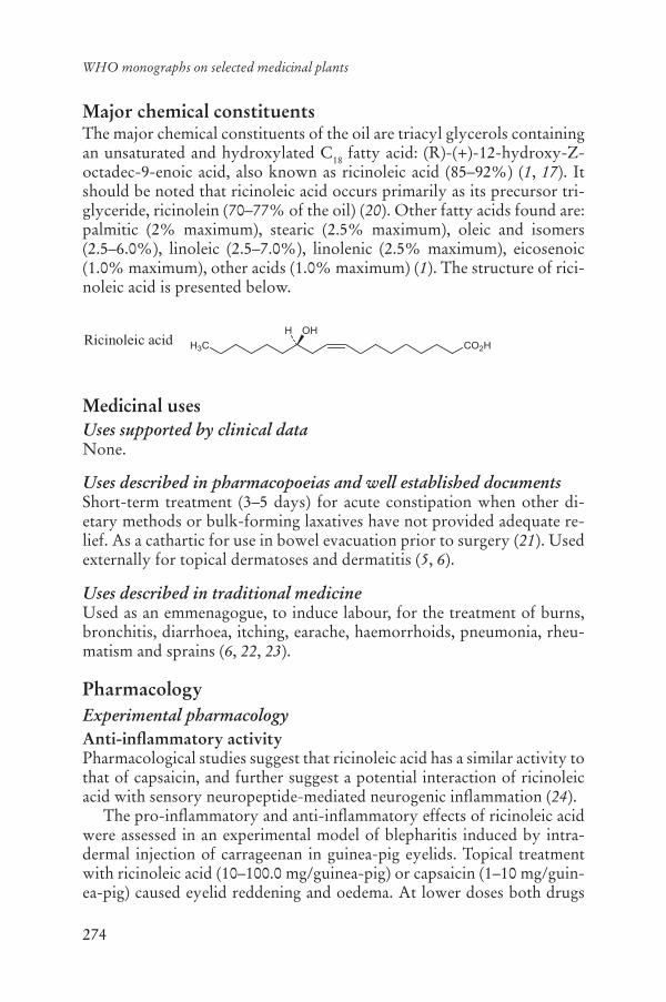
274
WHO monographs on selected medicinal plants
Major chemical constituentsThe major chemical constituents of the oil are triacyl glycerols containing an unsaturated and hydroxylated C18 fatty acid: (R)-(+)-12-hydroxy-Z-octadec-9-enoic acid, also known as ricinoleic acid (85–92%) (1, 17). It should be noted that ricinoleic acid occurs primarily as its precursor tri-glyceride, ricinolein (70–77% of the oil) (20). Other fatty acids found are: palmitic (2% maximum), stearic (2.5% maximum), oleic and isomers (2.5–6.0%), linoleic (2.5–7.0%), linolenic (2.5% maximum), eicosenoic (1.0% maximum), other acids (1.0% maximum) (1). The structure of rici-noleic acid is presented below.
Medicinal usesUses supported by clinical dataNone.
Uses described in pharmacopoeias and well established documentsShort-term treatment (3–5 days) for acute constipation when other di-etary methods or bulk-forming laxatives have not provided adequate re-lief. As a cathartic for use in bowel evacuation prior to surgery (21). Used externally for topical dermatoses and dermatitis (5, 6).
Uses described in traditional medicineUsed as an emmenagogue, to induce labour, for the treatment of burns, bronchitis, diarrhoea, itching, earache, haemorrhoids, pneumonia, rheu-matism and sprains (6, 22, 23).
PharmacologyExperimental pharmacologyAnti-inflammatory activityPharmacological studies suggest that ricinoleic acid has a similar activity to that of capsaicin, and further suggest a potential interaction of ricinoleic acid with sensory neuropeptide-mediated neurogenic inflammation (24).
The pro-inflammatory and anti-inflammatory effects of ricinoleic acid were assessed in an experimental model of blepharitis induced by intra-dermal injection of carrageenan in guinea-pig eyelids. Topical treatment with ricinoleic acid (10–100.0 mg/guinea-pig) or capsaicin (1–10 mg/guin-ea-pig) caused eyelid reddening and oedema. At lower doses both drugs
CO2H
OHH
H3CRicinoleic acid

275
Oleum Ricini
(ricinoleic acid 0.3–3 mg/guinea-pig and capsaicin 0.009–0.09 mg/guinea-pig) significantly potentiated the eyelid oedema induced by carrageenan. The tachykinin NK1 receptor antagonist FK 888 (0.59 mg/kg subcutane-ously) reduced carrageenan-induced eyelid oedema induced by either rici-noleic acid or capsaicin. Intravenous administration of the neutral endo-peptidase inhibitor, thiorphan (1.3 mg/kg body weight (bw)) significantly enhanced carrageenan-induced eyelid oedema produced by ricinoleic acid. However, repeated topical application of ricinoleic acid (0.9 mg/guinea-pig) or capsaicin (0.09 mg/guinea-pig) for 8 days inhibited carrageenan-induced eyelid oedema. This anti-inflammatory effect was accompanied by a reduction in the tachykinin content of the eyelids, as determined by radioimmunoassay. Ricinoleic acid, like capsaicin, appears to possess dual pro-inflammatory and anti-inflammatory properties; these are observed upon acute and repeated application, respectively (24).
A study comparing the anti-inflammatory activities of ricinoleic acid with those of capsaicin in several models of acute and subchronic inflam-mation was performed. Acute inflammation was induced by intradermal injection of carrageenan in mice or by histamine in the guinea-pig eyelid. Subchronic oedema was induced by injection of complete Freund’s adju-vant into the right ventral paw of mice. Tissue substance P was measured in the carrageenan experiments by radioimmunoassay. Acute topical ap-plication of ricinoleic acid (0.9 mg/mouse) or capsaicin (0.09 mg/mouse) significantly increased the mouse paw oedema induced by carrageenan, while repeated topical treatment with the same doses of both compounds for 8 days resulted in a marked inhibition of carrageenan-induced paw oe-dema matched by a reduction in levels of substance P in tissue. Similar ef-fects were found against histamine-induced eyelid oedema in guinea-pigs after acute or repeated application of ricinoleic acid or capsaicin. Ricino-leic acid and capsaicin given for 1–3 weeks reduced the established oedema induced by Freund’s adjuvant, a sub-chronic model of inflammation (25).
Induction of labourAn investigation exploring the effect of a diet including castor oil on the initiation of labour of pregnant rats was performed. The time of the ini-tiation of labour and course of delivery were observed in rats that received castor oil by gavage on days 18, 19 and 20 of gestation. The diet contain-ing castor oil induced the initiation of labour and shortened the course of the delivery in rats. Ricinoleic acid was the active component of castor oil in this study (26).
The effects of a diet containing castor oil on the synthesis of prosta-glandin E2 and the mechanism of labour induction were investigated in pregnant rats fed a diet containing castor oil on days 18 and 19 of gesta-

276
WHO monographs on selected medicinal plants
tion. At the time of death (day 20), blood from portal and peripheral veins, and samples from intestinal mucosa, amnion, amniotic cells and placenta were collected, and the tissues were cultured in the presence of ricinoleic acid or indometacin. The concentrations of prostaglandin E2 in the media or blood were measured by radioimmunoassay methods. The prostaglan-din E2 levels in the portal vein increased; the prostaglandin E2 levels in peripheral blood showed no significant changes; the prostaglandin E2 lev-els in the tissues of the intestinal mucosa, placenta, amnion and amniotic cells were increased. Ricinoleic acid stimulated the synthesis of prosta-glandin E2 in the tissues in vitro (27).
ToxicologyToxicity studies on castor oil have been performed by incorporating the oil into feed rations at concentrations as high as 10% of the diet. Animals, F344/N rats and B6C3F1 mice, of both sexes were fed the diet for 13 weeks. Exposure to castor oil at dietary concentrations as high as 10% in 13-week studies did not affect survival or body weight gains of rats or mice (10 per sex and dose). No biologically significant effects were noted in haematological analyses in rats. Small increases in total bile acids and in serum alkaline phosphatase were noted at various times during the studies in rats receiving the higher dietary concentrations of castor oil. Liver weights were increased in male rats receiving the 10% dietary concentra-tion and in male and female mice receiving diets containing 5% or 10% castor oil. However, no histopathological lesions were associated with these liver changes, nor were there any compound-related morphological changes in any organ in rats or mice. Thus, no significant adverse effects of castor oil administration were noted (28).
Epithelial cells, isolated from small intestine of hamsters, were used to measure in vitro cytotoxicity of the crude drug. The cytotoxicity of rici-noleic acid (castor oil) was assessed by:
— exclusion of trypan blue;— release of intracellular (prelabelled) 51Cr; and— inhibition of cellular uptake of 3-O-methylglucose.
Ricinoleate produced weak but dose-dependent (0.1–2.0 mM) cyto-toxicity as assessed by all three methods (29).
In an in vivo study, segments of rabbit ileum were examined by scan-ning electron microscopy after exposure to various compounds known to stimulate fluid secretion in the small intestine. After perfusion with rici-noleate (castor oil) at a concentration of 10 mM, striking changes were observed at villus tips and on the apicolateral surfaces of villi; erosions of the tips were confirmed by light microscopy of the same pieces of tissue
277
Oleum Ricini
examined by scanning electron microscopy. The changes that appeared after treatment with ricinoleate were partially reversed during perfusion with control buffer for 2 hours (30).
Clinical pharmacologyLaxative effectCastor oil is an anionic surfactant laxative. Its mechanism of action is pri-marily as a stool-wetting and stool-softening agent that allows the mixing of water, lipids and other faecal material by altering intestinal permeabil-ity and increasing net water and electrolyte secretions. The triglyceride of ricinoleic acid in castor oil is hydrolysed within the small intestine by pancreatic lipases to ricinoleic acid and glycerol. Ricinoleate acts as a local irritant that increases production of cyclic adenosine monophosphate re-sulting in extensive electrolyte secretion in the small intestine by reducing net absorption of fluid and electrolytes, which ultimately stimulates in-testinal peristalsis. Because ricinoleate acts in the small intestine, accumu-lation of fluid and evacuation takes place rapidly within 1–6 hours, and it continues until the compound is excreted via the colon. Colonic empty-ing is so complete that several days may elapse before a normal bowel movement occurs (21).
To examine the effects of oleic acid and ricinoleic acid on jejunal ab-sorption, steady-state jejunal perfusions were done in healthy volunteers. Taurocholate, used to solubilize the fatty acids, did not influence absorp-tion. Both fatty acids, at a concentration of 10 mM, reversed electrolyte and water net movement and induced reversible fluid secretion. Ricino-leic acid (the active principle of castor oil) was the more potent compound. However, ricinoleic acid was absorbed more slowly than oleic acid, and was associated with higher intraluminal concentrations (31).
A retrospective study was performed in patients who underwent elec-tive surgery for colorectal carcinomas. The patients were divided into two groups according to the method of colonic cleansing that was used. Group 1 (n = 154) used the traditional bowel preparation which included 30.0 ml castor oil given orally on the day before the operation, in addi-tion to three soap enemas. Patients in group 2 (n = 36) were given 500 ml 10% mannitol on the day before the operation. Infections due to wounds developed in 26 patients from group 1 versus 13 patients from group 2. The difference was statistically significant (16.9% versus 36.1%, p < 0.01). The differences in the incidence of anastomotic leaks and mortality rate between the two groups were not statistically significant. Castor-oil treatment reduced the number of postoperative infectious wound com-plications (32).
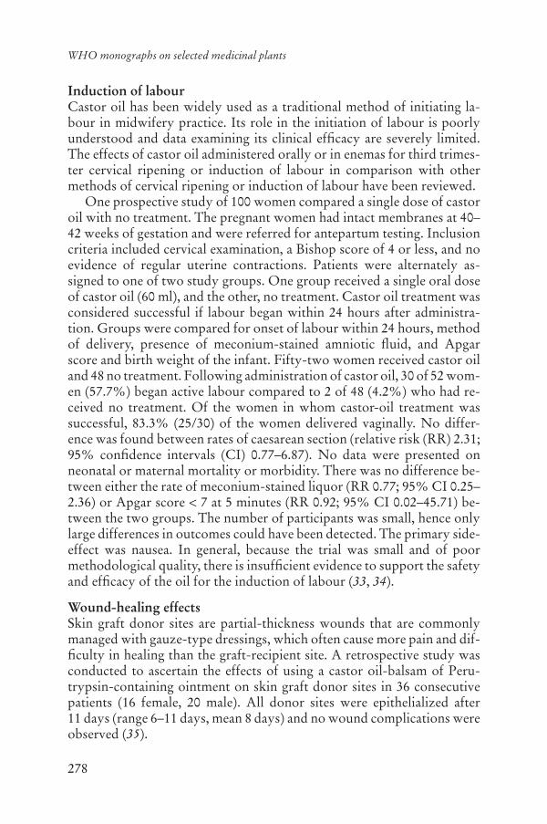
278
WHO monographs on selected medicinal plants
Induction of labourCastor oil has been widely used as a traditional method of initiating la-bour in midwifery practice. Its role in the initiation of labour is poorly understood and data examining its clinical efficacy are severely limited. The effects of castor oil administered orally or in enemas for third trimes-ter cervical ripening or induction of labour in comparison with other methods of cervical ripening or induction of labour have been reviewed.
One prospective study of 100 women compared a single dose of castor oil with no treatment. The pregnant women had intact membranes at 40–42 weeks of gestation and were referred for antepartum testing. Inclusion criteria included cervical examination, a Bishop score of 4 or less, and no evidence of regular uterine contractions. Patients were alternately as-signed to one of two study groups. One group received a single oral dose of castor oil (60 ml), and the other, no treatment. Castor oil treatment was considered successful if labour began within 24 hours after administra-tion. Groups were compared for onset of labour within 24 hours, method of delivery, presence of meconium-stained amniotic fluid, and Apgar score and birth weight of the infant. Fifty-two women received castor oil and 48 no treatment. Following administration of castor oil, 30 of 52 wom-en (57.7%) began active labour compared to 2 of 48 (4.2%) who had re-ceived no treatment. Of the women in whom castor-oil treatment was successful, 83.3% (25/30) of the women delivered vaginally. No differ-ence was found between rates of caesarean section (relative risk (RR) 2.31; 95% confidence intervals (CI) 0.77–6.87). No data were presented on neonatal or maternal mortality or morbidity. There was no difference be-tween either the rate of meconium-stained liquor (RR 0.77; 95% CI 0.25–2.36) or Apgar score < 7 at 5 minutes (RR 0.92; 95% CI 0.02–45.71) be-tween the two groups. The number of participants was small, hence only large differences in outcomes could have been detected. The primary side-effect was nausea. In general, because the trial was small and of poor methodological quality, there is insufficient evidence to support the safety and efficacy of the oil for the induction of labour (33, 34).
Wound-healing effectsSkin graft donor sites are partial-thickness wounds that are commonly managed with gauze-type dressings, which often cause more pain and dif-ficulty in healing than the graft-recipient site. A retrospective study was conducted to ascertain the effects of using a castor oil-balsam of Peru-trypsin-containing ointment on skin graft donor sites in 36 consecutive patients (16 female, 20 male). All donor sites were epithelialized after 11 days (range 6–11 days, mean 8 days) and no wound complications were observed (35).

279
Oleum Ricini
Adverse reactionsExanthema may occur. In this case, treatment with the oil should be dis-continued and should not be used again. Furthermore, castor oil may cause stomach upset, and at higher doses nausea, vomiting, painful ab-dominal cramps and severe diarrhoea with loss of water and electrolytes. In this case it is necessary to reduce the dosage or to discontinue use of the oil. Continuous use and misuse may increase fluid and electrolyte loss, requiring intravenous rehydration and replacement of electrolytes. Ex-cessive potassium loss may lead to disorders of heart function and muscle weakness (21).
A 26-year-old woman, who had had recurrent vulvo-vaginitis for 1 year, was treated with a topical pessary of meclocycline sulfosalicylate. After a 7-day treatment period, the following symptoms appeared: oede-ma of the labia, widespread irregular abdominal dermatitis in the right iliac fossa and numerous generalized small weals. These symptoms were treated with dexchlorpheniramine maleate without results and it was nec-essary to prescribe prednisone tablets. Patch tests at 4, 12, 24, 48 and 96 h found a positive response to hydrogenated castor oil, with associated ery-thema, infiltration and papules after 48 h.
There have been several reports of allergy to castor beans from which castor oil is derived. Cases of asthma have been reported not only in employees in the oil industry, but also in seamen and laboratory work-ers exposed to the beans. Three important allergens from the crude drug have been identified: a 2S storage albumin of 11 kDa, a 11S crystalloid protein with bands at 50 kDa and a protein doublet of 47 and 51 kDa. These allergens have been named Ric c 1, Ric c 2 and allergen 3. Castor oil extractors, fertilizer workers and farmers may acquire occupational dermatitis from handling plants or beans. Castor oil may be used in cos-metics, particularly lipsticks, because of its superior moisturizing char-acteristics, and may rarely produce allergic contact dermatitis. Sporadic case-reports of allergy have implicated ricinoleic acid as the causative agent (36).
A woman at 39 weeks gestation who had had a previous cesarean de-livery had severe abdominal pains and rupture of the membranes shortly after ingesting 5 ml of castor oil. Forty-five minutes later, repetitive vari-able decelerations prompted a cesarean delivery. During surgery, a por-tion of the umbilical cord was found to be protruding from a 2-cm rup-ture of the lower transverse scar (37).
Contact dermatitis and a number of cases of chelitis have also been reported following exposure of patients to the crude drug (38, 39).

280
WHO monographs on selected medicinal plants
ContraindicationsOleum Ricini should not be used as a laxative during pregnancy or while breastfeeding (5, 13), or in children under the age of 12 years.
Due to stimulation of bile flow, the oil should not be used by patients with biliary tract obstructions or other biliary disorders.
Oleum Ricini is contraindicated in patients with hypersensitivity or allergy to the oil, appendicitis, chronic inflammatory bowel disorders, undiagnosed abdominal pain or severe dehydration associated with the loss of salt and water.
It should not be used in cases of intestinal blockage and ileus.
WarningsInduction of labour: use of the crude drug in pregnancy (after 40 weeks of gestation) requires the supervision of a physician, midwife or other expe-rienced health care professional.
As a cathartic: intake of cathartic drugs for more than a few days may lead to a worsening of intestinal hypomotility. Do not take for more than 3–5 days without consulting a health care professional. Overdose may lead to nausea, vomiting, painful abdominal cramps and severe diarrhoea with loss of water and electrolytes. In case of overdose, a health care pro-fessional should be consulted regarding possible replacement of fluid and electrolytes.
PrecautionsDrug interactionsConcomitant use of castor oil with cardiac glycosides, antiarrhythmic drugs, diuretics, cortisol, liquorice, antihistamines and fat-soluble vita-mins may reduce the efficacy of these drugs or, in the case of cardiac gly-cosides, may increase the risk of adverse events due to fluid and electro-lyte losses.
Carcinogenesis, mutagenesis, impairment of fertilityGenetic toxicity studies were negative for induction of mutations in the Ames test using Salmonella typhimurium for induction of sister chroma-tid exchanges or chromosomal aberrations in Chinese hamster ovary cells, and for induction of micronuclei in the peripheral blood erythrocytes of mice fed a diet containing 10% seed oil for 13 weeks (28). No significant changes were noted in a screening study for male reproductive end-points, including sperm count and motility, and no changes were observed in the
281
Oleum Ricini
length of estrous cycles of rats or mice given diets containing castor oil at a concentration of up to 10% (28).
Pregnancy: non-teratogenic effectsOleum Ricini as a laxative is contraindicated in pregnancy before 40 weeks of gestation, due to potential induction of labour.
Nursing mothersOleum Ricini is contraindicated in breastfeeding mothers. Ricinoleic acid is absorbed into the bloodstream and is excreted into human breast milk. In suckling infants it has a purgative effect (13).
Paediatric useSee Contraindications.
Other precautionsNo information was found.
Dosage formsFixed oil, capsules (8).
StoragePreserve in well-closed containers, protected from light.
Posology(Unless otherwise indicated)As a laxative: single daily dose of 1–10 ml (40). Dose for induction of la-bour: under medical supervision, maximum single dose of 4–60 ml (33,34). Topical use: fixed oil (5).
ReferencesEuropean Pharmacopoeia1. , 5th ed. Strasbourg, Directorate for the Quality of Medicines of the Council of Europe (EDQM), 2005.The United States pharmacopeia,2. 25th rev. Rockville, MD, United States Pharmacopeia Convention, 2005.The Japanese pharmacopoeia3. , 14th ed. (English ed.). Tokyo, Ministry of Health and Welfare, 2001 (available at: http://jpdb.nihs.go.jp/jp14e/).The Korean Pharmacopoeia4. , 8th ed. (English ed.). Seoul, Korea Food and Drug Administration, 2004.
282
WHO monographs on selected medicinal plants
Pharmacopoeia of the People’s Republic of China5. . Vol. 1 (English ed). Guang-zhou, Guangdong Science and Technology Press, 2005.Iwu MM. 6. Handbook of African medicinal plants. Boca Raton, FL, CRC Press, 1993.African pharmacopoeia7. . Vol. 1, 1st ed. Lagos, Nigeria, Organization of Afri-can Unity, Scientific Technical & Research Commission, 1985.Youngken HW. 8. Textbook of pharmacognosy, 6th ed. Philadelphia, PA, Blak-iston, 1950.Medicinal plants in Thailand.9. Vol. I. Bangkok, Department of Pharmaceuti-cal Botany, Faculty of Pharmacy, Mahidol University, 1996.Ochse JJ, van den Brink RCB. 10. Vegetables of the Dutch East Indies (English ed.). Amsterdam, A. Asher, 1977.Medicinal plants in the South Pacific.11. Manila, World Health Organization Regional Office for the Western Pacific, 1998 (WHO Regional Publications, Western Pacific Series, No. 19).Medicinal plants in China12. . Manila, World Health Organization Regional Of-fice for the Western Pacific, 1989 (WHO Regional Publications, Western Pa-cific Series, No. 2).Nadkarni AK, ed. 13. Dr. K.M. Nadkarni’s Indian materia medica. Vol. 1. Bom-bay, Popular Prakshan, 1976.Kariyone T, Koiso R. 14. Atlas of medicinal plants. Osaka, Nihon Rinshosha, 1973.Bedevian AK. 15. Illustrated polyglottic dictionary of plant names. Cairo, Med-bouly Library, 1994.Anonymous.16. The review of natural products. 2nd ed. St. Louis, Facts and Comparisons, 2002.Bruneton J. 17. Pharmacognosy, phytochemistry, medicinal plants. Paris, Lavoisi-er, 1995.WHO guidelines for assessing quality of herbal medicines with reference to 18.contaminants and residues. Geneva, World Health Organization, 2007.Guidelines for predicting dietary intake of pesticide residues19. , 2nd rev. ed. Ge-neva, World Health Organization, 1997 (WHO/FSF/FOS/97.7).Hagers Handbuch der Drogen20. (CD ROM). Heidelberg, Springer Verlag, 2003 [in German].Molinoff PB et al., eds.21. Goodman & Gilman’s The pharmacological basis of therapeutics, 9th ed. New York, McGraw-Hill, 1996.Neuwinger HD. 22. Ricinus communis L. (Euphorbiaceae) in der Afrikanischen traditionellen Medizin. Drogenreport, 2003, 30:89–92.Scarpa A, Guerci A. Various uses of the castor oil plant (23. Ricinus communisL.). A review. Journal of Ethnopharmacology, 1982, 5:117–137.Vieira C et al. Pro- and anti-inflammatory actions of ricinoleic acid: similari-24.ties and differences with capsaicin. Naunyn-Schmiedeberg’s Archives of Pharmacology, 2001, 364:87–95.Vieira C et al. Effect of ricinoleic acid in acute and subchronic experimental 25.models of inflammation. Mediators of Inflammation, 2000, 9:223–228.

283
Oleum Ricini
Gao J et al. [Effect of castor oil-diet on the initiation of labor of pregnant rat.] 26.Zhongguo Yi Xue Ke Xue Yuan Xue Bao, 1998, 20:367–370 [in Chinese].Gao J, Sun N, Wang F. [Effects of castor oil-diet on the synthesis of prosta-27.glandin E2 in pregnant rats.] Zhonghua Fu Chan Ke Za Zhi, 1999, 34:147–149 [in Chinese].Irwin R. NTP technical report on the toxicity studies of castor oil (CAS No. 28.8001-79-4) in F344/N rats and B6C3F1 mice (dosed feed studies). Toxic Re-port Series, 1982, 12:1-B5.Gaginella TS et al. Cytotoxicity of ricinoleic acid (castor oil) and other intes-29.tinal secretagogues on isolated intestinal epithelial cells. Journal of Pharma-cology and Experimental Therapeutics, 1977, 201:259–266.Gaginella TS, Lewis JC, Phillips SF. Rabbit ileal mucosa exposed to fatty 30.acids, bile acids, and other secretagogues. Scanning electron microscopic ap-pearances. American Journal of Digestive Diseases, 1977, 22:781–790.Ammon HV, Thomas PJ, Phillips SF. Effects of oleic and ricinoleic acids on 31.net jejunal water and electrolyte movement. Perfusion studies in man. Jour-nal of Clinical Investigation, 1974, 53:374–379.Todorov AT, Mantchev ID, Atanasov TB. Traditional bowel preparation ver-32.sus osmotic agent mannitol for preoperative colonic cleansing in elective colorectal surgery. Folia Medica (Plovdiv), 2002, 44:36–39.Garry D et al. Use of castor oil in pregnancies at term. 33. Alternative Therapies in Health & Medicine, 2000, 6:77–79.Kelly AJ, Kavanagh J, Thomas J. Castor oil, bath and/or enema for cervical 34.priming and induction of labour. Cochrane Database Systematic Review,2001, 2:CD003099.Carson SN et al. Using a castor oil-balsam of Peru-trypsin ointment to assist in 35.healing skin graft donor sites. Ostomy Wound Management, 2003, 49:60–64.Di Berardino L, Della Torre F. Side effects to castor oil. 36. Allergy, 2003, 58:826.Sicuranza GB, Figueroa R. Uterine rupture associated with castor oil inges-37.tion. Journal of Maternal, Fetal and Neonatal Medicine, 2003, 13:133–134.le Coz CJ, Ball C. Recurrent allergic contact dermatitis and cheilitis due to 38.castor oil. Contact Dermatitis, 2000, 42:114–115.Fisher AA. Allergic cheilitis due to castor oil in lipstick. 39. Cutis, 1991, 47:389–390.Blumenthal M et al., eds. 40. The complete German Commission E monographs.Austin, TX, American Botanical Council, 1998.
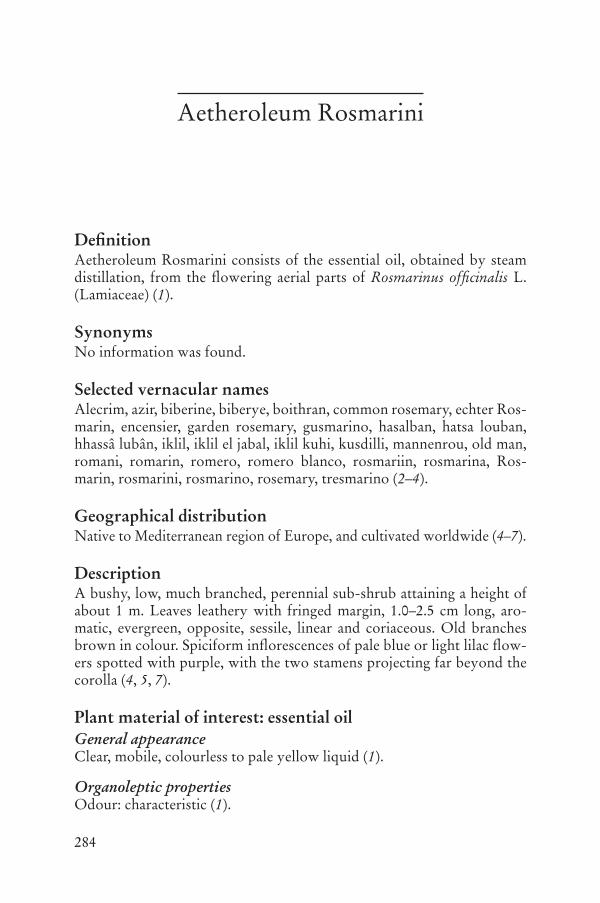
284
Aetheroleum Rosmarini
DefinitionAetheroleum Rosmarini consists of the essential oil, obtained by steam distillation, from the flowering aerial parts of Rosmarinus officinalis L. (Lamiaceae) (1).
SynonymsNo information was found.
Selected vernacular namesAlecrim, azir, biberine, biberye, boithran, common rosemary, echter Ros-marin, encensier, garden rosemary, gusmarino, hasalban, hatsa louban, hhassâ lubân, iklil, iklil el jabal, iklil kuhi, kusdilli, mannenrou, old man, romani, romarin, romero, romero blanco, rosmariin, rosmarina, Ros-marin, rosmarini, rosmarino, rosemary, tresmarino (2–4).
Geographical distributionNative to Mediterranean region of Europe, and cultivated worldwide (4–7).
DescriptionA bushy, low, much branched, perennial sub-shrub attaining a height of about 1 m. Leaves leathery with fringed margin, 1.0–2.5 cm long, aro-matic, evergreen, opposite, sessile, linear and coriaceous. Old branches brown in colour. Spiciform inflorescences of pale blue or light lilac flow-ers spotted with purple, with the two stamens projecting far beyond the corolla (4, 5, 7).
Plant material of interest: essential oilGeneral appearanceClear, mobile, colourless to pale yellow liquid (1).
Organoleptic propertiesOdour: characteristic (1).

285
Aetheroleum Rosmarini
Microscopic characteristicsNot applicable.
Powdered plant materialNot applicable.
General identity testsPhysicochemical properties, thin-layer and gas chromatography (1).
Purity testsMicrobiologicalNot applicable.
Foreign organic matterTo be established in accordance with national requirements.
Total ashNot applicable.
Acid-insoluble ashNot applicable.
Water-soluble extractiveNot applicable.
Loss on dryingNot applicable.
Pesticide residuesThe recommended maximum limit of aldrin and dieldrin is not more than 0.05 mg/kg (1). For other pesticides, see the European pharmacopoeia (1)and the WHO guidelines for assessing quality of herbal medicines with reference to contaminants and residues (8) and pesticide residues (9).
Heavy metalsFor maximum limits and analysis of heavy metals, consult the WHO guidelines for assessing quality of herbal medicines with reference to con-taminants and residues (8).
Radioactive residuesWhere applicable, consult the WHO guidelines for assessing quality of herbal medicines with reference to contaminants and residues (8).

286
WHO monographs on selected medicinal plants
Other purity testsRelative density: 0.895–0.920 (1).Refractive index: 1.464–1.473 (1).Optical rotation: -5° to + 8° (1).Acid value: not more than 1.0 (1).
Chemical assaysGas chromatographic analysis of Spanish type rosemary oil: -pinene(18–26%), camphene (8–12%), -pinene (2.0–6.0%), -myrcene (1.5–5.0%), limonene (2.5–5.0%), 1,8-cineol (16.0–25.0%), p-cymene (1.0–2.2%), camphor (13.0–21.0%), bornyl acetate (0.5–2.5%), -terpineol(1.0–3.5%), borneol (2.0–4.5%) and verbenone (0.7–2.5%) (1).
The oil of rosemary from Morocco and Tunisia contains: -pinene(9–14%), camphene (2.5–6.0%), -pinene (4.0–9.0%), -myrcene (1.0–2.0%), limonene (1.5–4.0%), 1,8-cineol (38.0–55.0%), p-cymene (0.8–2.5%), camphor (5.0–15.0%), bornyl acetate (0.1–1.5%), -terpineol(1.0–2.6%), borneol (1.5–5.0%) and verbenone (0.4%) (1).
Major chemical constituentsThe chief constituents of rosemary oil are camphor (5–31%), 1,8-cineol (15–55%), -pinene (9–26%), borneol (1.5–5.0%), camphene (2.5–12.0%),
-pinene (2.0–9.0%), limonene (1.5–5.0%), verbenone (2.2–11.1%), -caryophyllene (1.8–5.1%) and myrcene (0.9–4.5%) (1, 10, 11). The
structures of 1,8-cineole, borneol and camphor are presented below.
Medicinal usesUses supported by clinical dataNone.
Uses described in pharmacopoeias and well established documentsUsed orally for the treatment of dyspeptic complaints, and in external ap-plications for supportive management of rheumatic complaints and circu-latory disorders (12, 13). Although one pilot study has indicated that the
H3C
H3C
CH3
O
H
H3C
H3C
CH3
H
OH
H
and enantiomer and enantiomer
Borneol Camphor
O
CH3
CH3
CH3
1,8-Cineole

287
Aetheroleum Rosmarini
crude drug may enhance cognition (13), further data from randomized controlled clinical trials are required before any therapeutic recommen-dations can be made.
Uses described in traditional medicineUsed as a cholagogue, diaphoretic, digestant, diuretic, emmenagogue, laxative and a tonic (3, 5, 6). Also used in the management of headache, menstrual disorders, nervous menstrual complaints, tiredness, defective memory, sprains and bruises (14).
PharmacologyExperimental pharmacologyAntihepatotoxic activityThe hepatoprotective and antimutagenic effects of the essential oil were compared to those of ethanol leaf extracts in vivo using carbon tetra- chloride (CCl4) and cyclophosphamide as the hepatotoxic and mutagenic compounds. The results of the study showed that intragastric administra-tion of 1.5 g/kg body weight (bw) of the essential oil or the leaf extract to rats for 3 weeks protected the animals against CCl4-induced hepatotoxic-ity. The results were similar to those obtained using the control com-pound, silymarin (15). The results showed amelioration of most of the serum and liver parameters studied, and were confirmed by histopatho-logical examination of the liver tissue. The hepatoprotectant activity of essential oil was not as great as that of the leaf extract. However, intragas-tric pretreatment of mice for 7 days with the essential oil (1.1 mg/g bw), reduced cyclophosphamide-induced mitodepression in bone marrow cells (15).
Antimicrobial activityThe essential oil weakly inhibited the growth of Acinetobacter iwoffi, Ba-cillus subtilis, Erwinia carotovora, Shigella flexneri, Staphylococcus aureus,Streptococcus pyogenes and Yersinia enterocolitica when added undiluted or in a 1:1 dilution to the agar medium (16).
Antispasmodic effectsThe essential oil inhibited electrically stimulated muscular contractions in isolated guinea-pig ileum, but the effect of the lowest spasmolytic dose is preceded by an initial increase in the electrically stimulated contractile response. Pinene, which had weak spasmogenic properties, was consid-ered to be the active constituent (17). In guinea-pig ileum, addition of the essential oil to the bath medium inhibited acetyl choline-induced contrac-

288
WHO monographs on selected medicinal plants
tions, with a median inhibitory concentration (IC50) of 465 nl/ml of the oil and 41 nl/ml of 1,8-cineole (18).
Osteoclastic effectsThe essential oil, and its individual constituents, camphor, borneol, thy-mol, -pinene, -pinene and bornyl acetate, inhibited bone resorption when added to the food of rats. The monoterpenes borneol, thymol and camphor, were directly inhibitory in the osteoclast resorption pit assay. Within 30 minutes, borneol inhibited the formation of actin rings, a char-acteristic of resorbing osteoclasts indicating cell polarization. Both the in vitro and the in vivo effects of borneol were reversible (19).
Enzyme inductionModulation of cytochrome P450 isozymes and detoxification enzymes was compared after oral administration of the dried leaves, a dried dichlo-romethane leaf extract or the essential oil to rats. The animals received the leaves, extracts or essential oil in their rations at a concentration of 0.5% (w/w) for 2 weeks. The effects of the treatments were evaluated in assays for cytochrome P450 isozymes 1A, 2B, 2E1, glutathione S-transferase, nicotinamide adenine dinucleotide phosphate (NAD(P)H), quinone re-ductase and uridine diphosphate (UDP)-glucuronosyltransferase activi-ties and on protein levels (immunoblot analyses). The results demonstrat-ed that the essential oil selectively induced cytochrome P450, particularly isozyme 2B. The leaf extract enhanced both cytochrome P450 and de-toxification enzymes. A dichloromethane extract of the leaves acted as a monofunctional inducer, inducing glutathione S-transferase, quinone re-ductase and UDP-glucuronosyltransferase, in particular UDP-glucuro-nosyltransferase 1A6 (20).
ToxicologyThe embryotoxic effects of d-camphor were investigated in rats and rab-bits after intragastric administration for the treatment of hypotonic circu-latory dysregulations. No evidence of teratogenicity was observed when d-camphor was administered orally to pregnant rats during the fetal peri-od of organogenesis, at doses up to 1 g/kg bw per day, and to pregnant rabbits at doses up to 681 mg/kg bw per day. For rats, the no-observed-effect level for the fetal organism was above 1 mg/kg bw, and for rabbits, above 681 mg/kg bw. In rat dams a dose-dependent reduction in food in-take and salivation was noted at doses of 464 mg/kg bw and higher. The high dose of 1 mg/kg bw per day resulted in pronounced signs of toxicity such as clonic convulsion, pilo-erection, reduced motility and body weight gain. In rabbit dams, intragastric administration of a high dose (681 mg/kg
289
Aetheroleum Rosmarini
bw per day) resulted in reduced body weight gain and food consumption. No increased incidence of variations, retardations or malformations was observed in any of the offspring at any of the doses, not even at the highest dose tested (rat: 1000 mg/kg bw per day; rabbit: 681 mg/kg bw per day). The daily maximum dose of camphor for human therapeutic use is ap-proximately 1.43 mg/kg bw. Hence, under the present test conditions the therapeutic ratio is above 450 for the end-point of embryotoxicity, reflect-ing a wide margin of safety (21). Since d-camphor is only one of the chem-ical constituents of the essential oil, the relevance of these data to the toxi-cology of the essential oil needs to be investigated.
Clinical pharmacologyA clinical study to assess the olfactory impact of the essential oils of laven-der (Lavandula angustifolia) and rosemary (Rosmarinus officinalis) on cognitive performance and mood in healthy volunteers was performed (13). One hundred and forty-four participants were randomly assigned to one of three independent groups. Each of the subjects was asked to take the Cognitive Drug Research (CDR) computerized cognitive assessment bat-tery in a cubicle containing one of the two odours or no odour (control). Visual analogue mood questionnaires were completed before exposure to the odour and after completion of the test battery. The participants were misled as to the genuine aim of the study until the completion of testing to prevent expectancy effects from possibly influencing the data. The out-come variables from the nine tasks that constitute the CDR core battery feed into six factors that represent different aspects of cognitive function-ing. Analysis of performance revealed that lavender produced a significant decrement in performance of working memory, and impaired reaction times for both memory and attention-based tasks compared to the perfor-mance of the controls. In contrast, rosemary produced a significant en-hancement of performance for overall quality of memory and secondary memory factors, but also impaired speed of memory compared to controls. With regard to mood, comparisons of the change in ratings from baseline to post-test revealed that following the completion of the cognitive assess-ment battery, both the control group and the group exposed to lavender were significantly less alert than the subjects exposed to rosemary; how-ever, subjects in the control group were significantly less content than sub-jects who received the rosemary and lavender treatments (13).
Frontal electroencephalogram asymmetry shifting from baseline was examined in adults and infants exposed to lavender and rosemary oils by re-analysing previously published data from two studies. The results from 39 adults revealed significant electroencephalogram shifting in the laven-der-treated group, with greater relative left frontal electroencephalogram

290
WHO monographs on selected medicinal plants
activation (associated with greater approach behaviour and less depressed affect). The participants in the two groups exposed to the two aromas were further grouped by those with greater baseline, relative to left fron-tal electroencephalogram activation versus those with a greater baseline relative to right frontal activation. Collapsing across aroma groups, those with greater baseline, relative to right frontal activation, shifted left dur-ing exposure to the aroma. Those with greater baseline relative to left frontal activation did not change. In the group subjected to the rosemary aroma, those with greater baseline relative to right frontal electro-encephalogram activation shifted left during exposure to the aroma, while those with greater baselines relative to left frontal electroencephalogram activation shifted right. The second study, involving 27 full-term new-borns revealed no significant shifts in asymmetry in response to either aroma. However, when the aroma groups were collapsed, the right frontal electroencephalogram group exhibited significant shifting relative to left frontal electroencephalogram activation. This finding was similar to the findings in adults, suggesting that both lavender and rosemary may induce left frontal electroencephalogram shifting in adults and infants who show greater baselines relative to right frontal electroencephalogram activation (22).
The effects of lavender and rosemary oils on electroencephalogram ac-tivity, alertness and mood were assessed in 40 adults given 3 minutes of aromatherapy. Participants were also given simple mathematical compu-tations to do before and after the therapy. The lavender-treated group showed increased beta power, suggesting increased drowsiness; they also had less depressed mood and reported feeling more relaxed and performed the mathematical computations faster and more accurately following aro-matherapy. The rosemary-treated group, on the other hand, showed de-creased frontal alpha and beta power, suggesting increased alertness. They also had lower anxiety scores, reported feeling more relaxed and alert and they were only faster, not more accurate, at completing the mathematical computations after the aromatherapy session (23).
Adverse reactionsFollowing oral use gastrointestinal complaints and hypersensitivity reac-tions may occur rarely. Inhalation can occasionally cause irritation and very rarely laryngospasm (24).
External use may worsen bronchospasm. Rarely hypersensitivity reac-tions of the skin may occur. Photoaggravated allergic contact dermatitis (25, 26) and cheilitis (27) have been reported.

291
Aetheroleum Rosmarini
ContraindicationsAetheroleum Rosmarini is contraindicated in cases of hypersensitivity or allergy to the plant material. It should not be used in patients suffering from bronchial asthma or bronchitis or on damaged skin, such as in cases of burns, lesions or skin rashes.
WarningsDue to its irritant properties, the essential oil should not be used on the face or mucosa, and contact with the eyes should be avoided. After ap-plication of the essential oil, wash hands to avoid accidental contact with the face and eyes. As with all essential oils, do not exceed the recom-mended dose.
If there is persistence or worsening of rheumatic symptoms, e.g. in cases of redness, swelling or over-heating of joints, patients should seek advice from a health care practitioner.
PrecautionsDrug interactionsAlthough drug interactions have not been reported, cineole, the main con-stituent of the oil is known to induce liver metabolic enzymes in animals. Therefore, the oil may interact with other prescription medications.
Carcinogenesis, mutagenesis, impairment of fertilityThe crude drug is anti-mutagenic in rats treated with cyclophosph-amide (15).
Pregnancy: teratogenic effectsSee Toxicology.
Pregnancy: non-teratogenic effectsSee Toxicology.
Nursing mothersDue to the lack of safety data, the use of the crude drug during breastfeed-ing is not recommended.
Paediatric useDue to the lack of safety data, administration of the crude drug to chil-dren under the age of 12 years is not recommended.
Other precautionsNo information was found.

292
WHO monographs on selected medicinal plants
Dosage formsEssential oil for oral and external use (24) and aromatherapy (13, 22).Store in a cool place in an airtight container, protected from light (28).
Posology(Unless otherwise indicated)Daily dosage for oral administration: 1 ml of essential oil (24).External use: 6–10% essential oil in semi-solid and liquid preparations (24).
ReferencesEuropean Pharmacopoeia1. , 5th ed, Strasbourg, Directorate for the Quality of Medicines of the Council of Europe (EDQM), 2005.Bedevian AK. 2. Illustrated polyglottic dictionary of plant names. Cairo, Med-bouly Library, 1994.Farnsworth NR, ed. 3. NAPRALERT database. Chicago, University of Illinois at Chicago, IL (an online database available directly through the University of Illinois at Chicago or through the Scientific and Technical Network [STN] of Chemical Abstracts Services), 30 June 2005.Youngken HW. 4. Textbook of pharmacognosy, 6th ed. Philadelphia, PA, Blak-iston, 1950.Bruneton J. 5. Pharmacognosy, phytochemistry, medicinal plants. Paris, Lavoisi-er, 1995.Perry LM, Metzger J. 6. Medicinal plants of east and southeast Asia: Attributed properties and uses. Cambridge, MA, MIT Press, 1980.Wichtl M, eds. 7. Herbal drugs and phytopharmaceuticals, English ed. (transl Bisset NR). Boca Raton, FL, Medpharm, 1994.WHO guidelines for assessing quality of herbal medicines with reference to 8.contaminants and residues. Geneva, World Health Organization, 2007.Guidelines for predicting dietary intake of pesticide residues9. , 2nd rev. ed. Ge-neva, World Health Organization, 1997 (WHO/FSF/FOS/97.7).Salido S et al. Chemical composition and seasonal variations of rosemary oil 10.from southern Spain. Journal of Essential Oil Research, 2003, 15:10–14.Domokos J et al. Essential oil of rosemary (11. Rosmarinus officinalis L.) of Hungarian origin. Journal of Essential Oil Research, 1997, 9:41–45.Blumenthal M, Goldberg A, Brinckmann J, eds. 12. Herbal medicine: Expanded Commission E monographs. Austin, TX, American Botanical Council, 2000.Moss M et al. Aromas of rosemary and lavender essential oils differentially 13.affect cognition and mood in healthy adults. International Journal of Neuro-science, 2003, 113:15–38.Hagers Handbuch der Drogen14. (CD ROM). Heidelberg, Springer Verlag, 2003 [in German].
293
Aetheroleum Rosmarini
Fahim FA et al. Allied studies on the effect of 15. Rosmarinus officinalis L. on experimental hepatotoxicity and mutagenesis. International Journal of Food Sciences and Nutrition, 1999, 50:413–427.Mangena T, Muyima NYO. Comparative evaluation of the antimicrobial ac-16.tivities of essential oils of Artemisia afra, Pteronia incana and Rosmarinusofficinalis on selected bacteria and yeast strains. Letters in Applied Microbiol-ogy, 1999, 28:291–296.Lis-Balchin M et al. Comparison of the pharmacological and antimicrobial 17.actions of commercial plant essential oils. Journal of Herbs, Spices and Me-dicinal Plants, 1996, 4:69–82.Hof S, Ammon PT. Negative inotropic action of rosemary oil, 1,8-cineol, 18.and bornyl acetate. Planta Medica, 1989, 55:106–107.Mühlbauer RC et al. Common herbs, essential oils, and monoterpenes po-19.tently modulate bone metabolism. Bone, 2003, 32:372–380.Debersac P et al. Induction of cytochrome P450 and/or detoxication en-20.zymes by various extracts of rosemary: description of specific patterns. Foodand Chemical Toxicology, 2001, 39:907–918.Leuschner J. Reproductive toxicity studies of D-camphor in rats and rabbits. 21.Arzneimittelforschung, 1997, 47:124–128.Sanders C et al. EEG asymmetry responses to lavender and rosemary aromas 22.in adults and infants. International Journal of Neuroscience, 2002, 112:1305–1320.Diego MA et al. Aromatherapy positively affects mood, EEG patterns of 23.alertness and math computations. International Journal of Neuroscience,1998, 96:217–224.Blumenthal M et al., eds. 24. The complete German Commission E monographs: Therapeutic guide to herbal medicines. Austin, TX, American Botanical Council, 1998.Armisén M, Rodríguez V, Vidal C. Photoaggravated allergic contact derma-25.titis due to Rosmarinus officinalis cross-reactive with Thymus vulgaris. Con-tact Dermatitis, 2003, 48:52–53.Fernandez L et al. Allergic contact dermatitis from rosemary (26. Rosmarinusofficinalis L.). Contact Dermatitis, 1997, 37:248–249.Guin JD. Rosemary cheilitis: one to remember. 27. Contact Dermatitis, 2001, 45:63.Reynolds JEF ed. 28. Martindale: The extra pharmacopoeia, 13th ed. London, Pharmaceutical Press, 1999.

294
Folium Rosmarini
DefinitionFolium Rosmarini consists of the whole dried leaves of Rosmarinus offi-cinalis L. (Lamiaceae) (1).
SynonymsNo information was found.
Selected vernacular namesAlecrim, azir, biberine, biberye, boithran, common rosemary, echter Ros-marin, encensier, garden rosemary, gusmarino, hasalban, hatsa louban, hhassâ lubân, iklil, iklil el jabal, iklil kuhi, kusdilli, mannenrou, old man, romani, romarin, romero, romero blanco, rosmariin, Rosmarin, rosmari-na, rosmarini, rosmarino, rosemary, tresmarino (2–4).
Geographical distributionNative to the Mediterranean region of Europe, and cultivated world-wide (4–7).
DescriptionA bushy, low, much branched, perennial sub-shrub attaining a height of about 1 m. Leaves leathery with fringed margin, 1.0–2.5 cm long, aro-matic, evergreen, opposite, sessile, linear and coriaceous. Old branches brown in colour. Spiciform inflorescences of pale blue or light lilac flow-ers spotted with purple, with the two stamens projecting far beyond the corolla (4, 5, 7, 8).
Plant material of interest: dried leavesGeneral appearanceLeaves linear to linear-lanceolate, curved, 1–4 cm long, 2–4 mm wide; co-riaceous, greyish-green or occasionally brownish; margins entire and strongly revolute, apex obtuse, base tapering and non-petiolate; upper

295
Folium Rosmarini
surface dark green, reticulately pitted, lower surface tomentose. Occa-sional pieces of stems up to 4 cm long, 1–2 mm wide, dark brown to greenish, tomentose or woody with numerous opposite and decussate leaf scars (1).
Organoleptic propertiesOdour: strongly aromatic; taste: pungently aromatic, camphoraceous and bitter (1).
Microscopic characteristicsLeaf dorsiventral; upper epidermal cells polygonal with slightly thick-ened walls and occasional pits; lower epidermal cells sinuous; numerous diacytic stomata on the lower surface only; very abundant uniseriate, multicellular, much-branched covering trichomes on the lower epidermis, also glandular trichomes with a unicellular stalk and unicellular, bicellular or multicellular head occurring on both epidermises; hypodermis under-lying the upper epidermis composed of large, ovoid cells with thickened and beaded anticlinal walls; these cells extending across the lamina at in-tervals, separating the two-layered palisade into large, crescent-shaped areas, each with a group of spongy mesophyll (9).
Powdered plant materialGreyish-green to yellowish-green. Shows fragments of lower epidermis with straight to sinuous-walled cells and numerous diacytic stomata; frag-ments of the upper epidermis with straight-walled cells, slightly thick-ened and pitted, and an underlying hypodermis composed of large, ir-regular cells with thickened and beaded anticlinal walls; fragments in sectional view showing the hypodermal cells extending across the lamina at intervals, separating the one or two-layered palisade into large, cres-cent-shaped areas; numerous multicellular, extensively branched, cover-ing trichomes of the lower epidermis and rare conical covering trichomes of the upper epidermis; glandular trichomes of 2 types, the majority with a short, unicellular stalk and a radiate head composed of 8 cells, others, less abundant, with a unicellular stalk and a spherical, unicellular or bicel-lular head. Occasional cork fragments, fibres, vascular tissue and lignified parenchyma from the stems (1).
General identity testsMacroscopic and microscopic examinations (1), thin-layer chromatog-raphy (1), and high-performance liquid chromatography for phenolic acids (10).

296
WHO monographs on selected medicinal plants
Purity testsMicrobiologicalTests for specific microorganisms and microbial contamination limits are as described in the WHO guidelines for assessing quality of herbal medi-cines with reference to contaminants and residues (11).
Foreign organic matterNot more than 5% of stem, and not more than 2.0% other foreign matter (1).
Total ashNot more than 9.0% (1).
Acid-insoluble ashNot more than 1.5% (9).
Water-soluble extractiveNot less than 15.0% (9).
Water contentNot more than 10% (1).
Pesticide residuesThe recommended maximum limit of aldrin and dieldrin is not more than 0.05 mg/kg (1). For other pesticides, see the European pharmacopoeia (1)and the WHO guidelines for assessing quality of herbal medicines with reference to contaminants and residues (11) and pesticide residues (12).
Heavy metalsFor maximum limits and analysis of heavy metals, consult the WHO guidelines for assessing quality of herbal medicines with reference to con-taminants and residues (11).
Radioactive residuesWhere applicable, consult the WHO guidelines for assessing quality of herbal medicines with reference to contaminants and residues (11).
Chemical assaysNot less than 1.2% (v/w) of essential oil and not less than 3% total hy-droxycinnamic acid derivatives expressed as rosmarinic acid (1).
Major chemical constituentsContains up to 2.5% of essential oil, the chief constituents of which are cam-phor (5–21%), 1,8-cineole (15–55%), -pinene (9–26%), borneol (1.5–5.0%),
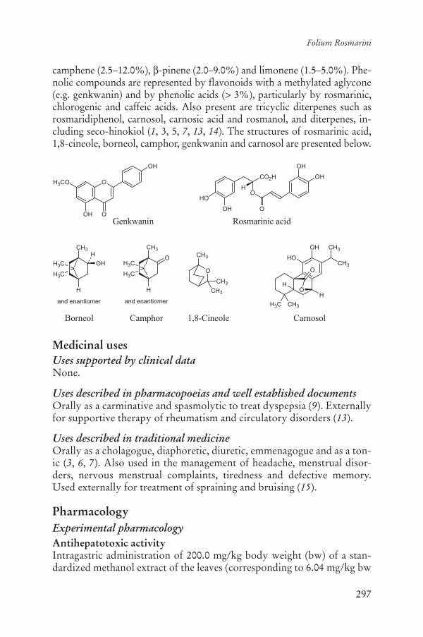
297
Folium Rosmarini
camphene (2.5–12.0%), -pinene (2.0–9.0%) and limonene (1.5–5.0%). Phe-nolic compounds are represented by flavonoids with a methylated aglycone (e.g. genkwanin) and by phenolic acids (> 3%), particularly by rosmarinic, chlorogenic and caffeic acids. Also present are tricyclic diterpenes such as rosmaridiphenol, carnosol, carnosic acid and rosmanol, and diterpenes, in-cluding seco-hinokiol (1, 3, 5, 7, 13, 14). The structures of rosmarinic acid, 1,8-cineole, borneol, camphor, genkwanin and carnosol are presented below.
Medicinal usesUses supported by clinical dataNone.
Uses described in pharmacopoeias and well established documentsOrally as a carminative and spasmolytic to treat dyspepsia (9). Externally for supportive therapy of rheumatism and circulatory disorders (13).
Uses described in traditional medicineOrally as a cholagogue, diaphoretic, diuretic, emmenagogue and as a ton-ic (3, 6, 7). Also used in the management of headache, menstrual disor-ders, nervous menstrual complaints, tiredness and defective memory. Used externally for treatment of spraining and bruising (15).
PharmacologyExperimental pharmacologyAntihepatotoxic activityIntragastric administration of 200.0 mg/kg body weight (bw) of a stan-dardized methanol extract of the leaves (corresponding to 6.04 mg/kg bw
OH
HO
CO2H
O
OH
OH
O
H
Rosmarinic acid
O
OOH
H3CO
OH
Genkwanin
H3C
H3C
CH3
O
H
H3C
H3C
CH3
H
OH
H
and enantiomer and enantiomer
Borneol Camphor
O
CH3
CH3
CH3
1,8-Cineole
O
O
OH
HOCH3
CH3
H
H3C CH3
H
Carnosol

298
WHO monographs on selected medicinal plants
of carnosol) to rats, 1 hour after treatment of the animals with carbon tetrachloride (CCl4), fully prevented CCl4-induced lipid peroxidation in the liver. The CCl4-induced increase in plasma bilirubin concentrations and alanine aminotransferase activity was completely normalized after treatment with the extract. The treatment also resulted in a significant recovery from CCl4-induced decrease in liver glycogen content. The ex-tract also increased liver cytosolic reduced glutathione activity and pro-duced an additional increase in plasma glutathione activity in rats treated with CCl4. Histological evaluation showed that the extract partially pre-vented CCl4-induced inflammation, necrosis and vacuolation (16).
The hepatoprotective and antimutagenic effects of an ethanol extract of the leaves were investigated using CCl4 and cyclophosphamide as the hepatotoxic and mutagenic compounds. The results indicated that intra-gastric administration of the ethanol extract (1.5 g/kg bw) to rats for 3 weeks produced a pronounced hepatoprotective effect as compared with silymarin (reference compound) due to the amelioration of most of the serum and liver parameters studied and confirmed by histopathologi-cal examination of the liver tissue. Pretreatment of mice for 7 days with the essential oil (1.1 mg/g bw) followed by intraperitoneal administration of cyclophosphamide significantly reduced the induced mitodepression in the bone marrow cells of the animals (17).
Anti-inflammatory activityTreatment of mouse macrophage RAW 264.7 cells with carnosol mark-edly reduced lipopolysaccharide-stimulated nitric oxide production in a concentration-related manner with an IC50 of 9.4 μM. Western blot, re-verse transcription–polymerase chain reaction, and northern blot analy-ses demonstrated that carnosol decreased lipopolysaccharide-induced inducible nitric oxide synthase mRNA and protein expression. Carnosol treatment reduced the translocation of nuclear factor-kappa B subunits and the binding activity of nuclear factor-B DNA in activated mac-rophages. Carnosol also inhibited inducible nitric oxide synthase and nuclear factor-B promoter activity in a transient transfection assay. These activities were due to down-regulation of inhibitor B kinase activity by carnosol (5 μM), which in turn inhibited lipopolysaccharide-induced phosphorylation as well as degradation of inhibitor B. Carnosol also in-hibited lipopolysaccharide-induced p38 and p44/42 mitogen-activated protein kinase activation at a higher concentration (20 μM). These results suggest that carnosol suppresses production of nitric oxide and inducible nitric oxide synthase gene expression by inhibiting activation of nuclear factor-B, and provide possible mechanisms for its anti-inflammatory activity (18).

299
Folium Rosmarini
Antioxidant activityOxidative damage to DNA, RNA, proteins and cell membranes occurs when the cellular concentration of reactive oxygen species exceeds the ca-pacity of the cell to eliminate them. The crude drug and its constituents have been shown to have both antioxidant and pro-oxidant properties both in vitro and in vivo (19–21). The antioxidant activity of six extracts of the leaves, with different compositions of six polyphenolic compounds (carnosic acid, carnosol, 12-O-methylcarnosic acid, rosmarinic acid, isoscutellarein 7-O-glucoside and genkwanin), were tested in aqueous (malonyldialdehyde formation) and lipid systems (Rancimat method). The results demonstrate that the extracts were excellent antioxidants in both systems at a concentration of 500 ppm (22). The ability of the phenolic diterpenes, carnosol, rosmanol and epirosmanol, to prevent oxidization of low-density lipoprotein in human blood and their ability to scavenge free radicals and superoxide anions was assessed in vitro. The antioxidant ac-tivities were evaluated by the thiobarbituric acid reactive substances (TBARS) assay and electron spin resonance method. The inhibition of the Cu2+-mediated oxidization of apo B formation in low-density lipoprotein was investigated by fluorescence spectroscopy. The results demonstrated that carnosol, rosmanol and epirosmanol inhibited lipid peroxidation and oxidized apo B formation in low-density lipoprotein in human blood. The median inhibitory concentration range was 7–10 μM (23). Supplementa-tion of the diet of mice with a dried hexane extract of the leaves (1% w/w of diet) for 1 week reduced the level of phospholipid hydroperoxides in the plasma, red blood cells (65–74% of the levels of non-supplemented control mice) and liver of the animals (24).
The antioxidant activities of carnosol and other compounds extracted from rosemary were compared. Carnosol showed potent antioxidative activity, and scavenged 1,1-diphenyl-2-picrylhydrazyl free radicals, as well as protecting DNA in the Fenton reaction (18).
An alcohol extract of the crude drug was assessed in vitro and in vivo for its ability to protect against free radical-induced skin damage. The extract inhibited oxidative alterations to skin surface lipids (25). The pro-tective effect of a 96% ethanol extract of the crude drug against oxidative DNA damage induced by hydrogen peroxide and visible light-excited methylene blue in colon cancer cells (CaCo-2) and hamster lung cells (V79) was investigated. The level of DNA damage (DNA strand-breaks) was measured using the comet assay. The extract reduced the genotoxic activity of both agents after long-term (24 h; 0.3 μg/ml) or short-term (2 h; 30.0 μg/ml) pre-incubation of cells, demonstrating that the extract

300
WHO monographs on selected medicinal plants
has a protective effect against oxidative damage to DNA as a consequence of scavenging of both hydroxyl radicals and singlet oxygen (26).
Antimicrobial activityA commercial extract of the leaves (no further details of extract given), dissolved in ethanol (100.0 mg/ml) was tested against foodborne micro-organisms. In the Gram-positive bacteria, the minimum inhibitory concen-tration of the ethanolic solution was 1% for Leuconostoc mesenteroides,0.5% for Listeria monocytogenes, 0.5% for Staphylococcus aureus, 0.13% for Streptococcus mutans and 0.06% for Bacillus cereus. It was fungistatic in Penicillium roquefortii and Botrytis cinerea. Up to 1% of the ethanolic solution had no activity against the Gram-negative bacteria Escherichiacoli, Salmonella enteritidis and Erwinia carotovora and on the yeasts Rho-dotorula glutinis and Cryptococcus laurentii. The antibacterial activity was associated with phenolic diterpenoids in the hexane extract of the leaves (27).
An extract of the leaves inhibited the growth of Shigella flexneri and S. sonnei with a minimum inhibitory concentration ranging from 0.5 to 1% (w/v) depending on the Shigella strain used (28).
Antinephrotoxic activityThe effects of rosmarinic acid on the suppression of mesangioprolifera-tive glomerulonephritis, induced by intravenous injection of rabbit anti-rat thymocyte serum to rats was assessed. Intragastric administration of rosmarinic acid to the rats at a dose of 100.0 mg/kg bw per day for 8 days decreased the quantity of proliferating cell nuclear antigen, fibronectin, type IV collagen and fibrin in the glomerulus. Superoxide dismutase ac-tivity of renal cortex homogenate was also significantly augmented in animals treated with rosmarinic acid (29).
Antitrypanosomal activityA methanol extract of the leaves at a concentration of 2.0 mg/ml inhibited the motility of cultured epimastigotes of Trypanosoma cruzi after 2 h of incubation. Two triterpene acids, oleanolic and ursolic acids, isolated from the extract were responsible for the activity. Ursolic acid stopped the movement of all T. cruzi epimastigotes at the minimum inhibitory con-centration of 40.0 μg/ml (88.0 μM) after 48 h of incubation. Oleanolic acid was less active, with a minimum inhibitory concentration of 250.0 μg/ml (550.0 μM) (30).
Antitumour activityThe effect of dietary intake of an extract of the leaves on 7,12-dimethylbenz[a]anthracene-induced mammary tumorigenesis and on the

301
Folium Rosmarini
in vivo formation of mammary 7,12-dimethylbenz[a]anthracene-DNA adducts was evaluated. Supplementation of a semi-purified diet with 1.0% by weight of the rosemary extract resulted in a significant (47%, p < 0.01) decrease in incidence of mammary tumours compared to controls. In sub-sequent studies, dietary supplementation with 0.5% and 1.0% rosemary extract inhibited total in vivo binding of 7,12-dimethylbenz[a]anthracene to mammary epithelial cell DNA by an average of 42%. This decrease in total binding was not due to a uniform decrease in the formation of all mammary 7,12-dimethylbenz[a]anthracene-DNA adducts (31).
Antiulcer activityAn ethanol (70%) extract was evaluated for antiulcerogenic activity in vivo. Intragastric administration of 100.0 mg/kg bw per day to 1.0 g/kg bw per day of the extract decreased the ulcerative lesion index produced by ethanol and reserpine in rats. No antisecretory activity was observed in the pyloric ligation model. Prior administration of L-NAME (N[G]-nitro L-arginine methyl ester), a nitric oxide-synthase inhibitor, did not reduce the antiulcerogenic activity of the extract in the ethanol-induced ulcer model, suggesting that the pharmacological mechanism has no rela-tionship with nitric oxide. When the animal groups were treated with in-domethacin using the same experimental model, the extract did not re-duce the antiulcerogenic activity, suggesting that the pharmacological mechanism has no relationship with prostaglandins. The previous treat-ment with N-ethylmaleimide, a thiol blocker, including mucosal nonpro-tein sulfhydryl groups, reduced the antiulcerogenic activity of the extract in the ethanol-induced ulcer model. This result suggests that the ethanol extract has active substances that increase the content of mucosal nonpro-tein sulfhydryl groups (32).
Diuretic effectsThe effects of aqueous extracts of the crude drug on the treatment of kid-ney function and diuresis in rats were determined. Outcomes assessed included urinary volume and the excretion of sodium, potassium and chloride. The concentration of electrolytes and urea in plasma and creati-nine clearance were also measured. Daily intragastric administration of the aqueous extracts of the leaves, at a dose of 10 ml/kg bw of an 8% or 16% extract in distilled water for 1 week, significantly enhanced diuresis in rats compared to the control group from the fifth day of treatment (p < 0.001). At a dose of 8%, the peak of urinary excretion of sodium, potassium and chloride was reached after 6 days of treatment (p < 0.001). A dose of 16% did not significantly affect the excretion of water and elec-trolytes over a similar period but significantly increased the urinary ex-

302
WHO monographs on selected medicinal plants
cretion of sodium and chloride on the seventh day and of potassium on the sixth day (p < 0.05). No change was observed in plasma electrolytes and urea in any group, except for a decrease in sodium and chloride con-centration in the group treated with the 16% extract of the crude drug. A decrease in creatinine clearance was observed after treatment with a daily dose of the 8% extract (33).
Enzyme inductionThe effects of an aqueous extract of the leaves and of rosmarinic acid were investigated on xenobiotic metabolizing enzymes in rat liver after dietary administration (0.5% daily rations) for 2 weeks. The modulation of phase I enzymes such as cytochrome P450 1A, 2B, 2E1, 3A and phase II en-zymes such as glutathione S-transferase, quinone reductase and uridine diphosphate (UDP)-glucuronosyltransferase was evaluated by measuring enzyme activities with specific substrates. The aqueous extract enhanced cytochrome P450 1A1, 2B1/2, 2E1 and glutathione S-transferase (espe-cially recombinant glutathione S-transferase A3/A5, M1 and M2), qui-none reductase and UDP-glucuronosyltransferase. No modification of xenobiotic metabolizing enzymes was observed in response to treatment with rosmarinic acid, indicating that other constituents of the leaves are responsible for this activity (34).
The ability of the leaves to modulate cytochrome P450 and detoxifica-tion enzymes in rat liver was evaluated by comparing the effects of dried leaves and leaf extracts with those of the essential oil. Rats received the powdered leaves or extracts of the leaves in their diet at 0.5% (w/w) for 2 weeks. The effects of such treatments were evaluated for cytochrome P450 isozymes 1A, 2B, 2E1, glutathione S-transferase, NAD(P)H, qui-none reductase and UDP-glucuronosyltransferase activities and on pro-tein levels (immunoblot analyses). The results demonstrated that essen-tial oil selectively induced cytochrome P450, particularly cytochrome P450 2B. The leaf extract enhanced both cytochrome P450 and detoxifi-cation enzymes. A dichloromethane extract of the leaves acted as a mono-functional inducer, inducing glutathione S-transferase, quinone reductase and UDP-glucuronosyltransferase, in particular UDP-glucuronosyl-transferase 1A6 (35).
An extract of the crude drug was fed to female mice at concentrations of 0.3% and 0.6% (by weight) for 4 weeks prior to determination of the activities of the detoxification enzymes glutathione-S-transferase and NAD(P)H, and quinone reductase in lung, liver and stomach. Liver ac-tivities of glutathione S-transferase and quinone reductase, and stomach glutathione S-transferase activity, were significantly increased in animals fed diets containing the extract. However, diets supplemented with the
303
Folium Rosmarini
extract did not affect lung glutathione S-transferase and quinone reductase activities (36).
Estrogenic effectsThe effects of a methanol extract of the leaves on the metabolism and ac-tion of estradiol and estrone were assessed in vivo. Treatment of female mice with 2% rosemary in an American Institute of Nutrition (AIN)-76A diet for 3 weeks increased the liver microsomal 2-hydroxylation of estra-diol and estrone by approximately 150%, increased their 6-hydroxylation by approximately 30% and inhibited the 16 -hydroxylation of estradiol by approximately 50%. Treatment of female CD-1 mice with 2% rose-mary in the diet for 3 weeks also stimulated the liver microsomal glucuroni-dation of estradiol and estrone by 54–67% and 37–56%, respectively. In further studies, feeding 2% rosemary in the diet to ovariectomized CD-1 mice for 3 weeks inhibited the uterotropic action of estradiol and estrone by 35–50% compared with animals fed a control diet (37).
Immune stimulant activityThe effect of an ethanol extract of the leaves on splenic mononuclear cell proliferation was determined in vivo. Rats were fed diets containing 0, 100, 200 or 400 ppm leaf extract or 400 ppm butylated hydroxytoluene in combination with 10 or 20% casein-enriched diets for 8 weeks. Splenic mononuclear cells were isolated from these animals and the mitogenic response to concanavalin A, phytohaemagglutinin and lipopolysaccha-ride was determined. Concanavalin A- and phytohaemagglutinin-stimu-lated proliferation of spleen cells in rats fed 10% casein and 200 ppm leaf extract was significantly higher than that of cells from the corresponding control animals. Other concentrations of the extract were not active, sug-gesting that the leaf extract does not have a generalized immune-enhanc-ing effect (38).
Rosmarinic acid induced apoptosis in a p56(lck) (Lck)-dependent manner. Lck(+) Jurkat T cells underwent apoptosis in response to treat-ment with rosmarinic acid , whereas Lck(-) Jurkat subclone J.CaM1.6 cellsdid not. J.CaM1.6 cells with various Lck mutants indicated that Lck SH2 domain, but not Lck kinase activity, was required for rosmarinic acid-induced apoptosis. Rosmarinic acid-mediated apoptosis involved a mitochondrial pathway as indicated by cytochrome c release and the complete blockage of apoptosis by an inhibitor of mitochondrial mem-brane depolarization. Both caspase-3 and caspase-8 were involved in ros-marinic acid-induced apoptosis and work downstream of mitochondria and caspase-9 in the order of caspase-9/caspase-3/caspase-8. In freshly isolated human peripheral blood mononuclear cells, rosmarinic acid

304
WHO monographs on selected medicinal plants
specifically induced apoptosis of Lck(+) subsets such as T and NK cells, but not Lck-deficient cells, including B cells and monocytes. Moreover, the ability of rosmarinic acid to kill T and NK cells was restricted to ac-tively proliferating cells, and was not seen in resting cells (39).
ToxicologyTo find out whether the crude drug induces abortion and/or interferes with the normal development of the concepts, doses of 26 mg of a 30% (w/v) aqueous extract (13 mg solids/ml) made with leaves, flowers and stem, were administered daily to rats by gavage during two different pe-riods of pregnancy. One group of animals (n = 12) received the extract from days 1 to 6 of pregnancy (pre-implantation period) and another group (n = 14) received the same extract from days 6 to 15 of pregnancy (organogenic period). Control groups (n = 12) received saline solution at the same volume and during the same periods as the comparable experi-mental groups. The animals were killed at term. The treatment of the dams during either the pre-implantation or the organogenic period did not cause significant changes in the post-implantation loss or in the num-ber of anomalies or malformations of the term fetuses, which also showed a similar degree of development to that of the control animals. The per-centage of pre-implantation loss in the group treated before embryo im-plantation increased compared to the control group, although the differ-ence was not statistically significant. This result suggests that rosemary extract may have an anti-implantation effect without interfering with the normal development of the concept after implantation (40).
Clinical pharmacologyOne of the postulated mechanisms of action of phenolic compounds as antioxidants is chelation of pro-oxidant metals, such as iron. Although the antioxidant activity is viewed as a positive effect, this activity may impair the utilization of dietary iron. A small clinical study assessed the effect of phenolic-rich extracts obtained from green tea or the crude drug on nonhaem-iron absorption. Twenty-four female volunteers consumed test meals on four separate occasions. The meals were identical except for the absence (meal A) or presence (meal B) of a phenolic-rich extract from green tea (study 1; n = 10) or rosemary (study 2; n = 14). The extracts (0.1 mM) were added to the meat component of the test meals. The meals were labelled with either 55Fe or 59Fe and were consumed on four consecu-tive days in the order ABBA or BAAB. Iron absorption was determined by measuring whole-body retention of 59Fe and the ratio of 55Fe or 59Fe activity in blood samples. The results demonstrated that the presence of phenolic-rich extracts resulted in decreased non-haem-iron absorption.

305
Folium Rosmarini
Mean (± standard deviation) iron absorption decreased from 12.1 ± 4.5% to 8.9 ± 5.2% (p < 0.01) in the presence of green tea extract and from 7.5 ± 4.0% to 6.4 ± 4.7% (p < 0.05) in the presence of the rosemary ex-tract (41).
Adverse reactionsThere is a single case-report of photoaggravated allergic contact dermati-tis, in which a patient developed contact dermatitis after handling the leaves of the plant on a sunny day (42–44). One case of cheilitis has been reported (45). A 56-year-old man developed occupational contact derma-titis of his hands, forearms, and face after coming into contact with an extract of the leaves. He reacted to carnosol, the main constituent of the extract (44).
ContraindicationsFolium Rosmarini is contraindicated in cases of hypersensitivity or aller-gy to the plant material.
WarningsNo information was found.
PrecautionsDrug interactionsWhile no drug interactions have been reported, an aqueous extract of the crude drug enhanced the activity of cytochrome P450 1A1, 2B1/2, 2E1 and glutathione S-transferase (especially recombinant glutathione S-transferase A3/A5, M1 and M2), quinone reductase and UDP-glucurono-syltransferase (34). Thus, drugs metabolized through these cytochrome P450 isozymes may be affected.
Carcinogenesis, mutagenesis, impairment of fertilityAn ethanol extract of the leaves was not mutagenic in the Hist revertant Salmonella typhimurium TA 1530 strain at a concentration of 5 g/ml(46). See also the study by Lemonica (40) under Toxicology.
Pregnancy: non-teratogenic effectsSee Toxicology.
Other precautionsNo information was found.

306
WHO monographs on selected medicinal plants
Dosage formsCrude drug for infusions, dry extracts, fluidextract and other Galenical preparations for internal and external use (47).
Posology(Unless otherwise specified)Daily dosage: for oral use 4–6 g of herb. Infusion: 2–4 g in 150 ml water three times daily. Fluidextract (1:1, 45% ethanol w/w) 1.5–3.0 ml daily. Tincture (1:5, 70% ethanol) 3–8.5 ml daily. Dry extract (4.5–5.5:1 w/w) 0.36–0.44 g, three times daily. External use: boil 50 g of herb in 1 l of wa-ter, add to one full bath (47).
ReferencesEuropean Pharmacopoeia1. , 5th ed. Strasbourg, Directorate for the Quality of Medicines of the Council of Europe (EDQM), 2005.Bedevian AK. 2. Illustrated polyglottic dictionary of plant names. Cairo, Med-bouly Library, 1994.Farnsworth NR, ed. 3. NAPRALERT database. Chicago, University of Illinois at Chicago, IL (an online database available directly through the University of Illinois at Chicago or through the Scientific and Technical Network [STN] of Chemical Abstracts Services), 30 June 2005.Youngken HW. 4. Textbook of pharmacognosy, 6th ed. Philadelphia, PA, Blak-iston, 1950.Bisset NR, Wichtl M, eds. 5. Herbal drugs and phytopharmaceuticals, English ed. Boca Raton, FL, Medpharm, 1994.Perry LM, Metzger J. 6. Medicinal plants of east and southeast Asia: Attributed properties and uses. Cambridge, MA, MIT Press, 1980.Bruneton J. 7. Pharmacognosy, phytochemistry, medicinal plants. Paris, Lavoisi-er, 1995.Boulos L. 8. Medicinal plants of North Africa. Algonac, Michigan, Reference Publications, 1983.British herbal pharmacopoeia9. . Exeter, British Herbal Medicine Association, 1996.Ziaková A, Brandsteterová E. Validation of HPLC determination of pheno-10.lic acids present in some Lamiaceae family plants. Journal of Liquid Chroma-tography and Related Technologies, 2003, 26:443–453.WHO guidelines for assessing quality of herbal medicines with reference to 11.contaminants and residues. Geneva, World Health Organization, 2007.Guidelines for predicting dietary intake of pesticide residues12. , 2nd rev. ed. Ge-neva, World Health Organization, 1997 (WHO/FSF/FOS/97.7).Blumenthal M et al., eds. 13. The complete German Commission E monographs: Therapeutic guide to herbal medicines. Austin, TX, American Botanical Council, 1998.

307
Folium Rosmarini
Cantrell CL et al. Seco-hinokiol, a new abietane diterpenoid from 14. Rosmari-nus officinalis. Journal of Natural Products, 2005, 68:98–100.Hagers Handbuch der Drogen15. (CD ROM). Heidelberg, Springer Verlag, 2003 [in German].Sotelo-Félix JI et al. Evaluation of the effectiveness of 16. Rosmarinus officinalis(Lamiaceae) in the alleviation of carbon tetrachloride-induced acute hepato-toxicity in the rat. Journal of Ethnopharmacology, 2002, 81:145–154.Fahim FA et al. Allied studies on the effect of 17. Rosmarinus officinalis L. on experimental hepatotoxicity and mutagenesis. International Journal of Food Sciences and Nutrition, 1999, 50:413–427.Lo AH et al. Carnosol, an antioxidant in rosemary, suppresses inducible ni-18.tric oxide synthase through down-regulating nuclear factor- B in mouse macrophages. Carcinogenesis, 2002, 23:983–991.Aruoma OI et al. Antioxidant and pro-oxidant properties of active rosemary 19.constituents: carnosol and carnosic acid. Xenobiotica, 1992, 22:257–268.Fuhrman B et al. Lycopene synergistically inhibits LDL oxidation in combi-20.nation with vitamin E, glabridin, rosmarinic acid, carnosic acid, or garlic. Antioxidant and Redox Signaling, 2000, 2:491–506.Gutiérrez ME et al. Interaction of tocopherols and phenolic compounds 21.with membrane lipid components: Evaluation of their antioxidant activity in a liposomal model system. Life Science, 2003, 72:2337–2360.Del Baño MJ et al. Phenolic diterpenes, flavones, and rosmarinic acid distri-22.bution during the development of leaves, flowers, stems, and roots of Ros-marinus officinalis: antioxidant activity. Journal of Agricultural and Food Chemistry, 2003, 51:4247–4253.Zeng HH et al. Antioxidant properties of phenolic diterpenes from 23. Rosmari-nus officinalis. Acta Pharmacologia Sinica, 2001, 22:1094–1098.Asai A, Nakagawa K, Miyazawa T. Antioxidative effects of turmeric, rose-24.mary and capsicum extracts on membrane phospholipid peroxidation and liver lipid metabolism in mice. Bioscience, Biotechnology and Biochemistry,1999, 63:2118–2122.Calabrese V et al. Biochemical studies of a natural antioxidant isolated from 25.rosemary and its application in cosmetic dermatology. International Journal of Tissue Reactions, 2000, 22:5–13.Slamenová D et al. Rosemary-stimulated reduction of DNA strand breaks 26.and FPG-sensitive sites in mammalian cells treated with H2O2 or visible light-excited methylene blue. Cancer Letters, 2002, 177:145–153.Del Campo J, Amiot MJ, Nguyen-TC. Antimicrobial effect of rosemary ex-27.tracts. Journal of Food Protection, 2000, 63:1359–1368.Bagamboula CF, Uyttendaele M, Debevere J. Antimicrobial effect of spices 28.and herbs on Shigella sonnei and Shigella flexneri. Journal of Food Protection,2003, 66:668–673.Makino T et al. Suppressive effects of rosmarinic acid on mesangioprolifera-29.tive glomerulonephritis in rats. Nephron, 2002, 92:898–904.

308
WHO monographs on selected medicinal plants
Abe F et al. Ursolic acid as a trypanocidal constituent in rosemary. 30. Biologicaland Pharmaceutical Bulletin, 2002, 25:1485–1487.Singletary KW, Nelshoppen JM. Inhibition of 7,12-dimethylbenz[c]anthracene 31.(DMBA)-induced mammary tumorigenesis and of in vivo formation of mammary DMBA-DNA adducts by rosemary extract. Cancer Letters, 1991, 60:169–175.Dias PC et al. Antiulcerogenic activity of crude hydroalcoholic extract of 32.Rosmarinus officinalis L. Journal of Ethnopharmacology, 2000, 69:57–62.Haloui M et al. Experimental diuretic effects of 33. Rosmarinus officinalis and Centaurium erythraea. Journal of Ethnopharmacology, 2000, 71:465–472.Debersac P et al. Effects of a water-soluble extract of rosemary and its puri-34.fied component rosmarinic acid on xenobiotic-metabolizing enzymes in rat liver. Food and Chemical Toxicology, 2001, 39:109–117.Debersac P et al. Induction of cytochrome P450 and/or detoxication en-35.zymes by various extracts of rosemary: description of specific patterns. Foodand Chemical Toxicology, 2001, 39:907–918.Singletary KW, Rokusek JT. Tissue-specific enhancement of xenobiotic de-36.toxification enzymes in mice by dietary rosemary extract. Plant Foods for Human Nutrition, 1997, 50:47–53.Zhu BT et al. Dietary administration of an extract from rosemary leaves en-37.hances the liver microsomal metabolism of endogenous estrogens and decreas-es their uterotropic action in CD-1 mice. Carcinogenesis, 1998, 19:1821–1827.Babu US, Wiesenfeld PL, Jenkins MY. Effect of dietary rosemary extract on 38.cell-mediated immunity of young rats. Plant Foods for Human Nutrition,1999, 53:169–174.Hur YG, Yun Y, Won J. Rosmarinic acid induces p5639. lck-dependent apoptosis in Jurkat and peripheral T cells via mitochondrial pathway independent from Fas/Fas ligand interaction. Journal of Immunology, 2004, 172:79–87.Lemonica IP, Damasceno DC, di-Stasi LC. Study of the embryotoxic effects 40.of an extract of rosemary (Rosmarinus officinalis L.). Brazilian Journal of Medical and Biological Research, 1996, 29:223–227.Samman S et al. Green tea or rosemary extract added to foods reduces nonheme-41.iron absorption. American Journal of Clinical Nutrition, 2001, 73:607–612.Armisén M, Rodríguez V, Vidal C. Photoaggravated allergic contact derma-42.titis due to Rosmarinus officinalis cross-reactive with Thymus vulgaris. Con-tact Dermatitis, 2003, 48:52–53.Fernandez L et al. Allergic contact dermatitis from rosemary (43. Rosmarinusofficinalis L.). Contact Dermatitis, 1997, 37:248–249.Hjorther AB et al. Occupational allergic contact dermatitis from carnosol, a natural-44.ly-occurring compound present in rosemary. Contact Dermatitis, 1997, 37:99–100.Guin JD. Rosemary cheilitis: one to remember. 45. Contact Dermatitis, 2001, 45:63.Alkofahi A et al. Biological activity of some Jordanian medicinal plant ex-46.tracts. Part II. Fitoterapia, 1997, 68:163–168.Blumenthal M, Goldberg A, Brinckmann J, eds. 47. Herbal medicine: Expanded Commission E monographs. Austin, TX, American Botanical Council, 2000.

309
Cortex Salicis
DefinitionCortex Salicis consists of the whole or fragmented dried bark from young branches of Salix alba L., S. daphnoides Vill., S. fragilis L., S. purpurea L., and other appropriate Salix species (Salicaceae) (1–4).
SynonymsNo information was found.
Selected vernacular namesSalix alba L.: Ak sõyüd ag, basket willow, bela vrba, beli, bid-e-maamou-li, caporniolo, derakht-e-bid, European willow, hopeapaju, hvid pil, is-bîdâr, kvitpil, osier blanc, paju, remmelgas, salcio bianco, salicastro, salcio da forche, salece, salgueiro-de-casa-roxa, saligastro, sargatillo, saule blanc, sauce blanco, Silberweide, sogut, solvpil, sufsaf abiad, tortiello, swallow tailed willow, vitpil, white willow (3, 5–10).
Salix daphnoides Vill.: Daphne willow, Reiweide, salicio nero, saule à bois glauque, saule faux daphné, saule noir, Schimmel Weide, vi, violet willow, wierzba wawrzynkolistna (3, 10).
Salix fragilis L.: Bid-khesht, brittle willow, Bruckweide, common crack willow, crack willow, Éva-fuz, hrökkvíòir, krhka vrba, Krackweide, kraakwilg, piilipuu, rabe remmelgas, salice, saliva, saule fragile, skjorpil, skörpil, sufsaf, törékeny fuz (3, 5, 8, 10).
Salix purpurea L.: Bidsorkh, morvár, osier rouge, purple osier, purple wil-low, purpur pil, Pupurweide, salcio da vimini, salcio rosso, sorkhbid, sauce Colorado, saule pourpre, wierzba purpurowa (3, 5, 10).
Geographical distributionNative to Europe and Asia, and naturalized in North America (6, 11).
DescriptionSalix species are woody trees, up to 25 m in height. Leaves are deciduous, generally petiolate and stipulate, simple, alternate to subopposite, linear

310
WHO monographs on selected medicinal plants
to widely obovate, margins entire to serrulate. Leaf underside is generally hairy or glaucous, rarely glabrous. Inflorescence consists of small uni-sexual catkins emerging, depending on species, either before, at the same time as, or after the leaves emerge; individual flowers inconspicuous, apetalous, subtended by a single fringed or hairy bract; sepals replaced by nectaries. Stamens 1–8, often 2 in staminate flower, and in pistillate flow-ers, the ovary is superior, bicarpellate, unilocular; style 1; stigmas 2, each 0–2-lobed. Fruit capsule, two-valved. Seeds many, comose (1). Salix albais a large tree with a short trunk, yellowish-brown branches and elliptic-lanceolate, acuminate and serrulate, ash-grey, sericious leaves. The fruit is a capsule dehiscent by 2 valves and contains numerous seeds, on each of which is a basal tuft of hair (11).
Plant material of interest: dried branch barkGeneral appearanceThe bark is 1–2 cm wide and 1–2 mm thick and occurs in flexible, elongat-ed, quilled or curved pieces. The outer surface is smooth or slightly wrin-kled longitudinally and greenish-yellow in the younger bark to brownish-grey in the older bark. The inner surface is smooth or finely striated longitudinally and white, pale yellow or reddish-brown, depending on the species. The fracture is short in the outer part and coarsely fibrous in the inner region, and is easily split longitudinally. The diameter of current year twigs is not more than 10 mm. The wood is white or pale yellow (2, 4).
Organoleptic propertiesOdour: slight; taste: astringent and bitter (1, 2, 4).
Microscopic characteristicsTwo or three rows of poorly developed cork cells with thickened outer walls; cortex of collenchymatous and parenchymatous cells. The latter contain cluster crystals of calcium oxalate, 20–25 μm in diameter and oc-casionally tannin. Phloem is characterized by tangential groups of ligni-fied fibres associated with a crystal sheath containing prismatic crystals of calcium oxalate. Simple, rounded starch granules 6–8 μm in diameter in the parenchymatous cells of the phloem and medullary rays (2).
Powdered plant materialPale yellow, greenish-yellow or light brown. Microscopically, bundles of narrow fibres, up to about 600 μm long, with very thick walls, lignified, and surrounded by a crystal sheath containing prism crystals of calcium oxalate; parenchyma of the cortex with thick, pitted and deeply beaded walls, and containing large cluster crystals of calcium oxalate; uniseriate

311
Cortex Salicis
medullary rays; thickened and suberized cork cells. Groups of brownish collenchyma from the bud may be present. Twigs show, additionally, frag-ments of lignified fibres and vessels from the xylem (2, 4).
General identity testsMacroscopic and microscopic examinations and thin-layer chromatogra-phy (1, 2, 4).
Purity testsMicrobiologicalTests for specific microorganisms and microbial contamination limits are as described in the WHO guidelines for assessing quality of herbal medi-cines with reference to contaminants and residues (12).
Foreign organic matterNot more than 3% of twigs with a diameter greater than 10 mm, and not more than 2% other foreign matter (1, 2, 4).
Total ashNot more than 10% (1, 2, 4).
Acid-insoluble ashNot more than 3% (1, 2).
Water-soluble extractiveNot less than 10% (1, 2).
Alcohol-soluble extractiveTo be established in accordance with national requirements.
Loss on dryingNot more than 11% (4).
Pesticide residuesThe recommended maximum limit of aldrin and dieldrin is not more than 0.05 mg/kg (4). For other pesticides, see the European pharmacopoeia (4)and the WHO guidelines for assessing quality of herbal medicines with reference to contaminants and residues (12) and pesticide residues (13).
Heavy metalsFor maximum limits and analysis of heavy metals, consult the WHO guidelines for assessing quality of herbal medicines with reference to con-taminants and residues (12).

312
WHO monographs on selected medicinal plants
Radioactive residuesWhere applicable, consult the WHO guidelines for assessing quality of herbal medicines with reference to contaminants and residues (12).
Chemical assaysNot less than 1.5% of total salicylate derivatives expressed as salicin by high-performance liquid chromatography (4).
Major chemical constituentsThe major biologically active constituents are the phenolic glycosides in-cluding salicin (approximately 1%), salicortin (up to 4.0%), 2´-O-acetylsalicortin (up to 10%), 2´-O-acetylsalicin (= fragilin, up to 4%), trem-ulacin (0.12–2%), 3´- and 4´-acetylsalicortin, populin and salireposide, which have collectively been designated as “salicylates” (6). Triandrin, vimalin, pi-cein and grandidentatin are non-saligenin structure-based phenolic com-pounds. Other significant constituents are flavonoids and tannins (1, 5, 6).
Total salicin content (after hydrolysis) varies according to species. Species rich in total salicin include S. daphnoides (2–10%), S. purpurea(4–8.5%), S. fragilis (2–10%) and S. alba (0.5–1%) (1, 6).
The structures of salicin, salicortin, 2´-O-acetylsalicortin and 6´-O-acetylsalicin (= fragilin) are presented below.
Medicinal usesUses supported by clinical dataUsed orally for the symptomatic treatment of fever and pain, and symp-tomatic treatment of mild rheumatic conditions (14–20).
Uses described in pharmacopoeias and well established documentsUsed orally for the treatment of the common cold (1).
Uses described in traditional medicineUsed orally for the treatment of constipation and urinary incontinence. Used externally for the treatment of warts (5).
Salicortin
2'-O-Acetylsalicortin
O
OH
O
HO
OH
O
OHR
O
OH
HO
HO
O
O
O
O O
HO*
and epimer at C*R
SalicinFragilin R = CO-CH3
R = HR = CO-CH3
R = H

313
Cortex Salicis
PharmacologyExperimental pharmacologyAnti-inflammatory activitySimilar to salicylates, such as acetylsalicylic acid, extracts of the crude drug are thought to act by inhibiting the activity of cyclooxygenase and thereby inhibiting the synthesis of prostaglandins, which play a role in inflamma-tion, fever and pain. Acetylsalicylic acid inhibits the synthesis of prosta-glandins through the acetylation of the enzyme cyclooxygenase. Salicylic acid, and the salicylates that lack an acetyl group, reduce prostaglandin synthesis via inhibition of the activity of cyclooxygenase II (1, 21).
Intragastric administration of 100.0 mg/kg body weight (bw) of trem-ulacin, a constituent of the bark, significantly inhibited carrageenan- induced hind paw oedema in rats (p < 0.001) (22). Intragastric administra-tion of 0.5 ml/kg bw of a 30% ethanol extract of the bark to rats reduced carrageenan-induced hind paw oedema, but was not effective in the adju-vant-induced arthritis or dextran-induced pedal oedema models (23).
Antipyretic activityIntragastric administration of 0.8 ml/kg bw of a 30% ethanol extract to rats, suppressed yeast-induced pyrexia (23). The antipyretic effects of salicin, saligenin (an aglycone of salicin) and salicylic acid (an active me-tabolite of salicin) were assessed in rats (24). After intragastric administra-tion of salicin to rats, the metabolite salicylic acid appeared slowly in the plasma and concentrations increased gradually, in contrast to the rapid appearance observed after oral administration of sodium salicylate or sali-genin. At a dose of 5 mM/kg bw, orally administered salicin did not affect the rectal temperature of afebrile rats while at the same dose, sodium sali-cylate and saligenin lowered body temperature significantly. However, salicin significantly reduced yeast-induced fever (p < 0.01), producing a normal body temperature, and completely prevented fever when admin-istered simultaneously with yeast. Salicin did not induce gastric lesions even at a dose of 5 mM/kg bw; conversely, sodium salicylate and saligenin induced severe gastric lesions in a dose-dependent manner when adminis-tered at doses of 1, 2.5 and 5 mM/kg bw (24).
PharmacokineticsThe pharmacokinetics of salicin, saligenin (an aglycone of salicin) and salicylic acid (an active metabolite of salicin) were determined in rats (24).Poor absorption of salicin and rapid absorption of salicylic acid and sali-genin were confirmed in this animal model. Only small amounts of sali-cylic acid and saligenin were detected in the intestinal tracts of rats 1 h
314
WHO monographs on selected medicinal plants
after oral administration. More than 50% of a salicin dose was recovered as salicin and saligenin from the intestinal tracts 1 h after treatment and 15.8% of the dose was still present as saligenin 4 h after administration. When given to germ-free rats, 19.8% of the salicin dose was recovered intact, mainly from the cecum, and no saligenin was detected even at 4 h after treatment. These results indicate that salicin is a pro-drug that is gradually transported to the lower part of the intestine, hydrolysed to saligenin by intestinal bacteria, and converted to salicylic acid after ab-sorption. It thus produces an antipyretic action without causing gastric injury (25).
ToxicologyIntragastric administration of a 40% ethanol extract of the bark to rats at a dose of 1.6 ml/kg bw for 13 weeks had no effect on kidney function, haematological parameters, liver function or cholesterol levels (26). His-tological evaluation of the animals after killing showed no pathological changes in the brain, heart, lungs, bone, kidneys, liver, reproductive or-gans, mammary tissue, stomach or intestines (26).
The median lethal dose range of a 30% ethanol extract of the bark was 28.0–42.0 ml/kg bw in mice (both sexes) (26).
Clinical pharmacologyAnticoagulant activityA study was performed to investigate the anticoagulant activity of a bark extract used in the treatment of 51 patients with chronic back pain. Thir-ty-five patients suffering from acute exacerbations of chronic low back pain received randomly and double-blind either the extract (correspond-ing to 240 mg salicin/day; n = 19) or a placebo (n = 16). A further 16 pa-tients with stable chronic ischaemic heart disease were given 100 mg ac-etylsalicylate per day. Platelet aggregation was studied after drawing blood from the treated patients using an aggregometer. Arachidonic acid (500 μg/ml), adenosine diphosphate (2 × 10⁻⁵ M) and collagen (0.18 μg/ml) were used as aggregating agents. The mean maximal arachidonic acid-induced platelet aggregation was 61%, 78% and 13% for the groups treated with extract, placebo and acetylsalicylate, respectively. Acetylsalicylate had a significant inhibitory effect on platelet aggregation compared to the ex-tract (p = 0.001) and placebo (p = 0.001). There was also a significant dif-ference between the placebo and the extract-treated groups in the maxi-mal platelet aggregation induced by arachidonic acid (p = 0.04) and ADP (p = 0.01). No statistical difference was found between platelet aggrega-tion in the three groups when collagen was added to the human platelets.

315
Cortex Salicis
Daily consumption of the extract with 240.0 mg salicin per day affected platelet aggregation to a lesser extent than acetylsalicylate (27).
Chronic lower back painStandardized extracts of the bark exhibited analgesic effects similar to those of higher doses of acetylsalicylic acid. A daily dose of 1572.0 mg of an extract of the crude drug (standardized to 15.2% salicin or 240.0 mgsalicin per day) was superior to placebo in the treatment of pain in pa-tients with osteoarthritis of the hip and the knee, and in patients with exacerbations of chronic low back pain. In two open studies, unspecified extracts of the crude drug exhibited similar efficacy to the normal treat-ment regime of nonsteroidal antirheumatic drugs and the efficacy was similar to that of refecoxib (18).
A randomized placebo-controlled clinical study evaluated the efficacy of a willow bark extract in 210 patients with chronic lower back pain. The patients were assigned to receive an orally administered willow bark ex-tract with either 120.0 mg (low dose) or 240.0 mg (high dose) of salicin, or a placebo, with tramadol as the sole rescue medication, in the 4-week trial. The principal outcome measure was the proportion of patients who were pain-free without tramadol for at least 5 days during the final week of the study. A total of 191 patients completed the study. The number of pain-free patients in the last week of treatment was 27 (39%) of 65 in the group receiving high-dose extract, 15 (21%) of 67 in the group receiving low-dose extract, and 4 (6%) of 59 in the group treated with placebo (p < 0.001).The response in the group treated with the high dose was evident after only 1 week of treatment. Significantly more patients in the group receiv-ing the placebo required tramadol during each week of the study (p < 0.001). One patient in the treatment group suffered a severe allergic reaction (14).
An open, non-randomized study (postmarketing surveillance) assessed the efficacy of an extract of the crude drug in three groups of patients aged 18–80 years presenting over an 18-month period with acute exacerbations of low back pain. The first group of 115 patients was prescribed a daily dose of the extract containing 120.0 mg of salicin. A second group of 112 patients was prescribed the extract equivalent to 240.0 mg salicin per day. A third “control” or “comparator” group of 224 patients received conventional therapy. In the patients who had received conventional ther-apy, the exacerbations had been shorter but the pain was more intense as judged by Arhus Index and Total Pain Index. After 4 weeks of treatment, about 40% of patients in the group treated with the extract equivalent to 240 mg salicin per day were free of pain. In the group treated with extract containing 120.0 mg of salicin, as a whole, about 19% of patients were

316
WHO monographs on selected medicinal plants
pain-free at 4 weeks, but only 8% of those who did not resort to supple-mentary treatment. In the group receiving conventional therapy, 18% of patients were pain-free. Better pain relief in the group treated with the equivalent of 240.0 mg salicin was accompanied by a reduction in the use of supplementary conventional treatments (15).
MigraineA placebo-controlled double-blind clinical trial involving 54 patients as-sessed the efficacy of a topical medication containing salicin for the treat-ment of either migraines and/or tension-type headaches. All the patients had headaches consistent with the International Classification of Head-ache Disorders (HIS) criteria for migraines and/or tension-type head-aches, and had suffered from headaches for at least 1 year up to approxi-mately 40 years. The patients were divided into two groups. One group received a placebo topical medication. The other group received a topical medication containing salicin. Neither preparation induced any stinging sensation. The patients were instructed to apply the medication, which was in a roll-on form, to the frontal region at the onset of a headache, take their usual oral or parenteral medications, apply a mask which covered the eyes and frontal region, and then lie down. They subsequently filled out forms rating the degree of relief which they attributed to the topical medications and the masks using a scale of 0 to 10. They were also asked if this form of treatment had helped or not. Seven of the 20 patients who received the placebo stated that the medication and mask helped. This group rated the treatment an average of 4.31 on the scale of 0 to 10. Twen-ty-eight of 34 patients who received salicin stated that it was effective. This group rated it 7.42 on the scale of 0 to 10 (p < 0.001). The majority of the patients treated with salicin stated that the duration of their headaches was significantly reduced, as was their need for analgesic and/or narcotic medications. This study demonstrates a significant difference between placebo and salicin in association with the photoprotective mask in treat-ing migraines and/or tension-type headaches with associated frontalis pain and photophobia (17).
OsteoarthritisA clinical study involving 78 patients assessed the efficacy of a chemically standardized willow bark extract in the treatment of osteoarthritis. Wil-low bark extract, at a dose corresponding to 240.0 mg salicin/day, was compared to a placebo in a 2-week, double-blind, randomized controlled trial. The primary outcome measure was the pain dimension of the West-ern Ontario and McMaster Universities (WOMAC) Osteoarthritis In-dex. Secondary outcome measures included the stiffness and physical

317
Cortex Salicis
function dimensions of the WOMAC, daily visual analogue scales (VAS) on pain and physical function, and final overall assessments by both pa-tients and investigators. The results demonstrated a statistically signifi-cant difference between the subjects who received active treatment and the placebo group in the WOMAC pain dimension (d = 6.5 mm, 95% confidence intervals (CI), 0.2–12.7 mm, p = 0.047); the WOMAC pain score was reduced by 14% from the baseline level after 2 weeks of active treatment, whereas there was an increase of 2% in the placebo group. The patient diary VAS confirmed the results, and the final overall assessments showed superiority of the willow bark extract over the placebo (patients’ assessment, p = 0.0002; investigators’ assessment, p = 0.0073) (19, 20).
A double-blind, randomized, controlled clinical trial assessed the effi-cacy of an extract of the bark (containing 17.6% total salicin), at a dose of 240.0 mg per day, in the treatment of patients with chronic arthritic pain. Eighty-two subjects with chronic arthritic pain were randomly assigned for 2 months without cross-over to receive either the extract or a placebo. The results demonstrated a small but statistically significant improvement in pain symptoms (p < 0.05), although the improvement was less in pa-tients with osteoarthritis. No other significant changes in any other mea-sures or in the use of other self-prescribed analgesics were observed (28).
The efficacy of a standardized willow bark extract was investigated in two randomized, controlled, double-blind trials with follow-up for 6 weeks, in patients with osteoarthritis and rheumatoid arthritis. One hundred and twenty-seven outpatients with osteoarthritis of the hip or knee and a pain score of at least 30 mm, and 26 outpatients with active rheumatoid arthritis were randomly allocated to one of three groups. Pa-tients in the first group received willow bark extract, corresponding to 240 mg of salicin/day; patients in the second group, diclofenac at a dose of 100 mg/day; and the third group, a placebo (n = 43, 43 and 41, respec-tively). The main outcome measure was the pain subscore. In the rheuma-toid arthritis trial, patients were randomly assigned to receive either wil-low bark extract, corresponding to 240 mg salicin/day (n = 13) or a placebo (n = 13). The main outcome measure was the patient’s assessment of pain rated on a 100-mm visual analogue scale. In the osteoarthritis trial, pain scores decreased by 8 mm (17%) in the group treated with willow bark and by 23 mm (47%) in the group treated with diclofenac, compared with 5 mm (10%) in the patients who received the placebo. The difference between the scores following treatment with willow bark extract and treatment with placebo was not statistically significant (–2.8 mm; 95% CI –12.1 to 6.4 mm; p = 0.55, analysis of covariance (ANCOVA)), but the difference between the outcomes of treatment with diclofenac and with

318
WHO monographs on selected medicinal plants
the placebo was highly significant (–18.0 mm; 95% CI –27.2 to –8.8 mm; p = 0.0002, ANCOVA). The results in the rheumatoid arthritis trial showed that the mean reduction of pain reported using the visual ana-logue scale was –8 mm (15%) in the group treated with willow bark com-pared with –2 mm (4%) in the placebo group. The difference was not statistically significant (estimated difference –0.8 mm; 95% CI –20.9 to 19.3 mm; p = 0.93, ANCOVA) (29).
PharmacokineticsThe pharmacokinetics of salicin and its major metabolites were deter-mined in humans after oral administration of an extract of the crude drug. The extract, corresponding to 240.0 mg salicin (1360 mg, 838 μmol), was administered to 10 healthy volunteers in two equal doses at times 0 h and 3 h. Over a period of 24 h, levels of salicylic acid and its metabolites in urine and serum, i.e. gentisic acid and salicyluric acid, were determined using reverse-phase high-performance liquid chromatography. Renal ex-cretion rate, elimination half-life and total bioavailability of salicylates were calculated. The results showed that salicylic acid was the major me-tabolite of salicin detected in the serum (86% of total salicylates), as well as salicyluric acid (10%) and gentisic acid (4%). Peak serum levels were reached within less than 2 h after oral administration. Renal elimination occurred predominantly in the form of salicyluric acid. Peak serum levels of salicylic acid were on average 1.2 mg/l, and the observed area under the serum concentration–time curve of salicylic acid was equivalent to that expected from an intake of 87.0 mg acetylsalicylic acid (30).
Adverse reactionsAllergic reactions such as pruritus, urticaria, asthma and gastrointestinal symptoms may occur (16). One case of an allergic reaction in a 32-year-old atopic patient who showed a severe anaphylactic reaction after the ingestion of a pollen compound containing the crude drug has been reported (31). One case of anaphylaxis resulting from the use of an extract of the crude drug in a patient with a history of an aspirin allergy has been reported (32).
ContraindicationsCortex Salicis is contraindicated in cases of hypersensitivity or allergy to the plant material or to salicylates (e.g. asthma, bronchial spasm, rhinitis or urticaria).
It is also contraindicated during pregnancy and lactation, in patients with salicylate intolerance and patients with impaired thrombocyte func-tion (7, 27), and in children under the age of 12 years (7).

319
Cortex Salicis
WarningsIn children under the age of 12 years, Cortex Salicis should only be used on the advice of a health care professional due to the possibility of Reye’s syndrome. In cases of a child or adolescent who has become very ill with severe vomiting, drowsiness or loss of consciousness following a viral in-fection, Reye’s syndrome should be suspected. This extremely rare, life-threatening disease requires immediate medical attention.
In cases of severe liver or renal dysfunction, coagulation disorders, gastric/duodenal ulcer and glucose-6-phosphate dehydrogenase deficien-cy, the product should only be taken under medical supervision.
Consult a health care professional in cases of fever (> 39 C), lasting longer than 3 days. A health care professional should also be consulted if acute conditions of swelling of joints, redness and impaired mobility per-sist or worsen during the first week of use of Cortex Salicis.
PrecautionsGeneralThe use of Cortex Salicis in patients with hypersensitivity to other non-steroidal anti-inflammatory drugs is not recommended. Use should be avoided in people with asthma because severe reactions (acute broncho-spasms) could be induced (16).
Drug interactionsCurrently there are no data indicating that the crude drug interacts with anticoagulant drugs such as coumarin and heparin. However, Cortex Sali-cis may increase the effects of anticoagulants. Patients on anticoagulant therapy should not use products containing the crude drug without the supervision of a health care professional (7).
Carcinogenesis, mutagenesis, impairment of fertilityOral administration of a 40% ethanol extract of the bark did not disrupt the estrous cycle, or inhibit ovulation or fertility when administered to female rats and rabbits at a dose of 1.6 ml/kg bw (26).
Pregnancy: teratogenic effectsOral administration of a 40% ethanol extract of the bark to female rats and rabbits at a dose of 1.6 ml/kg bw during pregnancy was not terato-genic (26).
Pregnancy: non-teratogenic effectsSee Contraindications.

320
WHO monographs on selected medicinal plants
Oral administration of a 40% ethanol extract of the bark was not em-bryotoxic when administered to rats and rabbits during pregnancy at a dose of 1.6 ml/kg bw (26).
Nursing mothersSee Contraindications.Salicylates cross the placenta and appear in breast milk (1).
Paediatric useSee Contraindications.
Other precautionsNo information was found.
Dosage formsCrude drug, dried hydroalcoholic or aqueous extracts, tinctures and fluidextracts (1, 16, 33).
Posology(Unless otherwise indicated)Adult oral daily dose: extracts, tinctures or fluidextracts equivalent to 120–240 mg of total salicin, or 6–12 g of powdered drug material as a de-coction (corresponding to 120–240 mg of total salicin) in two divided doses (1, 16, 33).
ReferencesUpton R et al., eds. Willow bark. 1. Salix spp. In: American herbal pharmaco-peia. Santa Cruz, CA, American Herbal Pharmacopeia, 1999.British herbal pharmacopoeia2. . Exeter, British Herbal Medicine Association, 1996.Bradley PR, ed. 3. British herbal compendium. Vol. 1. Dorset, British Herbal Medicine Association, 1992.European Pharmacopoeia4. , 5th ed. Strasbourg, Directorate for the Quality of Medicines of the Council of Europe (EDQM), 2005.Farnsworth NR, ed. 5. NAPRALERT database. Chicago, University of Illinois at Chicago, IL (an online database available directly through the University of Illinois at Chicago or through the Scientific and Technical Network [STN] of Chemical Abstracts Services), 30 June 2005.Wichtl M. 6. Herbal drugs and phytopharmaceuticals, English ed. (Bisset NG translator and ed.). Boca Raton, FL, CRC Press, 1994.Ernst E et al., eds. 7. The desktop guide to complementary and alternative med-icine. Edinburgh, Mosby, 2001.

321
Cortex Salicis
Zahedi E. 8. Botanical dictionary. Scientific names of plants in English, French,German, Arabic and Persian Languages. Tehran, University of Tehran Pub-lications, 1959.Bedevian AK. 9. Illustrated polyglottic dictionary of plant names. Cairo, Med-bouly Library, 1994.Parsa A. 10. Flore de l’Iran. Vol. VIII. Tehran, University of Tehran, 1960.Youngken HW. 11. Textbook of pharmacognosy, 6th ed. Philadelphia, PA, Blak-iston Company, 1950.WHO guidelines for assessing quality of herbal medicines with reference to 12.contaminants and residues. Geneva, World Health Organization, 2007.Guidelines for predicting dietary intake of pesticide residues13. , 2nd rev. ed. Ge-neva, World Health Organization, 1997 (WHO/FSF/FOS/97.7).Chrubasik S et al. Treatment of low back pain exacerbations with willow 14.bark extract: A randomized double-blind study. American Journal of Medi-cine, 2000, 109:9–14.Chrubasik S et al. Potential economic impact of using a proprietary willow 15.bark extract in outpatient treatment of low back pain: An open non-random-ized study. Phytomedicine, 2001, 8:241–251.European Medicines Agency. 16. Final proposal for a core data Salicis Cortex.London, EMEA, 2003.Hyson MI. Anticephalgic photoprotective premedicated mask. A report of a 17.successful double-blind placebo-controlled study of a new treatment for headaches with associated frontalis pain and photophobia. Headache, 1998, 38:475–477.März RW, Kemper F. Weidenrindenextrakt-Wirkungen und Wirksamkeit. 18.Erkenntnisstand zu Pharmakologie, Toxikologie und Klinik [Willow bark extract-effects and effectiveness. Status of current knowledge regarding phar-macology, toxicology and clinical aspects]. Wiener Medizinische Wochen-schrift, 2002, 152:354–359 [in German].Schmid B et al. Wirksamkeit und Verträglichkeit eines standardisierten Weid-19.enrindenextraktes bei Arthrose-Patienten: Randomisierte, Placebo-kontrolli-erte Doppelblindstudie [Effectiveness and tolerance of standardized willow bark extract in arthrosis patients. Randomized, placebo controlled double-blind study]. Zeitschrift für Rheumatologie, 2000, 59:314–320 [in German].Schmid B et al. Efficacy and tolerability of a standardized willow bark ex-20.tract in patients with osteoarthritis: Randomized, placebo-controlled, double blind clinical trial. Phytotherapy Research, 2001, 15:344–350.Wagner I et al. Influence of willow bark extract on cyclooxygenase activity 21.and on tumor necrosis factor- or interleukin 1 release in vitro and ex vivo.Clinical Pharmacology & Therapeutics, 2003, 73:272–274.Cheng GF et al. Anti-inflammatory effects of tremulacin, a salicin-related 22.substance isolated from Populus tomentosa Carr. Leaves. Phytomedicine,1994, 1:209–211.Leslie GB. A pharmacometric evaluation of nine bio-strath herbal remedies. 23.Medita, 1978, 8:3–19.

322
WHO monographs on selected medicinal plants
Fiebich BL, Appel K. Anti-inflammatory effects of willow bark extract. 24.Clinical Pharmacology & Therapeutics, 2003, 74:96–97.Akao T et al. Evaluation of salicin as an antipyretic prodrug that does not 25.cause gastric injury. Planta Medica, 2002, 68:714–718.Leslie GB, Salmon G. Repeated dose toxicity studies and reproductive stud-26.ies on nine bio-strath herbal remedies. Swiss Medicine, 1979, 1:1–3.Krivoy N et al. Effect of salicis cortex extract on human platelet aggregation. 27.Planta Medica, 2001, 67:209–212.Mills SY et al. Effect of a proprietary herbal medicine on the relief of chronic 28.arthritic pain: A double-blind study. British Journal of Rheumatology, 1996, 35:874–878.Biegert C et al. Efficacy and safety of willow bark extract in the treatment of 29.osteoarthritis and rheumatoid arthritis: results of 2 randomized double-blind controlled trials. Journal of Rheumatology, 2004, 31:2121–2130.Schmid B, Kötter I, Heide L. Pharmacokinetics of salicin after oral adminis-30.tration of a standardised willow bark extract. European Journal of Clinical Pharmacology, 2001, 57:387–391.Chivato T et al. Anaphylaxis induced by ingestion of a pollen compound. 31.Journal of Investigative Allergology and Clinical Immunology, 1996, 6:208–209.Boullata JI, McDonnell PJ, Oliva CD. Anaphylactic reaction to a dietary 32.supplement containing willow bark. Annals of Pharmacotherapy, 2003, 37:832–835.Blumenthal M et al., eds. 33. The complete German Commission E monographs: Therapeutic guide to herbal medicines. Austin, TX, American Botanical Council, 1998.

323
Fructus Tribuli
DefinitionFructus Tribuli consists of the dried fruits of Tribulus terrestris L. (Zygo-phyllaceae) (1–4).
SynonymsTribulus lanuginosus L. (5).
Selected vernacular namesAbrojo, abrojos, akanti, alaf-e-kanguereh, baijili, bastitaj, be tha gokharu, bethu, bhakhra, bullhead, burnut, burra gookeron, calthrop, caltrap, caltrop, cat’s head, chotagokhru, cows hoof, croix de chevalier, croix de malte, dars el agûz, dava-tehokourtdi, demirdiken, deshi gokhru, devil’s thorn, ekanty, eskrundki, espáákh, espigón, gai ma duong, gatha, ghota, goathead, gokharu gokhru, gokhur, gokshura, goksuraka, gokhri, gokhurkata, gokhyura, gukhura, hamabishi, herbe terrestre, ikshugandha, jili, jilisi, kandaai, khar-e-khasak khurd, khárk-tehârouk khárk-tehârpar, khárkhassak, khôrbâr, khokkrasun, kouleh-tighak, krunda, land caltrops, malteserkors, meetha gokhru, Mexican sanbur, michirkand, mithagokharu, mithogokharu, naal-khar, naam din, nana gokharu, nature’s viagara, neggilamullu, neggilu, nere-nchi, nerinjil, nerunjil, ookharu pakhda, outb, pakhda, pakhra, palleru, pal-leru kaya, palleruveru, pedda palgeru, punctur vine, qutiba, saligot terrestre, sannanaggilu, sannaneggilu, sarang, sarate, sekal-tali, sharwandi, sher sher, shitsurishi, small caltrop, svadamstraa, tahkandi, tat le, Teufelsdorm, Texas sandbur, traikantaka, tribolo commune, trikanta, tribule couché, tribule terrestre, tsi li, vejtidsel, zama (4–10).
Geographical distributionNative to the Mediterranean regions, but also found throughout the world (6).
DescriptionA perennial, decumbent, pubescent herb, branches 0.5–1.8 m long, tips ascending; longer leaves up to 7 cm long, with 6–9 pairs of leaflets, shorter

324
WHO monographs on selected medicinal plants
leaves up to 6 cm long with 3–5 pairs of leaflets, midrib ending in a mucro, 0.5–2 mm long; leaflets 6–22 mm by 2.5–9 mm, base obliquely rounded, apex subacute, both surfaces silky, subsessile, stipules falcate, 3–6 mmlong, acuminate; pedicel 2–4 cm long, sepals narrowly lanceolate, 7–11 mmlong, apex acute, appressed hairy, caducous, petals obovate-cuneate,1–2 cm by 1–1.5 cm, apex broadly rounded-truncate, bright yellow, sta-mens subequal, anthers 1 mm long; cocci 4–5, spines stout, sharp, 2 lateralones largest, pericarp rather thick, corky, seeds 1–3 in each mericarp (6).
Plant material of interest: dried fruitsGeneral appearanceA pentagonal fruit 7–12 mm in diameter consists of 5 mericarps, radially arranged. Each mericarp often splitting into single hatchet-shaped,3–6 mm long mericarp; dorsal surface yellowish-green, prominent, with longitudinal ribs and numerous spinelets, bearing symmetrically a pair of long spines and a pair of short spines; two lateral surfaces rough, with reticular striations, greyish-white. Texture hard. Each mericarp contains 1–3 seeds (1–3).
Organoleptic propertiesOdourless; taste: bitter and pungent (1, 2).
Microscopic characteristicsTransverse section of fruit shows small epidermal cells of each coccus rectangular; unicellular trichomes in abundance; rilesocarp 6–10 layers of large parenchymatous cells, rosette of calcium oxalate crystals abundant; mesocarp followed by 3–4 compact layers of small cells containing pris-matic crystals of calcium oxalate (4).
Powdered plant materialYellow-green. The fibres are lignified, with upper and lower layers in a criss-cross arrangement, a few scattered singly, sometimes fibre bundles connected with stone cells. Stone cells elongated-elliptical or subrounded, occurring in groups. Testa cells polygonal or subsquare, about 30 μm in diameter, walls reticulately thickened, lignified. Prisms of calcium oxalate 8–20 μm in diameter (1).
General identity testsMacroscopic and microscopic examinations (1, 2, 4), thin-layer chroma-tography (2) and microchemical test (3).
325
Fructus Tribuli
Purity testsMicrobiologicalTests for specific microorganisms and microbial contamination limits are as described in the WHO guidelines for assessing quality of herbal medi-cines with reference to contaminants and residues (11).
Foreign organic matterNot more than 4.0% fruit stalks and not more than 1% other foreign matter (2, 3).
Total ashNot more than 12% (1).
Acid-insoluble ashNot more than 1.5% (2).
Water-soluble extractiveNot less than 10% (4).
Alcohol-soluble extractiveNot less than 8.5% (2).
WaterNot more than 9% (1).
Loss on dryingNot less than 11% (2).
Pesticide residuesThe recommended maximum limit of aldrin and dieldrin is not more than 0.05 mg/kg (12). For other pesticides, see the European pharmacopoeia (12)and the WHO guidelines for assessing quality of herbal medicines with reference to contaminants and residues (11) and pesticide residues (13).
Heavy metalsFor maximum limits and analysis of heavy metals, consult the WHO guidelines for assessing quality of herbal medicines with reference to con-taminants and residues (11).
Radioactive residuesWhere applicable, consult the WHO guidelines for assessing quality of herbal medicines with reference to contaminants and residues (11).
Chemical assaysTo be established in accordance with national requirements.
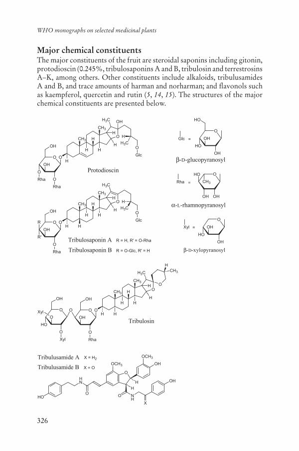
326
WHO monographs on selected medicinal plants
Major chemical constituentsThe major constituents of the fruit are steroidal saponins including gitonin, protodioscin (0.245%, tribulosaponins A and B, tribulosin and terrestrosins A–K, among others. Other constituents include alkaloids, tribulusamides A and B, and trace amounts of harman and norharman; and flavonols such as kaempferol, quercetin and rutin (5, 14, 15). The structures of the major chemical constituents are presented below.
CH3 H
CH3
H
HH
O
H
H
H3C
O
OH
O
R'
O
Rha
O
H
H
H3C
Glc
OH
O
OH
CH3
OH
HO
-L-rhamnopyranosyl
Rha =
O
OH
HO
HO
OH
-D-glucopyranosyl
Glc =
Tribulosaponin A
Tribulosaponin B
R
R = H, R' = O-Rha
R = O-Glc, R' = H
CH3 H
CH3
HH
O
H
H
H3C
O
OH
O
O
O
Rha
O
H
H
H3C
Glc
OH
-D-xylopyranosyl
O
OH
Xyl =
HO
OH
Protodioscin
OH
Rha
Tribulosin
CH3 H
CH3
HH
O
H
H
H3C
O
OH
O
O
Rha
H
OH
O
O
O
HO
O
Xyl
OH
O
CH3
H
HXyl
O
H
O
HN
NH
O
X
H
OH
OCH3
OHOCH3
HO
Tribulusamide A X = H2
X = OTribulusamide B

327
Fructus Tribuli
Medicinal usesUses supported by clinical dataNone.
Uses described in pharmacopoeias and well established documentsOrally for the treatment of cough, headache and mastitis (1).
Although clinical trials have assessed the use of the crude drug for the symptomatic treatment of angina pectoris and male infertility (16–22), randomized controlled clinical trials are needed before the use of the crude drug can be recommended for the treatment of these conditions.
Uses described in traditional medicineOrally for the treatment of abdominal distension, diarrhoea, kidney stones, nosebleeds and vitiligo (5, 23). Also used as an aphrodisiac, di-uretic, galactagogue, general tonic and uterine tonic (5).
PharmacologyExperimental pharmacologyAngiotensin-converting enzyme inhibitory effectsThe renin-angiotensin-aldosterone system, including angiotensin-converting enzyme plays an important role in the control of blood pres-sure and fluid volume (24, 25). An aqueous extract of the fruit (1 g fruit in 10 ml water) inhibited the activity of angiotensin-converting enzyme in vitro at a concentration of 0.33 mg/ml (24). The mechanism of action of the aqueous extract (500 mg fruit in water 2:1 w/v) was investigated in 2K1C hypertensive rats by measurement of circulatory and local angio-tensin converting enzyme activity in aorta, heart, kidney and lung. Hy-pertension was induced using a silver clip on the renal artery inserted by surgery. The animals were treated with a single oral daily dose of 10 mg/kg body weight (bw) of a lyophilized aqueous extract of the fruit for 4 weeks. The systolic blood pressure was increased in 2K1C rats as com-pared with control rats. The systolic blood pressure of rats treated with the crude drug extract was significantly decreased compared to hyperten-sive rats (p < 0.05). The angiotensin-converting enzyme activity in all tis-sues of 2K1C rats including aorta, heart, kidney and lung as well as in the serum was significantly increased compared to that of normal rats. The angiotensin-converting enzyme activity in all tissues of hypertensive rats treated with the fruit extract was significantly lower than that of control hypertensive rats (26).

328
WHO monographs on selected medicinal plants
Anti-inflammatory activityA methanol extract of the fruit inhibited cyclooxygenase 2 activity in lipopolysaccharide-induced mouse macrophages RAW264.7 by > 80% at a concentration of 10.0 μg/ml in vitro (27).
Diuretic activityAn aqueous extract of the fruit was dissolved in physiological saline and administered by gavage to rats to determine its diuretic effects. When ad-ministered at an intragastric dose of 5.0 g/kg bw, the extract induced di-uresis, and was slightly more effective than furosemide. The sodium, po-tassium and chloride ion concentrations in the urine of animals treated with the extract were also increased. Addition of the extract to the bath medium for strips of isolated guinea-pig ileum induced contractile activ-ity of guinea-pig ileum (28).
A dried ether extract of the fruit induced diuresis and increased creati-nine clearance in anaesthetized dogs; however no further details of this study were available (6).
Hepatoprotective activityAddition of the tribulusamides A and B, lignanamides isolated from a 70% ethanol extract of the fruit, to a medium containing primary cul-tured mouse hepatocytes at a concentration of 20 μM significantly (p < 0.05) prevented D-galactosamine/tumour necrosis factor alpha- induced cytotoxicity (14).
Stimulation of melanocyte proliferationAn aqueous fruit extract was tested in an assay using sulforhodamine B protein stain for cell number to determine if the extract was capable of stimulating melanocyte proliferation, and could be useful for the treat-ment of vitiligo. Significant stimulation (p < 0.05) of melanocyte prolifera-tion was observed, in the absence of tetradecanoyl phorbol acetate, using aqueous extracts of the fruit at concentrations of 0.01–1 mg/ml (29).
Reproductive effectsThe effect of an extract of the fruit on the isolated corpus cavernosal tis-sue of New Zealand white rabbits was studied. Twenty-four rabbits were randomly assigned to one of four experimental groups of six animals each. Group 1 served as control. Groups 2, 3 and 4 were treated with an oral dose of the extract at doses of 2.5 mg/kg bw, 5.0 mg/kg bw and 10.0 mg/kg bw, respectively, for 8 weeks. After killing the rabbits, the penile tissue was isolated and evaluated by the response to both contracting and relax-ing pharmacological agents and electrical field stimulation. The relaxant responses to electrical field stimulation, acetylcholine and nitroglycerin in

329
Fructus Tribuli
noradrenaline-precontracted corpus cavernosal strips from treated groups showed an increase in relaxation of a concentration-dependent nature when compared to that of the tissues from the control group. However, the contractile, anti-erectile response of corpus cavernosal tissue to nor-adrenaline and histamine did not differ significantly between the treatmentand the control groups. The relaxant responses to acetylcholine, nitro-glycerin and electrical field stimulation by more than 10%, 24% and 10%, at the doses of 2.5 mg/kg bw, 5.0 mg/kg bw and 10.0 mg/kg bw, respec-tively, compared to their control values and the lack of such effect on the contractile response to noradrenaline and histamine indicate that the ex-tract has a pro-erectile activity (30).
An extract of the fruit containing protodioscin was investigated in both normal and castrated rats to determine aphrodisiac effects. Rats were di-vided into five groups of eight animals each. The treatments were distilled water (normal and castrated animals), testosterone (normal and castrated animals, 10.0 mg/kg bw, subcutaneously, bi-weekly) and extract (castrated animals, 5.0 mg/kg bw, orally once daily for 8 weeks). Decreases in body weight and prostate weight were observed among the castrated groups of rats as compared to the normal rats. There was an overall reduction in the sexual behaviour parameters in the castrated rats as reflected by a decrease in mount and intromission frequencies and an increase in mount, intro-mission and ejaculation latencies as well as post-ejaculatory interval. Com-pared to the castrated control animals, treatment of castrated rats with ei-ther testosterone or the extract led to a mild to moderate improvement of the sexual behaviour parameters as demonstrated by an increase in mount frequency and intromission frequency and a decrease in mount latency, intromission latency and post-ejaculatory interval (29).
Formation of uroliths (urinary calculi)An ethanol extract of the fruit exhibited a dose-dependent protection against uroliths induced by glass bead implantation in albino rats. The extract provided significant protection against deposition of calculogenic material around the glass bead. It also protected against leukocytosis and elevation of serum urea levels (32).
ToxicityThe plant, when used as fodder, has been reported to cause photosensitiv-ity in livestock and is responsible for the diseases geeldikkop (in South Africa) and “bighead” (in Australia and the USA) in sheep. The condition is characterized by oedema of the head, fever and jaundice, leading to death (6). Two beta-carboline indoleamines (harman and norharman), present in trace amounts in the seeds of Tribulus terrestris have been im-

330
WHO monographs on selected medicinal plants
plicated in causing central nervous system effects in sheep that have grazed on Tribulus over a period of months. Harman and norharman appear to accumulate in tryptamine-associated neurons of the central nervous sys-tem and gradually interact irreversibly with a specific sequence of neu-ronal gene DNA. The extractable alkaloid content was 44.0 mg/kg dry matter. Synthetic harman and norharman administered subcutaneously to sheep at a dose of 54 mg/kg, caused nervous system effects similar to those caused by the fresh plant (33).
Behavioural, haematological, biochemical, functional and morpholog-ical studies on the acute, subchronic and chronic toxicities of proto- dioscin, one of the chemical constituents of the crude drug, showed no toxic manifestations under experimental conditions. No data were avail-able from which conclusions could be drawn concerning carcinogenic and teratogenic effect (33).
Clinical pharmacologySaponin-containing extracts of the fruit have been used to treat coronary heart disease (22). In a clinical observation study involving 406 patients and a cross-test (67 cases) the results showed that the extract was signifi-cantly more effective than the control for the treatment of angina pecto-ris, with a remission rate of 82.3% and 67.2% (p < 0.05), in the treatment and control groups, respectively. Electrocardiogram improvement was 52.7% in the treated group and 35.8% in the control group. The extract dilates the coronary artery and improves coronary circulation, and thus is more effective than the control treatment in improving the electrocardio-gram of myocardial ischaemia. No adverse reactions were observed on haematological parameters or hepatic and renal functions (22).
A clinical study was conducted to assess the effectiveness of a specific dosage and period of administration of protodioscin on sperm quality and quantity in men with moderate idiopathic oligozoospermia. This study also evaluated the effect of protodioscin on libido, erection, ejaculation and orgasm. The results demonstrated that oral treatment with the dose of 3 × 2 tablets per day for 60 days increased sperm quantity and quality in men diagnosed with moderate idiopathic oligozoospermia and restored libido, erection, ejaculation and orgasm of sexual intercourse. These re-sults were observed in more than 80% of the treated patients (18).
A double-blind study was done on 45 men with infertility due to oligo-asthenoteratozoospermia. Thirty-six men were treated with 500.0 mgpurified extract of the fruit containing protodioscin, orally 3 times daily for 3 months. The nine men in the control group were given a placebo for the same period of time. Spouses of eight of the men in the treated group became pregnant after treatment of their husbands, whereas no pregnan-

331
Fructus Tribuli
cies occurred in the spouses of the men in the control group. An improve-ment in the sperm morphology, acrosome morphology and reaction seemed to account for the increased fertility after treatment. In addition, the extract was shown to increase the level of dehydroepiandrosterone and might also have contributed to the activation of cell membrane recep-tors and the production of weak androgens (16).
An interesting correlation of dehydroepiandrosterone sulfate level with the incidence of low sex drive and higher occurrence of impotence was discovered in studies of patients diagnosed with diabetes mellitus. A clinical trial involving 30 men with erectile dysfunction, but not diabetes, 30 with neither erectile dysfunction nor diabetes and 15 men with both diabetes and erectile dysfunction was performed to assess the relationship between dehydroepiandrosterone sulfate and erectile dysfunction. The men were given an extract of the fruit at a dose of 3 × 250 mg per day for 3 weeks. The dehydroepiandrosterone sulfate levels, as well as other blood and liver parameters were evaluated. The results of the study showed a significant increase of dehydroepiandrosterone sulfate levels in subjects with and without diabetes after treatment, and a significant increase in the frequency of successful intercourse of 60% in subjects with or without diabetes and with or without erectile dysfunction (17).
Adverse reactionsNo information was found.
ContraindicationsFructus Tribuli is contraindicated in cases of hypersensitivity or allergy to the plant.
WarningsDue to the possibility of phototoxic reactions, patients using Fructus Tribuli should avoid excessive exposure to sunlight and use a sunscreen with a high sun protection factor (> 30) while taking the crude drug.
PrecautionsCarcinogenesis, mutagenesis, impairment of fertilityNo information was found.
Pregnancy: teratogenic effectsDue to the lack of safety data, use of the crude drug during pregnancy is not recommended.
332
WHO monographs on selected medicinal plants
Pregnancy: non-teratogenic effectsDue to the lack of safety data, use of the crude drug during pregnancy is not recommended.
Nursing mothersDue to the lack of safety data, use of the crude drug during breastfeeding is not recommended.
Paediatric useDue to the lack of safety data, use of the crude drug in children under the age of 12 years is not recommended.
Other precautionsNo information was found.
Dosage formsCrude drug and extracts.
Posology(Unless otherwise indicated)Oral daily dosage: 3–6 g of the powdered crude drug as a decoction (4);6–9 g in divided doses daily as a decoction (1).
ReferencesPharmacopoeia of the People’s Republic of China. 1. Beijing, Chemical Indus-try Press, 2005.The Japanese pharmacopoeia2. XIV, Supplement 2. Tokyo, Ministry of Health and Welfare, 2003.The Korean herbal pharmacopoeia, IV 3. (English ed.), 2002. Seoul, Korea Food and Drug Administration, 2003.The Ayurvedic Pharmacopoeia of India4. , Part I. Vol. I, 1st ed. New Delhi, Government of India Ministry of Health and Family Welfare, Department of Indian Systems of Medicine and Homeopathy, 1990 (reprinted 2001).Farnsworth NR, ed. 5. NAPRALERT database. Chicago, University of Illinois at Chicago, IL (an online database available directly through the University of Illinois at Chicago or through the Scientific and Technical Network [STN] of Chemical Abstracts Services), 30 June 2005.Van Valkenburg JLCH, Bunyapraphatsara N, eds. 6. Medicinal and poisonous plants – 2. Leiden, Backhuys, 2001 (PROSEA. Plant resources of South-east Asia, No. 12(2)).Al-Yahya MA et al. 7. Saudi plants: a phytochemical and biological approach.Riyadh, King Abdulaziz City for Science and Technology, King Saud Uni-versity Press, 1990.

333
Fructus Tribuli
Schlimmer JL. 8. Terminologie medico-pharmaceutique et francaise-persane[French-Persian medico-pharmaceutical terminology]. Tehran, University of Tehran, 1970.Bedevian AK. 9. Illustrated polyglottic dictionary of plant names. Cairo, Med-bouly Library, 1994.Parsa A. 10. Flore de l’Iran. Vol. VIII. Tehran, University of Tehran, 1960 (No. 613).WHO guidelines for assessing quality of herbal medicines with reference to 11.contaminants and residues. Geneva, World Health Organization, 2007.European Pharmacopoeia12. , 5th ed. Strasbourg, Directorate for the Quality of Medicines of the Council of Europe (EDQM), 2005.Guidelines for predicting dietary intake of pesticide residues13. , 2nd rev. ed. Ge-neva, World Health Organization, 1997 (WHO/FSF/FOS/97.7).Li JX et al. Tribulusamide A and B, new hepatoprotective lignanamides from 14.the fruits of Tribulus terrestris: indications of cytoprotective activity in mu-rine hepatocyte culture. Planta Medica, 1998, 64:628–631.Ganzera M, Bedir E, Khan IA. Determination of steroidal saponins in 15. Tribu-lus terrestris by reverse-phase high-performance liquid chromatography and evaporative light scattering detection. Journal of Pharmaceutical Sciences,2001, 90:1752–1758.Adimoelja A, Setiawan L, Djojotananjo T. 16. Tribulus terrestris (protodioscin) in the treatment of male infertility with idiopathic oligoasthenoteratozoo-spermia. In: Proceedings of the First International Conference of Medical Plants for Reproductive Medicine. Taipei, Taiwan, Province of China, 1995.Adimoelja A, Adaikan PG. Protodioscin from herbal plant 17. Tribulus terrestrisL improves the male sexual functions, probably via DHEA. In: Proceedingsof the 6th Biennial Asian-Pacific Meeting on Impotence in Kuala Lumpur, Malaysia. International Journal of Impotence Research 1997, 9(Suppl 1):20.Arsyad KM. Effect of protodioscin on the quantity and quality of sperms from 18.males with moderate idiopathic oligozoospermia. Medika, 1996, 2: 614–618.Yang et al. [Xinnao shutong therapy in 50 patients with cerebral arterioscle-19.rosis and the sequelae of cerebral thrombosis.] [New Drugs and Clinical Remedies], 1991, 10:92–95 [in Chinese].Chui SZ et al. [Xinnao shutong for coronary heart disease in 41 patients.] 20.[New Drugs and Clinical Remedies], 1992, 11:202–204 [in Chinese].Lu SB et al. [The clinic report of Xinnao shutong on myocardial infarction.] 21.[Acta Universitatis Medicinalis Secondae Shanghai], 1994, 14:78–79 [in Chinese].Wang B, Ma L, Liu T. [406 cases of angina pectoris in coronary heart disease 22.treated with saponin of Tribulus terrestris.] [Zhong Xi Yi Jie He Za Zhi],1990, 10:85–87 [in Chinese].Cai L et al. Steroidal saponins from 23. Tribulus terrestris. Planta Medica, 2001,67:196–198.Somanadhan B et al. An ethnopharmacological survey for potential angio-24.tensin converting enzyme inhibitors from Indian medicinal plants. Journal of Ethnopharmacology, 1999, 65:103–112.

334
WHO monographs on selected medicinal plants
Nakata K et al. Effects of an angiotensin-converting enzyme (ACE) inhibi-25.tor, SA446, on tissue ACE activity in normotensive, spontaneously hyper-tensive and renal hypertensive rats. Journal of Cardiovascular Pharmacology,1987, 9:305–310.Sharifi AM, Darabi R, Akbarloo N. Study of antihypertensive mechanism of 26.Tribulus terrestris in 2K1C hypertensive rats: role of tissue ACE activity. LifeSciences, 2003, 73:2963–2971.Hong CH et al. Evaluation of natural products on inhibition of inducible 27.cyclooxygenase (COX-2) and nitric oxide synthase (iNOS) in cultured mouse macrophage cells. Journal of Ethnopharmacology, 2002, 83:153–159.Al-Ali M et al. 28. Tribulus terrestris: preliminary study of its diuretic and con-tractile effects and comparison with Zea mays. Journal of Ethnopharmacol-ogy, 2003, 85:257–260.Lin ZX, Hoult JRS, Raman A. Sulphorhodamine B assay for measuring pro-29.liferation of a pigmented melanocyte cell line and its application to the evalu-ation of crude drugs used in the treatment of vitiligo. Journal of Ethnophar-macology, 1999, 66:141–150.Adaikan PG et al. Proerectile pharmacological effects of 30. Tribulus terrestrisextract on the rabbit corpus cavernosum. Annals of the Academy of Medicine Singapore, 2000, 29:22–26.Gauthaman K, Adaikan PG, Prasad RNV. Aphrodisiac properties of 31. Tribu-lus terrestris extract (protodioscin) in normal and castrated rats. Life Sciences,2002, 71:1385–1396.Anand R et al. Activity of certain fractions of 32. Tribulus terrestris fruits against experimentally induced urolithiasis in rats. Indian Journal of Experimental Biology, 1994, 32:548–552.Bourke CA, Stevens GR, Carrigan MJ. Locomotor effects on sheep of alka-33.loids identified in Australian Tribulus terrestris. Australian Veterinary Jour-nal, 1992, 69:163–165.

335
Flos Trifolii
DefinitionFlos Trifolii consists of the dried inflorescences of Trifolium pratense L. (Fabaceae) (1).
SynonymsNo information was found.
Selected vernacular namesAasristik, aka kurooba, aka tsumekusa, akerklee, basim ahmar, beebread, broad-leaved clover, cow clover, creeping clover, hong san ye cao, hong hua san ye cao, hong che zhou cao, klever krasnyi, klever lugovoi, mead-ow clover, peavine clover, puna-apila, purple clover, red clover, red-klover, redo kurooba, ribah, rode klaver, rodklover, rödklöver, Rot-od-kopklee, Rothe Kleeblumen, Rother klee, Rother Wiesen-Klee, Rotklee, rod-klee, trébol, trébol common, trébol rojo, trébol violeta, tréfle common, tréfle des prés, tréfle rouge, tréfle violet, trefoil, trevor, trevo-dos-prados, trevo-violeto, trifoglio pratense, trifoglio violetto, wild red clover, Wiesen-Klee, wiesenklee (2–8).
Geographical distributionNative to Europe. Found worldwide (7).
DescriptionA low-growing, common, perennial herb with ascending slender hairy stems bearing trifoliate leaves with broad, bristle-pointed stipules, the leaflets varying from ovate to obovate in outline, frequently notched at the apex, and showing a pale spot on their upper surface. The small but-terfly-shaped flowers are borne in ovoid heads with long or short pe-duncles; their colour varies from magenta to whitish (7).
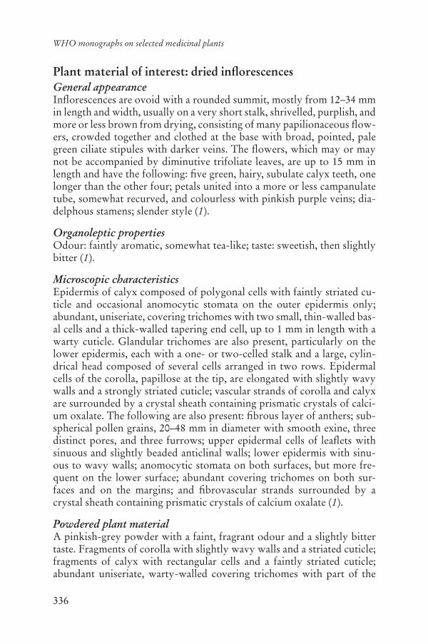
336
WHO monographs on selected medicinal plants
Plant material of interest: dried inflorescencesGeneral appearanceInflorescences are ovoid with a rounded summit, mostly from 12–34 mmin length and width, usually on a very short stalk, shrivelled, purplish, and more or less brown from drying, consisting of many papilionaceous flow-ers, crowded together and clothed at the base with broad, pointed, pale green ciliate stipules with darker veins. The flowers, which may or may not be accompanied by diminutive trifoliate leaves, are up to 15 mm in length and have the following: five green, hairy, subulate calyx teeth, one longer than the other four; petals united into a more or less campanulate tube, somewhat recurved, and colourless with pinkish purple veins; dia-delphous stamens; slender style (1).
Organoleptic propertiesOdour: faintly aromatic, somewhat tea-like; taste: sweetish, then slightly bitter (1).
Microscopic characteristicsEpidermis of calyx composed of polygonal cells with faintly striated cu-ticle and occasional anomocytic stomata on the outer epidermis only; abundant, uniseriate, covering trichomes with two small, thin-walled bas-al cells and a thick-walled tapering end cell, up to 1 mm in length with a warty cuticle. Glandular trichomes are also present, particularly on the lower epidermis, each with a one- or two-celled stalk and a large, cylin-drical head composed of several cells arranged in two rows. Epidermal cells of the corolla, papillose at the tip, are elongated with slightly wavy walls and a strongly striated cuticle; vascular strands of corolla and calyx are surrounded by a crystal sheath containing prismatic crystals of calci-um oxalate. The following are also present: fibrous layer of anthers; sub-spherical pollen grains, 20–48 mm in diameter with smooth exine, three distinct pores, and three furrows; upper epidermal cells of leaflets with sinuous and slightly beaded anticlinal walls; lower epidermis with sinu-ous to wavy walls; anomocytic stomata on both surfaces, but more fre-quent on the lower surface; abundant covering trichomes on both sur-faces and on the margins; and fibrovascular strands surrounded by a crystal sheath containing prismatic crystals of calcium oxalate (1).
Powdered plant materialA pinkish-grey powder with a faint, fragrant odour and a slightly bitter taste. Fragments of corolla with slightly wavy walls and a striated cuticle; fragments of calyx with rectangular cells and a faintly striated cuticle; abundant uniseriate, warty-walled covering trichomes with part of the

337
Flos Trifolii
end cell frequently broken off; glandular trichomes less frequent; sub-spherical pollen grains, scattered or associated with fragments of fibrous layer of anthers; abundant strands of vascular tissue with associated crys-tal sheath containing prismatic crystals of calcium oxalate; occasional portions of green leaf with wavy walled epidermis and anomocytic sto-mata (3).
General identity testsMacroscopic and microscopic examination, as well as thin-layer and high-performance liquid chromatography (1, 3).
Purity testsMicrobiologicalTests for specific microorganisms and microbial contamination limits are as described in the WHO guidelines for assessing quality of herbal medi-cines with reference to contaminants and residues (9).
The United States pharmacopeia requires the absence of Salmonellaspecies and Escherichia coli, with total aerobic microbial count not ex-ceeding 106 colony-forming units (cfu) per g, the total combined moulds and yeast count should not exceed 104 cfu per g, and the enterobacterial count should not be more than 1000 cfu per g (1).
Foreign organic matterNot more than 2% (1).
Total ashNot more than 10% (1).
Acid-insoluble ashNot more than 2% (1).
Water-soluble extractiveNot less than 15% (1).
Loss on dryingNot more than 12% (1).
Pesticide residuesThe recommended maximum limit of aldrin and dieldrin is not more than 0.05 mg/kg (10). For other pesticides, see the European pharmaco-poeia (10) and the WHO guidelines for assessing quality of herbal medi-

338
WHO monographs on selected medicinal plants
cines with reference to contaminants and residues (9) and pesticide resi-dues (11).
Heavy metalsFor maximum limits and analysis of heavy metals, consult the WHO guidelines for assessing quality of herbal medicines with reference to con-taminants and residues (9). The United States pharmacopeia stipulates to-tal heavy metals not more than 10 μg/g (1).
Radioactive residuesWhere applicable, consult the WHO guidelines for assessing quality of herbal medicines with reference to contaminants and residues (9).
Other purity testsChemical tests to be established in accordance with national requirements.
Chemical assaysNot less than 0.5% isoflavones, calculated on the dried basis as the sum of daidzein, genistein, formononetin and biochanin A (1).
Major chemical constituentsRich in isoflavonoids. The major active estrogenic isoflavonoids are bio-chanin A, daidzein, formononetin and genistein (5, 13, 14, 17). The struc-tures of these isoflavonoids are presented below.
Medicinal usesUses supported by clinical dataNone.
Uses described in pharmacopoeias and well established documentsAlthough numerous clinical trials have assessed the safety and efficacy of red clover extracts for the treatment of menopausal symptoms, hyperlipi-daemia, osteoporosis and prostate cancer (15–19), the data are as yet in-sufficient to support any of these indications. Further data from well-controlled clinical trials with sufficient numbers of subjects are needed before any therapeutic indications can be made.
Formononetin
O
O
HO
OR1 R2
R1 = R2 = HDaidzeinR1 = H R2 = CH3
R1 = OH R2 = HGenisteinBiochanin A R1 = OH R2 = CH3
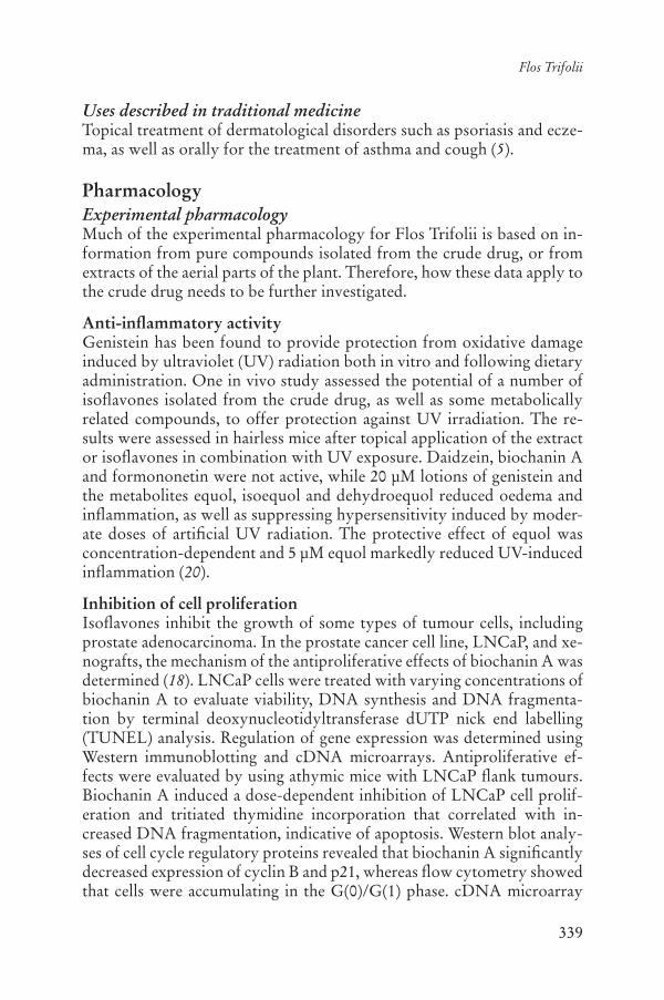
339
Flos Trifolii
Uses described in traditional medicineTopical treatment of dermatological disorders such as psoriasis and ecze-ma, as well as orally for the treatment of asthma and cough (5).
PharmacologyExperimental pharmacologyMuch of the experimental pharmacology for Flos Trifolii is based on in-formation from pure compounds isolated from the crude drug, or from extracts of the aerial parts of the plant. Therefore, how these data apply to the crude drug needs to be further investigated.
Anti-inflammatory activityGenistein has been found to provide protection from oxidative damage induced by ultraviolet (UV) radiation both in vitro and following dietary administration. One in vivo study assessed the potential of a number of isoflavones isolated from the crude drug, as well as some metabolically related compounds, to offer protection against UV irradiation. The re-sults were assessed in hairless mice after topical application of the extract or isoflavones in combination with UV exposure. Daidzein, biochanin A and formononetin were not active, while 20 μM lotions of genistein and the metabolites equol, isoequol and dehydroequol reduced oedema and inflammation, as well as suppressing hypersensitivity induced by moder-ate doses of artificial UV radiation. The protective effect of equol was concentration-dependent and 5 μM equol markedly reduced UV-induced inflammation (20).
Inhibition of cell proliferationIsoflavones inhibit the growth of some types of tumour cells, including prostate adenocarcinoma. In the prostate cancer cell line, LNCaP, and xe-nografts, the mechanism of the antiproliferative effects of biochanin A was determined (18). LNCaP cells were treated with varying concentrations of biochanin A to evaluate viability, DNA synthesis and DNA fragmenta-tion by terminal deoxynucleotidyltransferase dUTP nick end labelling (TUNEL) analysis. Regulation of gene expression was determined using Western immunoblotting and cDNA microarrays. Antiproliferative ef-fects were evaluated by using athymic mice with LNCaP flank tumours. Biochanin A induced a dose-dependent inhibition of LNCaP cell prolif-eration and tritiated thymidine incorporation that correlated with in-creased DNA fragmentation, indicative of apoptosis. Western blot analy-ses of cell cycle regulatory proteins revealed that biochanin A significantly decreased expression of cyclin B and p21, whereas flow cytometry showed that cells were accumulating in the G(0)/G(1) phase. cDNA microarray
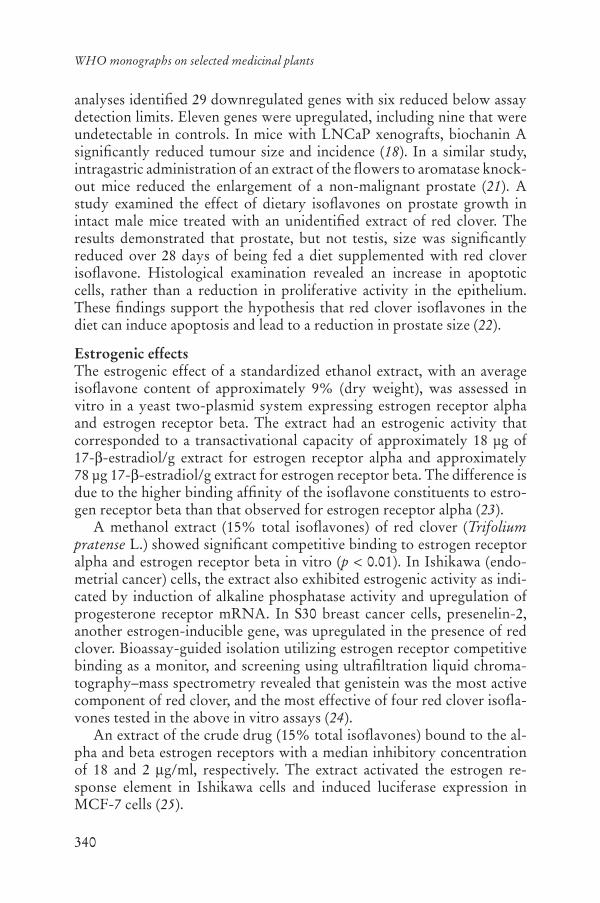
340
WHO monographs on selected medicinal plants
analyses identified 29 downregulated genes with six reduced below assay detection limits. Eleven genes were upregulated, including nine that were undetectable in controls. In mice with LNCaP xenografts, biochanin A significantly reduced tumour size and incidence (18). In a similar study, intragastric administration of an extract of the flowers to aromatase knock-out mice reduced the enlargement of a non-malignant prostate (21). A study examined the effect of dietary isoflavones on prostate growth in intact male mice treated with an unidentified extract of red clover. The results demonstrated that prostate, but not testis, size was significantly reduced over 28 days of being fed a diet supplemented with red clover isoflavone. Histological examination revealed an increase in apoptotic cells, rather than a reduction in proliferative activity in the epithelium. These findings support the hypothesis that red clover isoflavones in the diet can induce apoptosis and lead to a reduction in prostate size (22).
Estrogenic effectsThe estrogenic effect of a standardized ethanol extract, with an average isoflavone content of approximately 9% (dry weight), was assessed in vitro in a yeast two-plasmid system expressing estrogen receptor alpha and estrogen receptor beta. The extract had an estrogenic activity that corresponded to a transactivational capacity of approximately 18 μg of 17- -estradiol/g extract for estrogen receptor alpha and approximately 78 μg 17- -estradiol/g extract for estrogen receptor beta. The difference is due to the higher binding affinity of the isoflavone constituents to estro-gen receptor beta than that observed for estrogen receptor alpha (23).
A methanol extract (15% total isoflavones) of red clover (Trifolium pratense L.) showed significant competitive binding to estrogen receptor alpha and estrogen receptor beta in vitro (p < 0.01). In Ishikawa (endo-metrial cancer) cells, the extract also exhibited estrogenic activity as indi-cated by induction of alkaline phosphatase activity and upregulation of progesterone receptor mRNA. In S30 breast cancer cells, presenelin-2, another estrogen-inducible gene, was upregulated in the presence of red clover. Bioassay-guided isolation utilizing estrogen receptor competitive binding as a monitor, and screening using ultrafiltration liquid chroma-tography–mass spectrometry revealed that genistein was the most active component of red clover, and the most effective of four red clover isofla-vones tested in the above in vitro assays (24).
An extract of the crude drug (15% total isoflavones) bound to the al-pha and beta estrogen receptors with a median inhibitory concentration of 18 and 2 g/ml, respectively. The extract activated the estrogen re-sponse element in Ishikawa cells and induced luciferase expression in MCF-7 cells (25).

341
Flos Trifolii
An in vivo study assessed the estrogenic effects of the crude drug in ovariectomized rats (25). A red clover extract, standardized to contain 15% isoflavones was administered by gavage, at a dose of 250, 500 or 750 mg/kg body weight (bw) per day, to virgin, ovariectomized 50-day-old rats, for 21 days in the presence and absence of 17- -estradiol (50 μg/kg bw per day). The estrogenic effects assessed included an increase in uterine weight, vaginal cell cornification and branching of mammary gland ducts. The extract of the flowers produced a dose-dependent in-crease in uterine weight and differentiated vaginal cells at the two higher doses, but it did not stimulate cell proliferation in the mammary glands. Neither antiestrogenic nor additive estrogenic properties were observed in any of the tissues studied. These data suggest that red clover extract is weakly estrogenic in ovariectomized rats (26).
Thyroid effectsThe effects of isoflavones on the secretion of thyroid hormones as well as on the immunoreactivity to estrogen receptor alpha in the thyroid glands of ovariectomized ewes were studied. Eight ewes were fed 3.5 kg of 100% red clover silage daily for 14 days. Blood samples were collected before and on day 14 of exposure to phytoestrogens. After 5 months, four of the ewes were re-exposed to red clover silage as described above and the oth-er four served as controls. Ewes exposed to red clover silage had signifi-cantly higher plasma concentrations of total T(3) and free T(3) than ewes fed hay. The cross-sectional area of thyroid follicles tended to be larger in ewes fed red clover silage than in the control animals. Estrogen receptor alpha immunoreactivity was stronger in thyroid glands from ewes ex-posed to phytoestrogens than in ewes fed hay. Daily ingestion of 81–95 mgphytoestrogens per kg bw for 14 days stimulated secretion of thyroid hormones and tended to increase follicle size and estrogen receptor alpha immunoreactivity of thyroid glands (27).
Clinical pharmacologyA randomized, double-blind, placebo-controlled, cross-over trial involv-ing 51 perimenopausal and postmenopausal women assessed the effects of an extract of the crude drug for the treatment of hot flushes (28). The subjects had been amenorrhoeic for at least 6 months and had at least three hot flushes per day. The women received one tablet of either placebo or the extract (containing 40 mg total isoflavones, including genistein, 4.0 mg; daidzein, 3.5 mg; biochanin, 24.5 mg; and formononetin, 8 mg).Phase one of the trial lasted for 3 months, and was followed by a 1-month washout period. The subjects were then crossed over to the other arm for a further 14 weeks. All subjects were required to maintain a diary of

342
WHO monographs on selected medicinal plants
symptoms based on the Greene Menopause Score list. Of the initial 51 subjects entering the trial, 43 completed the study. At 12 weeks the frequency of hot flushes had decreased in both the group receiving pla-cebo and in the group receiving the treatment, by 18% and 20%, respec-tively. However, there were no statistically significant differences between groups in frequency of hot flushes or Greene Scores at any time point. No significant changes in body weight, steroid hormone binding globulin levels, blood counts, serum electrolytes, urea, creatinine or liver function were observed (28).
A second randomized, double-blind, placebo-controlled trial involved 37 perimenopausal and postmenopausal women and also assessed the ef-fects of an extract of the crude drug for the treatment of hot flushes (15).All subjects had been amenorrhoeic for at least 6 months and had at least three hot flushes per day. The women were randomly assigned to receive either one tablet of placebo or one of two doses of an extract (containing 40 mg or 160 mg of total isoflavones; the 40-mg tablet containing genistein, 4.0 mg; daidzein, 3.5 mg; biochanin, 24.5 mg; and formononetin, 8 mg)for 12 weeks. The outcomes measured were similar to those of the previ-ous study. During the 12-week treatment period, the frequency of hot flushes was reduced by 35% in the placebo group; by 29% in the group treated with 40 mg; and 34% in the group treated with 160 mg of the ex-tract. No significant changes in body weight, levels of steroid hormone binding globulin, blood counts, serum electrolytes, urea, creatinine or liver function were observed (15).
A third randomized, placebo-controlled trial involving 30 menopausal women who had had amenorrhoea for more than 12 months, and who were also experiencing more than five hot flushes per day, assessed the effect of the same extract as described in the previous two studies for the treatment of menopausal symptoms. All subjects participated in a single-blind phase in which they received placebo tablets for 4 weeks, and then they were randomly assigned to receive either placebo or 80 mg isofla-vones for a further 12 weeks. Efficacy was measured by the decrease in number of hot flushes per day and changes in the Greene Menopause Scale score. During the first 4 weeks of treatment with the placebo, the frequency of hot flushes decreased by 16%. During the subsequent dou-ble-blind phase, a further, statistically significant decrease of 44% was seen in the group treated with 80 mg of isoflavones (p < 0.01), whereas no further reduction occurred in women in the group treated with the pla-cebo. The Greene Menopause Scale score decreased by 13% in the group treated with the isoflavones and remained unchanged in the women treat-ed with the placebo (29).

343
Flos Trifolii
A fourth study compared the efficacy and safety of two products de-rived from red clover with that of a placebo in symptomatic menopausal women. This randomized, double-blind, placebo-controlled trial involved menopausal women, aged 45–60 years, who were experiencing at least 35 hot flushes per week. The study included women who had recently become postmenopausal (mean (standard deviation), 3.3 (4.5) years since menopause) experiencing on average 8.1 hot flushes per day. Women were excluded from the study if they were vegetarians, consumed soy products more than once per week, or took medications affecting isoflavone ab-sorption. After a 2-week placebo run-in, 252 participants were randomly assigned to receive either Promensil (82 mg of total isoflavones per day), Rimostil (57 mg of total isoflavones per day) or a placebo identical in ap-pearance to the clover products, and followed up for 12 weeks. The pri-mary outcome measured was the change in frequency of hot flushes as recorded by participants in their daily diaries. Secondary outcome mea-sures included changes in quality of life and adverse events. Of 252 par-ticipants, 246 (98%) completed the 12-week protocol. The reductions in mean daily count of hot flushes at 12 weeks were similar in all three groups: Promensil, 5.1; Rimostil, 5.4; and placebo, 5.0. In comparison with the group treated with the placebo, participants who received the Promensil (41%; 95% confidence interval (CI), 29%–51%; p = 0.03), but not those treated with Rimostil (34%; 95% CI, 22%–46%; p = 0.74) found that the treatment reduced hot flushes more rapidly. Improvements in quality of life and reports of adverse events were comparable in all three groups (19).
Data from two clinical trials, published only as abstracts, are also avail-able (30). A randomized, double-blind, placebo-controlled pilot study conducted in Peru assessed the efficacy of a red clover product in 30 post-menopausal women. The women were treated with 40 mg of the extract or a placebo for 4 months (31). A 75% reduction in hot flushes occurred in the treatment group; however, no data for the placebo group were re-ported. The second study was an uncontrolled trial (no placebo group) in 23 women with ammenorrhoea for 12 months (32). In patients treated with 40.0 mg of the extract, there was a 56% reduction in the frequency of hot flushes over a 2-month period. The severity of hot flushes decreased by 43% and the severity of night sweats decreased by 52%. No changes in endometrial thickness and no adverse effects were observed. Complete blood counts were all within normal limits (32).
The effects of a red clover-derived isoflavone extract on the Ki-67 pro-liferative marker of endometrial biopsies were assessed in a double-blind, randomized, controlled study involving 30 perimenopausal women. The

344
WHO monographs on selected medicinal plants
purpose of the study was to detect a decrease in the Ki-67 proliferative index during the late follicular phase after a 3-month course of 50.0 mg/day red clover isoflavones. The biopsies were timed as close as possible to days 7–11 of the menstrual cycle, and simultaneous measurements of trans-vaginal endometrial thickness, uterine artery Doppler, hormone profiles, lipids and bone markers were made. Of 30 women, two did not return for a second biopsy, and a third had an unsuccessful second biopsy. Four subjects were excluded from the intention-to-treat analysis because they did not have menstrual bleeding within the time frame of the study (3 subjects) or were tested on day 13 instead of between days 7 and 11 of the cycle (1 subject). There was no change in the Ki-67 proliferation index after treatment in either group. Eight women in the group given the placebo and eight in the treatment group had proliferative endometrial biopsies that were synchronized with estradiol levels at baseline and post-treatment, and analysis of these subjects revealed no detectable changein the relationship between estradiol levels and Ki-67 with treatment in either group. There was no change in fasting lipids, bone markers, uterine Doppler resistance or pulsatility index (33).
Effects on bone and cardiovascular systemSix trials have assessed the effects of isoflavones on total cholesterol and lipid levels (27, 34–38). A randomized, single-blind, cross-over study in-volving 21 premenopausal women with regular menstrual cycles assessed the effect of an extract of the crude drug on plasma lipids and oxidization of low-density lipoprotein cholesterol (34). Subjects were treated with a placebo or 86 mg of isoflavones per day for two menstrual cycles, after which they were crossed over to the other group. Fourteen women com-pleted the study and no differences in total cholesterol, triglycerides or oxidized low-density lipoprotein were observed.
A randomized, double-blind, placebo-controlled study involving 66 postmenopausal women with hypercholesterolaemia assessed the ef-fects of an ascending dose of the extract, 40 or 80 mg of isoflavones, on lipid profiles (35). Study participants were asked to follow a low isofla-vone diet for 3 months during the study. Treatment did not affect total cholesterol, low-density lipoprotein, high-density lipoprotein cholesterol or plasma triglycerides at any dose.
A double-blind placebo-controlled trial compared the effects of two doses of isoflavones, 40 and 80 mg, with the effects of a placebo on arte-rial compliance and plasma lipids in menopausal women. After a 3–4-week run-in period and a 5-week placebo phase, 26 women were ran-domly allocated to receive either 40 mg of the extract or the placebo for an additional 5 weeks, and then the dose was increased to 80 mg. Sixteen

345
Flos Trifolii
women completed the trial. The results demonstrated a significant im-provement in arterial compliance at both the 40- and 80-mg doses as com-pared with placebo (p < 0.05). No significant differences were seen be-tween the two doses (30, 36).
A double-blind study to evaluate the effects of varying doses of iso-flavones on lipid and bone metabolism in postmenopausal women was performed. An extract of red clover, containing genistein, daidzein, for-mononetin and biochanin was administered to 46 postmenopausal women after a single-blind placebo phase; this was followed by a single-blind wash-out phase. Patients were randomly assigned to receive 28.5 mg, 57 mg or 85.5 mg of isoflavones daily for 6 months. At 6 months, the serum high-density lipoprotein cholesterol was found to have risen significantly by 15.7–28.6% with the different doses (p = 0.007, p = 0.002 and p = 0.027 at doses of 28.5 mg, 57 mg and 85.5 mg, respectively), al-though the magnitude of the response was independent of the dose used. The serum apo-lipoprotein B fell significantly by 11.5–17.0% with the different doses (p = 0.005, p = 0.043, p = 0.007, respectively) and the magnitude of the response was independent of the dose used. The bone mineral density of the proximal radius and ulna rose significantly by 4.1% over 6 months in the women who received 57 mg/day (p = 0.002) and by 3.0% in the women who received 85.5 mg/day (p = 0.023) of isoflavones. The response to treatment with 28.5 mg/day of isoflavones was not significant. These results show that the administration of an iso-flavone combination extracted from red clover was associated with a sig-nificant increase in high-density lipoprotein cholesterol, a significant fall in apolipoprotein B, and a small but significant increase in the predomi-nantly cortical bone of the proximal radius and ulna after 6 months of treatment (37).
A double-blind, placebo-controlled study, involving 107 premeno-pausal, perimenopausal and postmenopausal women assessed the effects of an extract of the crude drug (corresponding to 40 mg of isoflavones per day) for 1 year on bone mineral density and content (39). The outcomes measured were changes in the lumbar spine and total hip bone mineral content and density. After 1 year, the bone mineral content and density of the lumbar spine (measured by dual energy X-ray absorptiometry (DEXA) scan) had decreased significantly (p < 0.01) in both groups. However, the decrease in the bone mineral content and density of the spine of premenopausal and perimenopausal women treated with the ex-tract was significantly lower than that in women who received the placebo (p < 0.02 and p < 0.01, respectively). No differences were observed in the postmenopausal women. No differences in bone mineral density of the
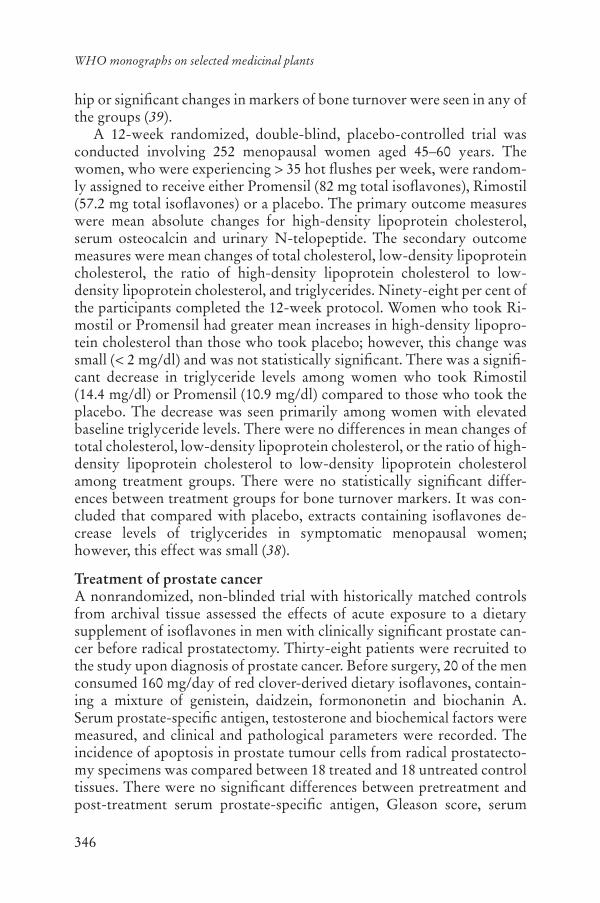
346
WHO monographs on selected medicinal plants
hip or significant changes in markers of bone turnover were seen in any of the groups (39).
A 12-week randomized, double-blind, placebo-controlled trial was conducted involving 252 menopausal women aged 45–60 years. The women, who were experiencing > 35 hot flushes per week, were random-ly assigned to receive either Promensil (82 mg total isoflavones), Rimostil (57.2 mg total isoflavones) or a placebo. The primary outcome measures were mean absolute changes for high-density lipoprotein cholesterol, serum osteocalcin and urinary N-telopeptide. The secondary outcome measures were mean changes of total cholesterol, low-density lipoprotein cholesterol, the ratio of high-density lipoprotein cholesterol to low- density lipoprotein cholesterol, and triglycerides. Ninety-eight per cent ofthe participants completed the 12-week protocol. Women who took Ri-mostil or Promensil had greater mean increases in high-density lipopro-tein cholesterol than those who took placebo; however, this change was small (< 2 mg/dl) and was not statistically significant. There was a signifi-cant decrease in triglyceride levels among women who took Rimostil (14.4 mg/dl) or Promensil (10.9 mg/dl) compared to those who took the placebo. The decrease was seen primarily among women with elevated baseline triglyceride levels. There were no differences in mean changes of total cholesterol, low-density lipoprotein cholesterol, or the ratio of high-density lipoprotein cholesterol to low-density lipoprotein cholesterol among treatment groups. There were no statistically significant differ-ences between treatment groups for bone turnover markers. It was con-cluded that compared with placebo, extracts containing isoflavones de-crease levels of triglycerides in symptomatic menopausal women; however, this effect was small (38).
Treatment of prostate cancerA nonrandomized, non-blinded trial with historically matched controls from archival tissue assessed the effects of acute exposure to a dietary supplement of isoflavones in men with clinically significant prostate can-cer before radical prostatectomy. Thirty-eight patients were recruited to the study upon diagnosis of prostate cancer. Before surgery, 20 of the men consumed 160 mg/day of red clover-derived dietary isoflavones, contain-ing a mixture of genistein, daidzein, formononetin and biochanin A. Serum prostate-specific antigen, testosterone and biochemical factors were measured, and clinical and pathological parameters were recorded. The incidence of apoptosis in prostate tumour cells from radical prostatecto-my specimens was compared between 18 treated and 18 untreated control tissues. There were no significant differences between pretreatment and post-treatment serum prostate-specific antigen, Gleason score, serum

347
Flos Trifolii
testosterone or biochemical factors in the treated patients (p > 0.05). Apoptosis in radical prostatectomy specimens from treated patients was significantly higher than in control subjects (p = 0.0018), specifically in regions of low to moderate-grade cancer (Gleason grade 1–3). No ad-verse events related to the treatment were reported (18).
Pharmacokinetics and pharmacodynamicsThe pharmacokinetics of red clover isoflavones were investigated after long-term administration as a once-daily dietary supplement. Fourteen subjects who had been consuming a low-isoflavone containing diet for 2 weeks were given an oral dose of two isoflavone tablets (approximately 80 mg of total isoflavones) daily for 2 weeks. Before the study day the participants fasted overnight and were then administered their last dose the next morning. Plasma samples were collected for a 48-hour period after the last dose. Plasma isoflavones were assayed by high-performance liquid chromatography. The results demonstrate that trough plasma lev-els were significantly higher for daidzein and genistein after long-term dosing than levels recorded prior to the commencement of the study and plasma levels of isoflavones after long-term dosing were in the range pre-viously reported in populations that consume an isoflavone-rich diet. The plasma half-lives observed after long-term administration were consistent with once-daily administration. Isoflavones have pharmacokinetic char-acteristics that suggest that once-daily administration is adequate when they are administered long-term (40).
The absorption of isoflavones varies substantially between individu-als. A single-blind, randomized, placebo-controlled, cross-over trial involving 14 subjects investigated whether isoflavone absorption differs depending on whether the isoflavones originated from soy beans or from red clover. Soy bean isoflavone glycosides and red clover isofla-vone aglycones were incorporated into a breakfast cereal and eaten daily for 2 weeks each, separated by a 2-week control or washout period. The excretion of isoflavones in urine was measured over a 24-hour period; approximately 25% of each isoflavone was recovered in urine, suggesting that similar amounts were absorbed irrespective of their glycoside/aglycone nature or the differing compositions of their sources (daidzein and genistein in soy beans and formononetin and biochanin in red clover). Although interindividual variability was high, there was less intraindividual variability; the amounts excreted when subjects consumed the two sources of isoflavone were correlated (r = 0.69; p = 0.007) (41).

348
WHO monographs on selected medicinal plants
Adverse reactionsIngestion of large amounts of clover in animal feed has been associated with a number of adverse effects in sheep in Australia. A publication on “clover disease” described symptoms of infertility, abnormal lactation, dystocia and prolapsed uterus, all of which were hypothetically attributed to the estrogenic effects of isoflavones (42). None of the controlled, clini-cal trials has reported adverse effects at doses up to 160 mg of isoflavones per day. Trifolium pratense does not contain coumarins, and therefore the concerns about blood coagulation are unfounded (43, 44).
ContraindicationsFlos Trifolii is contraindicated in cases of hypersensitivity or allergy to the crude drug. It is also contraindicated during pregnancy, breastfeeding and for children under the age of 12 years, and in cases of hormone- associated diseases, due to the potential hormonal effects.
WarningsDue to the potential estrogenic effects of the crude drug, patients with hormone-related disorders, estrogen-dependent cancers or a familial his-tory of estrogen-dependent cancers should contact a health care provider before use.
PrecautionsCarcinogenesis, mutagenesis, impairment of fertilityNo information was found.
Drug interactionsThere are conflicting data concerning an interaction of the crude drug with tamoxifen and other antiestrogenic drugs. Some studies suggest that specific isoflavones may enhance the ability of tamoxifen to inhibit the growth of estrogen-receptor-positive breast cancer cells (45–47). In ro-dent models, genistein has been shown to inhibit the efficacy of tamoxifen on the growth of estrogen-receptor-positive breast cancer cells implanted in ovariectomized mice, while other research shows that specific isofla-vones may be additive and work in a synergistic manner to prevent the development of chemically induced tumours and the growth of existing tumours (48–50). Therefore, the use of the crude drug or its preparations is not recommended in those being treated with tamoxifen and other anti-estrogenic drugs until further research has been done.
349
Flos Trifolii
Pregnancy: non-teratogenic effectsSee Contraindications.
Nursing mothersSee Contraindications.
Paediatric useSee Contraindications.
Dosage formsCrude drug and tablets.
Posology(Unless otherwise indicated)Oral dose: extracts of crude drug: 240–480 mg corresponding to 40–80 mg/day of isoflavones (15, 17, 28, 29).
ReferencesThe United States Pharmacopeia. 28. 1. Rockville, MD, United States Pharma-copeia Convention, 2005.Bedevian AK.2. Illustrated polyglottic dictionary of plant names. Cairo, Med-bouly Library, 1994.British herbal pharmacopoeia3. . Exeter, British Herbal Medicine Association, 1996.Bradley PR, ed. 4. British herbal compendium. Vol. 1. Bournemouth, British Herbal Medicine Association, 1992.Farnsworth NR, ed. 5. NAPRALERT database. Chicago, University of Illinois at Chicago, IL (an online database available directly through the University of Illinois at Chicago or through the Scientific and Technical Network [STN] of Chemical Abstracts Services), 30 June 2005.Ernst E et al, eds. 6. The desktop guide to complementary and alternative med-icine. Edinburgh, Mosby, 2001.Youngken HW. 7. Textbook of pharmacognosy. Philadelphia, Blakiston, 1950.Multilingual multiscript plant name database: Sorting Trifolium names8. . University of Melbourne (http://www.plantnames.unimelb.edu.au/Sorting/Trifolium.html).WHO guidelines for assessing quality of herbal medicines with reference to 9.contaminants and residues. Geneva, World Health Organization, 2007.European Pharmacopoeia10. , 5th ed. Strasbourg, Directorate for the Quality of Medicines of the Council of Europe (EDQM), 2005.Guidelines for predicting dietary intake of pesticide residues11. , 2nd rev. ed. Geneva, World Health Organization, 1997 (WHO/FSF/FOS/97.7).Newall CA, Anderson LA, Phillipson JD. 12. Herbal medicines, a guide for healthcare professionals. London, Pharmaceutical Press, 1996.
350
WHO monographs on selected medicinal plants
Lin LZ et al. LC-ESI-MS study of the flavonoid glycoside malonates of red 13.clover (Trifolium pratense). Journal of Agricultural Food Chemistry, 2000, 48:354–365.He XG, Lin LZ, Lian LZ. Analysis of flavonoids from red clover by liquid 14.chromatography-electrospray mass spectrometry. Journal of Chromatogra-phy – A, 1996, 755:127–132.Knight DC, Howes JB, Eden JA. The effect of Promensil, an isoflavone ex-15.tract, on menopausal symptoms. Climacteric, 1999, 2:79–84.Murkies AL et al. Dietary flour supplementation decreases post-menopausal 16.hot flushes: effect of soy and wheat. Maturitas, 1995, 21:189–195.NAMS – North American Menopause Society. The role of isoflavones in 17.menopausal health: consensus opinion of the North American Menopause Society. Menopause, 2000, 7:215–229.Jarred RA et al. Induction of apoptosis in low to moderate-grade human 18.prostate carcinoma by red clover-derived dietary isoflavones. Cancer Epi-demiology and Biomarkers Previews, 2002, 11:1689–1696.Tice JA et al. Phytoestrogen supplements: for the treatment of hot flashes: 19.the isoflavone clover extract (ICE) study. Journal of the American Medical Association, 2003, 290:207–214.Widyarini S et al. Isoflavonoid compounds from red clover (20. Trifolium prat-ense) protect from inflammation and immune suppression induced by UV radiation. Photochemistry and Photobiology, 2001, 74:465–470.Jarred RA et al. Anti-androgenic action by red clover-derived dietary isofla-21.vones reduces non-malignant prostate enlargement in aromatase knockout (ArKo) mice. Prostate, 2003, 56:54–64.Dornstauder E et al. Estrogenic activity of two standardized red clover ex-22.tracts (Menoflavon) intended for large scale use in hormone replacement ther-apy. Journal of Steroid Biochemistry and Molecular Biology, 2001, 78:67–75.Risbridger GP et al. The in vivo effect of red clover diet on ventral prostate 23.growth in adult male mice. Reproductive Fertility and Development, 2001, 13:325–329.Liu J et al. Evaluation of estrogenic activity of plant extracts for the potential 24.treatment of menopausal symptoms. Journal of Agriculture and Food Chem-istry, 2000, 49:2472–2479.Overk CR et al. Comparison of the in vitro estrogenic activities of com-25.pounds from hops (Humulus lupulus) and red clover (Trifolium pratense).Journal of Agriculture and Food Chemistry, 2005, 53:6246–6253.Burdette JE et al. 26. Trifolium pratense (red clover) exhibits estrogenic effects in vivo in ovariectomized Sprague-Dawley rats. Journal of Nutrition, 2002, 132:27–30.Madej A et al. Thyroid gland function in ovariectomized ewes exposed to 27.phytoestrogens. Journal of Chromatography B Analytical and Technological Biomedical Life Sciences, 2002, 777:281–287.Baber RJ et al. Randomized placebo-controlled trial of an isoflavone supple-28.ment and menopausal symptoms in women. Climacteric, 1999, 2:85–92.

351
Flos Trifolii
Van de Weijer P, Barentsen R. Isoflavones from red clover (Promensil) sig-29.nificantly reduce menopausal hot flush symptoms compared with placebo. Maturitas, 2002, 42:187–200.Fugh-Berman A, Kronenberg F. Red clover (30. Trifolium pratense) for meno-pausal women: current state of knowledge. Menopause, 2001, 8:333–337.Jeri AR, de Romana C. The effect of isoflavone phytoestrogens in relieving 31.hot flushes in Peruvian postmenopausal women (abstract). In: The Proceed-ings of the 9th International Menopause Society World Congress on the Menopause. Yokohama, Japan, 1999:129.Nachtigall LB et al. The effects of isoflavones derived from red clover on 32.vasomotor symptoms and endometrial thickness (abstract). In: The Proceed-ings of the 9th International Menopause Society World Congress on the Menopause. Yokohama, Japan, 1999, 128.Hale GE et al. A double-blind randomized study on the effects of red clover 33.isoflavones on the endometrium. Menopause, 2001, 8:338–346.Samman S et al. The effect of supplementation with isoflavones on plasma 34.lipids and oxidisability of low density lipoprotein in premenopausal women. Atherosclerosis, 1999, 47:277–283.Howes JB et al. The effects of dietary supplementation with isoflavones from 35.red clover on the lipoprotein profiles of postmenopausal women with mild to moderate hypercholesterolemia. Atherosclerosis, 2000, 152:143–147.Nestel PJ et al. Isoflavones from red clover improve systemic arterial compli-36.ance but not plasma lipids in menopausal women. Journal of Clinical Endo-crinology and Metabolism, 1999, 84:895–898.Clifton-Bligh PB et al. The effect of isoflavones extracted from red clover 37.(Rimostil) on lipid and bone metabolism. Menopause, 2001, 8:259–265.Knudson Schultz K et al. Effect of isoflavones on lipids and bone turnover 38.markers in menopausal women. Maturitas, 2004, 48:209–218.Atkinson C et al. The effects of isoflavone phytoestrogens on bone: prelimi-39.nary data from a large randomized controlled trial (abstract). In: Proceedingsof the Endocrine Society, Toronto, Canada, 2000:43.Howes J et al. Long-term pharmacokinetics of an extract of isoflavones from 40.red clover (Trifolium pratense). Journal of Alternative and Complementary Medicine, 2002, 8:135–142.Tsunoda N, Pomeroy S, Nestel P. Absorption in humans of isoflavones from 41.soy and red clover is similar. Journal of Nutrition, 2002, 132:2199–2201.Lewis’ dictionary of toxicology42. . Boca Raton, FL, CRC Press, 1998.Piersen CE et al. Chemical and biological characterization and clinical evalu-43.ation of botanical dietary supplements: A phase I red clover extract as a mod-el. Current Medicinal Chemistry, 2004, 11:1361–1374.Booth N et al. Confusion regarding anticoagulant coumarins in dietary sup-44.plements. Clinical Pharmacology and Therapeutics, 2004, 511–516.Jones JL et al. Genistein inhibits tamoxifen effects on cell proliferation and 45.cell cycle arrest in T47D breast cancer cells. American Surgery, 2002, 68:575–577.

352
WHO monographs on selected medicinal plants
Han D, Tachibana H, Yamada K. Inhibition of environmental estrogen-in-46.duced proliferation of human breast carcinoma MCF-7 cells by flavonoids. In Vitro Cell Development and Biology, 2001, 37:275–282.Tanos V et al. Synergistic inhibitory effects of genistein and tamoxifen on 47.human dysplastic and malignant epithelial breast cells in vitro. European Journal of Obstetrics and Gynecology Reproduction Biology, 2002, 37:188–194.Ju YH et al. Dietary genistein negates the inhibitory effect of tamoxifen on 48.growth of estrogen-dependent human breast cancer (MCF-7) cells implanted in athymic mice. Cancer Research, 2002, 62:2474–2477.Constantinou A et al. Consumption of soy products may enhance the breast 49.cancer preventive effects of tamoxifen. Proceedings of the American Associa-tion of Cancer Research, 2001, 42:826.Gotoh T et al. Chemoprevention of N-nitroso-N-methylurea-induced rat 50.mammary cancer by miso and tamoxifen, alone and in combination. JapaneseJournal of Cancer Research, 1998, 89:487–495.

353
Ramulus cum Uncis Uncariae
DefinitionRamulus cum Uncis Uncariae consists of the dried hook-bearing stem branch of Uncaria rhynchophylla (Miq.) Jacks, U. macrophylla Wall., U. hirsuta Havil., U. sinensis (Oliv.) Havil. or U. sessilifructus Roxb. (Rubiaceae) (1–3).
SynonymsUncaria rhynchophylla (Miq.) Jacks, Nauclea rhynchophylla Miq., Our-ouparia rhynchophylla Matsum (4).
Selected vernacular namesChoto-ko, chotoko, cho-to-kou, gout eng, kagikazura (1, 4, 5).
Geographical distributionIndigenous to China and Japan (6, 7).
DescriptionA deciduous twining shrub up to 10 m long. Branches brownish, gla-brous, occasionally bearing compressed, hooked thorns. Young stems slender, square to slightly angular, glabrous. Stipules of the plagiotropic shoot 6–10 mm long, those of the orthotropic shoot considerably larger, up to 30 mm long, inside glabrous with glandular hairs at the base, outside glabrous, margins entire, narrowly triangular, deeply bifid for over two thirds of the length, lobes narrowly triangular to triangular-lanceolate. Leaves opposite, ovate to ovate-oblong or elliptic to elliptic-oblong, 5–12 × 3–7 cm, membranous, glabrous on either side; apex acute to cus-pidate; base cuneate to truncate; lateral nerves 4–8 pairs, axils with sparse-ly hairy domatia, tertiary nerves curved, impressed, ultimate venation reticulate. Inflorescence an axillary or terminal peduncled solitary head. Flowers yellow, subsessile on the receptacle. Hypanthium 1–2 mm, densely hairy. Calyx 1 mm, pubescent; lobes oblong to slightly triangular, 0.5–1 mm, sparsely pubescent. Corolla tube 6–8 (6–10 mm), outside
354
WHO monographs on selected medicinal plants
glabrous or with a few scattered hairs; lobes oblong, 1.5–2.5 mm, outside glabrous or slightly farinose-pubescent, margins sometimes ciliate. Sta-mens 5. Ovary 2-celled. Fruiting head 14–18 mm in diameter, fruit a dry capsule. The plant entwines other trees with its hooked thorns (6, 8, 9).
Plant material of interest: dried hook-bearing stem branchGeneral appearanceCylindrical or subsquare, 1–4 cm long, 2–5 mm in diameter. Externally reddish-brown to dark brown or yellow-brown with fine longitudinal striations, glabrous, or yellowish-green to greyish-brown, sometimes with white dotted lenticels, covered with yellowish-brown pubescence. Transverse section: square to elliptical. Most nodes of stem branches with two opposite downward curved hooks/prickles (sterile peduncles), some with a hook only on one side and with raised scars on the other side; hooks relatively flattened or rounded, apex acute, base relatively broad; dotted scars of fallen petiole and ring-shaped stipule scars visible on the branch at the hook base; transverse section oblong to elliptical. Texture hard and tenacious, fracture yellowish-brown, bark fibrous, pith yellow-ish-white to light brown or hollowed (1, 2).
Organoleptic propertiesOdour: none; taste: slight (1, 2).
Microscopic characteristicsTransverse section of the prickle (hook) reveals vascular bundles in the cortex, unevenly distributed and arranged in a ring. Parenchyma cells in the secondary cortex containing crystals of calcium oxalate (2, 3).
Powdered plant materialTo be established in accordance with national requirements.
General identity testsMacroscopic examination (1–3), microchemical test (1–3), and high- performance liquid chromatography for the presence of characteristic oxindole alkaloids (2, 10–13).
Purity testsMicrobiologyTests for specific microorganisms and microbial contamination limits are as described in the WHO guidelines for assessing quality of herbal medi-cines with reference to contaminants and residues (14).

355
Ramulus cum Uncis Uncariae
ChemicalTo be established in accordance with national requirements.
Foreign organic matterNot more than 2% (3).
Total ashNot more than 4% (2, 3).
Acid-insoluble ashTo be established in accordance with national requirements.
Water-soluble extractiveTo be established in accordance with national requirements.
Alcohol-soluble extractiveNot more than 6% (1).
Loss on dryingNot more than 12% (2).
Pesticide residuesThe recommended maximum limit of aldrin and dieldrin is not more than 0.05 mg/kg (15). For other pesticides see the European pharmacopoeia (15)and the WHO guidelines for assessing quality of herbal medicines with reference to contaminants and residues (14) and pesticide residues (16).
Heavy metalsFor maximum limits and analysis of heavy metals, consult the WHO guidelines for assessing quality of herbal medicines with reference to con-taminants and residues (14).
Radioactive residuesWhere applicable, consult the WHO guidelines for assessing quality of herbal medicines with reference to contaminants and residues (14).
Chemical assayNot less than 0.03% of total rhynchophylline (rhynchophylline and hir-sutine) as determined by high-performance liquid chromatography and calculated on the dried basis (2).
Major chemical constituentsThe major constituents are indole alkaloids (1.7%) including the tetra-cyclic oxindoles rhynchophylline (0.02%), isorhynchophylline (0.05%),
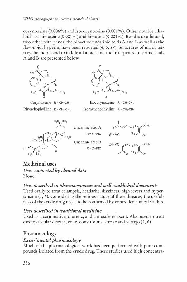
356
WHO monographs on selected medicinal plants
corynoxeine (0.006%) and isocorynoxeine (0.001%). Other notable alka-loids are hirsuteine (0.001%) and hirsutine (0.001%). Besides ursolic acid, two other triterpenes, the bioactive uncarinic acids A and B as well as the flavonoid, hyperin, have been reported (4, 5, 17). Structures of major tet-racyclic indole and oxindole alkaloids and the triterpenes uncarinic acids A and B are presented below.
Medicinal usesUses supported by clinical dataNone.
Uses described in pharmacopoeias and well established documentsUsed orally to treat eclampsia, headache, dizziness, high fevers and hyper-tension (1, 6). Considering the serious nature of these diseases, the useful-ness of the crude drug needs to be confirmed by controlled clinical studies.
Uses described in traditional medicineUsed as a carminative, diuretic, and a muscle relaxant. Also used to treat cardiovascular disease, colic, convulsions, stroke and vertigo (5, 6).
PharmacologyExperimental pharmacologyMuch of the pharmacological work has been performed with pure com-pounds isolated from the crude drug. These studies used high concentra-
O
R
NHN
OH3C
O
O
H H
H
CH3
Corynoxeine
O
R
NHN
OH3C
O
O
H H
H
CH3
Isocorynoxeine R = CH=CH2R = CH=CH2
Rhynchophylline IsorhynchophyllineR = CH2-CH3 R = CH2-CH3
H3C CH3
CH3H3C
CH3 CH3 CO2HH
H
H
HO
H
CO
OCH3
OH
OCH3
OHC
O
E-HMC
Z-HMCO
R
Uncarinic acid A
Uncarinic acid B
R = E-HMC
R = Z-HMC

357
Ramulus cum Uncis Uncariae
tions of the pure compounds and thus the applicability of these data to the crude drug and its preparations needs to be further investigated.
Antiarrhythmic activityThe effect of hirsutine, an indole alkaloid isolated from the crude drug, on membrane potentials of rabbit sino-atrial node and guinea-pig right ven-tricle and left atrium was investigated. Hirsutine at concentrations of 0.1–30.0 μM decreased the maximum rate of rise and prolonged action potential duration in sino-atrial node in vitro, as well as in atrial and ven-tricular preparations (18). Hirsutine, in concentrations of 1.0–3.0 μM pro-duced a dose-dependent relaxation of the isolated rat aorta precontracted with either norepinephrine or potassium chloride solution, suggesting that the compound dilated blood vessels through the inhibition of volt-age-dependent calcium channels (19). The compound, at a concentration of 30 μM, also decreased intracellular calcium concentrations in isolated vascular smooth muscle cells through the inhibition of norepinephrine-induced calcium influx (20).
Anticonvulsant activityIntragastric administration of a methanol extract of the crude drug to rats at a dose of 1.0 g/kg body weight (bw) inhibited kainic acid-induced epi-leptic seizures and reduced the levels of free radicals, as measured by lipid peroxidation in the brain (21).
Intraperitoneal administration of an ethyl acetate or methanol extract of the crude drug to mice at a dose of 70.0 mg/kg bw inhibited pentetra-zole-induced convulsions (22). Hyperin, a chemical constituent isolated from the extract, decreased the elevated activities of -aminobutyricacid-T, xanthine oxidase and lipid peroxide induced by pentetrazole in mouse brain tissue in vitro at a concentration of 25.0 mg/ml (23).
Intragastric administration of an aqueous extract of the crude drug to mice, at a dose of 1.0–3.0 g/kg bw, inhibited glutamate-induced convul-sions, but had no effect on convulsions induced by picrotoxin, strychnine or electroshock (24). The active constituents of the crude drug were iso-lated and identified as indole alkaloids, geissoschizine methyl ether and hirsutine. Intragastric administration of the alkaloids to mice at a dose of 100.0 mg/kg bw also inhibited glutamate-induced convulsions in a dose-dependent manner (24).
An aqueous extract of the crude drug at a concentration of 0.1 mg/mlprevented glutamate-induced neuronal death in cultured rat cerebellar granule cells through the inhibition of calcium influx into the cells (25). In cultured mouse cerebral neurons, glucose oxidase-induced toxicity was re-duced after treatment of the cells with 100 g/ml of the crude drug (26).

358
WHO monographs on selected medicinal plants
In cultured hippocampal neurons from neonatal mice, hydrogen per-oxide-induced neuronal damage was significantly reduced after treatment of the cells with 80 g/ml of the crude drug (27). A methanol extract of the crude drug protected against N-methyl-D-aspartate-induced excito-toxicity in cultured rat hippocampus slices at a concentration of 100 g/ml (28).
Antihypertensive activityAn alkaloid-containing extract of the crude drug reduced blood pressure in rats when administered by gavage at a dose of 50.0 mg/kg bw for 20 days, or when administered by intravenous injection to cats at a dose of 20.0 mg/kg bw for 20 days (29). A 50% methanol extract of the crude drug inhibited the activity of angiotensin-converting enzyme isolated from pig kidney at a concentration of 200.0 μg/ml in vitro (30).
An infusion of the crude drug inhibited norepinephrine-induced con-tractions of isolated rat aorta at a concentration of 0.12 mg/ml (31). En-dothelium-dependent relaxation induced by the extract of the crude drug was inhibited by N-monomethyl-L-arginine, but not indometacin or atro-pine, and was decreased when the endothelium was not present. The au-thors concluded that the extract relaxes the precontracted rat aorta through an endothelium-dependent mechanism involving nitric oxide (31).
Anti-inflammatory activityA decoction of the crude drug suppressed carrageenan-induced footpad oedema in rats when administered by subcutaneous injection at a dose of 10.0 ml/kg bw (32). An infusion of the crude drug inhibited the activity of prostaglandin synthetase in rabbit microsomes at a concentration of 750.0 μg/ml (33).
Antimicrobial activityAn ethanol extract of the crude drug was not active against Bacillus subti-lis, Candida albicans, Escherichia coli or Streptococcus pneumoniae invitro at concentrations up to 500.0 mg/disc (34).
Antioxidant activityIntraperitoneal administration of an ethanol extract of the crude drug at a dose of 1.0 g/kg bw to rats inhibited an increase in lipid peroxidation in the ipsilateral cortex induced by the injection of ferric chloride into the lateral cortex (35). The extract also induced an increase in the activity of superoxide dismutase in the mitochondrial fraction of the ipsilateral cor-tex (35). A methanol extract of the crude drug inhibited kainic acid- induced lipid peroxidation in rat brain tissues in vitro (21). An aqueous extract of the crude drug, at a concentration of 50.0 μg/ml, had strong
359
Ramulus cum Uncis Uncariae
scavenging activity against superoxide anion radicals in vitro, as measured by electron spin resonance spin-trapping techniques (36).
Depressant effects on the central nervous systemIntragastric administration of an aqueous extract of the crude drug (2.0 g/kg bw), or oxindole alkaloids isolated from the crude drug (10–100 mg/kgbw) to mice, significantly prolonged thiopental-induced sleep time (p < 0.05) (13, 37). Intragastric administration of an aqueous extract of the crude drug to mice at a dose of 2.0 g/kg bw significantly depressed loco-motor activity (p < 0.05) (37). Intragastric administration of three indole alkaloids isolated from the crude drug, corynoxine (30.0 mg/kg bw), corynoxine B (100.0 mg/kg bw) or isorhynchophylline (100.0 mg/kg bw) also significantly decreased locomotor activity (p < 0.05) (38).
The suppressant effects on the central nervous system were confirmed by a study in mice of extracts prepared from mixtures of the herb with and without the crude drug. Preparations containing the crude drug pro-longed thiopental-induced sleep, but the extract prepared from the mix-ture of herb without the crude drug was devoid of this activity (39). Intra-peritoneal administration of a 90% ethanol extract of the crude drug reduced spontaneous motor activity in mice (34).
Effects on neurotransmittersAn 80% ethanol extract of the crude drug inhibited serotonin reuptake by 97% at a concentration of 10.0 μg/ml in rat brain stem neurons (40).Uncarinic acids A and B, isolated from the crude drug inhibited the activ-ity of phosphatidylinositol-specific phospholipase C, a key enzyme in-volved in the signal transduction of growth factors, neurotransmitters and hormones, with a median inhibitory concentration of 35.66 and 44.55 μM, respectively (41). Hirsutine, at a concentration of 300 nM to 10.0 μM inhibited dopamine release induced by nicotine in rat pheochro-mocytoma PC12 cells (42). In a study in rats, dopamine release induced by potassium chloride was also inhibited by hirsutine at a concentration of 10.0 μM (41). Hirsutine also had antagonistic effects on the opioid re-ceptors and, at a concentration of 10.0 μM, reversed the inhibitory effect of morphine on twitch contraction in isolated guinea-pig ileum (43).
ToxicologyIntraperitoneal administration of a 90% ethanol extract of the crude drug to mice had a median lethal dose of 1.0 g/kg bw (34).
Clinical pharmacologyNo information was found.

360
WHO monographs on selected medicinal plants
Adverse reactionsNo information was found.
ContraindicationsAllergy to the plant material.
WarningsNo information was found.
PrecautionsGeneralNo information was found.
Drug interactionsNo information was found.
Drug and laboratory test interactionsNo information was found.
Carcinogenesis, mutagenesis, impairment of fertilityAn aqueous extract of the crude drug was not mutagenic in the Ames test at a concentration of 40.0–50.0 mg/plate in Salmonella typhimuriumstrains TA98 and TA100 (44, 45). The extract was also not mutagenic when administered by intraperitoneal injection to mice at a dose of 4.0 mg/kg bw, equal to 10–40 times the amount used in humans (44). An infusion of the crude drug inhibited aflatoxin B1-induced mutagenesis in Salmo-nella typhimurium strains TA98 and TA100 at a concentration of 40.0 mg/plate (46). Metabolic activation was required for activity.
Pregnancy: teratogenic effectsNo information was found.
Pregnancy: non-teratogenic effectsUse of the crude drug during pregnancy requires the supervision of a health care professional.
Nursing mothersNo safety data are available, thus the use of the crude drug by breastfeed-ing mothers is not recommended.
Paediatric useNo safety data are available, thus the use of the crude drug in children under the age of 12 years is not recommended.

361
Ramulus cum Uncis Uncariae
Other precautionsNo information was found.
Dosage formsCrude drug (1). Store in a dry place (1).
Posology(Unless otherwise indicated)Oral daily dose: 3–12 g of the crude drug, added to a decoction, taken in divided doses (1).
ReferencesPharmacopoeia of the People’s Republic of China. Vol. I1. (English ed.). Beijing,Chemical Industry Press, 2005.The Japanese pharmacopoeia2. , 14th ed., Suppl. 1 (English ed.). Tokyo, Minis-try of Health, Labour and Welfare, 2004.The Korean herbal pharmacopoeia3. (English ed.). Seoul, Korea Food and Drug Administration, 2002.Hänsel R et al., eds. 4. Hagers Handbuch der pharmazeutischen Praxis. Vol. 6,5th ed. Springer, Berlin.Farnsworth NR, ed. 5. NAPRALERT database. University of Illinois at Chi-cago, IL (an online database available directly through the University of Il-linois at Chicago or through the Scientific and Technical Network [STN) of Chemical Abstracts Services) 30 June 2005.Medicinal plants in China6. . Manila, Philippines, World Health Organization Regional Office for the Western Pacific, 1989 (WHO Regional Publications Western Pacific, Series No. 2).Perry LM, Metzger J. 7. Medicinal plants of east and southeast Asia: attributed properties and uses. Cambridge, MA, MIT Press, 1980.Ridsdale CE. A revision of 8. Mitragyna and Uncaria (Rubiaceae). Blumea,1978, 24:43–100.Kariyone T, Koiso R. 9. Atlas of medicinal plants. Osaka, Nihon Rinshosha, 1973.Laus G, Keplinger D. Separation of stereoisomeric oxindole alkaloids from 10.Uncaria tomentosa by high performance liquid chromatography. Journal of Chromatography A, 1994, 662:243–249.Laus G, Keplinger D. Radix Uncariae tomentosae (Willd.) DC – Eine monog-11.raphische Beschreibung. Zeitschrift für Phytotherapie, 1997, 18:122–126.Stuppner H, Sturm S, Konwalinka G. HPLC analysis of the main oxindole 12.alkaloids from Uncaria tomentosa. Chromatographia, 1992, 34:597–560.Sakakibara I et al. Chemical and pharmacological evaluations of Chinese 13.crude drug “Gou-teng”. Natural Medicines, 1998, 52:353–359.

362
WHO monographs on selected medicinal plants
WHO guidelines for assessing quality of herbal medicines with reference to 14.contaminants and residues. Geneva, World Health Organization, 2007.European Pharmacopoeia15. , 5th ed. Strasbourg, Directorate for the Quality of Medicines of the Council of Europe (EDQM), 2005.Guidelines for predicting dietary intake of pesticide residues (revised). 16. Ge-neva, World Health Organization, 1997 (WHO/FSF/FOS/97.7).Yamanaka E et al. Studies of plants containing indole alkaloids. IX. Quanti-17.tative analysis on the tertiary alkaloids in various parts of Uncaria rhyn-chophylla MIQ. Yakugaku Zasshi, 1983, 103:1028–1033.Masumiya H et al. Effects of hirsutine and dihydrocorynantheine on the ac-18.tion potentials of sino-atrial node atrium and ventricle. Life Sciences, 1999, 65:2333–2341.Yano S et al. Ca19. 2+ channel blocking effects of hirsutine, an indole alkaloid from Uncaria genus, in the isolated rat aorta. Planta Medica, 1991, 57:403–405.Horie S et al. Effects of hirsutine, an antihypertensive indole alkaloid from 20.Uncaria rhynchophylla, on intracellular calcium in rat thoracic aorta. LifeSciences, 1992, 50:491–498.Hsieh CL et al. Anticonvulsive and free radical scavenging actions of two 21.herbs, Uncaria rhynchophylla (MIQ) Jack and Gastrodia elata Bl., in kainic acid-treated rats. Life Sciences, 1999, 65:2071–2082.Kim DY et al. Anticonvulsant effect of Uncariae Ramulus et Uncus. II. Ef-22.fects of methanol extract and ethyl acetate fraction on neurotransmitters related components of brain. Korean Journal of Pharmacognosy, 1998, 29:179–186.Kim DY et al. Anticonvulsant effect of Uncariae Ramulus et Uncus. III. Ef-23.fects of ursolic acid and hyperin on neurotransmitters related components in brain tissue in vitro. Korean Journal of Pharmacognosy, 1998, 29:187–192.Mimaki Y et al. Anti-convulsion effects of Choto-san and Chotoko (Uncar-24.iae Uncis cum Ramulus) in mice, and identification of the active principles. Yakugaku Zasshi, 1997, 117:1011–1021.Shimada Y et al. Extract prepared from the hooks and stems of 25. Uncaria sin-ensis prevents glutamate-induced neuronal death in cultured cerebellar gran-ule cells. Journal of Traditional Medicines, 1998, 15:141–146.Kim HS et al. Effect of Ramulus et uncus uncariae on glucose-oxidase- 26.induced toxicity in cultured cerebral neurons. Korean Journal of Oriental Physiology and Pathology, 2002, 16:1016–1019.Lee JK et al. Effect of Ramulus et uncus uncariae on cultured hippocampal 27.neurons damaged by oxidative stress. Korean Journal of Oriental Physiology and Pathology, 2001, 15:677–681 [in Korean].Lee JS et al. Protective effect of methanol extract of 28. Uncaria rhynchophyllaagainst excitotoxicity induced by N-methyl-D-aspartate in rat hippocam-pus. Journal of Pharmacological Sciences, 2003, 92:70–73.Chang TH et al. Hypotensive effect of rhynchophylla total alkaloids and 29.rhynchophylline. National Medical Journal of China, 1978, 58:408–411.

363
Ramulus cum Uncis Uncariae
Inokuchi JI et al. Inhibitors of angiotensin converting enzyme in crude drugs. 30.I. Chemical and Pharmaceutical Bulletin, 1984, 32:3615–3619.Kuramochi T, Chu J, Suga T. Gou-teng (from31. Uncaria rhynchophyllaMiquel)-induced endothelium-dependent and -independent relaxations in the isolated rat aorta. Life Sciences, 1994, 54:2061–2069.Lin CC, Lin JM, Chiu HF. Studies on folk medicine “thang-kau-tin” from 32.Taiwan (I), the anti-inflammatory and liver-protectant effect. American Jour-nal of Chinese Medicine, 1992, 20:37–50.Kiuchi F et al. Inhibition of prostaglandin biosynthesis by the constitu-33.ents of medicinal plants. Chemical and Pharmaceutical Bulletin, 1983, 31:3391–3396.Woo WS, Lee EB, Han BH. Biological evaluation of Korean medicinal plants. 34.III. Archives of Pharmacal Research, 1979, 2:127–131.Liu J, Mori A. Antioxidant and free radical scavenging activities of 35. Gastrodiaelata Bl. and Uncaria rhynchophylla (Miq.) Jacks. Neuropharmacology, 1992, 31:1287–1298.Ohsugi M et al. Active-oxygen scavenging activity of traditional nourishing-36.tonic herbal medicines and active constituents of Rhodiola sacra. Journal of Ethnopharmacology, 1999, 67:111–119.Sakakibara I et al. Effect of oxindole alkaloids from the hooks of 37. Un-caria macrophylla on thiopental-induced hypnosis. Phytomedicine, 1998, 5:83–86.Sakakibara I et al. Effect on locomotion of indole alkaloids from the hooks 38.of Uncaria plants. Phytomedicine, 1999, 6:163–168.Jeenapongsa R, Tohda M, Watanabe H. Effects of Choto-san and Chotoko 39.on thiopental-induced sleeping time. Journal of Traditional Medicine, 2003, 20:165–167.Cho HM et al. Inhibitory effects of extracts from traditional herbal drugs on 40.5-hydroxytryptamine uptake in primary cultured rat brainstem neurons. Ko-rean Journal of Pharmacognosy, 1995, 26:349–354.Lee JS et al. Uncarinic acids: phospholipase C gamma-1 inhibitors from 41.hooks of Uncaria rhynchophylla. Bioorganic & Medicinal Chemistry Letters,1999, 9:1429–1432.Watano T et al. Non-competitive antagonism by hirsuteine of nicotinic re-42.ceptor-mediated dopamine release from rat pheochromocytoma cells. Japa-nese Journal of Pharmacology, 1993, 61:351–356.Horie S et al. Antagonistic effect of hirsutine, an alkaloid from 43. Uncaria rhyn-chophylla with hypotensive effect, on opioid receptors in the in vitro assay. Journal of Traditional Medicines, 1998, 15:360–361.Yamaoto H, Mizutani T, Nomura H. Studies on the mutagenicity of crude 44.drug extracts. I. Yakugaku Zasshi, 1982, 102:596–601.Yin XJ et al. A study on the mutagenicity of 102 raw pharmaceuticals used in 45.Chinese traditional medicine. Mutation Research, 1991, 260:73–82.Liu DX et al. Antimutagenicity screening of water extracts from 102 kinds of 46.Chinese medicinal herbs. Chung-kuo chung yao tsa chili, 1990, 15:617–622.
364
Cortex Viburni Prunifolii
DefinitionCortex Viburni Prunifolii consists of the dried root or stem, trunk or root bark of Viburnum prunifolium L. (Caprifoliaceae) (1–3).
SynonymsViburnum pyrifolium Poiret, V. prunifolium var. globosum Nash., V. len-tago var. pyrifolium (Poiret) Chapman, V. bushii Ashe, V. prunifolium var. bushii (Ashe) Palmer and Steyermark (3).
Selected vernacular namesAcerola negra, American sloe, American snowball, Amerikanische Vibur-num, Amerikanischer Schneeball, Amerikansk snebolles, baya negra, black haw, black hawthorn, Blommenbladt kvalkved, cramp bark, ploomilehine lodjapuu, sheepberry, sloe, sloe tree, stagberry, stagbush, sweet viburnum, viburno americano, Virginische Schneeball Schneebaum (2–5).
Geographical distributionNative to the USA (2–4, 6).
DescriptionA shrub or small tree up to 8 m in height. Bark is dark grey, lustrous and flaking. Leaf: deciduous, alternate, broadly elliptic to ovate-oblong, rarely lanceolate, 1.8–6.2 cm (mostly 2.3–4.0 cm) wide, 2.5–9.7 (mostly 3–6) cmlong, apex rounded or acute, base obtuse to decurrent, surface membra-nous or chartaceous, glabrous with stellate hairs along top midrib, and margins serrulate. Petioles 10–12 (occasionally 4–18) mm long, narrowly winged or not. Inflorescence: terminal, many-flowered cyme, flat-topped,3.5–12 cm across, bracts 5, peduncle up to 11 mm long. Flower: perfect, radially symmetric, spreading to broadly campanulate; sepals and petals 5,each whorl partially fused; corolla white, lobes 2–3 mm long; stamens 5, exerted, anthers yellow; ovary inferior, 3-carpellate with only 1 locule fer-

365
Cortex Viburni Prunifolii
tile, uni-ovulate, style short with 3 sessile stigmas. Fruit: drupe globose, blue-black, glaucous, 6–10 mm wide, 7–16 mm long, persistent (3, 6).
Plant material of interest: dried stem, trunk and root barkGeneral appearanceStem and trunk bark: irregularly and transversely curved or quilled or cut into irregularly shaped oblong chips of varying sizes (1.5–15 cm in length by 0.5–1.5 cm in diameter, and up to 6 mm in thickness). The outer surface of young bark is silvery grey with raised oval lenticels. The outer bark is usually covered with lichens and mosses. The outer surface of older bark is greyish-brown to black or reddish-brown where the cork has been abraded. When cork is present, usually on older bark, it is divided into ir-regular oblique, longitudinal, or transverse intersecting fissures.
The inner surface is longitudinally striated, pale yellowish to red-dish-brown, or pale yellow with reddish-brown blotches and streaks. The fracture is short and uneven showing greyish to blackish outer bark, greenish-brown to reddish-brown middle bark and light brown to whit-ish inner bark in which scattered groups of pale yellowish stone cells may be discerned with a hand lens (3, 6).
Root bark: transversely curved pieces or quills 1.5–10 cm long, up to 2 cmwide and 0.5–4 mm thick. The outer surface is greyish-brown or brown-ish-red where the cork is missing. The youngest pieces have slight longi-tudinal wrinkles, older bark has small rounded or oval lenticels and is ir-regularly wrinkled, fissured and scaly. The inner surface is pale yellowish to reddish-brown with longitudinal striations. The fracture is weak, brit-tle, short and uneven. The fractured surface shows greyish-brown or dark brown cork, brownish to brownish-red middle bark, and whitish inner bark with a number of pale yellow groups of stone cells visible with a hand lens. Fractured surfaces exhibit numerous minute calcium oxalate crystals that appear as glittering points (3, 6).
Organoleptic propertiesOdour: characteristically pungent, valerian-like; taste: astringent and bit-ter (3, 6).
Microscopic characteristicsStem bark: cork of variable width and composed of usually tangentially elongated cells with suberized to lignified walls. The cork layer consists of reddish-brown polygonal cells. The primary cortex consists of slightly thickened parenchyma cells containing calcium oxalate rosettes (10–20 μmin diameter). Oil droplets are present in the bark of young plants. When

366
WHO monographs on selected medicinal plants
viewed in cross-section, the secondary cortex is characterized by large subspherical groups of yellow sclereids and radially oriented medullary rays which are 1–2 cells broad. The parenchyma cells of the secondary cortex contain calcium oxalate prisms (up to 20 μm in length) and oil droplets. When viewed in longitudinal section, the sclereid aggregates are unusually long. The calcium oxalate crystals are arranged in longitudinal rows. Starch granules are infrequent, but when present, they are sub-spherical (2–6 μm in diameter). Scattered through the cortex are groups of stone cells and occasional isolated stone cells. Rifts or spaces of natural origin also occur in this region. Pericycle consists of parenchyma similar to the cortex, embedded with a few pericyclic fibres. The latter, when observed in longitudinal sections, possess an irregular lumen and obtuse or rounded ends. Phloem is of variable width, consisting of sieve tissue, phloem parenchyma and phloem rays; the phloem parenchyma contains starch grains, rosette aggregates and monoclinic prisms of calcium ox-alate, tannin, amorphous orange, yellow or olive brown masses and oil. The phloem rays vary from straight to slightly curved or wavy and, while mostly 1 to 2 cells in width, may be up to 3 cells in width, as best deter-mined from tangential-longitudinal sections through this region of the bark. Scattered through the phloem are numerous groups of stone cells. The margins of these groups are irregularly rounded, crenate, toothed and indented. Some of the phloem rays are intercepted in their outward course by groups of stone cells. As the stems grow older, secondary cork cambia originate successively in the cortex, pericycle and outer phloem forming wavy borke areas in these zones which contain groups of stone cells and, in the case of the pericyclic region, pericyclic fibres. In old stem bark, the medullary rays (phloem rays) reach outward to the periderm (3, 6).
Root bark: cork of variable thickness, composed of somewhat lignified cork cells which are tangentially elongated in cross-sections and polygonal in surface sections, many of which possess orange or yellowish-orange or brownish contents. Phellogen of tangentially elongated meristematic cells. A secondary cortex of a narrow zone of tangentially elongated parenchy-ma cells, some of which contain orange or brownish amorphous masses, tannin, oil globules, starch, monoclinic prisms and rosette aggregates of calcium oxalate. Groups of stone cells with irregularly indented margins and isolated stone cells scattered through the region. Phloem consists of a relatively broad zone comprising a matrix of parenchyma and sieve tubes separated into a number of oblong or curved phloem patches by medul-lary rays which, in cross-sections, run nearly straight or curve and con-verge in groups. The stone cell groups are numerous and frequently deep-ly notched, the individual stone cells are strongly lignified with prominent

367
Cortex Viburni Prunifolii
pore canals and rounded to irregular lumen, which frequently contain red-dish to brownish content. Bast fibres absent. The medullary rays are most-ly 1–2 cells wide, and contain either starch, tannin, orange-brownish amorphous masses or calcium oxalate crystals. Rosette aggregates of cal-cium oxalate found in the cells of the cortex, medullary rays and phloem are up to 54 μm in diameter. Monoclinic crystals also occur in these re-gions, but are fewer in number and only up to 25 μm in length. Starch grains present in these regions are simple and spheroidal to 2–3-compound, the individual grains being up to 23 μm in diameter (6).
Powdered plant materialStem bark: light brown to moderate yellowish-brown. Cork layer in sur-face view. Stone cells numerous, rounded or elongated, in groups or iso-lated, with thick, porous, lignified walls and with reddish to brownish lumen and up to 260 μm in length; numerous fragments of lignified cork with brownish walls and polygonal in shape; calcium oxalate in rosette aggregates and monoclinic prisms up to 57 μm in diameter or length; frag-ments composed of parenchyma cells containing starch grains or calcium oxalate in rosette aggregates or monoclinic prisms, oil globules or orange to olive brown-coloured amorphous masses; starch grains simple or 2- to 3-compound, the individual grains spheroidal, ovate, elliptical, pyriform or plano-convex, up to 23 μm in diameter or length; a few fragments hav-ing wood fibres with lignified walls, some with lumen of irregular width and with bordered pores (3, 6).
Root bark: to be established in accordance with national requirements.
General identity testsMacroscopic and microscopic examinations (3, 6) and thin-layer chroma-tography (1, 3).
Purity testsMicrobiologicalTests for specific microorganisms and microbial contamination limits are as described in the WHO guidelines for assessing quality of herbal medi-cines with reference to contaminants and residues (7).
Foreign organic matterNot more than 3% (1).
Total ashNot more than 12% (1).
368
WHO monographs on selected medicinal plants
Acid-insoluble ashNot more than 4% (1).
Water-soluble extractiveNot less than 10% (1).
Alcohol-soluble extractiveTo be established in accordance with national requirements.
Loss on dryingTo be established in accordance with national requirements.
Pesticide residuesThe recommended maximum limit of aldrin and dieldrin is not more than 0.05 mg/kg (8). For other pesticides, see the European pharmacopoeia (8)and the WHO guidelines for assessing quality of herbal medicines with reference to contaminants and residues (7) and pesticide residues (9).
Heavy metalsFor maximum limits and analysis of heavy metals, consult the WHO guidelines for assessing quality of herbal medicines with reference to con-taminants and residues (7).
Radioactive residuesWhere applicable, consult the WHO guidelines for assessing quality of herbal medicines with reference to contaminants and residues (7).
Chemical assaysTo be established in accordance with national requirements.
Major chemical constituentsThe chemical profile is somewhat diverse with iridoid glycosides (e.g. patrinoside, 2´-O-acetylpatrinoside and 2´-O-acetyl-dihydropenstemide),triterpenes (e.g. ursolic acid and ursolic acid acetate), coumarins (e.g. es-culetin, scopoletin and scopolin) and the rarely found biflavonoid, amen-toflavone, having been reported to be present in the crude drug (3, 5). Thestructures of patrinoside, esculetin and amentoflavone are presented below.
Medicinal usesUses supported by clinical dataNo information was found.

369
Cortex Viburni Prunifolii
Uses described in pharmacopoeias and well established documentsOrally for the treatment of dysmenorrhoea and asthma (1, 10, 11).
Uses described in traditional medicineUsed to treat menstrual irregularities and nervous tension. Also used as an antispasmodic, diuretic and tonic (5).
PharmacologyExperimental pharmacologyAntispasmodic activityThe data describing the antispasmodic effects of the crude drug are con-flicting. Up until 1940, the plant material used was not properly identified and was thought to be commonly adulterated with other Viburnum spe-cies or even the inert mountain maple bark (Acer spicatum) (12). Thus, the lack of activity observed prior to 1940 may have been due to the use of adulterated plant materials.
Oral administration of a hot aqueous extract of the bark to pregnant dogs (50.0 ml/animal) or pregnant rabbits (25 ml/animal) had no inhibi-tory or stimulatory effects on uterine contractions (13). Intravenous ad-ministration of a 70% ethanol extract to cats at a dose of 8 ml/animal did not relax spontaneous uterine spasms (13). In addition, a fluidextract and hot aqueous extract were not active in vitro in tests in rat uteri, when as-sayed at a concentration of 1:1000 or 2:1000 (14).
Scientific research performed after 1940 with properly identified plant materials has yielded more positive results. In 1941, a modified version of the oxytocic assay from the 11th edition of the United States Pharmaco-peia, in which the crude drug was tested for its ability to neutralize the oxytocic activity of the solution of posterior pituitary (United States
O
OOH
HO
OHOH
O
O
HO
OH
O
H
H
O
O
CH3
CH3
H
O
OH
HO
OH
O
HO
H
HO
HHO
O O
HO
HO
EsculetinAmentoflavone Patrinoside

370
WHO monographs on selected medicinal plants
Pharmacopeia) was investigated. Addition of 0.1 ml of an ethanol extract of the bark to a 100-ml bath chamber containing isolated guinea-pig uter-ine strips, reduced normal and resting tone, and decreased the rate and amplitude of spontaneous contractions (15). The antispasmodic effects were observed 5–15 minutes after addition and when the extract was re-moved the contractions resumed. A synergistic effect was observed in rat uteri treated with the fluidextract of the bark and 1 mg papaverine hydro-chloride (16). In a similar test, the essential oil of the bark reduced uterine contractions in isolated rat uteri (concentration not stated) (17).
An aqueous, 95% ethanol, methanol or fluidextract of the bark re-duced spontaneous uterine contractions in the isolated uterus from fe-male rats, at a concentration of 1:200, or 1mM (11, 18, 19). Methanol ex-tracts of the bark had antispasmodic effects in estrone-primed barium chloride-stimulated rat uterine horns in vitro (11). Scopoletin, isolated from the bark had antispasmodic properties in vitro in estrone-primed and barium chloride-induced rat uteri. The concentration needed to pro-duce a 50% decrease in the contraction amplitude of a single uterine horn was 0.09 mg/ml. Similar effects were observed in rodents in a study of oxytocin- and ergonovine-induced uterine contractions (20). A fluid- extract of the bark (concentration not stated) reduced spontaneous con-tractions in rat uteri in vitro. Addition of a 95% ethanol extract (concen-tration not stated) of the bark to the bath medium prevented barium chlo-ride and acetylcholine-induced spasms in isolated guinea-pig ileum (21).
A methanol extract (100 g dried bark), as well as the ethyl acetate and butanol fractions of the bark, inhibited spontaneous contractions of iso-lated rat uteri by 50% at concentrations of 2.0–200.0 μg/ml. A decrease in the tone and amplitude of acetylcholine-induced contractions was also observed. The median inhibitory concentration was 60 μg/ml for the crude extract, 36.0 μg/ml for the ethyl acetate fraction and 50 μg/ml for the butanol fraction. 2´-p-Coumaroyldihydropenstemide was the active constituent (22).
Clinical pharmacologyNo controlled clinical trials were found.
Adverse reactionsNo information was found.
ContraindicationsCortex Viburni Prunifolii is contraindicated in cases of hypersensitivity or allergy to the crude drug.

371
Cortex Viburni Prunifolii
WarningsUse of the crude drug during pregnancy should only take place under the supervision of a health care professional.
PrecautionsCarcinogenesis, mutagenesis, impairment of fertilityA 30% ethanol extract of the bark was not mutagenic in the Ames test at concentrations of 200 μl/disc in Salmonella typhimurium strains TA98 and TA100. Metabolic activation had no effect on the results (23).
Nursing mothersNo safety data are available, thus the use of the crude drug during breast-feeding is not recommended.
Paediatric useNo safety data are available, thus the use of the crude drug for children under the age of 12 years is not recommended.
Other precautionsNo information was found.
Dosage formsCrude drug, aqueous and ethanol extracts, fluidextracts and tinctures. Stor-age: protect from air, light, moisture, excessive heat and insect infestation (3).
Posology(Unless otherwise indicated)Powdered bark: 2.5–5 g as an infusion or decoction three times daily. Liq-uid extract (1:1) in 70% ethanol: dose 4–8 ml three times daily. Tincture (1:5 in 70% ethanol): 5–10 ml three times daily (1, 3).
ReferencesBritish herbal pharmacopoeia1. . Exeter, British Herbal Medicine Association, 1996.Farmacopea homeopática de los estados unidos mexicanos 2. [HomeopathicPharmacopoeia of the United States of Mexico]. Mexico City, Secretaría de Salud, Comisión Permanente de la Farmacopea de los Estados Unidos Mexi-canos, 1998 [in Spanish].Upton R et al., eds. Black haw bark. 3. Viburnum prunifolium. In: Americanherbal pharmacopeia. Santa Cruz, CA, American Herbal Pharmacopeia, 2000.

372
WHO monographs on selected medicinal plants
Bisset NR, Wichtl M, eds. 4. Herbal drugs and phytopharmaceuticals, English ed. Boca Raton, FL, Medpharm, 1994.Farnsworth NR, ed. 5. NAPRALERT database. Chicago, University of Illinois at Chicago, IL (an online database available directly through the University of Illinois at Chicago or through the Scientific and Technical Network [STN] of Chemical Abstracts Services), 30 June 2005.Youngken HW. 6. Textbook of pharmacognosy, 6th ed. Philadelphia, PA, Blak-iston, 1950.WHO guidelines for assessing quality of herbal medicines with reference to 7.contaminants and residues. Geneva, World Health Organization, 2007.European Pharmacopoeia8. , 5th ed. Strasbourg, Directorate for the Quality of Medicines of the Council of Europe (EDQM), 2005.Guidelines for predicting dietary intake of pesticide residues9. , 2nd rev. ed. Ge-neva, World Health Organization, 1997 (WHO/FSF/FOS/97.7).Evans Jr WE, Harne WG, Krantz Jr JC. A uterine principle from 10. Viburnum pruni-folium. Journal of Pharmacology and Experimental Therapeutics, 1942, 75:174–177.Jarboe CH et al. Uterine relaxant properties of 11. Viburnum. Nature, 1966, 212:837.Grote IW, Costello CH. 12. Viburnum. Drug and Cosmetic Industry, 1948, 63:169, 268–269.Hager BH, Becht FC. The action of 13. Viburnum prunifolium. Journal of Phar-macology and Experimental Therapeutics, 1919, 13:61–70.Pilcher JD, Delzell WR, Burman GE. The action of various “female” reme-14.dies on the excised uterus of the guinea-pig. Journal of the American Medical Association, 1916, 67:490–492.Munch JC, Pratt HJ. Studies on Viburnum15. . Pharmaceutical Archives, 1941, 12:88–91.Grote IW, Woods M. Studies on viburnum. III. The uterine sedative action of 16.various fractions. Journal of the American Pharmaceutical Association, 1947, 36:191–192.Costello CH, Lynn EV. An investigation of 17. Viburnum opulus. Journal of the American Pharmaceutical Association, 1943, 32:20–22.Baldini L, Brambilla G, Parodi S. Ricerche sull’azione uterine del 18. Viburnum pruni-folium. Archivio Italiano di Scienze Farmacologiche, 1963, 3:55–63 [in Italian].Butler CL, Mullen LE. Investigation of 19. Piscidia erythrina (Jamaica Dog-wood). Acta Phytotherapeutica, 1955, 2:1–6.Jarboe CH et al. Scopoletin, an antispasmodic component of 20. Viburnum opu-lus and V. prunifolium. Journal of Medicinal Chemistry, 1967, 10:488–489.Hörhammer L, Wagner H, Reinhardt H. Über neue Inhaltsstoffe aus den Riden 21.von Viburnum prunifolium L. (Amerikanisher Schneeball) und Viburnum opulusL. (Gemeiner Schneeball). Zeitschrift für Naturforschaffen, 1967, 226:768–776.Tomassini L et al. Iridoid glucosides from 22. Viburnum prunifolium. Planta Medica, 1999, 65:195.Schimmer O et al. An evaluation of 55 commercial plant extracts in the Ames 23.mutagenicity test. Pharmazie, 1994, 49:448–451.

373
Radix Withaniae
DefinitionRadix Withaniae consists of the dried roots of Withania somnifera (L.) Dunal. (Solanaceae) (1, 2).
SynonymsPhysalis somnifera L. (3).
Selected vernacular namesAchuvagandi, agol, ahan, aksin, amukkuram, amukkaramkizangu, amuk-kira, angarberu, a sh a ga n dha, asagand, asagandh, asagandh nagori, asa-gandha, asan, asana, askagandha as’vagandha, ashvagandha, ashvakandika, ashwaganda, ashwagandha, ashwaganha, asgand, asgandh, asgandha, as-ganhisrol, asoda, asun, asundha, asunyho, asuvagandi, asvagandha, asva-gandhi, aswagandha, aswal, aswgandh, babu, bâibru, bouzidân, dambari-co, ghoda,ghodakun,ghodasan, gisawa, gizawa, hayagandhã, hidi-budawa, hirchil, e-gaddy, hiremaddina-gaddy, hiremaddina-gida, Indian ginseng, juustumari, kakani hindi, kaknaj-e-hindi, kilangee, kuvia, lakri, ol asajet, oroval, penneru, pennerugadda, punir, samoah, sebbere-gola, sim-alfira-kh, sum-ul-far, sum-ul-firakh, techil, ubab, u’beb, ubuvimba, vajigandha, winter cherry, withania (1–7).
Geographical distributionWidespread from the Mediterranean coast to India in semi-arid habi-tats (4, 8).
DescriptionA woody herb or shrub, up to 2 m in height; growing from a long, tuber-ous taproot; stellatetomentose. Leaf: simple, 2–11 cm in length by 1.5–9.0 cm in width, exstipulate, petiole 6–20 mm long; blade elliptic to ovate-lanceolate, apex acute or rounded, base acute to long-decurrent, on vegetative shoots 8–10 cm long and alternate, on reproductive shoots 3–8 cm long and opposite, arranged in pairs of one large and one smaller

374
WHO monographs on selected medicinal plants
leaf; margin entire or wavy. Inflorescence: axillary, umbellate cyme of 2–25 yellow-green, short-pedicellate flowers. Flower: perfect, radially symmetrical, campanulate; calyx with 5 acute triangular lobes; corolla twice the length of the calyx, 7–8 mm long, with 5 lanceolate lobes, spread-ing or reflexed; stamens 5, slightly exserted, filaments alternate to petal lobes, partially fused to corolla; ovary superior, glabrous, stigma shal-lowly bifid. Fruit: berry; globose, 5–6 mm in diameter, orange-red, en-closed in green, membranous, inflated calyx approximately 2.5 cm in di-ameter and slightly 5-angled. Seeds: many, discoid, 2.5 mm in diameter, pale yellow (4, 9).
Plant material of interest: dried rootGeneral appearanceStraight and unbranched, the thickness varying with age. The main roots bear fibre-like secondary roots. The outer surface of the root is buff to grey-yellow with longitudinal wrinkles. The crown consists of 2–6 re-mains of the stem base. The base of the stem is green, variously thickened, cylindrical and longitudinally wrinkled. The roots break with a short un-even fracture (1, 2).
Organoleptic propertiesOdour: characteristic, horse-like; taste: sweetish, yet bitter and astringent and slightly mucilaginous (1, 2).
Microscopic characteristicsThe transverse section shows a narrow band of yellowish cork, exfoliated or crushed, a narrow cortex packed with starch grains; cork cambium of 2–4 diffused rows of cells; secondary cortex about 24 layers of compact parenchymatous cells; phloem consists of sieve tube, companion cells, phloem parenchyma; cambium 4–5 rows of tangentially elongated cells; secondary xylem hard, forming a closed vascular ring separated by multi-seriate medullary rays; a few xylem parenchyma (1, 2).
Powdered plant materialDusty white or grey to yellow-brown. Cork thin-walled; lignified, cubi-cal or elongated cells, often indistinct and collapsed, with yellowish-browncontents; 2–3 cells deep in smaller roots, up to 16 in larger primary roots. Parenchyma of the cortex composed of large thin-walled cells, packed with starch granules, and occasionally containing microsphenoidal crys-tals of calcium oxalate. Xylem elements are either tracheidal with bor-dered pits or, more rarely, reticulately thickened vessels. Fibres from xy-

375
Radix Withaniae
lem have thickened lignified walls and simple pits. Starch abundant, simple or 2–4-compound, with a pronounced irregularly shaped hilum (4, 9).
General identity testsMacroscopic and microscopic examinations (1), thin-layer chromatogra-phy (4) and high-performance liquid chromatography (10).
Purity testsMicrobiologicalTests for specific microorganisms and microbial contamination limits are as described in the WHO guidelines for assessing quality of herbal medi-cines with reference to contaminants and residues (11).
Foreign organic matterNot more than 2.0% (1, 2).
Total ashNot more than 7% (1, 2).
Acid-insoluble ashNot more than 1% (1, 2).
Water-soluble extractiveTo be established in accordance with national requirements.
Alcohol-soluble extractiveNot less than 15% (1, 2).
Loss on dryingTo be established in accordance with national requirements.
Pesticide residuesThe recommended maximum limit of aldrin and dieldrin is not more than 0.05 mg/kg (12). For other pesticides, see the European pharmaco-poeia (12) and the WHO guidelines for assessing quality of herbal med-icines with reference to contaminants and residues (11) and pesticide residues (13).
Heavy metalsFor maximum limits and analysis of heavy metals, consult the WHO guidelines for assessing quality of herbal medicines with reference to con-taminants and residues (11).

376
WHO monographs on selected medicinal plants
Radioactive residuesWhere applicable, consult the WHO guidelines for assessing quality of herbal medicines with reference to contaminants and residues (11).
Chemical assaysContains not less than 0.2% of total alkaloids determined by gravimetric method (1).
Major chemical constituentsThe major characteristic constituents are steroidal lactones collectively known as “withanolides” including withaferin A, 27-deoxywithaferin A, withanolide D, withanosides I–XI, and withasomniferols A–C. Alka-loids constitute the other major group of compounds found in this plant material. Among the alkaloids found in the root are anaferine, anahygrine, cuscohygrine, dl-isopelletierine, 3-tropyltigloate, tropane-3- -ol, 3- -tigloyl-oxy-tropane and tropine. Also present are saponins including sitoindosides VII–X (5, 8, 14). The structures of withaferin A, withano-lide D, withasomniferol A, cuscohygrine and dl-isopelletierine are presented below.
Medicinal usesUses supported by clinical dataAs an antistress agent to improve reaction time (15).
CH3 H
CH3
O
OHO
HH
H
H
O
CH3
OH
OH3C
H
OH H
Withasomniferol A
CH3 H
CH3
O
HH
O
CH3
OH3C
H
R1 H
OOH
HH
R2
Withanolide DWithaferin A R1 = H R2 = OH
R1 = OH R2 = H
NN
CH3
O
CH3
Cuscohygrine
H H NH
CH3
O
H
Isopelletierine
and enantiomer

377
Radix Withaniae
Uses described in pharmacopoeias and well established documentsAs a general tonic to increase energy, improve overall health and prevent disease in athletes and the elderly (1, 16).
Uses described in traditional medicineTreatment of bronchitis, dyspepsia, impotency, scabies and ulcers (5, 16).
PharmacologyExperimental pharmacologyAntistress activityThe activity of a standardized extract of the root (1:1 aqueous ethanol fraction containing the withanolide glycosides and withaferin A at a concentration of 28–30%) was investigated in a rat model of chronic stress. A mild, unpredictable foot-shock was administered once daily for 21 days to rats. These chronic stress-induced perturbations were attenu-ated by the intragastric administration of the crude drug, at a dose of 25.0 and 50.0 mg/kg body weight (bw) given 1 h before foot-shock for 21 days (17).
In a mouse model of chronic stress, the antioxidant effects of the root were assessed in the forced swimming test. Biochemical analysis revealed that chronic swimming significantly increased lipid peroxidation and de-creased glutathione levels in the brains of mice. The animal models also showed decreased levels of antioxidant defence enzymes, superoxide dismutase and catalase. Intragastric treatment with an extract of the crude drug, at a dose of 100.0 mg/kg bw significantly reduced lipid peroxidation and restored the glutathione levels decreased by chronic swimming in mice. Further, the treatment increased levels of superoxide dismutase in the forebrain and increased levels of catalase (18).
A withanolide-free aqueous fraction from the root (13 kg plant mat-erial in 70% ethanol, aqueous fraction) was evaluated for putative anti-stress activity. Intragastric administration of the preparation to immuno-compromised mice for 7 days increased antibody production with a median effective dose of 40 mg/kg bw. Thus, the fraction exhibited sig-nificant antistress activity in a dose-related manner. The same fraction was protective against chemically and physically induced stress in rats and mice (19).
Intragastric administration of sitoindosides IX and X, at a dose range of 50–200.0 mg/kg bw also produced antistress activity in albino mice and rats, and augmented learning acquisition and memory retention in both young and old rats (16).

378
WHO monographs on selected medicinal plants
Anti-inflammatory activityNumerous investigations have assessed the anti-inflammatory effects of the crude drug in vitro and in vivo (20–22). In one study, a suspension of powdered root (1 g/kg suspended in 2% acacia gum, 50 mg/ml) adminis-tered intragastrically to rats for 3 days, 1 hour before the injection of Freund’s complete adjuvant, reduced inflammation. Many serum proteins such as 2-glycoprotein, a major acute inflammatory phase protein and pre-albumin were decreased, indicating a reduction in acute inflammation (20). In another study by the same research group, 1 g/kg of the root sus-pension reduced the 2-macroglobulin in the serum of rats given sub-plantar injection of carrageenan suspension (21).
In one study, air pouch granuloma was induced by subcutaneous in-jections of 4 ml of carrageenan on the dorsum of rats which had been subcutaneously injected 1 day previously with 6 ml of air on the dorsum (22). The powdered crude drug was administered by gastric lavage at a dose of 1 g/kg bw for 3 days. Radioactive sodium sulfate was injected intraperitoneally on day 9 and the incorporation of radio-labelled sulfur in glycosaminoglycan, oxidative phosphorylation (ADP/O ratio), mg2+-dependent-ATPase enzyme activity and succinate dehydrogenase activity were determined in the mitochondria of the granuloma tissue. Adminis-tration of the root decreased the glycosaminoglycan content of the granu-loma tissue by 92%, compared with 43.6% following treatment with hy-drocortisone (15.0 mg/kg bw) and had no effect following treatment with phenylbutazone (100.0 mg/kg bw) (22).
In a further study, the effect of oral administration of the crude drug (root powder, 1 g/kg bw, daily for 15 days) on paw swelling and bony degenerative changes in Freund’s complete adjuvant-induced arthritis in rats was assessed. Intragastric administration of the powdered root to the rats caused a significant reduction in both paw swelling and degenerative changes as observed by radiological examination. The reductions were better than those produced by the reference drug, hydrocortisone (15.0 mg/kg bw) (16, 23).
Daily intragastric administration of 1.0 g/kg bw of the powdered root suspended in 2% gum acacia, was given 1 hour before the induction of inflammation by injection of Freund’s complete adjuvant in rats for 3 days. Phenylbutazone (100.0 mg/kg bw) was administered to another group of animals as a positive control. Assessment of acute phase reac-tants of the blood showed changes in the concentration of 2-glycopro-tein, major acute phase 1-protein, and pre-albumin in rats with inflam-mation, while in animals treated with the powdered root, the 2-

379
Radix Withaniae
glycoprotein and acute phase protein were decreased to undetectable levels (16).
Intragastric administration of the root, at doses of 500, 1000, 1500 or 1200 mg/kg bw, given orally as a suspension 3–4 hours prior to induction of inflammation, also caused a dose-dependent suppression of 2-macro-globulin (an indicator for anti-inflammatory drugs) in the serum of rats inflamed by subplantar injection of carrageenan. The maximum effect (about 75%) was seen at 1.0 g/kg bw. Actual measurements of inflamma-tion were not made (16). Glycowithanolides and a mixture of sitoindo-sides IX and X isolated from the crude drug were evaluated for their im-munomodulatory and central nervous system effects (antistress and improvements to memory and learning) in mice and rats. Both mixtures of compounds led to mobilization and activation of peritoneal macrophages, phagocytosis and increased activity of the lysosomal enzymes (16).
Anti-ischaemic activityAn in vivo study assessed the effect of the crude drug as a prophylactic treatment against stroke using the middle cerebral artery occlusion model in rats. Two groups of rats were pretreated with a hydroalcoholic extract of the crude drug (1.0 g/kg, administered by gastric lavage) for 15 or 30 days. The rats were then subjected to focal ischaemia by occlusion of the middle cerebral artery using an intraluminal thread. After 2 hours of middle cerebral artery occlusion, reperfusion was allowed by retracting the thread and the animals were assessed for ischaemic changes 30 min after reperfusion. Twenty-four hours later, rats were subjected to motor performance tests and were subsequently killed to enable estimation of the marker of oxidative stress, malondialdehyde. Significant motor im-pairment, with elevated levels of malondialdehyde, was observed in mid-dle cerebral artery-occluded control rats. Treatment with the crude drug for 30 days prevented motor impairment and significantly decreased the raised levels of malondialdehyde compared with rats treated with the vehicle. Treatment also attenuated the percentage hemispheric lesion area in diffusion-weighted imaging (17 ± 2%) compared with the vehicle-treated middle cerebral artery-occluded group (30 ± 4%) (24).
A study was conducted to evaluate the cardioprotective potential of a hydro-alcoholic extract of the roots, by assessing the effects of treatment on haemodynamic, histopathological and biochemical parameters in iso-prenaline- (isoproterenol-) induced myocardial necrosis in rats and to compare the effects with those of vitamin E (25). Rats were divided into six main groups: sham, isoprenaline control, Withania somnifera and vi-tamin E controls and Withania somnifera and vitamin E treatment groups. The root extract was administered at doses of 25, 50 and 100 mg/kg, and

380
WHO monographs on selected medicinal plants
vitamin E at a dose of 100 mg/kg, orally for 4 weeks. On days 29 and 30, the rats in the isoprenaline control group and the Withania somnifera and vitamin E treatment groups were given isoprenaline (85 mg/kg), subcuta-neously at an interval of 24 hours. On day 31, haemodynamic parameters were recorded before the animals were killed, and the hearts were sub-sequently removed and subjected to histopathological and biochemical studies. A significant decrease in glutathione (p < 0.05), activities of super-oxide dismutase, catalase, creatinine phosphokinase and lactate dehydro-genase (p < 0.01) as well as an increase in the level of the lipid peroxidation marker malonyldialdehyde (p < 0.01) was observed in the hearts of rats in the isoproterenol control group as compared to rats in the sham control group. Treatment with the root extract exerted a strong protective effect against isoprenaline-induced myonecrosis in rats. The dose of 50 mg/kgbw of the root extract had the greatest cardioprotective effect of the treat-ments studied (25).
Antioxidant activityDried ethanol extracts of the roots were tested for their total antioxidant activity in the iron (Fe3+) reducing assay, and found to be potent reduc-tants of Fe3+ at pH 5.5 (26). In an in vivo study, the powdered roots were assessed for their ability to protect neurons against excitotoxic lesions induced by kainic acid in mice. Mice were anaesthetized with ketamine and xylazine and kainic acid was administered by intra-hippocampal in-jections. The results showed an impairment of the function of the hip-pocampus region of brain after injection of kainic acid, and lipid peroxi-dation and protein carbonyl content were significantly increased in comparison with control animals (p < 0.05). The extract given 3 weeks prior to injections of kainic acid resulted in a decrease in neurotoxicity and measures of lipid peroxidation and protein carbonyl declined. The results of this study suggest that the crude drug mitigates the effects of excitotoxicity and oxidative damage in hippocampus by its antioxidative properties (26).
The glycowithanolides isolated from the crude drug were investigated for their preventive effect on the animal model of tardive dyskinesia, in-duced by once daily administration of the neuroleptic, haloperidol, for 28 days. Involuntary orofacial movements chewing movements, tongue protrusion and buccal tremors were assessed as parameters of tardive dys-kinesia. Intragastric administration of 100.0 and 200.0 mg of the root, concomitantly with haloperidol, for 28 days inhibited the induction of the neuroleptic tardive dyskinesia (27).

381
Radix Withaniae
The effects of the roots against oxidative stress in haloperidol-induced orofacial dyskinesia (haloperidol-induced vacuous chewing movements and tongue protrusion) were assessed in rats. Animals were treated for 21 days with intraperitoneal haloperidol (1 mg/kg); on day 22, vacuous chewing movements and tongue protrusions were counted during a 5-minute observation period. Coadministration of the extract (100–300 mg/kg bw) dose-dependently reduced haloperidol-orofacial dys-kinesia. Biochemical analysis revealed that chronic treatment with halo-peridol significantly increased lipid peroxidation and decreased forebrain levels of glutathione and the antioxidant defence enzymes, superoxide dismutase and catalase. Coadministration of the crude drug extract sig-nificantly reduced the lipid peroxidation and significantly reversed the decrease in forebrain superoxide dismutase and catalase levels, but had no significant effect on the haloperidol-induced decrease in forebrain gluta-thione levels (28).
The sitoindosides VII–X and withaferin A (glycowithanolide), were tested for antioxidant activity using the major free-radical scavenging en-zymes, superoxide dismutase, catalase and glutathione peroxidase levels in the rat brain frontal cortex and striatum. Active glycowithanolides of Withania somnifera (10.0 or 20.0 mg/kg bw) were administered by intra-peritoneal injection once daily for 21 days to groups of six rats. Dose-re-lated increases in all enzymes were observed; the increases were compa-rable to those seen following administration of deprenyl (2 g/kg bw intraperitoneally), indicating that the crude drug has an antioxidant effect in the brain which may be responsible for its diverse pharmacological properties (29).
In another study, an aqueous suspension of the crude drug was evalu-ated for its effect on stress-induced lipid peroxidation in mice and rabbits (30). Levels of lipid peroxidation in the blood were increased by intra-venous administration of lipopolysaccharides from Klebsiella pneumoniaeand peptidoglycans from Staphylococcus aureus. Simultaneous intragas-tric administration of the extract (100.0 mg/kg bw) prevented an increase in lipid peroxidation.
In another study, the powdered root was administered to mice at a dose of 0.7 and 1.4 g/kg bw, for 15 and 30 days, to determine its effects on lipid peroxidation, superoxide dismutase and catalase activities. Thirty days of treatment produced a significant decrease in lipid peroxidation, and an increase in both superoxide dismutase and catalase activities (31).The effect of an extract of the crude drug on the regulation of lead tox-icity and lipid peroxidative activity was investigated in liver and kidney tissues of rodents. Lead treatment of the animals for 20 days enhanced
382
WHO monographs on selected medicinal plants
hepatic and renal lipid peroxidation, whereas administration of the ex-tract at doses of 0.7 g/kg bw and 1.4 g/kg bw together with equivalent doses of lead acetate for 20 days significantly decreased lipid peroxidation and increased the activities of antioxidant enzymes, such as superoxide dismutase and catalase (32).
An in vivo study examined the attenuating effect of extracts of the root and of aloe vera on prevention of hippocampal and cortical cell degenera-tion due to oxidative damage in mice with streptozotocin-induced dia-betes. Doses of both plant extracts given to experimental animals were based on the evaluation of their total antioxidant activity and also their potency to reduce Fe3+. Lipid peroxidation and protein carbonyl were assayed in both regions of the brain and the changes in memory and motor behavi-oural functions in diabetic and control mice were observed. The results showed a significant increase in lipid peroxidation and protein carbonyl in the hippocampus and cortical regions of mice with streptozotocin- induced diabetes, as well as a significant impairment in both motor and memory behavioural functions in diabetic mice. However, when diabetic mice were supplemented with the extracts of the root and with aloe vera, the oxidative damage in both brain regions was reduced as marked by a significant decline in both lipid peroxidation and protein carbonyl. The combination of extracts of root and aloe vera was more effective in reduc-ing oxidative damage in brain regions than either of the plant extracts given alone. The combination of the extract and the aloe vera lowered the blood glucose level in mice with streptozotocin-induced diabetes. Mem-ory impairment and motor dysfunction were also lessened by supplemen-tation with the plant extracts (33).
Chemopreventive activityThe chemopreventive effect of an alcohol extract of the crude drug on 7,12-dimethylbenz[a]anthracene (DMBA)-induced skin cancer was in-vestigated in mice. The skin lesions were induced by the twice-weekly topical application of DMBA for 8 weeks to the shaved backs of mice. The alcohol extract was administered at the maximum oral tolerated dose of 400.0 mg/kg bw three times per week on alternate days 1 week before DMBA application and treatment was continued for 24 weeks. The re-sults showed a significant decrease in incidence and average number of skin lesions in mice that received the extract compared to those treated with DMBA alone at the end of week 24. A significant impairment was noticed in the levels of reduced glutathione, malondialdehyde, superoxide dismutase, catalase, glutathione peroxidase and glutathione S-transferase in skin lesions of DMBA-treated control mice compared with vehicle-

383
Radix Withaniae
treated mice. These parameters returned to near normal following admin-istration of the crude drug to DMBA-treated mice (34).
Another chemopreventive study of an alcohol extract of the crude drug against 20-methylcholanthrene-induced fibrosarcoma tumours in mice was performed. A single subcutaneous injection of 20-methyl-cholanthrene into the thigh region of the mice produced a high incidence (96%) of tumours. Intragastric administration of the extract at a dose of 400.0 mg/kg bw (1 week before injecting 20-methylcholanthrene and continued until 15 weeks thereafter) significantly reduced the incidence and volume of tumours and enhanced the survival of the mice, compared with mice injected with 20-methylcholanthrene. The tumour incidence was also delayed in the group treated with the extract when compared with mice injected with 20-methylcholanthrene without pretreatment with the extract. Liver biochemical parameters revealed a significant modulation of reduced glutathione, lipid peroxides, glutathione S-trans-ferase, catalase and superoxide dismutase in mice treated with the extract compared with mice injected with 20-methylcholanthrene (35).
Effects on memory and cognitionAn aqueous suspension (100 mg/ml of powdered root suspended in 2% acacia gum and water) was assessed for its effects on improving short-term memory by reversing the effects of memory deficits induced by sco-polamine and amnesia induced by electroconvulsive shock (36). Daily administration of 200 mg/kg bw of the suspension significantly reversed the scopolamine-induced delay in latency for the animals to reach the shock free zone and the number of errors in the passive avoidance step-down test (p < 0.001). Treatment of the animals with 100 mg/kg bw of the suspension produced a significant reduction in latency to reach the shock free zone (p < 0.05) (36).
Sitoindosides VII–X and withaferin A were isolated from an aqueous methanol extract of the roots. Rats were treated with 40 mg/kg bw (intra-peritoneally) of an equimolar mixture of these compounds for 7 days and the anticholinesterase activity was determined in brain slices. Acetyl- cholinesterase activity was increased in the lateral septum and globus pal-lidus indicating a possible enhancement of cognition (37).
Withanolides isolated from the crude drug inhibited acetylcholinest-erase and butyrylcholinesterase enzymes in a concentration-dependent fashion with IC50 values ranging between 20.5 and 85.2 M for acetylcho-linesterase and butyrylcholinesterase. Lineweaver-Burk as well as Dixon plots and their secondary replots indicated that the compounds were lin-ear mixed-type inhibitors of acetylcholinesterase and non-competitive inhibitors of acetylcholinesterase with Ki (dissociation constant) values

384
WHO monographs on selected medicinal plants
ranging between 20.0 and 45.0 M. All compounds were found to be non-competitive inhibitors of butyrylcholinesterase with Ki values ranging between 27.7 and 90.6 M (38).
Immune stimulant activityThe effects of the root were investigated in mice with myelosuppression induced by cyclophosphamide, azathioprin or prednisolone (39). Admin-istration of the root (100 mg/kg bw) prevented myelosuppression in mice treated with all three immunosuppressive drugs tested. A significant in-crease in haemoglobin concentration (p < 0.01), red blood cell count (p < 0.01), white blood cell count (p < 0.05), platelet count (p < 0.01), and body weight (p < 0.05) was observed in treated mice as compared with untreated (control) mice (39).
The effect of the crude drug on the cellular immune responses was studied in normal and in tumour-bearing animals. Intraperitoneal injec-tion of five doses of an extract of the crude drug (20 mg/dose/animal)enhanced the proliferation of lymphocytes, bone marrow cells and thymo-cytes in response to mitogens. Both phytohaemagglutinin and conca- navalin A mitogens administered concomitantly with crude drug treat-ment doubled the proliferation of lymphocytes, bone marrow cells and thymocytes. Splenocytes treated with the crude drug together with the mitogen led to a six-fold increase in lymphocyte proliferation. Natural killer cell activity was stimulated by the crude drug extract in both normal and tumour-bearing animals and it was found to be earlier than in the controls (48.92% cell lysis). Antibody-dependent cellular cytotoxicity was found to be enhanced in the group treated with the crude drug on the ninth day after treatment (65% cell lysis) (40). These authors have also reported that intraperitoneal administration of a 70% methanol extract of the roots to mice at a dose of 20 mg/animal enhanced total white blood cell count on day 10 after the administration of a single dose; bone mar-row cellularity also increased, and the delayed-type hypersensitivity reac-tion (Mantoux test) was reduced (41).
The immunomodulatory effects of extracts of the crude drug (suspen-sions in carboxymethyl cellulose) were investigated in mice to measure their effects on immune hyper-reactivity. The animal models used includ-ed antibody-mediated immune hyper-reactivity, as seen in active paw anaphylaxis with disodium chromoglycate and cell-mediated immune hyper-reactivity using the delayed-type hypersensitivity model with cyclophosphamide. The immunomodulatory effect was assessed in IgE-mediated anaphylaxis as the reduction of ovalbumin-induced paw oedema in animals treated with the crude drug suspension at doses of 150 and

385
Radix Withaniae
300.0 mg/kg bw. The positive control drug used was disodium chromo-glycate. Potentiation of the delayed-type hypersensitivity reaction was observed in animals treated with cyclophosphamide at a dose of 20.0 mg/kg bw, and the crude drug suspensions at a dose of 300–1000.0 mg/kg bw. A significant increase in white blood cell counts and platelet counts was observed in animals treated with the crude drug. A protective effect against cyclophosphamide-induced myelosuppression was observed in animals treated with suspensions of the crude drug at doses of 300–1000.0 mg/kg bw, and a significant increase in white blood cell counts and platelet counts was observed. Cyclophosphamide-induced immunosup-pression was counteracted by treatment with the same crude drug sus-pension. Treated animals showed an increase in haemagglutinating anti-body responses and haemolytic antibody responses towards sheep red blood cells (42).
In a study in mice, intraperitoneal administration of a methanol extract of the root (20.0 mg/kg bw) was found to significantly reduce leukopaenia induced by treatment with cyclophosphamide. On the twelfth day the to-tal white blood cell count in the group treated with cyclophosphamide was 3720 cells/mm2 and that of the group that received cyclophosphamide together with the root was 6120 cells/mm2. Treatment with the root and cyclophosphamide significantly increased the bone marrow cellularity (13.1 × 10⁶ cells/femur) compared to the group treated with cyclophosph-amide alone (8 × 10⁶ cells/femur) (p < 0.001). Administration of the extract increased the number of alpha-esterase positive cells in the bone marrow of animals treated with cyclophosphamide, compared with the animals in the group treated with cyclophosphamide alone (687/4000 cells) (43).
The mechanism underlying the immunostimulant effect of a methanol extract of the root was investigated by assessing nitric oxide production in J774 macrophages (44). At concentrations of 1–256 μg/ml the extract produced a significant and concentration-dependent increase in nitric ox-ide production, an effect which was abolished by N(G)nitro-L-argininemethyl ester, a non-selective inhibitor of nitric oxide synthase; dexa-methasone, an inhibitor of protein synthesis; and N(alpha-p)-tosyl-L-lysinechloromethyl ketone, an inhibitor of nuclear factor-kappa- activation. In addition, Western blot analysis showed that the methanol extract in-creased, in a concentration-dependent fashion, expression of inducible nitric oxide synthase protein. The results suggest that the crude drug may induce the synthesis of inducible nitric oxide synthase expression, prob-ably by acting at the transcriptional level, and the increased nitric oxide production by macrophages could account, at least in part, for the immuno-stimulant properties (44).

386
WHO monographs on selected medicinal plants
Neuroprotective activityEighteen compounds isolated from a methanol extract of the crude drug enhanced neurite outgrowth in human neuroblastoma cells. Double im-munostaining was performed in rat cortical neurons using antibodies to phosphorylated nuclear factor-H as an axonal marker, and to mitogen activated protein kinase as a dendritic marker. In cells treated with witha-nolide A, the length of nuclear factor-H-positive processes was signifi-cantly increased compared with that of vehicle-treated cells, whereas the length of mitogen activated protein kinase-positive processes was in-creased by withanosides IV and VI. These results suggest that axons are predominantly extended by withanolide A, and dendrites by withano-sides IV and VI (45).
The anti-parkinsonian effects of a root extract were investigated in rats pretreated with 100, 200 and 300 mg/kg bw of the root extract orally for 3 weeks. On day 21, 2 μl of 6-hydroxydopamine was infused into the right striatum while animals in the sham-operated group received 2 l of the vehicle solvent. Three weeks after being injected with 6-hydroxy- dopamine, the rats were tested for neurobehavioural activity and 5 weeks after treatment they were killed for the estimation of lipid peroxidation, reduced glutathione content, activities of glutathione S-transferase, gluta-thione reductase, glutathione peroxidase, superoxide dismutase and cata-lase, catecholamine content, dopaminergic D2-receptor binding and ty-rosine hydroxylase expression. The root extract reversed all the parameters in a dose-dependent manner indicating a potential for preventing neu-ronal injury in Parkinson disease (46).
ToxicityIn one study of its effects on the central nervous system, a 2% aqueous suspension of the powdered root, an alkaloid-containing extract of the root prepared in 10% propylene glycol using 2% gum acacia as the sus-pending agent, was used to determine acute toxicity (47). The acute me-dian lethal dose (LD50) was 465.0 mg/kg bw in rats and 432.0 mg/kg bw in mice. In an antistress-effect study, the acute toxicity of aqueous-meth-anol extracts of the crude drug and equimolar combinations of sitoindo-sides VII and VIII (SG-I) and withaferin-A (SG-2) were studied. The acute LD50 of SG-1 and SG-2 administered intraperitoneally to mice was 1076.0 mg/kg bw and 1564.0 mg/kg bw, respectively (48).
In one study of the effects of chronic administration, the root was boiled in water and administered to rats in their drinking-water for 8 months while monitoring body weight, general toxicity, well-being, number of pregnancies, litter size and weight of progeny (16). The es-timated dose given was 100.0 mg/kg bw per day. In the second part of

387
Radix Withaniae
the study, an estimated dose of 200 mg/kg bw per day was given for 4 weeks as above while monitoring body temperature, body weight, cortisol value in heparinized plasma and ascorbic acid content of the adrenals. The liver, spleen, lungs, kidneys, thymus, adrenals and stom-ach were examined histopathologically and were all found to be nor-mal. The initial average body weights of the animals in the group treat-ed with the crude drug (100.0 mg/kg bw per day) and in the control group on day 1 were 91 g and 106 g, which, after 4 weeks, had increased to 185 g (103%) and 178 g (67.9%), respectively. The rats treated with the crude drug gained more weight than those in the control group (no p value given). The percentage weight gain after 8 weeks of the same Withania somnifera treatment was 227% for the animals in the treated group and 145.3% for those in the control group. The relative body weight gain was significantly greater in the group treated with the crude drug than in the control group (p < 0.001). While it is not clear when the rats were mated, the average weights of the progeny at 1 month of age were 70 g and 45 g in the crude-drug-treated and con-trol groups, respectively, indicating healthier progeny in the treated group. In the 4-week study, the weight gain in the animals in the treat-ed group was comparable to that of those in the control group. The body temperature of animals in the group treated with the crude drug was 1.7 ºC lower than in the control animals. The treatment caused an increase in lung and liver weights and a decrease in adrenocortical ac-tivity as was evident from the reduction in adrenal weight and a sig-nificant reduction in plasma cortisol (p < 0.001). Histopathologically, all organs were normal, and no toxicity was observed (16).
Clinical pharmacologyA double-blind, placebo-controlled clinical trial assessed the effects of the root (250 mg twice daily) on psychomotor performance in 30 healthy vol-unteers (15). The effects were compared with those of Panax ginseng (100 mg twice daily). Test parameters included tapping, cancellation test, mental mathematical calculations, logical deductions, choice reaction times and auditory reactions. The performance of both groups was su-perior to that of subjects who received a placebo and the performance of subjects given the crude drug was superior to that of those given Panaxginseng after 40 days of treatment.
Adverse reactionsMay cause nausea, vomiting and diarrhoea (4).
388
WHO monographs on selected medicinal plants
ContraindicationsDue to the lack of safety data and the fact that the crude drug has been used in traditional medicine to induce abortion, its use during pregnancy or breastfeeding is contraindicated (4).
WarningsNo information was found.
PrecautionsDrug interactionsThe crude drug may potentiate the effects of barbiturates and reduce the effects of diazepam and clonazepam (4).
Carcinogenesis, mutagenesis, impairment of fertilityNo information was found.
Pregnancy: non-teratogenic effectsSee Contraindications.
Other precautionsNo information was found.
Dosage formsCrude drug, extracts and tinctures.
Posology(Unless otherwise indicated)Powdered crude drug: 3–6 g of the dried powdered root (1). Orally as an antistress agent: 250 mg twice daily (15).
ReferencesThe Ayurvedic pharmacopoeia of India1. . Part I, Vol. I, 1st ed. New Delhi, Government of India Ministry of Health and Family Welfare, Department of Indian Systems of Medicine and Homeopathy, 1990 (reprinted 2001).Unani pharmacopoeia of India2. . Part I, Vol. I. New Delhi, Ministry of Health and Family Welfare, Department of Indian Systems of Medicine and Hom-eopathy, 1999.Al-Yahya MA et al. 3. Saudi plants. A phytochemical and biological approach.Riyadh, King Abdulaziz City for Science and Technology, King Saud Uni-versity, 1990.
389
Radix Withaniae
Upton R, Petrone C, Swisher D, eds. Ashwagandha root. 4. Withania som-nifera. In: American herbal pharmacopeia. Santa Cruz, CA, American Herb-al Pharmacopeia, 2000.Farnsworth NR, ed. 5. NAPRALERT database. Chicago, University of Illinois at Chicago, IL (an online database available directly through the University of Illinois at Chicago or through the Scientific and Technical Network [STN] of Chemical Abstracts Services), 30 June 2005.Nadkarni AK, ed. 6. Dr. K.M. Nadkarni’s Indian materia medica. Vol. 1. Bom-bay, Popular Prakashan, 1976.Parsa A. 7. Flore de l’Iran. Vol. 16. Tehran, University of Tehran, 1997.Bruneton J. 8. Pharmacognosy, phytochemistry, medicinal plants. Paris, Lavoisi-er, 1995.Arambewela L, Silva R. (eds). Sri Lankan medicinal plant: monographs and 9.analysis. Vol. 4. Withania somnifera. Colombo, Industrial Technology Insti-tute (CISIR) and National Science Foundation, 1999:1–26.Ganzera M, Choudhary MI, Khan IA. Quantitative HPLC analysis of with-10.anolides in Withania somnifera. Fitoterapia, 2003, 74:68–76.WHO guidelines for assessing quality of herbal medicines with reference to 11.contaminants and residues. Geneva, World Health Organization, 2007.European pharmacopoeia12. , 5th ed. Strasbourg, Directorate for the Quality of Medicines of the Council of Europe (EDQM), 2005.Guidelines for predicting dietary intake of pesticide residues13. , 2nd rev. ed. Ge-neva, World Health Organization, 1997 (WHO/FSF/FOS/97.7).Zhao J et al. Withanolide derivatives from the roots of 14. Withania somniferaand their neurite outgrowth activities. Chemical and Pharmaceutical Bul- letin, 2002, 50:760–765.Karnick CR. 15. Pharmacopoeial standards of herbal plants. Delhi, Sri Satguru Publications, 1994.Mishra LC, Singh BB, Dagenais S. Scientific basis for the therapeutic use of 16.Withania somnifera (Ashwagandha): A review. Alternative Medicine Review,2000, 5:334–346.Bhattacharya SK, Muruganandam AV. Adaptogenic activity of 17. Withania somnifera: an experimental study using a rat model of chronic stress. Phar-macology Biochemistry and Behavior, 2003, 75:547–555.Singh A et al. Effect of natural and synthetic antioxidants in a mouse model 18.of chronic fatigue syndrome. Journal of Medicinal Food, 2002, 5:211–220.Singh B, Chandan BK, Gupta DK. Adaptogenic activity of a novel withano-19.lide-free aqueous fraction from the roots of Withania somnifera Dun. (Part II). Phytotherapy Research, 2003, 17:531–536.Anbalagan K, Sadique J. Influence of an Indian medicine (Ashwagandha) on 20.acute-phase reactants in inflammation. Indian Journal of Experimental Bio-logy, 1981, 19:245–249.Anbalagan K, Sadique J. Role of prostaglandins in acute phase proteins in 21.inflammation. Biochemical Medicine, 1984, 31:236–245.

390
WHO monographs on selected medicinal plants
Begum VH, Sadique J. Effect of 22. Withania Somnifera on glycosaminoglycan synthesis in carrageenin-induced air pouch granuloma. Biochemical Medi-cine and Metabolic Biology, 1987, 38:272–277.Begum VH, Sadique J. Long term effect of herbal drug 23. Withania somniferaon adjuvant induced arthritis in rats. Indian Journal of Experimental Biology,1988, 26:877–882.Chaudhary G et al. Evaluation of 24. Withania somnifera in a middle cerebral artery occlusion model of stroke in rats. Clinical and Experimental Pharma-cology and Physiology, 2003, 30:399–404.Mohanty I et al. Mechanisms of cardioprotective effect of 25. Withania som-nifera in experimentally induced myocardial infarction. Basic and Clinical Pharmacology and Toxicology, 2004, 94:184–190.Parihar MS, Hemnani T. Phenolic antioxidants attenuate hippocampal neu-26.ronal cell damage against kainic acid induced excitotoxicity. Journal of Bio-science, 2003, 28:121–128.Bhattacharya SK et al. Effect of 27. Withania somnifera glycowithanolides on a rat model of tardive dyskinesia. Phytomedicine, 2002, 9:167–170.Naidu PS, Singh A, Kulkarni SK. Effect of 28. Withania somnifera root extract on haloperidol-induced orofacial dyskinesia: possible mechanisms of action. Journal of Medicinal Food, 2003, 6:107–114.Bhattacharya29. SK, Stayan KS, Ghosal S. Antioxidant activity of glycowitha-nolides from Withania somnifera. Indian Journal of Experimental Biology,1997, 35:236–239.Dhuley JN. Effect of ashwagandha on lipid peroxidation in stress-induced 30.animals. Journal of Ethnopharmacology, 1998, 60:173–178.Panda S, Kar A. Evidence for free radical scavenging activity of Ashwagand-31.ha root powder in mice. Indian Journal of Physiology and Pharmacology,1997, 41:424–426.Chaurasia SS, Panda S, Kar A. 32. Withania somnifera root extract in the regula-tion of lead-induced oxidative damage in male mouse. Pharmacological Re-search, 2000, 41:663–666.Parihar MS et al. Susceptibility of hippocampus and cerebral cortex to oxida-33.tive damage in streptozotocin treated mice: prevention by extracts of Withania somnifera and Aloe vera. Journal of Clinical Neuroscience, 2004, 11:397–402.Prakash J, Gupta SK, Dinda AK. 34. Withania somnifera root extract prevents DMBA-induced squamous cell carcinoma of skin in Swiss albino mice. Nu-trition and Cancer, 2002, 42:91–97.Prakash J et al. Chemopreventive activity of 35. Withania somnifera in experi-mentally induced fibrosarcoma tumours in Swiss albino mice. PhytotherapyResearch, 2001, 15:240–244.Dhuley JN. Nootropic-like effect of Ashwagandha (36. Withania somnifera L.) in mice. Phytotherapy Research, 2001, 15:524–528.Choudhary MI et al. Withanolides, a new class of natural cholinesterase in-37.hibitors with calcium antagonistic properties. Biochemical and Biophysical Research Communications, 2005, 334:276–287.

391
Radix Withaniae
Schliebs R et al. Systemic administration of defined extracts from 38. Withania somnifera (Indian ginseng) and Shilajit differentially affects cholinergic but not glutamatergic and gabaergic markers in rat brain. Neurochemistry Inter-national, 1997, 30:181–190.Ziauddin M et al. Studies on the immunomodulatory effects of Ashwagand-39.ha. Journal of Ethnopharmacology, 1996, 50:69–76.Davis L, Kuttan G. Effect of 40. Withania somnifera on cell mediated immune responses in mice. Journal of Experimental and Clinical Cancer Research,2002, 21:585–590.Davis L, Kuttan G. Immunomodulatory activity of 41. Withania somnifera.Journal of Ethnopharmacology, 2000, 71:193–200.Agarwal R et al. Studies on immunomodulatory activity of 42. Withania som-nifera (Ashwagandha) extracts in experimental immune inflammation. Jour-nal of Ethnopharmacology, 1999, 67:27–35.Davis L, Kuttan G. Suppressive effect of cyclophosphamide-induced toxicity 43.by Withania somnifera extract in mice. Journal of Ethnopharmacology, 1998, 62:209–214.Iuvone T et al. Induction of nitric oxide synthase expression by 44. Withania somnifera in macrophages. Life Sciences, 2003, 72:1617–1625.Kuboyama T et al. Axon- or dendrite-predominant outgrowth induced by 45.constituents from Ashwagandha. Neuroreport, 2002, 13:1715–1720.Ahmad M et al. Neuroprotective effects of 46. Withania somnifera on 6-hy-droxydopamine induced Parkinsonism in rats. Human and Experimental Toxicology, 2005, 24:137–147.Malhotra CL et al. Studies on 47. Withania ashwagandha, Kaul. IV. The effect of total alkaloids on the smooth muscles. Indian Journal of Physiology and Pharmacology, 1965, 9:9–15.Singh N et al. 48. Withania Somnifera (Ashwagandha), a rejuvenating herbal drug which enhances survival during stress (an adaptogen). InternationalJournal of Crude Drug Research, 1982, 20:29–35.

392
Annex 1Participants of the Fourth WHO Consultation
on Selected Medicinal Plants Salerno-Paestum, Italy, 3–6 October 2005
Dr Anibal Amat, Departtamento de Pharmacognosy y Medical Botany, Facultad de Ciencias Exactas, Quimicas y Naturales, Universidad Na-cional de Misiones, Misiones, Argentina
Professor Elmira Amroyan, Head of Pharmacological Council, Arme-nian Drug & Medical Technology Scientific Centre, Yerevan, Republic of Armenia
Dr Ki-Ho Cho, Department of Cardiovascular & Neurologic Diseases (Stroke Center), Hospital of Oriental Medicine, Kyung Hee Univer-sity Medical Center, Seoul, Republic of Korea
Dr Anchalee Chuthaputti, Institute of Thai Traditional Medicine, De-partment for Development of Thai Traditional Medicine, Ministry of Public Health, Nonthaburi, Thailand
Dr Mariana Eve Costaguta, Scientific Assistant, Caribbean Pharmaco-poeia, Buenos Aires, Argentina
Professor Vincenso De Feo, Department of Pharmaceutical Sciences, Faculty of Pharmacy, State University of Salerno, Fisciano (Salerno), Italy
Professor Francesco De Simone, Dean, Faculty of Pharmacy, State Uni-versity of Salerno, Fisciano (Salerno), Italy (Co-Chairperson)
Dr Drissa Diallo, Director, Département du Médecine Traditionnelle, In-stitut Nationale du Recherche en Santé Publique, Bamako, Mali
Professor Peter Eagles, Chairperson, Medicines Control Council, Preto-ria, South Africa (Co-Chairperson)
Professor Hassan Farsam, Department of Medicinal Chemistry, Faculty of Pharmacy, Tehran University of Medical Sciences, Tehran, Islamic Republic of Iran
Dr Gabriel I. Giancaspro, Associate Director/LatinAmerican Specialist Department of Standards Development, United States Pharmacopeia, Rockville, MD, USA
Dr Benjamin Gilbert, FarManguinhos – FIOCRUZ, Rio de Janeiro, Brazil
393
Annex 1
Professor Alberto Giménez Turba, Director of IIFB, Universidad Mayor de San Andrés, La Paz, Bolivia (Co-Rapporteur)
Dr Yukihiro Goda, Director, Division of Pharmacognosy, Phytochemis-try and Narcotics, National Institute of Health Sciences, Ministry of Health, Labour and Welfare, Tokyo, Japan
Dr Tansir Ulhaq Haqqi, Research Officer, National Medicinal Plants Board, Department of Ayurveda, Yoga and Naturopathy, Unani, Sid-dha and Homoeopathy, Ministry of Health and Family Welfare, New Delhi, India
Professor Konstantin Keller, Department for International Pharmaceuti-cal Affairs, Federal Ministry of Health and Social Security, Bonn, Ger-many (Group Chairperson)
Professor Fritz Hubertus Kemper, Director, Environmental Specimen Bank for Human Tissue, University of Muenster, Munster, Germany (Group Chairperson)
Dr Jong-hwan Kim, Scientific Officer, Herbal Medicines Standardization Division, Medicines Evaluation Department, Korea Food and Drug Administration, Seoul, Republic of Korea
Dr I-hsin Lin, Chairperson, Committee on Chinese Medicine and Pharmacy, Department of Health, Taiwan, China
Dr Robin Marles, Manager, Research and Science Division, Natural Health Products Directorate, Health Canada, Ottawa, Canada (Co-Rapporteur)
Professor Emilio Minelli, Centre of Research in Medical Bioclimatology, Biotechnologies and Natural Medicine, State University of Milan, Mi-lan, Italy
Professor Tamàs Paàl, Director-General, National Institute of Pharmacy, Budapest, Hungary (Group Chairperson)
Dr Ju-Young Park, Senior Scientific Officer, Herbal Medicines Standard-ization Division, Medicines Evaluation Department, Korea Food and Drug Administration, Seoul, Republic of Korea
Dr Ain Raal, Associate Professor Docent of Pharmocognosy, Depart-ment of Pharmacy, The University of Tartu, Tartu, Estonia
Dr Farnaz Niaz Rathore, Drugs Controller, Ministry of Health, Islam-abad, Pakistan
Dr Lionel Robineau, Coordinator Caribbean Pharmacopoeia, TRAdi-tional Medicine for the IsLands (TRAMIL), Laboratoire de Biologie et Physiologie vegetales, Département de Biologie, UFR Sciences Ex-actes et Naturales, Pointe-à-Pitre, Guadeloupe, France
Professor Umbelto Solimene, Director, WHO Collaborating Centre for Traditional Medicine, Centre of Research in Medical Bioclimatology,

394
WHO monographs on selected medicinal plants
Biotechnologies and Natural Medicine, State University of Milan, Mi-lan, Italy
Professor Yang-Chang Wu, Graduate Institute of Natural Products, Kaohsiung Medical University (KMU), Taiwan, China
Dr Zhang Qian, Regulatory Scientist, Chinese Proprietary Medicines Unit, Centre for Drug Administration, Health Sciences Authority, Singapore, Singapore
WHO temporary advisersProfessor Raymond Boudet-Dalbin, Laboratoire de Chimie thérapeu-
tique, University of René, Paris, FranceProfessor Norman Farnsworth, Director of WHO Collaborating Centre
for Traditional Medicine, College of Pharmacy, University of Illinois at Chicago, Chicago, IL, USA (Group Rapporteur)
Professor Harry H. S. Fong, WHO Collaborating Centre for Traditional Medicine, College of Pharmacy, University of Illinois at Chicago, Chi-cago, IL, USA (Group Rapporteur)
Professor Gail B. Mahady, WHO Collaborating Centre for Traditional Medicine, College of Pharmacy, University of Illinois at Chicago, Chi-cago, USA, IL (Group Rapporteur)
WHO SecretariatMs Yukiko Maruyama, Scientist, Traditional Medicine, Department of
Technical Cooperation for Essential Drugs and Traditional Medicine, World Health Organization, Geneva, Switzerland
Dr Xiaorui Zhang, Coordinator, Traditional Medicine, Department of Technical Cooperation for Essential Drugs and Traditional Medicine, World Health Organization, Geneva, Switzerland

395
Annex 2Cumulative index
(in alphabetical order of plant name)
For the convenience of users of Volume 4, the monographs described in Vol-umes 1, 2, and 3 are also listed in this index. The numbers printed in bold type, preceding the page numbers, indicate the volume in which the indexed item is to be found. Monographs are listed in alphabetical order of the plant name.
AFructus Agni Casti, 4, 9Bulbus Allii Cepae, 1, 5Bulbus Allii Sativi, 1, 16Aloe, 1, 33Aloe Vera Gel, 1, 43Radix Althaeae, 2, 5Fructus Ammi Majoris, 3, 9Fructus Ammi Visnagae, 3, 23Herba Andrographidis, 2, 12Fructus Anethi, 3, 33Radix Angelicae Sinensis, 2, 25Aetheroleum Anisi, 3, 42Fructus Anisi, 3, 53Semen Armeniacae, 3, 64Flos Arnicae, 3, 77Radix Astragali, 1, 50Folium Azadirachti, 3, 88Oleum Azadriachti, 3, 102
BCortex Berberidis, 4, 30Gummi Boswellii, 4, 48Fructus Bruceae, 1, 59Radix Bupleuri, 1, 67
CFlos Calendulae, 2, 35Semen Cardamomi, 4, 61
Flos Carthami, 3, 114Flos Caryophylli, 2, 45Herba Centellae, 1, 77Flos Chamomillae, 1, 86Fructus Chebulae, 4, 71Rhizoma Cimicifugae Racemosae, 2, 55Cortex Cinnamomi, 1, 95Rhizoma Coptidis, 1, 105Folium cum Flore Crataegi, 2, 66Stigma Croci, 3, 126Semen Cucurbitae, 4, 83Rhizoma Curcumae Longae, 1, 115Folium Cynarae, 4, 92
ERadix Echinaceae, 1, 125Herba Echinaceae Purpureae, 1, 136Radix Eleutherococci, 2, 83Herba Ephedrae, 1, 145Aetheroleum Eucalypti, 2, 97Folium Eucalypti, 2, 106
FFructus Foeniculi, 3, 136Cortex Frangulae, 2, 114
GRadix Gentianae Luteae, 3, 150Radix Gentianae Scabrae, 3, 160

396
WHO monographs on selected medicinal plants
Folium Ginkgo, 1, 154Radix Ginseng, 1, 168Radix Glycyrrhizae, 1, 183Cortex Granati, 4, 108Pericarpium Granati, 4, 117Folium Guavae, 4, 127Gummi Gugguli, 3, 169
HFolium et Cortex Hamamelidis, 2, 124Radix Harpagophyti, 3, 182Semen Hippocastani, 2, 137Rhizoma Hydrastis, 3, 194Herba Hyperici, 2, 149
IRadix Ipecacuanhae, 3, 204Lichen Islandicus, 4, 140
LAetheroleum Lavandulae, 3, 219Flos Lavandulae, 3, 229Strobilus Lupuli, 3, 236
MFructus Macrocarponii, 4, 149Cortex Magnoliae, 4, 167Aetheroleum Melaleucae Alternifoliae,
2, 172Folium Melissae, 2, 180Aetheroleum Menthae Piperitae, 2, 188Folium Menthae Piperitae, 2, 199Herba Millefolii, 4, 179Fructus Momordicae, 4, 192Gummi Myrrha, 3, 247Fructus Myrtilli, 4, 210
OFolium Ocimi Sancti, 2, 206Oleum Oenotherae Biennis, 2, 217
PRadix Paeoniae, 1, 195Radix Panacis Quinquefolii, 4, 226Herba Passiflorae, 3, 257Cortex Phellodendron, 4, 244Rhizoma Picrorhizae, 4, 258Rhizoma Piperis Methystici, 2, 231Semen Plantaginis, 1, 202
Testa Plantaginis, 3, 268Radix Platycodi, 1, 213Cortex Pruni Africanae, 2, 246
RRadix Rauwolfiae, 1, 221Radix Rehmanniae, 3, 286Cortex Rhamni Purshianae, 2, 259Rhizoma Rhei, 1, 231Oleum Ricini, 4, 271Aetheroleum Rosmarini, 4, 284Folium Rosmarini, 4, 294
SCortex Salicis, 4, 309Flos Sambuci, 2, 269Fructus Schisandrae, 3, 296Radix Scutellariae, 3, 314Radix Senegae, 2, 276Folium Sennae, 1, 241Fructus Sennae, 1, 250Fructus Serenoae Repentis, 2, 285Fructus Silybi Mariae, 2, 300
THerba Tanaceti Parthenii, 2, 317Radix cum herba Taraxaci, 3, 328Herba Thymi, 1, 259Fructus Tribuli, 4, 323Flos Trifolii, 4, 335Semen Trigonellae foenugraeci, 3, 338
UCortex Uncariae, 3, 349Ramulus cum Uncis Uncariae, 4, 353Radix Urticae, 2, 329Folium Uvae Ursi, 2, 342
VHerba Valerianae, 1, 267Cortex Viburni Prunifolii, 4, 364
WRadix Withaniae, 4, 373
ZRhizoma Zingiberis, 1, 277Fructus Zizyphi, 3, 359

397
Annex 3Cumulative index
(in alphabetical order of plant material of interest)
For the convenience of users of Volume 4, the monographs described in Volumes 1, 2 and 3 are also listed in this index. The numbers printed in bold type, preceding the page numbers, indicate the volume number in which the indexed item is to be found. Monographs are listed in alpha-betical order of the plant material of interest.
AetheroleumAnisi, 3, 42Eucalypti, 2, 97Lavandulae, 3, 219Melaleucae Alternifoliae, 2, 172Menthae Piperitae, 2, 188Rosmarini, 4, 284
BulbusAllii Cepae, 1, 5Allii Sativi, 1, 16
CortexBerberidis, 4, 30Cinnamomi, 1, 95Frangulae, 2, 114Granati, 4, 108Hamamelidis, 2, 124 (see also Folium)Magnoliae, 4, 167Phellodendron, 4, 244Pruni Africanae, 2, 246Rhamni Purshianae, 2, 259Salicis, 4, 309Uncariae, 3, 349
Viburni Prunifolii, 4, 364
Dried juice of the leavesAloe, 1, 33
FloreCrataegi, 2, 66 (see also Folium)
FlosArnicae, 3, 77Calendulae, 2, 35Carthami, 3, 114Caryophylli, 2, 45Chamomillae, 1, 86Lavandulae, 3, 229Sambuci, 2, 269Trifolii, 4, 335
FoliumAzadirachti, 3, 88Crataegi, 2, 66 (see also Flore)Cynarae, 4, 92Eucalypti, 2, 106Ginkgo, 1, 154Guavae, 4, 127
398
WHO monographs on selected medicinal plants
Hamamelidis, 2, 124 (see also Cortex)Melissae, 2, 180Menthae Piperitae, 2, 199Ocimi Sancti, 2, 206Rosmarini, 4, 294Sennae, 1, 241Uvae Ursi, 2, 342
FructusAgni Casti, 4, 9Ammi Majoris, 3, 9Ammi Visnagae, 3, 23Anethi, 3, 33Anisi, 3, 53Bruceae, 1, 59Chebulae, 4, 71Foeniculi, 3, 136Macrocarponii, 4, 149Momordicae, 4, 192Myrtilli, 4, 210Schisandrae, 3, 296Sennae, 1, 250Serenoae Repentis, 2, 285Silybi Mariae, 2, 300Tribuli, 4, 323Zizyphi, 3, 359
GelAloe Vera, 1, 43
GummiBoswellii, 4, 48Gugguli, 3, 169Myrrha, 3, 247
HerbaAndrographidis, 2, 12Centellae, 1, 77Echinaceae Purpureae, 1, 136Ephedrae, 1, 145Hyperici, 2, 149Millefolii, 4, 179
Passiflorae, 3, 257Tanaceti Parthenii, 2, 317Taraxaci, 3, (see also Radix) 328Thymi, 1, 259Valerianae, 1, 267
LichenIslandicus, 4, 140
OleumAzadriachti, 3, 102Oenotherae Biennis, 2, 217Ricini, 4, 271
PericarpiumGranati, 4, 117
RadixAlthaeae, 2, 5Angelicae Sinensis, 2, 25Astragali, 1, 50Bupleuri, 1, 67Echinaceae, 1, 125Eleutherococci, 2, 83Gentianae Luteae, 3, 150Gentianae Scabrae, 3, 160Ginseng, 1, 168Glycyrrhizae, 1, 183Harpagophyti, 3, 182Ipecacuanhae, 3, 204Paeoniae, 1, 195Panacis Quinquefolii, 4, 226Platycodi, 1, 213Rauwolfiae, 1, 221Rehmanniae, 3, 283Scutellariae, 3, 314Senegae, 2, 276Taraxaci, 3, (see also Herba) 328Urticae, 2, 329Withaniae, 4, 373
RamulusUncariae, 4, 353 (see also Uncis)

399
Annex 3
RhizomaCimicifugae Racemosae, 2, 55Coptidis, 1, 105Curcumae Longae, 1, 115Hydrastis, 3, 194Picrorhizae, 4, 258Piperis Methystici, 2, 231Rhei, 1, 231Zingiberis, 1, 277
SemenArmeniacae, 3, 64Cardamomi, 4, 61Cucurbitae, 4, 83
Hippocastani, 2, 137Plantaginis, 1, 202Trigonellae Foenugraeci, 3, 338
StigmaCroci, 3, 126StrobilusLupuli, 3, 236
TestaPlantaginis, 3, 268
UncisUncariae, 4, 353 (see also Ramulus)
400
Annex 4Cumulative index of medicinal plants
(in alphabetical order of Latin binomial plant name)
For the convenience of users of Volume 4, the medicinal plants described in Volumes 1, 2, and 3 are also listed in this index. The plants are listed in alphabetical order of their Latin binomial name along with the family they belong to in square brackets. The specific monograph(s) in which the plant is defined is listed next to the white bullet. The numbers printed in bold type, preceding the page numbers, indicate the volume number in which the monograph is found.
AAchillea millefolium L. [Asteraceae] º Herba Millefolii, 4, 179Aesculus hippocastanum L.
[Hippocastanaceae] º Semen Hippocastani, 2, 137Allium cepa L. [Liliaceae] º Bulbus Allii Cepae, 1, 5Allium sativum L. [Liliaceae] º Bulbus Allii Sativi, 1, 16Aloe ferox Mill. and its hybrid with
A. africana Mill. and A. spicataBaker [Liliaceae]
º Aloe, 1, 33Aloe vera (L.) Burm. f. [Liliaceae] º Aloe, 1, 33 º Aloe Vera Gel, 1, 43Althaea officinalis L. [Malvaceae] º Radix Althaeae, 2, 5Ammi majus L. [Apiaceae] º Fructus Ammi Majoris, 3, 9
Ammi visnaga (L.) Lam. [Apiaceae] º Fructus Ammi Visnagae, 3, 23Andrographis paniculata (Burm. f.)
Nees [Acanthaceae] º Herba Andrographidis, 2, 12Anethum graveolens L. [Apiaceae] º Fructus Anethi, 3, 33Angelica sinensis (Oliv.) Diels
[Apiaceae] º Radix Angelicae Sinensis, 2, 25Arctostaphylos uva-ursi (L.) Spreng.
[Ericaceae] º Folium Uvae Ursi, 2, 342Arnica montana L. [Asteraceae] º Flos Arnicae, 3, 77Astragalus membranaceus (Fisch.)
Bunge [Fabaceae] º Radix Astragali, 1, 50Astragalus mongholicus Bunge
[Fabaceae] º Radix Astragali, 1, 50

401
Annex 4
Azadirachta indica A. Juss.[Meliaceae]
º Folium Azadirachti, 3, 88 º Oleum Azadirachti, 3, 102
BBerberis vulgaris L. [Berberidaceae] º Cortex Berberidis, 4, 30Boswellia serrata Roxb. ex Colebr.
[Burseraceae] º Gummi Boswellii, 4, 48Brucea javanica (L.) Merr.
[Simaroubaceae] º Fructus Bruceae, 1, 59Bupleurum falcatum L. [Apiaceae] º Radix Bupleuri, 1, 67Bupleurum falcatum L. var.
scorzonerifolium (Willd.) Ledeb. [Apiaceae]
º Radix Bupleuri, 1, 67
CCalendula officinalis L. [Asteraceae] º Flos Calendulae, 2, 35Carthamus tinctorius L.
[Asteraceae] º Flos Carthami, 3, 114Cassia senna L. [Fabaceae] º Folium Sennae, 1, 241 º Fructus Sennae, 1, 250Centella asiatica (L.) Urban.
[Apiaceae] º Herba Centellae, 1, 77Cephaelis acuminata (Benth.)
Karst. [Rubiaceae] º Radix Ipecacuanhae, 3, 204Cephaelis ipecacuanha (Brot.) A.
Rich. [Rubiaceae] º Radix Ipecacuanhae, 3, 204Cetraria islandica (L.) Acharius
s.1. [Parmeliaceae] º Lichen Islandicus, 4, 140
Chamomilla recutita (L.) Rauschert [Asteraceae]
º Flos Chamomillae, 1, 86Cimicifuga racemosa (L.) Nutt.
[Ranunculaceae]º Rhizoma Cimicifugae
Racemosae, 2, 55Cinnamomum cassia Blume
[Lauraceae] º Cortex Cinnamomi, 1, 95Cinnamomum verum J.S. Presl.
[Lauraceae] º Cortex Cinnamomi, 1, 95Commiphora molmol Engler
[Burseraceae] º Gummi Myrrha, 3, 247Commiphora mukul (Hook. ex
Stocks) Engl. [Burseraceae] º Gummi Gugguli, 3, 169Coptis chinensis Franch
[Ranunculaceae] º Rhizoma Coptidis, 1, 105Coptis deltoides C.Y. Cheng et
Hsiao [Ranunculaceae] º Rhizoma Coptidis, 1, 105Coptis japonica Makino
[Ranunculaceae] º Rhizoma Coptidis, 1, 105Crataegus laevigata (Poir.) DC
[Rosaceae]º Folium cum Flore Crataegi,
2, 66Crataegus monogyna Jacq. (Lindm)
[Rosaceae]º Folium cum Flore Crataegi,
2, 66Crocus sativus L. [Iridaceae] º Stigma Croci, 3, 126Cucurbita pepo L.
[Cucurbitaceae]º Semen Cucurbitae, 4, 83

402
WHO monographs on selected medicinal plants
Curcuma longa L. [Zingiberaceae]º Rhizoma Curcumae Longae,
1, 115Cynara cardunculus L. [Asteraceae] º Folium Cynarae, 4, 92
EEchinacea angustifolia D.C. var.
angustifolia [Asteraceae] º Radix Echinaceae, 1, 125Echinacea angustifolia D.C. var.
strigosa McGregor [Asteraceae] º Radix Echinaceae, 1, 125Echinacea pallida (Nutt.) Nutt.
[Asteraceae] º Radix Echinaceae, 1, 125Echinacea purpurea (L.) Moench
[Asteraceae]º Herba Echinaceae Purpu-
reae, 1, 136Elettaria cardamomum (L.) Maton
[Zingiberaceae] º Semen Cardamomi, 4, 61Eleutherococcus senticosus (Rupr.
& Maxim.) Maxim. [Araliaceae] º Radix Eleutherococci, 2, 83Ephedra sinica Stapf [Ephedraceae] º Herba Ephedrae, 1, 145Eucalyptus globulus Labill
[Myrtaceae]º Aetheroleum Eucalypti, 2, 97
º Folium Eucalypti, 2, 106
FFoeniculum vulgare Mill.
[Apiaceae] º Fructus Foeniculi, 3, 136
GGentiana lutea L. [Gentianaceae]
º Radix Gentianae Luteae, 3,150
Gentiana scabra Bunge[Gentianaceae]
º Radix Gentianae Scabrae, 3,160
Ginkgo biloba L. [Ginkgoaceae] º Folium Ginkgo, 1, 154Glycyrrhiza glabra L.
[Fabaceae] º Radix Glycyrrhizae, 1, 183Glycyrrhiza uralensis Fisch.
[Fabaceae] º Radix Glycyrrhizae, 1, 183
HHamamelis virginiana L.
[Hamamelidaceae]º Folium et Cortex Hamame-
lidis, 2, 124Harpagophytum procumbens DC.
ex Meiss. [Pedaliaceae] º Radix Harpagophyti, 3, 182Humulus lupulus L.
[Cannabaceae] º Strobilus Lupuli, 3, 236Hydrastis canadensis L. [Ranuncu-
laceae] º Rhizoma Hydrastis, 3, 194Hypericum perforatum L.
[Clusiaceae] º Herba Hyperici, 2, 149
LLavandula intermedia Loisel
[Lamiaceae]º Aetheroleum Lavandulae, 3,
219Lavandula angustifolia Mill.
[Lamiaceae]º Aetheroleum Lavandulae, 3,
219 º Flos Lavandulae, 3, 229
403
Annex 4
MMagnolia obovata Thunberg.
[Magnoliaceae] º Cortex Magnoliae, 4, 167Magnolia officinalis Rehder and
Wilson [Magnoliaceae] º Cortex Magnoliae, 4, 167Magnolia officinalis Rehder and
Wilson var. biloba Rehder andWilson [Magnoliaceae]
º Cortex Magnoliae, 4, 167Melaleuca alternifolia (Maiden and
Betche) Cheel [Myrtaceae]º Aetheroleum Melaleucae
Alternifoliae, 2, 172Melissa officinalis L.
[Lamiaceae] º Folium Melissae, 2, 180Mentha x piperita L. [Lamiaceae]
º Aetheroleum Menthae Pip-eritae, 2, 188
º Folium Menthae Piperitae, 2,199
Momordica charantia L.[Cucurbitaceae]
º Fructus Momordicae, 4, 192
NNeopicrorhiza scrophulariiflora
Hong [Scrophulariaceae] º Rhizoma Picrorhizae, 4, 258
OOcimum sanctum L. [Lamiaceae] º Folium Ocimi Sancti, 2, 206Oenothera biennis L. [Onagraceae]
º Oleum Oenotherae Biennis, 2, 217
PPaeonia lactiflora Pallas [Paeonaceae] º Radix Paeoniae, 1, 195
Panax ginseng C.A. Meyer[Araliaceae]
º Radix Ginseng, 1, 168Panax quinquefolius L. [Araliaceae]
º Radix Panacis Quinquefolii, 4, 226
Passiflora incarnata L. [Passifloraceae] º Herba Passiflorae, 3, 257Phellodendron amurense Rupr.
[Rutaceae] º Cortex Phellodendron, 4, 244Phellodendron chinense Schneid.
[Rutaceae] º Cortex Phellodendron, 4, 244Picrorhiza kurrooa Royle
[Scrophulariaceae] º Rhizoma Picrorhizae, 4, 258Pimpinella anisum L. [Apiaceae] º Aetheroleum Anisi, 3, 42 º Fructus Anisi, 3, 53Piper methysticum G. Forst.
[Piperaceae]º Rhizoma Piperis Methystici,
2, 231Plantago afra L. [Plantaginaceae] º Semen Plantaginis, 1, 202Plantago asiatica L. [Plantaginaceae] º Semen Plantaginis, 1, 202Plantago indica L. [Plantaginaceae] º Semen Plantaginis, 1, 202Plantago ovata Forsk. [Plantagin-
aceae] º Semen Plantaginis, 1, 202 º Testa Plantaginis, 3, 268Platycodon grandiflorum (Jacq.) A.
DC. [Campanulaceae] º Radix Platycodi, 1, 213Polygala senega L. [Polygalaceae] º Radix Senegae, 2, 276Polygala senega L. var. latifolia
Torrey et Gray [Polygalaceae]
404
WHO monographs on selected medicinal plants
º Radix Senegae, 2, 276Prunus africana (Hook. f.)
Kalkman [Rosaceae]º Cortex Pruni Africanae, 2,
246Prunus armeniaca L. [Rosaceae] º Semen Armeniacae, 3, 64Prunus armeniaca L. var. ansu Max-
im. or allied species [Rosaceae] º Semen Armeniacae, 3, 64Psidium guajava L. [Myrtaceae] º Folium Guavae, 4, 127Punica granatum L. [Lythraceae] º Cortex Granati, 4, 108 º Pericarpium Granati, 4, 117
RRauvolfia serpentina (L.) Benth. ex
Kurz [Apocynaceae] º Radix Rauwolfiae, 1, 221Rehmannia glutinosa Libosch.
[Scrophulariaceae] º Radix Rehmanniae, 3, 283Rehmannia glutinosa Libosch. var.
purpurea Makino [Scrophula-riaceae]
º Radix Rehmanniae, 3, 283Rhamnus frangula L. [Rhamnaceae] º Cortex Frangulae, 2, 114Rhamnus purshiana D.C.
[Rhamnaceae]º Cortex Rhamni Purshianae,
2, 259Rheum officinale Baill.
[Polygonaceae] º Rhizoma Rhei, 1, 231Rheum palmatum L. [Polygonaceae] º Rhizoma Rhei, 1, 231Ricinus communis L. [Euphorbi-
aceae] º Oleum Ricini, 4, 271Rosmarinus officinalis L. [Lamiaceae]
º Aetheroleum Rosmarini, 4,284
º Folium Rosmarini, 4, 294
SSalix alba L. [Salicaceae] º Cortex Salicis, 4, 309Salix daphnoides Vill. [Salicaceae] º Cortex Salicis, 4, 309Salix fragilis L. [Salicaceae] º Cortex Salicis, 4, 309Salix purpurea L. [Salicaceae] º Cortex Salicis, 4, 309Sambucus nigra L. [Caprifoliaceae] º Flos Sambuci, 2, 269Schisandra chinensis (Turcz.) Baill.
[Schisandraceae] º Fructus Schisandrae, 3, 296Scutellaria baicalensis Georgi
[Lamiaceae] º Radix Scutellariae, 3, 314Serenoa repens (Bartr.) Small.
[Arecaceae]º Fructus Serenoae Repentis,
2, 285Silybum marianum (L.) Gaertn.
[Asteraceae] º Fructus Silybi Mariae, 2, 300Syzygium aromaticum (L.) Merrill
et L.M. Perry [Myrtaceae] º Flos Caryophylli, 2, 45
TTanacetum parthenium (L.) Schultz
Bip. [Asteraceae]º Herba Tanaceti Parthenii, 2,
317Taraxacum officinale Weber ex
Wiggers [Asteraceae]º Radix cum Herba Taraxaci,
3, 328

405
Annex 4
Terminalia chebula Retz. [Com-bretaceae]
º Fructus Chebulae, 4, 71Terminalia chebula Retz. var. to-
mentella Kurt. [Combretaceae] º Fructus Chebulae, 4, 71Thymus vulgaris L. [Lamiaceae] º Herba Thymi, 1, 259Thymus zygis L. [Lamiaceae] º Herba Thymi, 1, 259Tribulus terrestris L.
[Zygophyllaceae] º Fructus Tribuli, 4, 323Trifolium pratense L. [Fabaceae] º Flos Trifolii, 4, 335Trigonella foenum-graecum L.
[Fabaceae]º Semen Trigonellae Foenu-
graeci, 3, 338
UUncaria hirsuta Havil. [Rubiaceae]
º Ramulus cum Uncis Uncari-ae, 4, 353
Uncaria macrophylla Wall. [Rubiaceae]
º Ramulus cum Uncis Uncari-ae, 4, 353
Uncaria rhynchophylla (Miq.) Jacks [Rubiaceae]
º Ramulus cum Uncis Uncari-ae, 4, 353
Uncaria sessilifructus Roxb.[Rubiaceae]
º Ramulus cum Uncis Uncari-ae, 4, 353
Uncaria sinensis (Oliv.) Havil. [Rubiaceae]
º Ramulus cum Uncis Uncari-ae, 4, 353
Uncaria tomentosa (Willd.) DC. [Rubiaceae]
º Cortex Uncariae, 3, 349Urtica dioica L. [Urticaceae] º Radix Urticae, 2, 329Urtica urens L. [Urticaceae] º Radix Urticae, 2, 329
VVaccinium macrocarpon Ait.
[Ericaceae]º Fructus Macrocarponii, 4, 149
Vaccinium myrtillus L. [Ericaceae] º Fructus Myrtilli, 4, 210Valeriana officinalis L. [Valerianaceae] º Radix Valerianae, 1, 267Viburnum prunifolium L.
[Caprifoliaceae]º Cortex Viburni Prunifolii, 4,
364Vitex agnus-castus L. [Lamiaceae] º Fructus Agni Casti, 4, 9
WWithania somnifera (L.) Dunal.
[Solanaceae] º Radix Withaniae, 4, 373
ZZingiber officinale Roscoe
[Zingiberaceae] º Rhizoma Zingiberis, 1, 277Zizyphus jujuba Mill. [Rhamnaceae] º Fructus Zizyphi, 3, 359Zizyphus jujuba var. inermis Rehd.
[Rhamnaceae] º Fructus Zizyphi, 3, 359

406
Annex 5Cumulative index of major chemical constituents
(by compound name in alphabetical order)
Cumulative index of major chemical compounds whose chemical structure drawings were presented in four volumes of WHO monographs on se-lected medicinal plants
Names are classified in alphabetical order. Some of the names are consid-ered as synonyms, they are followed by the name of the “leading name”.CAS RN is the number of the compound in the Chemical Abstracts Ser-vice database.Mol. form. is the molecular formula.Volume number (in bold) of the WHO monographs on selected medici-nal plants is given followed by __ and the page within the volume, where one can find the structure of the compound.The corresponding monograph name is given in the last column.
Name CAS RN Mol. form. Volume Monograph name
acetoxyvalerenic acid 84638-55-1 C-17 H-24 O-4 1_271 Radix Valerianae3-O-acetyl-11-oxo-
-boswellic acid67416-61-9 C-32 H-48 O-5 4_51 Gummi Boswellii
3-O-acetyl- -boswellic acid
5968-70-7 C-32 H-50 O-4 4_51 Gummi Boswellii
3-O-acetyl- -boswellic acid
89913-60-0 C-32 H-50 O-4 4_51 Gummi Boswellii
4-acetylguaiacol (see apocynine)2´-O-acetylsalicortin C-22 H-26 O-11 4_312 Cortex Salicisacevaltrate 25161-41-5 C-24 H-32 O-10 1_271 Radix Valerianaeachillicin 71616-00-7 C-17 H-22 O-5 4_184 Herba Millefoliiacintene A (see -pinene)actein 18642-44-9 C-37 H-56 O-11 2_58 Rhizoma Cimicifugae
Racemosaeacteol 27208-74-8 C-30 H-46 O-6 2_58 Rhizoma Cimicifugae
Racemosaeadhyperforin 143183-63-5 C-36 H-54 O-4 2_153 Herba Hyperici
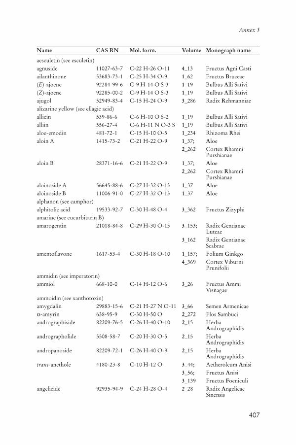
407
Annex 5
Name CAS RN Mol. form. Volume Monograph name
aesculetin (see esculetin)agnuside 11027-63-7 C-22 H-26 O-11 4_13 Fructus Agni Castiailanthinone 53683-73-1 C-25 H-34 O-9 1_62 Fructus Bruceae(E)-ajoene 92284-99-6 C-9 H-14 O S-3 1_19 Bulbus Alli Sativi(Z)-ajoene 92285-00-2 C-9 H-14 O S-3 1_19 Bulbus Alli Sativiajugol 52949-83-4 C-15 H-24 O-9 3_286 Radix Rehmanniaealizarine yellow (see ellagic acid)allicin 539-86-6 C-6 H-10 O S-2 1_19 Bulbus Alli Sativialliin 556-27-4 C-6 H-11 N O-3 S 1_19 Bulbus Alli Sativialoe-emodin 481-72-1 C-15 H-10 O-5 1_234 Rhizoma Rheialoin A 1415-73-2 C-21 H-22 O-9 1_37; Aloe
2_262 Cortex RhamniPurshianae
aloin B 28371-16-6 C-21 H-22 O-9 1_37; Aloe2_262 Cortex Rhamni
Purshianaealoinoside A 56645-88-6 C-27 H-32 O-13 1_37 Aloealoinoside B 11006-91-0 C-27 H-32 O-13 1_37 Aloealphanon (see camphor)alphitolic acid 19533-92-7 C-30 H-48 O-4 3_362 Fructus Zizyphiamarine (see cucurbitacin B)amarogentin 21018-84-8 C-29 H-30 O-13 3_153; Radix Gentianae
Luteae3_162 Radix Gentianae
Scabraeamentoflavone 1617-53-4 C-30 H-18 O-10 1_157; Folium Ginkgo
4_369 Cortex ViburniPrunifolii
ammidin (see imperatorin)ammiol 668-10-0 C-14 H-12 O-6 3_26 Fructus Ammi
Visnagaeammoidin (see xanthotoxin)amygdalin 29883-15-6 C-21 H-27 N O-11 3_66 Semen Armenicae
-amyrin 638-95-9 C-30 H-50 O 2_272 Flos Sambuciandrographiside 82209-76-5 C-26 H-40 O-10 2_15 Herba
Andrographidisandrographolide 5508-58-7 C-20 H-30 O-5 2_15 Herba
Andrographidisandropanoside 82209-72-1 C-26 H-40 O-9 2_15 Herba
Andrographidistrans-anethole 4180-23-8 C-10 H-12 O 3_44; Aetheroleum Anisi
3_56; Fructus Anisi3_139 Fructus Foeniculi
angelicide 92935-94-9 C-24 H-28 O-4 2_28 Radix AngelicaeSinensis

408
WHO monographs on selected medicinal plants
Name CAS RN Mol. form. Volume Monograph name
angelicone 37719-98-5 C-16 H-16 O-5 2_28 Radix AngelicaeSinensis
angelol G 83199-38-6 C-20 H-24 O-7 2_28 Radix AngelicaeSinensis
p-anisaldehyde 123-11-5 C-8 H-8 O-2 3_44; Aetheroleum Anisi3_56; Fructus Anisi3_139 Fructus Foeniculi
apigenin 520-36-5 C-15 H-10 O-5 1_89 Flos Chamomillaeapocynine 498-02-2 C-9 H-10 O-3 4_262 Rhizoma Picrorhizaearasaponin E1 (see ginsenoside Rb1)arbutin 497-76-7 C-12 H-16 O-7 2_345 Folium Uvae Ursiartecanin 29431-84-3 C-15 H-18 O-5 2_320 Herba Tanaceti
Partheniiasiatic acid 464-92-6 C-30 H-48 O-5 1_80 Herba Centellaeasiaticoside 16830-15-2 C-48 H-78 O-19 1_80 Herba Centellaeasterin (see cyanidin 3-monoglucoside)astragalin 480-10-4 C-21 H-20 O-11 2_39 Flos Calendulaeastragaloglucans 1_53 Radix Astragaliastragaloside I 84680-75-1 C-45 H-72 O-16 1_53 Radix Astragaliastragaloside II 84676-89-1 C-43 H-70 O-15 1_53 Radix Astragaliastragaloside III 84687-42-3 C-41 H-68 O-14 1_53 Radix Astragaliastragaloside IV 84687-43-4 C-41 H-68 O-14 1_53 Radix Astragaliastragaloside V 84687-44-5 C-47 H-78 O-19 1_53 Radix Astragaliastragaloside VI 84687-45-6 C-47 H-78 O-19 1_53 Radix Astragaliastragaloside VII 84687-46-7 C-47 H-78 O-19 1_53 Radix Astragaliaucubin 479-98-1 C-15 H-22 O-9 3_286; Radix Rehmanniae
4_13; Fructus Agni Casti4_262 Rhizoma Picrorhizae
aucubin p-hydroxybenzoate (see agnuside)azadirachtin 11141-17-6 C-35 H-44 O-16 3_91; Folium Azadirachti
3_104 Oleum Azadirachtibaicalein 491-67-8 C-15 H-10 O-5 3_316 Radix Scutellariaebaicalin 21967-41-9 C-21 H-18 O-11 3_316 Radix Scutellariaebaichanin A (see genistein)baldrinal 18234-46-3 C-12 H-10 O-4 1_271 Radix Valerianaebarbaloin (see aloin)barringtogenol C 13844-01-4 C-30 H-50 O-5 2_140 Semen Hippocastanibellaradine (see cuscohygrine)benzoaric acid (see ellagic acid)berbamine 478-61-5 C-37 H-40 N-2 O-6 4_33 Cortex Berberidisberbericinine (see palmatine)berberine 2086-83-1 C-20 H-18 N O-4 1_109; Rhizoma Coptidis
3_197; Rhizoma Hydrastis
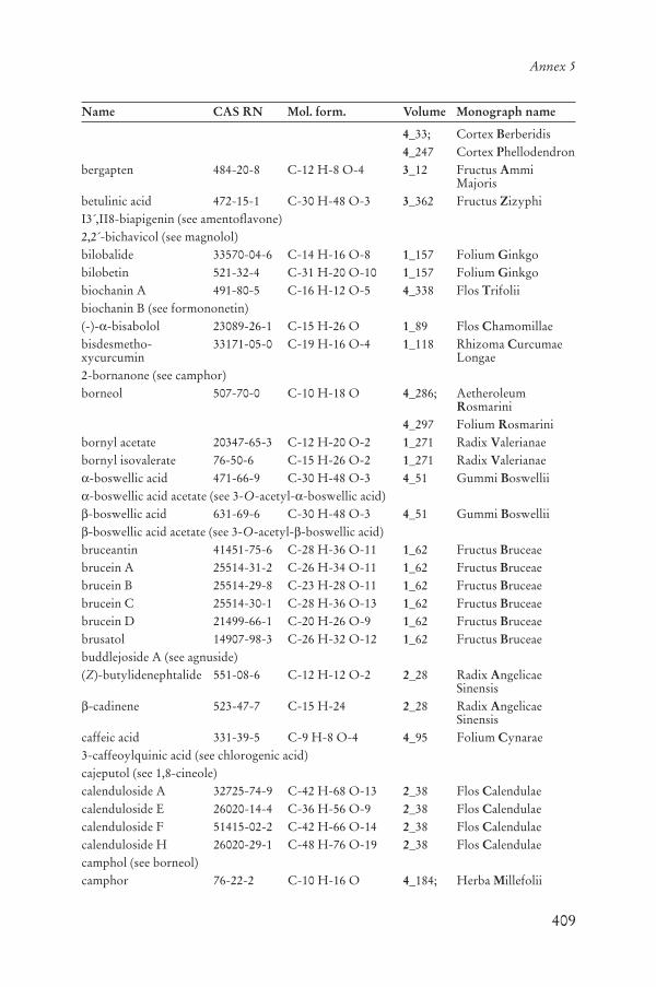
409
Annex 5
Name CAS RN Mol. form. Volume Monograph name
4_33; Cortex Berberidis4_247 Cortex Phellodendron
bergapten 484-20-8 C-12 H-8 O-4 3_12 Fructus AmmiMajoris
betulinic acid 472-15-1 C-30 H-48 O-3 3_362 Fructus ZizyphiI3´,II8-biapigenin (see amentoflavone)2,2´-bichavicol (see magnolol)bilobalide 33570-04-6 C-14 H-16 O-8 1_157 Folium Ginkgobilobetin 521-32-4 C-31 H-20 O-10 1_157 Folium Ginkgobiochanin A 491-80-5 C-16 H-12 O-5 4_338 Flos Trifoliibiochanin B (see formononetin)(-)- -bisabolol 23089-26-1 C-15 H-26 O 1_89 Flos Chamomillaebisdesmetho-xycurcumin
33171-05-0 C-19 H-16 O-4 1_118 Rhizoma CurcumaeLongae
2-bornanone (see camphor)borneol 507-70-0 C-10 H-18 O 4_286; Aetheroleum
Rosmarini4_297 Folium Rosmarini
bornyl acetate 20347-65-3 C-12 H-20 O-2 1_271 Radix Valerianaebornyl isovalerate 76-50-6 C-15 H-26 O-2 1_271 Radix Valerianae
-boswellic acid 471-66-9 C-30 H-48 O-3 4_51 Gummi Boswellii-boswellic acid acetate (see 3-O-acetyl- -boswellic acid)-boswellic acid 631-69-6 C-30 H-48 O-3 4_51 Gummi Boswellii-boswellic acid acetate (see 3-O-acetyl- -boswellic acid)
bruceantin 41451-75-6 C-28 H-36 O-11 1_62 Fructus Bruceaebrucein A 25514-31-2 C-26 H-34 O-11 1_62 Fructus Bruceaebrucein B 25514-29-8 C-23 H-28 O-11 1_62 Fructus Bruceaebrucein C 25514-30-1 C-28 H-36 O-13 1_62 Fructus Bruceaebrucein D 21499-66-1 C-20 H-26 O-9 1_62 Fructus Bruceaebrusatol 14907-98-3 C-26 H-32 O-12 1_62 Fructus Bruceaebuddlejoside A (see agnuside)(Z)-butylidenephtalide 551-08-6 C-12 H-12 O-2 2_28 Radix Angelicae
Sinensis-cadinene 523-47-7 C-15 H-24 2_28 Radix Angelicae
Sinensiscaffeic acid 331-39-5 C-9 H-8 O-4 4_95 Folium Cynarae3-caffeoylquinic acid (see chlorogenic acid)cajeputol (see 1,8-cineole)calenduloside A 32725-74-9 C-42 H-68 O-13 2_38 Flos Calendulaecalenduloside E 26020-14-4 C-36 H-56 O-9 2_38 Flos Calendulaecalenduloside F 51415-02-2 C-42 H-66 O-14 2_38 Flos Calendulaecalenduloside H 26020-29-1 C-48 H-76 O-19 2_38 Flos Calendulaecamphol (see borneol)camphor 76-22-2 C-10 H-16 O 4_184; Herba Millefolii
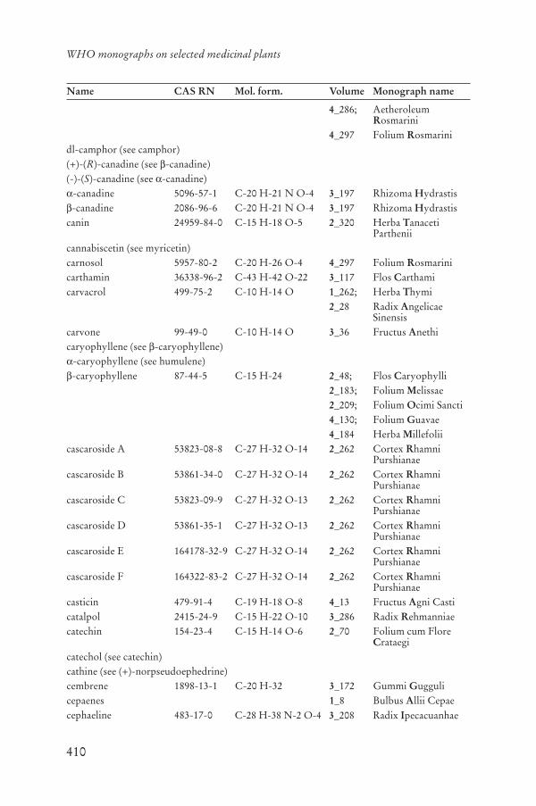
410
WHO monographs on selected medicinal plants
Name CAS RN Mol. form. Volume Monograph name
4_286; AetheroleumRosmarini
4_297 Folium Rosmarinidl-camphor (see camphor)(+)-(R)-canadine (see -canadine)(-)-(S)-canadine (see -canadine)
-canadine 5096-57-1 C-20 H-21 N O-4 3_197 Rhizoma Hydrastis-canadine 2086-96-6 C-20 H-21 N O-4 3_197 Rhizoma Hydrastis
canin 24959-84-0 C-15 H-18 O-5 2_320 Herba TanacetiParthenii
cannabiscetin (see myricetin)carnosol 5957-80-2 C-20 H-26 O-4 4_297 Folium Rosmarinicarthamin 36338-96-2 C-43 H-42 O-22 3_117 Flos Carthamicarvacrol 499-75-2 C-10 H-14 O 1_262; Herba Thymi
2_28 Radix AngelicaeSinensis
carvone 99-49-0 C-10 H-14 O 3_36 Fructus Anethicaryophyllene (see -caryophyllene)
-caryophyllene (see humulene)-caryophyllene 87-44-5 C-15 H-24 2_48; Flos Caryophylli
2_183; Folium Melissae2_209; Folium Ocimi Sancti4_130; Folium Guavae4_184 Herba Millefolii
cascaroside A 53823-08-8 C-27 H-32 O-14 2_262 Cortex RhamniPurshianae
cascaroside B 53861-34-0 C-27 H-32 O-14 2_262 Cortex RhamniPurshianae
cascaroside C 53823-09-9 C-27 H-32 O-13 2_262 Cortex RhamniPurshianae
cascaroside D 53861-35-1 C-27 H-32 O-13 2_262 Cortex RhamniPurshianae
cascaroside E 164178-32-9 C-27 H-32 O-14 2_262 Cortex RhamniPurshianae
cascaroside F 164322-83-2 C-27 H-32 O-14 2_262 Cortex RhamniPurshianae
casticin 479-91-4 C-19 H-18 O-8 4_13 Fructus Agni Casticatalpol 2415-24-9 C-15 H-22 O-10 3_286 Radix Rehmanniaecatechin 154-23-4 C-15 H-14 O-6 2_70 Folium cum Flore
Crataegicatechol (see catechin)cathine (see (+)-norpseudoephedrine)cembrene 1898-13-1 C-20 H-32 3_172 Gummi Guggulicepaenes 1_8 Bulbus Allii Cepaecephaeline 483-17-0 C-28 H-38 N-2 O-4 3_208 Radix Ipecacuanhae

411
Annex 5
Name CAS RN Mol. form. Volume Monograph name
ceramides 104404-17-3 2_333 Radix Urticaecetraric acid 489-49-6 C-20 H-18 O-9 4_143 Lichen Islandicuschamazulene 529-05-5 C-14 H-16 1_89; Flos Chamomillae
4_184 Herba Millefoliichebulagic acid 23094-71-5 C-41 H-30 O-27 4_74 Fructus Chebulaechebulanin 166833-80-3 C-27 H-24 O-19 4_74 Fructus Chebulaechebulinic acid 18942-26-2 C-41 H-32 O-27 4_74 Fructus Chebulaechicoric acid 6537-80-0 C-22 H-18 O-12 1_130; Radix Echinaceae
1_139 Herba EchinaceaePurpureae
chlorogenic acid 327-97-9 C-16 H-18 O-9 2_272; Flos Sambuci4_95 Folium Cynarae
chrysaloin 73889-54-0 C-21 H-22 O-8 2_262 Cortex RhamniPurshianae
chrysaloin A (see 11-deoxyaloin)chrysaloin B (see 11-deoxyaloin)chrysanthemin (see cyanidin 3-monoglucoside)chrysophanol 481-74-3 C-15 H-10 O-4 1_234 Rhizoma Rheichrysosplenol D 14965-20-9 C-18 H-16 O-8 4_13 Fructus Agni Casticichorigenin (see esculetin)cimicifugoside 66176-93-0 C-37 H-54 O-11 2_58 Rhizoma Cimicifugae
Racemosaecimigenol 3779-59-7 C-30 H-48 O-5 2_58 Rhizoma Cimicifugae
Racemosaecineole (see 1,8-cineole)1,8-cineole 470-82-6 C-10 H-18 O 2_99; Aetheroleum
Eucalypti2_109; Folium Eucalypti2_174; Aetheroleum
MelaleucaeAlternifoliae
3_36; Fructus Anethi4_69; Semen Cardamomi4_184; Herba Millefolii4_286; Aetheroleum
Rosmarini4_297 Folium Rosmarini
cinnamaldehyde 104-55-2 C-9 H-8 O 1_99 Cortex Cinnamomi6´-cinnamoylcatalpol (see picroside I)citral A (see geranial)citral b (see neral)citronellal 106-23-0 C-10 H-18 O 2_183 Folium Melissaecolumbamine 3621-36-1 C-20 H-20 N O-4 4_33 Cortex Berberidisconiferyl ferulate 63644-62-2 C-20 H-20 O-6 2_28 Radix Angelicae
Sinensis

412
WHO monographs on selected medicinal plants
Name CAS RN Mol. form. Volume Monograph name
corynoxeine 630-94-4 C-22 H-26 N-2 O-4 4_356 Ramulus cum Uncis Uncariae
coumarin 91-64-5 C-9 H-6 O-2 1_99 Cortex Cinnamomicrocetin (see -crocetin)
-crocetin 27876-94-4 C-20 H-24 O-4 3_129 Stigma Croci-crocetin 25368-09-6 C-21 H-26 O-4 3_129 Stigma Croci-crocetin 5892-54-6 C-22 H-28 O-4 3_129 Stigma Croci
crocin (see A-crocin)A-crocin 42553-65-1 C-44 H-64 O-24 3_129 Stigma CrociB-crocin 55750-84-0 C-38 H-54 O-19 3_129 Stigma CrociC-crocin 55750-85-1 C-32 H-44 O-14 3_129 Stigma CrociD-crocin 57710-64-2 C-32 H-44 O-14 3_129 Stigma CrociE-crocin 58050-17-2 C-26 H-34 O-9 3_129 Stigma Crocicrocin 2 (see B-crocin)crocin 3 (see C-crocin)crocin 4 (see D-crocin)cucurbitacin B 6199-67-3 C-32 H-46 O-8 4_262 Rhizoma Picrorhizaecucurbitacin D 3877-86-9 C-30 H-44 O-7 4_262 Rhizoma Picrorhizaecucurbitacin E 18444-66-1 C-32 H-44 O-8 4_262 Rhizoma Picrorhizaecucurbitacin F 5939-57-1 C-30 H-46 O-7 4_262 Rhizoma Picrorhizaecucurbitacin I 2222-07-3 C-30 H-42 O-7 4_262 Rhizoma Picrorhizaecucurbitacin R 55903-92-9 C-30 H-46 O-7 4_262 Rhizoma Picrorhizaecucurbitine 6807-92-7 C-5 H-10 N-2 O-2 4_86 Semen Cucurbitaecurcumin 458-37-7 C-21 H-20 O-6 1_118 Rhizoma Curcumae
Longaecurcumin I (see curcumin)curcumin II (see desmethoxycurcumin)curcumin III (see bisdesmethoxycurcumin)curzerene 17910-09-7 C-15 H-20 O 3_250 Gummi Myrrhacurzerenone 20493-56-5 C-15 H-18 O-2 3_250 Gummi Myrrhacuscohygrine 454-14-8 C-13 H-24 N-2 O 4_376 Radix Withaniaecyanidin 528-58-5 C-15 H-11 Cl O-6 4_152 Fructus
Macrocarponiicyanidin3-monoglucoside
7084-24-4 C-21 H-21 Cl O-11 4_213 Fructus Myrtilli
cyanidol (see cyanidin)cyclamin (see malvidin 3-monoglucoside)p-cymene 99-87-6 C-10 H-14 3_36 Fructus Anethicynarin 30964-13-7 C-25 H-24 O-12 1_130; Radix Echinaceae
4_95 Folium Cynaraecynaropicrin 35730-78-0 C-19 H-22 O-6 4_95 Folium Cynaraecynaroside 5373-11-5 C-21 H-20 O-11 4_13; Fructus Agni Casti
4_95 Folium Cynaraedaidzein 486-66-8 C-15 H-10 O-4 4_338 Flos Trifolii
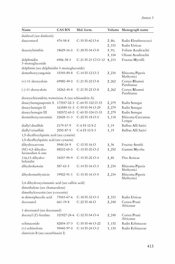
413
Annex 5
Name CAS RN Mol. form. Volume Monograph name
daidzeol (see daidzein)daucosterol 474-58-8 C-35 H-60 O-6 2_86; Radix Eleutherococci
2_333 Radix Urticaedeacetylnimbin 18609-16-0 C-28 H-34 O-8 3_91; Folium Azadirachti
3_104 Oleum Azadirachtidelphinidin3-monoglucoside
6906-38-3 C-21 H-21 Cl O-12 4_213 Fructus Myrtilli
delphinin (see delphinidin 3-monoglucoside)demethoxyyangonin 15345-89-8 C-14 H-12 O-3 2_234 Rhizoma Piperis
Methystici(+)-11-deoxyaloin 69980-49-0 C-21 H-22 O-8 2_262 Cortex Rhamni
Purshianae(-)-11-deoxyaloin 18262-45-8 C-21 H-22 O-8 2_262 Cortex Rhamni
Purshianaedeoxyschizandrin; wuweizisu A (see schisandrin A)desacylsenegasaponin A 173557-02-3 C-64 H-102 O-33 2_279 Radix Senegaedesacylsenegin II 163589-51-3 C-59 H-94 O-29 2_279 Radix Senegaedesacylsenegin III 180572-65-0 C-65 H-104 O-33 2_279 Radix Senegaedesmethoxycurcumin 22608-11-3 C-20 H-18 O-5 1_118 Rhizoma Curcumae
Longaediallyl disulfide 2179-57-9 C-6 H-10 S-2 1_19 Bulbus Alli Satividiallyl trisulfide 2050-87-5 C-6 H-10 S-3 1_19 Bulbus Alli Sativi1,5-dicaffeoylquinic acid (see cynarin)1,5-dicaffeylquinic acid (see cynarin)dihydrocarvone 5948-04-9 C-10 H-16 O 3_36 Fructus Anethi(9E)-4,5-dihydro-furanodien-6-one
88010-65-5 C-15 H-20 O-2 3_250 Gummi Myrrha
11 ,13-dihydro-helenalin
34257-95-9 C-15 H-20 O-4 3_80 Flos Arnicae
dihydrokawain 587-63-3 C-14 H-16 O-3 2_234 Rhizoma PiperisMethystici
dihydromethysticin 19902-91-1 C-15 H-16 O-5 2_234 Rhizoma PiperisMethystici
3,4-dihydroxycinnamic acid (see caffeic acid)dimethulene (see chamazulene)dimethylcrocetin (see -crocetin)
-dimorphecolic acid 73543-67-6 C-18 H-32 O-3 2_333 Radix Urticaedocosanol 661-19-8 C-22 H-46 O 2_248 Cortex Pruni
Africanae1-docosanol (see docosanol)docosyl (E)-ferulate 101927-24-6 C-32 H-54 O-4 2_248 Cortex Pruni
Africanaeechinacoside 82854-37-3 C-35 H-46 O-20 1_130 Radix Echinaceae(+)-echinolone 59440-97-0 C-14 H-24 O-2 1_130 Radix Echinaceaeelatericin B (see cucurbitacin I)

414
WHO monographs on selected medicinal plants
Name CAS RN Mol. form. Volume Monograph name
elatericine A (see cucurbitacin D)-elaterine (see cucurbitacin E)
eleutheroside A (see daucosterol)eleutheroside B 118-34-3 C-17 H-24 O-9 2_86 Radix Eleutherococcieleutheroside B1 16845-16-2 C-17 H-20 O-10 2_86 Radix Eleutherococcieleutheroside B4 (see (+)-sesamin)eleutheroside C 15486-24-5 C-8 H-16 O-6 2_86 Radix Eleutherococcieleutheroside D 79484-75-6 C-34 H-46 O-18 2_86 Radix Eleutherococcieleutheroside E 39432-56-9 C-34 H-46 O-18 2_86 Radix Eleutherococcieleutheroside E1 7374-79-0 C-28 H-36 O-13 2_86 Radix Eleutherococciellagic acid 476-66-4 C-14 H-6 O-8 4_120; Pericarpium Granati
4_130 Folium Guavaeemetine 483-18-1 C-29 H-40 N-2 O-4 3_208 Radix Ipecacuanhaeemodin (see frangula emodin)enin (see malvidin 3-monoglucoside)(-)-ephedrine 299-42-3 C-10 H-15 N O 1_148 Herba Ephedraeepi-catechin 490-46-0 C-15 H-14 O-6 2_70 Folium cum Flore
Crataegiepi-catechol (see epi-catechin)eremophilene 10219-75-7 C-15 H-24 2_183 Folium Melissaeescholine (see magnoflorine)esculetin 305-01-1 C-9 H-6 O-4 4_369 Cortex Viburni
Prunifoliiesculetol (see esculetin)estragole 140-67-0 C-10 H-12 O 3_139 Fructus Foeniculiestragole (see methylchavicol)eucalyptol (see 1,8-cineole)eugenol 97-53-0 C-10 H-12 O-2 1_99; Cortex Cinnamomi
2_48; Flos Caryophylli2_209 Folium Ocimi Sancti
eugenol acetate 93-28-7 C-12 H-14 O-3 2_48 Flos Caryophyllieutannin (see chebulinic acid)farnesyl acetate 4128-17-0 C-17 H-28 O-2 2_183 Folium Melissaefenchone 1195-79-5 C-10 H-16 O 3_139 Fructus Foeniculifeniculin (see gajaverin)(E)-ferulic acid 1135-24-6 C-10 H-10 O-4 2_28 Radix Angelicae
Sinensisflavosil (see formononetin)formononetin 485-72-3 C-16 H-12 O-4 2_58; Rhizoma Cimicifugae
Racemosae4_338 Flos Trifolii
fragilin 19764-02-4 C-15 H-20 O-8 4_312 Cortex Salicisfrangula emodin 518-82-1 C-15 H-10 O-5 1_234; Rhizoma Rhei

415
Annex 5
Name CAS RN Mol. form. Volume Monograph name
2_117 Cortex Frangulaefrangula emodin anthrone
491-60-1 C-15 H-12 O-4 2_117 Cortex Frangulae
frangulin A 521-62-0 C-21 H-20 O-9 2_117 Cortex Frangulaefrangulin B 14101-04-3 C-20 H-18 O-9 2_117 Cortex Frangulaefranguloside (see frangulin A)friedelin 559-74-0 C-30 H-50 O 2_249 Cortex Pruni
Africanaefumarprotocetraricacid
489-50-9 C-22 H-16 O-12 4_143 Lichen Islandicus
(5E,9Z)-furanodiene 19912-61-9 C-15 H-20 O 3_250 Gummi Myrrhafuranoeudesma-1,3-diene
87605-93-4 C-15 H-18 O 3_250 Gummi Myrrha
gallic acid 149-91-7 C-7 H-6 O-5 2_129; Folium et Cortex Hamamelidis
2_345; Folium Uvae Ursi4_74 Fructus Chebulae
gallogen (see ellagic acid)genistein 446-72-0 C-15 H-10 O-5 4_338 Flos Trifoliigenisteol (see genistein)genkwanin 437-64-9 C-16 H-12 O-5 4_297 Folium Rosmarinigentiopicrin (see gentiopicroside)gentiopicroside 20831-76-9 C-16 H-20 O-9 3_153; Radix Gentianae
Luteae3_162 Radix Gentianae
Scabraegeranial 141-27-5 C-10 H-16 O 2_183 Folium Melissaegeraniol 106-24-1 C-10 H-18 O 2_183 Folium Melissae[1]-gingerol 99742-03-7 C-12 H-16 O-4 1_280 Rhizoma Zingiberis[3]-gingerol 41743-67-3 C-14 H-20 O-4 1_280 Rhizoma Zingiberis[4]-gingerol 41743-68-4 C-15 H-22 O-4 1_280 Rhizoma Zingiberis[5]-gingerol 41743-69-5 C-16 H-24 O-4 1_280 Rhizoma Zingiberis[6]-gingerol 23513-14-6 C-17 H-26 O-4 1_280 Rhizoma Zingiberis[8]-gingerol 23513-08-8 C-19 H-30 O-4 1_280 Rhizoma Zingiberis[10]-gingerol 23513-15-7 C-21 H-34 O-4 1_280 Rhizoma Zingiberisginkgetin 481-46-9 C-32 H-22 O-10 1_157 Folium Ginkgoginkgolide A 15291-75-5 C-20 H-24 O-9 1_157 Folium Ginkgoginkgolide B 15291-77-7 C-20 H-24 O-10 1_157 Folium Ginkgoginkgolide C 15291-76-6 C-20 H-24 O-11 1_157 Folium Ginkgoginkgolide J 107438-79-9 C-20 H-24 O-10 1_157 Folium Ginkgoginkgolide M 15291-78-8 C-20 H-24 O-10 1_157 Folium Ginkgoginsenoside A1 (see pseudoginsenoside F11)ginsenoside Rb1 41753-43-9 C-54 H-92 O-23 1_171; Radix Ginseng
4_230 Radix PanacisQuinquefolii
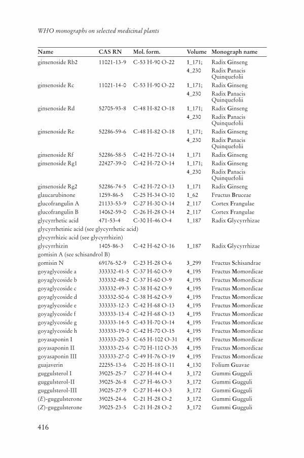
416
WHO monographs on selected medicinal plants
Name CAS RN Mol. form. Volume Monograph name
ginsenoside Rb2 11021-13-9 C-53 H-90 O-22 1_171; Radix Ginseng4_230 Radix Panacis
Quinquefoliiginsenoside Rc 11021-14-0 C-53 H-90 O-22 1_171; Radix Ginseng
4_230 Radix PanacisQuinquefolii
ginsenoside Rd 52705-93-8 C-48 H-82 O-18 1_171; Radix Ginseng4_230 Radix Panacis
Quinquefoliiginsenoside Re 52286-59-6 C-48 H-82 O-18 1_171; Radix Ginseng
4_230 Radix PanacisQuinquefolii
ginsenoside Rf 52286-58-5 C-42 H-72 O-14 1_171 Radix Ginsengginsenoside Rg1 22427-39-0 C-42 H-72 O-14 1_171; Radix Ginseng
4_230 Radix PanacisQuinquefolii
ginsenoside Rg2 52286-74-5 C-42 H-72 O-13 1_171 Radix Ginsengglaucarubinone 1259-86-5 C-25 H-34 O-10 1_62 Fructus Bruceaeglucofrangulin A 21133-53-9 C-27 H-30 O-14 2_117 Cortex Frangulaeglucofrangulin B 14062-59-0 C-26 H-28 O-14 2_117 Cortex Frangulaeglycyrrhetic acid 471-53-4 C-30 H-46 O-4 1_187 Radix Glycyrrhizaeglycyrrhetinic acid (see glycyrrhetic acid)glycyrrhizic acid (see glycyrrhizin)glycyrrhizin 1405-86-3 C-42 H-62 O-16 1_187 Radix Glycyrrhizaegomisin A (see schisandrol B)gomisin N 69176-52-9 C-23 H-28 O-6 3_299 Fructus Schisandraegoyaglycoside a 333332-41-5 C-37 H-60 O-9 4_195 Fructus Momordicaegoyaglycoside b 333332-48-2 C-37 H-60 O-9 4_195 Fructus Momordicaegoyaglycoside c 333332-49-3 C-38 H-62 O-9 4_195 Fructus Momordicaegoyaglycoside d 333332-50-6 C-38 H-62 O-9 4_195 Fructus Momordicaegoyaglycoside e 333333-12-3 C-42 H-68 O-13 4_195 Fructus Momordicaegoyaglycoside f 333333-13-4 C-42 H-68 O-13 4_195 Fructus Momordicaegoyaglycoside g 333333-14-5 C-43 H-70 O-14 4_195 Fructus Momordicaegoyaglycoside h 333333-19-0 C-42 H-70 O-15 4_195 Fructus Momordicaegoyasaponin I 333333-20-3 C-65 H-102 O-31 4_195 Fructus Momordicaegoyasaponin II 333333-23-6 C-70 H-110 O-35 4_195 Fructus Momordicaegoyasaponin III 333333-27-0 C-49 H-76 O-19 4_195 Fructus Momordicaeguajaverin 22255-13-6 C-20 H-18 O-11 4_130 Folium Guavaeguggulsterol I 39025-25-7 C-27 H-44 O-4 3_172 Gummi Gugguliguggulsterol-II 39025-26-8 C-27 H-46 O-3 3_172 Gummi Gugguliguggulsterol-III 39025-27-9 C-27 H-44 O-3 3_172 Gummi Gugguli(E)-guggulsterone 39025-24-6 C-21 H-28 O-2 3_172 Gummi Gugguli(Z)-guggulsterone 39025-23-5 C-21 H-28 O-2 3_172 Gummi Gugguli
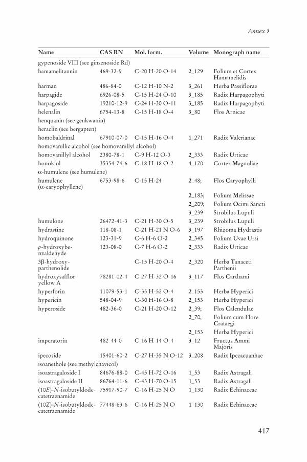
417
Annex 5
Name CAS RN Mol. form. Volume Monograph name
gypenoside VIII (see ginsenoside Rd)hamamelitannin 469-32-9 C-20 H-20 O-14 2_129 Folium et Cortex
Hamamelidisharman 486-84-0 C-12 H-10 N-2 3_261 Herba Passifloraeharpagide 6926-08-5 C-15 H-24 O-10 3_185 Radix Harpagophytiharpagoside 19210-12-9 C-24 H-30 O-11 3_185 Radix Harpagophytihelenalin 6754-13-8 C-15 H-18 O-4 3_80 Flos Arnicaehenquanin (see genkwanin)heraclin (see bergapten)homobaldrinal 67910-07-0 C-15 H-16 O-4 1_271 Radix Valerianaehomovanillic alcohol (see homovanillyl alcohol)homovanillyl alcohol 2380-78-1 C-9 H-12 O-3 2_333 Radix Urticaehonokiol 35354-74-6 C-18 H-18 O-2 4_170 Cortex Magnoliae
-humulene (see humulene)humulene( -caryophyllene)
6753-98-6 C-15 H-24 2_48; Flos Caryophylli
2_183; Folium Melissae2_209; Folium Ocimi Sancti3_239 Strobilus Lupuli
humulone 26472-41-3 C-21 H-30 O-5 3_239 Strobilus Lupulihydrastine 118-08-1 C-21 H-21 N O-6 3_197 Rhizoma Hydrastishydroquinone 123-31-9 C-6 H-6 O-2 2_345 Folium Uvae Ursip-hydroxybe-nzaldehyde
123-08-0 C-7 H-6 O-2 2_333 Radix Urticae
3 -hydroxy-parthenolide
C-15 H-20 O-4 2_320 Herba TanacetiParthenii
hydroxysaffloryellow A
78281-02-4 C-27 H-32 O-16 3_117 Flos Carthami
hyperforin 11079-53-1 C-35 H-52 O-4 2_153 Herba Hypericihypericin 548-04-9 C-30 H-16 O-8 2_153 Herba Hypericihyperoside 482-36-0 C-21 H-20 O-12 2_39; Flos Calendulae
2_70; Folium cum Flore Crataegi
2_153 Herba Hypericiimperatorin 482-44-0 C-16 H-14 O-4 3_12 Fructus Ammi
Majorisipecoside 15401-60-2 C-27 H-35 N O-12 3_208 Radix Ipecacuanhaeisoanethole (see methylchavicol)isoastragaloside I 84676-88-0 C-45 H-72 O-16 1_53 Radix Astragaliisoastragaloside II 86764-11-6 C-43 H-70 O-15 1_53 Radix Astragali(10E)-N-isobutyldode-catetraenamide
75917-90-7 C-16 H-25 N O 1_130 Radix Echinaceae
(10Z)-N-isobutyldode-catetraenamide
77448-63-6 C-16 H-25 N O 1_130 Radix Echinaceae

418
WHO monographs on selected medicinal plants
Name CAS RN Mol. form. Volume Monograph name
N-isobutyl-dodecatetraenamide
360762-35-2 C-16 H-25 N O 1_130 Radix Echinaceae
isocorynoxeine 51014-29-0 C-22 H-26 N-2 O-4 4_356 Ramulus cum Uncis Uncariae
7-isocorynoxeine (see isocorynoxeine)(E)-isoferulic acid 537-73-5 C-10 H-10 O-4 2_58 Rhizoma Cimicifugae
Racemosaeisofraxidin 486-21-5 C-11 H-10 O-5 2_86 Radix Eleutherococciisoginkgetin 548-19-6 C-32 H-22 O-10 1_157 Folium Ginkgoisoliquiritigenin 961-29-5 C-15 H-12 O-4 1_187 Radix Glycyrrhizaeisoliquiritin 5041-81-6 C-21 H-22 O-9 1_187 Radix Glycyrrhizaeisomitraphylline 4963-01-3 C-21 H-24 N-2 O-4 3_352 Cortex Uncariaeiso-orientin 4261-42-1 C-21 H-20 O-11 3_261 Herba Passifloraeisopelletierine 4396-01-4 C-8 H-15 N O 4_376 Radix Withaniaeisopteropodine 5171-37-9 C-21 H-24 N-2 O-4 3_352 Cortex Uncariaeisopunicine (see isopelletierine)isoquercitrin 21637-25-2 C-21 H-20 O-12 2_39; Flos Calendulae
2_153; Herba Hyperici2_272 Flos Sambuci
isorhamnetin 480-19-3 C-16 H-12 O-7 1_157 Folium Ginkgoisorhynchophylline 6859-01-4 C-22 H-28 N-2 O-4 4_356 Ramulus cum Uncis
Uncariaeisoschaftoside 52012-29-0 C-26 H-28 O-14 3_261 Herba Passifloraeisotussilagine 91108-32-6 C-10 H-17 N O-3 1_130; Radix Echinaceae
1_139 Herba EchinaceaePurpureae
isovaltrate 31078-10-1 C-22 H-30 O-8 1_271 Radix Valerianaeisovitexin 38953-85-4 C-21 H-20 O-10 3_261 Herba Passifloraejatrorrhizine 3621-38-3 C-20 H-20 N O-4 4_33; Cortex Berberidis
4_247 Cortex Phellodendronjujuboside B 55466-05-2 C-52 H-84 O-21 3_362 Fructus Zizyphikaempferol diglycoside coumarate
111957-48-3 C-36 H-36 O-17 1_157 Folium Ginkgo
kawain 500-64-1 C-14 H-14 O-3 2_234 Rhizoma PiperisMethystici
11-keto- -boswellic acid (see 11-oxo- -boswellic acid)11-keto- -boswellic acid acetate (see 3-O-acetyl-11-oxo- -boswellic acid)khellenin (see khellinin)khellin 82-02-0 C-14 H-12 O-5 3_26 Fructus Ammi
Visnagaekhellinin 17226-75-4 C-19 H-20 O-10 3_26 Fructus Ammi
Visnagaekhellinol 478-42-2 C-13 H-10 O-5 3_26 Fructus Ammi
Visnagaekhellol 478-79-5 C-13 H-10 O-5 3_26 Fructus Ammi
Visnagae
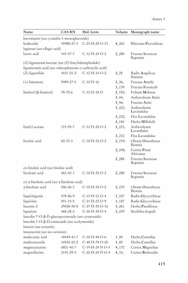
419
Annex 5
Name CAS RN Mol. form. Volume Monograph name
kuromanin (see cyanidin 3-monoglucoside)kutkoside 35988-27-3 C-23 H-28 O-13 4_262 Rhizoma Picrorhizaelagistase (see ellagic acid)lauric acid 143-07-7 C-12 H-24 O-2 2_288 Fructus Serenoae
Repentis(Z)-ligusticum lactone (see (Z)-butylidenephtalide)ligusticumic acid (see valerophenone-o-carboxylic acid)(Z)-ligustilide 4431-01-0 C-12 H-14 O-2 2_28 Radix Angelicae
Sinensis(+)-limonene 5989-27-5 C-10 H-16 3_36; Fructus Anethi
3_139 Fructus Foeniculilinalool ( -linalool) 78-70-6 C-10 H-18 O 2_183; Folium Melissae
3_44; Aetheroleum Anisi3_56; Fructus Anisi3_221; Aetheroleum
Lavandulae3_232; Flos Lavandulae4_184 Herba Millefolii
linalyl acetate 115-95-7 C-12 H-20 O-2 3_221; AetheroleumLavandulae
3_232 Flos Lavandulaelinoleic acid 60-33-3 C-18 H-32 O-2 2_219; Oleum Oenotherae
Biennis2_248; Cortex Pruni
Africanae2_288 Fructus Serenoae
Repentiscis-linoleic acid (see linoleic acid)linolenic acid 463-40-1 C-18 H-30 O-2 2_288 Fructus Serenoae
Repentiscis- -linolenic acid (see -linolenic acid)-linolenic acid 506-26-3 C-18 H-30 O-2 2_219 Oleum Oenotherae
Biennisliquiritigenin 578-86-9 C-15 H-12 O-4 1_187 Radix Glycyrrhizaeliquiritin 551-15-5 C-21 H-22 O-9 1_187 Radix Glycyrrhizaelucenin-2 29428-58-8 C-27 H-30 O-16 3_261 Herba Passifloraelupulone 468-28-0 C-26 H-38 O-4 3_239 Strobilus Lupuliluteolin 7-O- -D-glucopyranoside (see cynaroside)luteolin 7-O- -D-rutinoside (see scolymoside)lutexin (see orientin)lutonaretin (see iso-orientin)madecassic acid 18449-41-7 C-30 H-48 O-6 1_80 Herba Centellaemadecassoside 34540-22-2 C-48 H-78 O-20 1_80 Herba Centellaemagnocurarine 6801-40-7 C-19 H-24 N O-3 4_170 Cortex Magnoliaemagnoflorine 2141-09-5 C-20 H-24 N O-4 4_33; Cortex Berberidis
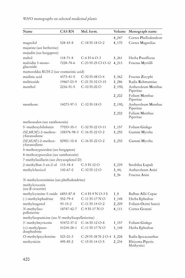
420
WHO monographs on selected medicinal plants
Name CAS RN Mol. form. Volume Monograph name
4_247 Cortex Phellodendronmagnolol 528-43-8 C-18 H-18 O-2 4_170 Cortex Magnoliaemajarine (see berberine)majudin (see bergapten)maltol 118-71-8 C-6 H-6 O-3 3_261 Herba Passifloraemalvidin 3-mono-glucoside
7228-78-6 C-23 H-25 Cl O-12 4_213 Fructus Myrtilli
mamorekku RUH 2 (see rosmarinic acid)maslinic acid 4373-41-5 C-30 H-48 O-4 3_362 Fructus Zizyphimelittoside 19467-03-9 C-21 H-32 O-15 3_286 Radix Rehmanniaementhol 2216-51-5 C-10 H-20 O 2_190; Aetheroleum Menthae
Piperitae2_202 Folium Menthae
Piperitaementhone 14073-97-3 C-10 H-18 O 2_190; Aetheroleum Menthae
Piperitae2_202 Folium Menthae
Piperitaemethoxsalen (see xanthotoxin)5´-methoxybilobetin 77053-35-1 C-32 H-22 O-11 1_157 Folium Ginkgo(5E,8R,9E)-2-methox-yfuranodiene
108376-98-3 C-16 H-22 O-2 3_250 Gummi Myrrha
(5E,8S,9E)-2-methox-yfuranodiene
82901-10-8 C-16 H-22 O-2 3_250 Gummi Myrrha
5-methoxypsoralen (see bergapten)8-methoxypsoralen (see xanthotoxin)7-methylaxillarin (see chrysosplenol D)2-methylbut-3-en-2-ol 115-18-4 C-5 H-10 O 3_239 Strobilus Lupulimethylchavicol 140-67-0 C-10 H-12 O 3_44; Aetheroleum Anisi
3_56 Fructus AnisiN-methylcoreximine (see phellodendrine)methylcrocetin(see -crocetin)methylcysteine S-oxide 6853-87-8 C-4 H-9 N O-3 S 1_8 Bulbus Allii Cepae(-)-methylephedrine 552-79-4 C-11 H-17 N O 1_148 Herba Ephedraemethyleugenol 93-15-2 C-11 H-14 O-2 2_209 Folium Ocimi SanctiN-methyliso-pelletierine
18747-42-7 C-9 H-17 N O 4_111 Cortex Granati
methylisopunicine (see N-methylisopelletierine)3´-methylmyricetin 53472-37-0 C-16 H-12 O-8 1_157 Folium Ginkgo(+)-methylpseu-doephedrine
51018-28-1 C-11 H-17 N O 1_148 Herba Ephedrae
O-methylpsychotrine 523-01-3 C-29 H-38 N-2 O-4 3_208 Radix Ipecacuanhaemethysticin 495-85-2 C-15 H-14 O-5 2_234 Rhizoma Piperis
Methystici

421
Annex 5
Name CAS RN Mol. form. Volume Monograph name
mitraphylline 509-80-8 C-21 H-24 N-2 O-4 3_352 Cortex Uncariaemitrinermine (see rhynchophylline)momordicoside F1 81348-81-4 C-37 H-60 O-8 4_195 Fructus Momordicaemomordicoside F2 81348-82-5 C-36 H-58 O-8 4_195 Fructus Momordicaemomordicoside G 81371-54-2 C-37 H-60 O-8 4_195 Fructus Momordicaemomordicoside I 81371-55-3 C-36 H-58 O-8 4_195 Fructus Momordicaemomordicoside K 81348-84-7 C-37 H-60 O-9 4_195 Fructus Momordicaemomordicoside L 81348-83-6 C-36 H-58 O-9 4_195 Fructus Momordicaemonomelittoside 20633-72-1 C-15 H-22 O-10 3_286 Radix Rehmanniaemukulol 41943-03-7 C-20 H-34 O 3_172 Gummi Guggulimyricetin 529-44-2 C-15 H-10 O-8 1_157; Folium Ginkgo
4_152 FructusMacrocarponii
myricetol (see myricetin)myrtillin (see delphinidin 3-monoglucoside)neoandrographolide 27215-14-1 C-26 H-40 O-8 2_15 Herba
Andrographidisneocarthamin 519-54-0 C-21 H-22 O-11 3_117 Flos Carthamineoolivil 77790-55-7 C-20 H-24 O-7 2_333 Radix Urticaeneprotine (see jatrorrhizine)neral 106-26-3 C-10 H-16 O 2_183 Folium Melissaenerol 106-25-2 C-10 H-18 O 2_183 Folium Melissaenimbin 5945-86-8 C-30 H-36 O-9 3_91; Folium Azadirachti
3_104 Oleum Azadirachtinopinene (see -pinene)(-)-norephedrine 492-41-1 C-9 H-13 N O 1_148 Herba Ephedrae(+)-norpseu-doephedrine
492-39-7 C-9 H-13 N O 1_148 Herba Ephedrae
cis- -ocimene 3338-55-4 C-10 H-16 2_28 Radix AngelicaeSinensis
oleanolic acid 508-02-1 C-30 H-48 O-3 2_333; Radix Urticae4_130 Folium Guavae
oleic acid 112-80-1 C-18 H-34 O-2 2_248 Cortex PruniAfricanae
olmelin (see biochanin A)orientin 28608-75-5 C-21 H-20 O-11 3_261 Herba Passiflorae11-oxo- -boswellicacid
17019-92-0 C-30 H-46 O-4 4_51 Gummi Boswellii
paeoniflorin 23180-57-6 C-23 H-28 O-11 1_197 Radix Paeoniaepalmatine 3486-67-7 C-21 H-22 N O-4 4_247 Cortex Phellodendronpanaxadiol 19666-76-3 C-30 H-52 O-3 1_171 Radix Ginsengpanaxatriol 32791-84-7 C-30 H-52 O-4 1_171 Radix Ginsengpanaxoside A (see ginsenoside Rg1)panaxoside Rb1 (see ginsenoside Rb1)

422
WHO monographs on selected medicinal plants
Name CAS RN Mol. form. Volume Monograph name
panaxoside Rc (see ginsenoside Rc)panaxoside Re (see ginsenoside Re)parthenolide 20554-84-1 C-15 H-20 O-3 2_320 Herba Tanaceti
Partheniipatrinoside 53962-20-2 C-21 H-34 O-11 4_369 Cortex Viburni
Prunifoliipelletierine 2858-66-4 C-8 H-15 N O 4_111 Cortex Granatipeonidin 134-01-0 C-16 H-13 Cl O-6 4_152 Fructus
Macrocarponiipeonidin 3-mono-glucoside
6906-39-4 C-22 H-23 Cl O-11 4_213 Fructus Myrtilli
peonidol chloride (see peonidin)petunidin 3-mono-glucoside
6988-81-4 C-22 H-23 Cl O-12 4_213 Fructus Myrtilli
(-)- -phellandrene 4221-98-1 C-10 H-16 3_36; Fructus Anethi3_139 Fructus Foeniculi
phellodendrine 6873-13-8 C-20 H-24 N O-4 4_247 Cortex Phellodendronphyscione 521-61-9 C-16 H-12 O-5 1_234 Rhizoma Rheipicrocrocin 138-55-6 C-16 H-26 O-7 3_129 Stigma Crocipicrosalvin (see carnosol)picroside I 27409-30-9 C-24 H-28 O-11 4_262 Rhizoma Picrorhizaepicroside II 39012-20-9 C-23 H-28 O-13 4_262 Rhizoma Picrorhizaepicroside III 64461-95-6 C-25 H-30 O-13 4_262 Rhizoma Picrorhizae
-pinene 80-56-8 C-10 H-16 3_36; Fructus Anethi3_139; Fructus Foeniculi4_130; Folium Guavae4_184 Herba Millefolii
-pinene 127-91-3 C-10 H-16 4_184 Herba Millefoliiplatycodin A 66779-34-8 C-59 H-94 O-29 1_216 Radix Platycodiplatycodin C 66779-35-9 C-59 H-94 O-29 1_216 Radix Platycodiplatycodin D 58479-68-8 C-57 H-92 O-28 1_216 Radix Platycodiplatycodin D2 66663-90-9 C-63 H-102 O-33 1_216 Radix Platycodiplenolin (see 11 , 13-dihydrohelenalin)polygalacin D 66663-91-0 C-57 H-92 O-27 1_216 Radix Platycodipolygalacin D2 66663-92-1 C-63 H-102 O-32 1_216 Radix Platycodiproanthocyanidintrimer
C-45 H-38 O-18 4_152 FructusMacrocarponii
proanthocyanidins C-30 H-26 O-14 2_129 Folium et Cortex Hamamelidis
procumbide 20486-27-5 C-15 H-22 O-10 3_185 Radix Harpagophytiprocyanidin 15514-06-4 C-30 H-26 O-12 2_129 Folium et Cortex
Hamamelidisprocyanidin B1 20315-25-7 C-30 H-26 O-12 2_70 Folium cum Flore
Crataegi
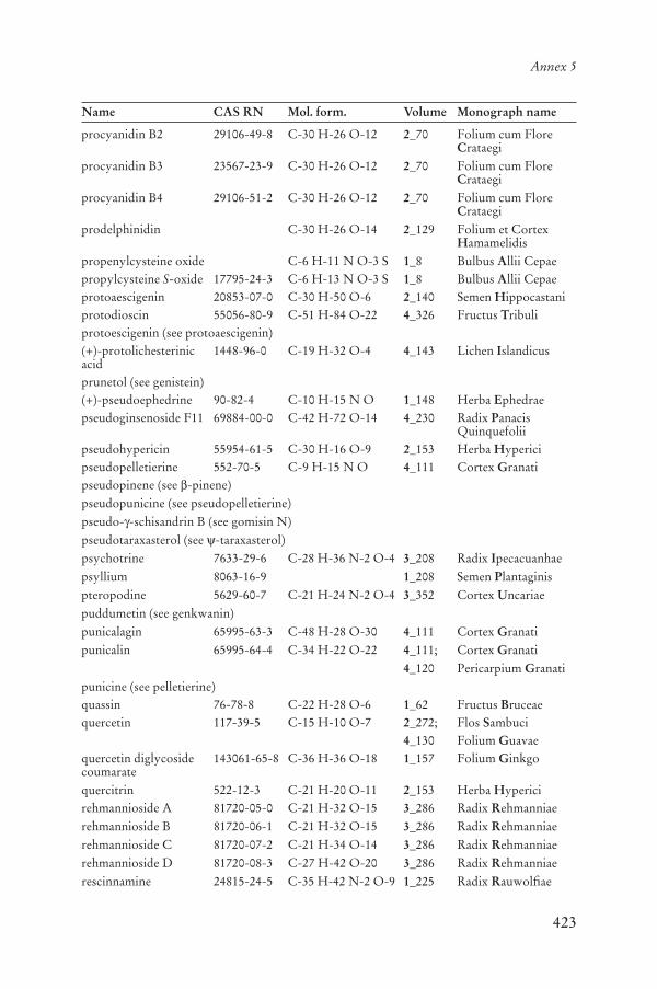
423
Annex 5
Name CAS RN Mol. form. Volume Monograph name
procyanidin B2 29106-49-8 C-30 H-26 O-12 2_70 Folium cum Flore Crataegi
procyanidin B3 23567-23-9 C-30 H-26 O-12 2_70 Folium cum Flore Crataegi
procyanidin B4 29106-51-2 C-30 H-26 O-12 2_70 Folium cum Flore Crataegi
prodelphinidin C-30 H-26 O-14 2_129 Folium et Cortex Hamamelidis
propenylcysteine oxide C-6 H-11 N O-3 S 1_8 Bulbus Allii Cepaepropylcysteine S-oxide 17795-24-3 C-6 H-13 N O-3 S 1_8 Bulbus Allii Cepaeprotoaescigenin 20853-07-0 C-30 H-50 O-6 2_140 Semen Hippocastaniprotodioscin 55056-80-9 C-51 H-84 O-22 4_326 Fructus Tribuliprotoescigenin (see protoaescigenin)(+)-protolichesterinicacid
1448-96-0 C-19 H-32 O-4 4_143 Lichen Islandicus
prunetol (see genistein)(+)-pseudoephedrine 90-82-4 C-10 H-15 N O 1_148 Herba Ephedraepseudoginsenoside F11 69884-00-0 C-42 H-72 O-14 4_230 Radix Panacis
Quinquefoliipseudohypericin 55954-61-5 C-30 H-16 O-9 2_153 Herba Hypericipseudopelletierine 552-70-5 C-9 H-15 N O 4_111 Cortex Granatipseudopinene (see -pinene)pseudopunicine (see pseudopelletierine)pseudo- -schisandrin B (see gomisin N)pseudotaraxasterol (see -taraxasterol)psychotrine 7633-29-6 C-28 H-36 N-2 O-4 3_208 Radix Ipecacuanhaepsyllium 8063-16-9 1_208 Semen Plantaginispteropodine 5629-60-7 C-21 H-24 N-2 O-4 3_352 Cortex Uncariaepuddumetin (see genkwanin)punicalagin 65995-63-3 C-48 H-28 O-30 4_111 Cortex Granatipunicalin 65995-64-4 C-34 H-22 O-22 4_111; Cortex Granati
4_120 Pericarpium Granatipunicine (see pelletierine)quassin 76-78-8 C-22 H-28 O-6 1_62 Fructus Bruceaequercetin 117-39-5 C-15 H-10 O-7 2_272; Flos Sambuci
4_130 Folium Guavaequercetin diglycoside coumarate
143061-65-8 C-36 H-36 O-18 1_157 Folium Ginkgo
quercitrin 522-12-3 C-21 H-20 O-11 2_153 Herba Hypericirehmannioside A 81720-05-0 C-21 H-32 O-15 3_286 Radix Rehmanniaerehmannioside B 81720-06-1 C-21 H-32 O-15 3_286 Radix Rehmanniaerehmannioside C 81720-07-2 C-21 H-34 O-14 3_286 Radix Rehmanniaerehmannioside D 81720-08-3 C-27 H-42 O-20 3_286 Radix Rehmanniaerescinnamine 24815-24-5 C-35 H-42 N-2 O-9 1_225 Radix Rauwolfiae

424
WHO monographs on selected medicinal plants
Name CAS RN Mol. form. Volume Monograph name
reserpine 50-55-5 C-33 H-40 N-2 O-9 1_225 Radix Rauwolfiaerhein 478-43-3 C-15 H-8 O-6 1_234 Rhizoma Rheirheinoside A 111545-28-9 C-27 H-30 O-17 1_234 Rhizoma Rheirheinoside B 111614-10-9 C-27 H-30 O-17 1_234 Rhizoma Rheirheinoside C 111545-29-0 C-27 H-30 O-16 1_234 Rhizoma Rheirheinoside D 111614-11-0 C-27 H-30 O-16 1_234 Rhizoma Rheirhimantin (see aucubin)rhodinal (see citronellal)rhynchophylline 76-66-4 C-22 H-28 N-2 O-4 4_356 Ramulus cum Uncis
Uncariae-18-rhyncophylline (see corynoxeine)
ricinic acid (see ricinoleic acid)ricinoleic acid 141-22-0 C-18 H-34 O-3 4_274 Oleum Riciniricinolic acid (see ricinoleic acid)rosemaric acid (see rosmarinic acid)rosmarinic acid 20283-92-5 C-18 H-16 O-8 2_183; Folium Melissae
4_297 Folium Rosmarinirutin 153-18-4 C-27 H-30 O-16 2_39; Flos Calendulae
2_70; Folium cum Flore Crataegi
2_153; Herba Hyperici2_272 Flos Sambuci
sabinene 3387-41-5 C-10 H-16 4_184 Herba Millefoliisafflor yellow A 85532-77-0 C-27 H-30 O-15 3_117 Flos Carthamisafflor yellow B 91574-92-4 C-48 H-54 O-27 3_117 Flos Carthamisafranal 116-26-7 C-10 H-14 O 3_129 Stigma Crocisaikogenin A 5092-09-1 C-30 H-48 O-4 1_70 Radix Bupleurisaikogenin D 5573-16-0 C-30 H-48 O-4 1_70 Radix Bupleurisaikogenin F 14356-59-3 C-30 H-48 O-4 1_70 Radix Bupleurisaikogenin G 18175-79-6 C-30 H-48 O-4 1_70 Radix Bupleurisaikosaponin A 20736-09-8 C-42 H-68 O-13 1_70 Radix Bupleurisaikosaponin B1 58558-08-0 C-42 H-68 O-13 1_70 Radix Bupleurisaikosaponin B2 58316-41-9 C-42 H-68 O-13 1_70 Radix Bupleurisaikosaponin B3 58316-42-0 C-43 H-72 O-14 1_70 Radix Bupleurisaikosaponin B4 58558-09-1 C-43 H-72 O-14 1_70 Radix Bupleurisaikosaponin D 20874-52-6 C-42 H-68 O-13 1_70 Radix Bupleurisalicin 138-52-3 C-13 H-18 O-7 4_312 Cortex Salicissalicin 6´-acetate (see fragilin)salicortin 29836-41-7 C-20 H-24 O-10 4_312 Cortex Salicissalicoside (see salicin)sanchinoside C1 (see ginsenoside Rg1)sanchinoside Rb1 (see ginsenoside Rb1)sanchinoside Re (see ginsenoside Re)

425
Annex 5
Name CAS RN Mol. form. Volume Monograph name
saponaretin (see isovitexin)schaftoside 51938-32-0 C-26 H-28 O-14 3_261 Herba Passifloraeschisandrin A 61281-38-7 C-24 H-32 O-6 3_299 Fructus Schisandraeschisandrin B 61281-37-6 C-23 H-28 O-6 3_299 Fructus Schisandraeschisandrol A 7432-28-2 C-24 H-32 O-7 3_299 Fructus Schisandraeschisandrol B 58546-54-6 C-23 H-28 O-7 3_299 Fructus Schisandraeschizandrin (see schisandrol A)(±)- -schizandrin (see schisandrin B)sciadopitysin 521-34-6 C-33 H-24 O-10 1_157 Folium Ginkgoscolymoside 20633-84-5 C-27 H-30 O-15 4_95 Folium Cynaraescopoletin 92-61-5 C-10 H-8 O-4 2_333 Radix Urticaesenecionine 130-01-8 C-18 H-25 N O-5 1_130; Radix Echinaceae
1_139 Herba EchinaceaePurpureae
(E)-senegasaponin A 162762-97-2 C-74 H-110 O-35 2_279 Radix Senegae(Z)-senegasaponin A 162613-72-1 C-74 H-110 O-35 2_279 Radix Senegae(E)-senegasaponin B 162870-58-8 C-69 H-102 O-31 2_279 Radix Senegae(Z)-senegasaponin B 162613-71-0 C-69 H-102 O-31 2_279 Radix Senegaesenegin II 34366-31-9 C-70 H-104 O-32 2_279 Radix Senegae(Z)-senegin II 162681-52-9 C-70 H-104 O-32 2_279 Radix Senegae(E)-senegin III 35906-36-6 C-75 H-112 O-35 2_279 Radix Senegae(Z)-senegin III 162681-53-0 C-75 H-112 O-35 2_279 Radix Senegaesennoside A 81-27-6 C-42 H-38 O-20 1_244; Folium Sennae
1_253 Fructus Sennaesennoside B 128-57-4 C-42 H-38 O-20 1_244; Folium Sennae
1_253 Fructus Sennaesennoside C 37271-16-2 C-42 H-40 O-19 1_244; Folium Sennae
1_253 Fructus Sennaesennoside D 37271-17-3 C-42 H-40 O-19 1_244; Folium Sennae
1_253 Fructus Sennaesennoside E 11137-63-6 C-44 H-38 O-23 1_244; Folium Sennae
1_253 Fructus Sennaesennoside F 52842-23-6 C-44 H-38 O-23 1_244; Folium Sennae
1_253 Fructus Sennae(+)-sesamin 607-80-7 C-20 H-18 O-6 2_86 Radix Eleutherococci[6]-shogaol 23513-13-5 C-17 H-24 O-3 1_280 Rhizoma Zingiberis[7]-shogaol 99742-07-1 C-18 H-26 O-3 1_280 Rhizoma Zingiberis[9]-shogaol 99742-08-2 C-20 H-30 O-3 1_280 Rhizoma Zingiberis[11]-shogaol 99742-09-3 C-22 H-34 O-3 1_280 Rhizoma Zingiberis[12]-shogaol 99742-10-6 C-23 H-36 O-3 1_280 Rhizoma Zingiberissilybin 22888-70-6 C-25 H-22 O-10 2_303 Fructus Silybi Mariaesilychristin 33889-69-9 C-25 H-22 O-10 2_303 Fructus Silybi Mariae
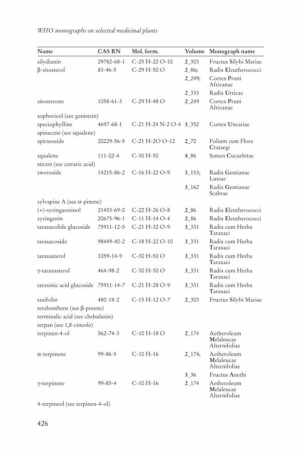
426
WHO monographs on selected medicinal plants
Name CAS RN Mol. form. Volume Monograph name
silydianin 29782-68-1 C-25 H-22 O-10 2_303 Fructus Silybi Mariae-sitosterol 83-46-5 C-29 H-50 O 2_86; Radix Eleutherococci
2_249; Cortex PruniAfricanae
2_333 Radix Urticaesitosterone 1058-61-3 C-29 H-48 O 2_249 Cortex Pruni
Africanaesophoricol (see genistein)speciophylline 4697-68-1 C-21 H-24 N-2 O-4 3_352 Cortex Uncariaespinacene (see squalene)spiraeoside 20229-56-5 C-21 H-2O O-12 2_70 Folium cum Flore
Crataegisqualene 111-02-4 C-30 H-50 4_86 Semen Cucurbitaestictin (see cetraric acid)sweroside 14215-86-2 C-16 H-22 O-9 3_153; Radix Gentianae
Luteae3_162 Radix Gentianae
Scabraesylvapine A (see -pinene)(+)-syringaresinol 21453-69-0 C-22 H-26 O-8 2_86 Radix Eleutherococcisyringenin 20675-96-1 C-11 H-14 O-4 2_86 Radix Eleutherococcitaraxacolide glucoside 75911-12-5 C-21 H-32 O-9 3_331 Radix cum Herba
Taraxacitaraxacoside 98449-40-2 C-18 H-22 O-10 3_331 Radix cum Herba
Taraxacitaraxasterol 1059-14-9 C-30 H-50 O 3_331 Radix cum Herba
Taraxaci-taraxasterol 464-98-2 C-30 H-50 O 3_331 Radix cum Herba
Taraxacitaraxinic acid glucoside 75911-14-7 C-21 H-28 O-9 3_331 Radix cum Herba
Taraxacitaxifolin 480-18-2 C-15 H-12 O-7 2_303 Fructus Silybi Mariaeterebenthene (see -pinene)terminalic acid (see chebulanin)terpan (see 1,8-cineole)terpinen-4-ol 562-74-3 C-10 H-18 O 2_174 Aetheroleum
MelaleucaeAlternifoliae
-terpinene 99-86-5 C-10 H-16 2_174; AetheroleumMelaleucaeAlternifoliae
3_36 Fructus Anethi-terpinene 99-85-4 C-10 H-16 2_174 Aetheroleum
MelaleucaeAlternifoliae
4-terpineol (see terpinen-4-ol)

427
Annex 5
Name CAS RN Mol. form. Volume Monograph name
-terpineol 98-55-5 C-10 H-18 O 4_64 Semen Cardamomi(+)- -terpinyl acetate 7785-54-8 C-12 H-20 O-2 4_64 Semen Cardamomi(R)- -terpinyl acetate (see (+)- -terpinyl acetate)tetracosanol 506-51-4 C-24 H-50 O 2_248 Cortex Pruni
Africanae1-tetracosanol (see tetracosanol)tetracosyl (E)-ferulate 101927-25-7 C-34 H-58 O-4 2_248 Cortex Pruni
Africanae(3S,9R)-tetrahydroridentin B
75991-58-1 C-15 H-24 O-4 3_331 Radix cum Herba Taraxaci
thalictrine (see magnoflorine)thalsine (see berberine)4(10)-thujene (see sabinene)thymol 89-83-8 C-10 H-14 O 1_262 Herba Thymitinctormine 149475-43-4 C-27 H-31 N O-14 3_117 Flos Carthamitribulosaponin A 311310-49-3 C-51 H-84 O-21 4_326 Fructus Tribulitribulosaponin B 311310-52-8 C-51 H-84 O-22 4_326 Fructus Tribulitribulosin 79974-46-2 C-55 H-90 O-25 4_326 Fructus Tribulitribulusamide A 218622-84-5 C-36 H-36 N-2 O-8 4_326 Fructus Tribulitribulusamide B 218622-86-7 C-36 H-34 N-2 O-9 4_326 Fructus Tribulitrigonelline 535-83-1 C-7 H-7 N O-2 3_341 Semen Trigonellae
Foenugraeci-turmerone 82508-15-4 C-15 H-22 O 1_118 Rhizoma Curcumae
Longaear-turmerone 532-65-0 C-15 H-20 O 1_118 Rhizoma Curcumae
Longae-turmerone 82508-14-3 C-15 H-22 O 1_118 Rhizoma Curcumae
Longae(-)-tussilagine 80151-77-5 C-10 H-17 N O-3 1_130; Radix Echinaceae
1_139 Herba EchinaceaePurpureae
umbellatin (see berberine)umbelliferone 93-35-6 C-9 H-6 O-3 2_28 Radix Angelicae
Sinensisuncarine C (see pteropodine)uncarine D (see speciophylline)uncarine E (see isopteropodine)uncarine F 14019-66-0 C-21 H-24 N-2 O-4 3_352 Cortex Uncariaeuncarinic acid A 206256-62-4 C-40 H-56 O-7 4_356 Ramulus cum Uncis
Uncariaeuncarinic acid B 128748-74-8 C-40 H-56 O-7 4_356 Ramulus cum Uncis
Uncariae
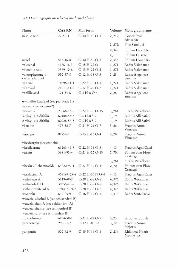
428
WHO monographs on selected medicinal plants
Name CAS RN Mol. form. Volume Monograph name
ursolic acid 77-52-1 C-30 H-48 O-3 2_249; Cortex PruniAfricanae
2_272; Flos Sambuci
2_345; Folium Uvae Ursi4_130 Folium Guavae
uvaol 545-46-0 C-30 H-50 O-2 2_345 Folium Uvae Ursivalerenal 4176-16-3 C-15 H-22 O 1_271 Radix Valerianaevalerenic acid 3569-10-6 C-15 H-22 O-2 1_271 Radix Valerianaevalerophenone-o-carboxylic acid
550-37-8 C-12 H-14 O-3 2_28 Radix AngelicaeSinensis
valtrate 18296-44-1 C-22 H-30 O-8 1_271 Radix Valerianaevaltroxal 71013-41-7 C-17 H-22 O-7 1_271 Radix Valerianaevanillic acid 121-34-6 C-8 H-8 O-4 2_28 Radix Angelicae
Sinensis6-vanilloylcatalpol (see picroside II)vicenin (see vicenin-2)vicenin-2 23666-13-9 C-27 H-30 O-15 3_261 Herba Passiflorae3-vinyl-1,2-dithiin 62488-53-3 C-6 H-8 S-2 1_19 Bulbus Alli Sativi2-vinyl-1,3-dithiin 80028-57-5 C-6 H-8 S-2 1_19 Bulbus Alli Sativivisnadin 477-32-7 C-21 H-24 O-7 3_26 Fructus Ammi
Visnagaevisnagin 82-57-5 C-13 H-10 O-4 3_26 Fructus Ammi
Visnagaevitexicarpin (see casticin)vitexilactone 61263-49-8 C-22 H-34 O-5 4_13 Fructus Agni Castivitexin 3681-93-4 C-21 H-20 O-10 2_70; Folium cum Flore
Crataegi3_261 Herba Passiflorae
vitexin 2´´-rhamnoside 64820-99-1 C-27 H-30 O-14 2_70 Folium cum Flore Crataegi
vitexlactam A 459167-05-6 C-22 H-35 N O-4 4_13 Fructus Agni Castiwithaferin A 5119-48-2 C-28 H-38 O-6 4_376 Radix Withaniaewithanolide D 30655-48-2 C-28 H-38 O-6 4_376 Radix Withaniaewithasomniferol A 194413-09-7 C-28 H-38 O-7 4_376 Radix Withaniaewogonin 632-85-9 C-16 H-12 O-5 3_316 Radix Scutellariaewuweizi alcohol B (see schisandrol B)wuweizichun A (see schisandrol A)wuweizichun B (see schisandrol B)wuweizisu B (see schisandrin B)xanthohumol 6754-58-1 C-21 H-22 O-5 3_239 Strobilus Lupulixanthotoxin 298-81-7 C-12 H-8 O-4 3_12 Fructus Ammi
Majorisyangonin 500-62-9 C-15 H-14 O-4 2_234 Rhizoma Piperis
Methystici

429
Annex 5
Name CAS RN Mol. form. Volume Monograph name
yatrorizine (see jatrorrhizine)zingiberene 495-60-3 C-15 H-24 1_118; Rhizoma Curcumae
Longae1_280 Rhizoma Zingiberis
zizyphus saponin I 77943-56-7 C-47 H-76 O-17 3_362 Fructus Zizyphizizyphus saponin II 77943-83-0 C-47 H-76 O-17 3_362 Fructus Zizyphizizyphus saponin III 77943-54-5 C-52 H-84 O-21 3_362 Fructus Zizyphizwiebelanes 126038-52-6 C-6 H-10 O S-2 1_8 Bulbus Allii Cepae
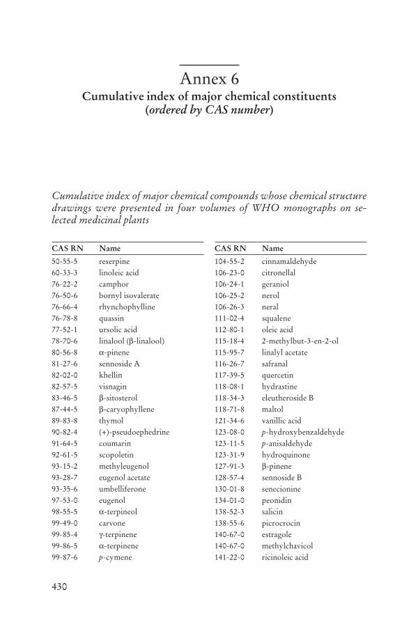
430
Annex 6Cumulative index of major chemical constituents
(ordered by CAS number)
Cumulative index of major chemical compounds whose chemical structure drawings were presented in four volumes of WHO monographs on se-lected medicinal plants
CAS RN Name
50-55-5 reserpine60-33-3 linoleic acid76-22-2 camphor76-50-6 bornyl isovalerate76-66-4 rhynchophylline76-78-8 quassin77-52-1 ursolic acid78-70-6 linalool ( -linalool)80-56-8 -pinene81-27-6 sennoside A82-02-0 khellin82-57-5 visnagin83-46-5 -sitosterol87-44-5 -caryophyllene89-83-8 thymol90-82-4 (+)-pseudoephedrine91-64-5 coumarin92-61-5 scopoletin93-15-2 methyleugenol93-28-7 eugenol acetate93-35-6 umbelliferone97-53-0 eugenol98-55-5 -terpineol99-49-0 carvone99-85-4 -terpinene99-86-5 -terpinene99-87-6 p-cymene
CAS RN Name
104-55-2 cinnamaldehyde106-23-0 citronellal106-24-1 geraniol106-25-2 nerol106-26-3 neral111-02-4 squalene112-80-1 oleic acid115-18-4 2-methylbut-3-en-2-ol115-95-7 linalyl acetate116-26-7 safranal117-39-5 quercetin118-08-1 hydrastine118-34-3 eleutheroside B118-71-8 maltol121-34-6 vanillic acid123-08-0 p-hydroxybenzaldehyde123-11-5 p-anisaldehyde123-31-9 hydroquinone127-91-3 -pinene128-57-4 sennoside B130-01-8 senecionine134-01-0 peonidin138-52-3 salicin138-55-6 picrocrocin140-67-0 estragole140-67-0 methylchavicol141-22-0 ricinoleic acid

431
Annex 6
CAS RN Name
141-27-5 geranial143-07-7 lauric acid149-91-7 gallic acid153-18-4 rutin154-23-4 catechin298-81-7 xanthotoxin299-42-3 (-)-ephedrine305-01-1 esculetin327-97-9 chlorogenic acid331-39-5 caffeic acid437-64-9 genkwanin446-72-0 genistein454-14-8 cuscohygrine458-37-7 curcumin463-40-1 linolenic acid464-92-6 asiatic acid464-98-2 -taraxasterol468-28-0 lupulone469-32-9 hamamelitannin470-82-6 1,8-cineole471-53-4 glycyrrhetic acid471-66-9 -boswellic acid472-15-1 betulinic acid474-58-8 daucosterol476-66-4 ellagic acid477-32-7 visnadin478-42-2 khellinol478-43-3 rhein478-61-5 berbamine478-79-5 khellol479-91-4 casticin479-98-1 aucubin480-10-4 astragalin480-18-2 taxifolin480-19-3 isorhamnetin481-46-9 ginkgetin481-72-1 aloe-emodin481-74-3 chrysophanol482-36-0 hyperoside482-44-0 imperatorin483-17-0 cephaeline483-18-1 emetine484-20-8 bergapten
CAS RN Name
485-72-3 formononetin486-21-5 isofraxidin486-66-8 daidzein486-84-0 harman489-49-6 cetraric acid489-50-9 fumarprotocetraric acid490-46-0 epi-catechin491-60-1 frangula emodin anthrone491-67-8 baicalein491-80-5 biochanin A492-39-7 (+)-norpseudoephedrine492-41-1 (-)-norephedrine495-60-3 zingiberene495-85-2 methysticin497-76-7 arbutin498-02-2 apocynine499-75-2 carvacrol500-62-9 yangonin500-64-1 kawain506-26-3 -linolenic acid506-51-4 tetracosanol507-70-0 borneol508-02-1 oleanolic acid509-80-8 mitraphylline518-82-1 frangula emodin519-54-0 neocarthamin520-36-5 apigenin521-32-4 bilobetin521-34-6 sciadopitysin521-61-9 physcione521-62-0 frangulin A522-12-3 quercitrin523-01-3 O-methylpsychotrine523-47-7 -cadinene528-43-8 magnolol528-58-5 cyanidin529-05-5 chamazulene529-44-2 myricetin532-65-0 ar-turmerone535-83-1 trigonelline537-73-5 (E)-isoferulic acid539-86-6 allicin545-46-0 uvaol

432
WHO monographs on selected medicinal plants
CAS RN Name
548-04-9 hypericin548-19-6 isoginkgetin550-37-8 valerophenone-o-carboxylic acid551-08-6 (Z)-butylidenephtalide551-15-5 liquiritin552-70-5 pseudopelletierine552-79-4 (-)-methylephedrine556-27-4 alliin559-74-0 friedelin562-74-3 terpinen-4-ol578-86-9 liquiritigenin587-63-3 dihydrokawain607-80-7 (+)-sesamin630-94-4 corynoxeine631-69-6 -boswellic acid632-85-9 wogonin638-95-9 -amyrin661-19-8 docosanol668-10-0 ammiol961-29-5 isoliquiritigenin1058-61-3 sitosterone1059-14-9 taraxasterol1135-24-6 (E)-ferulic acid1195-79-5 fenchone1259-86-5 glaucarubinone1405-86-3 glycyrrhizin1415-73-2 aloin A1448-96-0 (+)-protolichesterinic1617-53-4 amentoflavone1898-13-1 cembrene2050-87-5 diallyl trisulfide2086-83-1 berberine2086-96-6 -canadine2141-09-5 magnoflorine2179-57-9 diallyl disulfide2216-51-5 menthol2222-07-3 cucurbitacin I2380-78-1 homovanillyl alcohol2415-24-9 catalpol2858-66-4 pelletierine3338-55-4 cis- -ocimene3387-41-5 sabinene3486-67-7 palmatine
CAS RN Name
3569-10-6 valerenic acid3621-36-1 columbamine3621-38-3 jatrorrhizine3681-93-4 vitexin3779-59-7 cimigenol3877-86-9 cucurbitacin D4128-17-0 farnesyl acetate4176-16-3 valerenal4180-23-8 trans-anethole4221-98-1 (-)- -phellandrene4261-42-1 iso-orientin4373-41-5 maslinic acid4396-01-4 isopelletierine4431-01-0 (Z)-ligustilide4697-68-1 speciophylline4963-01-3 isomitraphylline5041-81-6 isoliquiritin5092-09-1 saikogenin A5096-57-1 -canadine5119-48-2 withaferin A5171-37-9 isopteropodine5373-11-5 cynaroside5508-58-7 andrographolide5573-16-0 saikogenin D5629-60-7 pteropodine5892-54-6 -crocetin5939-57-1 cucurbitacin F5945-86-8 nimbin5948-04-9 dihydrocarvone5957-80-2 carnosol5968-70-7 3-O-acetyl- -boswellic acid5989-27-5 (+)-limonene6199-67-3 cucurbitacin B6537-80-0 chicoric acid6753-98-6 humulene ( -caryophyllene)6754-13-8 helenalin6754-58-1 xanthohumol6801-40-7 magnocurarine6807-92-7 cucurbitine6853-87-8 methylcysteine S-oxide6859-01-4 isorhynchophylline6873-13-8 phellodendrine6906-38-3 delphinidin 3-monoglucoside

433
Annex 6
CAS RN Name
6906-39-4 peonidin 3-monoglucoside6926-08-5 harpagide6988-81-4 petunidin 3-monoglucoside7084-24-4 cyanidin 3-monoglucoside7228-78-6 malvidin 3-monoglucoside7374-79-0 eleutheroside E17432-28-2 schisandrol A7633-29-6 psychotrine7785-54-8 (+)- -terpinyl acetate8063-16-9 psyllium10219-75-7 eremophilene11006-91-0 aloinoside B11021-13-9 ginsenoside Rb211021-14-0 ginsenoside Rc11027-63-7 agnuside11079-53-1 hyperforin11137-63-6 sennoside E11141-17-6 azadirachtin13844-01-4 barringtogenol C14019-66-0 uncarine F14062-59-0 glucofrangulin B14073-97-3 menthone14101-04-3 frangulin B14215-86-2 sweroside14356-59-3 saikogenin F14907-98-3 brusatol14965-20-9 chrysosplenol D15291-75-5 ginkgolide A15291-76-6 ginkgolide C15291-77-7 ginkgolide B15291-78-8 ginkgolide M15345-89-8 demethoxyyangonin15401-60-2 ipecoside15486-24-5 eleutheroside C15514-06-4 procyanidin16830-15-2 asiaticoside16845-16-2 eleutheroside B117019-92-0 11-oxo- -boswellic acid17226-75-4 khellinin17795-24-3 propylcysteine S-oxide17910-09-7 curzerene18175-79-6 saikogenin G18234-46-3 baldrinal
CAS RN Name
18262-45-8 (-)-11-deoxyaloin18296-44-1 valtrate18444-66-1 cucurbitacin E18449-41-7 madecassic acid18609-16-0 deacetylnimbin18642-44-9 actein18747-42-7 N-methylisopelletierine18942-26-2 chebulinic acid19210-12-9 harpagoside19467-03-9 melittoside19533-92-7 alphitolic acid19666-76-3 panaxadiol19764-02-4 fragilin19902-91-1 dihydromethysticin19912-61-9 (5E,9Z)-furanodiene20229-56-5 spiraeoside20283-92-5 rosmarinic acid20315-25-7 procyanidin B120347-65-3 bornyl acetate20486-27-5 procumbide20493-56-5 curzerenone20554-84-1 parthenolide20633-72-1 monomelittoside20633-84-5 scolymoside20675-96-1 syringenin20736-09-8 saikosaponin A20831-76-9 gentiopicroside20853-07-0 protoaescigenin20874-52-6 saikosaponin D21018-84-8 amarogentin21133-53-9 glucofrangulin A21453-69-0 (+)-syringaresinol21499-66-1 brucein D21637-25-2 isoquercitrin21967-41-9 baicalin22255-13-6 guajaverin22427-39-0 ginsenoside Rg122608-11-3 desmethoxycurcumin22888-70-6 silybin23089-26-1 (-)- -bisabolol23094-71-5 chebulagic acid23180-57-6 paeoniflorin23513-08-8 [8]-gingerol

434
WHO monographs on selected medicinal plants
CAS RN Name
23513-13-5 [6]-shogaol23513-14-6 [6]-gingerol23513-15-7 [10]-gingerol23567-23-9 procyanidin B323666-13-9 vicenin-224815-24-5 rescinnamine24959-84-0 canin25161-41-5 acevaltrate25368-09-6 -crocetin25514-29-8 brucein B25514-30-1 brucein C25514-31-2 brucein A26020-14-4 calenduloside E26020-29-1 calenduloside H26472-41-3 humulone27208-74-8 acteol27215-14-1 neoandrographolide27409-30-9 picroside I27876-94-4 -crocetin28371-16-6 aloin B28608-75-5 orientin29106-49-8 procyanidin B229106-51-2 procyanidin B429428-58-8 lucenin-229431-84-3 artecanin29782-68-1 silydianin29836-41-7 salicortin29883-15-6 amygdalin30655-48-2 withanolide D30964-13-7 cynarin31078-10-1 isovaltrate32725-74-9 calenduloside A32791-84-7 panaxatriol33171-05-0 bisdesmethoxycurcumin33570-04-6 bilobalide33889-69-9 silychristin34257-95-9 11 ,13-dihydrohelenalin34366-31-9 senegin II34540-22-2 madecassoside35354-74-6 honokiol35730-78-0 cynaropicrin35906-36-6 (E)-senegin III35988-27-3 kutkoside
CAS RN Name
36338-96-2 carthamin37271-16-2 sennoside C37271-17-3 sennoside D37719-98-5 angelicone38953-85-4 isovitexin39012-20-9 picroside II39025-23-5 (Z)-guggulsterone39025-24-6 (E)-guggulsterone39025-25-7 guggulsterol I39025-26-8 guggulsterol-II39025-27-9 guggulsterol-III39432-56-9 eleutheroside E41451-75-6 bruceantin41743-67-3 [3]-gingerol41743-68-4 [4]-gingerol41743-69-5 [5]-gingerol41753-43-9 ginsenoside Rb141943-03-7 mukulol42553-65-1 A-crocin51014-29-0 isocorynoxeine51018-28-1 (+)-methylpseudoephedrine51415-02-2 calenduloside F51938-32-0 schaftoside52012-29-0 isoschaftoside52286-58-5 ginsenoside Rf52286-59-6 ginsenoside Re52286-74-5 ginsenoside Rg252705-93-8 ginsenoside Rd52842-23-6 sennoside F52949-83-4 ajugol53472-37-0 3´-methylmyricetin53683-73-1 ailanthinone53823-08-8 cascaroside A53823-09-9 cascaroside C53861-34-0 cascaroside B53861-35-1 cascaroside D53962-20-2 patrinoside55056-80-9 protodioscin55466-05-2 jujuboside B55750-84-0 B-crocin55750-85-1 C-crocin55903-92-9 cucurbitacin R55954-61-5 pseudohypericin

435
Annex 6
CAS RN Name
56645-88-6 aloinoside A57710-64-2 D-crocin58050-17-2 E-crocin58316-41-9 saikosaponin B258316-42-0 saikosaponin B358479-68-8 platycodin D58546-54-6 schisandrol B58558-08-0 saikosaponin B158558-09-1 saikosaponin B459440-97-0 (+)-echinolone61263-49-8 vitexilactone61281-37-6 schisandrin B61281-38-7 schisandrin A62488-53-3 3-vinyl-1,2-dithiin63644-62-2 coniferyl ferulate64461-95-6 picroside III64820-99-1 vitexin 2´´-rhamnoside65995-63-3 punicalagin65995-64-4 punicalin66176-93-0 cimicifugoside66663-90-9 platycodin D266663-91-0 polygalacin D66663-92-1 polygalacin D266779-34-8 platycodin A66779-35-9 platycodin C67416-61-9 3-O-acetyl-11-oxo- -
boswellic acid67910-07-0 homobaldrinal69176-52-9 gomisin N69884-00-0 pseudoginsenoside F1169980-49-0 (+)-11-deoxyaloin71013-41-7 valtroxal71616-00-7 achillicin73543-67-6 -dimorphecolic acid73889-54-0 chrysaloin75911-12-5 taraxacolide glucoside75911-14-7 taraxinic acid glucoside75917-90-7 (10E)-N-
isobutyldodecatetraenamide75991-58-1 (3S,9R)-tetrahydroridentin B77053-35-1 5´-methoxybilobetin77448-63-6 (10Z)-N-
isobutyldodecatetraenamide
CAS RN Name
77790-55-7 neoolivil77943-54-5 zizyphus saponin III77943-56-7 zizyphus saponin I77943-83-0 zizyphus saponin II78281-02-4 hydroxysafflor yellow A79484-75-6 eleutheroside D79974-46-2 tribulosin80028-57-5 vinyl-1,3-dithiin (2-)80151-77-5 (-)-tussilagine81348-81-4 momordicoside F181348-82-5 momordicoside F281348-83-6 momordicoside L81348-84-7 momordicoside K81371-54-2 momordicoside G81371-55-3 momordicoside I81720-05-0 rehmannioside A81720-06-1 rehmannioside B81720-07-2 rehmannioside C81720-08-3 rehmannioside D82209-72-1 andropanoside82209-76-5 andrographiside82508-14-3 -turmerone82508-15-4 -turmerone82854-37-3 echinacoside82901-10-8 (5E,8S,9E)-2-
methoxyfuranodiene83199-38-6 angelol G84638-55-1 acetoxyvalerenic acid84676-88-0 isoastragaloside I84676-89-1 astragaloside II84680-75-1 astragaloside I84687-42-3 astragaloside III84687-43-4 astragaloside IV84687-44-5 astragaloside V84687-45-6 astragaloside VI84687-46-7 astragaloside VII85532-77-0 safflor yellow A86764-11-6 isoastragaloside II87605-93-4 furanoeudesma-1,3-diene88010-65-5 (9E)-4,5-dihydrofuranodien-
6-one89913-60-0 3-O-acetyl- -boswellic acid

436
WHO monographs on selected medicinal plants
CAS RN Name
91108-32-6 isotussilagine91574-92-4 safflor yellow B92284-99-6 (E)-ajoene92285-00-2 (Z)-ajoene92935-94-9 angelicide98449-40-2 taraxacoside99742-03-7 [1]-gingerol99742-07-1 [7]-shogaol99742-08-2 [9]-shogaol99742-09-3 [11]-shogaol99742-10-6 [12]-shogaol101927-24-6 docosyl (E)-ferulate101927-25-7 tetracosyl (E)-ferulate104404-17-3 ceramides107438-79-9 ginkgolide J108376-98-3 (5E,8R,9E)-2-
methoxyfuranodiene111545-28-9 rheinoside A111545-29-0 rheinoside C111614-10-9 rheinoside B111614-11-0 rheinoside D111957-48-3 kaempferol diglycoside
coumarate126038-52-6 zwiebelanes128748-74-8 uncarinic acid B143061-65-8 quercetin diglycoside
coumarate143183-63-5 adhyperforin149475-43-4 tinctormine162613-71-0 (Z)-senegasaponin B162613-72-1 (Z)-senegasaponin A
CAS RN Name
162681-52-9 (Z)-senegin II162681-53-0 (Z)-senegin III162762-97-2 (E)-senegasaponin A162870-58-8 (E)-senegasaponin B163589-51-3 desacylsenegin II164178-32-9 cascaroside E164322-83-2 cascaroside F166833-80-3 chebulanin173557-02-3 desacylsenegasaponin A180572-65-0 desacylsenegin III194413-09-7 withasomniferol A206256-62-4 uncarinic acid A218622-84-5 tribulusamide A218622-86-7 tribulusamide B311310-49-3 tribulosaponin A311310-52-8 tribulosaponin B333332-41-5 goyaglycoside a333332-48-2 goyaglycoside b333332-49-3 goyaglycoside c333332-50-6 goyaglycoside d333333-12-3 goyaglycoside e333333-13-4 goyaglycoside f333333-14-5 goyaglycoside g333333-19-0 goyaglycoside h333333-20-3 goyasaponin I333333-23-6 goyasaponin II333333-27-0 goyasaponin III360762-35-2 N-isobutyldodecatetraenamide459167-05-6 vitexlactam A

437
Annex 7Cumulative index of major chemical constituents
(ordered by molecular formula)
Cumulative index of major chemical compounds whose chemical structure drawings were presented in four volumes of WHO monographs on se-lected medicinal plants
Mol. form. Name
C-4 H-9 N O-3 S methylcysteine S-oxideC-5 H-10 N-2 O-2 cucurbitineC-5 H-10 O 2-methylbut-3-en-2-olC-6 H-6 O-2 hydroquinoneC-6 H-6 O-3 maltolC-6 H-8 S-2 2-vinyl-1,3-dithiinC-6 H-8 S-2 3-vinyl-1,2-dithiinC-6 H-10 O S-2 allicinC-6 H-10 O S-2 zwiebelanesC-6 H-10 S-2 diallyl disulfideC-6 H-10 S-3 diallyl trisulfideC-6 H-11 N O-3 S alliinC-6 H-11 N O-3 S 1-propenylcysteine
oxideC-6 H-13 N O-3 S propylcysteine S-oxideC-7 H-6 O-2 p-hydroxybe-
nzaldehydeC-7 H-6 O-5 gallic acidC-7 H-7 N O-2 trigonellineC-8 H-8 O-2 p-anisaldehydeC-8 H-8 O-4 vanillic acidC-8 H-15 N O isopelletierineC-8 H-15 N O pelletierineC-8 H-16 O-6 eleutheroside CC-9 H-6 O-2 coumarinC-9 H-6 O-3 umbelliferone
Mol. form. Name
C-9 H-6 O-4 esculetinC-9 H-8 O cinnamaldehydeC-9 H-8 O-4 caffeic acidC-9 H-10 O-3 apocynineC-9 H-12 O-3 homovanillyl alcoholC-9 H-13 N O (-)-norephedrineC-9 H-13 N O (+)-norpseudo-
ephedrineC-9 H-14 O S-3 (E)-ajoeneC-9 H-14 O S-3 (Z)-ajoeneC-9 H-15 N O pseudopelletierineC-9 H-17 N O N-methylisopelletierineC-10 H-8 O-4 scopoletinC-10 H-10 O-4 (E)-ferulic acidC-10 H-10 O-4 (E)-isoferulic acidC-10 H-12 O trans-anetholeC-10 H-12 O estragoleC-10 H-12 O methylchavicolC-10 H-12 O-2 eugenolC-10 H-14 p-cymeneC-10 H-14 O carvacrolC-10 H-14 O carvoneC-10 H-14 O safranalC-10 H-14 O thymolC-10 H-15 N O (-)-ephedrineC-10 H-15 N O (+)-pseudoephedrine
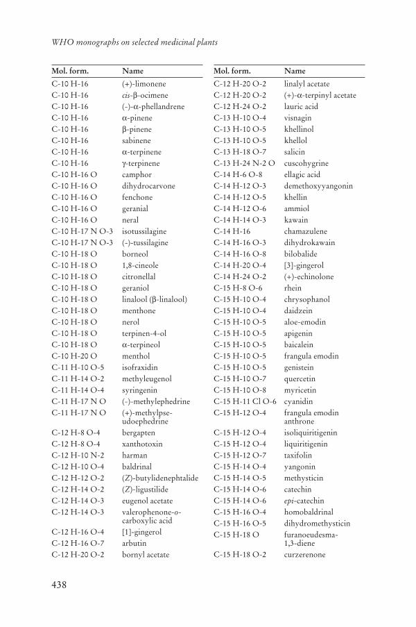
438
WHO monographs on selected medicinal plants
Mol. form. Name
C-10 H-16 (+)-limoneneC-10 H-16 cis- -ocimeneC-10 H-16 (-)- -phellandreneC-10 H-16 -pineneC-10 H-16 -pineneC-10 H-16 sabineneC-10 H-16 -terpineneC-10 H-16 -terpineneC-10 H-16 O camphorC-10 H-16 O dihydrocarvoneC-10 H-16 O fenchoneC-10 H-16 O geranialC-10 H-16 O neralC-10 H-17 N O-3 isotussilagineC-10 H-17 N O-3 (-)-tussilagineC-10 H-18 O borneolC-10 H-18 O 1,8-cineoleC-10 H-18 O citronellalC-10 H-18 O geraniolC-10 H-18 O linalool ( -linalool)C-10 H-18 O menthoneC-10 H-18 O nerolC-10 H-18 O terpinen-4-olC-10 H-18 O -terpineolC-10 H-20 O mentholC-11 H-10 O-5 isofraxidinC-11 H-14 O-2 methyleugenolC-11 H-14 O-4 syringeninC-11 H-17 N O (-)-methylephedrineC-11 H-17 N O (+)-methylpse-
udoephedrineC-12 H-8 O-4 bergaptenC-12 H-8 O-4 xanthotoxinC-12 H-10 N-2 harmanC-12 H-10 O-4 baldrinalC-12 H-12 O-2 (Z)-butylidenephtalideC-12 H-14 O-2 (Z)-ligustilideC-12 H-14 O-3 eugenol acetateC-12 H-14 O-3 valerophenone-o-
carboxylic acidC-12 H-16 O-4 [1]-gingerolC-12 H-16 O-7 arbutinC-12 H-20 O-2 bornyl acetate
Mol. form. Name
C-12 H-20 O-2 linalyl acetateC-12 H-20 O-2 (+)- -terpinyl acetateC-12 H-24 O-2 lauric acidC-13 H-10 O-4 visnaginC-13 H-10 O-5 khellinolC-13 H-10 O-5 khellolC-13 H-18 O-7 salicinC-13 H-24 N-2 O cuscohygrineC-14 H-6 O-8 ellagic acidC-14 H-12 O-3 demethoxyyangoninC-14 H-12 O-5 khellinC-14 H-12 O-6 ammiolC-14 H-14 O-3 kawainC-14 H-16 chamazuleneC-14 H-16 O-3 dihydrokawainC-14 H-16 O-8 bilobalideC-14 H-20 O-4 [3]-gingerolC-14 H-24 O-2 (+)-echinoloneC-15 H-8 O-6 rheinC-15 H-10 O-4 chrysophanolC-15 H-10 O-4 daidzeinC-15 H-10 O-5 aloe-emodinC-15 H-10 O-5 apigeninC-15 H-10 O-5 baicaleinC-15 H-10 O-5 frangula emodinC-15 H-10 O-5 genisteinC-15 H-10 O-7 quercetinC-15 H-10 O-8 myricetinC-15 H-11 Cl O-6 cyanidinC-15 H-12 O-4 frangula emodin
anthroneC-15 H-12 O-4 isoliquiritigeninC-15 H-12 O-4 liquiritigeninC-15 H-12 O-7 taxifolinC-15 H-14 O-4 yangoninC-15 H-14 O-5 methysticinC-15 H-14 O-6 catechinC-15 H-14 O-6 epi-catechinC-15 H-16 O-4 homobaldrinalC-15 H-16 O-5 dihydromethysticinC-15 H-18 O furanoeudesma-
1,3-dieneC-15 H-18 O-2 curzerenone

439
Annex 7
Mol. form. Name
C-15 H-18 O-4 helenalinC-15 H-18 O-5 artecaninC-15 H-18 O-5 caninC-15 H-20 O curzereneC-15 H-20 O (5E,9Z)-furanodieneC-15 H-20 O ar-turmeroneC-15 H-20 O-2 (9E)-4,5-dihydro-
furanodien-6-oneC-15 H-20 O-3 parthenolideC-15 H-20 O-4 11 ,13-dihydro-
helenalinC-15 H-20 O-4 3 -hydroxy-
parthenolideC-15 H-20 O-8 fragilinC-15 H-22 O -turmeroneC-15 H-22 O -turmeroneC-15 H-22 O valerenalC-15 H-22 O-2 valerenic acidC-15 H-22 O-4 [4]-gingerolC-15 H-22 O-9 aucubinC-15 H-22 O-10 catalpolC-15 H-22 O-10 monomelittosideC-15 H-22 O-10 procumbideC-15 H-24 -cadineneC-15 H-24 -caryophylleneC-15 H-24 eremophileneC-15 H-24 humulene
( -caryophyllene)C-15 H-24 zingibereneC-15 H-24 O-4 (3S,9R)-tetrahy-
droridentin BC-15 H-24 O-9 ajugolC-15 H-24 O-10 harpagideC-15 H-26 O (-)- -bisabololC-15 H-26 O-2 bornyl isovalerateC-16 H-12 O-4 formononetinC-16 H-12 O-5 biochanin AC-16 H-12 O-5 genkwaninC-16 H-12 O-5 physcioneC-16 H-12 O-5 wogoninC-16 H-12 O-7 isorhamnetinC-16 H-12 O-8 3´-methylmyricetinC-16 H-13 Cl O-6 peonidin
Mol. form. Name
C-16 H-14 O-4 imperatorinC-16 H-16 O-5 angeliconeC-16 H-18 O-9 chlorogenic acidC-16 H-20 O-9 gentiopicrosideC-16 H-22 O-2 (5E,8R,9E)-2-methoxy-
furanodieneC-16 H-22 O-2 (5E,8S,9E)-2-methoxy-
furanodieneC-16 H-22 O-9 swerosideC-16 H-24 O-4 [5]-gingerolC-16 H-25 N O N-isobutyldode-
catetraenamideC-16 H-25 N O (10E)-N-isobutyldode-
catetraenamideC-16 H-25 N O (10Z)-N-isobutyldode-
catetraenamideC-16 H-26 O-7 picrocrocinC-17 H-20 O-10 eleutheroside B1C-17 H-22 O-5 achillicinC-17 H-22 O-7 valtroxalC-17 H-24 O-3 [6]-shogaolC-17 H-24 O-4 acetoxyvalerenic acidC-17 H-24 O-9 eleutheroside BC-17 H-26 O-4 [6]-gingerolC-17 H-28 O-2 farnesyl acetateC-18 H-16 O-8 chrysosplenol DC-18 H-16 O-8 rosmarinic acidC-18 H-18 O-2 honokiolC-18 H-18 O-2 magnololC-18 H-22 O-10 taraxacosideC-18 H-25 N O-5 senecionineC-18 H-26 O-3 [7]-shogaolC-18 H-30 O-2 linolenic acidC-18 H-30 O-2 -linolenic acidC-18 H-32 O-2 linoleic acidC-18 H-32 O-3 -dimorphecolic acidC-18 H-34 O-2 oleic acidC-18 H-34 O-3 ricinoleic acidC-19 H-16 O-4 bisdesme-
thoxycurcuminC-19 H-18 O-8 casticinC-19 H-20 O-10 khellininC-19 H-22 O-6 cynaropicrinC-19 H-24 N O-3 magnocurarine

440
WHO monographs on selected medicinal plants
Mol. form. Name
C-19 H-30 O-4 [8]-gingerolC-19 H-32 O-4 (+)-protolichesterinic
acidC-20 H-18 N O-4 berberineC-20 H-18 O-5 desmethoxycurcuminC-20 H-18 O-6 (+)-sesaminC-20 H-18 O-9 cetraric acidC-20 H-18 O-9 frangulin BC-20 H-18 O-11 guajaverinC-20 H-20 N O-4 columbamineC-20 H-20 N O-4 jatrorrhizineC-20 H-20 O-6 coniferyl ferulateC-20 H-20 O-14 hamamelitanninC-20 H-21 N O-4 -canadineC-20 H-21 N O-4 -canadineC-20 H-24 N O-4 magnoflorineC-20 H-24 N O-4 phellodendrineC-20 H-24 O-4 -crocetinC-20 H-24 O-7 angelol GC-20 H-24 O-7 neoolivilC-20 H-24 O-9 ginkgolide AC-20 H-24 O-10 ginkgolide BC-20 H-24 O-10 ginkgolide JC-20 H-24 O-10 ginkgolide MC-20 H-24 O-10 salicortinC-20 H-24 O-11 ginkgolide CC-20 H-26 O-4 carnosolC-20 H-26 O-9 brucein DC-20 H-30 O-3 [9]-shogaolC-20 H-30 O-5 andrographolideC-20 H-32 cembreneC-20 H-34 O mukulolC-21 H-18 O-11 baicalinC-21 H-20 O-6 curcuminC-21 H-20 O-9 frangulin AC-21 H-20 O-10 isovitexinC-21 H-20 O-10 vitexinC-21 H-20 O-11 astragalinC-21 H-20 O-11 cynarosideC-21 H-20 O-11 iso-orientinC-21 H-20 O-11 orientinC-21 H-20 O-11 quercitrinC-21 H-20 O-12 hyperoside
Mol. form. Name
C-21 H-20 O-12 isoquercitrinC-21 H-20 O-12 spiraeosideC-21 H-21 Cl O-11 cyanidin 3-mono-
glucosideC-21 H-21 Cl O-12 delphinidin
3-monoglucosideC-21 H-21 N O-6 hydrastineC-21 H-22 N O-4 palmatineC-21 H-22 O-5 xanthohumolC-21 H-22 O-8 chrysaloinC-21 H-22 O-8 (-)-11-deoxyaloinC-21 H-22 O-8 (+)-11-deoxyaloinC-21 H-22 O-9 aloin AC-21 H-22 O-9 aloin BC-21 H-22 O-9 isoliquiritinC-21 H-22 O-9 liquiritinC-21 H-22 O-11 neocarthaminC-21 H-24 N-2 O-4 isomitraphyllineC-21 H-24 N-2 O-4 isopteropodineC-21 H-24 N-2 O-4 mitraphyllineC-21 H-24 N-2 O-4 pteropodineC-21 H-24 N-2 O-4 speciophyllineC-21 H-24 N-2 O-4 uncarine FC-21 H-24 O-7 visnadinC-21 H-26 O-4 -crocetinC-21 H-27 N O-11 amygdalinC-21 H-28 O-2 (E)-guggulsteroneC-21 H-28 O-2 (Z)-guggulsteroneC-21 H-28 O-9 taraxinic acid glucosideC-21 H-30 O-5 humuloneC-21 H-32 O-9 taraxacolide glucosideC-21 H-32 O-15 melittosideC-21 H-32 O-15 rehmannioside AC-21 H-32 O-15 rehmannioside BC-21 H-34 O-4 [10]-gingerolC-21 H-34 O-11 patrinosideC-21 H-34 O-14 rehmannioside CC-22 H-16 O-12 fumarprotocetraric acidC-22 H-18 O-12 chicoric acidC-22 H-23 Cl O-11 peonidin 3-mono-
glucosideC-22 H-23 Cl O-12 petunidin 3-mono-
glucoside
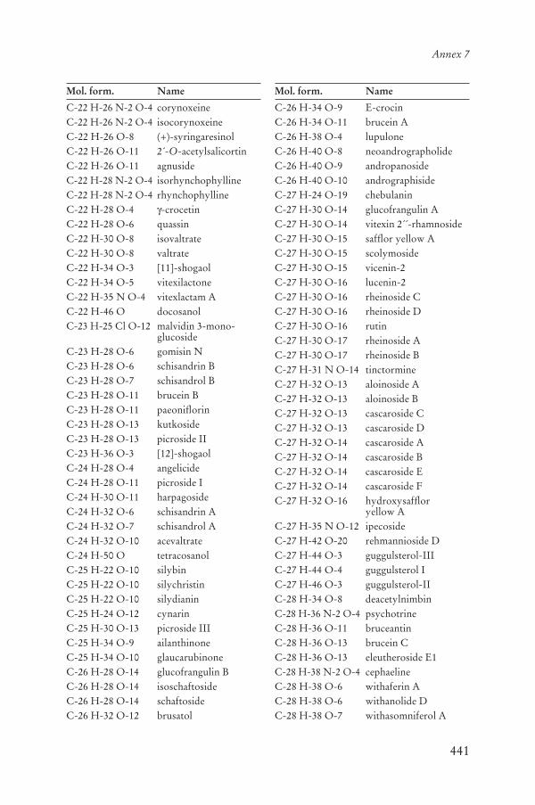
441
Annex 7
Mol. form. Name
C-22 H-26 N-2 O-4 corynoxeineC-22 H-26 N-2 O-4 isocorynoxeineC-22 H-26 O-8 (+)-syringaresinolC-22 H-26 O-11 2´-O-acetylsalicortinC-22 H-26 O-11 agnusideC-22 H-28 N-2 O-4 isorhynchophyllineC-22 H-28 N-2 O-4 rhynchophyllineC-22 H-28 O-4 -crocetinC-22 H-28 O-6 quassinC-22 H-30 O-8 isovaltrateC-22 H-30 O-8 valtrateC-22 H-34 O-3 [11]-shogaolC-22 H-34 O-5 vitexilactoneC-22 H-35 N O-4 vitexlactam AC-22 H-46 O docosanolC-23 H-25 Cl O-12 malvidin 3-mono-
glucosideC-23 H-28 O-6 gomisin NC-23 H-28 O-6 schisandrin BC-23 H-28 O-7 schisandrol BC-23 H-28 O-11 brucein BC-23 H-28 O-11 paeoniflorinC-23 H-28 O-13 kutkosideC-23 H-28 O-13 picroside IIC-23 H-36 O-3 [12]-shogaolC-24 H-28 O-4 angelicideC-24 H-28 O-11 picroside IC-24 H-30 O-11 harpagosideC-24 H-32 O-6 schisandrin AC-24 H-32 O-7 schisandrol AC-24 H-32 O-10 acevaltrateC-24 H-50 O tetracosanolC-25 H-22 O-10 silybinC-25 H-22 O-10 silychristinC-25 H-22 O-10 silydianinC-25 H-24 O-12 cynarinC-25 H-30 O-13 picroside IIIC-25 H-34 O-9 ailanthinoneC-25 H-34 O-10 glaucarubinoneC-26 H-28 O-14 glucofrangulin BC-26 H-28 O-14 isoschaftosideC-26 H-28 O-14 schaftosideC-26 H-32 O-12 brusatol
Mol. form. Name
C-26 H-34 O-9 E-crocinC-26 H-34 O-11 brucein AC-26 H-38 O-4 lupuloneC-26 H-40 O-8 neoandrographolideC-26 H-40 O-9 andropanosideC-26 H-40 O-10 andrographisideC-27 H-24 O-19 chebulaninC-27 H-30 O-14 glucofrangulin AC-27 H-30 O-14 vitexin 2´´-rhamnosideC-27 H-30 O-15 safflor yellow AC-27 H-30 O-15 scolymosideC-27 H-30 O-15 vicenin-2C-27 H-30 O-16 lucenin-2C-27 H-30 O-16 rheinoside CC-27 H-30 O-16 rheinoside DC-27 H-30 O-16 rutinC-27 H-30 O-17 rheinoside AC-27 H-30 O-17 rheinoside BC-27 H-31 N O-14 tinctormineC-27 H-32 O-13 aloinoside AC-27 H-32 O-13 aloinoside BC-27 H-32 O-13 cascaroside CC-27 H-32 O-13 cascaroside DC-27 H-32 O-14 cascaroside AC-27 H-32 O-14 cascaroside BC-27 H-32 O-14 cascaroside EC-27 H-32 O-14 cascaroside FC-27 H-32 O-16 hydroxysafflor
yellow AC-27 H-35 N O-12 ipecosideC-27 H-42 O-20 rehmannioside DC-27 H-44 O-3 guggulsterol-IIIC-27 H-44 O-4 guggulsterol IC-27 H-46 O-3 guggulsterol-IIC-28 H-34 O-8 deacetylnimbinC-28 H-36 N-2 O-4 psychotrineC-28 H-36 O-11 bruceantinC-28 H-36 O-13 brucein CC-28 H-36 O-13 eleutheroside E1C-28 H-38 N-2 O-4 cephaelineC-28 H-38 O-6 withaferin AC-28 H-38 O-6 withanolide DC-28 H-38 O-7 withasomniferol A
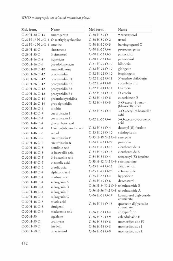
442
WHO monographs on selected medicinal plants
Mol. form. Name
C-29 H-30 O-13 amarogentinC-29 H-38 N-2 O-4 O-methylpsychotrineC-29 H-40 N-2 O-4 emetineC-29 H-48 O sitosteroneC-29 H-50 O -sitosterolC-30 H-16 O-8 hypericinC-30 H-16 O-9 pseudohypericinC-30 H-18 O-10 amentoflavoneC-30 H-26 O-12 procyanidinC-30 H-26 O-12 procyanidin B1C-30 H-26 O-12 procyanidin B2C-30 H-26 O-12 procyanidin B3C-30 H-26 O-12 procyanidin B4C-30 H-26 O-14 proanthocyanidinsC-30 H-26 O-14 prodelphinidinC-30 H-36 O-9 nimbinC-30 H-42 O-7 cucurbitacin IC-30 H-44 O-7 cucurbitacin DC-30 H-46 O-4 glycyrrhetic acidC-30 H-46 O-4 11-oxo- -boswellic acidC-30 H-46 O-6 acteolC-30 H-46 O-7 cucurbitacin FC-30 H-46 O-7 cucurbitacin RC-30 H-48 O-3 betulinic acidC-30 H-48 O-3 -boswellic acidC-30 H-48 O-3 -boswellic acidC-30 H-48 O-3 oleanolic acidC-30 H-48 O-3 ursolic acidC-30 H-48 O-4 alphitolic acidC-30 H-48 O-4 maslinic acidC-30 H-48 O-4 saikogenin AC-30 H-48 O-4 saikogenin DC-30 H-48 O-4 saikogenin FC-30 H-48 O-4 saikogenin GC-30 H-48 O-5 asiatic acidC-30 H-48 O-5 cimigenolC-30 H-48 O-6 madecassic acidC-30 H-50 squaleneC-30 H-50 O -amyrinC-30 H-50 O friedelinC-30 H-50 O taraxasterol
Mol. form. Name
C-30 H-50 O -taraxasterolC-30 H-50 O-2 uvaolC-30 H-50 O-5 barringtogenol CC-30 H-50 O-6 protoaescigeninC-30 H-52 O-3 panaxadiolC-30 H-52 O-4 panaxatriolC-31 H-20 O-10 bilobetinC-32 H-22 O-10 ginkgetinC-32 H-22 O-10 isoginkgetinC-32 H-22 O-11 5´-methoxybilobetinC-32 H-44 O-8 cucurbitacin EC-32 H-44 O-14 C-crocinC-32 H-44 O-14 D-crocinC-32 H-46 O-8 cucurbitacin BC-32 H-48 O-5 3-O-acetyl-11-oxo-
-boswellic acidC-32 H-50 O-4 3-O-acetyl- -boswellic
acidC-32 H-50 O-4 3-O-acetyl- -boswellic
acidC-32 H-54 O-4 docosyl (E)-ferulateC-33 H-24 O-10 sciadopitysinC-33 H-40 N-2 O-9 reserpineC-34 H-22 O-22 punicalinC-34 H-46 O-18 eleutheroside DC-34 H-46 O-18 eleutheroside EC-34 H-58 O-4 tetracosyl (E)-ferulateC-35 H-42 N-2 O-9 rescinnamineC-35 H-44 O-16 azadirachtinC-35 H-46 O-20 echinacosideC-35 H-52 O-4 hyperforinC-35 H-60 O-6 daucosterolC-36 H-34 N-2 O-9 tribulusamide BC-36 H-36 N-2 O-8 tribulusamide AC-36 H-36 O-17 kaempferol diglycoside
coumarateC-36 H-36 O-18 quercetin diglycoside
coumarateC-36 H-54 O-4 adhyperforinC-36 H-56 O-9 calenduloside EC-36 H-58 O-8 momordicoside F2C-36 H-58 O-8 momordicoside IC-36 H-58 O-9 momordicoside L
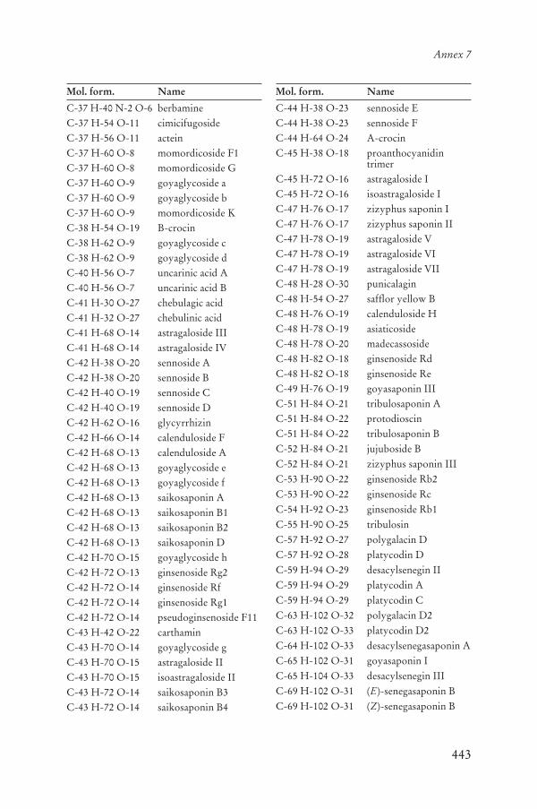
443
Annex 7
Mol. form. Name
C-37 H-40 N-2 O-6 berbamineC-37 H-54 O-11 cimicifugosideC-37 H-56 O-11 acteinC-37 H-60 O-8 momordicoside F1C-37 H-60 O-8 momordicoside GC-37 H-60 O-9 goyaglycoside aC-37 H-60 O-9 goyaglycoside bC-37 H-60 O-9 momordicoside KC-38 H-54 O-19 B-crocinC-38 H-62 O-9 goyaglycoside cC-38 H-62 O-9 goyaglycoside dC-40 H-56 O-7 uncarinic acid AC-40 H-56 O-7 uncarinic acid BC-41 H-30 O-27 chebulagic acidC-41 H-32 O-27 chebulinic acidC-41 H-68 O-14 astragaloside IIIC-41 H-68 O-14 astragaloside IVC-42 H-38 O-20 sennoside AC-42 H-38 O-20 sennoside BC-42 H-40 O-19 sennoside CC-42 H-40 O-19 sennoside DC-42 H-62 O-16 glycyrrhizinC-42 H-66 O-14 calenduloside FC-42 H-68 O-13 calenduloside AC-42 H-68 O-13 goyaglycoside eC-42 H-68 O-13 goyaglycoside fC-42 H-68 O-13 saikosaponin AC-42 H-68 O-13 saikosaponin B1C-42 H-68 O-13 saikosaponin B2C-42 H-68 O-13 saikosaponin DC-42 H-70 O-15 goyaglycoside hC-42 H-72 O-13 ginsenoside Rg2C-42 H-72 O-14 ginsenoside RfC-42 H-72 O-14 ginsenoside Rg1C-42 H-72 O-14 pseudoginsenoside F11C-43 H-42 O-22 carthaminC-43 H-70 O-14 goyaglycoside gC-43 H-70 O-15 astragaloside IIC-43 H-70 O-15 isoastragaloside IIC-43 H-72 O-14 saikosaponin B3C-43 H-72 O-14 saikosaponin B4
Mol. form. Name
C-44 H-38 O-23 sennoside EC-44 H-38 O-23 sennoside FC-44 H-64 O-24 A-crocinC-45 H-38 O-18 proanthocyanidin
trimerC-45 H-72 O-16 astragaloside IC-45 H-72 O-16 isoastragaloside IC-47 H-76 O-17 zizyphus saponin IC-47 H-76 O-17 zizyphus saponin IIC-47 H-78 O-19 astragaloside VC-47 H-78 O-19 astragaloside VIC-47 H-78 O-19 astragaloside VIIC-48 H-28 O-30 punicalaginC-48 H-54 O-27 safflor yellow BC-48 H-76 O-19 calenduloside HC-48 H-78 O-19 asiaticosideC-48 H-78 O-20 madecassosideC-48 H-82 O-18 ginsenoside RdC-48 H-82 O-18 ginsenoside ReC-49 H-76 O-19 goyasaponin IIIC-51 H-84 O-21 tribulosaponin AC-51 H-84 O-22 protodioscinC-51 H-84 O-22 tribulosaponin BC-52 H-84 O-21 jujuboside BC-52 H-84 O-21 zizyphus saponin IIIC-53 H-90 O-22 ginsenoside Rb2C-53 H-90 O-22 ginsenoside RcC-54 H-92 O-23 ginsenoside Rb1C-55 H-90 O-25 tribulosinC-57 H-92 O-27 polygalacin DC-57 H-92 O-28 platycodin DC-59 H-94 O-29 desacylsenegin IIC-59 H-94 O-29 platycodin AC-59 H-94 O-29 platycodin CC-63 H-102 O-32 polygalacin D2C-63 H-102 O-33 platycodin D2C-64 H-102 O-33 desacylsenegasaponin AC-65 H-102 O-31 goyasaponin IC-65 H-104 O-33 desacylsenegin IIIC-69 H-102 O-31 (E)-senegasaponin BC-69 H-102 O-31 (Z)-senegasaponin B

444
WHO monographs on selected medicinal plants
Mol. form. Name
C-70 H-104 O-32 senegin IIC-70 H-104 O-32 (Z)-senegin IIC-70 H-110 O-35 goyasaponin IIC-74 H-110 O-35 (E)-senegasaponin AC-74 H-110 O-35 (Z)-senegasaponin AC-75 H-112 O-35 (E)-senegin IIIC-75 H-112 O-35 (Z)-senegin III

445

446

Selected WHO publications of related interest
Information on medicinal plants:
WHO monographs on selected medicinal plants, Volume 3 (ISBN 978 92 4 154702 4), 2007
WHO monographs on selected medicinal plants, Volume 2(ISBN 92 4 154537 2), 2002
WHO monographs on selected medicinal plants, Volume 1 (ISBN 92 4 154517 8), 1999
Quality assurance and control of herbal medicines:
WHO Guidelines on good agricultural and collection practices (GACP) for medicinal plants(ISBN 92 4 154627 1), 2003
WHO good agricultural and collection practices (GACP) monograph on Artemisia annua L.(ISBN 978 92 4 159443 1), 2006
Quality control methods for medicinal plant materials(ISBN 92 4 154510 0), 1998
Basic tests for drugs: pharmaceutical substances, medicinal plant materials and dosage forms(ISBN 92 4 154513 5), 1998
WHO guidelines for assessing quality of herbal medicines with reference to contaminants and residues (ISBN 978 92 4 159444 8), 2007
WHO guidelines for good manufacturing practices (GMP) for herbal medicines (ISBN 978 92 4 154716 1), 2007
Regulation, evaluation and safety monitoring of herbal medicines:
Summary report of the global survey on national policy on traditional medicine and complementary/alternative medicine and regulation of herbal medicines
(ISBN 92 4 159323 7), 2005
WHO guidelines on safety monitoring and pharmacovigilance of herbal medicines (ISBN 92 4 159221 4), 2004
General Guidelines for Methodologies on Research and Evaluation of Traditional Medicine(WHO/EDM/TRM/2000.1), 2000
Consumer information:
WHO guidelines on development of consumer information on proper use of traditional medicine and complementary/alternative medicine
(ISBN 92 4 159170 6), 2004
Further information on WHO technical documents in the field of traditional medicine including those listed above, can be found at the address below:
http://www.who.int/medicinedocs/en/cl/CL1.1.1.11/
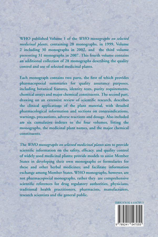
WHO published Volume 1 of the WHO monographs on selected medicinal plants, containing 28 monographs, in 1999, Volume 2 including 30 monographs in 2002, and the third volume presenting 31 monographs in 2007. This fourth volume contains an additional collection of 28 monographs describing the quality control and use of selected medicinal plants.
Each monograph contains two parts, the first of which provides pharmacopoeial summaries for quality assurance purposes, including botanical features, identity tests, purity requirements, chemical assays and major chemical constituents. The second part, drawing on an extensive review of scientific research, describes the clinical applications of the plant material, with detailed pharmacological information and sections on contraindications, warnings, precautions, adverse reactions and dosage. Also included are six cumulative indexes to the four volumes, listing the monographs, the medicinal plant names, and the major chemical constituents.
The WHO monographs on selected medicinal plants aim to provide scientific information on the safety, efficacy, and quality control of widely used medicinal plants; provide models to assist Member States in developing their own monographs or formularies for these and other herbal medicines; and facilitate information exchange among Member States. WHO monographs, however, are not pharmacopoeial monographs, rather they are comprehensive scientific references for drug regulatory authorities, physicians, traditional health practitioners, pharmacists, manufacturers, research scientists and the general public.
ISBN 978 92 4 154705 5