Embed Size (px)
Citation preview

ROBIN T. VAVACHAN
TRYPANOSOMA CRUZICHAGAS’ DISEASE

INTRODUCTION• Carlos Chagas- investigated.
• Caused by Trypanosoma cruzi• 10 million people infected - mostly in Latin America.
• But it has now spread to other continents.
• Chagas disease is curable if treatment is initiated soon after infection.
• Habitat – in humans- amastigote(intracellular parasites) and trypomastigote (peripheral blood) forms.

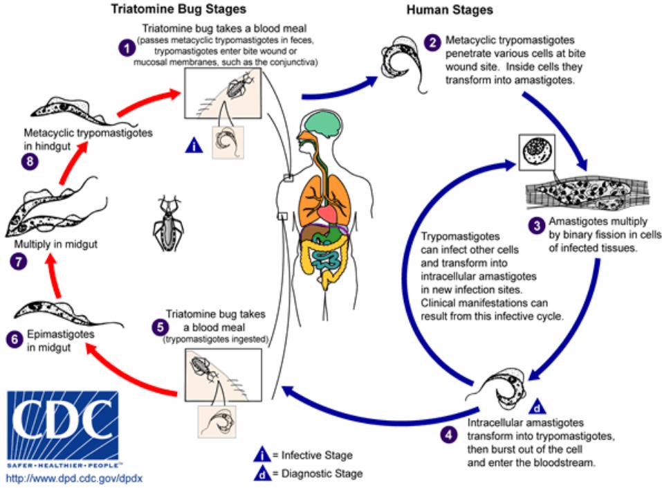
TRYPANOSOMA CRUZI AND CHAGAS’ DISEASE • Transmitted by the insect vector
Triatoma infestans (reduviid bug)• Reduviid bugs live in mud filled walls
of huts in rural areas• The bug bites and transmits the
disease.• Amastigote form in midgut and
metacyclic trypomastigote form in hindgut ---of the bug
Triatoma infestans (Reduviid bug)
Trypanosoma cruzi with human erythrocytes
4
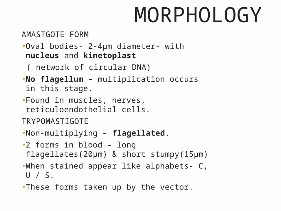
MORPHOLOGY AMASTGOTE FORM• Oval bodies- 2-4μm diameter- with nucleus and
kinetoplast ( network of circular DNA) • No flagellum – multiplication occurs in this stage.• Found in muscles, nerves, reticuloendothelial cells.
TRYPOMASTIGOTE• Non-multiplying – flagellated.• 2 forms in blood – long flagellates(20μm) & short
stumpy(15μm)• When stained appear like alphabets- C, U / S.• These forms taken up by the vector.

5
Morhology (epimastigote form)• Found in the vector.
• Has kinetoplast adjacent to nucleus.
• An undulating membrane runs along the anterior half of the parasite.
• Multiply by binary fission(hindgut).
6
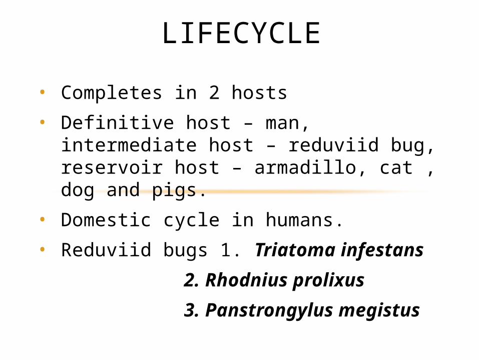
• Completes in 2 hosts
• Definitive host – man, intermediate host – reduviid bug, reservoir host – armadillo, cat , dog and pigs.
• Domestic cycle in humans.
• Reduviid bugs 1. Triatoma infestans 2. Rhodnius prolixus 3. Panstrongylus megistus
LIFECYCLE
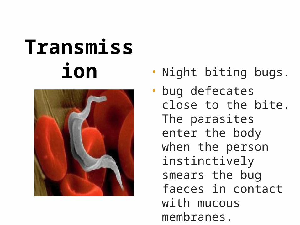
• Night biting bugs.
• bug defecates close to the bite. The parasites enter the body when the person instinctively smears the bug faeces in contact with mucous membranes.
• Also blood transfusion, organ transplantation & vertical transmission.
Transmission
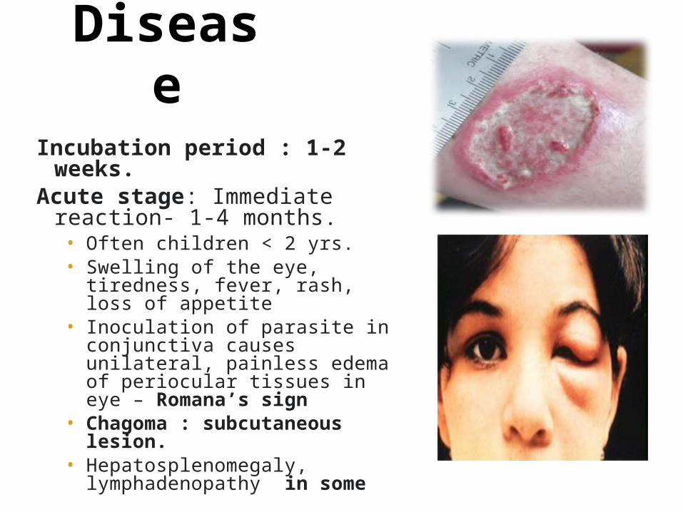
Chagas Disease
Incubation period : 1-2 weeks.Acute stage: Immediate reaction- 1-
4 months.• Often children < 2 yrs.• Swelling of the eye, tiredness,
fever, rash, loss of appetite• Inoculation of parasite in
conjunctiva causes unilateral, painless edema of periocular tissues in eye – Romana’s sign
• Chagoma : subcutaneous lesion.• Hepatosplenomegaly,
lymphadenopathy in some

10
Chronic Chagas disease:• Years after initial infection.
• inducing an inflammatory response, cellular destruction, fibrosis of muscles and nerves, that control tone of hollow organs like heart, oesophagus, colon etc.(intracellular amastigotes destroy the intramural neurons )
• Leads to cardiomyopathy, megaesophagus, megacolon.
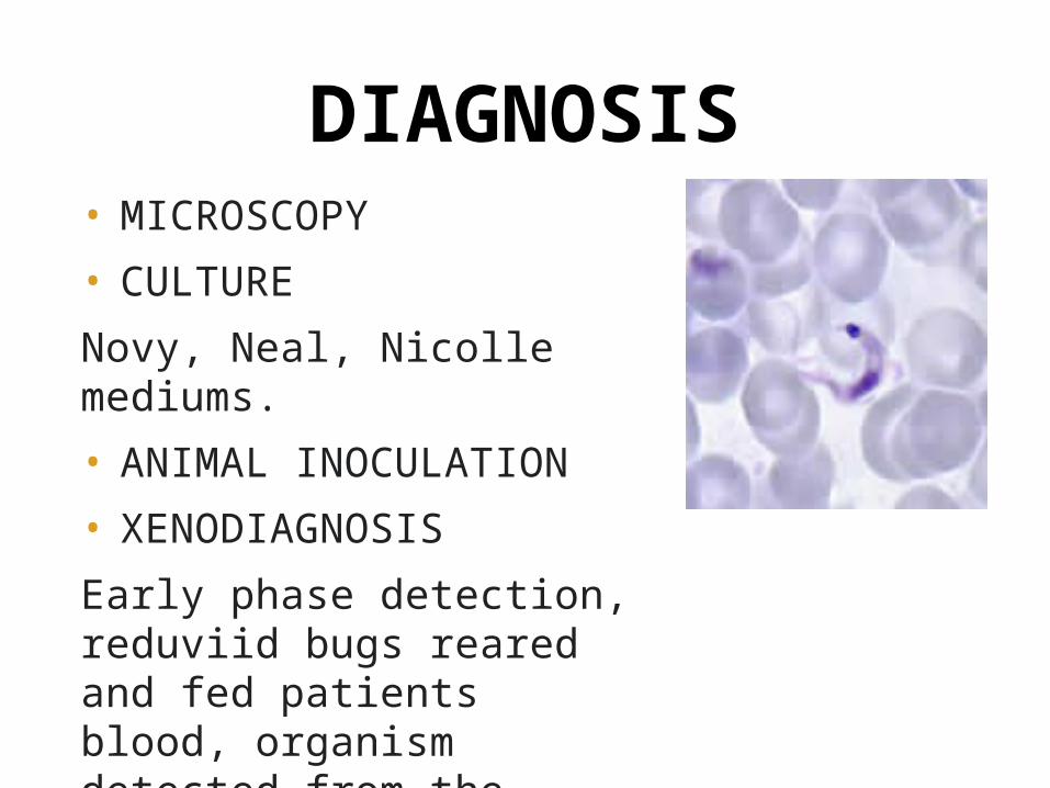
• MICROSCOPY
• CULTURE
Novy, Neal, Nicolle mediums.
• ANIMAL INOCULATION
• XENODIAGNOSIS
Early phase detection, reduviid bugs reared and fed patients blood, organism detected from the faeces of bug.
DIAGNOSIS
12
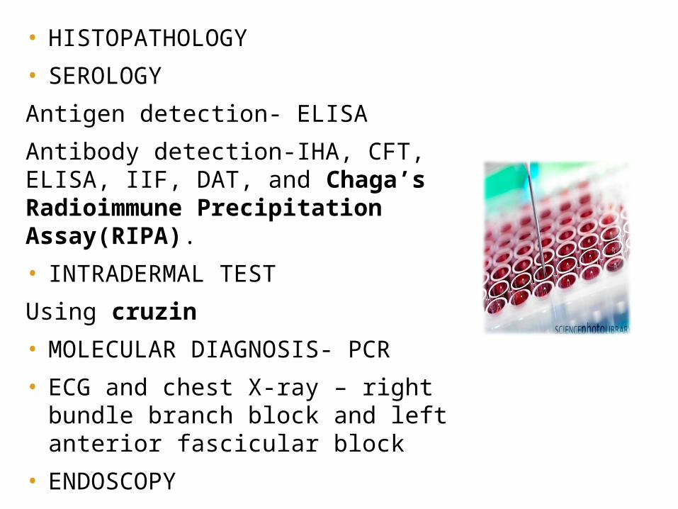
• HISTOPATHOLOGY
• SEROLOGY
Antigen detection- ELISA
Antibody detection-IHA, CFT, ELISA, IIF, DAT, and Chaga’s Radioimmune Precipitation Assay(RIPA).• INTRADERMAL TEST
Using cruzin• MOLECULAR DIAGNOSIS- PCR
• ECG and chest X-ray – right bundle branch block and left anterior fascicular block
• ENDOSCOPY
TREATMENT• No effective specific treatment available.
• benznidazole and nifurtimox chemotherapy( SUCCESS IN BOTH ACUTE AND CHRONIC)
• Early detection and speedy treatment are the keys to an effective remedy
• Only kill extracellular trypanosomes & not intracellular.
• NIFUTRIMOX : 8-10 mg/kg – adults & 15 mg/kg – children, 4 doses a day for 90-120 days orally.
• BENZNIDAZOLE : 5-10 mg/kg for 60 days orally.
PROPHYLAXIS• Insecticide spraying .
• personal preventive measures such as bed nets, insect repellent.
• good hygiene practices in food preparation, transportation, storage and consumption.
• screening of blood donors, testing of organ, tissue or cell donors and receivers.
• screening of new-borns from infected mothers, and siblings of infected children.

DR.T.V.RAO MD 15
THANKS


![20060503268 - apps.dtic.mil · TGK2], and 3 each for Leishmania major [LMK1, LMK2, & LMK3] and Trypanasoma cruzi [TCK1, TCK2, & TCK3] see table). We have cloned complete cDNAs of](https://img.pdfslide.net/doc/110x75/5f64cbaf04c3ed0e7e4cfc0e/20060503268-appsdticmil-tgk2-and-3-each-for-leishmania-major-lmk1-lmk2.jpg)