Embed Size (px)
Citation preview
11.3 The Kidney and Osmoregulation
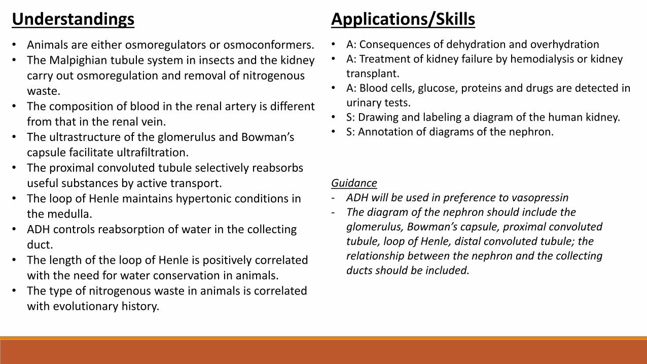
Understandings
• Animals are either osmoregulators or osmoconformers.• The Malpighian tubule system in insects and the kidney
carry out osmoregulation and removal of nitrogenous waste.
• The composition of blood in the renal artery is different from that in the renal vein.
• The ultrastructure of the glomerulus and Bowman’s capsule facilitate ultrafiltration.
• The proximal convoluted tubule selectively reabsorbs useful substances by active transport.
• The loop of Henle maintains hypertonic conditions in the medulla.
• ADH controls reabsorption of water in the collecting duct.
• The length of the loop of Henle is positively correlated with the need for water conservation in animals.
• The type of nitrogenous waste in animals is correlated with evolutionary history.
Applications/Skills• A: Consequences of dehydration and overhydration• A: Treatment of kidney failure by hemodialysis or kidney
transplant.• A: Blood cells, glucose, proteins and drugs are detected in
urinary tests.• S: Drawing and labeling a diagram of the human kidney.• S: Annotation of diagrams of the nephron.
Guidance- ADH will be used in preference to vasopressin- The diagram of the nephron should include the
glomerulus, Bowman’s capsule, proximal convoluted tubule, loop of Henle, distal convoluted tubule; the relationship between the nephron and the collecting ducts should be included.
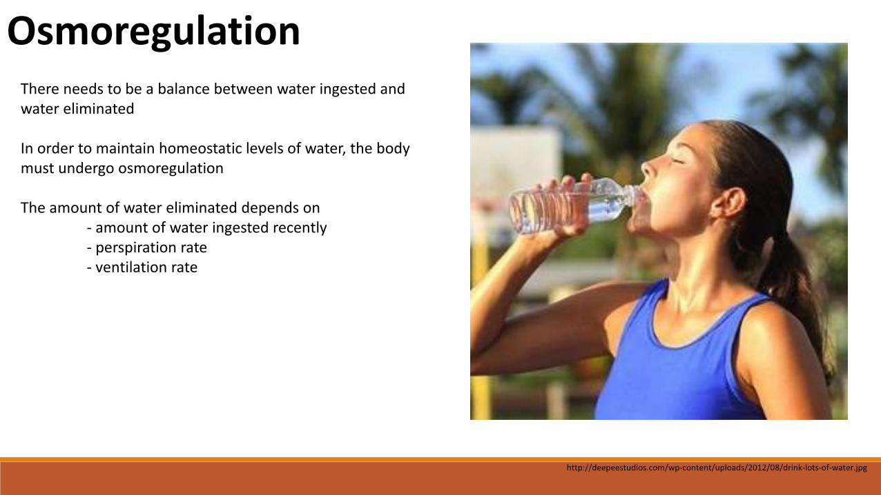
OsmoregulationThere needs to be a balance between water ingested and water eliminated
In order to maintain homeostatic levels of water, the body must undergo osmoregulation
The amount of water eliminated depends on - amount of water ingested recently- perspiration rate- ventilation rate
http://deepeestudios.com/wp-content/uploads/2012/08/drink-lots-of-water.jpg
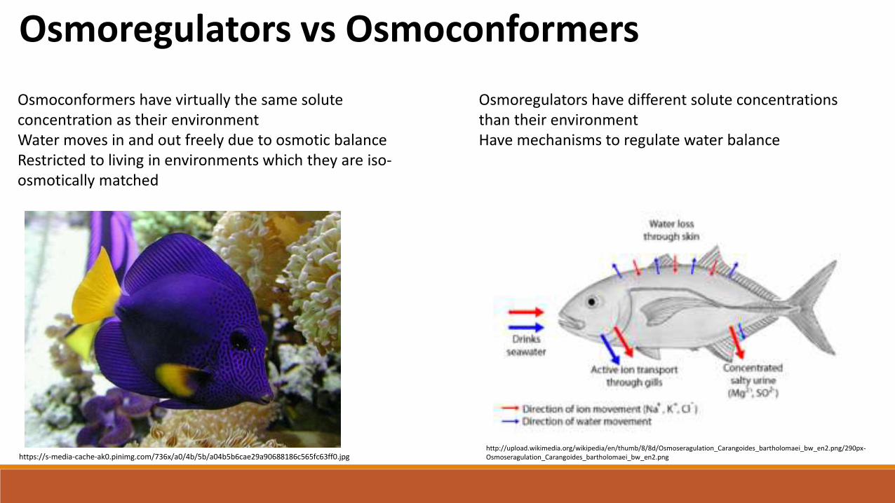
Osmoregulators vs Osmoconformers
Osmoconformers have virtually the same solute concentration as their environmentWater moves in and out freely due to osmotic balanceRestricted to living in environments which they are iso-osmotically matched
Osmoregulators have different solute concentrations than their environmentHave mechanisms to regulate water balance
https://s-media-cache-ak0.pinimg.com/736x/a0/4b/5b/a04b5b6cae29a90688186c565fc63ff0.jpghttp://upload.wikimedia.org/wikipedia/en/thumb/8/8d/Osmoseragulation_Carangoides_bartholomaei_bw_en2.png/290px-Osmoseragulation_Carangoides_bartholomaei_bw_en2.png
Nitrogenous Waste Products
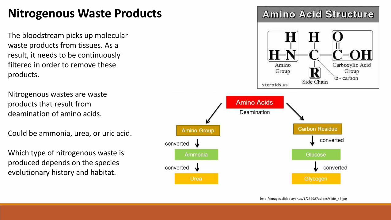
The bloodstream picks up molecular waste products from tissues. As a result, it needs to be continuously filtered in order to remove these products.
Nitrogenous wastes are waste products that result from deamination of amino acids.
Could be ammonia, urea, or uric acid.
Which type of nitrogenous waste is produced depends on the species evolutionary history and habitat.
http://images.slideplayer.us/1/257987/slides/slide_45.jpg
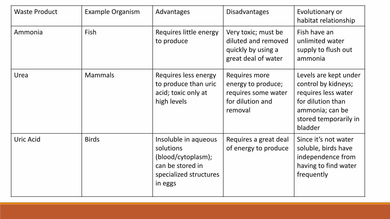
Waste Product Example Organism Advantages Disadvantages Evolutionary or habitat relationship
Ammonia Fish Requires little energy to produce
Very toxic; must be diluted and removed quickly by using a great deal of water
Fish have an unlimited water supply to flush out ammonia
Urea Mammals Requires less energy to produce than uric acid; toxic only at high levels
Requires more energy to produce; requires some waterfor dilution and removal
Levels are kept under control by kidneys; requires less waterfor dilution than ammonia; can be stored temporarily in bladder
Uric Acid Birds Insoluble in aqueous solutions (blood/cytoplasm); can be stored in specialized structures in eggs
Requires a great deal of energy to produce
Since it’s not watersoluble, birds have independence from having to find water frequently

Malpighian Tubules
http://bio1152.nicerweb.com/Locked/media/ch44/44_12MalpighianTubules_L.jpg
Small tubes that lie in pools of blood in an insects body cavity
Closed at one end (distal end)
Opens into the insects gut (proximal end)
Components of the insects blood enter the tubules
Nitrogenous wastes, excess water, and salt ions remain in the tubules and move to the proximal end that empties into the gut; gets eliminated
Useful substances are transported back to the blood
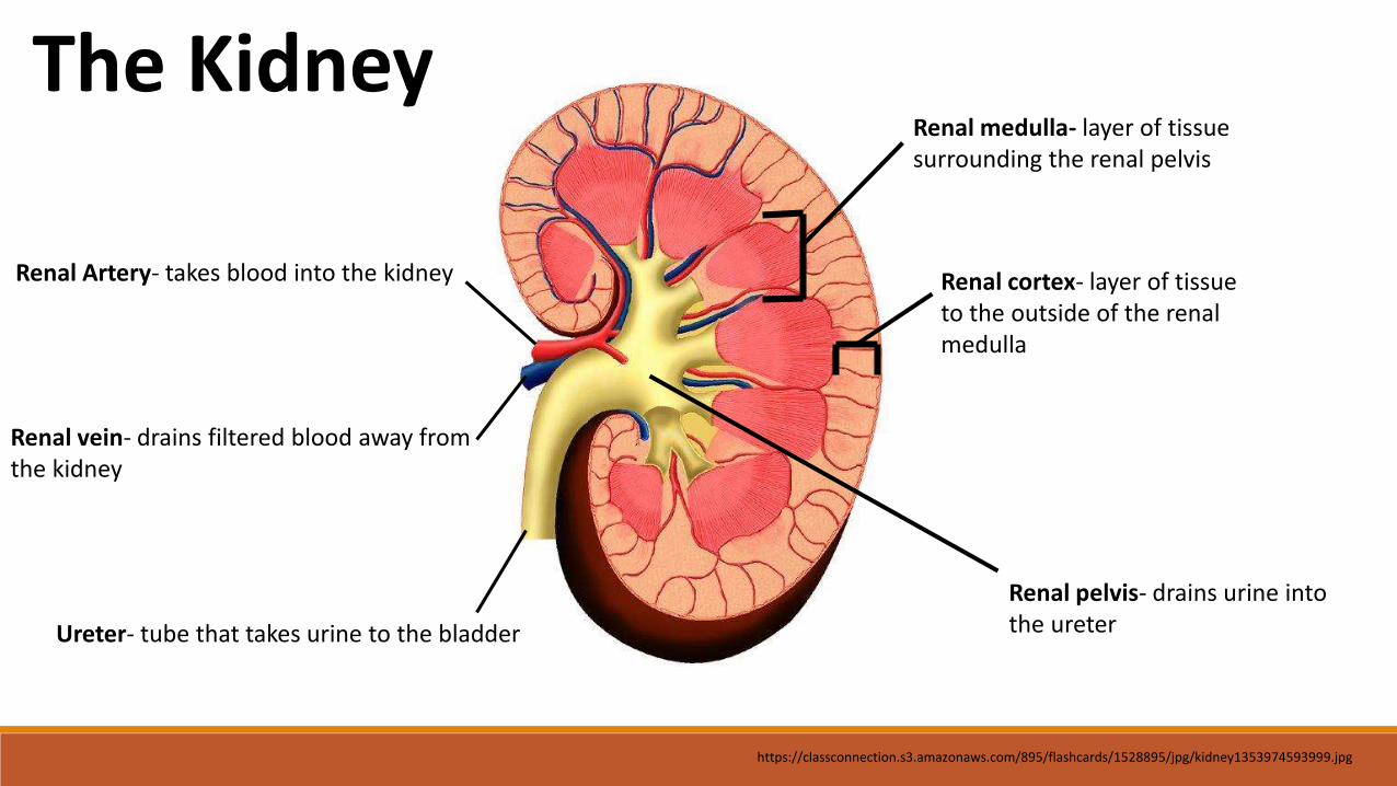
Renal medulla- layer of tissue surrounding the renal pelvis
Renal Artery- takes blood into the kidney
Renal vein- drains filtered blood away from the kidney
Ureter- tube that takes urine to the bladder
Renal cortex- layer of tissue to the outside of the renal medulla
Renal pelvis- drains urine into the ureter
The Kidney
https://classconnection.s3.amazonaws.com/895/flashcards/1528895/jpg/kidney1353974593999.jpg
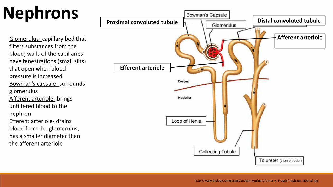
Proximal convoluted tubule Distal convoluted tubuleNephrons
Glomerulus- capillary bed that filters substances from the blood; walls of the capillaries have fenestrations (small slits) that open when blood pressure is increasedBowman’s capsule- surrounds glomerulusAfferent arteriole- brings unfiltered blood to the nephronEfferent arteriole- drains blood from the glomerulus; has a smaller diameter than the afferent arteriole
Afferent arteriole
Efferent arteriole
http://www.biologycorner.com/anatomy/urinary/urinary_images/nephron_labeled.jpg
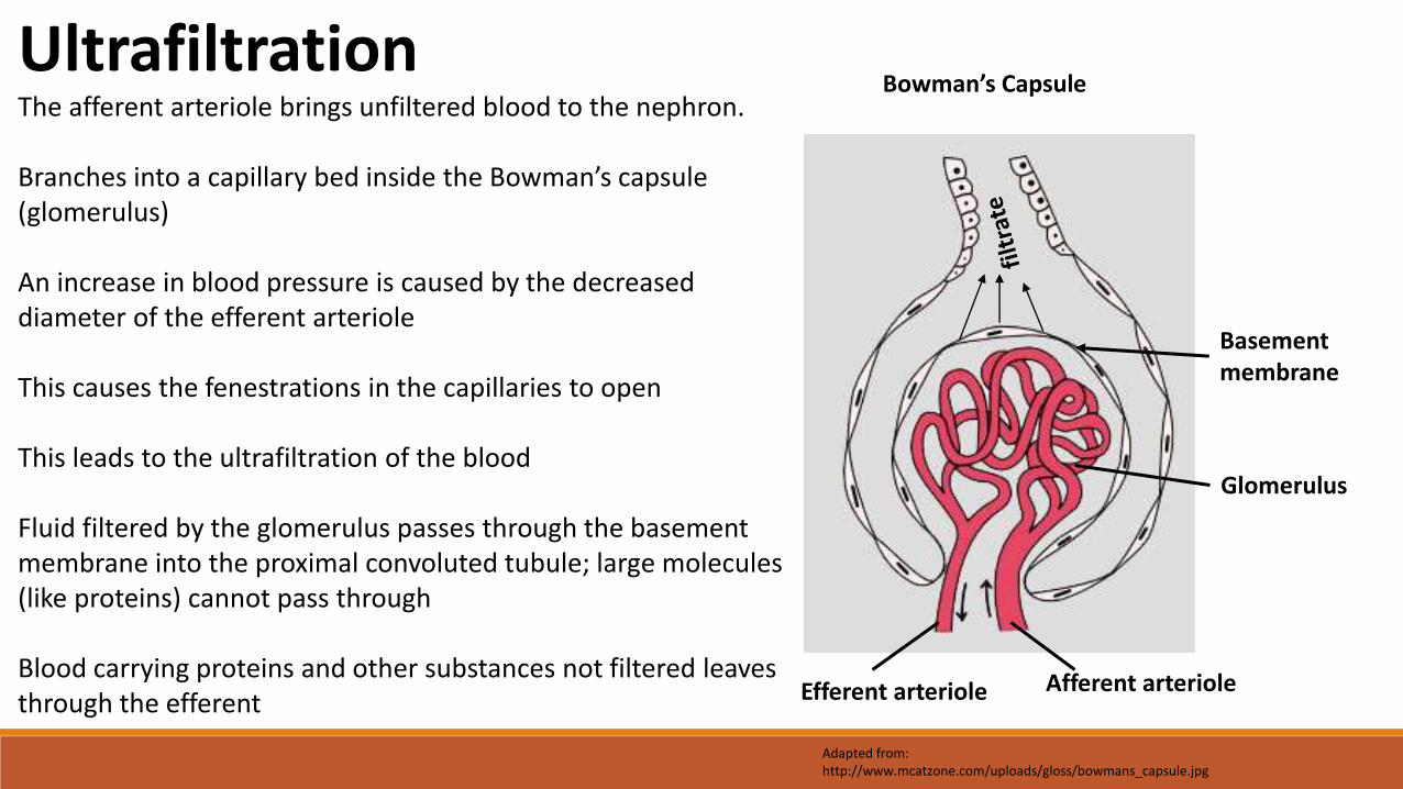
Bowman’s Capsule
Afferent arterioleEfferent arteriole
Glomerulus
The afferent arteriole brings unfiltered blood to the nephron.
Branches into a capillary bed inside the Bowman’s capsule (glomerulus)
An increase in blood pressure is caused by the decreased diameter of the efferent arteriole
This causes the fenestrations in the capillaries to open
This leads to the ultrafiltration of the blood
Fluid filtered by the glomerulus passes through the basement membrane into the proximal convoluted tubule; large molecules (like proteins) cannot pass through
Blood carrying proteins and other substances not filtered leaves through the efferent
Basement membrane
Ultrafiltration
Adapted from:http://www.mcatzone.com/uploads/gloss/bowmans_capsule.jpg
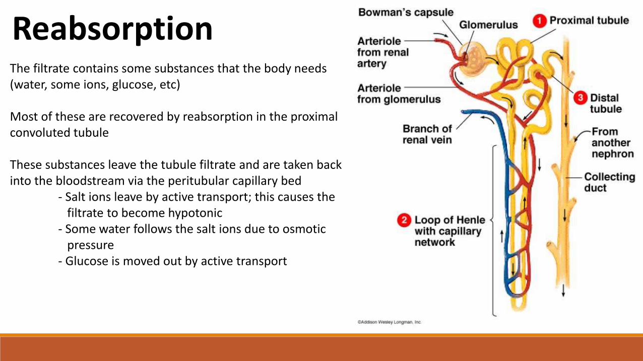
ReabsorptionThe filtrate contains some substances that the body needs (water, some ions, glucose, etc)
Most of these are recovered by reabsorption in the proximal convoluted tubule
These substances leave the tubule filtrate and are taken back into the bloodstream via the peritubular capillary bed
- Salt ions leave by active transport; this causes the filtrate to become hypotonic
- Some water follows the salt ions due to osmotic pressure
- Glucose is moved out by active transport
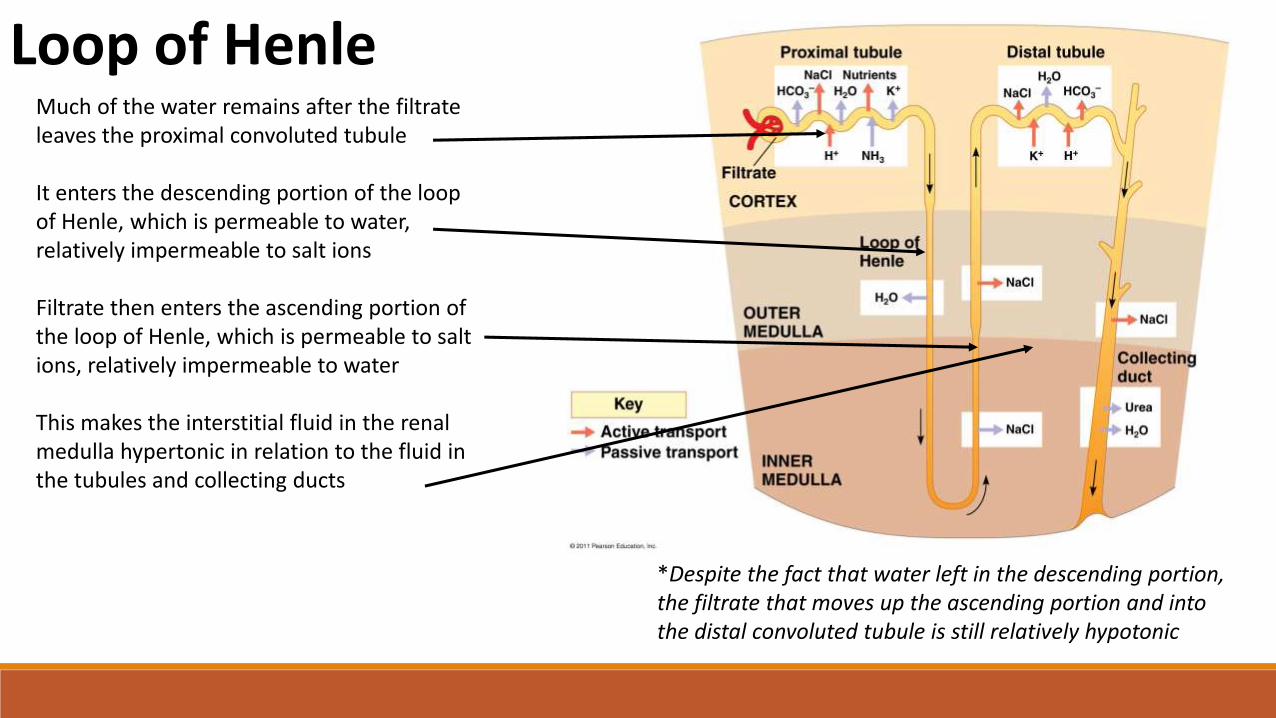
Loop of HenleMuch of the water remains after the filtrate leaves the proximal convoluted tubule
It enters the descending portion of the loop of Henle, which is permeable to water, relatively impermeable to salt ions
Filtrate then enters the ascending portion of the loop of Henle, which is permeable to salt ions, relatively impermeable to water
This makes the interstitial fluid in the renal medulla hypertonic in relation to the fluid in the tubules and collecting ducts
*Despite the fact that water left in the descending portion, the filtrate that moves up the ascending portion and into the distal convoluted tubule is still relatively hypotonic
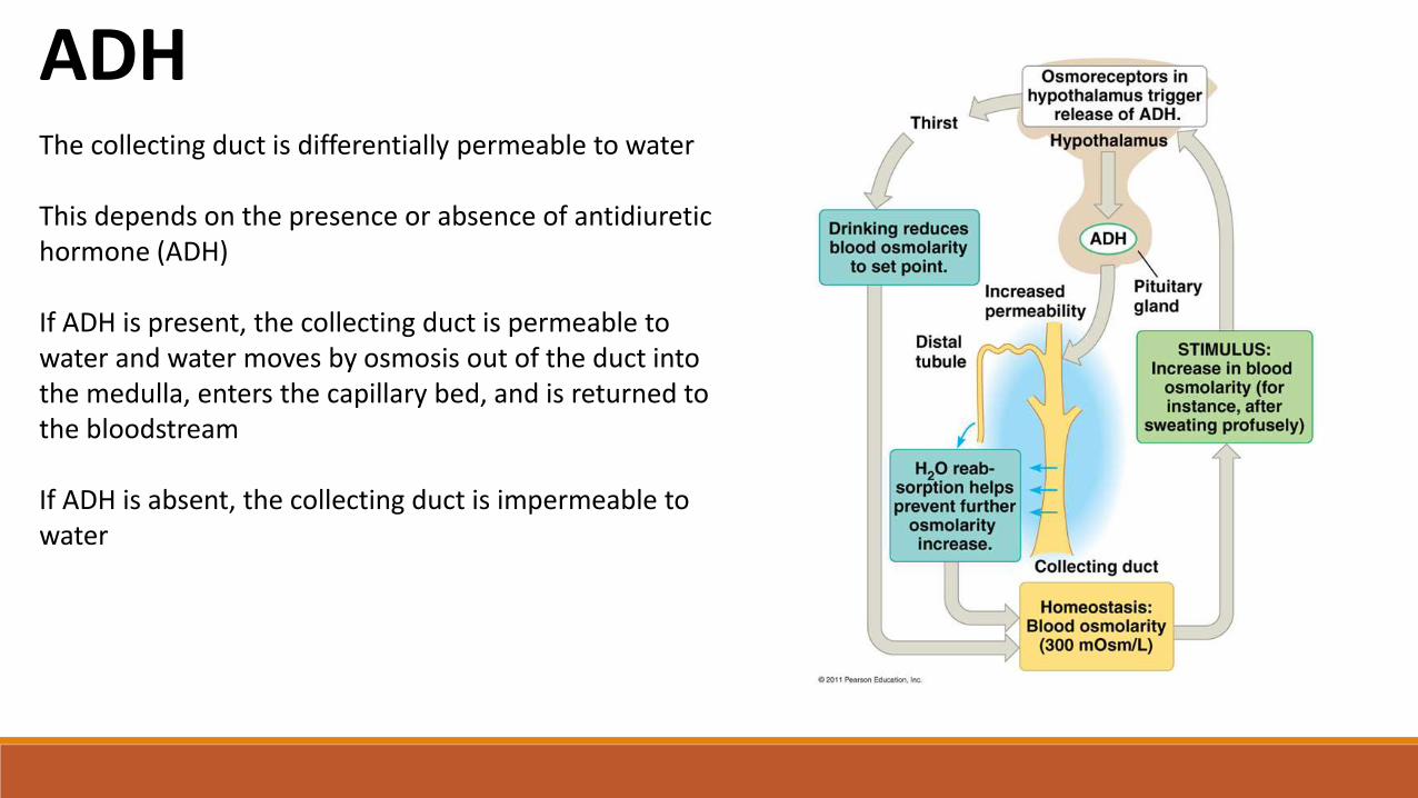
ADHThe collecting duct is differentially permeable to water
This depends on the presence or absence of antidiuretic hormone (ADH)
If ADH is present, the collecting duct is permeable to water and water moves by osmosis out of the duct into the medulla, enters the capillary bed, and is returned to the bloodstream
If ADH is absent, the collecting duct is impermeable to water
http://www.patana.ac.th/secondary/science/IBtopics/IB%20Excretion(12)/12.1/variation.gif
Water Conservation
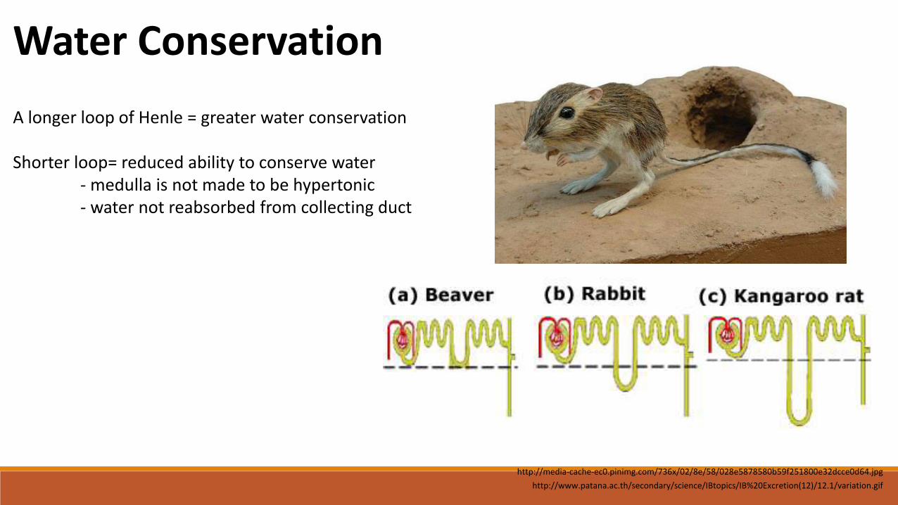
A longer loop of Henle = greater water conservation
Shorter loop= reduced ability to conserve water - medulla is not made to be hypertonic - water not reabsorbed from collecting duct
http://media-cache-ec0.pinimg.com/736x/02/8e/58/028e5878580b59f251800e32dcce0d64.jpg
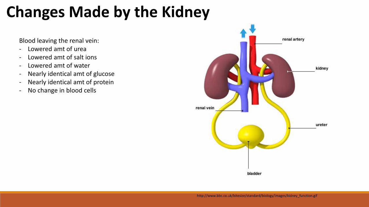
Changes Made by the Kidney
Blood leaving the renal vein:- Lowered amt of urea- Lowered amt of salt ions- Lowered amt of water- Nearly identical amt of glucose- Nearly identical amt of protein - No change in blood cells
http://www.bbc.co.uk/bitesize/standard/biology/images/kidney_function.gif
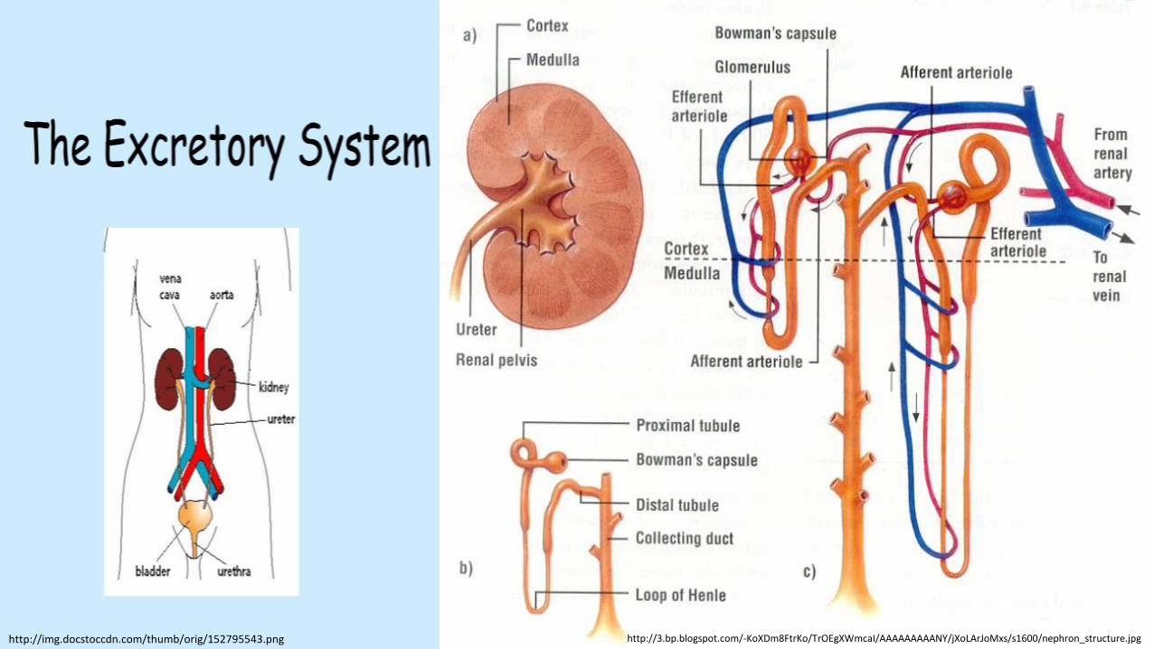
http://3.bp.blogspot.com/-KoXDm8FtrKo/TrOEgXWmcaI/AAAAAAAAANY/jXoLArJoMxs/s1600/nephron_structure.jpghttp://img.docstoccdn.com/thumb/orig/152795543.png
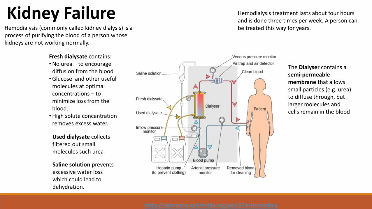
Kidney Failure
https://commons.wikimedia.org/wiki/File:Hemodialysis-en.svg
Hemodialysis (commonly called kidney dialysis) is a process of purifying the blood of a person whose kidneys are not working normally.
Hemodialysis treatment lasts about four hours and is done three times per week. A person can be treated this way for years.
The Dialyser contains a semi-permeable membrane that allows small particles (e.g. urea) to diffuse through, but larger molecules and cells remain in the blood
Used dialysate collects filtered out small molecules such urea
Fresh dialysate contains:•No urea – to encourage
diffusion from the blood•Glucose and other useful
molecules at optimal concentrations – to minimize loss from the blood.•High solute concentration
removes excess water.
Saline solution prevents excessive water loss which could lead to dehydration.
http://www.kalingahospital.com/data/images/transplant1.jpg
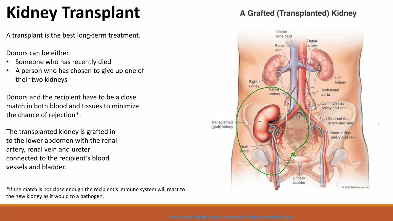
A transplant is the best long-term treatment.
Donors can be either:• Someone who has recently died• A person who has chosen to give up one of
their two kidneys
Donors and the recipient have to be a close match in both blood and tissues to minimize the chance of rejection*.
*If the match is not close enough the recipient's immune system will react to the new kidney as it would to a pathogen.
The transplanted kidney is grafted in to the lower abdomen with the renal artery, renal vein and ureter connected to the recipient’s blood vessels and bladder.
Kidney Transplant
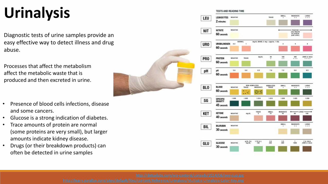
http://learn.parallax.com/sites/default/files/content/Reference/Urinalysis/Multistix-Urinalysis-Color-Key.png
http://detoxlabs.com/wp-content/uploads/2014/08/pee-cup.jpg
Urinalysis
• Presence of blood cells infections, disease and some cancers.
• Glucose is a strong indication of diabetes.• Trace amounts of protein are normal
(some proteins are very small), but larger amounts indicate kidney disease.
• Drugs (or their breakdown products) can often be detected in urine samples
Diagnostic tests of urine samples provide an easy effective way to detect illness and drug abuse.
Processes that affect the metabolism affect the metabolic waste that is produced and then excreted in urine.
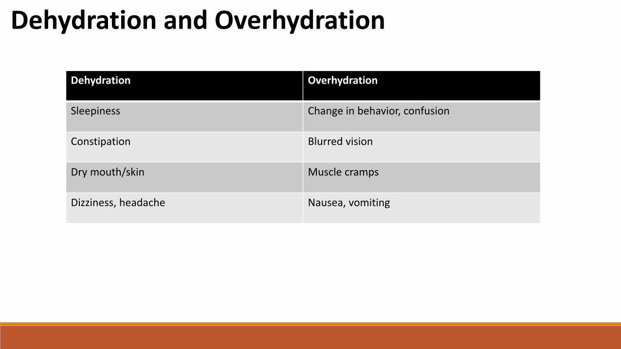
Dehydration and Overhydration
Dehydration Overhydration
Sleepiness Change in behavior, confusion
Constipation Blurred vision
Dry mouth/skin Muscle cramps
Dizziness, headache Nausea, vomiting
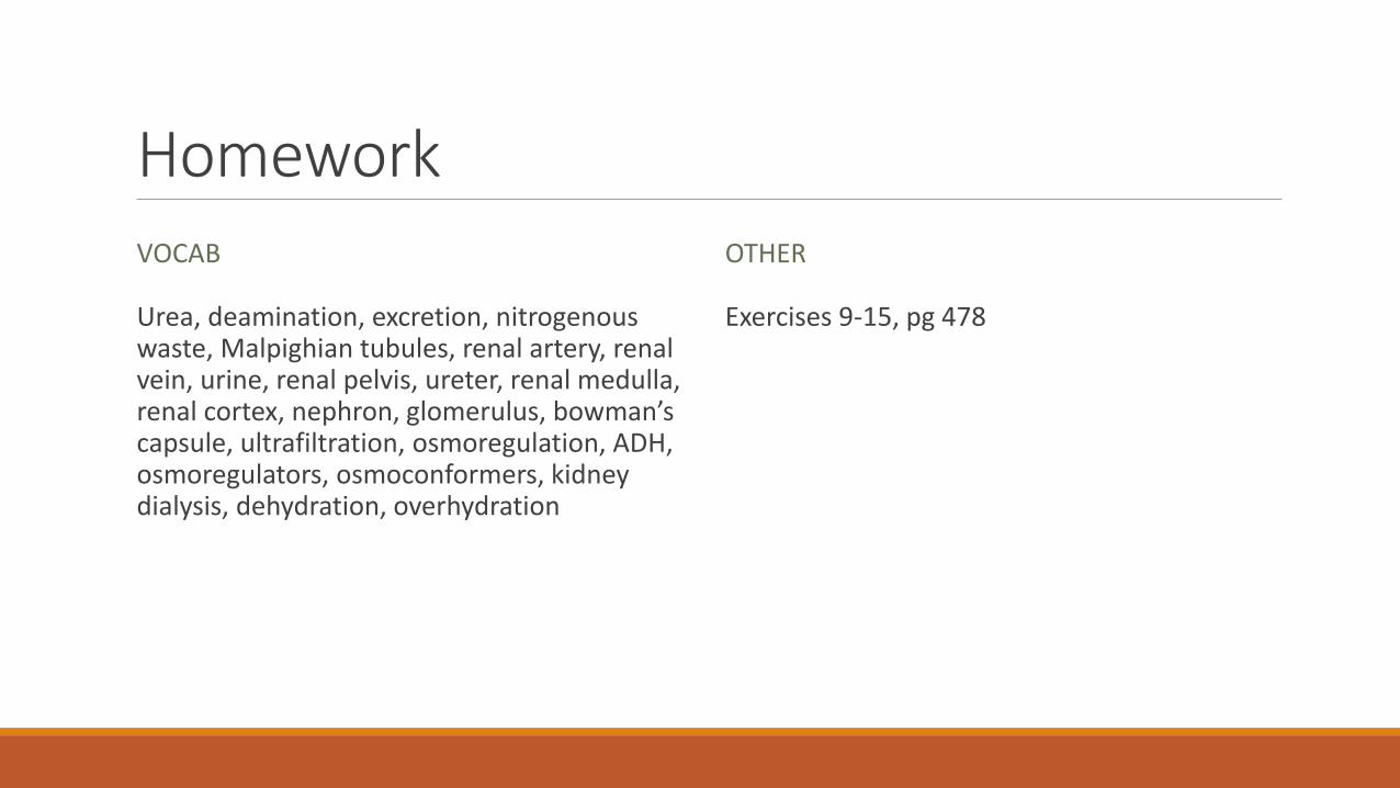
HomeworkVOCAB
Urea, deamination, excretion, nitrogenous waste, Malpighian tubules, renal artery, renal vein, urine, renal pelvis, ureter, renal medulla, renal cortex, nephron, glomerulus, bowman’s capsule, ultrafiltration, osmoregulation, ADH, osmoregulators, osmoconformers, kidney dialysis, dehydration, overhydration
OTHER
Exercises 9-15, pg 478