Embed Size (px)
Citation preview
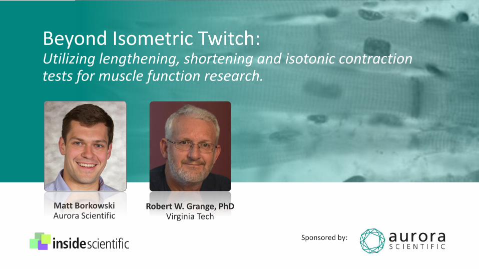
Beyond Isometric Twitch: Utilizing lengthening, shortening and isotonic contraction tests for muscle function research.
Matt BorkowskiAurora Scientific
Robert W. Grange, PhDVirginia Tech
Sponsored by:

InsideScientific is an online educational environment designed for life science researchers. Our goal is to aid in
the sharing and distribution of scientific information regarding innovative technologies, protocols, research
tools and laboratory services.

Thank you to our event sponsor
Aurora Scientific, a trusted
provider of instrumentation
for research in muscle
physiology, neuroscience
and material science.

Utilizing lengthening, shortening and isotonic contraction tests for muscle function research.
Matt Borkowski
Sales & Support Manager
Aurora Scientific
Copyright 2015 M. Borkowski, Aurora Scientific & InsideScientific. All Rights Reserved.

THE JOURNEY OF A THOUSAND MILES BEGINS WITH A SINGLE STEP
-- Lao Tzu

• Aurora has served the muscle community for nearly 20 years.
• Test systems and solutions ranging from single cells up to the whole animal.
• Friendly, reliable support.
Cell
Whole Animal
Fiber
Whole Muscle
About Aurora Scientific

A NEW CHAPTER BEGINS…

• Aurora Scientific believes in providing solutions for the complete characterization of muscle.
• To completely characterize the muscle, the 3 types of contractions must be used.
Complete Characterization
concentric
isometric
eccentric
What is complete characterization?

What is complete characterization?
• Isometric: Contraction at a constant muscle length.
• Eccentric: Contraction while the muscle is lengthening.
• Concentric: Contraction while the muscle is shortening (can be isotonic).
Complete Characterization
concentric
isometric
eccentric
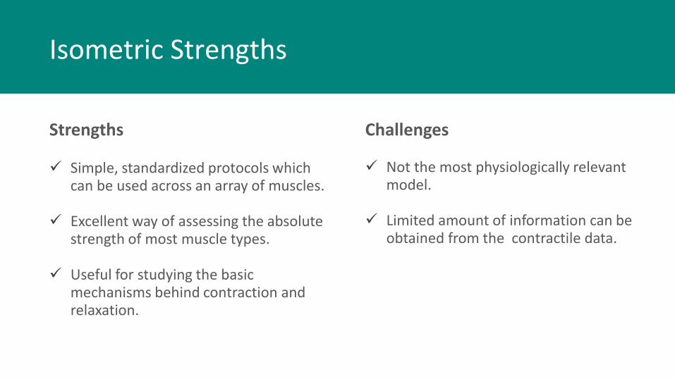
Strengths
Simple, standardized protocols which can be used across an array of muscles.
Excellent way of assessing the absolute strength of most muscle types.
Useful for studying the basic mechanisms behind contraction and relaxation.
Challenges
Isometric Strengths
Not the most physiologically relevant model.
Limited amount of information can be obtained from the contractile data.
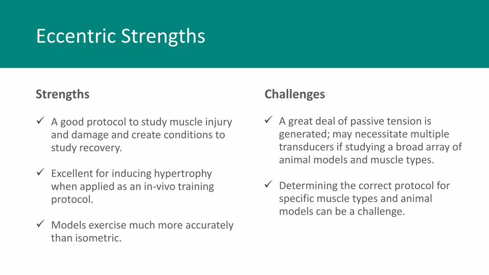
Strengths
A good protocol to study muscle injury and damage and create conditions to study recovery.
Excellent for inducing hypertrophy when applied as an in-vivo training protocol.
Models exercise much more accurately than isometric.
Eccentric Strengths
A great deal of passive tension is generated; may necessitate multiple transducers if studying a broad array of animal models and muscle types.
Determining the correct protocol for specific muscle types and animal models can be a challenge.
Challenges

Strengths
Offers a wealth of information within the contractile data: Power, Work, Force-Velocity relationship can all be measured.
Isotonic protocols closely mirror real life work and exercise.
Either force or velocity can be controlled, allowing for multiple ways to test a particular hypothesis.
Concentric Strengths
Can be technically challenging to implement.
Requires a good deal of configuration for different muscle types.
Challenges
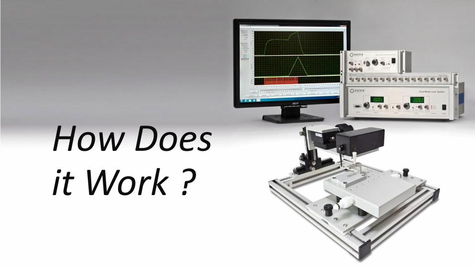
How Does it Work ?
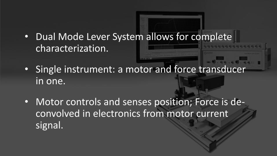
• Dual Mode Lever System allows for complete characterization.
• Single instrument: a motor and force transducer in one.
• Motor controls and senses position; Force is de-convolved in electronics from motor current signal.
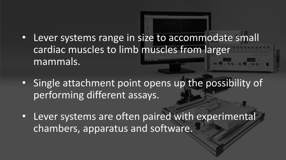
• Lever systems range in size to accommodate small cardiac muscles to limb muscles from larger mammals.
• Single attachment point opens up the possibility of performing different assays.
• Lever systems are often paired with experimental chambers, apparatus and software.

One attachment point: 3 experiment types
In vitroIn situIn vivo
Click Here to see specific Aurora Muscle Physiology Apparatus

The foot is secured in a foot-plate mounted to the dual mode lever system.
Percutaneous or subcutaneous electrodes can elicit muscle contraction.
Aggregate torque of the plantar or dorsiflexors of the lower limb can be measured.
Resistance of the pedal can be adjusted to create isotonic resistance.
Foot pedal can rotate in conjunction with contraction to create eccentric or concentric conditions.
In vivo –Ankle Torsion
Courtesy of Yan lab, UVA
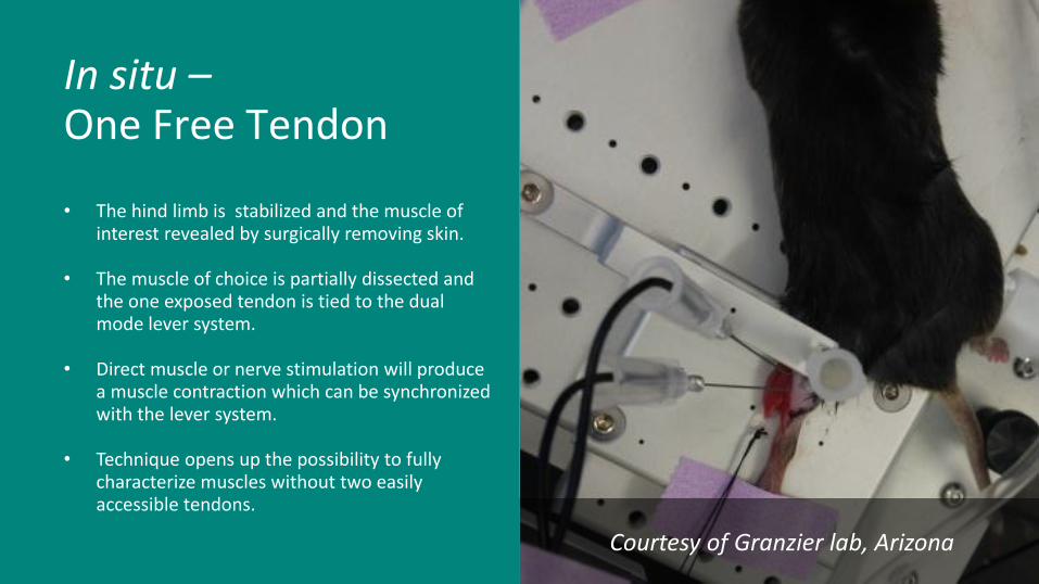
Courtesy of Granzier lab, Arizona
• The hind limb is stabilized and the muscle of interest revealed by surgically removing skin.
• The muscle of choice is partially dissected and the one exposed tendon is tied to the dual mode lever system.
• Direct muscle or nerve stimulation will produce a muscle contraction which can be synchronized with the lever system.
• Technique opens up the possibility to fully characterize muscles without two easily accessible tendons.
In situ –One Free Tendon

• Muscle dissected from animal and sutured at both free tendons.
• Muscle activated via field stimulation.
• Classical, vertical bath configuration means only one tendon can attach to an instrument.
• Only the dual mode lever system permits tension & length to be recorded and controlled in this orientation.
In vitro –Isolated Muscle
Courtesy of Barton Lab, UFL

Who can benefit from going beyond isometric?
Muscle Physiologists
Exercise Scientists
Bioengineers & Biologists
Metabolic & Cardiovascular Scientists
Geneticists
Neuroscientists
Pharmacologists & Biochemists
Anyone studying muscle mechanics
Isometric and Dynamic Muscle Function Assessment
Robert W. Grange, PhD
Department of Human Nutrition, Foods and Exercise,
Virginia Tech
Copyright 2015 R.W Grange, Aurora Scientific & InsideScientific. All Rights Reserved.
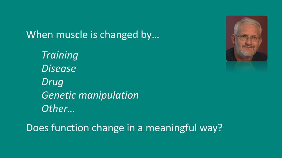
When muscle is changed by…
TrainingDiseaseDrugGenetic manipulationOther…
Does function change in a meaningful way?
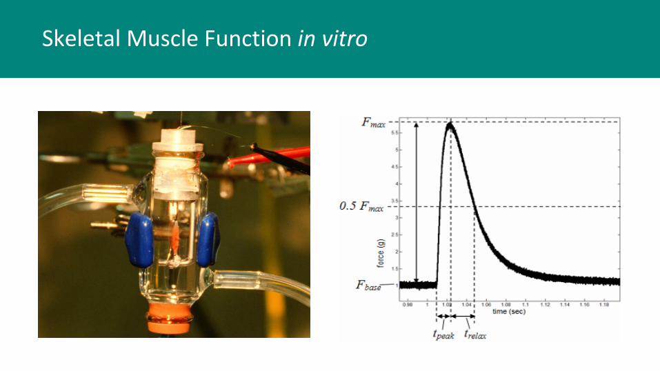
Skeletal Muscle Function in vitro
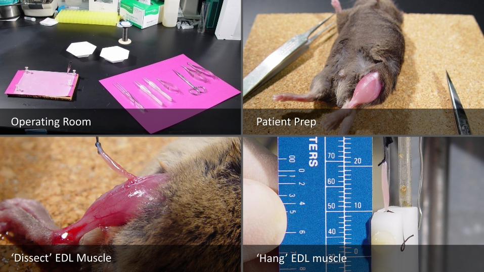
Patient Prep
‘Hang’ EDL muscle
Operating Room
‘Dissect’ EDL Muscle

Servomotor and Isometric Force Transducers
Dual-mode Servomotor For Dynamic Contractions
Isometric Force Transducer
Stepper Motor For Maintaining L0
Muscle Clamps
- +
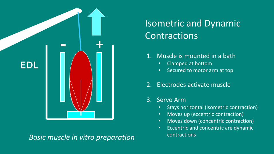
EDL
Isometric and Dynamic Contractions
Basic muscle in vitro preparation
1. Muscle is mounted in a bath• Clamped at bottom• Secured to motor arm at top
2. Electrodes activate muscle
3. Servo Arm • Stays horizontal (isometric contraction)• Moves up (eccentric contraction)• Moves down (concentric contraction)• Eccentric and concentric are dynamic
contractions

Torque = Force x Moment arm
Ma
Force
Skeletal Muscle Function in vivo
Mouse Hindlimb
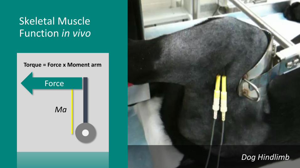
Torque = Force x Moment arm
Ma
Force
Skeletal Muscle Function in vivo
Dog Hindlimb

Skeletal Muscle Fundamentals

Epstein M, Herzog W.. Philos Trans R Soc Lond B Biol Sci 2003;358:1445-1452.
Anatomy
Anatomy
• Muscle• Fascicles• Fibers• Myofibrils• Sarcomere (functional unit of muscle)
• Z lines• Myosin and Actin: contraction
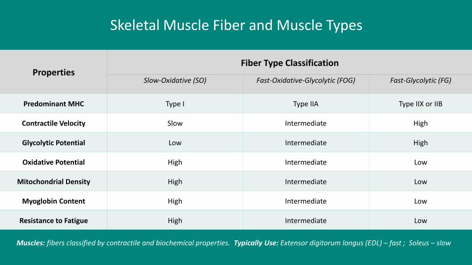
Muscles: fibers classified by contractile and biochemical properties. Typically Use: Extensor digitorum longus (EDL) – fast ; Soleus – slow
Skeletal Muscle Fiber and Muscle Types
PropertiesFiber Type Classification
Slow-Oxidative (SO) Fast-Oxidative-Glycolytic (FOG) Fast-Glycolytic (FG)
Predominant MHC Type I Type IIA Type IIX or IIB
Contractile Velocity Slow Intermediate High
Glycolytic Potential Low Intermediate High
Oxidative Potential High Intermediate Low
Mitochondrial Density High Intermediate Low
Myoglobin Content High Intermediate Low
Resistance to Fatigue High Intermediate Low

Coupling
DHPRRyR
Excitation – ContractionCoupling
• Excitation: Action potential…
• Via T-tubule…
• Releases calcium from RYR of Sarcoplasmic Reticulum…
• Calcium binds Troponin…
• Myosin and Actin interact:
• Contraction
-dF/dt
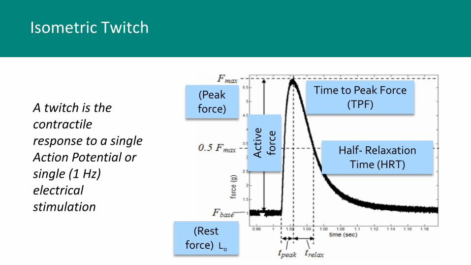
(Rest force) Lo
(Peak force)
Act
ive
forc
e
Time to Peak Force (TPF)
Half- Relaxation Time (HRT)
Isometric Twitch
A twitch is the contractile response to a single Action Potential or single (1 Hz) electrical stimulation

Force Summation
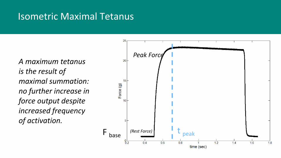
Tetanus
t peakF base
Peak Force
(Rest Force)
Isometric Maximal Tetanus
A maximum tetanus is the result of maximal summation: no further increase in force output despite increased frequency of activation.
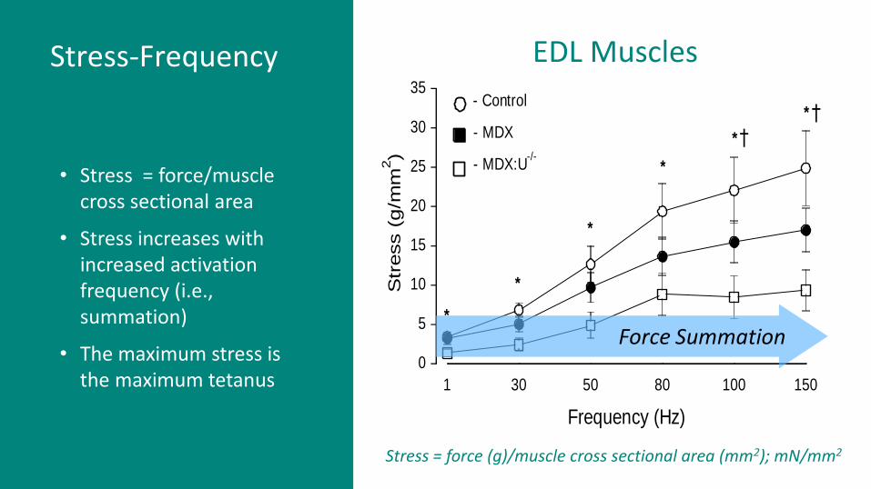
1 30 50 80 100 150
Frequency (Hz)
0
5
10
15
20
25
30
35
Str
ess (
g/m
m2)
- Control
- MDX
- MDX:U-/-
*
*
*
*
*
*
†
†
Stress = force (g)/muscle cross sectional area (mm2); mN/mm2
EDL Muscles
Force Summation
Stress-Frequency
• Stress = force/muscle cross sectional area
• Stress increases with increased activation frequency (i.e., summation)
• The maximum stress is the maximum tetanus
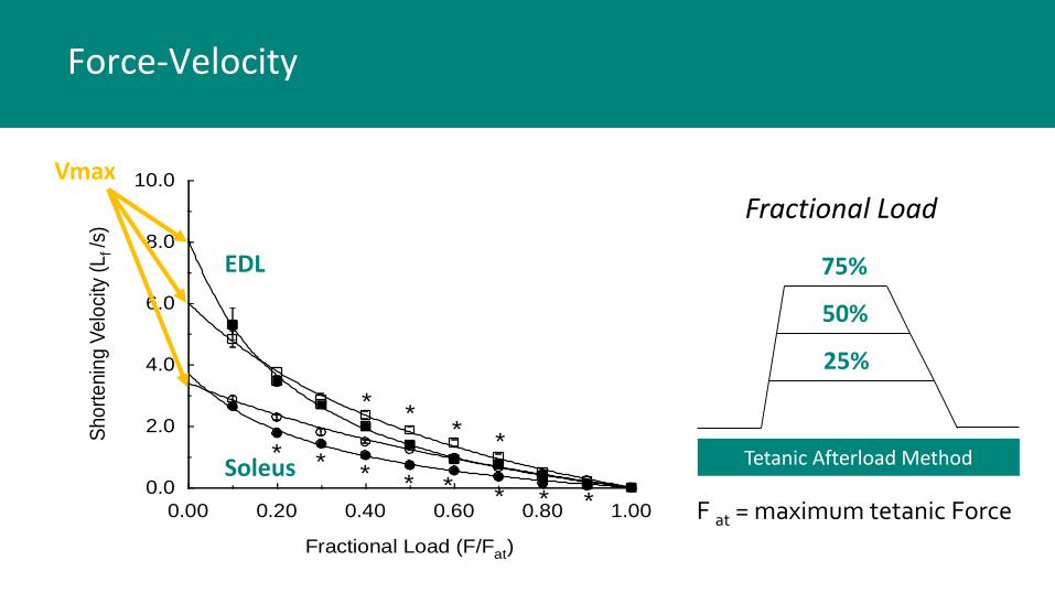
0.00 0.20 0.40 0.60 0.80 1.00
Fractional Load (F/Fat)
0.0
2.0
4.0
6.0
8.0
10.0
Sho
rte
nin
g V
elo
city
(L
f /s)
**
**
* ** * * * * *
25%
50%
75%
Fractional Load
Tetanic Afterload Method
F at = maximum tetanic Force
Vmax
EDL
Soleus
Force-Velocity

EDL
Soleus
Power –FxV; Work/Time
• Peak power typically occurs at a fractional load of 0.40 (40%) of maximum load

EDL vs Soleus Muscle Fatigues More Quickly

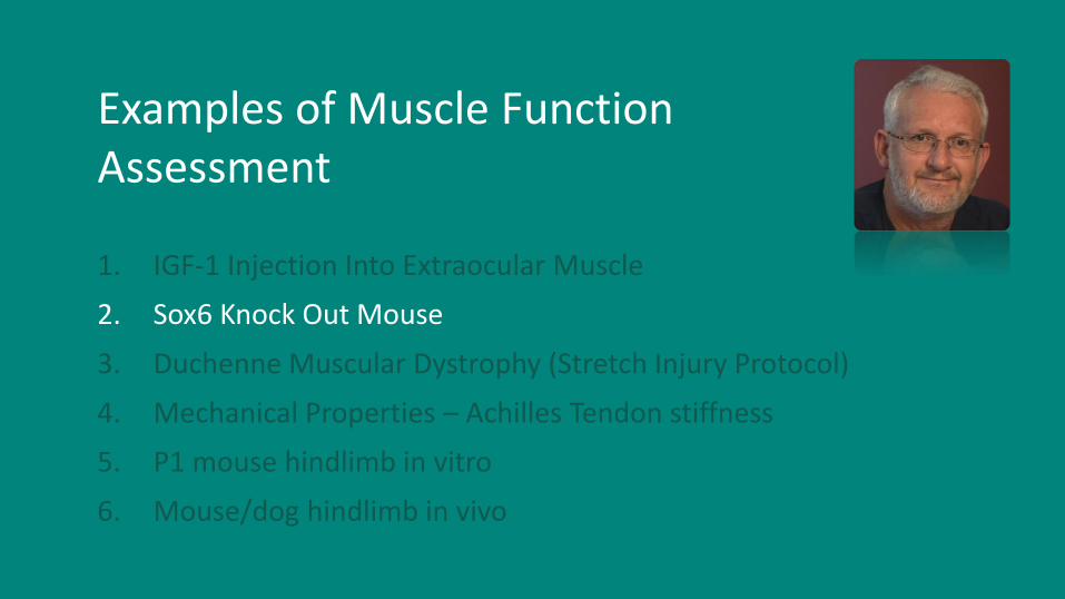
Examples of Muscle Function Assessment
1. IGF-1 Injection Into Extraocular Muscle
2. Sox6 Knock Out Mouse
3. Duchenne Muscular Dystrophy (Stretch Injury Protocol)
4. Mechanical Properties – Achilles Tendon stiffness
5. P1 mouse hindlimb in vitro
6. Mouse/dog hindlimb in vivo

Increased extraocular muscle strength with direct injection of insulin-like growth factor-I. Anderson, Christiansen, Grandt, Grange, McLoon. Invest. Opthalmol Vis Sci 47(6):2461-7, 2006.
CONCLUSIONS: Direct muscular injection of IGF-I
effectively increases EOM force
generation without the potential
biomechanical hazards of surgery
such as permanently altered muscle
length or insertional position on the
globe.
• 25 ug IGF-1 • rabbit superior rectus
muscle • assess after 1 week
Increased Extraocular Muscle Strength
Examples of Muscle Function Assessment
1. IGF-1 Injection Into Extraocular Muscle
2. Sox6 Knock Out Mouse
3. Duchenne Muscular Dystrophy (Stretch Injury Protocol)
4. Mechanical Properties – Achilles Tendon stiffness
5. P1 mouse hindlimb in vitro
6. Mouse/dog hindlimb in vivo

Concerted regulation of myofiber-specific gene expression and muscle performance by the transcriptional repressor Sox6. Quiat, Voelker, Pei, Grishin, Grange, Bassel-Duby, Olson. PNAS 108(25):10196-201, 2011
Inhibition of Sox6 leads to a fast to slow phenotype shift
Sox6 - a transcriptional repressor of slow fiber phenotype
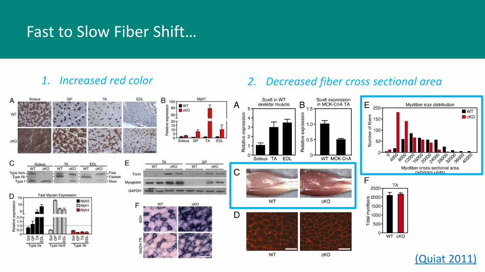
(Quiat 2011)
1. Increased red color
Fast to Slow Fiber Shift…
2. Decreased fiber cross sectional area
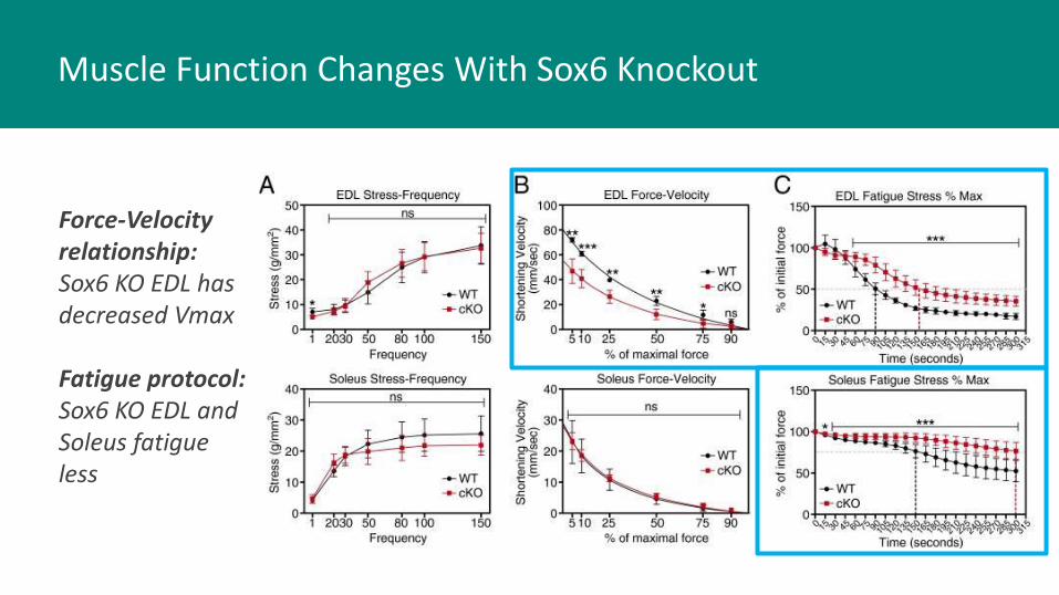
Force-Velocity relationship: Sox6 KO EDL has decreased Vmax
Fatigue protocol: Sox6 KO EDL and Soleus fatigue less
Muscle Function Changes With Sox6 Knockout

Examples of Muscle Function Assessment
1. IGF-1 Injection Into Extraocular Muscle
2. Sox6 Knock Out Mouse
3. Duchenne Muscular Dystrophy (Stretch Injury Protocol)
4. Mechanical Properties – Achilles Tendon stiffness
5. P1 mouse hindlimb in vitro
6. Mouse/dog hindlimb in vivo

SarcolemmaDGC
Ervasti, J.M., J.Biol.Chem., 2003. 278(16): p. 13591-13594.
Fiber DGC Absent From Sarcolemma:
Dystrophic muscle membrane more
“leaky”
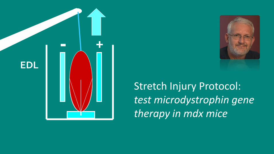
Duchenne Muscular Dystrophy

- +EDL
Procion Orange(Mr 631)
Stretch – Injury Protocol
Fiber
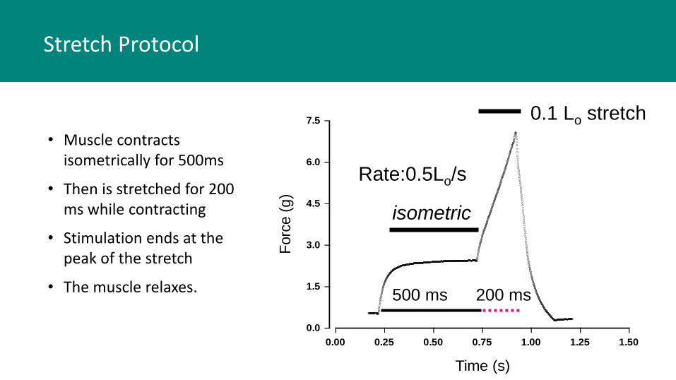
0.00 0.25 0.50 0.75 1.00 1.25 1.50
Time (s)
0.0
1.5
3.0
4.5
6.0
7.5
Forc
e (
g)
isometric
0.1 Lo stretch
Rate:0.5Lo/s
500 ms 200 ms
Stretch Protocol
• Muscle contracts isometrically for 500ms
• Then is stretched for 200 ms while contracting
• Stimulation ends at the peak of the stretch
• The muscle relaxes.

Fast-twitch skeletal muscles of dystrophic mouse pups are resistant to injury from acute mechanical stress. Grange, Gainer, Marschner, Talmadge, and Stull.
Am J Physiol Cell Physiol 283(4):C1090-101, 2002.
No stretch
Five stretches
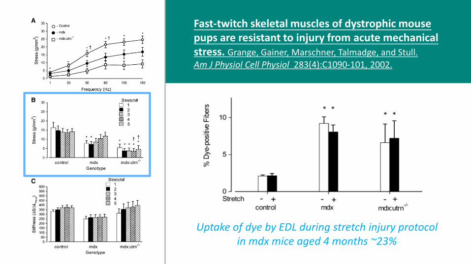
Uptake of dye by EDL during stretch injury protocol in mdx mice aged 4 months ~23%
Fast-twitch skeletal muscles of dystrophic mouse pups are resistant to injury from acute mechanical stress. Grange, Gainer, Marschner, Talmadge, and Stull.
Am J Physiol Cell Physiol 283(4):C1090-101, 2002.
- +
EDL
Stretch Injury Protocol: test microdystrophin gene therapy in mdx mice
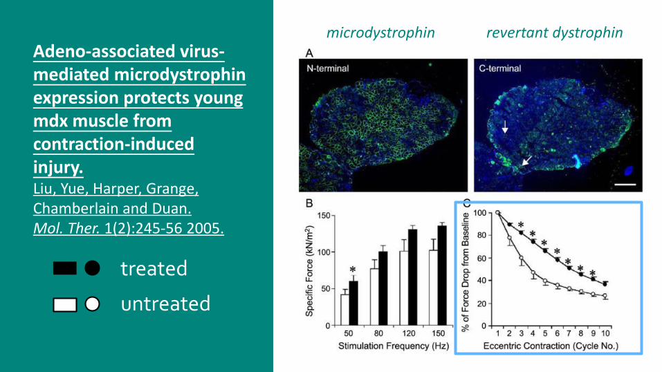
Adeno-associated virus-mediated microdystrophinexpression protects young mdx muscle from contraction-induced injury. Liu, Yue, Harper, Grange, Chamberlain and Duan. Mol. Ther. 1(2):245-56 2005.
microdystrophin revertant dystrophin
treated
untreated

Examples of Muscle Function Assessment
1. IGF-1 Injection Into Extraocular Muscle
2. Sox6 Knock Out Mouse
3. Duchenne Muscular Dystrophy (Stretch Injury Protocol)
4. Mechanical Properties – Achilles Tendon stiffness
5. P1 mouse hindlimb in vitro
6. Mouse/dog hindlimb in vivo

Stiffness – change in force during a change in muscle length
• for stress-strain assessment of achillestendon to determine stiffness in mouse pup aged 15 days
• Grange Lab 2-12-2013
Hindlimb Prep

Examples of Muscle Function Assessment
1. IGF-1 Injection Into Extraocular Muscle
2. Sox6 Knock Out Mouse
3. Duchenne Muscular Dystrophy (Stretch Injury Protocol)
4. Mechanical Properties – Achilles Tendon stiffness
5. P1 mouse hindlimb in vitro
6. Mouse/dog hindlimb in vivo

- +EDL
P1 Hindlimb
Force Assessment in Mouse Pup Hindlimbs

KLHL40 deficiency destabilizes thin filament proteins and promotes nemalinemyopathy
Ankit Garg,1 Jason O’Rourke,1 ChengzuLong,1 Jonathan Doering,2 GianinaRavenscroft,3 Svetlana Bezprozvannaya,1 Benjamin R. Nelson,1 Nadine Beetz,1 Lin Li,4 She Chen,4 Nigel G. Laing,3 Robert W. Grange,2 Rhonda Bassel-Duby,1 and Eric N. Olson1 J Clin Invest. 2014;124(8):3529–3539.
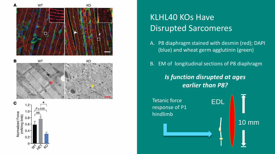
A. P8 diaphragm stained with desmin (red); DAPI (blue) and wheat germ agglutinin (green)
B. EM of longitudinal sections of P8 diaphragm
KLHL40 KOs Have Disrupted Sarcomeres
Tetanic force response of P1 hindlimb
Is function disrupted at ages earlier than P8?
10 mm
EDL
Examples of Muscle Function Assessment
1. IGF-1 Injection Into Extraocular Muscle
2. Sox6 Knock Out Mouse
3. Duchenne Muscular Dystrophy (Stretch Injury Protocol)
4. Mechanical Properties – Achilles Tendon stiffness
5. P1 mouse hindlimb in vitro
6. Mouse/dog hindlimb in vivo

Mouse Plantar Flexor Torque in vivo
Torque = Force x Moment arm
Ma
Force

Age- 9 weeks Age- 17 weeks
The Advantage of In Vivo:
Assess muscle function over time between conditions
Age- 21 weeks Age- 25 weeks

Dog Plantar Flexor Torque in vivo
Torque = Force x Moment arm
Ma
Force
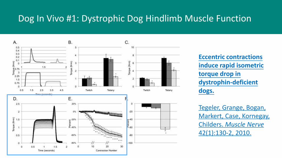
Eccentric contractions induce rapid isometric torque drop in dystrophin-deficient dogs.
Tegeler, Grange, Bogan, Markert, Case, Kornegay, Childers. Muscle Nerve 42(1):130-2, 2010.
Dog In Vivo #1: Dystrophic Dog Hindlimb Muscle Function

Gene Therapy Prolongs Survival and Restores Function in Murine and Canine Models of Myotubular Myopathy
Martin K Childers1,2,†, Romain Joubert3, Karine Poulard3, Christelle Moal3, Robert W Grange4, Jonathan A Doering4, Michael W Lawlor5,6, Branden E. Rider5, Thibaud Jamet3, Nathalie Danièle3, Samia Martin3, Christel Rivière3, Thomas Soker6, Caroline Hammer3, Laetitia Van Wittenberghe3, Mandy Lockard7, Xuan Guan7, Melissa Goddard7, Erin Mitchell7, Jane Barber7, J. KoudyWilliams7, David L Mack1, Mark E Furth8, Alban Vignaud3, Carole Masurier3, Fulvio Mavilio3, Philippe Moullier3,9,10, Alan H Beggs5,†, and Anna Buj-Bello3,†
Sci Transl Med. 2014 January 22; 6(220): 220ra10. doi:10.1126/scitranslmed.3007523.
Dog In Vivo #2: MTM-Deficient Dog HindlimbMuscle Function

“Loss-of-function mutations in the myotubularin gene (MTM1) cause X-linked myotubular myopathy (XLMTM), a fatal, congenital pediatric disease that affects the entire skeletal musculature.” Childers et al., 2014
Dr. Childers (University of Washington) has a colony of XLMTM dogs.MTM1 encodes a lipid phosphatase; primarily effects skeletal muscle:
• Hypotrophic fibers• Muscle structural abnormalities• Generalized weakness
There is no known cure… what are the functional outcomes of gene therapy?

Childers et al., Sci Transl Med. 6(220), 2014
Hindlimb infusion of XLMTM dogs with AAV8-MTM1 (canine)
AAV-infused Non-infused
VL
CT
Baseline age: 9 wks
6 wks post-inf
8 wks post-inf
14 wkspost-inf
1 year post-inf

Acknowledgements
The authors and co-authors listed herein: thank you for providing me the opportunity to contribute to your great work!
Audentes
Therapeutics

?
Muscle function:
Physiological Interpretation is Essential (PIE)
PIE

Robert W. Grange, PhD
Virginia [email protected]
Matt Borkowski
Aurora [email protected]
Thank You!For additional information on Aurora Scientific instruments specially designed for muscle function research please visit:
http://www.aurorascientific.com/