Embed Size (px)
Citation preview

SARDARHUSSAIN,,AssistantProfessor,Biotechnology,[email protected]
LECTURE NOTES IN CELL BIOLOGY Module 1: cellular organization of organisms
Historical perspective of cell biology, cell as structure and functional unit of living organisms, cellular organization in prokaryotes and eukaryotes, compartmentalization of cell, cell
fractionation ,cell membrane and permeability

Cell Biology notes by, Sardar Hussain, Asst. Prof. GSC, CTA.
1
The cell theory, or cell doctrine, states that all organisms are composed of similar units of organization,
called cells. The concept was formally articulated in 1839 by Schleiden & Schwann and has remained as the
foundation of modern biology. The idea predates other great paradigms of biology including Darwin’s theory of
evolution (1859), Mendel’s laws of inheritance (1865), and the establishment of comparative biochemistry
(1940).
First Cells Seen in Cork
While the invention of the telescope made the Cosmos accessible to human observation, the microsope
opened up smaller worlds, showing what living forms were composed of. The cell was first discovered and
named by Robert Hooke in 1665. He remarked that it looked strangely similar to cellula or small rooms which
monks inhabited, thus deriving the name. However what Hooke actually saw was the dead cell walls of plant
cells (cork) as it appeared under the microscope. Hooke’s description of these cells was published in
Micrographia . The cell walls observed by Hooke gave no indication of the nucleus and other organelles found
in most living cells. The first man to witness a live cell under a microscope was Antonvan Leeuwenhoek, who in
1674 described the algae Spirogyra. Van Leeuwenhoek probably also saw bacteria.
Formulation of the Cell Theory
In 1838, Theodor Schwann and Matthias Schleiden were enjoying after-dinner coffee and talking about
their studies on cells. It has been suggested that when Schwann heard Schleiden describe plant cells with
nuclei, he was struck by the similarity of these plant cells to cells he had observed in animal tissues. The two
scientists went immediately to Schwann’s lab to look at his slides. Schwann published his book on animal and
plant cells (Schwann 1839) the next year, a treatise devoid of acknowledgments of anyone else’s contribution,
including that of Schleiden (1838). He summarized his observations into three conclusions about cells:
The cell is the unit of structure, physiology, and organization in living things.
The cell retains a dual existence as a distinct entity and a building block in the construction of
organisms.
Cells form by free-cell formation, similar to the formation of crystals (spontaneous generation).
We know today that the first two tenets are correct, but the third is clearly wrong. The correct interpretation
of cell formation by division was finally promoted by others and formally enunciated in Rudolph Virchow’s
powerful dictum, Omnis cellula e cellula , “All cells only arise from pre-existing cells”.
Modern Cell Theory
All known living things are made up of cells.
The cell is structural & functional unit of all living things.
All cells come from pre-existing cells by division. (Spontaneous Generation does not occur).
Cells contains hereditary information which is passed from cell to cell during cell division.
All cells are basically the same in chemical composition.
All energy flow (metabolism & biochemistry) of life occurs within cells.
Historical perspectives of Cell Biology
Cell Biology notes by, Sardar Hussain, Asst. Prof. GSC, CTA.
2
As with the rapid growth of molecular biology in the mid-20th century, cell biology research exploded in
the 1950’s. It became possible to maintain, grow, and manipulate cells outside of living organisms. The first
continuous cell line to be so cultured was in 1951 by George Otto Gey and coworkers, derived from cervical
cancer cells taken from Henrietta Lacks, who died from her cancer in 1951. The cell line, which was eventually
referred to as HeLa cells, have been the watershed in studying cell biology in the way that the structure of DNA
was the significant breakthrough of molecular biology.
In an avalanche of progress in the study of cells, the coming decade included the characterization of the
minimal media requirements for cells and development of sterile cell culture techniques. It was also aided by
the prior advances in electron microscopy, and later advances such as development of transfection methods,
discovery of green fluorescent protein in jellyfish, and discovery of small interfering RNA (siRNA), among
others.
A Timeline
1595 – Jansen credited with 1st compound microscope
1655 – Hooke described ‘cells’ in cork.
1674 – Leeuwenhoek discovered protozoa. He saw bacteria some 9 years later.
1833 – Brown descibed the cell nucleus in cells of the orchid.
1838 – Schleiden and Schwann proposed cell theory.
1840 – Albrecht von Roelliker realized that sperm cells and egg cells are also cells.
1856 – N. Pringsheim observed how a sperm cell penetrated an egg cell.
1858 – Rudolf Virchow (physician, pathologist and anthropologist) expounds his famous conclusion: omnis
cellula e cellula , that is cells develop only from existing cells [cells come from preexisting cells]
1857 – Kolliker described mitochondria.
1879 – Flemming described chromosome behavior during mitosis.
1883 – Germ cells are haploid, chromosome theory of heredity.
1898 – Golgi described the golgi apparatus.
1938 – Behrens used differential centrifugation to separate nuclei from cytoplasm.
1939 – Siemens produced the first commercial transmission electron microscope.
1952 – Gey and coworkers established a continuous human cell line.
1955 – Eagle systematically defined the nutritional needs of animal cells in culture.
1957 – Meselson, Stahl and Vinograd developed density gradient centrifugation in cesium chloride solutions for
separating nucleic acids.
1965 – Ham introduced a defined serum-free medium. Cambridge Instruments produced the first commercial
scanning electron microscope.
1976 – Sato and colleagues publish papers showing that different cell lines require different mixtures of
hormones and growth factors in serum-free media.
1981 – Transgenic mice and fruit flies are produced. Mouse embryonic stem cell line established.
1995 – Tsien identifies mutant of GFP with enhanced spectral properties
1998 – Mice are cloned from somatic cells.
1999 – Hamilton and Baulcombe discover siRNA as part of post-transcriptional gene silencing (PTGS) in
plants
Cell Biology notes by, Sardar Hussain, Asst. Prof. GSC, CTA.
3
References:
Landmark Papers in Cell Biology: Selected Research Articles Celebrating Forty Years of The American
Society for Cell Biology. 2000. Cold Spring Harbor Laboratory Press.
Mazzarello P. A unifying concept: the history of cell theory. Nat Cell Biol. 1999. 1(1):E13-5.
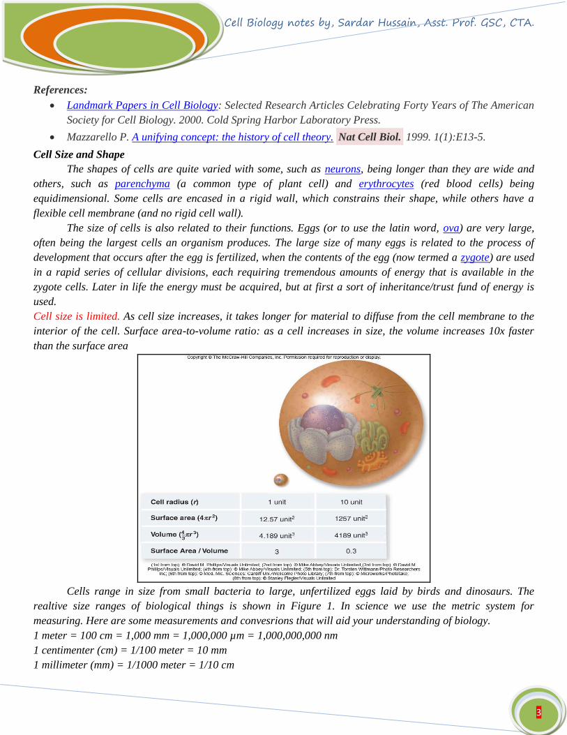
Cell Size and Shape
The shapes of cells are quite varied with some, such as neurons, being longer than they are wide and
others, such as parenchyma (a common type of plant cell) and erythrocytes (red blood cells) being
equidimensional. Some cells are encased in a rigid wall, which constrains their shape, while others have a
flexible cell membrane (and no rigid cell wall).
The size of cells is also related to their functions. Eggs (or to use the latin word, ova) are very large,
often being the largest cells an organism produces. The large size of many eggs is related to the process of
development that occurs after the egg is fertilized, when the contents of the egg (now termed a zygote) are used
in a rapid series of cellular divisions, each requiring tremendous amounts of energy that is available in the
zygote cells. Later in life the energy must be acquired, but at first a sort of inheritance/trust fund of energy is
used.
Cell size is limited. As cell size increases, it takes longer for material to diffuse from the cell membrane to the
interior of the cell. Surface area-to-volume ratio: as a cell increases in size, the volume increases 10x faster
than the surface area
Cells range in size from small bacteria to large, unfertilized eggs laid by birds and dinosaurs. The
realtive size ranges of biological things is shown in Figure 1. In science we use the metric system for
measuring. Here are some measurements and convesrions that will aid your understanding of biology.
1 meter = 100 cm = 1,000 mm = 1,000,000 µm = 1,000,000,000 nm
1 centimenter (cm) = 1/100 meter = 10 mm
1 millimeter (mm) = 1/1000 meter = 1/10 cm

Cell Biology notes by, Sardar Hussain, Asst. Prof. GSC, CTA.
4
1 micrometer (µm) = 1/1,000,000 meter = 1/10,000 cm
1 nanometer (nm) = 1/1,000,000,000 meter = 1/10,000,000 cm
Figure 1. Sizes of viruses, cells, and organisms. Images from Purves et al., Life: The Science of Biology, 4th
Edition, by Sinauer Associates (www.sinauer.com) and WH Freeman (www.whfreeman.com), used with
permission.
All living organisms (bacteria, blue green algae, plants and animals) have cellular organization and
may contain one or many cells. The organisms with only one cell in their body are called unicellular organisms
(bacteria, blue green algae, some algae, Protozoa, etc.). The organisms having many cells in their body are
called multicellular organisms (fungi, most plants and animals). Any living organism may contain only one type
of cell either
A. Prokaryotic cells;
B. Eukaryotic cells.
Cellular organization in prokaryotes and eukaryotes

Cell Biology notes by, Sardar Hussain, Asst. Prof. GSC, CTA.
5
The terms prokaryotic and eukaryotic were suggested by Hans Ris in the 1960’s. This classification is
based on their complexcity. Further based on the kingdom into which they may fall i.e the plant or the animal
kingdom, plant and animal cells bear many differences. These will be studied in detail in the upcoming sections.
Prokaryotic cells
Prokaryote means before nucleus in Greek. They include all cells which lack nucleus and other
membrane bound organelles. Mycoplasma, virus, bacteria and cyanobacteria or blue-green algae are
prokaryotes.
Most prokaryotes range between 1 μm to 10 μm, but they can vary in size from 0.2 μm to 750 μm
(Thiomargarita namibiensis). They belong to two taxonomic domains which are the bacteria and the archaea.
Most prokaryotes are unicellular, exceptions being myxobacteria which have multicellular stages in their life
cycles. They are membrane bound mostly unicellular organisms lacking any internal membrane bound
organelles. A typical prokaryotic cell is schematically illustrated in Figure 1. Though prokaryotes lack cell
organelles they harbor few internal structures, such as the cytoskeletons, ribosomes, which translate mRNA to
proteins. Membranous organelles are known in some groups of prokaryotes, such as vacuoles or membrane
systems devoted to special metabolic properties, e.g., photosynthesis or chemolithotrophy. In addition, some
species also contain protein-enclosed microcompartments, which have distinct physiological roles
(carboxysomes or gas vacuoles).
Figure 1: Schematic diagram of a prokaryotic cell
The individual structures depicted in Figure 1 are as follows
Flagella: It is a long, whip-like protrusion found in most prokaryotes that aids in cellular locomotion. Besides
its main function of locomotion it also often functions as a sensory organelle, being sensitive to chemicals and
temperatures outside the cell.
Capsule: The capsule is found in some bacterial cells, this additional outer covering protects the cell when it is
engulfed by phagocytes and by viruses, assists in retaining moisture, and helps the cell adhere to surfaces and
nutrients. The capsule is found most commonly among Gram-negative bacteria. Escherichia coli, Klebsiella
pneumoniae Haemophilus influenzae, Pseudomonas aeruginosa and Salmonella are some examples Gram-

Cell Biology notes by, Sardar Hussain, Asst. Prof. GSC, CTA.
6
negative bacteria possessing capsules. Whereas examples of Gram positive bacteria are Bacillus megaterium,
Streptococcus pneumoniae, Streptococcus pyogenes.
Cell wall: Cell wall is the outermost layer of most cells that protects the bacterial cell and gives it shape. One
exception is Mycoplasma which lacks cell wall. Bacterial cell walls are made of peptidoglycan which is made
from polysaccharide chains cross-linked by unusual peptides containing D-amino acids. Bacterial cell walls are
different from the cell walls of plants and fungi which are made of cellulose and chitin, respectively. The cell
wall of bacteria is also distinct from that of Archaea, which do not contain peptidoglycan.
The cell wall is essential to the survival of many bacteria. The antibiotic penicillin is able to kill bacteria
by preventing the cross-linking of peptidoglycan and this causes the cell wall to weaken and lyse. Lysozyme
enzyme can also damage bacterial cell walls.
There are broadly speaking two different types of cell wall in bacteria, called Gram-positive and Gram-
negative. The names originate from the reaction of cells to the Gram stain, a test long-employed for the
classification of bacterial species. Gram-positive bacteria possess a thick cell wall containing many layers of
peptidoglycan and teichoic acids. In contrast, Gram-negative bacteria have a relatively thin cell wall consisting
of a few layers of peptidoglycan surrounded by a second lipid membrane containing lipopolysaccharides and
lipoproteins. These differences in structure can produce differences in property as antibiotic susceptibility. For
example vancomycin can kill only Gram-positive bacteria and is ineffective against Gram-negative pathogens,
such as Pseudomonas aeruginosa or Haemophilus influenzae
Cell membrane: Cell membrane surrounds the cell's cytoplasm and regulates the flow of substances in and out
of the cell. It will be discussed in detail in one of the coming chapters.
Cytoplasm: The cytoplasm of a cell is a fluid in nature that fills the cell and is composed mainly of 80% water
that also contains enzymes, salts, cell organelles, and various organic molecules. The details will be discussed
in forthcoming chapter.
Ribosomes: Ribosomes are the organelles of the cell responsible for protein synthesis. Details of ribosomes will
be explained in coming chapter.
Nucleiod Region: The nucleoid region is possessed by a prokaryotic bacterial cell. It is the area of the
cytoplasm that contains the bacterial DNA molecule.
Plasmids: The term plasmid was first introduced by the American molecular biologist Joshua Lederberg in
1952. A plasmid is a DNA molecule (mostly in bacteria) that is separate from, and can replicate independently
of, the chromosomal DNA. They are double-stranded and circular. Plasmids usually occur naturally in
bacteria, but are sometimes found in eukaryotic organisms. Their sizes vary from 1 to over 1,000 kbp. The
number of identical plasmids in a single cell can range anywhere from one to thousands under some
circumstances and it is represented by the copy number. Plasmids can be considered mobile because they are
often associated with conjugation, a mechanism of horizontal gene transfer. Plasmids that can coexist within a
bacterium are said to be compatible. Plasmids which cannot coexist are said to be incompatible and after a few
generations are lost from the cell. Plasmids that encode their own transfer between bacteria are termed
conjugative. Non-conjugative plasmids do not have these transfer genes but can be carried along by
conjugative plasmids via a mobilisation site. Functionally they carry genes that code for a wide range of
metabolic activities, enabling their host bacteria to degrade pollutant compounds, and produce antibacterial
proteins. They can also harbour genes for virulence that help to increase pathogenicity of bacteria causing

Cell Biology notes by, Sardar Hussain, Asst. Prof. GSC, CTA.
7
diseases such as plague, dysentery, anthrax and tetanus. They are also responsible for the spread of antibiotic
resistance genes that ultimately have an impact on the treatment of Pili.
Pili: Pili are hair-like structures on the surface of the cell that help attach to other bacterial cells. Shorter pili
called fimbriae help bacteria attach to various surfaces. A pilus is typically 6 to 7 nm in diameter. The types of
pili are Conjugative pili and Type IV pili. Conjugative pili allow the transfer of DNA between bacteria, in the
process of bacterial conjugation. Some pili, called type IV pili, generate motile forces.
Morphology of prokaryotic cells
Prokaryotic cells have various shapes; the four basic shapes are (Figure 3):
• Cocci - spherical
• Bacilli - rod-shaped
• Spirochaete - spiral-shaped
• Vibrio - comma-shape
Eukaryotic cells
Plant cells are eukaryotic cells that differ in several key aspects from the cells of other eukaryotic organisms.
Their distinctive features include the following organelles:
1. Vacuole: It is present at the centre and is water-filled volume enclosed by a membrane known as the
tonoplast. The function is to maintain the cell's turgor, pressure by controlling movement of molecules between
the cytosol and sap, stores useful material and digests waste proteins and organelles.
2. Cell Wall: It is the extracellular structure surrounding plasma membrane. The cell wall is composed of
cellulose, hemicellulose, pectin and in many cases lignin, is secreted by the protoplast on the outside of the cell
membrane. This contrasts with the cell walls of fungi (which are made of chitin), and of bacteria, which are
made of peptidoglycan. An important function of the cell wall is that it controls turgity. The cell wall is divided
into the primary cell wall and the secondary cell wall. The Primary cell wall: extremely elastic and the
secondary cell wall forms around primary cell wall after growth are complete.
3. Plasmodesmata: Pores in the primary cell wall through which the plasmalemma and endoplasmic reticulum
of adjacent cells are continuous.
4. Plastids: The plastids are chloroplasts, which contain chlorophyll and the biochemical systems for light
harvesting and photosynthesis. A typical plant cell (e.g., in the palisade layer of a leaf) might contain as many
as 50 chloroplasts. The other plastids are amyloplasts specialized for starch storage, elaioplasts specialized for
fat storage, and chromoplasts specialized for synthesis and storage of pigments. As in mitochondria, which have
a genome encoding 37 genes, plastids have their own genomes of about 100–120 unique genes and, it is
presumed, arose as prokaryotic endosymbionts living in the cells of an early eukaryotic ancestor of the land
plants and algae

Cell Biology notes by, Sardar Hussain, Asst. Prof. GSC, CTA.
8
A typical plant cell
Plant cell types
Parenchyma cells: These are living cells that have diverse functions ranging from storage and support to
photosynthesis and phloem loading (transfer cells). Apart from the xylem and phloem in its vascular bundles,
leaves are composed mainly of parenchyma cells. Some parenchyma cells, as in the epidermis, are specialized
for light penetration and focusing or regulation of gas exchange, but others are among the least specialized
cells in plant tissue, and may remain totipotent, capable of dividing to produce new populations of
undifferentiated cells, throughout their lives. Parenchyma cells have thin, permeable primary walls enabling the
transport of small molecules between them, and their cytoplasm is responsible for a wide range of biochemical
functions such as nectar secretion, or the manufacture of secondary products that discourage herbivory.
Parenchyma cells that contain many chloroplasts and are concerned primarily with photosynthesis are called
chlorenchyma cells. Others, such as the majority of the parenchyma cells in potato tubers and the seed
cotyledons of legumes, have a storage function
Collenchyma cells: Collenchyma cells (Figure 2b) are alive at maturity and have only a primary wall. These
cells mature from meristem derivatives that initially resemble parenchyma, but differences quickly become
apparent. Plastids do not develop, and the secretory apparatus (ER and Golgi) proliferates to secrete
additional primary wall. The wall is most commonly thickest at the corners, where three or more cells come in
contact, and thinnest where only two cells come in contact, though other arrangements of the wall thickening
are possible. Pectin and hemicellulose are the dominant constituents of collenchyma cell walls of dicotyledon
angiosperms, which may contain as little as 20% of cellulose in Petasites. Collenchyma cells are typically quite
elongated, and may divide transversely to give a septate appearance. The role of this cell type is to support the
plant in axes still growing in length, and to confer flexibility and tensile strength on tissues. The primary wall
lacks lignin that would make it tough and rigid, so this cell type provides what could be called plastic support –
support that can hold a young stem or petiole into the air, but in cells that can be stretched as the cells around

Cell Biology notes by, Sardar Hussain, Asst. Prof. GSC, CTA.
9
them elongate. Stretchable support (without elastic snap-back) is a good way to describe what collenchyma
does. Parts of the strings in celery are collenchymas
Sclerenchyma cells: Sclerenchyma cells (from the Greek skleros, hard) are hard and tough cells with a
function in mechanical support. They are of two broad types – sclereids or stone cells and fibres. The cells
develop an extensive secondary cell wall that is laid down on the inside of the primary cell wall. The secondary
wall is impregnated with lignin, making it hard and impermeable to water. Thus, these cells cannot survive for
long' as they cannot exchange sufficient material to maintain active metabolism. Sclerenchyma cells are
typically dead at functional maturity, and the cytoplasm is missing, leaving an empty central cavity.
parenchyma collenchyma sclerenchyma
Animal cells: An animal cell is a form of eukaryotic cell that makes up many tissues in animals. Figure 7
depicts a typical animal cell. The animal cell is distinct from other eukaryotes, most notably plant cells, as they
lack cell walls and chloroplasts, and they have smaller vacuoles. Due to the lack of a rigid cell wall, animal
cells can adopt a variety of shapes, and a phagocytic cell can even engulf other structures. There are many
different cell types. For instance, there are approximately 210 distinct cell types in the adult human body.
Cell Biology notes by, Sardar Hussain, Asst. Prof. GSC, CTA.
10
Cell organelles in animal cell
Cell membrane: Plasma membrane is the thin layer of protein and fat that surrounds the cell, but is inside the
cell wall. The cell membrane is semipermeable, allowing selective substances to pass into the cell and blocking
others.
Nucleus: They are spherical body containing many organelles, including the nucleolus. The nucleus controls
many of the functions of the cell (by controlling protein synthesis) and contains DNA (in chromosomes). The
nucleus is surrounded by the nuclear membrane and possesses the nucleolus which is an organelle within the
nucleus - it is where ribosomal RNA is produced.
Golgi apparatus: It is a flattened, layered, sac-like organelle involved in packaging proteins and carbohydrates
into membrane-bound vesicles for export from the cell.
Ribosome and Endoplasmic reticulum: Ribosomes are small organelles composed of RNA-rich cytoplasmic
granules that are sites of protein synthesis and Endoplasmic reticulum are the sites of protein maturation and
they can be divided into the following types:
a. Rough endoplasmic reticulum: These are a vast system of interconnected, membranous, infolded and
convoluted sacks that are located in the cell's cytoplasm (the ER is continuous with the outer nuclear
membrane). Rough ER is covered with ribosomes that give it a rough appearance. Rough ER transport
materials through the cell and produces proteins in sacks called cisternae (which are sent to the Golgi body, or
inserted into the cell membrane).
b. Smooth endoplasmic reticulum: These are a vast system of interconnected, membranous, infolded and
convoluted tubes that are located in the cell's cytoplasm (the ER is continuous with the outer nuclear
membrane). The space within the ER is called the ER lumen. Smooth ER transport materials through the cell. It
contains enzymes and produces and digests lipids (fats) and membrane proteins; smooth ER buds off from
rough ER, moving the newly-made proteins and lipids to the Golgi body and membranes.
Mitochondria: These are spherical to rod-shaped organelles with a double membrane. The inner membrane is
infolded many times, forming a series of projections (called cristae). The mitochondrion converts the energy
stored in glucose into ATP (adenosine triphosphate) for the cell.
Lysosome: Lysosomes are cellular organelles that contain the hydrolase enzymes which breaks down waste
materials and cellular debris. They can be described as the stomach of the cell. They are found in animal cells,
while in yeast and plants the same roles are performed by lytic vacuoles.Lysosomes digest excess or worn-out
organelles, food particles, and engulf viruses or bacteria. The membrane around a lysosome allows the
digestive enzymes to work at the 4.5 pH they require. Lysosomes fuse with vacuoles and dispense their enzymes
into the vacuoles, digesting their contents. They are created by the addition of hydrolytic enzymes to early
endosomes from the Golgi apparatus.
Centrosome: They are small body located near the nucleus and has a dense center and radiating tubules. The
centrosomes are the destination where microtubules are made. During mitosis, the centrosome divides and the
two parts move to opposite sides of the dividing cell. Unlike the centrosomes in animal cells, plant cell
centrosomes do not have centrioles.
Peroxisome
Peroxisomes are organelles that contain oxidative enzymes, such as D-amino acid oxidase, ureate oxidase, and
catalase. They may resemble a lysosome, however, they are not formed in the Golgi complex. Peroxisomes are
distinguished by a crystalline structure inside a sac which also contains amorphous gray material. They are

Cell Biology notes by, Sardar Hussain, Asst. Prof. GSC, CTA.
11
self-replicating, like the mitochondria. Components accumulate at a given site and they can be assembled into a
peroxisome. Peroxisomes function to rid the body of toxic substances like hydrogen peroxide, or other
metabolites. They are a major site of oxygen utilization and are numerous in the liver where toxic byproducts
accumulate.
Vacuoles and vesicles
Vacuoles are single-membrane organelles that are essentially part of the outside that is located within the cell.
The single membrane is known in plant cells as a tonoplast. Many organisms will use vacuoles as storage
areas. Vesicles are much smaller than vacuoles and function in transporting materials both within and to the
outside of the cell.
Differences between plant and animal cells
1. Animal cells are generally small in size. Plant cells are larger than animal cells.
2. Cell wall is absent. The plasma membrane of plant cells is
Surrounded by a rigid cell wall of cellulose.
3. Except the protozoan Euglena no animal cell
possesses plastids. Plastids are present.
4. Vacuoles in animal cells are many and small. Most mature plant cells have a large central sap
vacuole.
5. Animal cells have a single highly complex Golgi
Plant cells have many simpler units of and prominent
Golgi apparatus. Apparatus, called dictyosomes.
6. Animal cells have centrosome and centrioles. Plant cells lack centrosome and centrioles
Interesting Facts:
1. There are anywhere from 75 to 100 trillion cells in the human body.
2. There are more bacterial cells in the body than human cells.
3. Thiomargarita namibiensis is the largest bacterium ever discovered, found in the ocean
Sediments of the continental shelf of Namibia and can be seen through the naked eye.
4. An unfertilized Ostrich egg is the largest single cell.
5. The smallest cell is a type of bacteria known as mycoplasma. Its diameter is 0.001 mm. 6. The Longest Cell in
your body is the motor neuron cell, which is located in the spinal cord, near the central nervous system.
Cellular compartments in cell biology comprise all of the closed parts within the cytosol of a eukaryotic
cell, usually surrounded by a single or double lipid layer membrane. These compartments are often, but not
always, defined as membrane enclosed regions. The formation of cellular compartments is called
compartmentalization.
Both organelles, the mitochondria and chloroplasts (in photosynthetic organisms), are compartments that are
believed to be of endosymbiotic origin. Other compartments such as peroxisomes, lysosomes, the endoplasmic
Compartmentalization of eukaryotic cells

Cell Biology notes by, Sardar Hussain, Asst. Prof. GSC, CTA.
12
reticulum, the cell nucleus or the Golgi apparatus are not of endosymbiotic origin. Smaller elements like
vesicles, and sometimes even microtubules can also be counted as compartments.
It was thought that compartmentalization is not found in prokaryotic cells. But the discovery of carboxysomes
and many other metabolosomes revealed that prokaryotic cells are capable of making compartmentalized
structures, though these are in most cases not surrounded by a lipid bilayer, but of pure proteinaceous built.
Types:
In general there are 4 main cellular compartments, they are:
1.The nuclear compartment comprising the nucleus
2.The intercisternal space which comprises the space between the membranes of the endoplasmic
reticulum (which is continuous with the nuclear envelope)
3.Organelles (the mitochondrion in all eukaryotes and the plastid in phototrophic eukaryotes)
4.The cytosol
Functions - Compartments have three main roles.
One is to establish physical boundaries for biological processes that enables the cell to carry out
different metabolic activities at the same time. This may include keeping certain biomolecules within a region,
or keeping other molecules outside. Within the membrane-bound compartments, different intracellular pH,
different enzyme systems, and other differences are isolated from other organelles and cytosol. With
mitochondria, the cytosol has an oxidizing environment which converts NADHto NAD+. With these cases, the
compartmentalization is physical.
Another is to generate a specific micro-environment to spatially or temporally regulate a biological
process. As an example, a yeast vacuole is normally acidified by proton transporters on the membrane.
A third role is to establish specific locations or cellular addresses for which processes should occur. For
example, a transcription factor may be directed to a nucleus, where it can promote transcription of certain
genes. In terms of protein synthesis, the necessary organelles are relatively near one another. The nucleolus
within the nuclear envelope is the location of ribosome synthesis. The destination of synthesized ribosomes for
protein translation is rough endoplasmic reticulum (rough ER), which is connected to and shares the same
membrane with the nucleus. The Golgi body is also near the rough ER for packaging and redistributing.
Likewise, intracellular compartmentalization allows specific sites of related eukaryotic cell functions isolated
from other processes and therefore efficient.
Establishment Often, cellular compartments are defined by membrane enclosure. These membranes
provide physical barriers to biomolecules. Transport across these barriers is often controlled in order to
maintain the optimal concentration of biomolecules within and outside of the compartment
Eukaryotic cells are complex and contain many kinds of membrane organelles. For instance, they
contain nuclei, mitochondria, vacuoles etc. Two methods exist to study the organelles in more detail. The first
is by using a variety of techniques to visualize the nuclei while still inside the cells by microscopy (i.e. cell
staining and immunofluorescence). The second method involves suspending the cells in solution, and breaking
them open (lysing the cells). Then the various organelles are then separated from each other by centrifugation,
which then allows them to be used for further study. It is this second method that we will use in this laboratory.
Cell fractionation
Cell Biology notes by, Sardar Hussain, Asst. Prof. GSC, CTA.
13
A procedure called cell fractionation is used to break open the cells and separate the various
organelles. To perform cell fractionation, we first will suspend our cells in solution, and then we break open
the cells, or lyse them. This will release the organelles inside into solution. Next, we can separate the
organelles by centrifuging our solution. By using centrifugation, we can easily separate the various organelles,
since the various organelles are of different mass, and density (for instance, nuclei are significantly heavier
than mitochondria etc.). During centrifugation, different organelles will pellet at the bottom at specific speeds
based on the mass and densities of the organelles. For instance, the heavier (larger) the organelles, the less
velocity is needed to pellet the organelle. The lighter (smaller) the organelle, centrifugation must occur at a
greater velocity to pellet the organelle.
Therefore, if we want to separate nuclei from mitochondria, we will centrifuge at low speed. At low
speed, the nuclei will pellet at the bottom, while the mitochondria will stay suspended in the solution. The left
over solution after centrifugation is called the supernatant, and will contain lighter organelles (i.e.
mitochondria and dissolved proteins). If we then want to separate the mitochondria from the rest of the
supernatant, we can centrifuge at the appropriate speed that would pellet the mitochondria, and then remove
the resulting supernatant.
If we first centrifuged or cell suspension at the speed appropriate to pellet mitochondria, we would
bring the mitochondria to the bottom of the tube. However, we would pellet everything that is heavier than the
mitochondria (i.e. nuclei etc.). Therefore, in order to get nuclei separated from mitochondria, we must
centrifuge at the lower speed first to obtain the nuclei, and centrifuge the supernatant at the higher speed to
collect the mitochondria. This type of separation protocol is called differential centrifugation. In this
procedure, it is possible to separate the organelles to purification because objects a similar size to our desired
organelles will also pellet at the same speeds. However, these objects are significantly less concentrated in our
suspension. Therefore, our pellets will are enriched for the organelle we are attempting to isolate.
Each pellet that is isolated can then be resuspended by adding solution, and can be called a fraction (for
instance, the resuspended nuclear pellet is considered the nuclear fraction). Additionally, a sample of the
original suspension is considered a fraction, and is called the crude fraction, as it contains all the organelles
and soluble proteins. Lastly, a sample of the final supernatant is also considered a fraction and is called the
soluble fraction, and contains everything that was not pelleted by centrifugation.
Cell Fractionation means separating different parts and organelles of a cell, so that they can be studied
in detail. All the processes of cell metabolism (such as respiration or photosynthesis) have been studied in this
way. The most common method of fractionating cells is to use differential centrifugation.A more sophisticated
separation can be performed by density gradient centrifugation. In this, the cell-free extract is centrifuged in a
dense solution (such as sucrose or caesium chloride). The fractions don't pellet, but instead separate out into
layers with the densest fractions near the bottom of the tube. The desired layer can then be pipetted off. This is
the technique used in the Meselson-Stahl experiment, and it is also used to separate the two types of ribosomes.
The terms 70S and 80S refer to their positions in a density gradient
How is subcellular farctionation done?

Cell Biology notes by, Sardar Hussain, Asst. Prof. GSC, CTA.
14
1. Cut tissue (e.g. liver, heart, leaf, etc) in ice-cold
isotonic buffer. Cold to stop enzyme reactions, isotonic to
stop osmosis, and buffer to stop pH changes.
2. Grind tissue in a blender to break open cells.
3. Filter. This removes insoluble tissue (e.g. fat,
connective tissue, plant cell walls, etc). This filtrate is not
called a cell-free extract, and is capable of carrying out
most of the normal cell reactions.
4. Centrifuge filtrate at low speed
(1 000 x g for 10 min)
5. Centrifuge supernatant at medium speed
(10 000 x g for 30 min)
6. Centrifuge supernatant at high speed
(100 000 x g for 1 hour)
7. Centrifuge supernatant at very high speed
(300 000 x g for 3 hours)
8. Supernatant is now organelle-free cytoplasm
Cell Biology notes by, Sardar Hussain, Asst. Prof. GSC, CTA.
15
2. Cell membrane and permeability
Overview: Life at the Edge
The plasma membrane separates the living cell from its nonliving surroundings.
This thin barrier, 8 nm thick, controls traffic into and out of the cell.
Like all biological membranes, the plasma membrane is selectively permeable, allowing some substances to
cross more easily than others.
Concept 2.1 Cellular membranes are fluid mosaics of lipids and proteins
The main macromolecules in membranes are lipids and proteins, but carbohydrates are also important.
The most abundant lipids are phospholipids.
Phospholipids and most other membrane constituents are amphipathic molecules.
Amphipathic molecules have both hydrophobic regions and hydrophilic regions.
The arrangement of phospholipids and proteins in biological membranes is described by the fluid mosaic
model.
Membrane models have evolved to fit new data.
Models of membranes were developed long before membranes were first seen with electron microscopes in the
1950s.
In 1915, membranes isolated from red blood cells were chemically analyzed and found to be composed of lipids
and proteins.
In 1925, E. Gorter and F. Grendel reasoned that cell membranes must be a phospholipid bilayer two molecules
thick.
The molecules in the bilayer are arranged such that the hydrophobic fatty acid tails are sheltered from water
while the hydrophilic phosphate groups interact with water.
Actual membranes adhere more strongly to water than do artificial membranes composed only of
phospholipids.
One suggestion was that proteins on the surface of the membrane increased adhesion.
In 1935, H. Davson and J. Danielli proposed a sandwich model in which the phospholipid bilayer lies between
two layers of globular proteins.
Early images from electron microscopes seemed to support the Davson-Danielli model, and until the 1960s, it
was widely accepted as the structure of the plasma membrane and internal membranes.
Further investigation revealed two problems.
First, not all membranes were alike. Membranes differ in thickness, appearance when stained, and percentage
of proteins.
Membranes with different functions differ in chemical composition and structure.
Second, measurements showed that membrane proteins are not very soluble in water.
Membrane proteins are amphipathic, with hydrophobic and hydrophilic regions.
If membrane proteins were at the membrane surface, their hydrophobic regions would be in contact with water.
In 1972, S. J. Singer and G. Nicolson presented a revised model that proposed that the membrane proteins are
dispersed and individually inserted into the phospholipid bilayer.
Cell Biology notes by, Sardar Hussain, Asst. Prof. GSC, CTA.
16
AP Biology
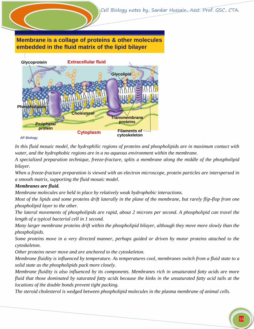
Membrane is a collage of proteins & other molecules
embedded in the fluid matrix of the lipid bilayer
Extracellular fluid
Cholesterol
Cytoplasm
Glycolipid
Transmembraneproteins
Filaments ofcytoskeleton
Peripheralprotein
Glycoprotein
Phospholipids
In this fluid mosaic model, the hydrophilic regions of proteins and phospholipids are in maximum contact with
water, and the hydrophobic regions are in a no aqueous environment within the membrane.
A specialized preparation technique, freeze-fracture, splits a membrane along the middle of the phospholipid
bilayer.
When a freeze-fracture preparation is viewed with an electron microscope, protein particles are interspersed in
a smooth matrix, supporting the fluid mosaic model.
Membranes are fluid.
Membrane molecules are held in place by relatively weak hydrophobic interactions.
Most of the lipids and some proteins drift laterally in the plane of the membrane, but rarely flip-flop from one
phospholipid layer to the other.
The lateral movements of phospholipids are rapid, about 2 microns per second. A phospholipid can travel the
length of a typical bacterial cell in 1 second.
Many larger membrane proteins drift within the phospholipid bilayer, although they move more slowly than the
phospholipids.
Some proteins move in a very directed manner, perhaps guided or driven by motor proteins attached to the
cytoskeleton.
Other proteins never move and are anchored to the cytoskeleton.
Membrane fluidity is influenced by temperature. As temperatures cool, membranes switch from a fluid state to a
solid state as the phospholipids pack more closely.
Membrane fluidity is also influenced by its components. Membranes rich in unsaturated fatty acids are more
fluid that those dominated by saturated fatty acids because the kinks in the unsaturated fatty acid tails at the
locations of the double bonds prevent tight packing.
The steroid cholesterol is wedged between phospholipid molecules in the plasma membrane of animal cells.

Cell Biology notes by, Sardar Hussain, Asst. Prof. GSC, CTA.
17
At warm temperatures (such as 37°C), cholesterol restrains the movement of phospholipids and reduces
fluidity.
At cool temperatures, it maintains fluidity by preventing tight packing.
Thus, cholesterol acts as a “temperature buffer” for the membrane, resisting changes in membrane fluidity as
temperature changes.
To work properly with active enzymes and appropriate permeability, membranes must be about as fluid as
salad oil.
Cells can alter the lipid composition of membranes to compensate for changes in fluidity caused by changing
temperatures.
For example, cold-adapted organisms such as winter wheat increase the percentage of unsaturated
phospholipids in their membranes in the autumn.
This prevents membranes from solidifying during winter.
Membranes are mosaics of structure and function.
A membrane is a collage of different proteins embedded in the fluid matrix of the lipid bilayer.
Proteins determine most of the membrane’s specific functions.
The plasma membrane and the membranes of the various organelles each have unique collections of proteins.
There are two major populations of membrane proteins.
Peripheral proteins are not embedded in the lipid bilayer at all.
Instead, they are loosely bound to the surface of the protein, often connected to integral proteins.
Integral proteins penetrate the hydrophobic core of the lipid bilayer, often completely spanning the membrane
(as transmembrane proteins).
The hydrophobic regions embedded in the membrane’s core consist of stretches of nonpolar amino acids, often
coiled into alpha helices.
Where integral proteins are in contact with the aqueous environment, they have hydrophilic regions of amino
acids.
On the cytoplasmic side of the membrane, some membrane proteins connect to the cytoskeleton.
On the exterior side of the membrane, some membrane proteins attach to the fibers of the extracellular matrix.
The proteins of the plasma membrane have six major functions:
Transport of specific solutes into or out of cells.
Enzymatic activity, sometimes catalyzing one of a number of steps of a metabolic pathway.
Signal transduction, relaying hormonal messages to the cell.
Cell-cell recognition, allowing other proteins to attach two adjacent cells together.
Intercellular joining of adjacent cells with gap or tight junctions.
Attachment to the cytoskeleton and extracellular matrix, maintaining cell shape and stabilizing the location of
certain membrane proteins.
Membrane carbohydrates are important for cell-cell recognition.
The plasma membrane plays the key role in cell-cell recognition.
Cell-cell recognition, the ability of a cell to distinguish one type of neighboring cell from another, is crucial to
the functioning of an organism.
This attribute is important in the sorting and organization of cells into tissues and organs during development.
It is also the basis for rejection of foreign cells by the immune system.

Cell Biology notes by, Sardar Hussain, Asst. Prof. GSC, CTA.
18
Cells recognize other cells by binding to surface molecules, often carbohydrates, on the plasma membrane.
Membrane carbohydrates are usually branched oligosaccharides with fewer than 15 sugar units.
They may be covalently bonded to lipids, forming glycolipids, or more commonly to proteins, forming
glycoproteins.
The oligosaccharides on the external side of the plasma membrane vary from species to species, from individual
to individual, and even from cell type to cell type within the same individual.
This variation distinguishes each cell type.
The four human blood groups (A, B, AB, and O) differ in the external carbohydrates on red blood cells.
Membranes have distinctive inside and outside faces.
Membranes have distinct inside and outside faces. The two layers may differ in lipid composition. Each protein
in the membrane has a directional orientation in the membrane.
The asymmetrical orientation of proteins, lipids and associated carbohydrates begins during the synthesis of
membrane in the ER and Golgi apparatus.
Membrane lipids and proteins are synthesized in the endoplasmic reticulum. Carbohydrates are added to
proteins in the ER, and the resulting glycoproteins are further modified in the Golgi apparatus. Glycolipids are
also produced in the Golgi apparatus.
When a vesicle fuses with the plasma membrane, the outside layer of the vesicle becomes continuous with the
inside layer of the plasma membrane. In that way, molecules that originate on the inside face of the ER end up
on the outside face of the plasma membrane.
Concept 2.2 Membrane structure results in selective permeability
A steady traffic of small molecules and ions moves across the plasma membrane in both directions.
For example, sugars, amino acids, and other nutrients enter a muscle cell, and metabolic waste products leave.
The cell absorbs oxygen and expels carbon dioxide.
It also regulates concentrations of inorganic ions, such as Na+, K+, Ca2+, and Cl?, by shuttling them across
the membrane.
However, substances do not move across the barrier indiscriminately; membranes are selectively permeable.
The plasma membrane allows the cell to take up many varieties of small molecules and ions and exclude others.
Substances that move through the membrane do so at different rates.
Movement of a molecule through a membrane depends on the interaction of the molecule with the hydrophobic
core of the membrane.
Hydrophobic molecules, such as hydrocarbons, CO2, and O2, can dissolve in the lipid bilayer and cross easily.
The hydrophobic core of the membrane impedes the direct passage of ions and polar molecules, which cross the
membrane with difficulty.
This includes small molecules, such as water, and larger molecules, such as glucose and other sugars.
An ion, whether a charged atom or molecule, and its surrounding shell of water also has difficulty penetrating
the hydrophobic core.
Proteins assist and regulate the transport of ions and polar molecules.
Specific ions and polar molecules can cross the lipid bilayer by passing through transport proteins that span
the membrane.
Some transport proteins, called channel proteins, have a hydrophilic channel that certain molecules or ions can
use as a tunnel through the membrane.

Cell Biology notes by, Sardar Hussain, Asst. Prof. GSC, CTA.
19
For example, the passage of water through the membrane can be greatly facilitated by channel proteins known
as aquaporins.
Other transport proteins, called carrier proteins, bind to molecules and change shape to shuttle them across the
membrane.
Each transport protein is specific as to the substances that it will translocate.
For example, the glucose transport protein in the liver will carry glucose into the cell but will not transport
fructose, its structural isomer.
Concept 2.3 Passive transport is diffusion of a substance across a membrane with no energy investment
Diffusion is the tendency of molecules of any substance to spread out in the available space.
Diffusion is driven by the intrinsic kinetic energy (thermal motion or heat) of molecules.
Movements of individual molecules are random.
However, movement of a population of molecules may be directional.
Imagine a permeable membrane separating a solution with dye molecules from pure water. If the membrane
has microscopic pores that are large enough, dye molecules will cross the barrier randomly.
The net movement of dye molecules across the membrane will continue until both sides have equal
concentrations of the dye.
At this dynamic equilibrium, as many molecules cross one way as cross in the other direction.
In the absence of other forces, a substance will diffuse from where it is more concentrated to where it is less
concentrated, down its concentration gradient.
No work must be done to move substances down the concentration gradient.
Diffusion is a spontaneous process that decreases free energy and increases entropy by creating a randomized
mixture.
Each substance diffuses down its own concentration gradient, independent of the concentration gradients of
other substances.
The diffusion of a substance across a biological membrane is passive transport because it requires no energy
from the cell to make it happen.
The concentration gradient itself represents potential energy and drives diffusion.
Because membranes are selectively permeable, the interactions of the molecules with the membrane play a role
in the diffusion rate.
Diffusion of molecules of limited permeability through the lipid bilayer may be assisted by transport proteins.
Osmosis is the passive transport of water.
Differences in the relative concentration of dissolved materials in two solutions can lead to the movement of
ions from one to the other.
The solution with the higher concentration of solutes is hypertonic relative to the other solution.
The solution with the lower concentration of solutes is hypotonic relative to the other solution.
These are comparative terms.
Tap water is hypertonic compared to distilled water but hypotonic compared to seawater.
Solutions with equal solute concentrations are isotonic.
Imagine that two sugar solutions differing in concentration are separated by a membrane that will allow water
through, but not sugar.
The hypertonic solution has a lower water concentration than the hypotonic solution.
Cell Biology notes by, Sardar Hussain, Asst. Prof. GSC, CTA.
20
More of the water molecules in the hypertonic solution are bound up in hydration shells around the sugar
molecules, leaving fewer unbound water molecules.
Unbound water molecules will move from the hypotonic solution, where they are abundant, to the hypertonic
solution, where they are rarer. Net movement of water continues until the solutions are isotonic.
The diffusion of water across a selectively permeable membrane is called osmosis.
The direction of osmosis is determined only by a difference in total solute concentration.
The kinds of solutes in the solutions do not matter.
This makes sense because the total solute concentration is an indicator of the abundance of bound water
molecules (and, therefore, of free water molecules).
When two solutions are isotonic, water molecules move at equal rates from one to the other, with no net
osmosis.
The movement of water by osmosis is crucial to living organisms.
Cell survival depends on balancing water uptake and loss.
An animal cell (or other cell without a cell wall) immersed in an isotonic environment experiences no net
movement of water across its plasma membrane.
Water molecules move across the membrane but at the same rate in both directions.
The volume of the cell is stable.
The same cell in a hypertonic environment will lose water, shrivel, and probably die.
A cell in a hypotonic solution will gain water, swell, and burst.
For organisms living in an isotonic environment (for example, many marine invertebrates), osmosis is not a
problem.
The cells of most land animals are bathed in extracellular fluid that is isotonic to the cells.
Organisms without rigid walls have osmotic problems in either a hypertonic or hypotonic environment and
must have adaptations for osmoregulation, the control of water balance, to maintain their internal environment.
For example, Paramecium, a protist, is hypertonic to the pond water in which it lives.
In spite of a cell membrane that is less permeable to water than other cells, water still continually enters the
Paramecium cell.
To solve this problem, Paramecium cells have a specialized organelle, the contractile vacuole, which functions
as a bilge pump to force water out of the cell.
The cells of plants, prokaryotes, fungi, and some protists have walls that contribute to the cell’s water balance.
A plant cell in a hypotonic solution will swell until the elastic cell wall opposes further uptake.
At this point the cell is turgid (very firm), a healthy state for most plant cells.
Turgid cells contribute to the mechanical support of the plant.
If a plant cell and its surroundings are isotonic, there is no movement of water into the cell. The cell becomes
flaccid (limp), and the plant may wilt.
The cell wall provides no advantages when a plant cell is immersed in a hypertonic solution. As the plant cell
loses water, its volume shrinks. Eventually, the plasma membrane pulls away from the wall. This plasmolysis is
usually lethal.
Specific proteins facilitate passive transport of water and selected solutes.
Many polar molecules and ions that are normally impeded by the lipid bilayer of the membrane diffuse
passively with the help of transport proteins that span the membrane.

Cell Biology notes by, Sardar Hussain, Asst. Prof. GSC, CTA.
21
The passive movement of molecules down their concentration gradient via transport proteins is called
facilitated diffusion.
Two types of transport proteins facilitate the movement of molecules or ions across membranes: channel
proteins and carrier proteins.
Some channel proteins simply provide hydrophilic corridors for the passage of specific molecules or ions.
For example, water channel proteins, aquaporins, greatly facilitate the diffusion of water.
Many ion channels function as gated channels. These channels open or close depending on the presence or
absence of a chemical or physical stimulus.
If chemical, the stimulus is a substance other than the one to be transported.
For example, stimulation of a receiving neuron by specific neurotransmitters opens gated channels to allow
sodium ions into the cell.
When the neurotransmitters are not present, the channels are closed.
Some transport proteins do not provide channels but appear to actually translocate the solute-binding site and
solute across the membrane as the transport protein changes shape.
These shape changes may be triggered by the binding and release of the transported molecule.
In certain inherited diseases, specific transport systems may be defective or absent.
Cystinuria is a human disease characterized by the absence of a protein that transports cysteine and other
amino acids across the membranes of kidney cells.
An individual with cystinuria develops painful kidney stones as amino acids accumulate and crystallize in the
kidneys.
Concept 2.4 Active transport uses energy to move solutes against their gradients
Some transport proteins can move solutes across membranes against their concentration gradient, from the side
where they are less concentrated to the side where they are more concentrated.
This active transport requires the cell to expend metabolic energy.

Cell Biology notes by, Sardar Hussain, Asst. Prof. GSC, CTA.
22
Active transport enables a cell to maintain its internal concentrations of small molecules that would otherwise
diffuse across the membrane.
Active transport is performed by specific proteins embedded in the membranes.
ATP supplies the energy for most active transport.
ATP can power active transport by transferring a phosphate group from ATP (forming ADP) to the transport
protein.
This may induce a conformational change in the transport protein, translocating the solute across the
membrane.
The sodium-potassium pump actively maintains the gradient of sodium ions (Na+) and potassium ions (K+)
across the plasma membrane of animal cells.
Typically, K+ concentration is low outside an animal cell and high inside the cell, while Na+ concentration is
high outside an animal cell and low inside the cell.
The sodium-potassium pump maintains these concentration gradients, using the energy of one ATP to pump
three Na+ out and two K+ in.
Some ion pumps generate voltage across membranes.
All cells maintain a voltage across their plasma membranes.
Voltage is electrical potential energy due to the separation of opposite charges.
The cytoplasm of a cell is negative in charge compared to the extracellular fluid because of an unequal
distribution of cations and anions on opposite sides of the membrane.
The voltage across a membrane is called a membrane potential, and ranges from? 50 to? 200 millivolts (mV).
The inside of the cell is negative compared to the outside.
The membrane potential acts like a battery.
The membrane potential favors the passive transport of cations into the cell and anions out of the cell.
Two combined forces, collectively called the electrochemical gradient, drive the diffusion of ions across a
membrane.
One is a chemical force based on an ion’s concentration gradient.
The other is an electrical force based on the effect of the membrane potential on the ion’s movement.
An ion does not simply diffuse down its concentration gradient but diffuses down its electrochemical gradient.

Cell Biology notes by, Sardar Hussain, Asst. Prof. GSC, CTA.
23
For example, there is a higher concentration of Na+ outside a resting nerve cell than inside.
When the neuron is stimulated, a gated channel opens and Na+ diffuse into the cell down their electrochemical
gradient. The diffusion of Na+ is driven by their concentration gradient and by the attraction of cations to the
negative side of the membrane.
Special transport proteins, electrogenic pumps, generate the voltage gradient across a membrane.
The sodium-potassium pump in animals restores the electrochemical gradient not only by the active transport of
Na+ and K+, setting up a concentration gradient, but because it pumps two K+ inside for every three Na+ that
it moves out, setting up a voltage across the membrane.
The sodium-potassium pump is the major electrogenic pump of animal cells.
In plants, bacteria, and fungi, a proton pump is the major electrogenic pump, actively transporting H+ out of
the cell.
Proton pumps in the cristae of mitochondria and the thylakoids of chloroplasts concentrate H+ behind
membranes.
These electrogenic pumps store energy that can be accessed for cellular work.
In cotransport, a membrane protein couples the transport of two solutes.
A single ATP-powered pump that transports one solute can indirectly drive the active transport of several other
solutes in a mechanism called cotransport.
As the solute that has been actively transported diffuses back passively through a transport protein, its
movement can be coupled with the active transport of another substance against its concentration gradient.
Plants commonly use the gradient of hydrogen ions generated by proton pumps to drive the active transport of
amino acids, sugars, and other nutrients into the cell.
One specific transport protein couples the diffusion of protons out of the cell and the transport of sucrose into
the cell. Plants use the mechanism of sucrose-proton cotransport to load sucrose into specialized cells in the
veins of leaves for distribution to nonphotosynthetic organs such as roots.
Concept 2.5 Bulk transport across the plasma membrane occurs by exocytosis and endocytosis

Cell Biology notes by, Sardar Hussain, Asst. Prof. GSC, CTA.
24
Small molecules and water enter or leave the cell through the lipid bilayer or by transport proteins.
Large molecules, such as polysaccharides and proteins, cross the membrane via vesicles.
During exocytosis, a transport vesicle budded from the Golgi apparatus is moved by the cytoskeleton to the
plasma membrane.
When the two membranes come in contact, the bilayers fuse and spill the contents to the outside.
Many secretory cells use exocytosis to export their products.
During endocytosis, a cell brings in macromolecules and particulate matter by forming new vesicles from the
plasma membrane.
Endocytosis is a reversal of exocytosis, although different proteins are involved in the two processes.
A small area of the plasma membrane sinks inward to form a pocket.
As the pocket deepens, it pinches in to form a vesicle containing the material that had been outside the cell.
There are three types of endocytosis: phagocytosis (“cellular eating”), pinocytosis (“cellular drinking”), and
receptor-mediated endocytosis.
In phagocytosis, the cell engulfs a particle by extending pseudopodia around it and packaging it in a large
vacuole.
The contents of the vacuole are digested when the vacuole fuses with a lysosome.
In pinocytosis, a cell creates a vesicle around a droplet of extracellular fluid. All included solutes are taken into
the cell in this nonspecific process.
Receptor-mediated endocytosis allows greater specificity, transporting only certain substances.
This process is triggered when extracellular substances, or ligands, bind to special receptors on the membrane
surface. The receptor proteins are clustered in regions of the membrane called coated pits, which are lined on
their cytoplasmic side by a layer of coat proteins.
Binding of ligands to receptors triggers the formation of a vesicle by the coated pit, bringing the bound
substances into the cell.
Receptor-mediated endocytosis enables a cell to acquire bulk quantities of specific materials that may be in low
concentrations in the environment.
Human cells use this process to take in cholesterol for use in the synthesis of membranes and as a precursor for
the synthesis of steroids.
Cholesterol travels in the blood in low-density lipoproteins (LDL), complexes of protein and lipid.
These lipoproteins act as ligands to bind to LDL receptors and enter the cell by endocytosis.
In an inherited disease called familial hypercholesterolemia, the LDL receptors are defective, leading to an
accumulation of LDL and cholesterol in the blood.
This contributes to early atherosclerosis.


![Module 2: Foundations in Biology Revision Exam …Cell...Module 2: Foundations in Biology Revision Exam Questions 2.1.6: Cell division The College of Richard Collyer MDT 04/2016 [6]](https://img.pdfslide.net/doc/110x75/5aa38daa7f8b9a1f6d8eb150/module-2-foundations-in-biology-revision-exam-cellmodule-2-foundations.jpg)