Embed Size (px)
Citation preview

Volume 4, Issue 1, 2015
May, 2015 ISSN: 2319-8796
International Journal of
NanoScience & Technology
This Journal is an academic
and peer-reviewed publication
(Print ISSN 2319-8796 )
Cite this volume
as 4(1)IJNST(2015) and so on....
An International Refereed Journal
www.manishanpp.com
International Journal of
NanoScience & Technology
International Journal of
NanoScience & Technology

COVERAGE OF THE JOURNAL
OBJECTIVE OF THE JOURNAL
CALL FOR PAPERSWe invite you to submit high quality papers for review and possible publication in all areas of nano science and technology which includes Nano-electronics and Systems, Nano-imaging and Molecular Manipulation and Devices, Nano-optics,Nanomachining, Nano-Catalysts, Nanomachining, Nano-Mechanics. All authors must agree on the content of the manuscript and its submission for publication in this Journal before it is submitted to us. Manuscripts should be submitted by e-mail to the Editor at [email protected] .
To promote and encourage specially YOUNG SCIENTISTS to take active part in research and get acquainted with the latest development and research in the field of nano science and technology. To promote cooperation in the pursuit of research in general and to exchange and contribute to the progress in the field of nano science and technology in particular.
Following types of papers are invited for publication in this Journal :-a) Original Research Papers of Scientific values b) Review Papersc) Short Communications d) Case Reports e) Letters to the Editor f) As you see
TYPES OF PAPERS ARE INVITED
REVIEWERS PROCESSAll manuscripts are reviewed by an editor and members of the Editorial Board or qualified outside REVIEWERS. Deci-sions will be made as rapidly as possible and the Journal strive to return reviewer’s comments to authors within 6 weeks. The Editorial Board will re-review manuscripts that are accepted pending reviewers. It is the goal of the this Journal to publish manuscripts within 4 weeks after submission after getting O K report from the Author.CONTACT US
ABOUT THE JOURNAL
1. Nano-electronics and Systems 2. Nano-imaging and Mo-lecular Manipulation and Devices3. Nano-optics 4. Nanomachining5. Nano-composites 6. Nano Bio-electronics and Molecular Nanotechnology7. Nano-Catalysts 8. Nano-Magnetism9. Nano-Mechanics 10. Nano-Biotechnology11. Nano-toxicology 12. Nano- food technology13. Nano-medicine 14. Nano-Photonics15. Nano-Structured Materials 16. Nano-Functional Mate-rials17. Nano-Electronics 18. Nano- Devices, Sensors and bio-sensors & Novel Materials in the Nano Regime19. Application and Commercial aspects of Nanotechnology
20. Carbon based Nanostructures: fullerenes, nanotubes, grapheme21. Computational Nano-Science Simulation Design and Modeling 22. Different Methods for Growth of Nanostruc-tures23. Nano-manufacturing and nano-engineering 24. Nano-fabrication25. Nanometerlogy and applications 26. Green nanotech-nology27. Morphology, characterisation, properties and behav-iour of Nanomaterials and nanostructures of Organic, Inor-ganic, and Biomedical of Nanomaterials Doped Nanoma-terials Modification of Nanomaterials28. Any other related topics
INTERNATIONAL JOURNAL OF NANOSCIENCE AND TECHNOLOGY is a biannual an academic and peer-reviewed Journal published by ACADEMIC AND RESEARCH PUBLICATIONS in Collaboration with JPMS Society. JPMS Societyis a Society registered under the Societies Registration Act and its Registration No. is 1649/1986-87. It was published from year i.e. 2012. The ISSN of the JOURNAL is 2319-8796.
For quick reply, please note the address and contact them directly by Post or email:-
For all publication matters related to the Journals Acceptance letter for publication of articles , Invoice, Reprints etc. should be sent directly to the PUBLICATION EDITOR whose address is as follows :
To,
Mrs. M anisha Verma, B.Sc., B.Tech.(Electronics)
Publication Editor (Chief Executive Director)Academic And Research Publications
Office: 22,Gaur Galaxy, Sec 5, Vaishali, Ghaziabad (UP)201010Email : [email protected] , www.manishanpp.com
Academic And Research Publications
For publication of your article, Acceptance letter, Review Reports , Status Report , and all other queries For publication of your article, Acceptance letter, Review Reports , Status Report , and all other queries related to your articles, should be sent directly to the Editor-in-Chief , whose address is as follows:Prof. M anik Sinha, The Editor-In-Chief
Email : [email protected] , Contact at : 09415155631

I J N S TInternational Journal
Of Nanoscience & Technology
Volume 4, Issue 1, 2015
‟ Ŧħis journal is Indexed/abstracted in Indian Science Abstract along with National/or International abstracting /Indexing
services if covered in these secondary publications”© Journal on Nanoscience and Technology. All rights reserved. No portion of material can be reproduced in part or
full without the prior permission of the Editor.Note : The views expressed herein are the opinions of contributors and do not reflect the stated policies of the
Academic And Research Publications & JPMS SOCIETY
May, 2015
Academic And Research PublicationsOffice: 22,Gaur Galaxy, Sec 5, Vaishali, Ghaziabad (UP)201010
Cite this volume as 4(1)IJNST(2015) and so on. . . .
This Journal is an academic and peer-reviewed publication (Print ISSN 2319-8796 )
An International Refereed Journal
www.manishanpp.com

Volume 4, 2015 Issue 1, 2015Editorial Board
Chief Editor Prof. Dr. techn. Murthy CHAVALI S.S.S. Yadav
M.Sc. Tech., P.G.D.C.A., C. Ger. (Austria), Ph.D. Tech. (Austria), Post-doc. Fellow (USA, Japan, Taiwan), C. Jap. (Japan), M.A.C.S., M.A.I.A.A.,M.I.L.A., M.S.A.E., M.I.S.A.S., M.O.S.A., M.I.F.S.A., M.E.A.C.R.,
PROFESSOR - Analytical Chemistry & Nanotechnology,RESEARCH COORDINATOR (DSH),Div. of Chemistry, Dept. of Sciences and Humanities,VIGNAN
University,Vadlamudi 522 213, Guntur, Andhra Pradesh, INDIA. E-mail : [email protected]
Coordinating EditorDr. Kavyanjali ShuklaFLAT-E, 5/F, BLOCK - 13,
SOUTH HORIZONS, AP LEI CHAU, HONG KONG
E-mail: shuklakavyanjali@[email protected]
Assistant EditorsDr. Khushwinder Kaur
Department of Chemistry Punjab University, Chandigarh Email :
:[email protected] Dr. Pervinder Kaur ,
Department of Agronomy Punjab Agricultural,University,
Ludhiana, Punjab. Email : [email protected]
Dr. Archna Srivastava, Asso. Prof. Rungta College of
Engg & Tech Bhilai (C.G.)Email:
Dr. Badal Kumar MandalProfessor
Trace Elements Speciation Research Laboratory Environmental and Analytical Chemistry Division
School of Advanced Sciences VIT UniversityVellore 632014, Tamil Nadu, IndiaE-mail: [email protected]. Mihir Kumar Purkait
Associate Professor Department of Chemical Engineering Indian
Institute of Technology, Guwahati Guwahati - 781039 Assam (INDIA)
Email :[email protected] Prof. Mandava V. Basaveswara Rao
Professor, Department of Chemistry, Krishna University . Machilipatnam -521 001, Krishna
Dist Andhra Pradesh, India.Email; [email protected] ;
[email protected] Dr. Masanori Kikuchi
International Center for Materials NanoArchitectonics (MANA),
National Institute for Materials Science, 1-1, Namiki, Tsukuba, Ibaraki 305-0044,
Japan Email: [email protected]
© Journal on Nanoscience and Technology. All rights reserved. No portion of material can be reproduced in part or full without the prior permission of the Editor.Note : The views expressed herein are the opinions of contributors and do not reflect the stated policies of the Academic And Re-search Publications.. Correspondence: All enquiries, editorial, business and any other, may be addressed to: The Editor-in-chief, International Journal of Nanoscience and Technology (IJNST), H.Office: EC 41, Maya Enclave, New Delhi -110064.
E-mail : [email protected], www.manishanpp.com.ISSN : 2319-8796
Members of Editorial Board
Associate EditorsProf. Dr. Wu, Ren-jang
Department of Applied Chemistry, Providence ,University, 200 Chung-Chi Rd., Shalu, 43301 Taichung, Taiwan
ROC ,TAIWAN, ROC Email:
Prof. M. SRIDHARAN, Dept. Electronics & Communica-
tion Engn. / SEEE , SASTRA UNIVERSITY
Thanjavur - 613 401, Tamil Nadu.(INDIA)Email:
Prof. Javier RodriguezViejoNanomaterials and Microsystems Group
Department of Physics Universitat Autonoma de Barcelona 08193 - Bellaterra. Barcelona Spain
E-mail: [email protected] Prof. Fumio Watari
in Hokkaido University, JapanEmail; [email protected]
Prof. Yang-Kyu Choi Department of Electrical Engineering Korea Advanced ,Institute of Science and Technol-ogy 291 Daehak-ro, Yuseong-gu ,Daejeon
305-701 Republic of Korea E-mail: [email protected]
Dr.P.Sujata Devi, Nano-Sericulture Materials Diagram, Central Glass ,Research Institute,196 Raja S C Mallik
Roas, Kolkata-700032. Email: [email protected]
Dr. Maqsood Ahmad, Asst.Professor,King Abdullah institute for Nanotechnology,King Saud University, BOX-
2454,Riyadh - 11451,Saudi Arabia.Email ; [email protected] ;
[email protected] Laxmi Upadhya, MNNIT, Allahabad. U P
Email : laxman7mnnit@gmail .com
International Journal of
Nanoscience & Technology
Editor-in-ChiefProf. Manik Sinha
Former Dean, Faculty of Law,Dr R.M.L Awadh University, Faizabad (UP),
Senior Advocate, Govt Of India, High Court, LucknowEmail: [email protected]
Publication Editor
Er. Manisha Verma, B. Sc., B. tech.Publication Editor(Chief Executive)
Academic And Research Publications 22, Gaur Galaxy , Plot No 5, Sec-5, Vaishali ,
Ghaziabad (U.P.)- 201010(INDIA)Email : [email protected],
www.manishanpp.com.
May, 2015

International Journal of
Nanoscience & TechnologyVolume No. 4 Issue No. 1, 2015
C o n t e n t sMay, 2015
Page No.S. No. Title
1. Synthesis and Characterization of Magnetic Cobalt Nanoparticles 01 using Pluronic P123 As Surfactant at Room Temperature
Prerna Sen and Badal Kumar Mandal
2. Colorimetric Detection of Hg+2 Using 07 Diastase Capped Silver Nanoparticles
Sireesh babu Maddinedi, Badal Kumar Mandal and Kiran Kumar Anna
3. Size Controlled Synthesis of Silver 16 Nanoparticles - a review Sireesh babu Maddinedi, Badal Kumar Mandal and Kiran Kumar Anna
4. Incorporation of Organoselenides and Selenolactams in Microemulsions: 23 Effect On Stability, Percolation and Size Khushwinder Kaur
5. Let's Talk Nanotechnology : 43 Some Applications and Scope For Future Research
Dr. Kavyanjali Shukla

© Journal on Nanoscience and Technology. All rights reserved. No portion of material can be reproduced in part or full without the prior permission of the Editor.
www.manishanpp.com

1 ••
Int. J. Nano. Sci. & Tech. Vol. 4 (1) 2015, pp. ISSN: 2319-8796
www.manish
anpp.com
SYNTHESIS AND CHARACTERIZATION OF MAGNETIC COBALT NANOPARTICLES USING PLURONIC P123 AS
SURFACTANT AT ROOM TEMPERATUREPrerna Sen and Badal Kumar Mandal*
Trace Elements Speciation Research Laboratory, Environmental and Analytical Chemistry Division, School of Advanced Sciences, VIT University, Vellore 632014, India.
* email:[email protected]
(Date of Receipt : 24-04-2015; Date of Acceptance for Publication : 05-05-2015)
PAGE : 6 REFERENCES : 13
The properties of nanoparticles are ex-tremely important because as the size of the particle decreases the properties are significantly altered from those of their bulk [1]. Magnetic nanoparticles, es-pecially Co, Fe, Ni and their alloys, have attracted increasing interest among re-searchers due to their promising appli-cations in high-density magnetic record-ing media [2]. Among these magnetic metal nanoparticles, the cobalt nano-
particles exhibit single-domain magnet-ism, high-saturation magnetization, and high coercive force; therefore, these have great potential applications in the field of magnetic record and memory [3]. The crystal phase of nanoparticles is a key factor for application in magnetic recording, The Co nanoparticles has a richer crystal phase diagram with three nearly isoenergetic crystal structures: face-centered cubic (FCC), hexagonally close-packed (HCP), and epsilon [2]. Jey-
ଁୗhe aim of the present work is to synthesize cobalt nanoparticles (CoNPs) at room temperature using pluronic P-123 as surfactant. This method is fast and simple which can be done under room temperature. Compared with other methods it is cost effec-tive and easy to prepare.The synthesis of cobalt nanoparticles by reduction of cobalt salt with sodium borohydride as a reducing agent with the combination of ethanol and stabilizing agent P123. Characterization of cobalt nanoparticles were carried out by XRD, SEM and EDX. The size of the nanoparticles is predominantly within the range of 50-100 nm.
INTRODUCTION
Key Words : Synthesis of Co nanoparticles, Synthesize Cobalt Nanoparticles (CoNPs), P123 (Pluronic acid) , Magnetic Nanoparticles.
1-6
2 ••
Int. J. Nano. Sci. & Tech. Vol. 4 (1) 2015, pp. ISSN: 2319-8796
www.manish
anpp.com
adevan et. al., reported that cobalt nan-oparticles have the advantage of lower annealing temperatures (300-500 0C) with high magnetism [4]. The magnetic properties of NP’s are extremely de-pendent on sample type, magnetization direction and cystallinity of the samples [5]. The key factor for the magnetic property is the presence and absence of magnet-ic domain. The magnetic properties of nanoparticles are determined by many factors: (a) the chemical composition, (b) the type and the degree of defective-ness of the crystal lattice, (c) the particle size and shape, (d) the morphology, (e) the interaction of the particle with the surrounding matrix and the neighboring particles [6]. Recently, these CoNPs have been synthesized by a variety of meth-ods including thermal decomposition, gas vapor condensation, and reduction of cobalt salt. Generally, in most types of nanoparticles are prepared by these method (sol–gel processing, hot spray-ing, evaporation condensation, matrix isolation, laser-induced vapor phase reactions and aerosols) control of size and size distribution is not possible [7]. For most of these strategies, however, complicated procedures, sophisticated equipment and rigorous experimental conditions are required. Thus, further development of the synthetic tech-niques into practical routes to large quantities of nanostructures from a di-versified range of materials, rapidly, and at reasonably low costs, still requires great inventiveness. In contrast, uncon-ventional methods based on chemical synthesis might provide an alternative and interesting approach for generating nanostructures in terms of material di-versity, cost, and the potential for high-volume production [8]. Magnetic metal-
MATERIALS AND METHODS
1-6
lic nanoparticles can be synthesized from metal salts using strong reducing agents, namely, alkali metal disper-sions in ethers or hydrocarbons, alkali metal complexes with organic electron acceptors (e.g., naphthalene), sodium borohydride (NaBH4) and other com-plex hydrides. By using NaBH4 in aque-ous solutions at room temperature, both homo- (Fe, Co, Ni) and heterometallic (FeCo, FeCu, CoCu) nanoparticles were obtained as amorphous powders con-taining substantial amounts of boron [9]. Shao et. al., reported synthesis of CoNPs using nontoxic cobalt acetate tetrahy-drate as a precursor material [10]. Shin reported NaBH4 reduction technique for CoNPs synthesis using cobalt chloride as precursor in aqueous solution [11].In the present project work, an easy and inexpensive chemical synthesis route has been proposed. The synthesis of CoNPs by reduction of cobalt salt with NaBH4 as the reducing reagent with the combination of ethanol and sur-factants (P123) has been carried out at room temperature. The samples are characterized by X-ray Diffraction (XRD) and SEM measurements. The elemen-tal analysis and chemical composition are studied by Energy Dispersive X-ray Spectroscopy (EDX).
Cobalt acetate, P123 (Pluronic acid) and ethanol were obtained from Aldrich chem-icals. Milli-Q water was used throughout the work.
Experimental Work and Methodology

3 ••
Int. J. Nano. Sci. & Tech. Vol. 4 (1) 2015, pp. ISSN: 2319-8796
www.manish
anpp.com
1-6
Figure 1 : Experimental Setup for the synthesis of CoNPs.
CoNPs were prepared through chemi-cal reduction of cobalt acetate Co(CH3COO)2 in ethanol. To prevent the product from aggregation, a surfactant has to be added at the beginning of each synthesis. In a typical experimen-tal design, 3.7gms of cobalt acetate was dissolved in ethanol. Pluronic acid (P123) was dissolved in ethanol. The above prepared solutions were added in a three-necked round bottom flask fit-ted with a mechanical stirrer at the rate of 500 rpm under constant stirring as shown in the above diagram (Fig. 1). On stirring a pink colour emulsion was ob-tained. Then NaBH4 solution was add-ed dropwise to the above solutions at room temperature under stirring at 500 rpm for 30 min. The solution turned to black colour which showed the reduc-tion of cobalt acetate. The black colour solution was allowed to settle down at room temperature. The black precipi-
tate of the CoNPs was separated by centrifugation at 5000 rpm for 10 min. The remaining solution was decanted, and the precipitate was rinsed with eth-anol twice followed by centrifugation in the same rpm. The supernatant was decanted and the CoNPs were isolated in wet form. The processes of filtration and pulverization were the significant and controlling steps. The black precip-itate was filtered through 0.22 micron filter paper using suction pump and the wet product was taken out in watch glass for the process of pulverization and drying done at cooled condition to avoid ignition of the particles. Finally, air stable CoNPs powder was obtained at room temperature. The CoNPs synthe-sized by this method were very stable for a long time.
Reaction Mechanism
The reduction of transition metal ions by NaBH4 is a ubiquitous reaction useful for the production of ultrafine particles of met-als and metal borides. However, NaBH4 reduction chemistry is complex and the nature of the products depends strongly on the reaction conditions used [12-13].
When the cobalt salt was dissolved in ethanol, Co(HOR)6
2+ was formed with a pink colour (Reaction 1). Reac-tion 2 shows that the replacement of Co(HOR)6
2+ by NaBH4 and the colour of the solution turned to black with the evolution of H2 gas.
Synthesis of Co nanoparticles

4 ••
Int. J. Nano. Sci. & Tech. Vol. 4 (1) 2015, pp. ISSN: 2319-8796
www.manish
anpp.com
CoNPs was characterized by the tech-niques such as XRD, SEM and EDX. Fig-ure 2 shows the typical XRD pattern of CoNPs synthesized. The XRD pattern shows broad peaks which indicate that our sample is amorphous in nature. To
1-6
RESULTS AND DISCUSSIONCharacterization of CoNPs
narrow down the peaks annealing of sample is done at 800 0C for 4 hr. The XRD pattern shows broader peaks due to the outer covering of organic surfactants on the surfaces on CoNPs even after heating at 800 0C. It clearly indicates that the sam-ple is amorphous due to its hard covering which can prevent the oxidation of metal in air and avoid aggregation.
Figure 2: XRD diffractogram of CoNPs

5 ••
Int. J. Nano. Sci. & Tech. Vol. 4 (1) 2015, pp. ISSN: 2319-8796
www.manish
anpp.com
1-6
Figure 3: Energy Dispersive X-ray (EDX) microgram of CoNPs
The EDX spectra of the CoNPs shows that it consists of 86.42% of cobalt and 13.58% of oxygen (weight %) while 63.33% of cobalt and 36.67% of oxygen (Atomic %). The typical SEM image of the synthesized CoNPs (Fig. 4) shows
size within less than 100 nm. Due to magnetic dipole-dipole interactions of the individual particles more aggrega-tion of the nanoparticles causes the large surface area and bigger sized particles.
Figure 4: SEM microgram of cobalt nanoparticles.

6 ••
Int. J. Nano. Sci. & Tech. Vol. 4 (1) 2015, pp. ISSN: 2319-8796
www.manish
anpp.com
••••••• ••••••••
CONCLUSION
The CoNPs prepared by borohydride re-duction method using surfactants are very stable at room temperature for long time. In the borohydride reduction method for the synthesis of nanoparticles ethanol is used as the solvent and P123, are used as a capping agent. P123 is a good chelat-ing agent and also a good stabilizer. P123 is a protective copolymer which stabilizes the CoNPs by preventing their aggrega-tion and affords protective coatings by preventing their re-oxidation. Finally, the particles obtained were stable and black in color. XRD results showed that the co-balt nanoparticles synthesized was amor-phous in nature. The EDX spectra of the CoNPs showed that it consisted of 86.42% cobalt and SEM images showed its par-ticle size less than 100 nm. Hence P123 may be a smart choice to prepare stable and smaller sized particles of magnetic CONPs at room temperature.
REFERENCES
1. S.S. Kalyan Kamal, P.K. Sahoo, M. Premkumar, N.V. Rama Rao, T. Jagadeesh Kumar, B. Sreedhar, A.K. Singh, S. Ram and K. Chandra Sekhar, (2009). Journal of Alloys and Compounds 474 214–218.
2. H. Shao, Y. Huang, H.-S. Lee, Y. J. Suh, and C. O. Kim (2006), Current Applied Physics 6S1 e195–e197.
3. Huiping Shao Yuqiang Huang Hy-
oSook Lee, Yong Jae Suh and Chong Oh Kim (2006), Journal of Magnetism and Magnetic Materials 304 e28–e30.
4. B. Jeyadevan, K. Urakawa, A. Hobo, N. Chinnasamy, K. Shinoda, K.Tohji, D.D.J. Djayaprawira, M. Tsunoda, M. Takahashi (2003) , Jpn. J. Appl. Phys. 42 L350-L352.
5. H.Q. Cao, Z. Xu, H. Sang, D. Sheng, C.Y. Tie (2001), Adv. Mater. 13 121-123.
6. S.B gubin et. al., Russian chemical review 74(6) (2005) 489-520.
7. V. Pallai, D.O. Shah, J. Magn. Magn. Mater 163 (1996) 243-248.
8. Y. Xia, J.A. Rogers, K.E. Paul, G.M. Whi-tesides (1999), Chem. Rev. 99 1823-1827.
9. S Sun, C B Murray (1999), J. Appl. Phys. 85 4325-4230.
10. N. Patel, A. Miotello, V. Bello (2011), Applied Catalysis B: Environmental 103 31–38.
11. S P Gubin, and Yu A Koksharov (2002), Neorg. Mater, 38 1287-1304.
12. G.N.Glavee, K.J. Klabunde, C.M. So-rensen, and G.C.Hadjipanayis (1993), Langmuir 9 162-169.
13. G.N. Glavee, K.J. Klabunde, C.M. So-rensen, and G.C. Hadjipanayis (1994), Langmuir 10 4726-4730.
1-6

7 ••
Int. J. Nano. Sci. & Tech. Vol. 4 (1) 2015, pp. ISSN: 2319-8796
www.manish
anpp.com
COLORIMETRIC DETECTION OF Hg+2 USING DIASTASE CAPPED SILVER NANOPARTICLES
Sireesh babu Maddinedi, Badal Kumar Mandal* and Kiran Kumar Anna
Trace Elements Speciation Research Laboratory, Environmental and Analytical Chemistry Division, School of Advanced Sciences, VIT University, Vellore 632014
Email: [email protected]
(Date of Receipt : 16-04-2015; Date of Acceptance for Publication : 27-05-2015)
Now-a-days, metal nanoparticles such as gold and silver have received more attention among all the nanomateri-als because of their optical, electronic and magnetic properties. Due to these extraordinary properties, silver nano-particles (AgNPs) has applications in optoelectronics (1), pharmaceuticals (2), catalysis (3,4), electronics (5), antimicrobi-al products (6,7), sensing (8), therapeutics (9-11), and photonics (12). Recently, con-sumer products containing AgNPs are commonly available in the market be-cause of their antimicrobial activity to-wards large range of fungi, bacteria and low cost of production. For instance, Ag-
NPs are being used in coatings, clothes, food , bandages and containers as dis-infectants and deodorizers (13,14) as well as an insecticide for pest control (15).On the other hand, the selective col-orimetric detection of hazardous heavy metal ions present in the environment is of growing significance. For instance, divalent mercury ion (Hg+2) is one of the most common toxic heavy met-als existing in the nature and causes health threats affecting central nervous system, brain and kidney (16,17). Accord-ing to the United States Environmental Protection Agency (USEPA) the recom-mended levels of Hg+2 in water is 2 ppb, whereas the World Health Organization
Ðiastase capped silver nanoparticles (AgNPs) were prepared via a green reduction route and used as colorimetric sensors for the detection of Hg+2. The synthesized Ag-NPs were characterized by using UV-visible, XRD and TEM analysis. The obtained Ag-NPs showed selective colorimetric detection of potentially toxic Hg2+ ions in aqueous media at micromolar level. The proposed colorimetric method i.e. sensing system has effectively been used for the detection of Hg+2 in tap water samples.
INTRODUCTION
Key Words : Silver Nanoparticles, Colorimetric Sensors, Mercury, Diastase.
7-15
PAGE : 09 REFERENCES : 28
[email protected],*The Corresponding Author

8 ••
Int. J. Nano. Sci. & Tech. Vol. 4 (1) 2015, pp. ISSN: 2319-8796
www.manish
anpp.com
(WHO) and Bureau of Indian Standards (BIS) suggested it as 1 ppb. Hence, it is important to detect and determine the Hg2+ levels in the biological and envi-ronment samples with very good selec-tivity and sensitivity. Traditional methods used for the direct quantitative detection of mercury in-cludes the gas chromatography, cold vapor atomic fluorescence spectrometry and absorption spectroscopy (18,19). But, these methods are time consuming and expensive instruments are needed with complex sample preparation methods. Therefore, it is greatly advantageous to develop simple and rapid methods for measuring Hg+2 levels in the environ-ment. However, several reports have been published for the immediate de-tection of Hg+2 using chromophores (20), noble metal NPs (21-23), fluorophores (24), polymer (25) to evade the complex sam-ple preparation and instrumentation.Herein, we describe the use of diastase, a thiol group containing enzyme as re-ducing and stabilizing agent for the synthesis of AgNPs in alkaline pH. The prepared AgNPs are stable and acted as selective colorimetric sensor for the detection of Hg2+ in aqueous media.
7-15
Experimental Section
Silver nitrate, sodium hydroxide, metal salts and all other organic solvents were purchased from sigma aldrich chemicals, Bangalore. Metal stock solutions were prepared by dissolving metal salts in dou-ble distilled water and were further used whenever necessary. Diastase was pur-
chased from CDH (P) Ltd., Delhi, India.
Diastase capped AgNPs were prepared as per our reported procedure else-where (26). In brief, 1 mL diastase solution (1%) of pH 12 was added to 5 mL of 1 mM AgNO3 solution and stirred at room tem-perature for about 10 min using a magnet-ic stirrer. Formation of AgNPs was known by the change of colour from colourless to reddish brown and was monitored by using UV-visible spectroscopy.
Preparation of Silver Nanoparticles
General Procedure for the Colorimetric Determination of Hg2+
To examine the colorimetric detection of Hg+2, the as-synthesized AgNPs were di-luted three times with double distilled wa-ter. Investigation of ability of diastase sta-bilized AgNPs for detection of Hg+2 was carried out by the addition of 200 µL of 1 mM metal ion solutions to the 300 µL of three times diluted AgNPs solution. The changes in absorption were monitored at room temperature using UV-visible spec-trophotometer and all the readings were taken after 5 minutes of metal ions addi-tion to AgNPs solution.
Preparation of Simulated Water Samples
Tap water samples were collected and filtered through a cellulose nitrate filter paper and a suitable volume of Hg+2 solu-tions was spiked to make simulated real samples. The analysis of spiked water samples was carried out by the proposed procedure using UV-visible spectropho-tometer.RESULTS AND DISCUSSION
Diastase, an enzyme containg thiol groups have used for the synthesis of AgNPs, played the key role as both reduc-
MATERIALS AND METHODS

9 ••
Int. J. Nano. Sci. & Tech. Vol. 4 (1) 2015, pp. ISSN: 2319-8796
www.manish
anpp.com
7-15
ing and stabilizing agent. When diastase solution was added to the AgNO3 under alkaline conditions, the colour of the solution changed from colorless to yellow and then to reddish brown due to the surface plasmon resonance of the AgNPs. From the UV-visible spectra of as-synthesized AgNPs, the presence
of an intense SPR absorption peak at 414 nm indicated the change of Ag+ to Ago (Fig.1A). The free thiol groups exist-ing on the diastase surface might be responsible for the reduction of AgNO3 to AgNPs and the formed NPs were stabilized by the carboxylic groups of diastase.
Figure 1: (A) UV–visible spectra (B) XRD pattern of AgNPs synthesized with diastase
The purified AgNPs were character-ized by using powder XRD. Figure 1B shows the XRD pattern of the diastase stabilized AgNPs. The formed AgNPs showed different diffraction peaks at 2θ values of 38.1, 44.2, 64.39, 77.35 and 81.37 which corresponds to the charac-teristic planes (111), (200), (220), (311)
and (222) respectively, indicating the face centered cubic crystal structure of
AgNPs (JCPDS NO: 96-901-1608). The morphology of the prepared NPs was studied under TEM. Figure 2 shows the TEM images of the AgNPs prepared with the diastase. TEM images represent the presence of anisotropic spherical and rod shaped nanoparticles. It also shows the formation of rod shaped nanopar-ticles by the aggregation of spherical smaller nanoparticles.

10 ••
Int. J. Nano. Sci. & Tech. Vol. 4 (1) 2015, pp. ISSN: 2319-8796
www.manish
anpp.com
Figure 2: TEM images of the diastase capped AgNPs
7-15
Colorimetric Determination of Hg+2
In order to study the selectivity of AgNPs towards other metal ions, we examined the color change with different metal ions such as Cu+2, Pb+2, Mn+2, Co+2, Cd+2, Ni+2, Fe+2, Ba+2, Zn+2, Cr+2 at 1mM concen-tration each. The AgNPs solution existed as reddish brown with the addition of 1mM each of these metal ions except Hg+2 ions. After addition of 1mM of Hg+2 ions it resulted in the disappearance of the color of AgNPs solution immediately. UV–visi-ble absorption spectra showed the disap-pearance of peak at 414 nm (Figure 3A), which may be due to the binding of Hg+2
ions onto the surface of AgNPs to move diastase enzyme aside.
Further, the colorimetric responses of the
AgNPs system towards Hg2+ were stud-ied at different pH of 4, 5, 6, 7, 8, 9 and 10. A noticeable color disappe-arance was observed for AgNPs at pH ‘4’ and ‘5’ with the addition of Hg2+ (80 µM). Addition of Hg+2 (80 µM) to AgNPs at pH ‘4’ and ‘5’ resulted in the large decrease in the absorbance, compared to that at higher pH (6, 7, 8, 9, 10) indicating that pH ‘4’ and ‘5’ are the optimum condition for AgNPs sensing system for detection of Hg+2 (Figure 3B). This may be attributed to the fact that in the basic solutions the presence of hydroxyl groups may interact with the metal ions and results in the decreased interaction between mercury ions and AgNPs surface. Hence, pH ‘4’ and ‘5’ are the most favorable conditions for the present sensing assay.

11 ••
Int. J. Nano. Sci. & Tech. Vol. 4 (1) 2015, pp. ISSN: 2319-8796
www.manish
anpp.com
7-15
Figure 3: (A) UV-visible spectra showing the colorimetric response of AgNPs towards various cations (B) Response of AgNPs system upon addition of Hg2+ ions (80 µM) at various pH
Further, different concentrations of Hg+2
ions were added to AgNPs solution, in order to assess the limit of detectable concentration of Hg+2 in aqueous solu-tions. UV-visible absorption spectra of AgNPs and the related color change were recorded after the addition of dif-ferent concentrations of Hg+2 (Figure 4A). The distinctive SPR peak of AgNPs was used to determine the limit of de-tection for Hg+2 by using UV–visible
spectroscopy. From Figure 4A, it is confirmed that the concentration of Hg+2 < 80 µM did not show complete color change of AgNPs. The absorb-ance at 414 nm decreased with in-creasing concentration of Hg+2 added, suggested more Hg+2 ions binding to AgNPs surface (27). These results con-clude that the lowest concentration of Hg+2 that can be detected by AgNPs colorimetrically was 80 µM.
Figure 4: (A) UV–vis absorption response of AgNPs system upon addition of Hg2+ ions (50– 90 µM) (B) Possible mechanism of Hg+2 detection by AgNPs.

12 ••
Int. J. Nano. Sci. & Tech. Vol. 4 (1) 2015, pp. ISSN: 2319-8796
www.manish
anpp.com
7-15
Synthesized AgNPs were characterized after and before the addition of Hg+2 ions using UV-visible spectroscopy. Typi-cal SPR absorption peak of Ag nanocol-loids after 2 times dilution was found at 414 nm and the solution was yellowish in color without the presence of Hg+2, but became colorless with the addition of 80 µM of Hg+2. Based on the UV-visible data, the sensing mechanism of AgNPs may be based on the redox reaction be-tween AgNPs and Hg+2. The standard reduction potential of Hg+2 (0.85 V) is higher than the Ag+ (0.8 V) and hence the added Hg+2 will be bound on the surface
of AgNPs to move the diastase molecules away from the surface of AgNPs, result-ing the redox reaction between Hg+2 and silver. Mercury (II) ions may be reduced radiolytically in aqueous AgNPs solu-tion and a mercury layer will be formed around AgNPs. Figure 3A also showed the peak broadening and blue shift of the SPR band, when 80 µM of Hg+2 was added to the AgNPs dispersion, which might be the result of adsorbed metallic mercury atoms formed by the reaction of Hg+2 with AgNPs (28). The proposed mech-anism of Hg+2 detection by AgNPs system is shown in Figure 4B.
Figure 5: UV–vis response of AgNPs system towards Hg+2 in tap water matrix
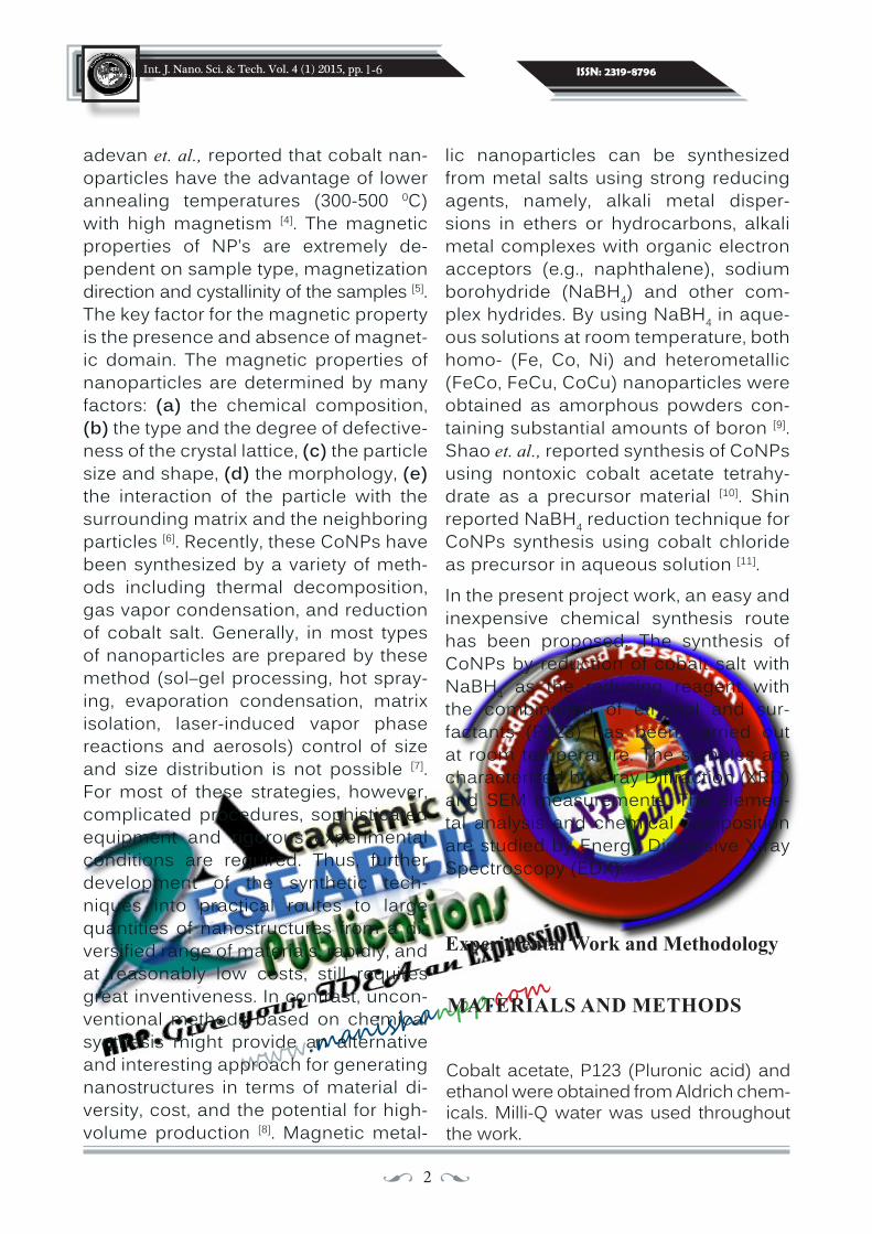
Further, the diastase stabilized AgNPs system was evaluated for the determi-nation of Hg2+ in tap water matrix. The amount of Hg+2 in the environmental samples such as tap water is very less and hence a potential practical assay is necessary. These tap water samples were free of Hg+2 or the amount may be very less for detection. Hence, the samples were spiked with various con-centrations of mercury within the range of 1-100 µM and the atomic absorption
spectroscopic data were taken to know the concentrations of unknown solutions which was found to be 44, 52, 67, 82 µM (Figure 6, Table 1). Figure 5 shows the ability of AgNPs sensing system for the detection of Hg+2 in the tap water. The limit of detection of AgNPs based sens-ing system for the detection of Hg+2 ions in tap water samples is 80 µM. This result further suggested the great potential of present method for the sensing of Hg+2 in tap water samples.
13 ••
Int. J. Nano. Sci. & Tech. Vol. 4 (1) 2015, pp. ISSN: 2319-8796
www.manish
anpp.com
7-15
Figure 6: Calibration plot of standard Hg+2 solution (30, 50, 70, 90 µM) obtained by atomic absorption spectroscopy
Table 1 AAS absorbance values of simulated Hg+2 samples prepared in tap water matrix

14 ••
Int. J. Nano. Sci. & Tech. Vol. 4 (1) 2015, pp. ISSN: 2319-8796
www.manish
anpp.com
CONCLUSION
In this paper we have showed the effi-ciency of diastase capped AgNPs for the colorimetric detection of Hg+2. This AgNPs system showed high selectivity for Hg+2 ions over other alakaline, alkali and tran-sition metal ions with a limit of detection of 80 µM L−1. The high stability and easy synthesis of the protein directed synthe-sized AgNPs allocate this method to be very simple to apply. This method offers low cost and simple way of Hg+2 detection in the environmental samples without any further modifications.
ACKNOWLEDGEMENT
REFERENCES
1. Y. Cui and C. M. Lieber (2001). Science. 291 851–3.
2. E. Ulkür, O. Oncul, H. Karagoz, E. Yeniz and B. Celiköz (2005). Burns. 31 874–7.
3. N. R. Jana, T. K. Sau and T. Pal (1999). J. Phys. Chem. B. 103 115–121.
4. Z. J. Jiang, C. Y. Liu and L. W. Sun (2005). J. Phys. Chem. B. 109 1730–5.
5. G. N. R. Tripathi (2003). J. Am. Chem. Soc. 125 1178–9.
6. V. K. Sharma, R. a Yngard and Y. Lin (2009). Adv. Colloid Interface Sci. 145 83–96.
The authors are grateful to VIT University, Vellore 632014 for the help and platform given to do this work.
7. I. Sondi and B. Salopek-Sondi (2004). J. Colloid Interface Sci. 275 177–82.
8. A. D. M. Farland, R. P. V. Duyne (2003). Nano Lett. 3 1057–1062.
9. Y. Li, K. Hindi, K. M. Watts, J. B. Tay-lor, K. Zhang, Z. Li, D. A. Hunstad, C. L. Cannon, W. J. Youngs and K. L. Wooley (2010). Chem. Commun.. 46 121–3.
10. C. Shao, B. Yuan, H. Wang, Q. Zhou, Y. Li, Y. Guan and Z. Deng (2011). J. Mater. Chem. 21 2863.
11. J. Jain, S. Arora, J. M. Rajwade, P. Om-ray, S. Khandelwal and K. M. Paknikar (2009). Mol. Pharm. 6 1388-1401.
12. Y. Zhang, R. Huang, X. Zhu, L. Wang and C. Wu (2012). Chinese Sci. Bull. 57 238–246.
13. T. M. Benn and P. Westerhoff (2008). Environ. Sci. Technol. 42 4133–4139.
14. J. Gensel, T. Borke, N. P. Pérez, A. Fery, D. V Andreeva, E. Betthausen, A. H. E. Müller, H. Möhwald and E. V Skorb (2012). Adv. Mater. 24 985–9.
15. Q. Li, S. Mahendra, D. Y. Lyon, L. Bru-net, M. V Liga, D. Li and P. J (2008). J. Alvarez, Water Res. 42 4591–602.
16. T. W. Clarkson, L. Magos and G. J. My-ers (2003), N. Engl. J. Med. 349 1731–7.
17. Y. Wang, F. Yang and X. Yang (2010).
7-15

15 ••
Int. J. Nano. Sci. & Tech. Vol. 4 (1) 2015, pp. ISSN: 2319-8796
www.manish
anpp.com
Biosens. Bioelectron. 25 1994–8.
18. S. Yoon, A. E. Albers, A. P. Wong and C. J. Chang. J. Am (2005). Chem. Soc. 127 16030–1.
19. J. Wang and B. Liu (2008). Chem. Com-mun. 39 4759–61.
20. E. Coronado, R. Gala, C. Martı, E. Pal-omares, J. R. Durrant and M. Gratzel (2005). J. Am. Chem. Soc. 127 838–842.
21. J.S. Lee, M. S. Han and C. a Mirkin (2007). Angew. Chem. Int. Ed. Engl. 46 4093–6.
22. G. K. Darbha, A. K. Singh, U. S. Rai, E. Yu, H. Yu and P. Chandra Ray (2008). J. Am. Chem. Soc. 130 8038–43.
23. D. Li, A. Wieckowska and I. Willner
(2008). Angew. Chem. Int. Ed. Engl. 47 3927–31.
24. Y. Yang, K. Yook and J. Tae (2005). J. Am. Chem. Soc. 1 16760–16761.
25. Y. Zhao and Z. Zhong (2006). J. Am. Chem. Soc. 128 9988–9.
26. S.B. Maddinedi, B. K. Mandal, CURR NANOSCI. (in press).
27. Y. Guo, Z. Wang, W. Qu, H. Shao and X. Jiang (2011). Biosens. Bioelectron. 26 4064–9.
28. K. Farhadi, M. Forough, R. Molaei, S. Hajizadeh and A. Rafipour (2012). Sen-sors Actuators B Chem. 161 880–885.
7-15
••••••• ••••••••

16 ••
Int. J. Nano. Sci. & Tech. Vol. 4 (1) 2015, pp. ISSN: 2319-8796
www.manish
anpp.com
SIZE CONTROLLED SYNTHESIS OF SILVER NANOPARTICLES - a review
Sireesh babu Maddinedi, Badal Kumar Mandal* and Kiran Kumar Anna
Trace Elements Speciation Research Laboratory, Environmental and Analytical Chemistry Division, School of Advanced Sciences,
VIT University, Vellore 632014, India.
Email: [email protected]
(Date of Receipt : 06-04-2015; Date of Acceptance for Publication : 30-05-2015)
Recently, the development of techniques for the preparation of new class of na-noscale materials is becoming greatest importance in nanotechnology. This is mainly because of the day by day raising in the significant role of nanomaterials in various fields of science such as elec-tronics, chemistry, biology and physics. The unique extraordinary mechanical, thermal electronic, optical and biologi-cal properties made them unusual from other bulk scale materials. But still the synthesis of nanomaterials with desired features is the greatest challenge in the field of nanoscience.Generally nanomaterials are featured
by (i) Quantum confinement effects (ii) large number of particles per unit weight, and (iii) high surface to volume ratio. All these features of nanomaterials result in extremely diverse properties from those of bulk materials. For example, the prop-erties of materials that change from bulk to nanometer range are optical property (1), magnetic properties (2) and solubili-ty (3), melting temperature (4). For exam-ple, gold nanoparticles (AuNPs) shows ruby red or blue color depending on their size whereas the conventional gold is yellow (5). It is well known that the large surface area per unit weight is a sig-nificant feature of nanomaterials.In general, methods for synthesis of na-
Šize controlled synthesis of metal nanoparticles remains a narrow part of research in the field of nanotechnology even after the development of various methods for their synthesis. The intrinsic properties of the nanoparticles can be controlled by tuning their size and shape, which has generated great potential applications in several fields of engineering and science. This review mainly emphasises on different methods reported so far for the production of silver nanoparticles (AgNPs) with tunable sizes. This review also discusses about the various parameters responsible for tuning the size of AgNPs.
INTRODUCTION
Key Words : Size Controlled Synthesis, Silver NPs, Tuning of Size.
16-22
PAGE : 7 REFERENCES : 50
[email protected],*The Corresponding Author
17 ••
Int. J. Nano. Sci. & Tech. Vol. 4 (1) 2015, pp. ISSN: 2319-8796
www.manish
anpp.com
nomaterials can be largely classified into two different categories: (i) top down (6,7) and (ii) bottom up (8, 9) approaches. Gen-erally, top down approach includes the physical synthesis methods such as laser etching (10), lithographic techniques (6, 7) and ball milling (11), where the materials in bulk state are scaled down until the na-noscale or desired size is obtained. But, the problems such as hardness with ball milling techniques and high cost involve-ment of lithographic techniques in de-signing very small nanomaterials made them less suitable for industrial applica-tions (6, 7, 11). In contrast, bottom up ap-proach commonly includes the biological and chemical synthetic methods. Here, the nanomaterials of required shape and size can be synthesized from their bulk precursors using a biological or chemi-cal reduction reaction. During the course of reduction, the building blocks of the nanomaterials will be formed first which later will be assembled to form the final nanostructure. The chemical and physi-cal parameters can be changed in order to control the nucleation and growth rate, and also the addition of external capping agent is done to stop the growth process in attaining the nanomaterials of required shape and size (12). Additionally, the nano-particles (NPs) of small size can be sepa-rated from large size by using centrifuga-tion (13) in solution phase synthesis.
Nanoparticles are the nanoscale materi-als that exist in the size range between 1-100 nm. During the Roman period, metal nanoparticles were used as deco-rants in glasses, where glasses showed different colors when they were viewed through reflected and transmitted light due to the presence of NPs (14). Noble metal NPs have been commonly
used in different applications such as sur-face enhanced Raman scattering (SERS) (15) and Plasmonics (16, 17, 18). Additionally, they have been used in various bio-appli-cations such as antimicrobial (19), immu-nological labeling (20, 21) and X-ray contrast agents (22). On the other hand, Palladium (Pd) and Platinum (Pt) NPs have been established as very good catalysts for dif-ferent hydrogenation reactions. Hence, the growing use of metal NPs in different areas of science is becoming a promis-ing area of attention. Metal NPs (MNPs) of different sizes can be fabricated using various biological and chemical meth-ods. Moreover, MNPs exhibits different properties with change in their size. For example, the phenomenon of Surface plasmon resonance (SPR) exhibited by noble MNPs varies with the size of gold and silver NPs. Hence there is a raising demand for the synthesis of MNPs of de-sired size and shape.
Ahmad et. al., have prepared the AgNPs of different size by ultrasonication method using k-carrageenan as reducing agent. They have found that the irradiation time played the key role in tuning the NPs size (23). In the same way, Bonatt et. al., have developed a temperature dependent method for tuning the size and shape of the AgNPs using the fruit seed extract of cashew-apple. They have proposed that the size of NPs increased with increase in the temperature of the reaction system (24). Zong et. al., have proposed a kineti-cally controlled seed mediated synthesis method for size tuning of AgNPs using glucose as reducing agent. The average sizes of the obtained NPs were found be-tween 20-200 nm (25). Panacek et. al., have reported the synthe-sis of size controlled AgNPs within the
16-22

18 ••
Int. J. Nano. Sci. & Tech. Vol. 4 (1) 2015, pp. ISSN: 2319-8796
www.manish
anpp.com
16-22
diameter range from 28 to 77 nm using D-maltose as reducing and stabilizing agent. The comparative catalytic activity of the prepared AgNPs against the meth-ylene blue degradation showed the high efficiency of small size NPs over larger ones (26). Similarly, Suganya et. al., have developed a Spirulina platensis extract mediated biosynthetic method for the synthesis of AgNPs of different sizes. Au-thors have shown the comparative anti-microbial activity of synthesized NPs and proved the improved activity of AgNPs with decrease in size. They also reported that the activity was size and dose de-pendent (27).
Alternatively, reaction conditions such as pH also played an important role in tun-ing the size of NPs. For example, Para-meshwaran et. al., have varied the pH of the reaction mixture containing beta vulgaris peel extract and AgNO3 to ob-tain the NPs of different sizes (28). Jeong et. al., have synthesized AgNPs of two different sizes (10 nm, 100 nm) using so-dium citrate as reducing agent and their cytotoxic and antimicrobial properties were studied. Change in the concentra-tion of sodium citrate used for the reduc-tion played key role in the preparation of nanoparticles of required size. The com-parative cytotoxicity and antimicrobial activity studied against the Methylobac-terium bacteria suggested the size and dose dependant activity of NPs. Authors suggested that the size and amount of the AgNPs were important parameters to be considered before using in the com-mercial products such as blood-contact medical devices (29).Chen et. al., prepared a highly disper-sive AgNPs of different sizes by chemical method where PVP w as used as reduc-
ing agent and particles size was tuned by varying the solvent (water, ethanol and ethylene glycol) used to carry out the reaction. Introduction of organic sol-vent such as ethanol decreased the size of the NPs formed which may be due to the decrease in the collision between the AgNPs. It was also found that the smaller NPs were obtained with ethylene gly-col which because of the longer chain length of ethylene glycol compared to ethanol (30). Goswami et. al., have pre-pared AgNPs of different sizes via a chemical method by using NaBH4 as re-ducing agent in the presence of PVP and sodium citrate. Morphology of the NPs was controlled by varying the amount of the stabilizer PVP during synthesis pro-cess. They have found that the smaller NPs showed the good electrochemical response towards the cholesterol sens-ing (31).
Zahran et. al., have proposed a new method to prepare AgNPs of different sizes using sodium alginate. They have tuned the size of the nanoparticles by varying the temperature of reaction sys-tem (60-80 oC). This may be due to the higher temperature played an key role in accelerating the redox reaction be-tween silver ions and sodium alginate which finally led to the change in the size of the NPs (32).
Xu et. al., have prepared flower-like Ag mesostructures with the size range of 200 nm to 700 nm via a simple seed-mediated approach. This size-tunable synthesis was easily attained by adjust-ing the concentration of Ag seeds. The formation of Ag mesostructures was at-tained by the assembly of primary Ag na-nosheets, which exhibited a flower-like

19 ••
Int. J. Nano. Sci. & Tech. Vol. 4 (1) 2015, pp. ISSN: 2319-8796
www.manish
anpp.com
16-22
architecture. Compare to the previous approach for flower-like Ag mesostruc-tures, the present method was simple and reliable using green procedure and size controllability. These flower-like Ag mesostructures revealed large surface area and was served as highly efficient catalysis for the NaBH4 reduction of p-nitrophenol to p-aminophenol (33). Lin et. al., have synthesized highly fluores-cent AgNPs with various sizes by the mi-croemulsion method. The size of the Ag-NPs was controlled easily by altering the molar ratio of water to surfactant in mi-croemulsion. The molar ratio and reac-tion time could influence the maximum absorbance wavelength and the absorb-ance of AgNPs, as well as the fluores-cence emission intensity (34).
Reithofer et. al., have synthesized a size controlled AgNPs in UV irradiation meth-od with using ultra short peptides such as Ac-LIVAGKNH2 (35). Tang et. al., have syn-thesized AgNPs in polyol process where PVP was used as a capping and PEG as a reducing agent. Ratio of the PVP to PEG played an important role in tuning the size of the AgNPs. Higher the PVP to PEG ratio bigger size NPs formation was observed (36). Oh et. al., have synthesized AgNPs of controlled size and shape in seed-mediated growth method by using ascorbic acid and citrate. The size of the nanostructures was varied by changing the concentration of citrate and ascorbic acid. Larger size NPs was obtained when the concentration of ascorbic acid and citrate ions was higher. Catalytic studied have shown that the large surface area of the bumpy nanostructures would be highly attractive as efficient heterogene-ous catalyst (37). Piella et. al., have shown that the varia-
tion in the concentration of tannic acid in the reaction meadium resulted in the Ag-NPs of different sizes. The average sizes of the obtained NPs varied from 10.1±0.9 nm to 46.1±8.3 nm. The catalytic studies showed the high catalytic efficiency of smaller NPs (38). Kang et. al.,have devel-oped a PVA mediated synthesis of size controlled AgNPs by electron irradiation method. In this method particle size was tuned by varying the energy of electron beam and current. Particle size was in-creased with increase in the dose of electron beam and current. They have also studied the increase in the sizes of the NPs by increase in the concentration of PVA (39).
Similarly, Zhang et. al., have synthesized silver nanocrystals with uniform sizes in droplet micro reactors through seed-me-diated growth using glycolaldehyde as a reducing agent. The average diameter of the obtained NPs was 30-100 nm (40). Zao et. al., have used sodium borohydride so-lution for the synthesis of AgNPs of dif-ferent sizes in chemical route and found that with increasing the volume of silver seed used in reaction the obtained size of the NPs was decreased (41). Jacob et. al., have synthesized AgNPs of different sizes by chemical method using water-ethylene glycol mixture. They have var-ied the concentration of ethylene and water to tune the size of NPs. Increasing the water-ethylene glycol ratios the for-mation of silver atom cluster decreased, resulting the decrease in the size of NPs (42).Meneses et. al., have prepared a size and shape controlled synthesis of AgNPs via a chemical method using the secondary amines. They have identified that the ad-dition of 1 or 5 equivalents of ethylenedi-

20 ••
Int. J. Nano. Sci. & Tech. Vol. 4 (1) 2015, pp. ISSN: 2319-8796
www.manish
anpp.com
CONCLUSIONS
16-22
REFERENCES
1. K.L. Kelly, E. Coronado, L.L. Zhao and G.C. Schatz (2003). J. Phys. Chem. B. 107 668–677.
2. G.M. Pastor, J. Dorantes-Davila and K.H. Bennemann (1989). Phys. Rev. 40 7642-7654.
3. Shirinyan, A.S., A.M. Gusak, and M. Wautelet (2005). Acta Materialia. 53 5025-5032.
4. M. Zhang, M.Y. Efremov, F. Schi-ettekatte, E.A. Olson, A.T. Kwan, S.L. Lai, T. Wisleder, J.E. Greene and L.H. Allen (2000). Phys. Rev. B. 62 10548-10557.
5. S. Eustis and M.A. El-Sayed (2006). Chem. Soc. Rev. 35 209-217.
6. 6. G.M. Wallraff and W.D. Hinsberg (1999). Chem. Rev. 99 1801−1821.
7. R. Garcia, V. Martinez and J. Martinez (2006). Chem. Soc. Rev. 35 29–38.
8. M. Daniel and D. Astruc (2004). Chem.
Production of AgNPs of tunable sizes is a promising and interesting area of re-search in the field of nanoscience and nanotechnology. In this paper we have discussed about various physical, chem-ical and biological methods available for the size controlled synthesis of AgNPs. Despite to the methods available so far, it is still difficult and challenge to pre-pare AgNPs of desirable size because of their complicated growth mechanism and detailed chemistry involved. Hence
amine as additional capping agent no-tably decreased the average size of the particles (43). Cassar et. al., synthesized size tunable monodispersed AgNPs by green method using plant seeds. In this method different quantities of seed ex-tract were added to the AgNO3 solution for tuning the size (44). Mikhlin et. al., have used the different molar ratios of AgNO3 and NaBH4 for tuning the size of NPs (45). Liu et. al., have developed Cacumen Platycladi extract mediated synthesis of AgNPs of different sizes in micro reactor method. Size of the NPs increased with increase in the extract quantity which might be due to the increased availabil-ity of plant biomolecules for capping (46). Mashayekh et. al., have prepared differ-ent sizes of nanoparticles in pulsed laser ablation method. In this method, the par-ticles size was tuned with increasing the laser pulse influence (47). Yin et. al., have prepared AgNPs of different sizes in mi-crowave method using formaldehyde as reducing agent. Varying the concentra-tion of AgNO3 tuned the size of NPs (48). Wan et. al., have used reaction tempera-ture as an important parameter for tun-ing the size of NPs (49).
more research should be focused to un-derstand the growth mechanism of size controlled metal NPs, which helps to es-tablish the general understanding of the structures of different chemical reagents and biomolecules required to obtain NPs of desired size and shape. It is also nec-essary for the development of new low cost and nontoxic reproducible methods for the tuning the size of AgNPs. Addi-tionally, future studies should focus on the remarkable properties of NPs with variation in their size and shape.

21 ••
Int. J. Nano. Sci. & Tech. Vol. 4 (1) 2015, pp. ISSN: 2319-8796
www.manish
anpp.com
Rev. 104 293−346.
9. C. Burda, X. Chen, R. Narayanan and M.A. El-sayed (2005). Chem. Rev. 105 1025−1102.
10. X. Li, Y. He and M.T. Swihart (2004). Langmuir. 20 4720–4727.
11. R. Hakim, K. Damak, M. Gemmi, S. Luin, R. Maalej and A. Toncelli (2015). J. Phys. Chem. C. 119 2844−2851
12. P.Y. Xia (2009). Angew Chem Int Ed Engl. 48 60–103.
13. A. Singh, R. Pasricha and M. Sastry (2012). Analyst. 137 3083–3090.
14. Raton, C., and R. Holiday (2009). Gold Science and Applications, CRC Press, New York. p. 446.
15. R. Tian, M. Li, H. Teng, H. Luo, D. Yan and M. We(2015). J. Mater. Chem. C.
16. B.S.A. Maier, M.L. Brongersma, P.G. Kik, S. Meltzer, A.A.G. Requicha and H.A. At-water (2001). Adv. Mater. 13 1501–1505.
17. X. Huang, P.K. Jain and I.H. El-sayed (2008). Lasers Med Sci. 23 217–228.
18. H. Zhang, Y. Li, I.A. Ivanov, Y. Qu, Y. Huang and X. Duan (2010). Angew. Chem. Int. Ed. 49 2865–2868.
19. X. Li, S.M. Robinson, A. Gupta, K. Saha, Z. Jian and D.F. Moyano (2014). ACS NANO. 8 10682–10686.
20. S. Xu, X. Ji, W. Xu, X. Li, L. Wang and Y. Bai (2004). Analyst. 129 63–68.
16-22
21. I.H. El-sayed, X. Huang and M.A. El-sayed (2005). Nano Lett. 5 829-834.
22. L.E. Cole and R.K. Roeder (2013). Na-nomedicine. 10 321–341.
23. S. Shafii, W. Lihua, M.R. Nordin and L.K. Yong (2012). J Chem Eng Process Technol. 3 1–8.
24. M. B. Ahmad, R.F. Elsupikhe and K. Shameli (2015). Springer 1931-7.
25. C.C. Bonatto and L.P. Silva (2014). Ind. Crop. Prod. 58 46–54.
26. R. Zong, X. Wang and Y. Zhu (2014). Phys. Chem. Chem. Phys. 16 4236–4241.
27. A. Panacek, R. Prucek, J. Hrbac, T. Nevecna, J. Steffkova, R. Zboril and L. Kvitek (2014). Chem. Mater. 26 1332 −1339.
28. K.S. Suganya, K. Govindaraju, V.G. Ku-mar, T.S. Dhas, V. Karthick and G. Sin-garavelu (2015). Spectrochim.ACTA PART A Mol. Biomol. Spectrosc.144 266-72.
29. R. Parameshwaran, S. Kalaiselvam and R. Jayavel (2013). Mater. Chem. Phys. 140 135–147.
30. Y. Jeong, D.W. Lim and J. Choi (2014). Adv. Mater. Sci. Eng 1–6.
31. Chen, Z. Li and C. Xiao (2013), J Mater Sci: Mater Electron. 24 1469–1474.
32. P Goswami and N. Borah (2014). J. Chem. Pharm. Res. 6 697–704.
33. M.K Zahran, H.B. Ahmed, and M.H.

22 ••
Int. J. Nano. Sci. & Tech. Vol. 4 (1) 2015, pp. ISSN: 2319-8796
www.manish
anpp.com••••••• ••••••••
El-rafie (2014). Carbohydr. Polym. 111 10–17.
34. M. Xu and Y. Zhang (2014), Mater. Lett. 130 9–13.
35. J. Lin, L. Xue, Q. An and Y. Yan (2012). J Nanopart Res. 14 1-9.
36. M.R. Reithofer, A. Lakshmanan, A.T. Ping, J.M. Chin and C.A. Hauser (2014). Biomaterials. 35 7535-42.
37. Y. Tang, W. He, S. Wang, Z. Tao and L. Cheng (2014). Cryst Eng Comm. 16 4431-40.
38. J . Oh, D.Y. Kim and J. Lee (2014). Bull. Korean Chem. Soc. 35 1001–1004.
39. J. Piella, N.G. Bastus, F. Merkoci and V. Puntes (2014). Chem. Mater. 26 2836−2846.
40. H.S. Kang, B. Kim, J.H. Park, H.W. Kim and Y.H. Koo (2013). Bull. Korean Chem. Soc. 34 3899–3902.
41. L. Zhang, Y. Wang, L. Tong and Y. Xia (2013). Langmuir. 29 15719−15725
42. Zao Y.I., Z. Jian-bo, H.E. Hua, X.U. Xi-bin, L.U.O. Bing-chi and L.I. Xi-bo (2012). Trans. Nonferrous Met. Soc. China. 22 865–872.
43. J.A. Jacob, S. Kapoor, N. Biswas and T. Mukherjee (2007). Colloids and Surfaces A: Physicochem. Eng. Aspects. 301329–334.
44. E.R. Meneses, V. Montiel-palma, M.A.D. Crespo and M.G.I. lopez (2015). J. Al-loy. Comp. 1–11.
45. R.N. Cassar, D. Graham, I. Larmour, A.W. Wark and K. Faulds (2014). Vib. Spectrosc. 71 41–46.
46. Y.L. Mikhlin, E.A. Vishnyakova, A.S. Ro-manchenko, S. V Saikova, M.N. Likhat-ski and Y. V Larichev (2014). Appl. Surf. Sci. 297 75–83.
47. H. Liu, J. Huang, D. Sun, L. Lin and W. Lin (2012). Chem. Eng. J. 209 568–576.
48. M. Mashayekh and D. Dorranian (2014). Int. J. Light Electron Opt. 125 5612–5617.
49. H. Yin, T. Yamamoto, Y. Wada and S. Yanagida (2004). Mater. Chem. Phys. 83 66–70.
50. Y. Wan, Z. Guo, X. Jiang, K. Fang, X. Lu and Y. Zhang (2013). J. Colloid Interface Sci. 394 263–268.
16-22

23 ••
Int. J. Nano. Sci. & Tech. Vol. 4 (1) 2015, pp. ISSN: 2319-8796
www.manish
anpp.com
INCORPORATION OF ORGANOSELENIDES AND SELENOLACTAMS IN MICROEMULSIONS:
Effect on Stability, Percolation and Size
Department of Chemistry and Centre of Advanced Studies,Panjab University, Chandigarh – 160 014 (India)
Email: [email protected](Date of Receipt : 05-04-2015; Date of Acceptance for Publication : 02-05-2015)
Aqueous solubility is one of the major indicators for solubility in intestinal flu-ids which serves as the pathway for various metabolic and bioavailability issues. A significant portion of biologi-cally active organic compounds such as β-lactam and seleno-β-lactam, suffer from the problem of poor water solubil-ity, use of organic media for solubiliza-tion, easy degradation and low stability. Hence, one of the biggest challenges
these days is to device new methods to enhance the solubility and stability of these compounds. Microemulsions are known to provide superior encap-sulation vehicles for hosting such com-pounds. Although microemulsions/reverse micelles, formed in organic de-tergent solutions, are less interesting from a biological point of view than mi-celles or liposomes that form in water, they offer the advantage of a compul-sory compartmentation. In fact, all wa-ter-soluble components added to the
À new methodology has been developed for the solubilization of organoselenides and organolactams by assimilating them in water/AOT+lecithin(LC)/isooctane mixed mi-croemulsions. The stability of the system has been characterized by particle size and poly dispersity index measurements. The system has also been characterized for its spectral and physiochemical behavior indicating different type of water molecules as-sociated with the surfactant head group and contributing to the formation of water pool. UV-vis absorption spectroscopy results indicate organoselenide moiety affects the bonding properties of the formulated microemulsion. The dynamics of the system has also been commented upon using conductivity measurements.
INTRODUCTION
Key Words : Microemulsions, Organoselenides, Selenolactams, Physicochemical Behavior, Spectroscopic Studies and Thermodynamic Behavior.
23-42
PAGE : 20 REFERENCES : 31

24 ••
Int. J. Nano. Sci. & Tech. Vol. 4 (1) 2015, pp. ISSN: 2319-8796
www.manish
anpp.com
23-42
hydrocarbon detergent system must go into the water pool of the reverse micelles—and in this sense reverse mi-celles provide one with a well-defined microreactor. Secondly, the flexibility of the microemulsion system offers an opportunity for dissolving moleculues under certain conditions so that they themselves can ‘choose’ an optimal mi-croenvironment for their functioning. Thirdly, because of their ability to con-trol the size and properties of the water pool, the reverse micelles are interesting model candidates to mimic the water pockets that are often found in various bioaggregates such as proteins, mem-branes, and mitochondria. Fourthly, they not only encapsulate biomolecules but also protect them from degradation. Therefore, encapsulation in microemul-sion results in the enhancement of sol-ubility, stability, catalytic activity etc [1-4]. Certain enzymes are known to manifest enhanced activity at certain hydration levels [5]. However, the reports involving solubilization of water-insoluble organic compounds are few [6-9]. Mehta et. al., [6-8] have recently carried out incorporation of pyrimidine and pyridine based organ-ochalcogenides in microemulsion me-dia. The effect of position of Se atom in the organoselenide on their encapsula-tion properties has been analyzed.
β-lactam constitutes to be one of the most widely employed class of antibiot-ics. Another class of molecules that is of great interest is seleno-β-lactams. The problem of ever-increasing bacterial re-sistance to β-lactam antibiotics poses a new challenge for the use of these antibiotics. Attempts to solve this diffi-culty have been made by exploring new β-lactam chemistry by the skeletal mod-
ification of naturally occurring β-lactam antibiotics [10]. However, the aspect of their solubility and stability in aqueous media is still unfamiliar and needs to be explored more.Therefore, water/AOT+Lecithin/ isooc-tane system was chosen for the pre-sent study to elucidate the relationship between the microenviroments and the properties of water insoluble orga-noselnides/selenolactams. The pre-sent work has been devoted to study and understand the behavior of orga-noselenides and selenolactams in w/o microemulsion media from both experi-mental and theoretical point of view. Efforts have been made to assimilate organoselenides and selenolactams in AOT+Lecithin based microemulsion media. The choice of the system is based on the fact that the combination of anionic/cationic with a zwitterionic surfactant shows better water solubili-zation capacity as compared to single surfactant system. The results are ex-pected to open a new gateway on the knowledge of organoselenide and or-ganolactam interactions, stability in re-versed micelles, which may be useful in designing synthetic routes for green synthesis and drug delivery applica-tions of such synthetic lactams using microemulsion media [11,12].Experimental
MATERIALS AND METHODSThe preparation methodology involves the use of chemicals such AOT (Sodium bis(2-ethylhexyl) sulfosuccinate), LC (Lecithin), extracted from soybeans as surfactants obtained from Fluka and Sigma Aldrich with purity> 99% and > 95% respectively. The surfactants were used as supplied. Isooctane used was obtained from E-Mer-
25 ••
Int. J. Nano. Sci. & Tech. Vol. 4 (1) 2015, pp. ISSN: 2319-8796
www.manish
anpp.com
23-42
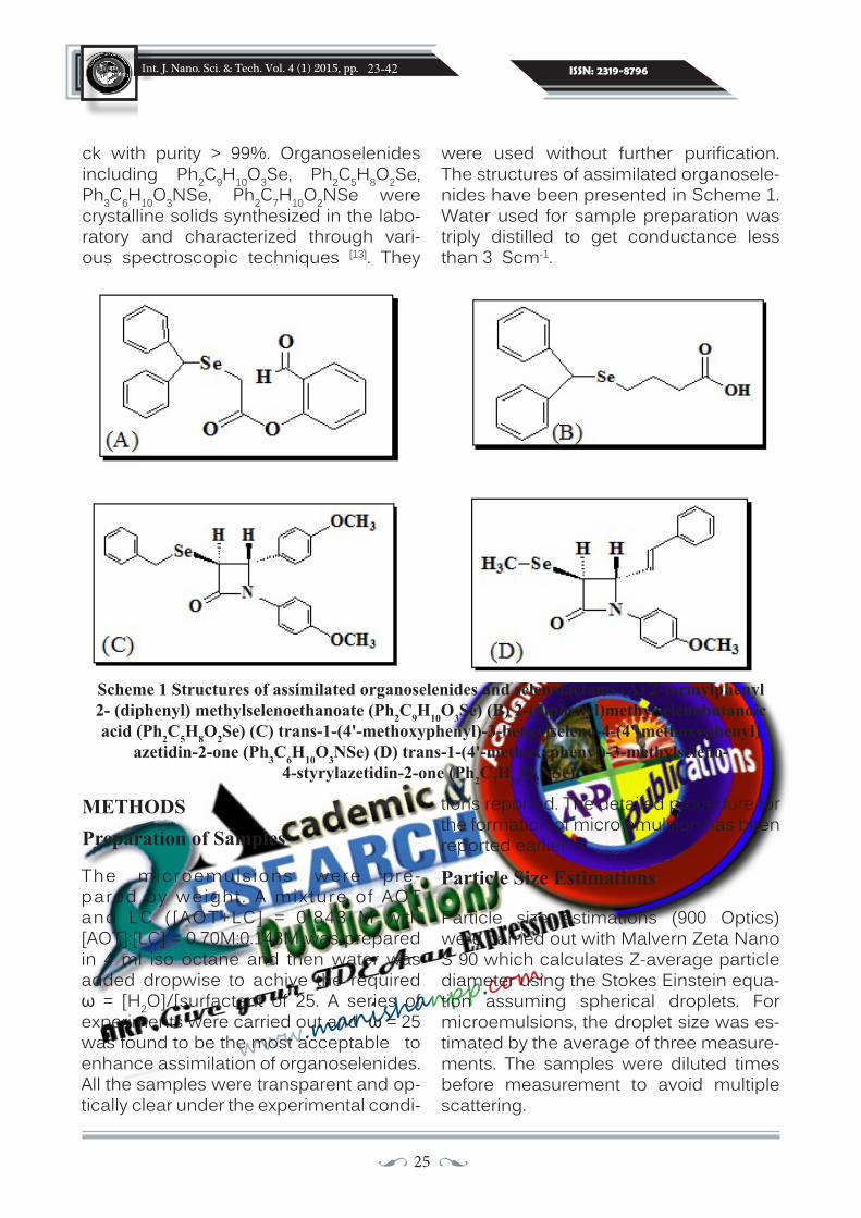
ck with purity > 99%. Organoselenides including Ph2C9H10O3Se, Ph2C5H8O2Se, Ph3C6H10O3NSe, Ph2C7H10O2NSe were crystalline solids synthesized in the labo-ratory and characterized through vari-ous spectroscopic techniques [13]. They
Scheme 1 Structures of assimilated organoselenides and selenolactams (A) 2-formylphenyl 2- (diphenyl) methylselenoethanoate (Ph2C9H10O3Se) (B) 2-(diphenyl)methylselenobutanoic acid (Ph2C5H8O2Se) (C) trans-1-(4'-methoxyphenyl)-3-benzylseleno-4-(4'-methoxyphenyl)
azetidin-2-one (Ph3C6H10O3NSe) (D) trans-1-(4'-methoxyphenyl)-3-methylseleno-4-styrylazetidin-2-one (Ph2C7H10O2NSe).
were used without further purification. The structures of assimilated organosele-nides have been presented in Scheme 1. Water used for sample preparation was triply distilled to get conductance less than 3 Scm-1.
METHODSPreparation of Samples
The microemulsions were pre-pared by weight . A mixture of AOT and LC ([AOT+LC] = 0.843 M with [AOT]:[LC] = 0.70M:0.143M was prepared in 4 ml iso octane and then water was added dropwise to achive the required ω = [H2O]/[surfactant of 25. A series of experiments were carried out and ω = 25 was found to be the most acceptable to enhance assimilation of organoselenides. All the samples were transparent and op-tically clear under the experimental condi-
tions reported. The detailed procedure for the formation of micro emulsion has been reported earlier [7].
Particle Size Estimations
Particle size estimations (900 Optics) were carried out with Malvern Zeta Nano S 90 which calculates Z-average particle diameter using the Stokes Einstein equa-tion assuming spherical droplets. For microemulsions, the droplet size was es-timated by the average of three measure-ments. The samples were diluted times before measurement to avoid multiple scattering.

26 ••
Int. J. Nano. Sci. & Tech. Vol. 4 (1) 2015, pp. ISSN: 2319-8796
www.manish
anpp.com
23-42
Spectroscopic Measurements FTIR spectroscopy
FTIR spectra were recorded in the frequen-cy range of 4400 - 350 cm-1 with the help of Perkin-Elmer (RX1) FTIR spectrometer us-ing AgCl plates. For each sample 50 scans were recorded with the spectral resolution of 2 cm−1. The data was repeated in trip-licate to reduce the error, and one of the data was chosen for detailed study.Conductivity MeasurementsElectrical conductivity measurements of the samples were carried out with PICO digital conductivity meter operating at 50 Hz from Labindia instruments with an absolute accuracy of ±3% and precision of ±0.1%. The cell constant used was 1.0 cm-1. The temperature was kept constant with the help of RE320 Ecoline thermostat with an accuracy of ±0.01K.
For temperature percolation, experimen-tal data were collected at different con-centrations viz., 2.5, 5, 15 and 30 mM for Ph2C9H10O3Se, Ph2C5H8O2Se, Ph3C6H10O3NSe and Ph2C7H10O2NSe in the temperature range of 30 °C to -50 °C. The composition of the microemulsion was kept constant at with [surfactant] = 0.743 and [AOT]:[LC] = 0.70M:0.143M and ω = 25.0. All the samples were prepared by weight and were transparent and optically clear under the conditions of conductivity reported.
RESULTS AND DISCUSSION
Particle size and stability studies
Assessment of the literature [14] depicts that AOT+Lecithin droplets are usually ellipsoi-dal so identifying the spatial size distribu-tion of the system becomes difficult. As a result the projected profiles are no longer uniform and an analytical solution is not possible. So the data obtained is apparent [15]. However, it has been reported by [16]
that on dilution, the interparticular interac-tions are minimized and the droplet struc-ture can pass from a reversed spherical droplet to a reversed rod-shaped droplet, hexagonal phase, lamellar phase, cubic phase and various other structures un-til, after appropriate dilution, a spherical droplet will be formed again. So the parti-cle size measurements of the reverse mi-celle with and without the addition of or-ganoselenides have been carried out with dilution to minimize the scattering effects asuuming spherical droplets. The results have been tabulated in Table 1 with rep-resentative plot of Ph2C9H10O3Se shown in Fig. 1. An increase in the mean diameter of reverse micellar droplet with the addition of organoselenides has been observed. The size follows the order: without addi-tive < Ph3C6H10O3NSe < Ph2C9H10O3Se < Ph2C5H8O2Se < Ph2C7H10O2NSe. The drop-let size observed in the present case is also higher and this is possible if one considers the formation of non spherical structure in the microemulsion with large effective diameters [17,18] The observed PDI values lies in the range of 0.38-0.46. Evaluation of the sample stability after 15 days indicate a slight increase in size and PDI values. This indicates that net attractive forces hydrophobic or vanderwaals are stronger than the repulsive forces and hence the droplets tend to flocculate. However no visible evidence of gravitational sepra-tion or creaming was observed during the storage period. The observed trend is opposite to the conductivity data, where the larger droplet size is considered to fa-vor percolation. This can be explained by considering different mechanisms of the two measured processes. As percolation, apart from depending upon droplet size, depends on the number of other factors such as feasibility of formation of tran-sient tube, effective overlap etc. have to be taken into consideration to explain the process completely.

27 ••
Int. J. Nano. Sci. & Tech. Vol. 4 (1) 2015, pp. ISSN: 2319-8796
www.manish
anpp.com
Table 1. Estimated diameter of the droplets at = 25 effected by the addition of organoselenides/selenolactams = 5mM
23-42
Assessment of the literature [14] depicts that AOT+Lecithin droplets are usually ellipsoidal so identifying the spatial size distribution of the system becomes dif-ficult. As a result the projected profiles are no longer uniform and an analytical solution is not possible. So the data ob-tained is apparent [15]. However, it has been reported by [16] that on dilution, the interparticular interactions are minimized and the droplet structure can pass from a reversed spherical droplet to a reversed rod-shaped droplet, hexagonal phase, lamellar phase, cubic phase and vari-ous other structures until, after appropri-ate dilution, a spherical droplet will be formed again. So the particle size meas-urements of the reverse micelle with and without the addition of organoselenides have been carried out with dilution to minimize the scattering effects asuuming spherical droplets. The results have been tabulated in Table 1 with representative plot of Ph2C9H10O3Se shown in Fig.1. An increase in the mean diameter of re-verse micellar droplet with the addition of organoselenides has been observed. The size follows the order: without ad-
ditive < Ph3C6H10O3NSe < Ph2C9H10O3Se < Ph2C5H8O2Se < Ph2C7H10O2NSe. The droplet size observed in the present case is also higher and this is possible if one considers the formation of non spherical structure in the microemulsion with large effective diameters [17,18]. The observed PDI values lies in the range of 0.38-0.46. Evaluation of the sample stability after 15 days indicate a slight increase in size and PDI values. This indicates that net attrac-tive forces hydrophobic or vanderwaals are stronger than the repulsive forces and hence the droplets tend to floc-culate. However no visible evidence of gravitational sepration or creaming was observed during the storage period. The observed trend is opposite to the con-ductivity data, where the larger droplet size is considered to favor percolation. This can be explained by considering dif-ferent mechanisms of the two measured processes. As percolation, apart from de-pending upon droplet size, depends on the number of other factors such as feasibil-ity of formation of transient tube, effective overlap etc. have to be taken into consid-eration to explain the process completely.
Molar Solubilization Ratio(χ) and micelle/aqueous phase partitioning of organochalcogens (K)

28 ••
Int. J. Nano. Sci. & Tech. Vol. 4 (1) 2015, pp. ISSN: 2319-8796
www.manish
anpp.com
23-42
Solubilization of a substance can be esti-mated using three parameters, the molar solubilization ratio ( χ) and the micelle-wa-ter partition coefficient (K) [19] and oil/water partition coefficient (P). The oil /water par-tition co-efficient was determined by dis-solving 2mg organoselenide/selenolac-tam in isooctane. Water was then added in 1 : 1 ratio(v/v). The mixture was shaken for 10 min and centrifuged for 1 h.The two layers were separated and the content of organoselenide in aqueous layer was assayed by UV–visible spectrophotom-eter. The final content of organoselnide/selenolactam in the lipophilic phase was calculated by subtracting the content of organoselnide/selenolactam in aqueous phase from initial loaded content of or-ganoselnide in lipophilic phase. Further, the effect of presence of surfactant on the partition of organoselenide/selnolactam in oil/water was also studied. The value of partition coefficient in Oil/water system are given in Table 2.
The value is characterized as the amount of solute (organoselenide/selenolactam) that can be solubilized by one mole of micellar surfactant. It illustrates the abil-ity of the surfactant to solubilize the orga-noselenide and is given by
where [St] is the total apparent solubility of the solubilizate, [Scmc] is the apparent solubility of the organoselenide at cmc, which is taken as their water solubility, [S], because it changes only very slightly up to the cmc of the surfactant. Ct is the molar concentration of surfactant in so-
lution and cmc is the critical micelle concentration. is equal to the ratio of organoselenide concentration solubi-lized in micelles to the surfactant con-centration in the micellar form and is obtained from the slope of the curves that results when solubilizate concen-tration is plotted against the surfactant concentration. The variation of solubili-ties for Ph2C9H10O3Se, Ph2C5H8O2Se, Ph3C6H10O3NSe, Ph2C7H10O2NSe are plotted in Fig 2.
The aqueous solubilities of organosele-nides/selenolactams compounds am-plify linearly with surfactant concen-tration, demonstrating their solubility enrichment over that in water. This phe-nomenon is presumably associated to the micellar solubilization. The values from the above plots are tabulated in Table 2 for the added organoselenides and selenolactams respectively. To fur-ther examine the efficiency of solubili-zation, the micelle-water partition coef-ficient, K, of the solubilizate flanked by the micelle and aqueous phases has also been evaluated [20] via. equation 2.
The obtained value of Ph2C5H8O2Se, Ph2C9H10O3Se, are higher than Ph2C7H10O2NSe, Ph3C6H10O3NSe. The low value of Log P observed with the introduc-tion of surfactant indicates the presence of organoselnide at the interface This also indicates that the intrinsic solute proper-ties and its specific interactions with the micellar pseudo-phase contribute to the distribution of organodiselenide between the two pseudophases.
{ }]cmcC[]S[]S[
t
cmct
−−
=χ
{ }]S[
]S[]S[K t −=

29 ••
Int. J. Nano. Sci. & Tech. Vol. 4 (1) 2015, pp. ISSN: 2319-8796
www.manish
anpp.com
23-42
The obtained value of Ph2C5H8O2Se, Ph2C9H10O3Se, are higher than Ph2C7H10O2NSe, Ph3C6H10O3NSe. The low value of Log P observed with the introduction of surfactant indicates the presence of organoselnide at the inter-
Table 2 Molar Solubilization Ratio ( χ) and micelle-water partition coefficient (K) for Ph2C9H10O3Se, Ph2C9H10O3Se, Ph3C6H10O3NSe, Ph2C7H10O2NSe
using AOT+LC as surfactant
face This also indicates that the intrinsic solute properties and its specific inter-actions with the micellar pseudo-phase contribute to the distribution of organ-odiselenide between the two pseudo-phases.
Spectroscopic Measurements
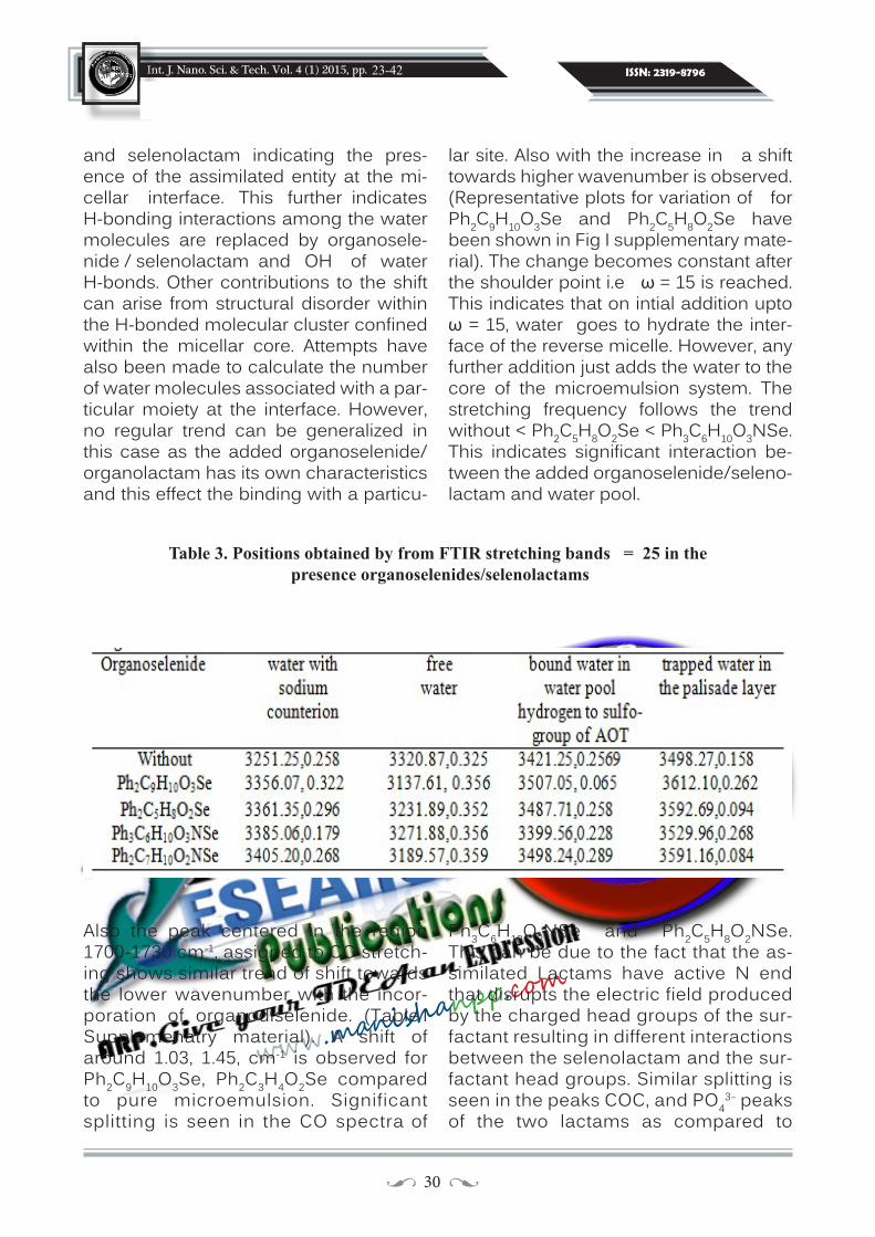
FTIR spectroscopy
In any reverse micellar formulation, the information about the water pool is ob-tained from the OH band [21]. It has already been reported by Mehta et. al., [22] that the water close to interface exhibits behavior markedly different from that of bulk wa-ter as it processes restricted mobility and a depressed freezing point and lacks the normal hydrogen-bonded structure pre-sent in the bulk [23]. As this water is com-posed of four different states, viz., bound water (two types), trapped water, and free water, it is reasonable to assume that the total peak area corresponding to the wa-ter band is the sum of the peak areas of the different states of water [24]. Thus, total peak area is given by
If PF is the fraction of free water corre-sponding to the 3290 ± 20 cm-1 peak, PB is the fraction of bound water correspond-ing to the 3490 ± 20 cm-1 peak, and PT (wa-ter bound to the surfactant head group and counterion) is the fraction of trapped water corresponding to the 3610 ± 10 cm-1
peak, then their respective values can be calculated from the following relations
Figure 3 depicts the changes occurring in OH band by the addition of organosele-nide/selenolactam at ω = 25 and the data has been tabulated in Table 3. De-convulation of the band shows four sub peaks revealing different kinds of water molecules in one band. The peak for free water has been found to show blue shift with the incorporation of organoselenide GTotal= G1+G2+G3 ............ (3)
= = =PFG1
GTOTAL
__ G2 G3__ __GTOTAL GTOTAL
PB PT
30 ••
Int. J. Nano. Sci. & Tech. Vol. 4 (1) 2015, pp. ISSN: 2319-8796
www.manish
anpp.com
23-42
and selenolactam indicating the pres-ence of the assimilated entity at the mi-cellar interface. This further indicates H-bonding interactions among the water molecules are replaced by organosele-nide / selenolactam and OH of water H-bonds. Other contributions to the shift can arise from structural disorder within the H-bonded molecular cluster confined within the micellar core. Attempts have also been made to calculate the number of water molecules associated with a par-ticular moiety at the interface. However, no regular trend can be generalized in this case as the added organoselenide/organolactam has its own characteristics and this effect the binding with a particu-
lar site. Also with the increase in a shift towards higher wavenumber is observed. (Representative plots for variation of for Ph2C9H10O3Se and Ph2C5H8O2Se have been shown in Fig I supplementary mate-rial). The change becomes constant after the shoulder point i.e ω = 15 is reached. This indicates that on intial addition upto ω = 15, water goes to hydrate the inter-face of the reverse micelle. However, any further addition just adds the water to the core of the microemulsion system. The stretching frequency follows the trend without < Ph2C5H8O2Se < Ph3C6H10O3NSe. This indicates significant interaction be-tween the added organoselenide/seleno-lactam and water pool.
Table 3. Positions obtained by from FTIR stretching bands = 25 in the presence organoselenides/selenolactams
Also the peak centered in the region 1700-1730 cm-1, assigned to CO stretch-ing shows similar trend of shift towards the lower wavenumber with the incor-poration of organodiselenide. (Table I Supplemenatry material). A shift of around 1.03, 1.45, cm-1 is observed for Ph2C9H10O3Se, Ph2C3H4O2Se compared to pure microemulsion. Significant splitting is seen in the CO spectra of
Ph3C6H10O3NSe and Ph2C5H8O2NSe. This can be due to the fact that the as-similated Lactams have active N end that disrupts the electric field produced by the charged head groups of the sur-factant resulting in different interactions between the selenolactam and the sur-factant head groups. Similar splitting is seen in the peaks COC, and PO4
3- peaks of the two lactams as compared to

31 ••
Int. J. Nano. Sci. & Tech. Vol. 4 (1) 2015, pp. ISSN: 2319-8796
www.manish
anpp.com
23-42
other compounds. However, no shifts were seen in the spectra of SO3
- reveal the entrapping of organoselenide in the micellar interface. The results further depict the presence of specific interac-tions between the interfacial water mol-ecules and the organoselenide moie-ties. The presence of organoselenides perturbs the H-bonding capabilities of the OH, CO and C-O-C groups of the sur-factant molecules. Significant changes can be seen in CH spectra indicating the presence of the organoselenides in the continuous medium ruling out the presence of interactions of the or-ganoselenide with this part of the AOT molecule.
Conductivity Measurements
The variation conductivity with tem-perature measured at different orga-noselenide concentrations has been presented in Fig. 4. The threshold tem-perature c has been determined from dlog /d vs , where maxima corresponds to percolation threshold. Usual proce-dure reported earlier by the authors has been followed [6-8]. The threshold values of c at different concentrations have been listed in Table 4. The results de-pict that c increase with the increase in concentration of the added orga-noselenide/selenolactam. The value of c for all concentration under study var-ies as Ph2C5H8O2Se ~ Ph2C7H10O2NSe< Ph2C9H10O3Se < Ph3C6H10O3NSe < with-out additive. The Sigmoidal Boltzmann fitting (SBE) procedure reported by Hait et. al., [25] has also been employed to get the percolation threshold. Fitted curves of conductance - have been depicted in Fig. 4.
The efficacy of additive has been cal-
culated using Eq. (5) as reported in (25,26)
Where θc0 and θc
a, C have their usual meaning. Eq. (5) has been used to calcu-late the c data of systems listed in Table 4. The values of the slope (Sk) obtained have also been included in table 4. On the basis of values of Sk, the efficacy of the additive in favoring percolation follows the order: Ph2C7H10O2NSe > Ph2C5H8O2Se > Ph2C9H10O3Se. Species yielding nega-tive slopes are percolation assistants, and solutes yielding positive slopes are percolation retardants [26].
The mutual contact and bridging be-tween droplets is due to the presence of additives either at the interface (e.g., hy-drotopes and bile salts) [27] or in the core of the droplets (e.g., poly (ethylene gly-col) s) [28]. The results depict that as a re-sult of low water solubility the added or-ganoselenide/selenolactam molecules are restricted mainly in the surfactant film. This results in the straight bridging of nanodroplets in case of Ph2C5H8O2Se and Ph2C9H10O3Se and thus the chan-nel formation is favored at higher orga-noselenide concentration wheras at low concentration the bridging is not much efficient. However, selenolactam; Ph3C6H10O3NSe and Ph2C7H10O2NSe contains active N-end, which helps the, the molecule adheres to the droplet inter-face. This configuration can result in the eclipsed bridging of nanodroplets due to cyclization but this favors the percolation process. With the increase in concentra-tion of organoselenide/selenolactam, the intearfacial rigidity increases and this reduces the droplet association therby increasing the percolation threshold.
However, the results are contrary to oth-
θca = θc
0 + SKC

32 ••
Int. J. Nano. Sci. & Tech. Vol. 4 (1) 2015, pp. ISSN: 2319-8796
www.manish
anpp.com
er reports [6,7] published on organosele-nides where the percolation process is delayed. This is because of the steric hindrance offered by C10H6N2Br2Se2 and C10H4N2Br4Se2 [6,7] due to the presence of two and four bromine atoms/orga-noselenide is very high as compared to the phenyl / β Lactam ring in the present case. As a result of which straightforward bridging is observed whereas in the pre-sent case the bridging is somewhat stag-gered. Therefore an efficient intradroplet contact is established which results in material exchange. Similar results have been observed by Moulik et. al., [27] to ex-plain the effect of hydrotopes on droplet percolation.
The activation energy for conductance percolation has been estimated by us-ing Arrhenius equation. The calculated Ep [29,30] values, using do not follow a spe-cific trend. The results are also contrary to the earlier reported results [27], which depicts that percolation assisting de-rivatives should have higher EP values. A plausible explanation for these results may be put forward as argued by Moulik et. al., [27] for the systems AOT/hydrocar-bon/water in the presence of aromatic methoxy hydrotopes. The fission of the
bulky droplets into a pair of singlets with associated transfer of counter ions re-quired high activation energy. The bridg-ing of the droplet with an efficient bind-ing additive may reduce θc but it is the process of separation that decides the magnitude of Ep. The magnitude of Ep is thus essentially controlled by the sepa-rability of the droplets from the “fused” to the “free” state. This is because of dif-ferent structures and physicochemical properties of the added moieties in com-parison to the previous studies. Bulkier organoselenide/selenolactam mole-cules redisperse around the droplets as-sociated with the transport of the Na+ ion. The activated state of mass transfer thus yields higher ∆Ep. The value of ∆Ep fol-lows an intriguing correlation with θc for the respective set of systems. The phe-nomenon of percolation is essentially different from the normal mode of con-duction, and the magnitude of ∆Ep largely depends on the separability of the drop-lets. This is because of different struc-tures and physicochemical properties of the added moieties in comparison to the previous studies. Also in addition to clus-tering, fusion, mass transfer, and fission, there may be other steric and interacting factors for the process of percolation.
Table 4. Percolation threshold and activation energy parameters for temperature induced percolation of water/AOT+LC/Isooctane microemulsion system with varying concentrations
of organoselenide/selenolactam at ω = 25.
Thermodynamics of droplet clustering, association mode
23-42

33 ••
Int. J. Nano. Sci. & Tech. Vol. 4 (1) 2015, pp. ISSN: 2319-8796
www.manish
anpp.com
For percolation to occur droplet cluster-ing is an essential feature. It is governed by various factors such as droplet densi-ty, size and the type of additive. The pro-cess reported by Moulik group [25,27] and Alexandridis et. al., [31] states that the dilu-tion of percolating microemulsion lowers the conductivity rapidly until the clusters dissociate into individual droplets be-low θc. The phenomenon corresponds to the process of demicellization when the surfactant concentration is lowered below the critical micellization con-centration (cmc). The standard free energy, G0
cl and the values of thermody-namic parameters have been calculated using standard equations
where Xd is the mole fraction of the drop-lets corresponding to the percolation threshold. Estimated values of G0
cl for different concentrations for temperature induced percolation are listed in Table 5. The negative value of G0
cl indicates spon-taneous formation of clusters. The results further reveal a more negative value of G0
cl in the presence of organoselenide/selenolactams.
Application of Gibbs - Helmholtz equation
and finally the standard entropy of cluster formation per mole of the droplet, S0
cl is given by the relation
In order to estimate H0cl, G0
cl is plotted as a function of inverse percolation tem-perature and the slope of G0
cl/ θc vs 1/Tc plot yields the value of H0
cl (Fig. 5). The value has been obtained by least squre curve fitting. The obtained positive val-ues of H0
cl and S0cl accounts for a strong
heat-absorbing step in the percolation process. The values of H0
cl and S0cl fol-
low the order: without > Ph3C6H10O3NSe > Ph2C9H10O3Se > Ph2C7H10O2NSe > Ph2C5H8O2Se. The low positive enthalpy change in the presence of selenides than in the absence indicates a relatively less organized i.e. easily disruptable mi-cro surrounding of the droplets of Ph2C5H8O2Se and Ph2C7H10O2NSe where-as it is more organized in Ph2C9H10O3Se and Ph3C6H10O3NSe as compared to the system without organoselenide.
allows calculating the standard enthalpy of cluster formation, H0
cl
∆ G0cl =RT ln Xd .....(6 )
ΔH0cl = ∂ (ΔG0
cl/Tc) / ∂(1/Tc) .....(7)
ΔS0cl = ( ΔH0
cl - ∆G0cl ) /Tc .....(8)
23-42

34 ••
Int. J. Nano. Sci. & Tech. Vol. 4 (1) 2015, pp. ISSN: 2319-8796
www.manish
anpp.com
Table 5. Energetic parameters of droplet clustering for water/AOT+LC/Isooctane microemulsion at ω = 25, Organoselenide = 3 mM and varying S = [oil]/[surfactant]
10 100 10000
2
4
6
8
10
12
Inten
sity(%
)
Size (d.nm)
Fig. 1. Estimated droplet size of Ph2C9H10O3Se.
Figures
23-42

35 ••
Int. J. Nano. Sci. & Tech. Vol. 4 (1) 2015, pp. ISSN: 2319-8796
www.manish
anpp.com
0.1 0.2 0.3 0.4 0.50.00
0.05
0.10
0.15
0.20
0.25
Or
gano
selen
ide/Se
lenola
ctam
(mM)
Ct(mM)
Ph2C10H9O3Se Ph3C6H10O3NSe Ph2C7H10O2NSe Ph2C5H8O2Se
Fig.2. Variation of Solubility of added organoselenides/selenolactams with total surfactant concentration (Ct).
3000 3200 3400 3600
0.4
0.5
0.6
0.7
0.8
0.9
1.0(B)(A)
Abso
rban
ce
wavenumber3000 3200 3400 3600
Fig. 3. Deconvulated FTIR spectra of water/AOT/LC/isoctane at ω = 25 (A) Ph2C9H10O2Se (B) Ph2C3H4O2Se.
23-42

36 ••
Int. J. Nano. Sci. & Tech. Vol. 4 (1) 2015, pp. ISSN: 2319-8796
www.manish
anpp.com
-1
0
1
2
3
4
θ (K)
logσ
2.5mM 5mM 15mM 20mM Without
(B)
2.5mM 10mM 20mM Without
300 305 310 315 320 325-1
0
1
2
3
(C)
2.5mM Without
300 305 310 315 320 325
(D)
(A)
2.5mM 10mM Without
Fig. 4. Variation of specific conductance with temperature for different organoselenides (A) Ph2C9H10O2Se (B) Ph2C5H8O2Se (C) Ph3C6H10O3NSe (D) Ph2C7H10O2NSe
(solid lines show SBE fitting).
3.06 3.09 3.12 3.15 3.18 3.21-84
-82
-80
-78
-76
-74
-72Ph2C7H10O2NSeR=0.989
∆G0 cl/θ c(JK
-1 mol-1 )
103θ-1(K-1)
Fig. 5. oclG∆ / cθ as a function of θ-1 to evaluate o
clH∆ in the presence of Ph2C5H8O2NSe = 5 mM and ω = 25.
23-42

37 ••
Int. J. Nano. Sci. & Tech. Vol. 4 (1) 2015, pp. ISSN: 2319-8796
www.manish
anpp.com
Graphical AbstractA new methodology has been developed for the stabilization of organoselenides/selenolactums by
incorporating them in mixed surfactant microemulsion system.
1 10 1000
2
4
6
8
10
12
Inte
nsity
(%)
size (d.nm)
Table I Positions obtained by from FTIR stretching bands water/AOT+LC/isooctane microemulsion at ω = 30 in the presence organoselenides
23-42

38 ••
Int. J. Nano. Sci. & Tech. Vol. 4 (1) 2015, pp. ISSN: 2319-8796
www.manish
anpp.com
5 10 15 20 25 30 35
3400
3380
3360
3340
3320
3300
Without Ph2C5H8O2Se Ph2C9H10O3Se
Abso
rban
ce
ω=[H2O]/[Surfactant]
Fig. I. FT IR spectra of -OH functionality as a function of water/surfactant molar ratio
23-42
H2O/

39 ••
Int. J. Nano. Sci. & Tech. Vol. 4 (1) 2015, pp. ISSN: 2319-8796
www.manish
anpp.com
REFERENCES
1. A.V. Pietrini, P. L. Luisi (2002), Cir-cular dichroic properties and aver-age dimensions of DNA-containing
CONCLUSIONS
In conclusion, we have detected that the incorporation of organoselenides/seleno-lactams in microemulsions significantly affects the water pool properties and the electric percolation phenomenon. Knowl-edge of the mixed surfactant systems could have many applications in mate-rial synthesis. The expanded range of micellar geometries and functionalities available in mixed surfactant systems not only makes them useful for applications but also provides opportunities for fun-damental studies of self assembly. The rational for using microemulsions as a medium for solubilzation of organosele-nides is to provide alternative route for solubilization of these compounds and thus help in organic synthesis. Conduc-tivity and spectroscopy results indicate the presence of specific interactions be-tween organoselenides, AOT and Lecithin and water molecules and the location of the organoselenide at the interface. FTIR results also indicate the presence of four types of water molecules. The clustering of the microemulsion droplets is shown to be endothermic. The microenvironment of the system in the absence of selenolac-tams is found to be more organized than in their presence.
ACKNOWLEDGEMENT
K.K is thankful to DST for Inspire Fellow-ship and UGC for startup grant.
reverse micellar aggregates, Bio-chimical Biophysica Acta 1562 57 – 62.
2. H. Stamatis, A. Xenakis, F.N. Kolisis (1999), Bioorganic reactions in mi-croemulsions: the case of lipases, Biotechnology Advances 17 293–318.
3. J. H. Yu, Y. X. Weng, X. S. Wang, L. Zhang, B. W. Zhang, Yi. Cao (2003), The triplet excited state changes of amphiphilic porphyrins with dif-ferent side-chain length in AOT re-verse micelles, Journal of Photochem-istry and Photobiology A 156139-144.
4. A. Kalaitzaki, M. Poulopoulou, A. Xenakis, V. Papadimitriou (2014), Surfactant-rich biocompatible mi-croemulsions as effective carriers of methylxanthine drugs, Colloids Surf. A 442 80-87.
5. M. Maestro (1989), Enzymatic activity in reverse micelles-some modellistic considerations on bell-shaped curves, J. Mol. Liq. 42 71-82.
6. S.K. Mehta, K. Kaur, K.K. Bhasin (2010), Modification of interface of water/AOT/isooctane reverses mi-celles by halogenated organodis-elenides, Colloids Surf. A 355 37-43.
7. S.K. Mehta, K. Kaur, Rishu and K.K. Bhasin (2009), Mixed anionic and zwitterionic surfactant based mi-croemulsions as vehicles for solu-bilizing organodiselenides, J. Col-loid Interf. Sci. 336 322–328.
23-42

40 ••
Int. J. Nano. Sci. & Tech. Vol. 4 (1) 2015, pp. ISSN: 2319-8796
www.manish
anpp.com
8. S.K. Mehta, K. Kaur (2010), Spec-troscopic investigations of aromat-ic heterocyclic compounds in re-verse micellar media, Colloids Surf. A 368 37-43.
9. I. Brovchenko, A. Krukau, A. Oleinik-ova, A. K. Mazur (2007), Water Clus-tering and Percolation in Low Hy-dration DNA Shells, J. Phys. Chem. 111 3258-3266.
10. S.S. Bari, A. Bhalla (2010), Spiro-cyclic β-Lactams: Synthesis and Biological Evaluation of Novel Het-erocycles, Top Heterocycl Chem. 22 49-99.
11. M. Hager, K. Holmberg (2000), A substitution reaction in an oil-in-water microemulsion catalyzed by a phase transfer catalyst, Tetrahe-dron Lett. 41 1245-1248.
12. K. Holmberg (2003), Organic reac-tions in microemulsions. Curr. Opin. Colloid Interface Sci., 8 187–196.
13. S.S. Bari, A. Bhalla, Y. Nagpal, S.K.Mehta, K.K. Bhasin (2010), Syn-thesis and characterization of nov-el trans-3-benzyl/(diphenyl)me-thyl/naphthyl seleno substituted monocyclic β-lactams: X-ray struc-ture of trans-1-(4′-methoxyphenyl)-3-(d iphenyl )methylseleno-4-(4 ′ -methoxypheny l )azet id in -2 -one , Journal of Organometallic Chemistry 695 1979-1980.
14. S.S. Narayanan, S. S.Sinha, R. Sarkar, S. K. Pal (2008), Picosec-ond to nanosecond reorganization of water in AOT/Lecithin mixed reverse micelles of different mor-phology, Chemical Physics Letters, 452 99-104.
15. A. Thyabat, N. J. Miles (2006), An improved estimation of size distri-bution from particle profile meas-urements, Powder Technology 166 52–160.
16. I. Capek (2001), Microemulsion po-lymerization of styrene in The pres-ence of a cationic emulsifier, Adv. Colloid Interf. Sci. 92 195-233.
17. V. Papadimitriou, S. Pispas, S. Syriou, A. Pournara, M. Zoump-anioti (2008), T.G. Sotiroudis, A.K.Schurtenberger, C. Cavaco, Biocompatible Microemulsions Based on Limonene: Formulation, Structure, and Applications, Lang-muir 24 3380-3386.
18. P. Schurtenberger, C. Cavaco (1994), Polymer-like lecithin re-verse micelles. 1. A light scattering study, Langmuir 10 100-108.
19. A. A. Dar, G. M. Rather, A. R. Das (2007), Mixed Micelle Formation and Solubilization Behavior toward Polycyclic Aromatic Hydrocarbons of Binary and Ternary Cationic-Nonionic Surfactant Mixtures, J. Phys Chem. B 111 3122.
23-42

41 ••
Int. J. Nano. Sci. & Tech. Vol. 4 (1) 2015, pp. ISSN: 2319-8796
www.manish
anpp.com
20. S. K. Mehta, K. K.Kaur, K. K Bhasin (2010), Modification of interface of water/AOT/isooctane reverse mi-celles by halogenated organodis-elenides, Colloids Surf. A, 355 37-44.
21. G. Cavallaro, G. La Manna, V.T. Liveri, F. Aliotta, M. E. Fontanella (1995), Structural Investigation of Water/Lecithin/Cyclohexane Mi-croemulsions by FT-IR Spectrosco-py, J. Colloid Interf. Sci. 176 281–285.
22. K. Kaur, N. Bhatia, S.K. Mehta (2012), Formation of cyclodextrin-stabilized nanoemulsions and mi-croemulsions and exploitation of their solubilization behavior, RSC Advances 2 8467.
23. E. Caponetti, D. C.Martino, F. Fer-rante, L. Pedone, A. Ruggirello, V.T. Liveri (2003), Structure of Urea clusters confined in AOT reverse micelles, Langmuir 19 4913–4922.
24. G.W. Zhou, G.Z. Li, W. J. Chen (2002), Fourier Transform Infrared Investigation on Water States and the Conformations of Aerosol-OT in Reverse Microemulsions, Lang-muir, 18 4566-4571.
25. S.K. Hait, A. Sanyal, S. P. Moulik (2002), Physicochemical Studies on Microemulsions. 8. The Effects of Aromatic Methoxy Hydrotropes on Droplet Clustering and Under-standing of the Dynamics of Con-ductance Percolation in Water/Oil
Microemulsion Systems, J. Phys. Chem. B 106 12642-126450.
26. S. K.Mehta, S. Sharma, K.K. Bhasin (2005), On the Temperature Per-colation in a w/o Microemulsion in the Presence of Organic Deriva-tives of Chalcogens, J. Phys. Chem. B,109 9751-9759.
27. S.K. Hait, S.P.Moulik, M. P.Rodgers, S. E.Burke, R. Palepu (2001), Phys-icochemical Studies on Microe-mulsions. 7. Dynamics of Percola-tion and Energetics of Clustering in Water/AOT/Isooctane and Water/AOT/Decane w/o Microemulsions in Presence of Hydrotopes (Sodium Salicylate, α-Naphthol, β-Naphthol, Resorcinol, Catechol, Hydroqui-none, Pyrogallol and Urea) and Bile Salt (Sodium Cholate), J. Phys. Chem. B 105 7145-7154.
28. W. Meier (1996), Poly(oxyethylene) Adsorption in Water/Oil Microe-mulsions: A Conductivity Study, Lang-muir 12 1188-1192.
29. I. Chakraborty, S. P. Moulik (2005), Physicochemical studies on mi-croemulsions 9. Conductance percolation of AOT-derived W/O microemulsion with aliphatic and aromatic hydrocarbon oils, J. Col-loid Interface Sci. 289 530–540.
30. B.K. Paul, and R.K. Mitra (1998), Conductivity of reverse micellar systems of water/AOT + Brij-56 or
23-42

42 ••
Int. J. Nano. Sci. & Tech. Vol. 4 (1) 2015, pp. ISSN: 2319-8796
www.manish
anpp.com
••••••• ••••••••
Bri j -58/IPM and their percola-t ion under varied concentra-t ions of amphiphi les and di f fer-ent addit ives, Colloids Surf . A 273 129-140.
31. P. Alexandridis, J. F. Holzwarth, T.A. Hatton (1995), Thermodynam-ics of droplet clustering in perco-lating AOT water in oil microemul-sions, J. Phys. Chem. A 99 8222-8232.
23-42

43 ••
Int. J. Nano. Sci. & Tech. Vol. 4 (1) 2015, pp. ISSN: 2319-8796
www.manish
anpp.com
LETTER TO THE EDITOR
Let's Talk Nanotechnology : Some Applications and Scope For Future Research
Dr. Kavyanjali Shukla
Flat E, Floor 5, Tower 13, South horizons, Ap lei chau, HongKong.
Dr. Kavyanjali Shukla holds a PhD. in Zoology. She is an author, researcher, an avid science communicator and an environmentalist who has a keen interest in
volunteering for various social causes. She is also working as Coordinating Edi-tor, International Journal of Nanoscience and Technology (IJNST)and Geo-
sciences (IJGST), Ʌcademic & Ɍesearch Ƥublications house, India.
Šeptember, 1, 2014, I got the opportunity to deliver a science talk among the young and enthusiastic students of Christ University, Bangalore. The topic of dis-cussion was " The fundamentals and applications of Nanoscience and technology- an overview; subtopic : possible application of nanotechnologies in amoebic keratitis. " I started off as how we used to study science during the early, historical times to what and how we study it today. How the methods of studying science have really evolved, with each revolution-ary discovery and how we have used our past knowledge to decipher new techniques for further studies. Nanoscience is a the study of controlling matter at an atomic and molecular scale. Generally nanotechnology deals with structures sized between 1 to 100 nanometer and involves developing or modifying materials or devices within that size. Prefix ‘nano’ is derived from the Greek word meaning dwarf. One nanometer (nm) is equal to one-billionth of a meter, 10-9 m. By definition of Hilary Thomson, 2008, “Na-notechnology has been called a molecular revolution—innovation so profound it will allow us to rebuild our world molecule by molecule. The unprecedented benefits of such control over matter have the potential to permeate every aspect of our lives. But so do the risks.” The term ‘nanotechnol-ogy’ was not used until 1974, when Norio Taniguchi, a researcher at the University of Tokyo, Japan used it to refer to the ability to engineer materials precisely at the nano-metre level. We often use two terms, Nanoscience and nanotechnology, here, we may understand nanoscience as the study of phenomena and manipulation of materials at atomic, molecular and macromolecular scales, where properties differ significantly from those at a larger scale, while, Nanotechnology is more of a phenomenon that enables the designing, production of structures, devices and systems that have novel applications. This is done by controlling the size and shape of the material at the na-nometer (10-9 m of a meter) scale technology.
It is now, that we have to understand that nanotechnology might be our very near future. In order to understand it, we need to be first acquainted by it. What is nano science? Why we have to know about it ? What benefits do we derive from studying
43-49

44 ••
Int. J. Nano. Sci. & Tech. Vol. 4 (1) 2015, pp. ISSN: 2319-8796
www.manish
anpp.com
it, these all queries must be flashing in our minds and these will be dealt in details in order to clear our doubts. But, let us first understand the objectives of the present discussion. As of now, I have found two objectives that I discussed among the under-graduate students of Christ university. These are as follows:
• To realize that nanotechnology is our future and so to discuss its developments and applications and
• To spread awareness regarding the need of knowledge upgradation for future ca-reer prospects and career goals.
Let's acknowledge the fact that nanotechnology or nanoscience have always existed in nature. It is only that now we are discovering it and inventing novel things by deriv-ing inspiration from these living beings that are a result of a phenomenon called adap-tation and being favored by nature.
One such example is of a lotus flower. The leaves of lotus flower, Nymphaea sp. is wa-terproof. The reason behind this waterproof quality is the presence of very small (nano projections) throughout the surface. So, when water falls on a leaf surface, the droplets simply sits on these enormously small projections and then rolls off through the entire surface taking along with it, all the dirt as well. Now, this same technique have been borrowed from nature and some special nano fibres have been developed for cloth making, that have the properties of being waterproof and dirt resistant. Such a cloth is now utilized for making garments such as shirts, which are now also, readily available in the market. Such garments are water and dirt resistant.
Nanotechnology exists in nature since time immemorial
Lotus petals and leavesare water proof because ofnanostructures
Electron microscopicview of lotus leaves
How a waterdroplet rolls on aleaf, taking of allthe dirt
The same principle isnow utilized formaking of nano fibresfor cloth making, suchclothes will bewaterproof and will notget dirty by sweat anddirt……..its ALREADYIN THE MARKET
A SLIDE FROM MY POWERPOINT PRESENTATION AT CHRIST UNIVERSITY
43-49

45 ••
Int. J. Nano. Sci. & Tech. Vol. 4 (1) 2015, pp. ISSN: 2319-8796
www.manish
anpp.com
Another, such example is that of a water striders, that have remarkable non-wetting legs that enable them to stand and move quickly on water. Each leg is covered with large numbers of angled tiny hairs (microsetae) with tiny nano-grooves. Air is trapped in spaces in the microsetae and nanogrooves to form a cushion where the leg touches the water. This prevents the leg from getting wet. So, now, we may understand how these naturally occurring nanostruc-tures have actually inspired the modern day scientists for novel inventions. Besides, there are many such examples of nanostructure and their utilities that are found in nature. The exterior of the toucan beak that is made up of overlapping nanosized tiles of keratin, the same protein that makes up hair, fingernails, and horn. The interior of the beak is a rigid foam made of a network of nanosized bony fibres connected by membranes. This allows the beak to absorb high-energy impacts. (http://pubs.acs.org/cen/news/83/i50/8350toucan.html). Beetles and flies also have nanostructures that help them stick to walls, ceil-ings and what appear to be smooth surfaces. 500,000 hairs per toe. Hundreds of nanoprojections (spatulae) per hair. Adhesive force in one foot = 100 new-tons.
Then, there have been some extremely revolutionary inventions in the past dec-ade or so, that we need to be acquainted with and much of the research work is still being carried off. Some of these researches includes, Nanocompos-ites used in making paints, such paints are scratch-resistant, light-weight, and rust-proof, and generates improvements in strength and reductions in weight, which lead to fuel savings and increased longevity. And in 2001, Toyota started using nanocomposites in a bumper that makes it 60% lighter and twice as resistant to denting and scratching Stain-repellent Eddie Bauer Nano-areTM , with surface fibers of 10 to 100 nanometers, uses a process that coats each fiber of fabric with "nano-whiskers." Developed by Nano-Tex. Then, there are, sunscreens are utilizing nano particles that are extremely effective at absorb-ing UV light, Due to the particle size, they spread more easily, cover better, and save money since you use less. They are transparent, unlike traditional screens which are white. These sunscreens are so successful that by 2001 they had captured 60% of the Australian sunscreen market.
One of the most desirable application of nanotechnology that is being explored is in food packaging. "food", being, one of the most important commodity for survival of living beings. Various methods of nano packaging are contributing towards food protection and extension of its shelf life. As we all know that, food Packaging is meant for the protection of food from harmful substances and extending the shelf life. Key characteristics in nano packaging materials are enhanced external, mechanical and thermal properties, protection from any chemical and microbiological effects, additional safety and functionality. Be-sides, these, the products should be Inert, Cheap, Lightweight, Easy to dispose or reuse, Impervious to environmental storage and transport conditions, resist-ant to physical abuse. Few examples of such inventions include, Waxy coat-ings used in fruits, vegetables, meat, to increase shelf life, Sonotec, U.S., has developed an anti bacterial coating for such foods, Nanocrystals embedded
43-49

46 ••
Int. J. Nano. Sci. & Tech. Vol. 4 (1) 2015, pp. ISSN: 2319-8796
www.manish
anpp.com
in plastic beer bottles creates a molecular barrier that helps preventing oxygen to escape from the bottles giving the brew a 6 month shelf life. Nanosensor packaging done to incorporate RFID (radio frequency identification) tags for anti-counterfieting processes. Nansulates used to coat dairy processing tanks and pipes to protect them from corrossion and insulate against heat loss. Use of carbon nanotubes as a sensor to detect toxic substances and spoilage of food, is developed by Georgia tech, U.S. Developing bioswitch for “release on command” preservative packaging. This is intelligent packaging that will re-lease preservative if the food begins to spoil. Silver zeolites used to create anti bacterial polymer composites that offer added stability and slow release of silver ion into stored foods sustains the antimicrobial activity. Chitosan films loaded with AGNPs have better tensile and gas barrier properties.
Nanotech will involve Interlocking ideas, applied knowledge and mechanisms from different fields to fill in the knowledge and data gaps. besides this infor-mation I also discussed how these novel nanotechnological advancements could favor ocular (eye) researches and be beneficial for ocular diseases such as amoebic keratitis, that is also one of my research area. countries pioneer-ing in nanotechnological researches, with China pioneering most of the utility based research projects.
Delivering such a talk was a great experience, with students sharing their keen enthusiasm and putting up queries to me. Would like to thank the academic staff of Christ University for making the arrangements. Wish to deliver my next talk again, seeing the interest of the students and university staff, whenever I, next visit India and find time to share some more information, in a more inter-active way .
Besides delivering my own talk, I also got the opportunity to attend two, ex-tremely informative lectures at Hong Kong. To meet two such distinguished scientists named Dr. Anella Seddon and other a named, Dr. Jamie Gallengar.
Dr. Anella Seddon, is presently working with the University of Bristol and I got to meet her through British council, who organized her lecture at Hong Kong, at their office. It was a great opportunity to attend her lecture, meet her in person and discuss about nanotechnology and its upcoming developments. Though her interface with the audience was very interactive, I immensely liked the powerpoint preparation of her work and the information displayed. She not only explained the phenomenon in a simple language, but also cited some practical examples as well experiments involving the audience. that served to be an easy way of learning as well as memorizing. She also discussed about the new products that are reaching the market these days and how are they beneficial. This included a whole new range of cosmetics, sunscreens, sport related items, such as tennis rackets, shirts. etc. She also focused her talk on long lived problem of water purification and making it fit for drinking at a large scale. How graphene will act like a miracle material, I came to know through
43-49

47 ••
Int. J. Nano. Sci. & Tech. Vol. 4 (1) 2015, pp. ISSN: 2319-8796
www.manish
anpp.com
this lecture only. She also pointed some energy related needs of today's world and provided an answer to them, for example, she told that if we could gather/ harvest all the sunlight reaching the earth for one hour, we will be able to pro-vide earth with one full year of energy requirement, so nanotechnology will be helpful in meeting energy demands, but, we still don't know how to do it!! but we may certainly work in the direction to find answers to these questions.
She also touched the areas of drug delivery, where she told us a way how nanobox containing a chemotherapy drug inside will be working only on the cancerous cells leaving the healthy ones, undamaged. these nanoboxes will be "locked" from outside and will be only recognized by cancerous cells.
She also discussed how Stanford university is developing self healing skin. After discussing these many ideas, I asked her couple of questions regarding water purification, with special reference to ballast water purification. To which she replied how researchers are being carried out these days to combat such a problem.
The other such interactive lecture was of Dr. Jamie Gallenger. He is a multi-talented person. He is a science communicator, a scientist, a dancer and also works as an Public engagement officer for university of Glasgow. He is an enthusiastic person and very happy to meet new people. He did his PhD. in nano, thermoelectronics. in his own words " they are clever little devices; they can take waste heat and convert it directly to useful electricity imagine, just by walking around the waste heat, that could be harvested and used to electrical devices, that we carry ! I try and make them work a little bit better than they already do."
It was an academically gratifying experience, meeting these two wonderful personalities, both of them working in the areas of nanotechnology. I found it to be worthy of sharing with the readers of this journal and the editor. Such kind of talks not only upgrade our knowledge but also open up our brain's windows, through which we acquire and learn and discover new pathways that lead to new discoveries. I also found that how the scientist are working in the areas of food, environmental science, biological science and medicine, incorporating nanotechnology and trying to find solutions to the problems. We have a good and promising scope for nano science research in present time as well as future. I look forward to meeting these people again and also to attend more of such informative talks and also delivering more talks, ex-changing ideas in various waysthanking you,
dated: 19th May, 2015 Yours sincerely, Dr. Ϗavyanjali Shukla
43-49

48 ••
Int. J. Nano. Sci. & Tech. Vol. 4 (1) 2015, pp. ISSN: 2319-8796
www.manish
anpp.com
Dr. Kavyanjali SHUKLA with Dr. Jamie Gallenger at HK science museum
Dr. Anella Seddon, University of Bristol, delivering her lecture at the British council,Hong Kong.
43-49
49 ••
Int. J. Nano. Sci. & Tech. Vol. 4 (1) 2015, pp. ISSN: 2319-8796
www.manish
anpp.com
Dr. Kavyanjali Shukla with some of the student listeners
43-49

© Journal on Nanoscience and Technology. All rights reserved. No portion of material can be reproduced in part or full without the prior permission of the Editor.
w w w . m a n i s h a n p p . c o m
w w w . m a n i s h a n p p . c o m

© Journal on Nanoscience and Technology. All rights reserved. No portion of material can be reproduced in part or full without the prior permission of the Editor.
Prof. T.R.C Sinha a true lover of science and always concerned about the environment. It was his dream to publish scientific journals and he started this project with the same enthusiasm as he had done for eve-ry project he undertook.We are guided by the vision of Prof. Sinha and endeavour to make his dream of promoting science and help young scientists, publish their articles and encourage them in their research.
Manisha Verma

52 ••
Int. J. Nano. Sci. & Tech. Vol. 4 (1) 2015, pp. ISSN: 2319-8796
www.manish
anpp.com
International Journal of
Nanoscience & Technology(Bi-Annual Journal)
2015
Name:
SUBSCRIPTION ORDER FORM
Address:
Pin:
Tel: Fax:
E-mail:
Signature Date
New Renewal
MODE OF PAYMENTPayment should be made online. The amount is to be credit in the Bank The details of the bank are as follows:-
Name of the Account holder : Academic And Research PublicationsName of the bank : PUNJAB NATIONAL BANK, VAISHALI BRANCH, GHAZIABAD, IFSC: PUNB0405300 Current Account No. 4053002100011106
Published by
Academic And Research PublicationsH.Office: EC 41, Maya Enclave, New Delhi -110064

53 ••
Int. J. Nano. Sci. & Tech. Vol. 4 (1) 2015, pp. ISSN: 2319-8796
www.manish
anpp.com
SUBSCRIPTION RATES
INDIVIDUAL COPY for CO-AUTHORS Only
Inland ForeignRs. 500/- US$ 40ANNUAL SUBSCRIPTION
(2 Issues Per Year)
Individuals ForeignRs. 800/- US$ 80
Institutions ForeignRs.1500/- US$ 100
ADVERTISEMENT RATES
Full Page
Colour : Rs. 35,000.00Black/White : Rs. 25,000.00
Half Page
Colour : Rs. 25,000.00Black/White : Rs. 15,000.00
(Four weeks to be allowed aftersubmission of advertising material)
ARP ,

54 ••
Int. J. Nano. Sci. & Tech. Vol. 4 (1) 2015, pp. ISSN: 2319-8796
www.manish
anpp.com
This journal is a collaborative effort which aims to increase
awareness and understanding of scientific research, This is
a peer reviewed biannual journal which gives a
platform to fresh ideas and past experience about
developements in India and outside. The journal has Open
Access to its content and makes knowledge available to the
public in general and research scholars in specific. Through
this journal we welcome greater global exchange of
knowledge, ideas among intellectuals, researchers and
scholars. The journal offers an open forum for discussion.
I will like to appreciate the efforts of all authors, co-authors,
reviewers and member of Editorial Board who have
contributed immencely towards this journal.
Manisha Verma
Dear Reader,
55 ••
Int. J. Nano. Sci. & Tech. Vol. 4 (1) 2015, pp. ISSN: 2319-8796
www.manish
anpp.com
PUBLICATION ETHICS: Submission of a manuscript implies:
that the work described has not been published before
(except in the form of an abstract or as part of a pub-
lished lecture, or thesis) that it is not under consideration
for publication elsewhere; that if and when the manu-
script is accepted for publication, the authors agree to
automatic transfer of the copyright to the publisher.
The International Journal of scientific research is com-
mitted to uphold the highest standards of publication
ethics. All authors submitting their works to the IJS&T for
publication as original articles attest that the submitted
works represent their authors' contributions and have
not been copied or plagiarized in whole or in part from
other works. The authors acknowledge that they have
disclosed all and any actual or potential conflicts of in-
terest with their work or partial benefits associated with
it.
Multiple submissions: It is unethical to submit the same
manuscript to more than one journal at the same time.
Doing this wastes the time of editors and peer reviewers,
and can damage the reputation of journals if published
in more than one.

56 ••
Int. J. Nano. Sci. & Tech. Vol. 4 (1) 2015, pp. ISSN: 2319-8796
www.manish
anpp.com

57 ••
Int. J. Nano. Sci. & Tech. Vol. 4 (1) 2015, pp. ISSN: 2319-8796
www.manish
anpp.com

58 ••
Int. J. Nano. Sci. & Tech. Vol. 4 (1) 2015, pp. ISSN: 2319-8796
www.manish
anpp.com

59 ••
Int. J. Nano. Sci. & Tech. Vol. 4 (1) 2015, pp. ISSN: 2319-8796
www.manish
anpp.com

60 ••
Int. J. Nano. Sci. & Tech. Vol. 4 (1) 2015, pp. ISSN: 2319-8796
www.manish
anpp.com

61 ••
Int. J. Nano. Sci. & Tech. Vol. 4 (1) 2015, pp. ISSN: 2319-8796
www.manish
anpp.com

62 ••
Int. J. Nano. Sci. & Tech. Vol. 4 (1) 2015, pp. ISSN: 2319-8796
www.manish
anpp.com
Notes

63 ••
Int. J. Nano. Sci. & Tech. Vol. 4 (1) 2015, pp. ISSN: 2319-8796
www.manish
anpp.com
Notes

64 ••
Int. J. Nano. Sci. & Tech. Vol. 4 (1) 2015, pp. ISSN: 2319-8796
www.manish
anpp.com
The articles are not printed in gratis. A nominal charge@of Rs 500=00 per printed page is charged and is printed in B/W colour. For any coloured page, is separately charged. Charges for the same is quoted on request. Five Reprints and a copy of Journal is provided to the Corresponding author. Extra copies of the Journal for coauthor only are supplied on charge @ Rs 400-00 per copy and shall be sent along with the copy of the Correspondent Author.
PRINTING CHARGE
For quick reply, please note the address and contact them directly by Post or email:-a) For publication of your article, Acceptance letter, Invoice, sending of Cheques/Drafts, for sending Review Reports ,Status Report about your article , and all other queries related to your articles, should be sent directly to the Editor-in-Chief , whose address is as follows:
CONTACT US
REMITTANCE
REQUESTAuthors are requested to keep a copy of the Mss. till it is published in the Journal.
The Research Article should be in the following order
Guidelines to AuthorsInternational Journal of Nanoscience & Technology
Benefit to Authors
Submission of Article(s)All manuscripts are to be submitted in English , typed double-spaced throughout the text It is requested that manuscripts be sent by e-mail only. Authors will be requested to substantiate the need if it exceeds the maximum number of pages.All manuscript should be submitted to the Editor-in-Chief directly.
Following benefits shall be available to the authors, if your article is published in the journals of this PUBLISHER.Authors can use their published articles in any way they like for the dissipation of knowledge free of charge. However it can not be used for any commercial use without prior permission. Authors can have access on all articles published by this Publisher. Wide publicity and reach by getting indexed in many free indexing services.
This journal is published by the Academic And Research Publications(ARP), New Delhi twice in a year. The emphasis is to involve a large community of scientists specially JUNIOR SCIENTISTS and scholars from India and abroad in de-veloping a framework of discussion and debate on conservation and sustainable development. Research articles , Review articles Book reviews, Interviews, Short communication , Letters to the Editor ,Case reports and News items related to the subject are accepted for publication. The PUBLISHER is aiming to publish your article as rapidly as possible pro-vide a rapid publishing platform, each and every point is vital to reduce the editorial work flow. Authors are requested to extend their maximum cooperation for speedy actions to be taken at this end. Hence they are requested to check the guidelines thoroughly before submitting their article(s)and check that all the required informations are mentioned and strictly according to the guide lines.
In the 1st Part:1) Title 2)Name of the organisation where work was carried out 3)Name of the Author with whom correspondence is to be made along with his/her Mob.No. 4)Date of Submission 5)No. of Tables 6)No .of Photographs 7)Numbers of Graphs 8)No. of References 9)Name and addresses of at least Three referees along with their Email I/Ds and Mob Nos.In the 2nd Part :1) Title of The Article 2) Name and addresses of all the Author(s) along with their Email I/Ds and Mob.Nos. 3) Abstract 4) Introduction 5) Materials and methods 6) Results 7) Discussions (if any) 8) Acknowledgements 10) References (Under References heading, Name of the Journals should be in italic letters) and (11) Name of The journal.Tablesto be included should have a heading, giving the substance, and should be typed double-spaced on separate sheets. They should also be numbered in serial order. Figures either drawn manually or by computer should be in black ink and the lettering on them should be large enough to stand reduction. Photographs in colour should have sharp contrast. Legends for figures and plates should be typed in numerical order on separate sheets, one for figures and one for plates.References
A. For articles in a Journal:-Walsh, J.E. (2008) Climate of the Arctic Marine Environment. Ecological Applications. 18. pp. 3-22.B. For Books:-Ward, D.R. (2002) Water Wars: drought, floods, folly and politics of thirst: River head Books. New York. p. 12.C. Chapter in a book:-Andrews, T.J., Clo ugh, B.F. and Muller, G.J. (1984). Photosynthetic gas exchange properties and carbon isotope ratios of some mangroves in North Queensland. In: H.J. Teas (Ed.), Physiology and Management of Mangroves. W. Junk. The Hague. pp. 15-23. From website:-National Oceans and Atmospheric Administration (NOAA). 1995. Regional Perspectives: Indian Ocean. www.ncdc.noaa.gov /paleo.outreach /coral/sor/sor_indian.html , accessed on July 13, 2008.While giving reference of more than two authors in the text, after, the name of the first author, et.al., should be used, followed by the year of publication.
The literature cited should list the author’s name, year of publication, title of the paper, and the Journal titles(italic let-ters) which should be cited in full (no abbreviation) with volume number and page numbers, as indicated below:
All remittances are to be on line credited in the account of Academic And Research Publications in the PUNJAB NATIONAL BANK, VAISHALI BRANCH, GHAZIABAD, IFSC: PUNB0405300 Current Account No. 4053002100011106 and inform the at [email protected] accordingly.
Prof. Manik Sinha-THE EDITOR-IN-CHIEF, H.Office: EC 41, Maya Enclave, New Delhi -110064. Email:[email protected], [email protected]. www.manishanpp.com.
An International Refereed Journal

Volume No. 4, Issue No. 1, 2015
C o n t e n t sReviewed Articles
May, 2015
ACADEMIC & RESEARCH PUBLICATIONSAn International Refereed Journal ACADEMIC & RESEARCH PUBLICATIONS
H.Office: EC 41, Maya Enclave, New Delhi -110064
Email :[email protected] , [email protected]
www.manishanpp.com
ARP
ARPJoin us on facebook
1. Synthesis and Characterization of Magnetic Cobalt Nanoparticles 01 using Pluronic P123 As Surfactant at Room Temperature
Prerna Sen and Badal Kumar Mandal
2. Colorimetric Detection of Hg+2 Using 07 Diastase Capped Silver Nanoparticles
Sireesh babu Maddinedi, Badal Kumar Mandal and Kiran Kumar Anna
3. Size Controlled Synthesis of Silver 16 Nanoparticles - a review Sireesh babu Maddinedi, Badal Kumar Mandal and Kiran Kumar Anna
4. Incorporation of Organoselenides and Selenolactams in Microemulsions: 23 Effect On Stability, Percolation and Size Khushwinder Kaur
5. Let's Talk Nanotechnology : 43 Some Applications and Scope For Future Research
Dr. Kavyanjali Shukla