Embed Size (px)
Citation preview
Hindawi Publishing CorporationNeural PlasticityVolume 2013, Article ID 306432, 11 pageshttp://dx.doi.org/10.1155/2013/306432
Clinical StudyMaking Memories: The Development of Long-Term VisualKnowledge in Children with Visual Agnosia
Tiziana Metitieri,1 Carmen Barba,1 Simona Pellacani,2
Maria Pia Viggiano,1,3 and Renzo Guerrini1,3
1 Pediatric Neurology Unit, Children’s Hospital A. Meyer, University of Florence, Viale Pieraccini 24, 50139 Firenze, Italy2 IRCCS Stella Maris, Viale del Tirreno 331, Calambrone, 56018 Pisa, Italy3 Department of Neuroscience, Psychology, Pharmacology and Child Health, University of Florence, Viale Pieraccini 6,50139 Firenze, Italy
Correspondence should be addressed to Tiziana Metitieri; [email protected]
Received 4 May 2013; Accepted 11 September 2013
Academic Editor: Małgorzata Kossut
Copyright © 2013 Tiziana Metitieri et al. This is an open access article distributed under the Creative Commons AttributionLicense, which permits unrestricted use, distribution, and reproduction in any medium, provided the original work is properlycited.
There are few reports about the effects of perinatal acquired brain lesions on the development of visual perception. These studiesdemonstrate nonseverely impaired visual-spatial abilities and preserved visual memory. Longitudinal data analyzing the effects ofcompromised perceptions on long-term visual knowledge in agnosics are limited to lesions having occurred in adulthood.The studyof children with focal lesions of the visual pathways provides a unique opportunity to assess the development of visual memorywhen perceptual input is degraded. We assessed visual recognition and visual memory in three children with lesions to the visualcortex having occurred in early infancy. We then explored the time course of visual memory impairment in two of them at 2 yearsand 3.7 years from the initial assessment. All children exhibited apperceptive visual agnosia and visual memory impairment. Weobserved a longitudinal improvement of visual memory modulated by the structural properties of objects. Our findings indicatethat processing of degraded perceptions frombirth results in impoverishedmemories.The dynamic interaction between perceptionand memory during development might modulate the long-term construction of visual representations, resulting in less severeimpairment.
1. Introduction
Visual agnosia is a modality-specific disorder of objectrecognition caused by a lesion involving the visual cortex [1].As originally described by Lissauer [2] the disorder cannotbe attributed to poor sensory processing, and recognition ofobjects through other modalities can be relatively preserved.Most of our understanding of visual agnosia derives frominvestigations of adults with visual agnosia acquired afteryears of normal functioning [3]. Previous findings suggestthat long-termmemory and recognition can be concurrentlyor differentially impaired [1]. In the majority of cases ofagnosia both memory and perception are clearly impaired[4], though, in some, the representations stored in memoryare preserved even for objects that cannot be recognized[5–7]. However, data from agnosics exploring the dynamic
influence of degraded perceptions on the updating of long-term visual knowledge are limited. The longitudinal inves-tigation of the profoundly agnosic H.J.A. demonstrated asubtle deterioration in his ability to draw objects frommemory over time [8]. These data might suggest an interplaybetween perception andmemory rather than their functionalindependence [8]: when perceptual processing is impaired,visual memory may gradually decline, due to less fine-tuningof the system to the visual properties of objects.
Of particular interest may be the observation of thesedynamic changes in children with lesions which occurredaround birth in whom perceptual impairments lead to weakupdating of visual memories that starts early in life. Reportedobservations of childhood visual agnosia are scarce, and, inmost of them, the causative lesion had occurred after visualrecognition of objects had been consolidated [9, 10]. Studies

2 Neural Plasticity
exploring the effects of perinatal acquired brain lesions onthe development of visual perception are relatively rare [11–15].These studies have described nonseverely impaired visualabilities following bilateral early damage to the primaryvisual cortex. Selective impairment of visual recognition andpreserved visual memory are commonly reported findings.Performance on visual tasks and the different degrees ofimprovement at follow-up reveal the possible mechanisms ofthe visual system’s developmental plasticity in child agnosia[11]. The study of visual object recognition in children withearly brain damage might offer the opportunity of exploringthe interaction between perception and memory in thecourse of development.
We investigated visual recognition and visual memory inthree children with occipital lobe lesions (the striate cortexand to different degrees the dorsal and ventral streams) hav-ing occurred in early infancy and causing different patterns ofperceptual impairment. We attempted to define their visualrecognition deficits according to the classification of agnosiaused in adults [1, 3]. These children also manifested visualmemory impairment, which appeared to bemodulated by thestructural properties of objects. We also explored the timecourse of the visual memory impairment in two children wereexamined 2 years and 3.7 years from the initial assessment.
2. Methods
Between 2008 and 2009, each child underwent a neu-ropsychological testing battery, evaluating general cognitiveabilities, language, visual perception, and visual memory.Two children (G. and R.) were reassessed for language, visualperception, and visual memory tasks between 2011 and 2012.
For tasks where normative data were not available, weassessed as controls two groups of age-matched childrenhaving normal or corrected to normal vision. Performanceson the Navon task were compared to those of a group of10 children (mean age = 11.97, SD = 1.72, 4F/6M) with nohistory of neurological disease (healthy controls). In orderto compare the specificity of the effect of degraded visualperception on visual memory, we included a second groupof four control subjects (mean age = 12.25, SD = 1.26) withunilateral early-onset brain lesions, normal general cognitiveabilities, and no evidence of visual agnosia (neurologicalcontrols). These individuals characteristics were as follows:F. (male, 14 years, right handed) had left mesial temporalsclerosis and seizures; M. (male, 11 years, right handed) hadright temporal polar lesion and was a candidate for epilepsysurgery; A. (female, 12 years, right handed) had left posteriorquadrantic dysplasia; C. (male 12 years, right handed) hadleft temporal lobe dysplasia and was a candidate for epilepsysurgery.
Informed consent was obtained before each assessment,in accordance with the Declaration of Helsinki and with therequirements of the Ethical committee of our Hospital.
3. Case Histories
Clinical characteristics of the three subjects are summarizedin Table 1.
L., a 17-year-old right-handed boy, exhibited lethargy,hypotonia, and convulsions since the second day after birth.Hypoglycemia (plasma glucose level: 9mg/L) was detected.He was treated with i.v. glucose, phenobarbital, and benzodi-azepines. Recurrent hypoglycemia and convulsions occurreduntil 6 months of age, when he started exhibiting left hemi-clonic seizures in euglycemia. A combination of vigabatrinand valproate was effective in controlling seizures. At 9 yearsof age he started to exhibit weekly drug-resistant seizureswith unresponsiveness, head deviation to the left, hypotonia,and falls. Interictal EEG showed left temporoparietooccip-ital spikes and slow waves with contralateral spread. MRIrevealed a linear hyperintense signal involving bilaterally theparietooccipital and calcarine sulci (Figure 1). Visual fieldtesting revealed bilateral inferior quadrantanopia. When hewas 11 years old he attracted our attention during clini-cal evaluation of epilepsy. Neuropsychological assessment,including visual recognition and visual memory testing, wasconducted when he was 13 years old. After that year his familydiscontinued follow-up.
G., a 14-year-old right-handed boy, had experienced heartrate deceleration in the 36th week of pregnancy and wasdelivered by caesarean section. Apgar scores were 0–7 at 1 and5 minutes after delivery. He had normal psychomotor devel-opment. At age 2, he experienced a right unilateral, prolongedseizure, and between age 6 and 8, three additional nocturnalseizures with right arm jerking. Since age 8 he exhibiteddiurnal seizures with initial visual hallucinations, pallor,cyanosis, right arm jerkingwith head, and eye deviation to theright. EEG showed bilateral asynchronous parietooccipitalspikes. Brain MRI revealed high signal intensity in the leftparietooccipital and calcarine sulci with atrophy of the leftoccipital lobe (Figure 1). Visual field testing revealed rightinferior quadrantanopia. Carbamazepine treatment achievedcomplete seizure control. His last seizure occurred at age 9.5.Neuropsychological assessment, including visual recognitionand visual memory testing, was performed when he was 10years old and repeated at 13 years 7 months.
R., a 15-year-old right-handed boy who had an unevent-ful family and personal history, was diagnosed with righthomonymous hemianopia at age 7. Three years later, seizuresappeared, characterized by amaurosis and unresponsiveness.Brain MRI revealed an atrophic lesion involving the leftmesial temporooccipital cortex (Figure 1). Carbamazepinewas started with reduction of seizure frequency. From age 11he experienced weekly episodes of head and eye deviation tothe right, stiffening with subsequent fall and prolonged post-ictal aphasia. Antiepileptic drug treatment was ineffective.He progressively developed expressive language impairmentand learning difficulties. A protocol for presurgical evaluationof epilepsy was started. Prolonged video-EEG recordingscaptured his habitual seizures which were accompanied by aleft predominant ictal discharge.
Functional MRI demonstrated left hemispheric dom-inance for language. Invasive EEG using subdural gridsdemonstrated that the seizure onset zone correspondedto the lesional area. At age 13.7, left temporal lobectomywas performed, sparing the superior (T1) and middle (T2)temporal gyri, and parietooccipital corticectomy. Histology

Neural Plasticity 3
Table 1: Clinical characteristics for each child∗.
L. G. R.Age (years) 12 10 12(i) at seizure onset/(ii) at last seizure 0,2/6; 9/still present 6/9,5 6/9,5
Birth and delivery Hypoglycemia (neonatalconvulsions) Perinatal hypoxia Uneventful
Visual field Bilateral inferior quadrantanopia Right inferior quadrantanopia Right hemianopia
Interictal EEG abnormalitiesLeft temporoparietooccipitalspikes and slow waves withcontralateral spread
Bilateral asynchronoustemporoparietooccipital spikesand diffuse spike and wavedischarges
Diffuse spikes and waves whileawake and diffuse polyspikedischarges during sleep, leftpredominance
Ictal EEG NA NA Diffuse spike discharge with leftpredominance
Seizure semiologyUnresponsiveness, headdeviation to the left, hypotonia,and falls
(a) During sleep: right armjerks and ocular revulsion(b) While awake: visualhallucinations, pallor, cyanosis,right arm jerks, and head andeye deviation to the right
(a) Amaurosis, abnormal eyemovement, and unresponsiveness;(b) Head and eye deviation to theright, stiffening with frequent falls;postictal aphasia
Treatment VPA + ESM + TPM CBZ LTG + OxCBZ∗NA: not available; CBZ: carbamazepine; ESM: ethosuccimide; LTG: lamotrigine; OxCBZ: oxcarbazepine; TPM: topiramate; VPA: valproic acid.
revealed focal cortical dysplasia type IIId. Postoperative EEGwas normal. He is seizure-free for 18 months after surgery.He developed mild expressive language impairment requir-ing speech therapy. Visual field defect and verbal memoryimpairment were unchanged in postsurgical evaluation andare documented in the Results section. Neuropsychologicalassessment, including visual recognition and visual memorytesting, was conducted when he was 12 years old and repeatedat the age of 14, four months after surgery.
4. Results
4.1. Neuropsychological Assessment at Baseline. This report isdivided into two subsections. In this subsection we presentdata of the first neuropsychological testing administered toG., L, and R. In the next subsection we report data on follow-up evaluation of G. and R.
4.1.1. Intelligence. General cognitive abilities were testedusing the Wechsler Intelligence Scale for Children-Revised[16]. Each subject scored within average limits on the verbalscale and below average on the performance scale. L. and R.had Verbal IQs in the borderline range (VIQ = 74 and 77)and G. had a Verbal IQ in the average range (VIQ = 94).Performance IQs were mildly below average for L. and R.(PIQ = 64 and 67) and in the borderline range for G (PIQ= 74).
4.1.2. Verbal Fluency. Spontaneous speech was fluent orrelatively fluent with appropriate articulation, phonology,vocabulary, syntax, and prosody for each subject. Verbalfluency was within average range [17] for L. and G. for eitherphonemic (𝑧 = −0.05 and 𝑧 = −1.5) or semantic (𝑧 = −1.65and 𝑧 = −1.03) condition; R. had a score below average onphonemic and on semantic fluency (𝑧 = −2.47, 𝑧 = −1.90).
4.1.3. Visual Discrimination. In a task similar to the Efronshape test [18], we presented black geometrical shapes ona white background, using a LCD monitor. In the firstcondition, ten squares with different sizes (from 2.6 to 6.2∘)were presented in pairs, for 12 trials. Within a pair the sizevaried according to seven levels of difference (from 0.0 to1.2∘). In the second condition, a single shape (square of sixsizes, 4.0 to 7.0∘ or rectangle of six dimensions, 3.25 × 3.5 to6.5 × 9.0
∘) was presented, for 12 trials.All children performed correctly at deciding if two
squares were similar (L. = 100%, G. = 92%, R. = 83%, 𝜒2(2) =2.12, 𝑃 = .35) and if the shape corresponded to a square or arectangle (L. = 92%, G. = 83%, R. = 75%; 𝜒2(2) = 1.17, 𝑃 =.56). R. performance was slightly worse in the above taskrequiring fine-form discrimination.
In the shape discrimination task [17] children were askedto select in ten trials which of 9 abstract line drawingsmatched the target on the top of the sheet. All of themperformed correctly (L. = 90%, G. = 80%, R. = 100%).
4.1.4. Cancellation and Counting. In the visual search taskL. correctly crossed 22/24 stars (92%, the two stars missedwere one in the left and the other in the right part of thesheet), G. correctly cancelled 19/24 stars (79%, missed starswere not lateralized), and R. cancelled 19/24 stars (79%),missing stars towards the right side of the sheet, thus showingcontralesional visuospatial hemineglect.
In the dot counting task, a number of black dots rangingfrom 3 to 19, with 3 to 7mm distance from each other and6mm diameter, were printed on 10 white sheets.
L. performed the task without errors (10/10), G. countedcorrectly 9/10 dots configurations, and R. counted correctly6/10 dots configurations, showing difficulties in countingmore than 12 dots per sheet.
4 Neural Plasticity
L
G
R
(a) (b) (c)
(a) (b) (c)
(a) (b) (c)
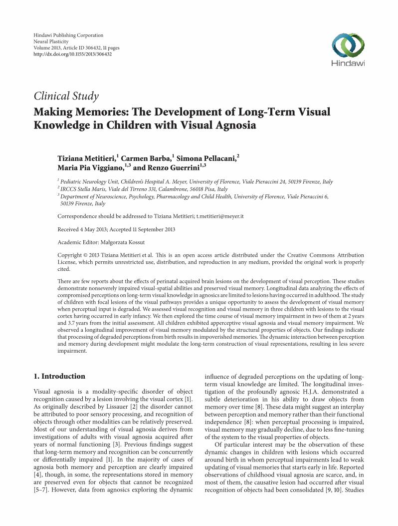
Figure 1: MRI scan for each patient. L.: (a), (b) Brain MRI (axial T2 and sagittal T1 weighted images) showing bilateral periventricularhyperintensities in the occipital lobes (white arrows) and dilatation of the occipital horn of the lateral ventricle (white arrowhead). G.: (a)–(c). Brain MRI (axial FLAIR, coronal and axial T2 weighted images) showing bilateral periventricular hyperintensities in the parietooccipitalareas (see white arrowheads and arrows). R.: (a) Brain MRI (axial T1 weighted image) showing cortical-subcortical atrophy and abnormalcortical sulcation in the left temporooccipital area (white arrow). (b), (c). Brain MRI (axial and sagittal FLAIR weighted images) showingincreased signal intensity in the left calcarine cortex and dilatation of the occipital horn of the left lateral ventricle (white arrowheads).
4.1.5. Naming
Drawings. L. correctly named 21/60 (35%, below average)drawings of the Boston Naming Test (BNT) [19, 20]. Errorsweremainly visual (25%), semantic (13%), and “do not know”responses (18%). Visual errors were incorrect generalizationsfrom a perceived or unperceived detail (e.g.: pretzel > hand-cuffs, wreath > bow, stilts > ski, unicorn > horse, palette >paintbrush).
G. correctly named 35/60 (58%, average) drawings on theBNT. Errors were mainly visual (16%), semantic (8%), andadequate circumlocutions (8%). Visual errors were incorrectgeneralizations from a perceived or unperceived detail (e.g.:knocker > slingshot, asparagus > stick, trellis > ladder).
R. correctly named 34/60 drawings (57%, below average).Errors were mainly visual (12%), semantic (12%), and “do notknow” responses (8%).
G. and R. performed a tachistoscopic presentation of theBNT drawings 4–6 months later. Stimuli remained on the
LCD screen for 500 msec. G. showed a slight increase incorrect naming (40/60, 67%), while R. showed a relevantdecrease (22/60, 37%) in total accuracy.
Silhouettes. Children were requested to identify 48 blackdrawings (24 living and 24 nonliving objects) on display untila first attempt at naming them.
L. correctly named 23 silhouettes (48%) without differ-ences in naming living (12/24) or nonliving objects (11/24).G. identified 41 silhouettes (85%; 22/24 living and 19/24nonliving). R. identified 29 silhouettes (60%; 16/24 living and13/24 nonliving).
Overlapping Figures. Children were presented 12 series of 4geometrical figures and 12 series of 4 letters. The overlapconcerned only one hidden figure while the remaining threehad at least some uncovered contours.
L. correctly performed on 10/12 (83%) patterns of geomet-rical figures and 9/12 (75%) patterns of overlapping letters.

Neural Plasticity 5
G. correctly identified geometrical figures on 11/12 (92%)trials and letters on 11/12 (92%) trials. R. correctly identifiedgeometrical figures on 10/12 (83%) trials and letters on 10/12(83%) trials.
Famous Faces. Sixteen pictures of the best known Italiansoccer playerswere presented on the LCD screen. All childrenwere habitual soccer fans, knew themain teams, and collectedstickers of the Italian national team.
L. recognized 1/16 (6%) football players showing a deficitin the recognition of famous faces. G. recognized 9/16 (56%)football players and R. 8/16 (50%).
4.1.6. Copying. The Rey Figure [21] was used to assessdrawing abilities in copying (Figure 2). L. realized a verygood copy (score 31/36), but with a very slow process,beginning with the triangle-rectangle at the left bottom ofthe figure and adding segment by segment until the end, forabout 20 minutes. G. had difficulties in copying the globalstructure and only reproduced some details with generalspatial contiguity. He obtained a score of 12.5/36, belowaverage. R. showed difficulties in this task with closing-inphenomenon and obtained a score of 16/36, below average.
4.1.7. Reading. Each child was asked to perform two taskstaken from the Battery for the Evaluation of Dyslexia andDysorthographia (DDE-2) [22]. In the first task they wererequested to read four lists of words as quickly as they could,and in the second task to read three lists of nonwords.
G. performed at normal levels for time and accuracy. L.and R. were very slow in reading either words and nonwords.Their reading speed fell below the norm for children at thesame school grade.
4.1.8. Writing. All three children were able to write correctlywords and sentences [22], spontaneously andunder dictation.
4.1.9. Navon Test. Target letters (S or T) were present ata global and/or a local level. The global letter subtended3.276∘
× 3.66∘ of visual angle (in width and height, resp.), and
each local letter subtended 0.56∘ × 0.48∘. The interelementdistance was 0.1∘.
There were two conditions: a global condition, in whichthe subject had to respond to global letters, ignoring localones, and a local condition in which the subject had torespond to local letters, ignoring global ones. Three types oftrials were presented: congruent (global letter was the sameas the local letter), incongruent (global and local letters weredifferent), or neutral (the target letter was associated withthe letter X). The stimuli were presented on a PC runningE-Prime software (Version 1.2) [23]. Participants respondedvia the keyboard, pressing one of the two keys assigned tothe target letters. The order of the different conditions wasbalanced. There were 72 trials for each condition. Table 2shows error rates for L., G., R., and healthy controls for theglobal-local task.
A global interference effect, where local congruent lettersproduce a high proportion of accuracy than local incongru-ent, was observed for healthy control subjects (𝜒2(2) = 21.27,
𝑃 < .001). The reverse effect, local precedence, was notobserved for healthy controls in the global condition (𝜒2(2) =2.71, 𝑃 = .26). L., G. and R. exhibited an exaggeratedglobal interference effect in the local condition (𝜒2(2) = 9.15,𝑃 < .05). They also sustained a local interference effectin the global condition (𝜒2(2) = 6.99, 𝑃 < .05), althoughthis effect emerged only for G., and R. accuracy rates. Thecontrast between the processing of local and global aspectsof hierarchical forms demonstrated a strong bias to attend toglobal shape in the local condition for L., G., and R. However,the results for both G. and R. indicate also a bias to attend tolocal elements.These two childrenwere generally impaired intheir processing of hierarchical forms. This suggests that G.and R. find it difficult to attend to hierarchical forms mainlywhen there is competition of the local/global levels, than intrials in which competition is absent.
4.1.10. VerbalMemory. L. andG. obtained average scores (𝑧 =−.28 and 𝑧 = .65) in paired-associated learning [17], whereasR. learned only few and simple associations after the threepresentation of the same list of ten pairs of words. His scorewas below average (𝑧 = −2.66).
Incidental semantic memory was assessed presenting 20animal names. Each child was requested to name the color ofthe animal but was not instructed to remember the items. Atthe end of the incidental learning phase, children were askedto report as many animals as they could. In the recall phaseG. reported 10/20 animals, performing within average; L. andR. performed below average (5/20 and 6/20, resp.).
4.1.11. Visual Memory
Rey Figure Recall.All children failed the recall of the complexfigure (mean score = 3, SD = 1.32). L. could recall local detailsbut failed to integrate them to the global structure, whereasG. and R. drawings were limited to very few details with nospatial relations with respect to the model (Figure 2).
Neurological control subjects recall was in the normalrange (Mean score = 17.75, SD = 7.24).
Drawing From Memory. Subjects had to draw three objects:a flower, a house, and a bicycle (Figure 3). All children com-plained about the difficulties of drawing while accomplishingthe task. They performed quite well in drawing a typicalrepresentation of a flower and a house. For the bicycle L. andR. omitted more visual properties, limiting their drawings tothe emerging details (wheels). R. at the end of his task said “Iwould not ride that bike!”.
Benton Visual Retention Test. Children were presented 10geometrical designs of the Benton Visual Retention Test,(BVRT) Part A [24] for 10 seconds and asked to reproducethem immediately afterwards. In this task the verbal com-ponent is important because subjects may remember thenames of geometric figures without relying on stored visualrepresentations to perform the task.
L. performed at average (8/10) and G. and R. werebelow average (4/10 for both). Neurological control subjectsperformed at average (Mean score = 8, SD = 0.82).

6 Neural Plasticity
Recall
L
G
R
Copy
Figure 2: Rey’s figure: copy and recall after a 15-minute delay performed by L., G., and R. The good copy realized by L. required a very slowprocess, beginning with the triangle-rectangle at the left top of the figure and adding segment by segment until the end. G. reproduced somedetails with general spatial contiguity. R. closed in on model and showed difficulties in drawing both details and global shape. All childrenhad poor recall: L. could recall local details but failed to integrate them into an integrated whole, whereas G. and R. drawings were limited tovery few details.
Table 2: Percentage of error for responses in the Navon task by local and global conditions.
Local GlobalCongruent Incongruent Neutral Congruent Incongruent Neutral
Healthy controls 1,4 8,8 1,4 1,4 3,7 3,7L. 4,2 20,8 16,7 4,2 4,2 8,3G. 8,3 20,8 4,2 0 20,8 0R. 4,2 25 12,5 4,2 16,7 8,3

Neural Plasticity 7
Flower Bicycle
L
G
R
Figure 3: Examples of drawings frommemory performed by L., G.,and R. (a flower, a house, and a bicycle). Drawings were based ontypical representations of objects. L. and R. limited their drawingsof a bicycle to the emerging details (wheels).
Following the same procedure used for the BVRT weadministered two more visual memory tasks (stimulusimages courtesy of Michael J. Tarr, Center for the NeuralBasis of Cognition and Department of Psychology, CarnegieMellon University, http://www.tarrlab.org/).
Visual Retention of Objects. Children were presented fivedrawings of objects, each on the center of amidsheet, selectedfor their high familiarity and for being composed of at leasttwo different parts. They were broom, boat, egg in eggcup,balloon, and bomb. These objects may benefit from verbalrehearsal strategies for storage in memory. Each figure wasassigned a value ranging from 0 to 2 depending upon thedegree to which it was correctly drawn in local details, globalform, and orientation.
L. andG. obtained a score of 2/10 andR. scored 1/10 (mean= 1.67, SD = 0.58). Their performance was significantly worsethan that observed in neurological control subjects (mean =7.75, SD =.96; t(5) = 9.64, 𝑃 < .001). See examples in Figure 4.
Model Control 1 Model Control 2 Broom Egg in eggcup
L
R
G
Figure 4: Two examples of immediate visual memory for objectsperformed by L., G., and R. The model is shown at the top, withthe same object recalled by two neurological control subjects onthe right. L., G., and R. had difficulties in reproducing the visualproperties of each model and their drawings were limited at mostto the typical representation of objects.
Visual Retention of Abstract Drawings. Children were pre-sented 7 abstract designs which differed in their visual andspatial characteristics. These designs may not benefit fromverbal rehearsal strategies to be stored inmemory. Each figurewas assigned a value of 0 to 2 depending upon the degree towhich it was correctly drawn in local details, global form, andorientation.
Children were unable to draw from memory the abstractpatterns (Figure 5), omitting or placing erroneously someparts (Mean score = 1.83, SD = .29). Neurological controlsubjects obtained a mean score of 9.25 (SD = .64; t(5) = 18.24,𝑃 < .001).
Although perceptual tasks were performed at differentlevels and degrees of impairment, each child revealed severeimpairment in visual memory, slightly modulated by visual-semantic properties of recalled objects. Table 3 summarizesthe neuropsychological results for this section.
Each child performed relatively well on tests assessingbasic aspects of visual perception. Their impairment atconstructing a perceptual representation from vision and the

8 Neural Plasticity
Table 3: Performance of each agnosic child on perception and memory tasks.
L. G. R.Efron tasks Not impaired Not impaired Not impaired, some errorsShape discrimination Not impaired Not impaired Not impairedDot counting Not impaired Not impaired ImpairedStar cancellation Not impaired Impaired, not lateralized misses Impaired, right misses
Boston naming test Below average, visual errors Average, visual errors Below average, visual andsemantic errors
BNT at 500ms — 12.5% improvement 35%Worsening
SilhouettesImpairedNo differences forliving/nonliving
Not impairedLiving objects better
IntermediateNo differences forliving/nonliving
Overlapping figures Slightly impaired Not impaired Slightly impairedFamous faces Impaired Not impaired Not impairedRey figure copying Average, but very slow process Impaired ImpairedReading Slow Normal SlowWriting Relatively normal Relatively normal Relatively normal
Navon task Global effect exaggerated Global and local effectexaggerated
Global and local effectexaggerated
Verbal memory Not impaired Not impaired ImpairedRey figure memory Impaired Impaired ImpairedImmediate visual memory(i) Benton VRT Impaired Impaired Impaired(ii) Objects Impaired Impaired Impaired(iii) Abstract designs Impaired Impaired Impaired
consequent inability to copy or identify a drawing are con-sistent with apperceptive agnosia [3, 25]. Children evidencedqualitative differences in copying abilities and integration ofshape elements into perceptual wholes. L. was impaired atintegrating parts with whole shapes, and his performance isconsistent with integrative agnosia: he performed correctly inthe Efron test, reproduced an accurate copy of Rey’s complexfigure, had an abnormally strong bias to attend to globalshape in the Navon task, though his performance was notfacilitated by silhouettes in naming objects. R. impairment isconsistent with visual-form agnosia: he was less accurate inthe Efron test, his drawings were inaccurate, and he had anabnormally strong bias to attend to local and global elementsin the Navon task. G. showedmild deficits in both integrationand segmentation of complex visual objects.
4.2. Follow-Up for G. and R. G. and R. were reexamined after3.7 years and 2 years, respectively, from the first evaluation.G. was 13 years old and R was 14 years old. R. completed thefollow-up evaluation four months after neurosurgery.
4.2.1. Boston Naming Test. G. correctly named 45/60 (75%)drawings, with a 17% improvement from the first eval-uation. Errors were mainly visual and semantic. R. cor-rectly named 9/40 drawings (22.5%), manifesting a relevantdecrease (−34.5%) in performance. Errors were mainly “donot know” responses (30%) and adequate circumlocutions(27.5%). While the improvement in naming observed forG. might be interpreted as a normal developmental effect,
R. impairment in naming was likely related to languageimpairment that occurred after surgery.
4.2.2. Copying: Rey Figure. G. improved his abilities in copy-ing the global and local features, obtaining a score of 23.5/36,now falling within average, with an increment of 46.8% onthe previous score. Similarly, R. showed an improvement of48.4% on the actual score (of 31/36, average), compared tothe first assessment. These improvements (Figure 6) are notexplained by practice effect (at least two years have passedsince the first assessment) nor by a developmental effect(normative data show slight differences in scores from age 11to adulthood).
4.2.3. Visual Memory
Rey Figure Recall.G. increased his recall of the complex figure(12 versus 3.5, +70.8%), though his performance remainedbelow average (𝑧 = −2.82). R. showed a greater improvement(18.5 versus 1.5, +91.9%) and his performance fell withinaverage (𝑧 = −1.15), with no difference from neurologicalcontrols (Mean score = 17.75, SD = 7.24). See Figure 6.
Benton Visual Retention Test. G. and R. were still belowaverage (3/10 and 6/10, resp.), showing no improvement in therecall of geometrical drawings.
Visual Retention of Objects. G. obtained a score of 7/10, witha five-point improvement from the initial assessment (2/10).

Neural Plasticity 9
Model Control 3 Model Control 4 Number 1 Number 6
R
G
L
Figure 5: Two examples of immediate visual memory for abstractdesigns performed by L., G., and R. The model is shown at the top,with the same object recalled by two neurological control subjectson the right. All three children were unable to draw from memorya coherent pattern of a model that did not allow verbal rehearsalstrategies.
Copy Recall
G
R
Figure 6: Rey’s figure: Copy and Recall performed by G. and R. atfollow up. A relevant improvement was observed in their drawings,both in the details and in their into a global shape, compared to thefirst assessment.
Broom Egg in eggcup
R
G
G
R
Number 1 Number 6
Figure 7: Examples of immediate visual memory for objects andabstract drawings performed by G. and R. at follow-up. Bothchildren improved the quality of their drawings from memoryby adding more details in an integrated whole, compared to theprevious testing.
R. reported a score of 2/10, one point higher than previouslyobserved (1/10). Two examples are illustrated in Figure 7.
Visual Retention of Abstract Drawings. Both childrenimproved the quality of their drawings from memory forthe seven abstract patterns (Figure 7): G. with a score from1 to 5/14 and R. from 0 to 4/14, compared to the previousassessment. However, their scores were still below the averageobserved in neurological control subjects.
These results demonstrated a global improvement inperceptual and memorial processes over time. There wasa general increase in performance at follow-up from theprevious assessment in almost all tasks, with the exception ofthe BentonVisual RetentionTest. Improvementwas higher inmemory tasks relying on the storage of structural propertiesof objects. When memory was based only on pure visualproperties this incremental effect was limited. However, R.

10 Neural Plasticity
showed a deterioration in naming and no change in visualretention of objects.
5. Discussion
We are presenting behavioural data of three children withvisual agnosia and visual memory impairment, caused bybrain lesions of the visual cortex which occurred in earlyinfancy.
Children exhibited relatively normal sensory and seman-tic memory functioning, with several difficulties in objectrecognition. They also exhibited deficits in copying, nam-ing drawings, and integrating object features. We classifiedtheir impairments in two types of apperceptive agnosia. R.exhibited visual-form agnosia, L. integrative agnosia, and G.mild difficulties in segmentation and integration processesthat share some characteristics with that observed in L.Resemblance between these findings and those describedin adult agnosics [3, 26] provides further evidence that theassociations between specific impairment and site of lesionare concordant in both adults and children [27].
Reports on childhood agnosia are relatively rare. Theseinclude both acquired cases of early [11, 14] and late onset[9, 10, 12] origin. Kiper and collaborators [11] describedtwo patients (one of them was evaluated during childhood)with bilateral perinatal lesions of the primary visual cortex.They exhibited recognition deficits, which were classifiedas apperceptive agnosia at different degrees of severity, andmanifested different levels of adaptive plasticity over time[11]. Amicuzi and collaborators [14] reported a 4.6-year-old girl with bilateral occipital damage and visual agnosiawhomanifested selective deficit of figure-ground segregation,impaired visual recognition, and abnormal moving throughspace. Both case reports emphasized the “fuzzy boundaries”[28] between visual-form agnosia and integrative agnosia, asfar as children’s behavior and domain-specific functions areconcerned. Compared to adults, the differences between thetwo types of agnosia are even less distinguishable, possiblybecause developmental neurocognitive mechanisms are flex-ible and adaptive [27]. Nonetheless, these studies indicatethat the core deficit of apperceptive agnosia can indeed beidentified in children with acquired lesions of the visualcortex.
In previously reported observations of childhoodagnosia, no evidence of visual memory impairment, asassessed by standard tests, was uncovered [11, 14]. Weobserved degraded object recognition and associated deficitsof visual memory in apperceptive agnosia. All three childrenexhibited impaired visual memory and poor drawing frommemory with, however, some evidence that residual visualknowledge might be built up relying on the structuralproperties of objects.
As interpreted in the longitudinal investigation of theadult agnosic H.J.A., when perceptual inputs are degraded,as in apperceptive agnosia, visual memory may decline,due to impaired fine-tuning of the system to the visualproperties of objects [8]. Conversely, we observed a lon-gitudinal improvement of visual memory, modulated byobject properties, suggesting that underlying developmental
mechanisms may act from infancy to late childhood. Theimpairment was more severe for visual-spatial compared tovisual-object properties, showing a dissociation in processingof visual representations. These findings are consistent withthe hypothesis of a common representation of an image thatis processed in the visual buffer [29]. At this level, a selectivedamage may occur to the spatial and object mechanismsthat act on the image [30]. Developmental processes mightselectively adapt and modulate these mechanisms to theneuroanatomical substrate for image construction. Indeed,the improvement we observed in R. and G. visual memoryat follow-up was more apparent when the task relied on thestructural properties of objects and less so when memoryconcerned only pure visual-spatial properties of objects.These preliminary findings have important implications forunderstanding the development and construction of visualrepresentations.More longitudinal studies are needed to shedlight on the dynamic interaction between visual perceptionand visual memory during development, when the occipitalcortex is damaged from birth.
6. Conclusions
Impairments of visual recognition are relatively overlooked inchildren, nevertheless they have a crucial impact on learningas well as mobility and independent living.
Our findings indicate that impaired processing of visualimages in childhood agnosia results in impoverished visualmemories. During the course of development the dynamicinteraction between perception andmemorymightmodulatethe long-term construction of visual representations, result-ing in less severe impairment.
As suggested by previous reports, developmental brainplasticity and compensatory behavioral adaptations allow theemergence of surprising abilities in daily life [12]. Therefore,an early diagnosis of visual agnosia is crucial for planningspecific rehabilitation interventions that may foster the pro-gressive adaptation leading to a developmental process that isunique in a child with brain damage.
Early neuropsychological rehabilitation in childhoodvisual agnosia should train visual and multimodal envi-ronmental exploration, improving recognition and memorythrough the processes of adaptation and compensation thatare the hallmark of development following perinatal neuralpathology.
Conflict of Interests
The authors declare that there are no sources of financialsupport and there is no conflict of interests.
References
[1] E. De Renzi, “Agnosia,” in Handbook of Clinical and Exper-imental Neuropsychology, G. Denes and L. Pizzamiglio, Eds.,Psychology Press, 1999.
[2] H. Lissauer, “Ein Fall von Seelenblindheit nebst einemBeitrage zur Theorie derselben,” Archiv fur Psychiatrie und

Neural Plasticity 11
Nervenkrankheiten, vol. 21, pp. 222–270, 1890, translated inCog-nitive Neuropsychology, vol. 5, pp. 157–192, 1988, Commentaryby T. Shallice and M. Jackson, “Lissauer on agnosia,” CognitiveNeuropsychology, vol. 5, pp. 153–156, 1988.
[3] M. J. Farah, Visual Agnosia, The MIT Press, Cambridge, UK,2004.
[4] L. Trojano and D. Grossi, “A critical review of mental imagerydefects,” Brain and Cognition, vol. 24, no. 2, pp. 213–243, 1994.
[5] M. Behrmann, G. Winocur, and M. Moscovitch, “Dissociationbetween mental imagery and object recognition in a brain-damaged patient,” Nature, vol. 359, no. 6396, pp. 636–637, 1992.
[6] P. Servos and M. A. Goodale, “Preserved visual imagery invisual form agnosia,”Neuropsychologia, vol. 33, no. 11, pp. 1383–1394, 1995.
[7] P. Bartolomeo, A.-C. Bachoud-Levi, B. De Gelder et al.,“Multiple-domain dissociation between impaired visual per-ception and preservedmental imagery in a patient with bilateralextrastriate lesions,” Neuropsychologia, vol. 36, no. 3, pp. 239–249, 1998.
[8] M. J. Riddoch, G. W. Humphreys, T. Gannon, W. Blott, and V.Jones, “Memories are made of this: the effects of time on storedvisual knowledge in a case of visual agnosia,” Brain, vol. 122, no.3, pp. 537–559, 1999.
[9] A. W. Young and H. D. Ellis, “Childhood prosopagnosia,” Brainand Cognition, vol. 9, no. 1, pp. 16–47, 1989.
[10] A. Schiavetto, J.-C. Decarie, J. Flessas, G. Geoffroy, and M.Lassonde, “Childhood visual agnosia: a seven-year follow-up,”Neurocase, vol. 3, no. 1, pp. 1–17, 1997.
[11] D. C. Kiper, P. Zesiger, P. Maeder, T. Deonna, and G. M.Innocenti, “Vision after early-onset lesions of the occipital cor-tex: I. Neurospychological and psychophysical studies,” NeuralPlasticity, vol. 9, no. 1, pp. 1–25, 2002.
[12] S. Le, D. Cardebat, K. Boulanouar et al., “Seeing, since child-hood, without ventral stream: a behavioural study,” Brain, vol.125, no. 1, pp. 58–74, 2002.
[13] P. Joy and R. Brunsdon, “Visual agnosia and prosopagnosia inchildhood: a prospective case study,” Child Neuropsychology,vol. 8, no. 1, pp. 1–15, 2002.
[14] I. Amicuzi, M. Stortini, M. Petrarca et al., “Visual recognitionand visually guided action after early bilateral lesion of occipitalcortex: a behavioral study of a 4.6-year-old girl,”Neurocase, vol.12, no. 5, pp. 263–279, 2006.
[15] E. Funnell and J. Wilding, “Development of a vocabularyof object shapes in a child with a very-early-acquired visualagnosia: a unique case,” Quarterly Journal of ExperimentalPsychology, vol. 64, no. 2, pp. 261–282, 2011.
[16] D. Wechsler, Manual for the Wechsler Intelligence Scale forChildren—Revised, Psychological Corporation, New York, NY,USA, 1974, translated in V. Rubini and F. Padovani, Scaladi Intelligenza Wechsler per Bambini Riveduta, OrganizzazioniSpeciali, Firenze, Italy, 1986.
[17] P. Bisiacchi, M. Cendron, M. Gugliotta, P. E. Tressoldi, and C.Vio, BVN 5-11 Batteria di Valutazione Neuropsicologica Per L’etaEvolutiva, Erickson, Trento, Italy, 2005.
[18] R. Efron, What Is Perception? Boston Studies in Philosophy ofScience, 1968.
[19] E. F. Kaplan, H. Goodglass, and S. Weintraub,The Boston Nam-ing Test. Experimental, Experimental edition, Lea & Febiger,Philadelphia, Pa, USA, 1983.
[20] D. Riva, F. Nichelli, and M. Devoti, “Developmental aspects ofverbal fluency and confrontationnaming in children,”Brain andLanguage, vol. 71, no. 2, pp. 267–284, 2000.
[21] P. A. Osterrieth, “Le test de copie d’une figure complex:Contribution a l’etude de la perception et de la memoir (TheComplex Figure Copy Test),” in Archives De Psychologie, J.Corwin, Bylsma, and F. W. Translated, Eds., vol. 30, pp. 286–356, The Clinical Neuropsychologist, 1993.
[22] G. Sartori, R. Job, and P. E. Tressoldi, DDE-2. Batteria PerLa Valutazione delLa Dislessia E delLa Disortografia Evolutiva-2. (Battery For the Evaluation of developmental Dyslexia andDysorthographia), Giunti, Firenze, Italy, 2007.
[23] W. Schneider, A. Eschmann, and A. Zuccolotto, E-Prime User’sGuide, Psychology Software Tools, Pittsburgh, Pa, USA, 2002.
[24] A. L. Benton, Revised Visual Retention Test Manual, The Psy-chological Corporation, New York, NY, USA, 1974, translatedin S. Ferracuti and E. Cannoni, Benton Visual Retention Test—Revised, Giunti Organizzazioni Speciali, Firenze, Italy, 2000.
[25] J. J. Marotta andM. Behrmann, “Agnosia,” in Encyclopedia of theHuman Brain, V. S. Ramachandran, Ed., Academic Press, SanDiego, Calif, USA, 2002.
[26] M. J. Riddoch, G. W. Humphreys, N. Akhtar, H. Allen, R. M.Bracewell, and A. Schofield, “A tale of two agnosias: distinctionsbetween form and integrative agnosia,”Cognitive Neuropsychol-ogy, vol. 25, no. 1, pp. 56–92, 2008.
[27] J. Stiles, J. Reilly, B. Paul, and P. Moses, “Cognitive developmentfollowing early brain injury: evidence for neural adaptation,”Trends in Cognitive Sciences, vol. 9, no. 3, pp. 136–143, 2005.
[28] E. De Renzi and F. Lucchelli, “The fuzzy boundaries of apper-ceptive agnosia,” Cortex, vol. 29, no. 2, pp. 187–215, 1993.
[29] S. M. Kosslyn, “Mental images and the brain,” Cognitive Neu-ropsychology, vol. 22, no. 3-4, pp. 333–347, 2005.
[30] M. J. Riddoch andG.W.Humphreys, “The retrieval andmanip-ulation of visual memories: evidence from neuropsychology,” inVisual Memory, S. J. Luck and A. Hollingworth, Eds., OxfordUniversity Press, Oxford, UK, 2008.

Submit your manuscripts athttp://www.hindawi.com
Neurology Research International
Hindawi Publishing Corporationhttp://www.hindawi.com Volume 2014
Alzheimer’s DiseaseHindawi Publishing Corporationhttp://www.hindawi.com Volume 2014
International Journal of
ScientificaHindawi Publishing Corporationhttp://www.hindawi.com Volume 2014
Hindawi Publishing Corporationhttp://www.hindawi.com Volume 2014
BioMed Research International
Hindawi Publishing Corporationhttp://www.hindawi.com Volume 2014
Research and TreatmentSchizophrenia
The Scientific World JournalHindawi Publishing Corporation http://www.hindawi.com Volume 2014
Hindawi Publishing Corporationhttp://www.hindawi.com Volume 2014
Neural Plasticity
Hindawi Publishing Corporationhttp://www.hindawi.com Volume 2014
Parkinson’s Disease
Hindawi Publishing Corporationhttp://www.hindawi.com Volume 2014
Research and TreatmentAutism
Sleep DisordersHindawi Publishing Corporationhttp://www.hindawi.com Volume 2014
Hindawi Publishing Corporationhttp://www.hindawi.com Volume 2014
Neuroscience Journal
Epilepsy Research and TreatmentHindawi Publishing Corporationhttp://www.hindawi.com Volume 2014
Hindawi Publishing Corporationhttp://www.hindawi.com Volume 2014
Psychiatry Journal
Hindawi Publishing Corporationhttp://www.hindawi.com Volume 2014
Computational and Mathematical Methods in Medicine
Depression Research and TreatmentHindawi Publishing Corporationhttp://www.hindawi.com Volume 2014
Hindawi Publishing Corporationhttp://www.hindawi.com Volume 2014
Brain ScienceInternational Journal of
StrokeResearch and TreatmentHindawi Publishing Corporationhttp://www.hindawi.com Volume 2014
Neurodegenerative Diseases
Hindawi Publishing Corporationhttp://www.hindawi.com Volume 2014
Journal of
Cardiovascular Psychiatry and NeurologyHindawi Publishing Corporationhttp://www.hindawi.com Volume 2014