Embed Size (px)
Citation preview
Non-pharmacological Management of Atrial
Fibrillation
BY- DR SARITA CHOUDHARY
• AF is a supraventricular tachyarrhythmia with uncoordinated atrial activation and consequently ineffective atrial contraction Electrocardiogram (ECG) include: 1) irregular R-R intervals 2) absence of distinct repeating P waves, and 3) irregular atrial activity.
• Most frequent sustained arrhythmia of clinical significance.• It may occur due to reversible factors such as alcohol
ingestion, hyperthyroidism,or pulmonary embolism.• In 70-80% of patients, atrial fibrillation is associated with
organic heart disorders coronary heart disease, hypertension with left ventricular hypertrophy, valve disease, or congenital heart defects.
• Symptoms range from no symptoms to fatigue, palpitations, dyspnea, hypotension, syncope, or HF
• Nonvalvular AF increases the risk of stroke 5 times and AF in the setting of mitral stenosis increases the risk of stroke 20 times over patients in sinus rhythm.
• Thromboembolism occurring with AF is associated with a greater risk of recurrent stroke, more severe disability, and mortality . Silent AF is also associated with ischemic stroke.
• AF is also associated with a 3-fold risk of HF and 2-fold increased risk of both dementia and mortality.
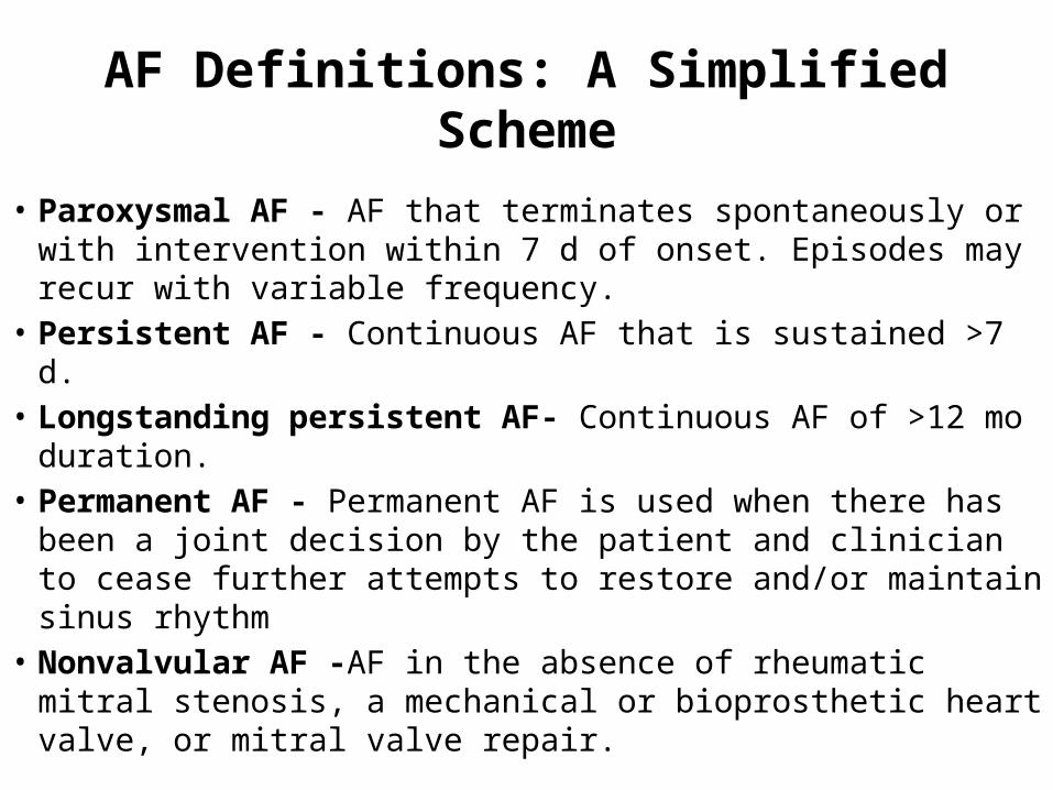
AF Definitions: A Simplified Scheme
• Paroxysmal AF - AF that terminates spontaneously or with intervention within 7 d of onset. Episodes may recur with variable frequency.
• Persistent AF - Continuous AF that is sustained >7 d.• Longstanding persistent AF- Continuous AF of >12 mo
duration.• Permanent AF - Permanent AF is used when there has been a
joint decision by the patient and clinician to cease further attempts to restore and/or maintain sinus rhythm
• Nonvalvular AF -AF in the absence of rheumatic mitral stenosis, a mechanical or bioprosthetic heart valve, or mitral valve repair.
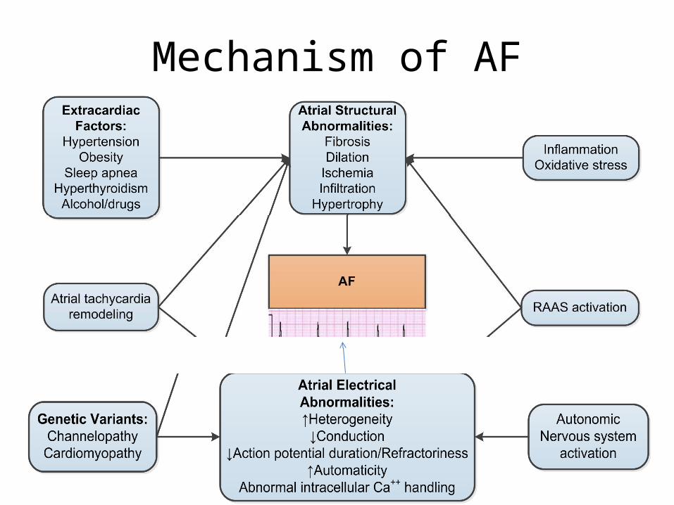
Mechanism of AF
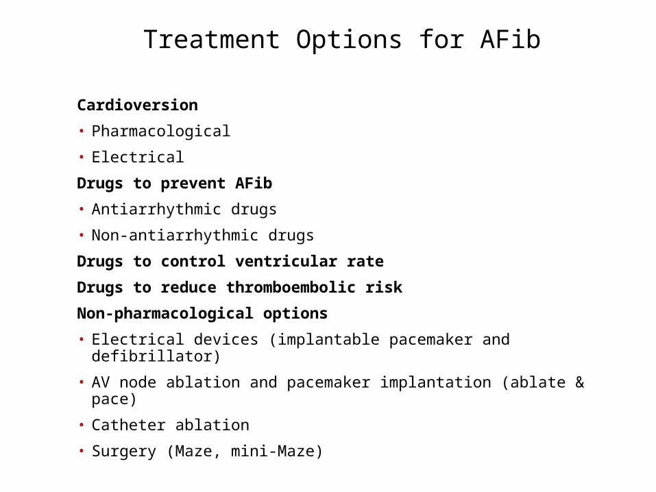
Treatment Options for AFib
Cardioversion
• Pharmacological
• Electrical
Drugs to prevent AFib
• Antiarrhythmic drugs
• Non-antiarrhythmic drugs
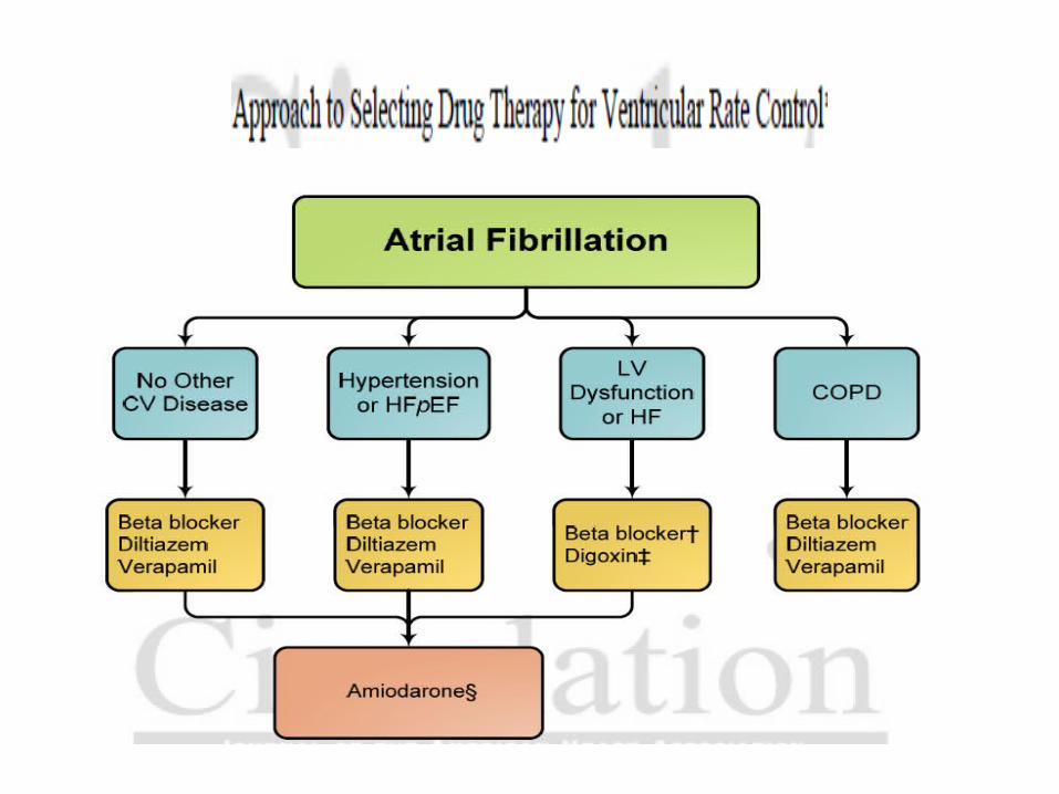
Drugs to control ventricular rate
Drugs to reduce thromboembolic risk
Non-pharmacological options
• Electrical devices (implantable pacemaker and defibrillator)
• AV node ablation and pacemaker implantation (ablate & pace)
• Catheter ablation
• Surgery (Maze, mini-Maze)
• The interest for non-pharmacological therapy has emerged due to the limited antiarrhythmic drugs and their frequent proarrhythmic effects.
• Thus, reasons for choosing nonpharmacological therapy may be paroxysmal atrial fibrillation with very frequent attacks and severe symptoms, chronic atrial fibrillation without adequate rate control leaving the patient at risk of developing tachycardiomyopathy, or patients who experience intolerable side effects of otherwise effective drug therapy.
Electrophysiologic Mechanisms• AF requires both a trigger for initiation and an appropriate
anatomic substrate for maintenance, both of which are potential targets for therapy.
• Triggers of AF- Ectopic focal discharges often initiate AF Rapidly firing foci initiating paroxysmal AF arise most commonly from LA myocardial sleeves that extend into the pulmonary veins. These observations led to the development of pulmonary vein isolation as the cornerstone for radiofrequency catheter ablation strategies
• Although the pulmonary veins are the most common sites for ectopic focal triggers, triggers can also arise elsewhere, including the posterior LA, ligament of Marshall, coronary sinus, venae cavae, septum, and appendages.
• Atrial myocardial fibers are oriented in disparate directions around the pulmonary veins and the posterior LA, with considerable anatomic variability among individuals.
• Conduction abnormalities that promote re-entry are likely due to relatively depolarized resting potentials in pulmonary vein myocytes.
• Re-entry is further favored by abbreviated action potentials and refractoriness in pulmonary vein myocytes
• Isolated pulmonary vein myocytes also demonstrate abnormal automaticity and triggered activity that could promote rapid focal firing.
• Maintenance of AF-Theories proposed to explain the perpetuation and maintenance of AF include
1) multiple independent re-entrant wavelets associated with heterogeneous conduction and refractoriness
2) ≥1 rapidly firing foci and 3) ≥1 rotors, or spiral wave re-entrant circuits. With a
single rapid focus or rotor excitation, wave fronts may encounter refractory tissue and break up during propagation, resulting in irregular or fibrillatory conduction). Both rapid focal firing and re-entry may be operative during AF.
• These presumed mechanisms have driven the development of therapies.
• The atrial maze procedure and ablation lines may interrupt paths for multiple wavelets and spiral re-entry.
• Using a biatrial phase mapping approach, a limited number of localized, rapid drivers were identified in a small group of patients with various types of AF . *
• In most cases, these localized sources appeared to be reentrant, while in others they were consistent with focal triggers and radiofrequency catheter ablation targeting of these sites often terminated or slowed AF.
• Narayan SM, Krummen DE, Shivkumar K, et al. Treatment of atrial fibrillation by the ablation of localized sources: CONFIRM (Conventional Ablation for Atrial Fibrillation With or Without Focal Impulse and Rotor Modulation) trial. J Am Coll Cardiol. 2012;60:628-36.
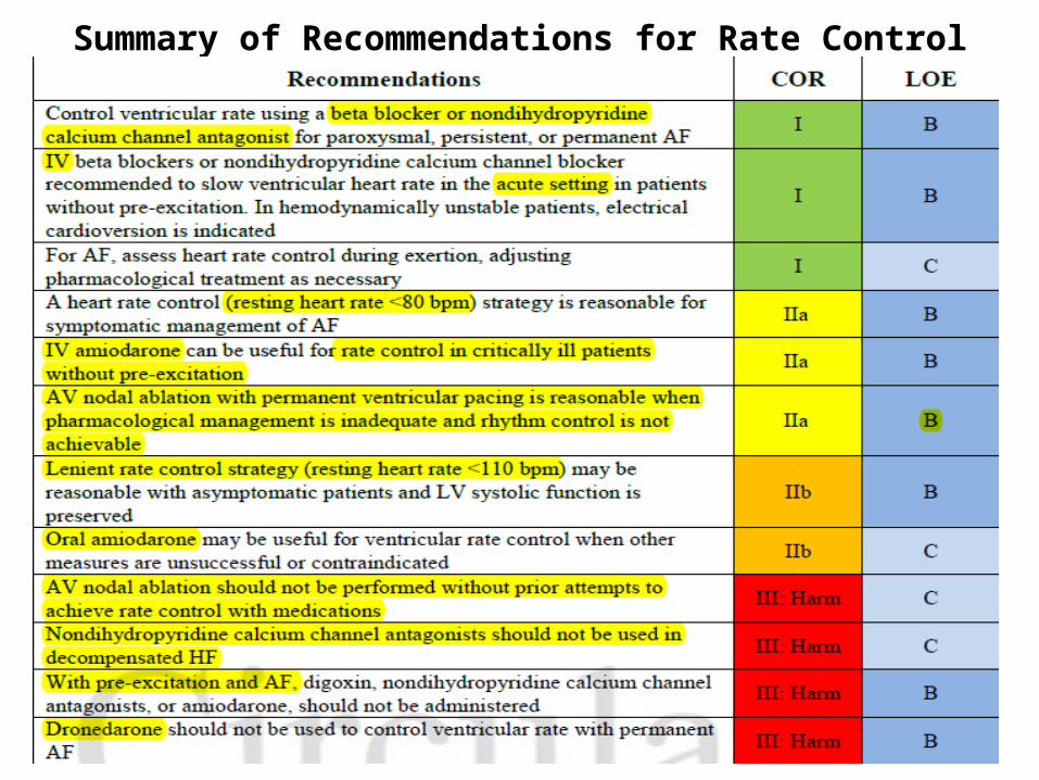
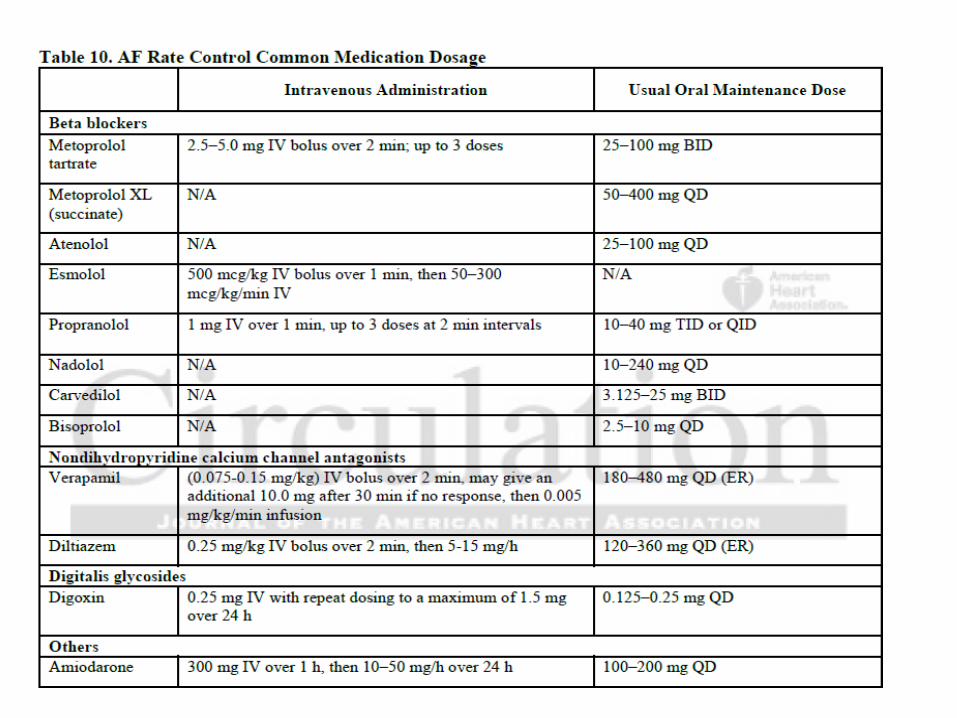
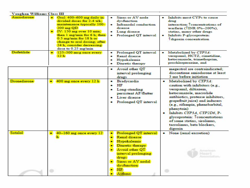
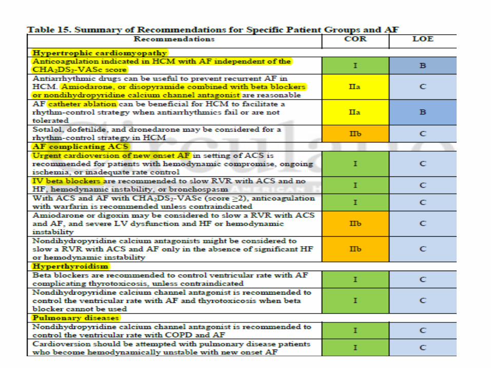
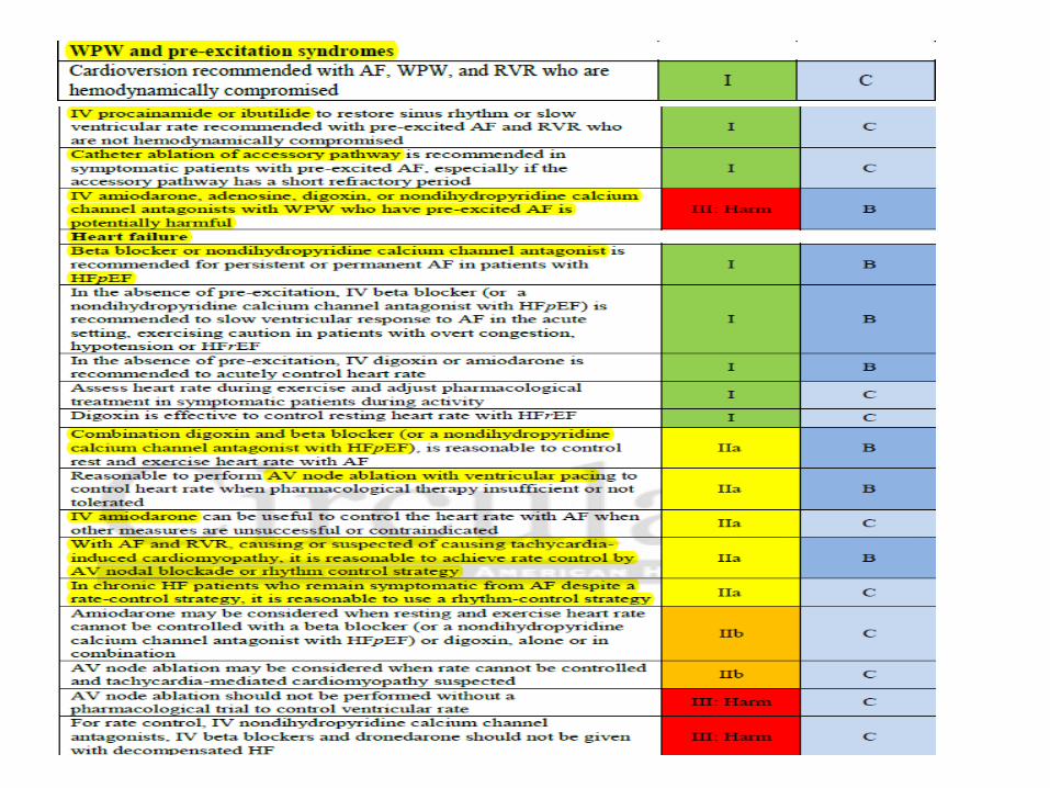
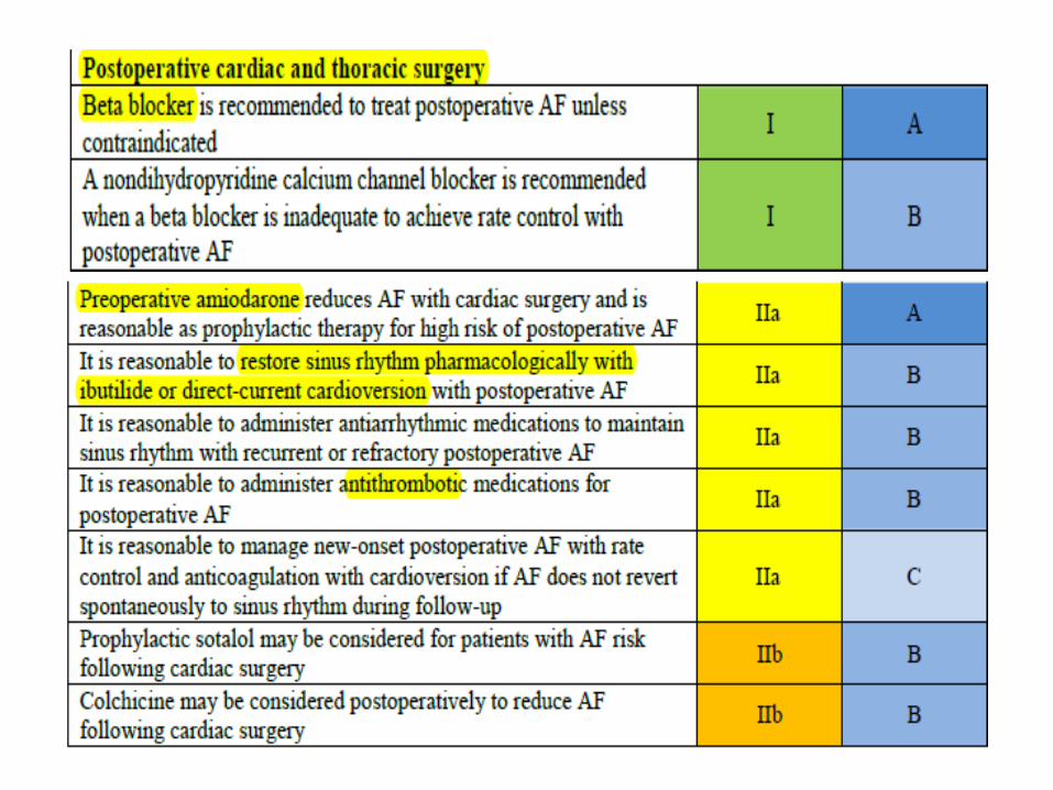
Summary of Recommendations for Rate Control
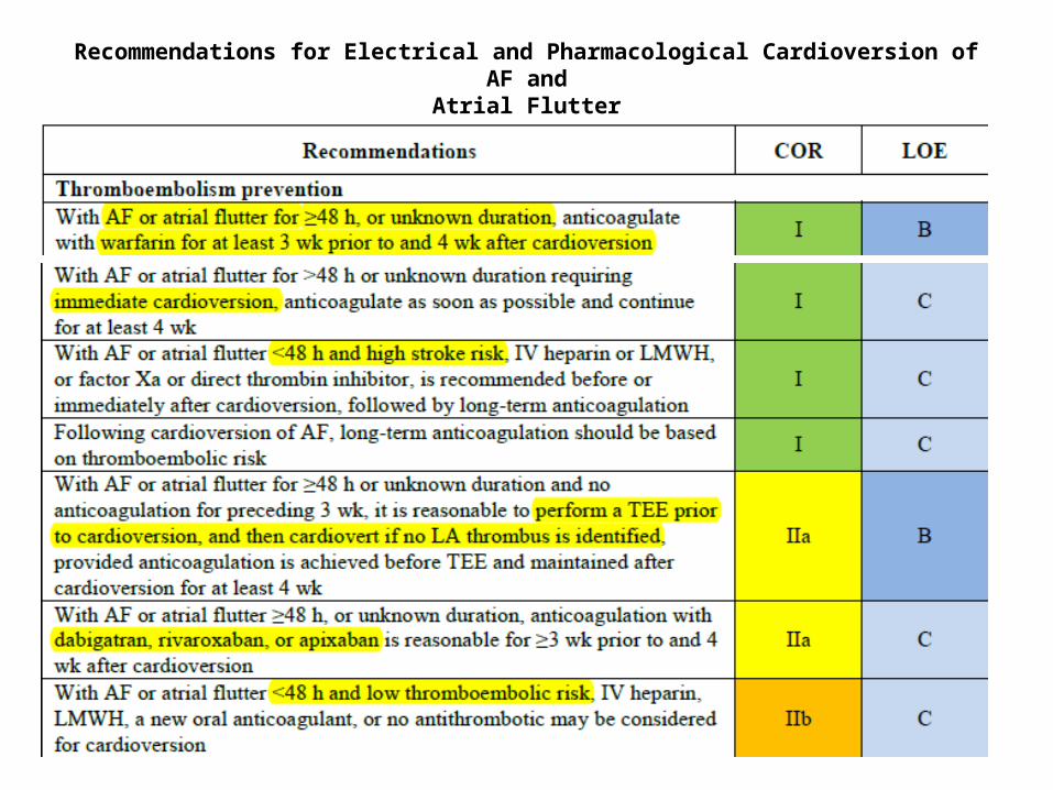
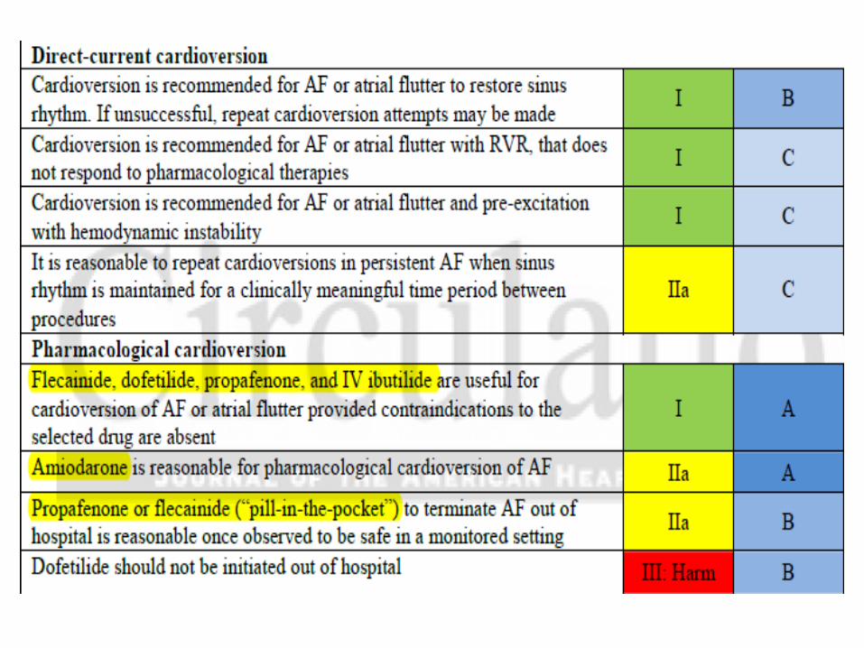
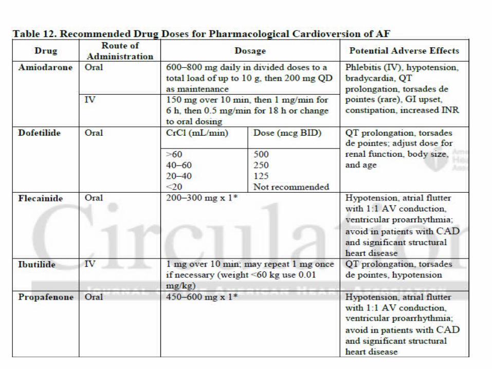
Recommendations for Electrical and Pharmacological Cardioversion of AF andAtrial Flutter
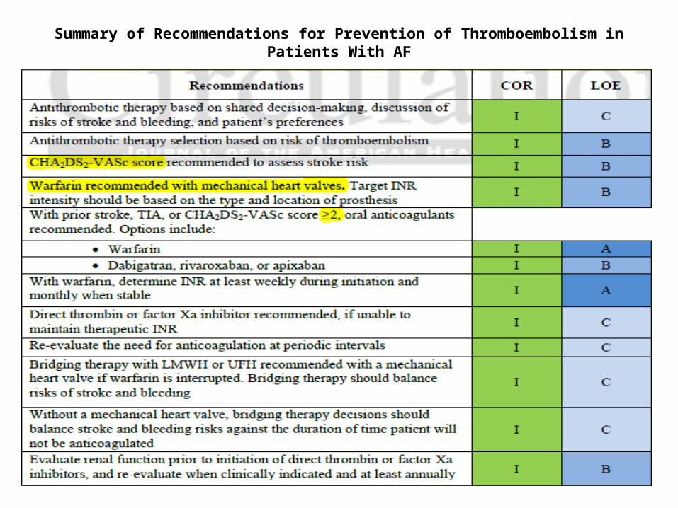
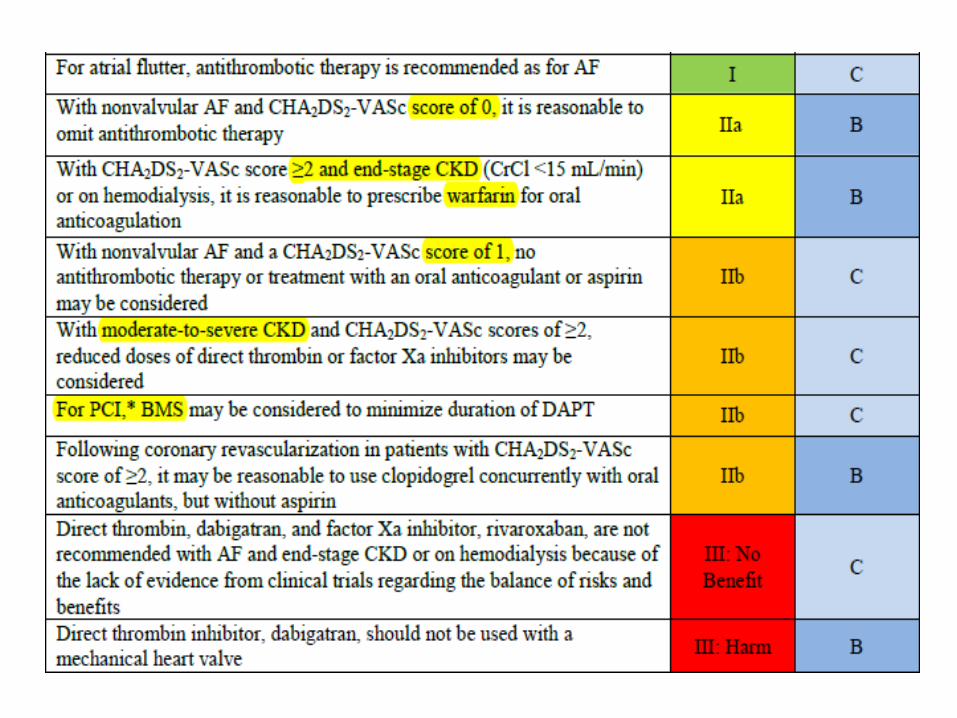
Summary of Recommendations for Prevention of Thromboembolism in Patients With AF
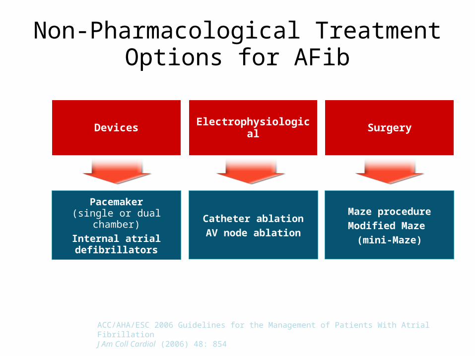
SurgeryElectrophysiologicalDevices
Pacemaker(single or dual chamber)
Internal atrialdefibrillators
Catheter ablationAV node ablation
Non-Pharmacological Treatment Options for AFib
Maze procedureModified Maze
(mini-Maze)
ACC/AHA/ESC 2006 Guidelines for the Management of Patients With Atrial FibrillationJ Am Coll Cardiol (2006) 48: 854
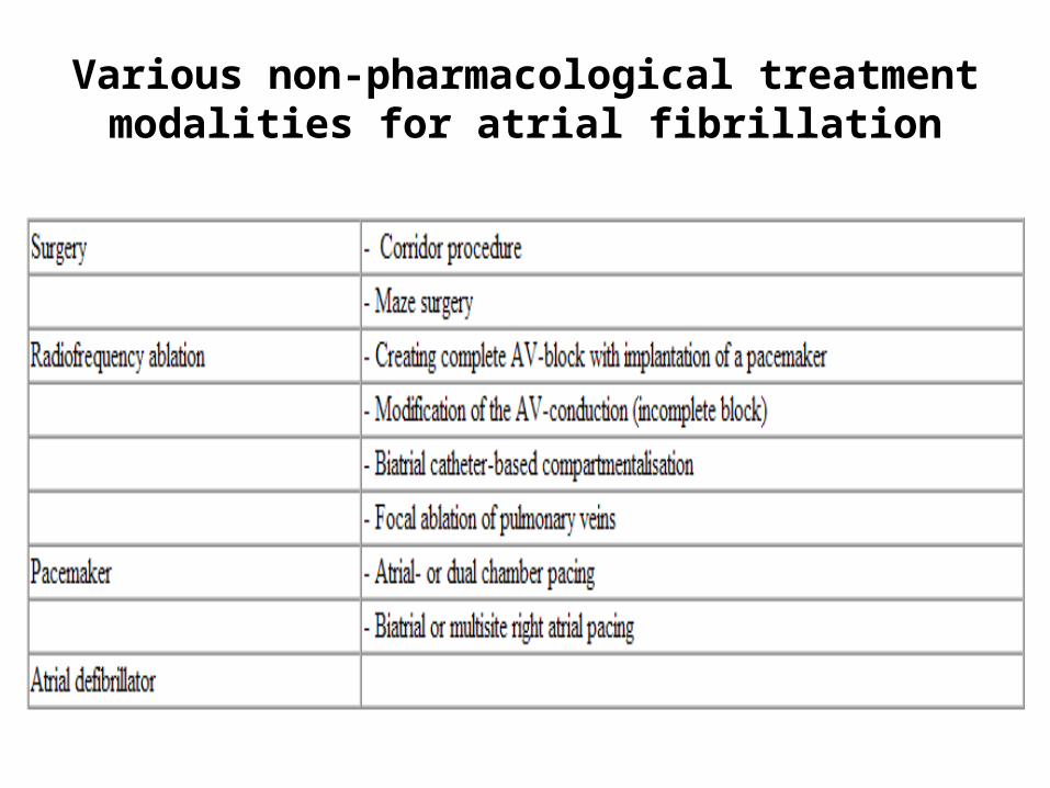
Various non-pharmacological treatment modalities for atrial fibrillation
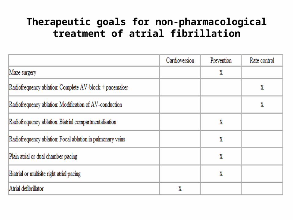
Therapeutic goals for non-pharmacological treatment of atrial fibrillation
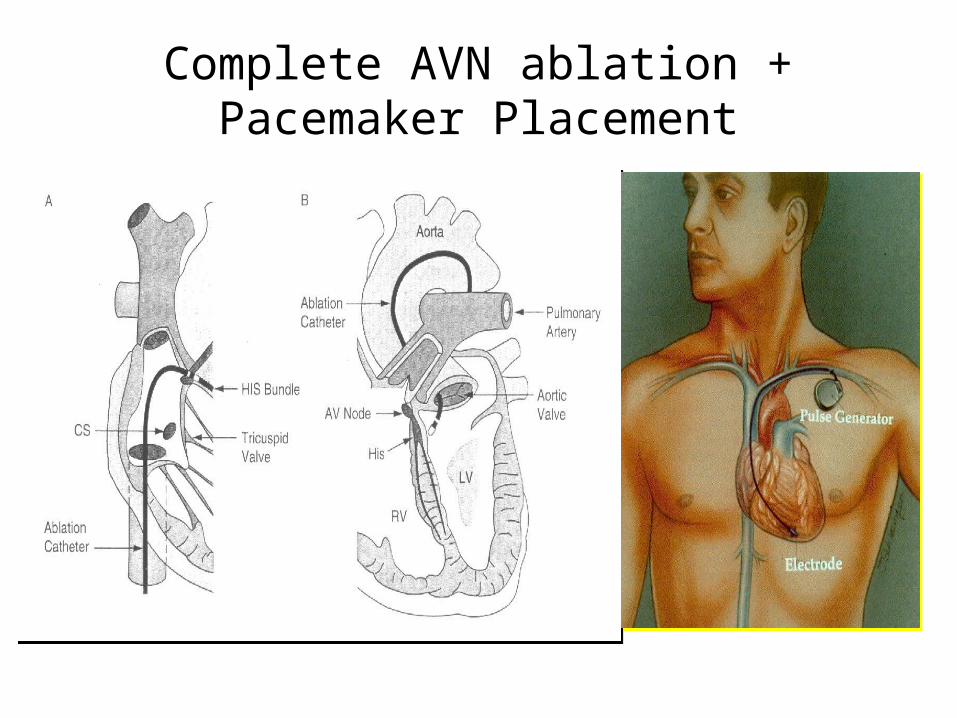
Complete AVN ablation + Pacemaker Placement
Atrioventricular Junction Ablation
• Transvenous ablation of the atrioventricular (AV) junction was first performed with direct-current shocks in 1981.
• Radiofrequency energy replaced direct-current shocks for AV junction ablation because of improved efficacy and safety.
• advantage is its success rate approaches 100%. • It is associated with a low incidence of adverse
effects.
• AV nodal ablation with permanent pacemaker implantation effectively controls and regularizes ventricular heart rate
• Patients most likely to benefit include those with tachycardia- induced cardiomyopathy with ventricular rate control refractory to medical therapy .
• It is usually reserved for elderly patients as it leads to pacemaker dependency.
• Patients with symptoms refractory to medical therapy who are treated with AV nodal ablation and permanent pacemaker implantation have an improvement in cardiac symptoms, quality of life, ventricular function and health care utilization.*
• Brignole M, Menozzi C, Gianfranchi L, et al. Assessment of atrioventricular junction ablation and VVIR pacemaker versus pharmacological treatment in patients with heart failure and chronic atrial fibrillation: a randomized, controlled study. Circulation. 1998;98:953-60.
• Kay GN, Ellenbogen KA, Giudici M, et al. The Ablate and Pace Trial: a prospective study of catheter ablation of the AV conduction system and permanent pacemaker implantation for treatment of atrial fibrillation. APT Investigators. J Interv Card Electrophysiol. 1998;2:121-35
• With this approach, no rate control medications are necessary, but anticoagulation to prevent thromboembolism is required as atrial systole is not restored ,based on the patient’s stroke risk as assessed by the CHA2DS2-VASc system.
• Lifelong pacemaker dependency with its potential complications.
• Time-permitting, pacemaker implantation may be performed 4 to 6 weeks prior to the AV node ablation to ensure proper pacemaker function as malfunction due to lead dislodgement can be catastrophic.
• Postablation, the ventricular pacing rate is usually set between 90 bpm and 100 bpm and then gradually tapered over several months
• Sudden death secondary to torsades de pointes or ventricular fibrillation has been reported after AV junction ablation. This outcome is possibly related to increased dispersion of ventricular refractoriness produced by sudden heart rate slowing and ventricular pacing
• RV apical pacing also creates a ventricular activation sequence that can lead to depressed ventricular function.* In patients with left ventricular ejection fraction (LVEF) <35% and symptoms of HF, implantation of a biventricular pacing system is recommended.
• Curtis AB, Worley SJ, Adamson PB, et al. Biventricular pacing for atrioventricular block and systolic dysfunction. N Engl J Med. 2013;368:1585-93.
Advantages:100% efficacy85% symptomatic improvementEliminates need for rate control drugs
Disadvantages:Pacemaker dependant
Need for anticoagulationGood Candidates:
Tachy / Brady SyndromeCHF with BiV deviceMedication refractory / intolerantElderly
ACC/AHA 2014 AF guideline• AV nodal ablation with permanent ventricular pacing is reasonable to control
the heart rate when pharmacological therapy is inadequate and rhythm control is not achievable -Class IIa(Level of Evidence: B)
• Class III: Harm -AV nodal ablation with permanent ventricular pacing should not be performed to improve rate control without prior attempts to achieve rate control with medications
AV Node Modification• The objective of partial AV node ablation is to modify the conduction properties slowing
the ventricular rate in AF without the need for pacemaker dependency. • Unfortunately, AV node modification is technically difficult and long-term results often
unpredictable. • Radiofrequency energy applied to the right posterior or midseptal regions results in a
significant and sustained reduction in ventricular rate during AF in 60% to 85% of patients. • But it is associated with a 21% incidence of either early or late AV block.• Compared with AV junction ablation, the advantage of AV node modification is the
achievement of rate control during AF without the need for long-term pacing. • However, it has not achieved widespread use because of a high recurrence rate and
because symptoms caused by an irregular rhythm may persist.• An attempt at AV node modification is most appropriate in patients who are appropriate
candidates for AV junction ablation and who wish to avoid pacemaker implantation.• Because of a high risk of AV block, AV node modification should not be recommended in
patients who consider the possibility of pacemaker implantation unacceptable.
• Williamson BD, Man CK, Daoud E, et al. Radiofrequency catheter modification of atrioventricular conduction to control the ventricular rate during atrial fibrillation. N Engl J Med. 1994;331:910 –917
Atrial Pacing to Prevent AFVagotonic AF• Coumel et al* demonstrated the efficacy of atrial pacing in patients with AF of
vagal origin. These patients have AF triggered by sinus bradycardia or atrial bigeminy, usually at rest, during sleep, or after meals.
• Patients with sick sinus syndrome may need a pacemaker to prevent severe bradycardia with syncope. Atrial fibrillation is a common part of the sick sinus syndrome (tachy-bradyarrhythmia).
• Antiarrhythmic therapy may exacerbate sick sinus syndrome and require pacemaker implantation.
• Atrial pacing prevents long pauses and atrial escape beats, which may trigger atrial fibrillation and atrioventricular synchrony is possible
• Atrial pacing cannot be justified on the basis of existing data in patients with nonvagotonic AF or those without sinus node disease.
*Coumel P, Friocourt P, Mugica J, et al. Long-term prevention of vagal atrial arrhythmias by atrial pacing at 90/minute: experience with 6 cases. Pacing Clin Electrophysiol. 1983;6:552–560.
• Dual-site atrial pacing (left atrial pacing was achieved by an electrode in the middle or distal part of the coronary sinus) results in more homogeneous atrial depolarization and repolarization than conventional pacing, and it is more efficacious than single-site pacing for preventing AF.*
• The primary role of pacemakers in the treatment of patients with AF is for treatment of symptomatic bradycardia, which is often related to underlying sick sinus syndrome.
• For patients with sick sinus syndrome who need pacing, atrial or dual chamber pacing significantly decreases the incidence of subsequent AF compared with RV pacing.**
• Friedman PA, Hill MRS, Hammill SC, et al. Randomized prospective pilot study of long-term dual-site atrial pacing for prevention of atrial fibrillation. Mayo Clin Proc. 1998;73:848–854.
**Tracy CM, Epstein AE, Darbar D, et al. 2012 ACCF/AHA/HRS Focused Update Incorporated Into the ACCF/AHA/HRS 2008 Guidelines for Device-Based Therapy of Cardiac Rhythm Abnormalities: A Report of the American College of Cardiology Foundation/American Heart Association Task Force on Practice Guidelines and the Heart Rhythm Society. J Am Coll Cardiol. 2013;61:e6-e75.
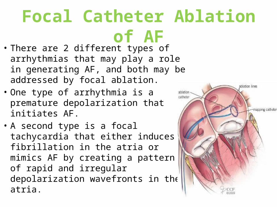
Focal Catheter Ablation of AF• There are 2 different types of
arrhythmias that may play a role in generating AF, and both may be addressed by focal ablation.
• One type of arrhythmia is a premature depolarization that initiates AF.
• A second type is a focal tachycardia that either induces fibrillation in the atria or mimics AF by creating a pattern of rapid and irregular depolarization wavefronts in the atria.
• Although focal sources of AF may be found in the right atrium, left atrium, coronary sinus, superior vena cava, or vein of Marshall, 95% of foci are located within a pulmonary vein.
• The pulmonary veins are covered by myocardial sleeves formed by one or more layers of myocardial fibers oriented in circular, longitudinal, oblique, or spiral directions. These sleeves vary from 2 to 25 mm in length, with a mean length of '10 to 20 mm in the superior pulmonary veins and 5 to 10 mm in the inferior pulmonary veins.
• The difference in the length of the sleeves may explain why arrhythmogenic foci are found more often in the superior than in the inferior pulmonary veins.
• The arrhythmogenic nature of these myocardial sleeves may be due in part to their embryonic origin from the same substrate that gives rise to the conduction system, which may be subject to abnormal automaticity.
• However, it is unclear whether the arrhythmias that arise in pulmonary veins are most often due to automaticity, reentry, or triggered activity, and it is possible that more than one mechanism plays a role in generating these arrhythmias.
Focal Ablation Within Pulmonary Veins• One approach to the ablation of a pulmonary vein arrhythmia
that triggers or simulates AF is to target the site of origin of the arrhythmia within the pulmonary vein directly.
• Localization of the site of origin of the arrhythmia is guided by the endocardial activation time
• The most common complications associated with the focal ablation of arrhythmias arising within the pulmonary veins are pericardial effusion (4%), transient ischemic episodes (2%), and symptomatic pulmonary vein stenosis (2%)
• The early experience with the focal ablation of pulmonary vein arrhythmias indicates that the recurrence rate is high and the success rate only modest, even in experienced laboratories.
Pulmonary Vein Isolation• Electrically isolating the pulmonary veins by circumferential
ablation at their ostia.• This would be effective in preventing recurrent AF caused by
multiple pulmonary vein foci, even if new foci emerge at some future time.
• Two techniques have been used to isolate pulmonary veins electrically. One technique involves the delivery of multiple, contiguous point applications of radiofrequency energy in a circumferential fashion in the left atrium near the ostia of the pulmonary veins.
• Another technique uses ultrasound energy to create a circumferential lesion with a catheter that has an ultrasound transducer mounted near its tip. The transducer is inside an inflatable balloon that is used to occlude the pulmonary vein and to stabilize the transducer at the left atrial–pulmonary vein junction.
• Presently, empirically isolate all four PVs not at the ostium but outside the tubular portion of the PV to avoid the risk of venous stenosis and improve procedural efficacy.
• Because the PV is funnel-shaped with a large proximal end (referred to as the antrum), which blends into the posterior wall of the LA.
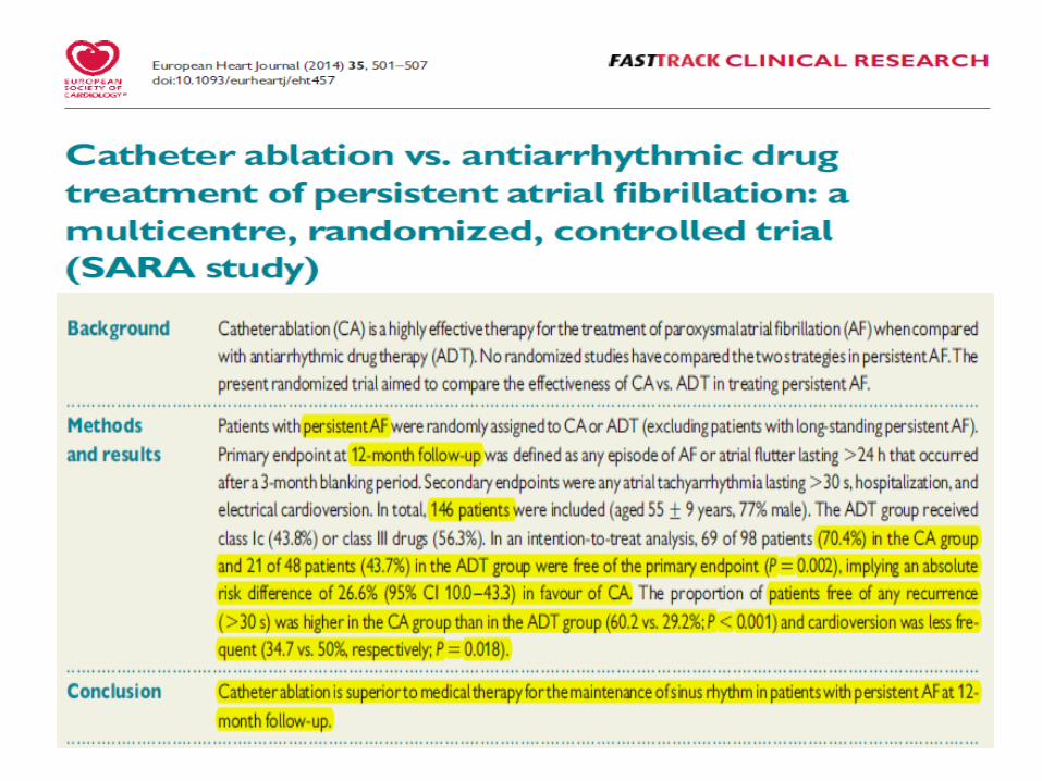
• Success rates for catheter-based ablation are lower in patients with persistent atrial fibrillation than paroxysmal AF.
• Antral encirclement of PVs in cases with long-standing persistent AF underwent a single- procedure, showed a drug-free success rate ranging from 37% to 56% at approximately 1 year .Integration of repeat procedures increased the drug-free success rate to 59%. The combination of drugs and multiple procedures yielded a success rate of approximately 77%. *
• In addition, the chances of a successful outcome are lower in those with marked dilation of the left atrium.
• Brooks AG, Stiles MK, Laborderie J, et al. Outcomes of long-lasting persistent atrial fibrillation ablation: a systematic review. Heart Rhythm 2010;7: 835–46
Segmental Isolation of Pulmonary Veins• The myocardial fibers that envelope the pulmonary veins may not be
present along the entire circumference of the pulmonary vein ostia.• Therefore, to eliminate conduction in and out of a pulmonary vein,
ablation along the entire circumference of the ostium may not be necessary.
• Instead, radiofrequency energy can be targeted to the segments of the ostium at which muscle fibers are present.
• These sites are identified by the presence of high-frequency depolarizations, which likely represent pulmonary vein muscle potentials which may encompass as little as 25% of the circumference of the ostium .
• If no ectopy is present during the ablation procedure, it may be appropriate to perform empiric segmental isolation of the left and right superior and left inferior pulmonary veins, because these are the most common sources of the arrhythmias that trigger AF.
• Success rate of 90% in patients with paroxysmal AF and with minimal or no risk of pulmonary vein stenosis.
• The end point of the procedure is to electrically isolate the pulmonary veins - pulmonary vein isolation or PVI.
• Major advantages of this technique are that it eliminates the need for detailed mapping of all pulmonary vein foci and the procedure can be performed with conventional ablation catheters and does not require specialized tools such as a balloon ultrasound ablation catheter.
• At present, the best candidates for the elimination of focal triggers of AF are symptomatic, drug-refractory patients with paroxysmal AF and normal or only mildly enlarged left atria.
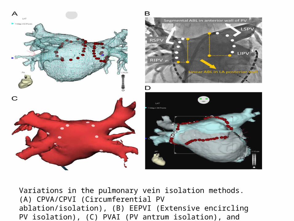
Variations in the pulmonary vein isolation methods. (A) CPVA/CPVI (Circumferential PV ablation/isolation), (B) EEPVI (Extensive encircling PV isolation), (C) PVAI (PV antrum isolation), and (D) BOX isolation
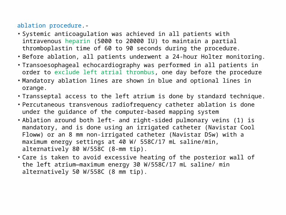
ablation procedure.-• Systemic anticoagulation was achieved in all patients with intravenous heparin
(5000 to 20000 IU) to maintain a partial thromboplastin time of 60 to 90 seconds during the procedure.
• Before ablation, all patients underwent a 24-hour Holter monitoring.• Transoesophageal echocardiography was performed in all patients in order to
exclude left atrial thrombus, one day before the procedure • Mandatory ablation lines are shown in blue and optional lines in orange. • Transseptal access to the left atrium is done by standard technique.• Percutaneous transvenous radiofrequency catheter ablation is done under the
guidance of the computer-based mapping system• Ablation around both left- and right-sided pulmonary veins (1) is mandatory,
and is done using an irrigated catheter (Navistar Cool Floww) or an 8 mm non-irrigated catheter (Navistar DSw) with a maximum energy settings at 40 W/ 558C/17 mL saline/min, alternatively 80 W/558C (8-mm tip).
• Care is taken to avoid excessive heating of the posterior wall of the left atrium—maximum energy 30 W/558C/17 mL saline/ min alternatively 50 W/558C (8 mm tip).
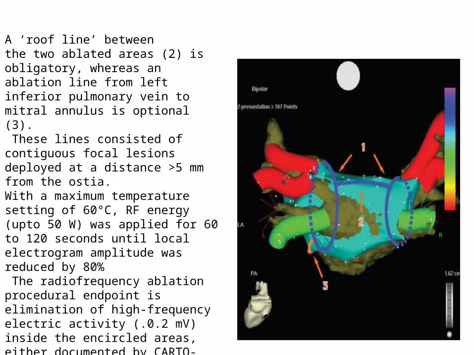
A ‘roof line’ betweenthe two ablated areas (2) is obligatory, whereas an ablation line from left inferior pulmonary vein to mitral annulus is optional (3). These lines consisted ofcontiguous focal lesions deployed at a distance >5 mm from the ostia. With a maximum temperature setting of 60°C, RF energy (upto 50 W) was applied for 60 to 120 seconds until local electrogram amplitude was reduced by 80% The radiofrequency ablation procedural endpoint is elimination of high-frequencyelectric activity (.0.2 mV) inside the encircled areas, either documented by CARTO-mapping or by the use of Lasso catheter.

s
Left atrium obtained by multislice computed tomography segmentation and non-fluoroscopic navigation after atrial fibrillation ablation (circumferentialpulmonary vein isolation) ABL, ablation catheter; CC circular catheter; CSC, coronary sinus catheter; LAA, left atrial appendage; LIPV, left inferior pulmonary vein;LSPV, left superior pulmonary vein; RIPV, right inferior pulmonary vein; RSPV, right superior pulmonary vein

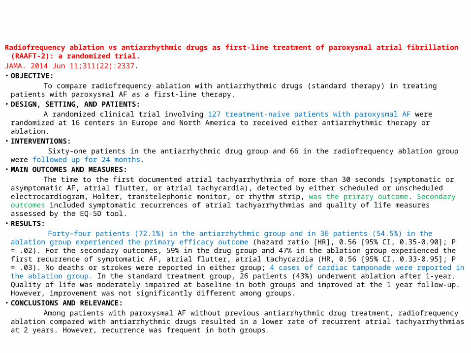
Radiofrequency ablation vs antiarrhythmic drugs as first-line treatment of paroxysmal atrial fibrillation (RAAFT-2): a randomized trial.
JAMA. 2014 Jun 11;311(22):2337.• OBJECTIVE: To compare radiofrequency ablation with antiarrhythmic drugs (standard therapy) in treating patients with paroxysmal AF as a
first-line therapy.• DESIGN, SETTING, AND PATIENTS: A randomized clinical trial involving 127 treatment-naive patients with paroxysmal AF were randomized at 16 centers in Europe
and North America to received either antiarrhythmic therapy or ablation. • INTERVENTIONS: Sixty-one patients in the antiarrhythmic drug group and 66 in the radiofrequency ablation group were followed up for 24 months.• MAIN OUTCOMES AND MEASURES: The time to the first documented atrial tachyarrhythmia of more than 30 seconds (symptomatic or asymptomatic AF, atrial flutter,
or atrial tachycardia), detected by either scheduled or unscheduled electrocardiogram, Holter, transtelephonic monitor, or rhythm strip, was the primary outcome. Secondary outcomes included symptomatic recurrences of atrial tachyarrhythmias and quality of life measures assessed by the EQ-5D tool.
• RESULTS: Forty-four patients (72.1%) in the antiarrhythmic group and in 36 patients (54.5%) in the ablation group experienced the primary
efficacy outcome (hazard ratio [HR], 0.56 [95% CI, 0.35-0.90]; P = .02). For the secondary outcomes, 59% in the drug group and 47% in the ablation group experienced the first recurrence of symptomatic AF, atrial flutter, atrial tachycardia (HR, 0.56 [95% CI, 0.33-0.95]; P = .03). No deaths or strokes were reported in either group; 4 cases of cardiac tamponade were reported in the ablation group. In the standard treatment group, 26 patients (43%) underwent ablation after 1-year. Quality of life was moderately impaired at baseline in both groups and improved at the 1 year follow-up. However, improvement was not significantly different among groups.
• CONCLUSIONS AND RELEVANCE: Among patients with paroxysmal AF without previous antiarrhythmic drug treatment, radiofrequency ablation compared with
antiarrhythmic drugs resulted in a lower rate of recurrent atrial tachyarrhythmias at 2 years. However, recurrence was frequent in both groups.
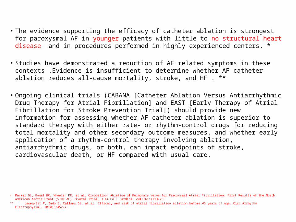
• The evidence supporting the efficacy of catheter ablation is strongest for paroxysmal AF in younger patients with little to no structural heart disease and in procedures performed in highly experienced centers. *
• Studies have demonstrated a reduction of AF related symptoms in these contexts .Evidence is insufficient to determine whether AF catheter ablation reduces all-cause mortality, stroke, and HF . **
• Ongoing clinical trials (CABANA [Catheter Ablation Versus Antiarrhythmic Drug Therapy for Atrial Fibrillation] and EAST [Early Therapy of Atrial Fibrillation for Stroke Prevention Trial]) should provide new information for assessing whether AF catheter ablation is superior to standard therapy with either rate- or rhythm-control drugs for reducing total mortality and other secondary outcome measures, and whether early application of a rhythm-control therapy involving ablation, antiarrhythmic drugs, or both, can impact endpoints of stroke, cardiovascular death, or HF compared with usual care.
• Packer DL, Kowal RC, Wheelan KR, et al. Cryoballoon Ablation of Pulmonary Veins for Paroxysmal Atrial Fibrillation: First Results of the North American Arctic Front (STOP AF) Pivotal Trial. J Am Coll Cardiol. 2013;61:1713-23.
** Leong-Sit P, Zado E, Callans DJ, et al. Efficacy and risk of atrial fibrillation ablation before 45 years of age. Circ Arrhythm Electrophysiol. 2010;3:452-7.

• The decision whether to pursue catheter ablation depends on a large number of variables, including the type of AF (paroxysmal versus persistent verses longstanding persistent), degree of symptoms, presence of structural heart disease, candidacy for alternative options such as rate control or antiarrhythmic drug therapy, likelihood of complications, and patient preference .
• It is important to recognize that most patients enrolled in trials of AF catheter ablation have generally been younger, healthy individuals with symptomatic paroxysmal AF refractory to ≥1 antiarrhythmic medication.
• The safety and efficacy of catheter ablation are less well established for other populations of patients, especially patients with longstanding persistent AF, very elderly patients, and patients with significant HF including tachycardia-induced cardiomyopathy
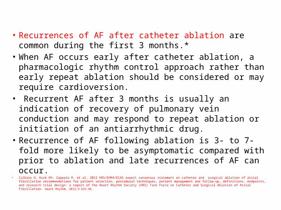
• Recurrences of AF after catheter ablation are common during the first 3 months.*
• When AF occurs early after catheter ablation, a pharmacologic rhythm control approach rather than early repeat ablation should be considered or may require cardioversion.
• Recurrent AF after 3 months is usually an indication of recovery of pulmonary vein conduction and may respond to repeat ablation or initiation of an antiarrhythmic drug.
• Recurrence of AF following ablation is 3- to 7-fold more likely to be asymptomatic compared with prior to ablation and late recurrences of AF can occur.
• Calkins H, Kuck KH, Cappato R, et al. 2012 HRS/EHRA/ECAS expert consensus statement on catheter and surgical ablation of atrial fibrillation recommendations for patient selection, procedural techniques, patient management and follow-up, definitions, endpoints, and research trial design: a report of the Heart Rhythm Society (HRS) Task Force on Catheter and Surgical Ablation of Atrial Fibrillation. Heart Rhythm. 2012;9:632-96.
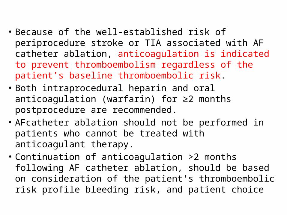
• Because of the well-established risk of periprocedure stroke or TIA associated with AF catheter ablation, anticoagulation is indicated to prevent thromboembolism regardless of the patient’s baseline thromboembolic risk.
• Both intraprocedural heparin and oral anticoagulation (warfarin) for ≥2 months postprocedure are recommended.
• AFcatheter ablation should not be performed in patients who cannot be treated with anticoagulant therapy.
• Continuation of anticoagulation >2 months following AF catheter ablation, should be based on consideration of the patient's thromboembolic risk profile bleeding risk, and patient choice
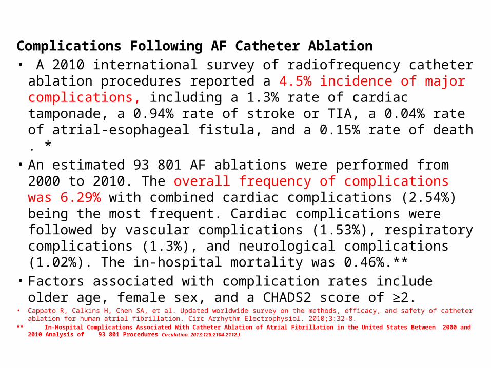
Complications Following AF Catheter Ablation• A 2010 international survey of radiofrequency catheter ablation
procedures reported a 4.5% incidence of major complications, including a 1.3% rate of cardiac tamponade, a 0.94% rate of stroke or TIA, a 0.04% rate of atrial-esophageal fistula, and a 0.15% rate of death . *
• An estimated 93 801 AF ablations were performed from 2000 to 2010. The overall frequency of complications was 6.29% with combined cardiac complications (2.54%) being the most frequent. Cardiac complications were followed by vascular complications (1.53%), respiratory complications (1.3%), and neurological complications (1.02%). The in-hospital mortality was 0.46%.**
• Factors associated with complication rates include older age, female sex, and a CHADS2 score of ≥2.
• Cappato R, Calkins H, Chen SA, et al. Updated worldwide survey on the methods, efficacy, and safety of catheter ablation for human atrial fibrillation. Circ Arrhythm Electrophysiol. 2010;3:32-8.
** In-Hospital Complications Associated With Catheter Ablation of Atrial Fibrillation in the United States Between 2000 and 2010 Analysis of 93 801 Procedures Circulation. 2013;128:2104-2112.)
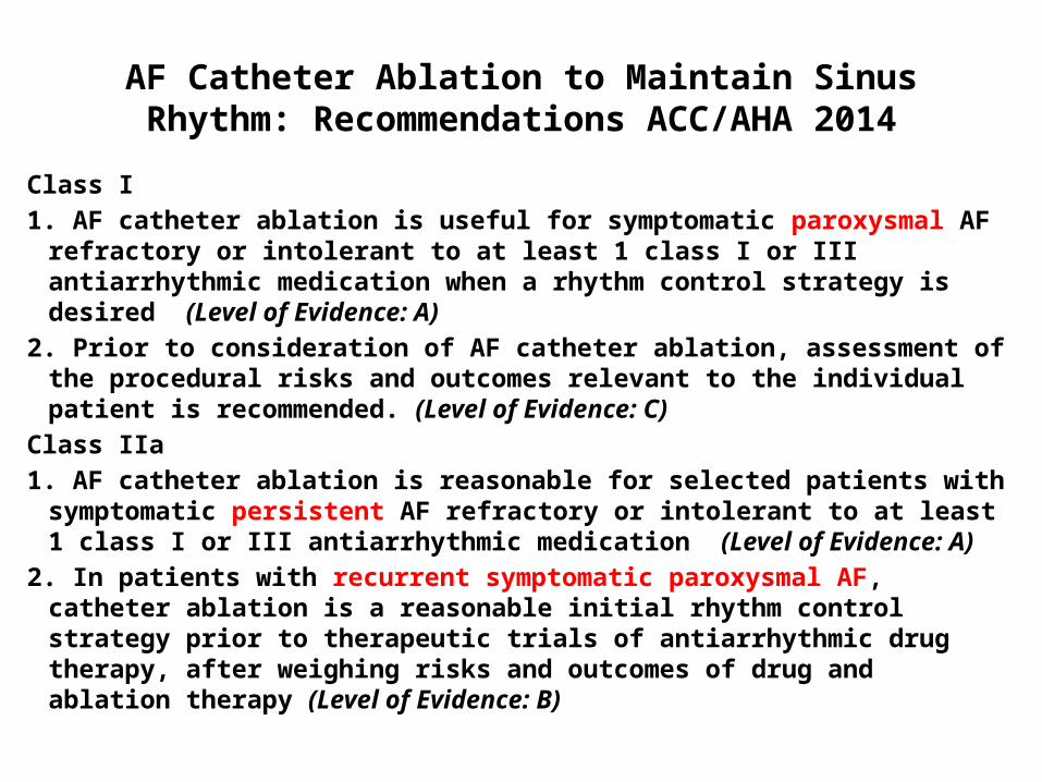
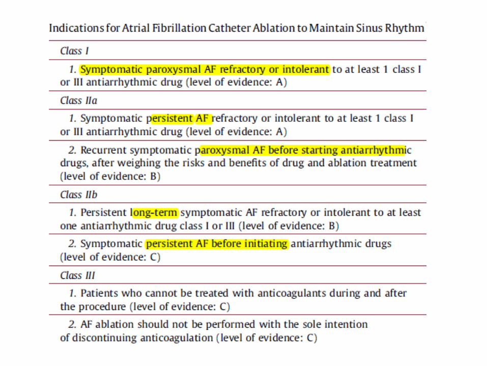
AF Catheter Ablation to Maintain Sinus Rhythm: Recommendations ACC/AHA 2014
Class I1. AF catheter ablation is useful for symptomatic paroxysmal AF refractory
or intolerant to at least 1 class I or III antiarrhythmic medication when a rhythm control strategy is desired (Level of Evidence: A)
2. Prior to consideration of AF catheter ablation, assessment of the procedural risks and outcomes relevant to the individual patient is recommended. (Level of Evidence: C)
Class IIa1. AF catheter ablation is reasonable for selected patients with
symptomatic persistent AF refractory or intolerant to at least 1 class I or III antiarrhythmic medication (Level of Evidence: A)
2. In patients with recurrent symptomatic paroxysmal AF, catheter ablation is a reasonable initial rhythm control strategy prior to therapeutic trials of antiarrhythmic drug therapy, after weighing risks and outcomes of drug and ablation therapy (Level of Evidence: B)
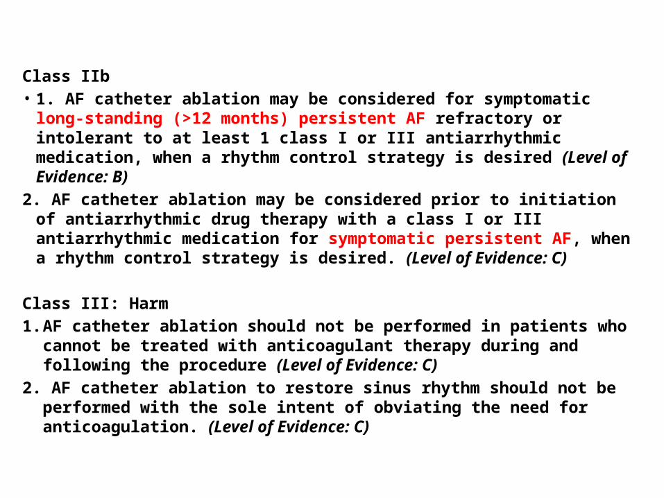
Class IIb• 1. AF catheter ablation may be considered for symptomatic long-standing (>12
months) persistent AF refractory or intolerant to at least 1 class I or III antiarrhythmic medication, when a rhythm control strategy is desired (Level of Evidence: B)
2. AF catheter ablation may be considered prior to initiation of antiarrhythmic drug therapy with a class I or III antiarrhythmic medication for symptomatic persistent AF, when a rhythm control strategy is desired. (Level of Evidence: C)
Class III: Harm1. AF catheter ablation should not be performed in patients who cannot be
treated with anticoagulant therapy during and following the procedure (Level of Evidence: C)
2. AF catheter ablation to restore sinus rhythm should not be performed with the sole intent of obviating the need for anticoagulation. (Level of Evidence: C)
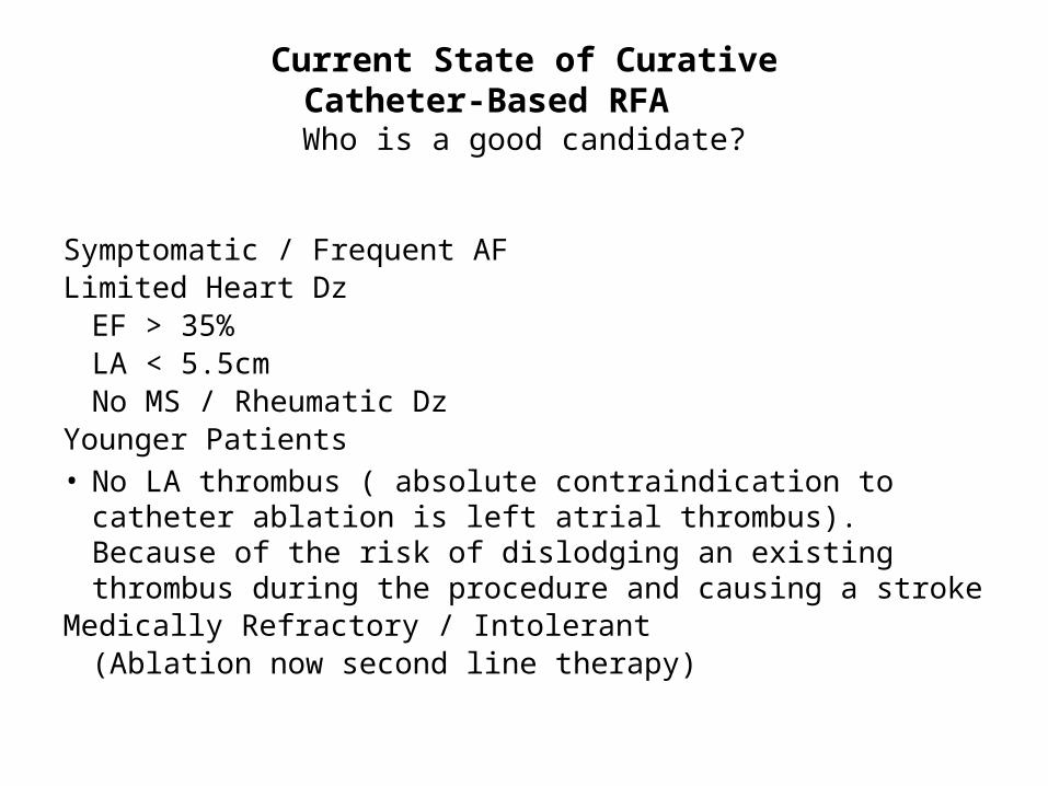
Current State of CurativeCatheter-Based RFA
Who is a good candidate?
Symptomatic / Frequent AFLimited Heart Dz
EF > 35%LA < 5.5cmNo MS / Rheumatic Dz
Younger Patients• No LA thrombus ( absolute contraindication to catheter ablation
is left atrial thrombus). Because of the risk of dislodging an existing thrombus during the procedure and causing a stroke
Medically Refractory / Intolerant(Ablation now second line therapy)

A-Fib vs. EP Labs
Internal Atrial Defibrillator• An implantable atrial defibrillator in theory is an excellent modality for the
detection and rapid conversion of AF.• It involves putting in transvenous leads in the right atrium and also in
coronary sinus which are connected to the defibrillator located subcutaneously in the infraclavicular region.
• The device is usually triggered at a preset delivery energy varying from 0.1 to 10.0 joules. However, a majority of patients reported significant discomfort with energy >2.0 joules.
• Safe and have an 80% efficacy in terminating AF.• The drawbacks include discomfort from the shocks, early recurrence of AF,
and technical problems with the device.• Because frequent shocks are undesirable, the ideal candidate for an
implanted atrial defibrillator has infrequent episodes of symptomatic, drug-refractory AF. However, if episodes of AF are rare, it is difficult to justify implanting a device.
• At present, the implanted atrial defibrillator is available only in combination with a ventricular defibrillator.
Nonpharmacologic Stroke Prevention
• A combination of the LAA’s unique geometry and atrial fibrillation leads to low blood flow velocity and stasis, which are precursors to thrombus formation.
• Only 50–60% of patients who clinically should be prescribed warfarin are actually taking it
• 14-44 % of AF patients have a contraindication to anticoagulation• Even with adequate anticoagulation, the risk of stroke is not abolished.• The annual incidence of stroke in patients therapeutically warfarinised
is 2–5%• Thus increased interest in alternative treatment strategies to help reduce
the risk of stroke in these patients.• The main focus of this interest is on the left atrial appendage (LAA) as It
has been shown that more than 90% of thrombi in patients with nonrheumatic, nonvalvular AF are located within this cavity
Percutaneous Approaches to Occlude the LAA• Exclusion of the LAA, both surgically and with
devices, has been attempted with the goal of reducing thromboembolism in patients with AF.
• There are 2 general approaches to occlude the LAA using percutaneous approaches. The first strategy involves implantable devices that are inserted percutaneously into the LAA with the goal of occluding or plugging the LAA. Devices for LAA occlusion include the WATCHMAN Device and the Amplatzer Cardiac Plug.
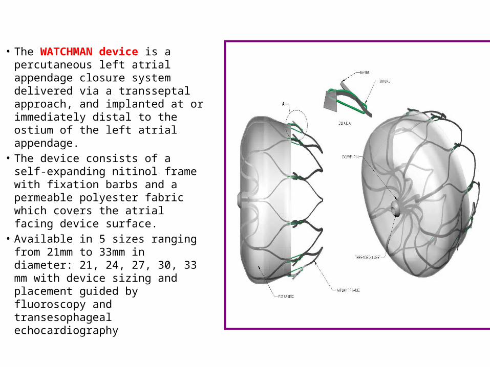
• The WATCHMAN device is a percutaneous left atrial appendage closure system delivered via a transseptal approach, and implanted at or immediately distal to the ostium of the left atrial appendage.
• The device consists of a self-expanding nitinol frame with fixation barbs and a permeable polyester fabric which covers the atrial facing device surface.
• Available in 5 sizes ranging from 21mm to 33mm in diameter: 21, 24, 27, 30, 33 mm with device sizing and placement guided by fluoroscopy and transesophageal echocardiography
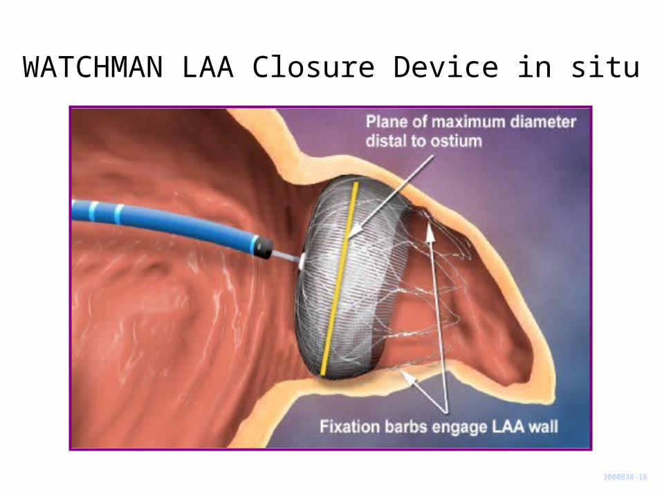
WATCHMAN LAA Closure Device in situ
3000838-18

PROTECT AF trial• Prospective, randomized study of WATCHMAN LAA Device vs. Long-term Warfarin Therapy
• 2:1 allocation ratio device to control
• Key Inclusion Criteria• Age 18 years or older• Documented non-valvular AF• Eligible for long-term warfarin therapy, and no other conditions that would require long-
term warfarin therapy• Calculated CHADS2 score > 1
• 800 Patients enrolled from Feb 2005 to Jun 2008– Device Group (463)– Control Group (244)– Roll-in Group (93)
• 59 Enrolling Centers (U.S. & Europe)• Follow-up Requirements
– TEE follow-up at 45 days, 6 months and 1 year– Clinical follow-up biannually up to 5 years– Regular INR monitoring while taking warfarin
Reddy VY, Doshi SK, Siever H, et al. Percutaneous Left Atrial Appendage Closure for Stroke Prophylaxis in Patients with Atrial Fibrillation: 2.3 Year Follow-Up of the PROTECT AF Trial (Watchman left atrial appendage system for embolic protection in patients with atrial fibrillation) trial. Circulation. 2013;127:720-9
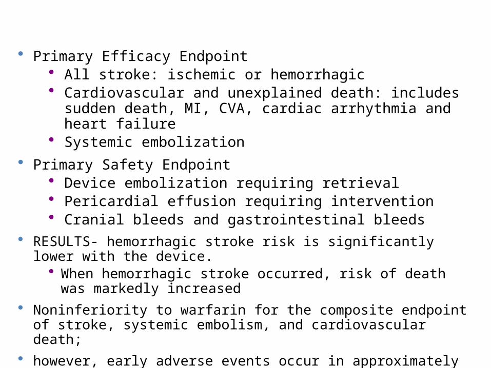
• Primary Efficacy Endpoint• All stroke: ischemic or hemorrhagic • Cardiovascular and unexplained death: includes sudden death, MI,
CVA, cardiac arrhythmia and heart failure • Systemic embolization
• Primary Safety Endpoint • Device embolization requiring retrieval• Pericardial effusion requiring intervention• Cranial bleeds and gastrointestinal bleeds
• RESULTS- hemorrhagic stroke risk is significantly lower with the device.• When hemorrhagic stroke occurred, risk of death was markedly increased
• Noninferiority to warfarin for the composite endpoint of stroke, systemic embolism, and cardiovascular death;
• however, early adverse events occur in approximately 10% of patients including pericardial bleeding
• After WATCHMAN device placement continue aspirin 81–325 mg daily indefinitely and clopidegrel 75 mg for 6 months.
• In light of the permeability of the device membrane to blood, warfarin is prescribed with a target International Normalised Ratio (INR) of 2.0 to 3.0 for a period of 45 days. Discontinued at 45 days if follow up TEE shows either complete closure of the left atrial appendage <5mm of peridevice flow.
• No infective endocarditis prophyaxis required.
• The second strategy is to tie off the LAA using an epicardial snare, referred to as the LARIAT device. This device received FDA approval in 2009 for facilitation of suture placement and knot tying for use in surgical applications in which soft tissues are being approximated. The initial experience **with this device appeared promising, with 97% acute obliteration of the LAA as confirmed by TEE and a favorable safety profile
• The LARIAT device’s long-term outcomes, requiring RCTs to study reduced stroke risk and safety, are not yet defined.
• The device requires subxiphoid pericardial access that may not be achievable in the presence of pericardial adhesions, it can provoke pericarditis that can be severe, and it is not suitable for all LAA anatomies.
• Aspirin continued indefinately , OAC given if no contraindication
** Bartus K, Han FT, Bednarek J, et al. Percutaneous left atrial appendage suture ligation using the LARIAT device in patients with atrial fibrillation: initial clinical experience. J Am Coll Cardiol. 2013;62:108-18.
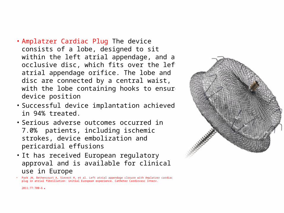
• Amplatzer Cardiac Plug The device consists of a lobe, designed to sit within the left atrial appendage, and an occlusive disc, which fits over the left atrial appendage orifice. The lobe and disc are connected by a central waist, with the lobe containing hooks to ensure device position
• Successful device implantation achieved in 94% treated.
• Serious adverse outcomes occurred in 7.0% patients, including ischemic strokes, device embolization and pericardial effusions
• It has received European regulatory approval and is available for clinical use in Europe
• Park JW, Bethencourt A, Sievert H, et al. Left atrial appendage closure with Amplatzer cardiac plug in atrial
fibrillation: initial European experience. Catheter Cardiovasc Interv. 2011;77:700-6.
Surgical Treatment of AFMaze Procedure• The surgical maze procedure was introduced in 1987 • Creating anatomic barriers in the atria may decrease the number
of circulating wavelets to a level below the critical number required to sustain AF and are often effective in restoring and maintaining sinus rhythm long-term.
• It is typically performed in association with mitral valve or coronary bypass surgery
• Another intraoperative approach is cryoablation limited to the posterior left atrium. Linear cryolesions connecting the 4 pulmonary veins and the posterior mitral annulus were effective in restoring sinus rhythm long-term in 69% of patients with chronic AF.
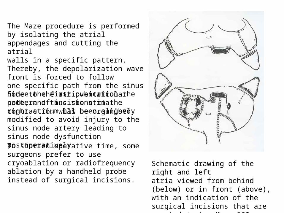
Schematic drawing of the right and leftatria viewed from behind (below) or in front (above), with an indication of the surgical incisions that are created during Maze III operation.
The Maze procedure is performed by isolating the atrial appendages and cutting the atrialwalls in a specific pattern. Thereby, the depolarization wave front is forced to followone specific path from the sinus node to the atrioventricular node, and thus the atrial contraction will be organised.
Since the first pubication the pattern of incisions in the right atrium has been slightly modified to avoid injury to the sinus node artery leading to sinus node dysfunction postoperatively
To shorten operative time, somesurgeons prefer to use cryoablation or radiofrequency ablation by a handheld probe instead of surgical incisions.
• 74% to 90% of patients are in sinus rhythm at 2 to 3 years postoperatiely. The operative mortality rate is 1%, but up to 6% of patients have required a pacemaker. In 90% of patients, the right and left atria regain mechanical function.*
• The FAST (Atrial Fibrillation Catheter Ablation Versus Surgical Ablation Treatment) trial compared the outcomes of catheter ablation and surgical ablation in a randomized study design .Patients either had left atrial dilation and hypertension (42 patients, 33%) or failed prior catheter ablation (82 patients, 67%). Freedom from atrial arrhythmias was greater after surgical ablation compared with catheter ablation, but the complication rate after surgical ablation was higher. **
• McCarthy PM, Gillinov AM, Castle L, et al. The Cox-Maze procedure: the Cleveland Clinic experience. Semin Thorac Cardiovasc Surg. 2000; 12:25–29.• Cox JL, Ad N, Palazzo T, et al. Current status of the Maze procedure for the treatment of atrial fibrillation. Semin Thorac Cardiovasc Surg. 2000; 12:15–19** Boersma LV, Castella M, van BW, et al. Atrial fibrillation catheter ablation versus surgical ablation treatment (FAST): a 2-center randomized clinical trial.
Circulation. 2012;125:23-30.
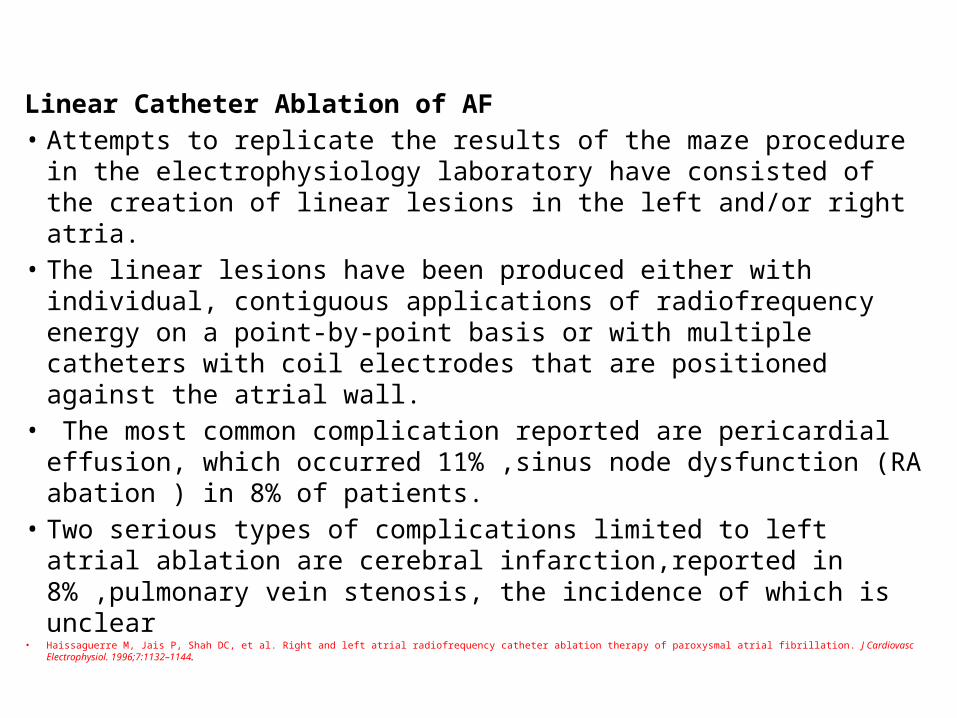
Linear Catheter Ablation of AF• Attempts to replicate the results of the maze procedure in the
electrophysiology laboratory have consisted of the creation of linear lesions in the left and/or right atria.
• The linear lesions have been produced either with individual, contiguous applications of radiofrequency energy on a point-by-point basis or with multiple catheters with coil electrodes that are positioned against the atrial wall.
• The most common complication reported are pericardial effusion, which occurred 11% ,sinus node dysfunction (RA abation ) in 8% of patients.
• Two serious types of complications limited to left atrial ablation are cerebral infarction,reported in 8% ,pulmonary vein stenosis, the incidence of which is unclear
• Haissaguerre M, Jais P, Shah DC, et al. Right and left atrial radiofrequency catheter ablation therapy of paroxysmal atrial fibrillation. J Cardiovasc Electrophysiol. 1996;7:1132–1144.
• Decisions regarding the choice of catheter-based or surgical ablation must be made on the basis of patient preference, and institutional experience and outcomes with each therapy
Surgery Maze Procedures: Recommendations• Class IIa- An AF surgical ablation procedure is reasonable
for selected patients with AF undergoing cardiac surgery for other indications. (Level of Evidence: C)
• Class IIb-A stand-alone AF surgical ablation procedure may be reasonable for selected patients with highly symptomatic AF not well managed with other approaches (Level of Evidence: B)
Surgical excision of the LAA may be considered in patients undergoing cardiac surgery. Class IIb (Level of Evidence: C)
• Removal of the LAA—yields inconsistent results and the anatomy of the LAA is quite variable . The circumflex coronary artery lies proximate to the base of the LAA and epicardial and endocardial-based surgical techniques to occlude the LAA are often inadequate because of surgeon concern regarding damage to the circumflex artery
• The results of surgical occlusion of the LAA remain suboptimal, with incomplete occlusion in ≥50% and thrombus was identified in ≥25% of patients with unsuccessful LAA occlusion .*So continued need for anticoagulation in patients who have undergone surgical LAA ligation.
• Kanderian AS, Gillinov AM, Pettersson GB, et al. Success of surgical left atrial appendage closure: assessment by transesophageal echocardiography. J Am Coll Cardiol. 2008;52:924-9.

Conclusion• In the minority of patients in whom AF cannot be adequately managed
by pharmacological therapy, the most appropriate type of nonpharmacological therapy must be selected on an individualized basis.
• For example, in a cardiomyopathy patient with chronic AF and an uncontrolled ventricular rate, AV junction ablation may be the optimal therapy.
• In a patient with the sick sinus syndrome, atrial-based pacing (perhaps biatrial) may be the most appropriate option.
• In a patient who requires an implantable cardioverter-defibrillator and whom has occasional episodes of AF, implantation of a device capable of both atrial and ventricular defibrillation should be considered.
• If the patient has idiopathic AF, best technique currently available for curing paroxysmal AF (assuming it originates from the pulmonary veins) is segmental isolation of the pulmonary veins by discrete applications of radiofrequency energy at the ostia, guided by pulmonary vein potentials.
• For ablation of long-standing persistent AF, there is a consensus that PVI is the first-line approach, similar to paroxysmal AF.
• Since ablation of chronic AF cases is still challenging, early treatment of paroxysmal AF before transformation to the persistent or chronic form is mandatory.
• In the occasional patient who is disabled by chronic AF, referral for surgical treatment may be appropriate.
• However, at present, the most appropriate option for most patients with idiopathic chronic AF may be the use of pharmacological therapy to minimize symptoms as much as possible, prevent tachycardia-induced cardiomyopathy, and avoid thromboembolic complications.
• Nonpharmacological Approaches to Atrial Fibrillation Melvin M. Scheinman, MD; Fred Morady, MD Clinical Cardiology: New Frontiers Circulation April 24, 2001• Current strategiesfornon-pharmacologicaltherapyoflong-standing persistentatrialfibrillation Teiichi Yamane,MD, Journal of Arrhythmia 28 (2012) 155–161

THANK U ....
Selected Risk Factors and Biomarkers for AF• Increasing age• Hypertension • Diabetes mellitus • MI • VHD • HF • Obesity• Obstructive sleep apnea• Cardiothoracic surgery• Smoking• Exercise• Alcohol use• Hyperthyroidism
•Increased pulse pressure •European ancestry•Family history•Genetic variantsElectrocardiographic•LVHEchocardiographic•LA enlargement•Decreased LV fractional shortening •Increased LV wall thickness Biomarkers•Increased CRP •Increased BNP